Embed Size (px)
Citation preview
Jaundiced cats can provide some diagnostic and therapeutic dilemmas, this update will try to make decision making simpler.
What causes jaundice?
Are there any factors in the signalment or history and physical examination that can help to narrow the possibilities? Age Younger animals: FIP, lymphocytic cholangitis more common Young- middle aged: Pancreatitis, neutrophilic cholangitis and hepatic lipidosis are more common Middle-older aged: Neutrophilic cholangitis, pancreatitis, hepatic lipidosis and neoplasia are more com-mon. History Hepatic lipidosis is often seen in overweight cats after a period of recent weight loss/anorexia. FIP is more common in pure breed/pedigree cats and from multi-cat households or with a history of some “stressor” several weeks-months before presentation. Whilst it is typically seen in young cats (>50% are <2yrs old), we can see it in older cats. Young cats with weight loss and a good appetite may have lymphocytic cholangitis, clearly in older cats with these signs other differentials including hyperthyroidism are considered.
Any toxic exposure? Physical findings Abdominal pain can be present with pancreatitis, neutrophilic cholangitis, cholecystitis Pyrexia can be present with neutrophilic cholangitis, sepsis, FIP Ascites can be present with FIP, lymphocytic cholangitis, neoplasia Hepatomegaly can be present with lymphocytic cholangitis, lipidosis and neoplasia
rowereferrals.co.uk Tel: 01454 521000
Pre hepatic Hepatic Post hepatic
• Haemolysis • Inflammatory/cholangitis • Pancreatitis• Immune • Neutrophilic (NC) • Cholecystitis• Non immune • Lymphocytic (LC) • Cholecystitis• Infectious • Hepatic lipidosis (associated with NC)• Toxic • FIP • Hepatobiliary mass• Hypophosphataemia • Amyloidosis • Dudodenal mass
• Sepsis• Hepatotoxcity
Clinical Advice SheetHow to approach the yellow cat?
Kate Murphy BVSc (Hons) DSAM DipECVIM-CA FRCVS PGCert(HE) EBVS® European Veterinary Specialist in Small Animal Internal Medicine & RCVS Specialist in Small Animal MedicineElisa J. Best BVSc Cert SAS MRCVS RCVS Advanced Practitioner in Small Animal SurgeryMargaret Costello MVB DVR DipECVDI MRCVS EBVS® European Specialist in Veterinary Diagnostic Imaging
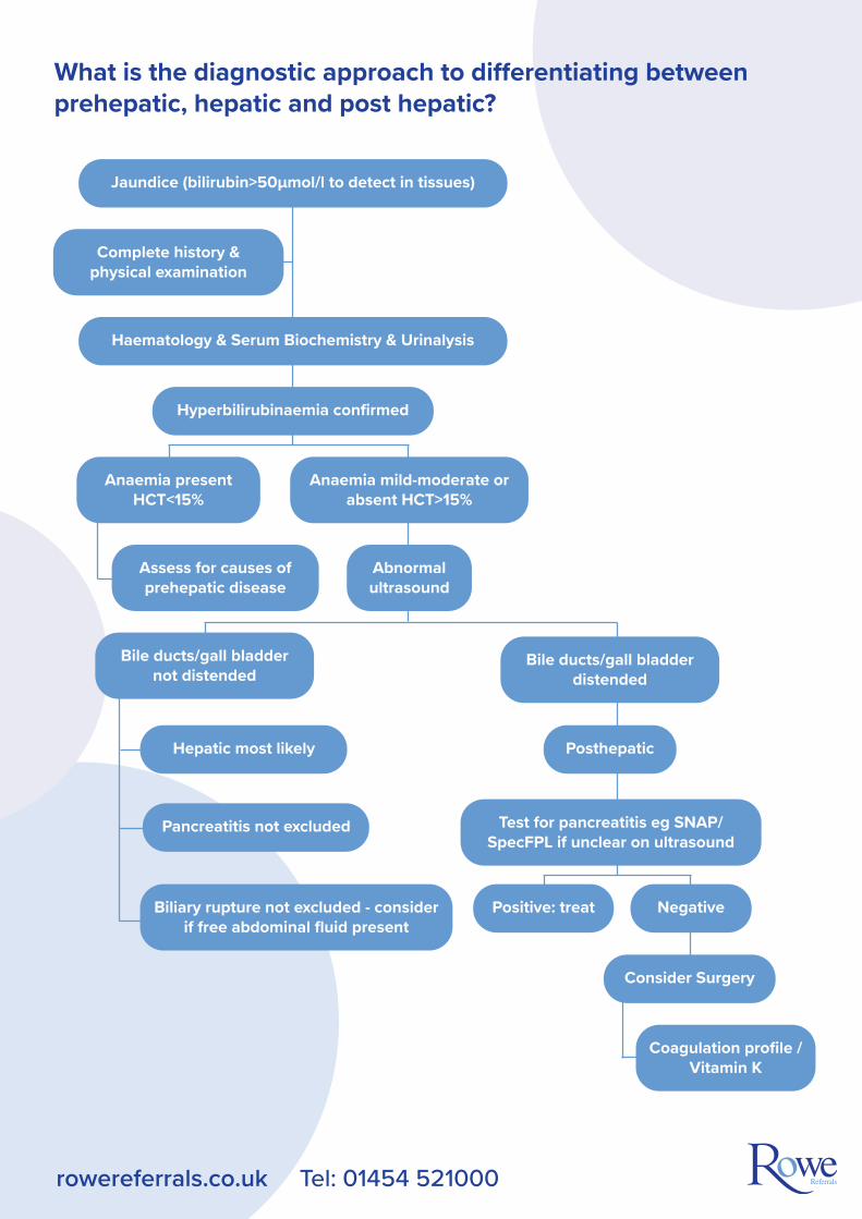
What is the diagnostic approach to differentiating between prehepatic, hepatic and post hepatic?
rowereferrals.co.uk Tel: 01454 521000
Jaundice (bilirubin>50µmol/l to detect in tissues)
Complete history & physical examination
Haematology & Serum Biochemistry & Urinalysis
Hyperbilirubinaemia confirmed
Anaemia present HCT<15%
Anaemia mild-moderate or absent HCT>15%
Assess for causes of prehepatic disease
Abnormal ultrasound
Bile ducts/gall bladder not distended
Bile ducts/gall bladder distended
Posthepatic
Test for pancreatitis eg SNAP/SpecFPL if unclear on ultrasound
Coagulation profile / Vitamin K
Hepatic most likely
Pancreatitis not excluded
Biliary rupture not excluded - consider if free abdominal fluid present
Negative
Consider Surgery
Positive: treat
Does the degree of hyperbilirubinaemia help? Bilirubin is normally <15µmol/l
Tissue discolouration is not seen until bilirubin is >50µmol/l Prehepatic jaundice, FIP, pancreatitis, sepsis, amyloidosis rarely causes bilirubin >100µmol/ Hepatic lipidosis and post-hepatic obstruction can cause very marked increases in bilirubin even 200µmol/l and these need urgent assessment to assess if surgery is indicated for obstructive dis-ease and to institute supportive therapy for lipidotic cats.
Does the degree of hyperbilirubinaemia help?Mild - moderate anaemia is common in cats with chronic or inflammatory disease and therefore care not to overinterpret anaemia and label cases as haemolytic is important
Haemolytic anaemia, causing jaundice will be associated with acute and severe anaemia typically with HCT <15%.
Inflammatory leukogram/left shift can be seen with NC and sepsis amongst many other causes
Any increase in ALT in the cat is important and should be investigated, it can be caused by non-hepatic disease e.g. Hyperthyroidism or primary liver disease when ALP and GGT will normally also be in-creased.
A significant increase in ALP with a normal GGT is a major flag for possible hepatic lipidosis.
An increased bilirubin, without changes in ALT/ALP/GGT in pyrexic cat particularly if associated with hyperglobulinaemia is a concern for FIP. High globulins can also be seen in LC but are associated with abnormal liver enzymes.
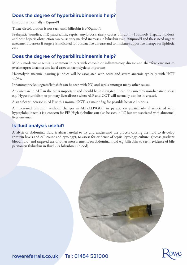
Is fluid analysis useful?Analysis of abdominal fluid is always useful to try and understand the process causing the fluid to de-velop (protein levels and cell count and cytology), to assess for evidence of sepsis (cytology, culture, glucose gradient blood:fluid) and targeted use of other measurements on abdominal fluid e.g. bilirubin to see if evidence of bile peritonitis (bilirubin in fluid >2x bilirubin in blood).
rowereferrals.co.uk Tel: 01454 521000
What ultrasound features should be looked for? Radiographs can be useful as a general survey to assess the size of the liver and to assess serosal detail. They are unlikely to provide much information about the gall bladder as this is of similar opacity to the liver and silhouettes with it. Sometimes you may see radiopaque choleliths in the position of the gall bladder, however this finding alone, can be incidental and is not an indication for surgery unless com-bined with clinical, biochemical and ultrasonographic evidence of obstruction. Very rarely you may see gas associated with gas producing organisms. It can be useful to identify the position of the duodenum to see if any evidence of a mass/mass effect here, however masses resulting in obstruction are usually too small to see on radiographs. Chest radiography may be performed if you are suspicious of neoplasia as a cause of the jaundice and wish to check for metastatic disease, but this is likely to be seen only late in the progression of disease and lymph node or other solid organ metastasis would be more common.
The disadvantages/limitations of radiography include
• Choleliths may not be evident (may not always be mineralised or sufficiently mineralised)
• The gall bladder cannot be differentiated from the liver parenchyma
• They provide no information about the common bile duct
It would be inadvisable to elect for surgery of biliary disease on the basis of radiographs alone.
High quality ultrasound, if available can be considered the next step after laboratory work and radiog-raphy can always be performed later if indicated from the ultrasound.
Ultrasound of the hepatobiliary system in the cat:
Feline challenges include the small size of the patient, many are very thin, and you are examining small structures (duodenal papilla, pancreatic duct, common bile duct)
This requires a good quality machine and a high frequency probe to optimise the exam.
During the ultrasound as well as a general assessment of the abdomen specific focus in these patients will be on:
Liver
• Normally within the costal arch
• Liver lobe edges are sharp or rounded.
• Echogenicity should be compared to falciform fat, the fat is usually isoechoic or hyperechoic to the liver but in fat cats the liver may be hyperechoic
• Parenchyma should be smooth, interrupted by portal veins, the larger ones have slightly brightwalls
rowereferrals.co.uk Tel: 01454 521000
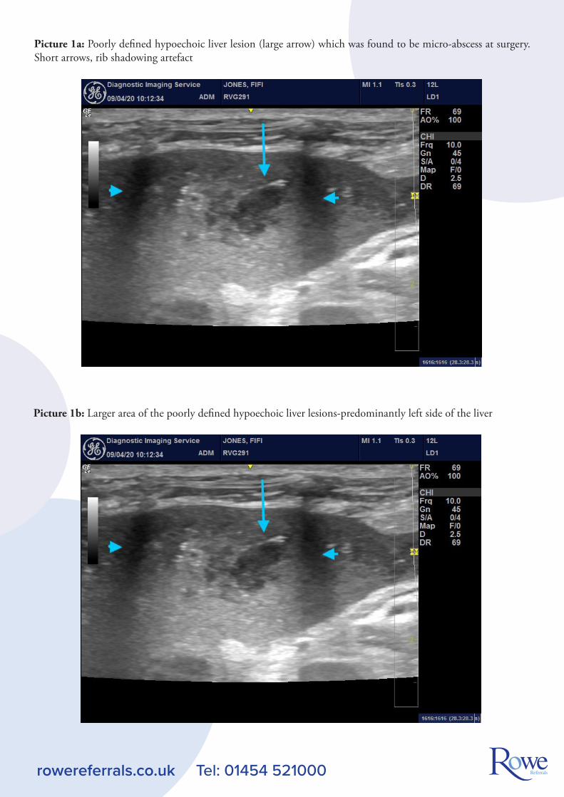
Picture 1a: Poorly defined hypoechoic liver lesion (large arrow) which was found to be micro-abscess at surgery. Short arrows, rib shadowing artefact
rowereferrals.co.uk Tel: 01454 521000
Picture 1b: Larger area of the poorly defined hypoechoic liver lesions-predominantly left side of the liver
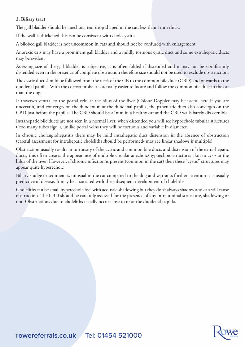
2. Biliary tract
The gall bladder should be anechoic, tear drop shaped in the cat, less than 1mm thick.
If the wall is thickened this can be consistent with cholecystitis
A bilobed gall bladder is not uncommon in cats and should not be confused with enlargement
Anorexic cats may have a prominent gall bladder and a mildly tortuous cystic duct and some extrahepatic ducts may be evident
Assessing size of the gall bladder is subjective, it is often folded if distended and it may not be significantly distended even in the presence of complete obstruction therefore size should not be used to exclude ob-struction.
The cystic duct should be followed from the neck of the GB to the common bile duct (CBD) and onwards to the duodenal papilla. With the correct probe it is actually easier to locate and follow the common bile duct in the cat than the dog.
It traverses ventral to the portal vein at the hilus of the liver (Colour Doppler may be useful here if you are uncertain) and converges on the duodenum at the duodenal papilla, the pancreatic duct also converges on the CBD just before the papilla. The CBD should be <4mm in a healthy cat and the CBD walls barely dis-cernible.
Intrahepatic bile ducts are not seen in a normal liver, when distended you will see hypoechoic tubular structures (“too many tubes sign”), unlike portal veins they will be tortuous and variable in diameter
In chronic cholangiohepatitis there may be mild intrahepatic duct distension in the absence of obstruction (careful assessment for intrahepatic choleliths should be performed- may see linear shadows if multiple)
Obstruction usually results in tortuosity of the cystic and common bile ducts and distension of the extra-hepatic ducts; this often creates the appearance of multiple circular anechoic/hypoechoic structures akin to cysts at the hilus of the liver. However, if chronic infection is present (common in the cat) then these “cystic” structures may appear quite hyperechoic
Biliary sludge or sediment is unusual in the cat compared to the dog and warrants further attention it is usually predictive of disease. It may be associated with the subsequent development of choleliths.
Choleliths can be small hyperechoic foci with acoustic shadowing but they don’t always shadow and can still cause obstruction. The CBD should be carefully assessed for the presence of any intraluminal struc-ture, shadowing or not. Obstructions due to choleliths usually occur close to or at the duodenal papilla.
rowereferrals.co.uk Tel: 01454 521000
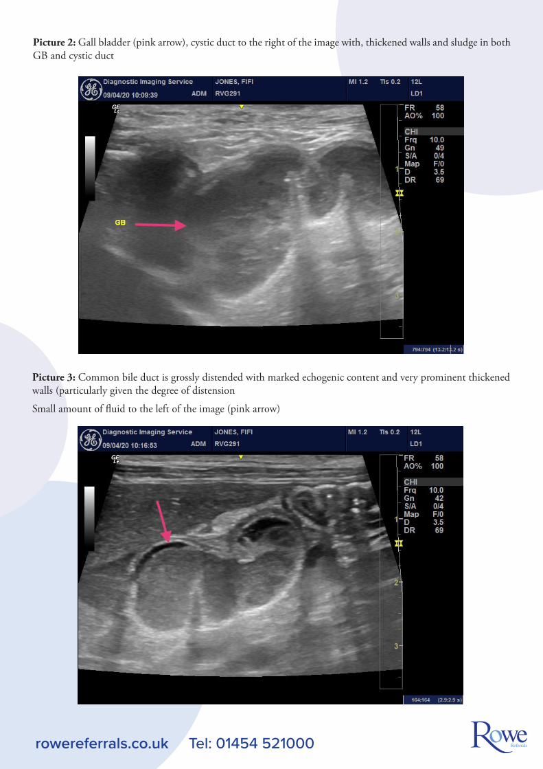
Picture 2: Gall bladder (pink arrow), cystic duct to the right of the image with, thickened walls and sludge in both GB and cystic duct
rowereferrals.co.uk Tel: 01454 521000
Picture 3: Common bile duct is grossly distended with marked echogenic content and very prominent thickened walls (particularly given the degree of distension
Small amount of fluid to the left of the image (pink arrow)
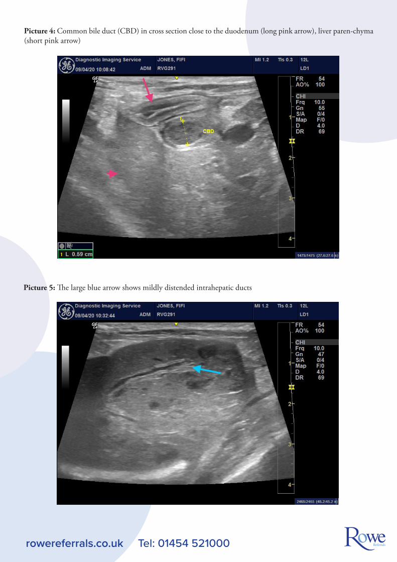
Picture 4: Common bile duct (CBD) in cross section close to the duodenum (long pink arrow), liver paren-chyma (short pink arrow)
rowereferrals.co.uk Tel: 01454 521000
Picture 5: The large blue arrow shows mildly distended intrahepatic ducts
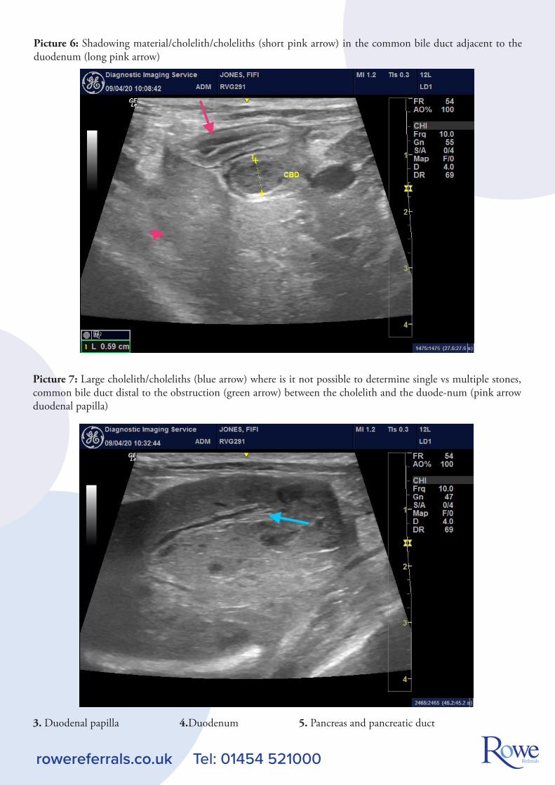
Picture 6: Shadowing material/cholelith/choleliths (short pink arrow) in the common bile duct adjacent to the duodenum (long pink arrow)
rowereferrals.co.uk Tel: 01454 521000
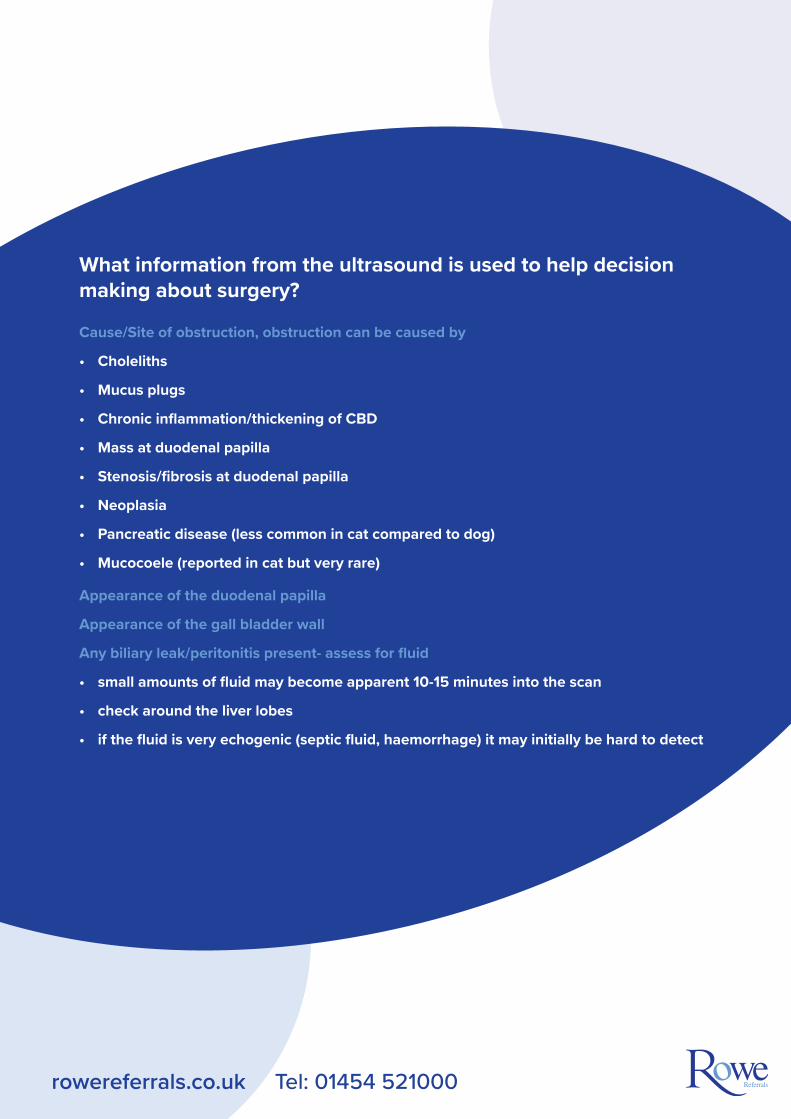
Picture 7: Large cholelith/choleliths (blue arrow) where is it not possible to determine single vs multiple stones, common bile duct distal to the obstruction (green arrow) between the cholelith and the duode-num (pink arrow duodenal papilla)
3. Duodenal papilla 4.Duodenum 5. Pancreas and pancreatic duct
rowereferrals.co.uk Tel: 01454 521000
What information from the ultrasound is used to help decision making about surgery?
Cause/Site of obstruction, obstruction can be caused by
• Choleliths
• Mucus plugs
• Chronic inflammation/thickening of CBD
• Mass at duodenal papilla
• Stenosis/fibrosis at duodenal papilla
• Neoplasia
• Pancreatic disease (less common in cat compared to dog)
• Mucocoele (reported in cat but very rare)
Appearance of the duodenal papilla
Appearance of the gall bladder wall
Any biliary leak/peritonitis present- assess for fluid
• small amounts of fluid may become apparent 10-15 minutes into the scan
• check around the liver lobes
• if the fluid is very echogenic (septic fluid, haemorrhage) it may initially be hard to detect
What surgical options are there? Surgical management is primarily aimed at relieving the obstruction and managing bile peritonitis should it already be present.
Obstruction: Ultrasound examination has hopefully already located the site of the obstruction and poten-tially the cause. If the obstruction cannot be relieved, then biliary diversion surgery is indicated; however, it is always preferable to remove the obstruction and restore normal biliary flow if possible. The first step is to perform a duodenotomy and locate the duodenal papilla which lies approximately 2 cm aboral to the pylorus in cats. The common bile duct should then be cannulated using a 3.5-5 french soft tube such as an irrigating cannula, IV cannula with stylet removed or a nasogastric feeding tube. Sterile saline is then used to perform gentle flushing to try and remove the obstruction, which may just be biliary sediment and mu-cus. Sometimes, gall stones are the cause of the obstruction and they may be firmly lodged in the duode-nal papilla. In this situation, a very small incision (sphincterotomy) of a few mm into the papilla may be sufficient to allow the stone to move. If the stone is lodged in the common bile duct, attempts should be made to flush the stone into the gallbladder and then perform a cholecystectomy to remove it, but this may not always be possible. If the stone will not move with flushing the options are either to incise into the common bile duct to remove it (choledochotomy) or to perform biliary diversion surgery.
rowereferrals.co.uk Tel: 01454 521000
The common bile duct in a cat is usually too small to perform a choledochotomy without microsurgery techniques, however in the case of obstruction the common bile duct may be very distended allowing this to be performed.
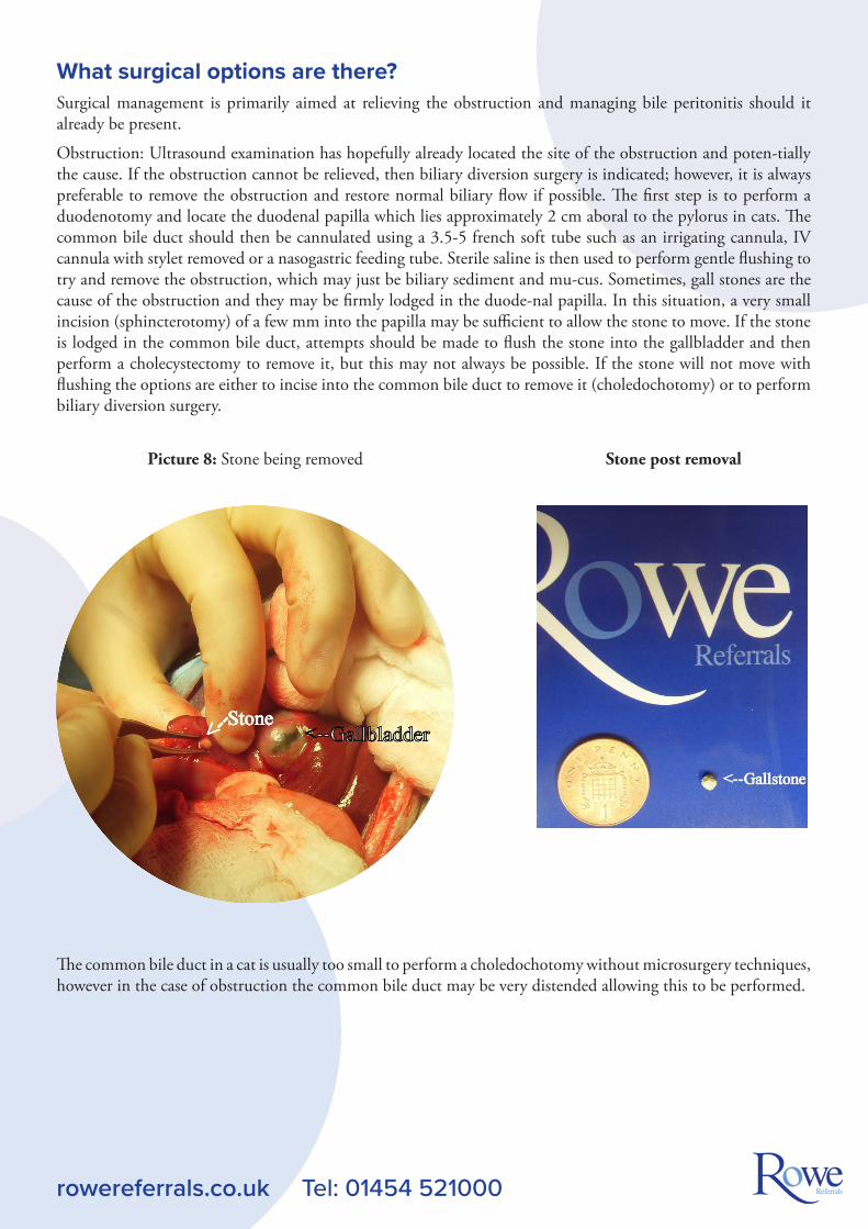
Picture 8: Stone being removed Stone post removal
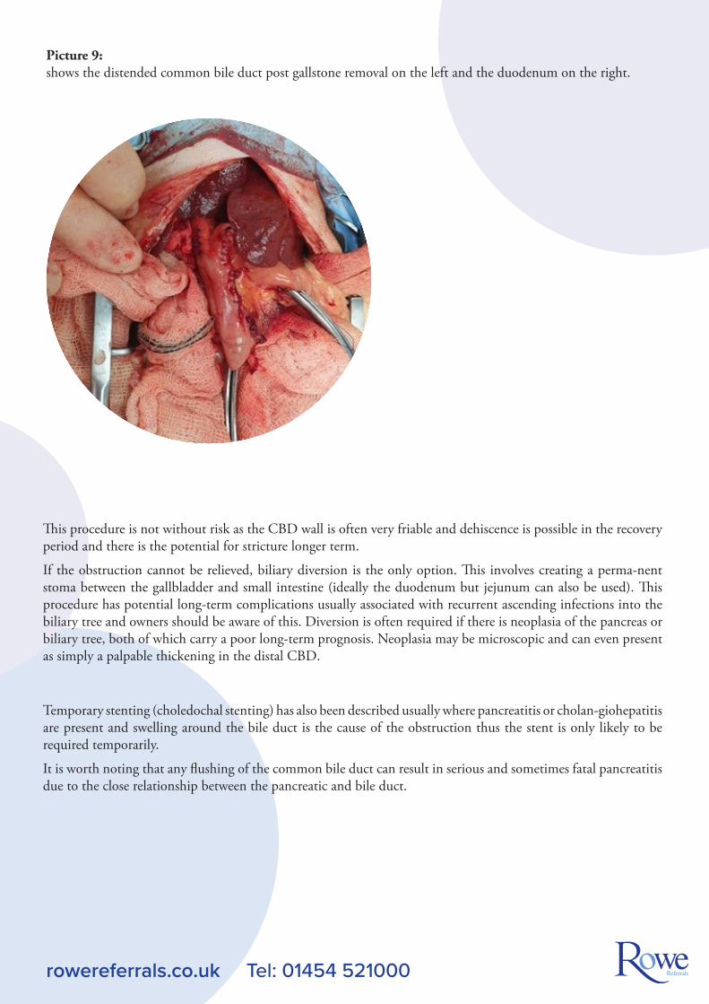
Picture 9: shows the distended common bile duct post gallstone removal on the left and the duodenum on the right.
rowereferrals.co.uk Tel: 01454 521000
This procedure is not without risk as the CBD wall is often very friable and dehiscence is possible in the recovery period and there is the potential for stricture longer term.
If the obstruction cannot be relieved, biliary diversion is the only option. This involves creating a perma-nent stoma between the gallbladder and small intestine (ideally the duodenum but jejunum can also be used). This procedure has potential long-term complications usually associated with recurrent ascending infections into the biliary tree and owners should be aware of this. Diversion is often required if there is neoplasia of the pancreas or biliary tree, both of which carry a poor long-term prognosis. Neoplasia may be microscopic and can even present as simply a palpable thickening in the distal CBD.
Temporary stenting (choledochal stenting) has also been described usually where pancreatitis or cholan-giohepatitis are present and swelling around the bile duct is the cause of the obstruction thus the stent is only likely to be required temporarily.
It is worth noting that any flushing of the common bile duct can result in serious and sometimes fatal pancreatitis due to the close relationship between the pancreatic and bile duct.
Bile peritonitis:
The two main aims are to stop the leakage of bile (if the leak can be found) which may involve suturing a tear in the CBD or removing a leaking gall bladder.
The second and equally important part of management is placement of an abdominal drain. Bile peritoni-tis is highly irritant to the peritoneum and can cause a chemical peritonitis or if bile is infected a septic peritonitis and can be fatal.
From personal experience if a drain is placed, bile peritonitis is not the death sentence that one hears about, even if the leak can’t be located. It is likely that very small leaks will likely seal on their own which is helpful where there may be biliary rupture deep in the liver tissue which is surgically inaccessible.
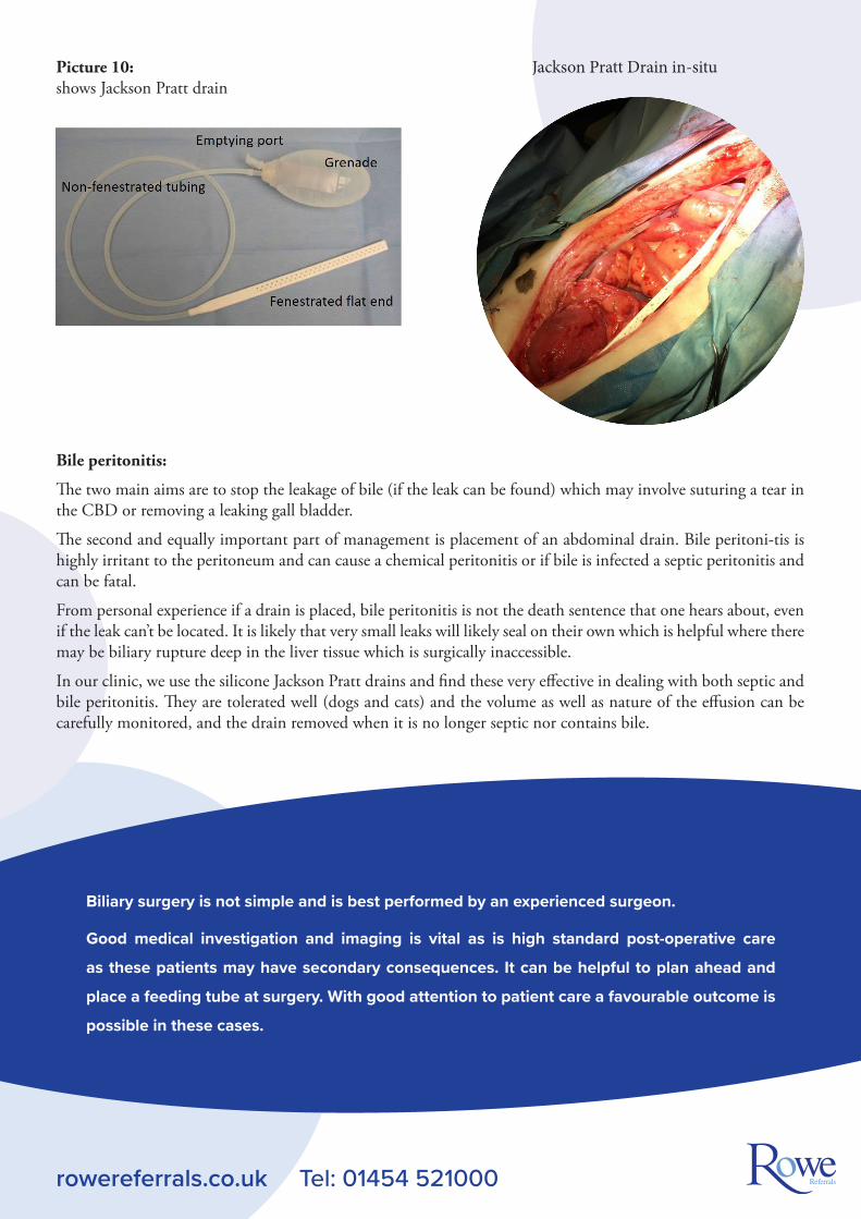
In our clinic, we use the silicone Jackson Pratt drains and find these very effective in dealing with both septic and bile peritonitis. They are tolerated well (dogs and cats) and the volume as well as nature of the effusion can be carefully monitored, and the drain removed when it is no longer septic nor contains bile.
rowereferrals.co.uk Tel: 01454 521000
Picture 10: shows Jackson Pratt drain
Jackson Pratt Drain in-situ
Biliary surgery is not simple and is best performed by an experienced surgeon.
Good medical investigation and imaging is vital as is high standard post-operative care
as these patients may have secondary consequences. It can be helpful to plan ahead and
place a feeding tube at surgery. With good attention to patient care a favourable outcome is
possible in these cases.