Embed Size (px)
Citation preview
Roberts, T. et al. (2013). Cleidocranial dysplasia: a review of the dental, historical, and practical
implications with an overview of the South African experience.
Oral Surgery, Oral Medicine, Oral Pathology, Oral Radiology, 115(1): 46 – 55
http://dx.doi.org/10.1016/j.oooo.2012.07.435
University of the Western Cape Research Repository [email protected]
Cleidocranial dysplasia: a review of the dental, historical, and practical
implications with an overview of the South African experience
Tina Roberts, Lawrence Stephen and Peter Beighton
Abstract
Cleidocranial dysplasia (CCD) is an uncommon but well-known genetic skeletal
condition. Several hundred affected persons are members of a large extended family in
the Cape Town Mixed Ancestry community of South Africa. The clinical manifestations
are often innocuous, but hyperdontia and other developmental abnormalities of the
teeth are a major feature and may require special dental management.
Over the past 40 years, the authors have encountered more than 100 affected persons
in Cape Town. Emphasis has been on dental management, but medical, genetic, and
social problems have also been addressed. In this article, we have reviewed the
manifestations of the disorder in the light of our own experience, and performed a
literature search with emphasis on the various approaches to dental management and
treatment options in CCD. Advances in the understanding of the biomolecular
pathogenesis of CCD are outlined and the international and local history of the disorder
is documented.
The purpose of this article was to review the dental manifestations and management of
cleidocranial dysplasia (CCD) [OMIM 119600]. The history, genetic background, and
general manifestations of CCD are also outlined and an overview is presented.
The disorder is a genetic skeletal dysplasia in which hypoplasia of the clavicles and deficient
ossification of the anterior fontanelle are the major features. Affected persons have a
characteristic facial appearance with a bulky forehead, hypertelorism, and midfacial
hypoplasia.1 General health is usually good and the intellect is unimpaired.2 The adverse
general health effects of CCD are usually not very severe or debilitating and there is no
associated impairment in cognitive or intellectual functioning in affected persons.2
A variety of dental problems may occur in CCD. In particular, supernumerary teeth
(hyperdontia) in the primary and secondary dentition may lead to dental crowding and
malocclusion. Retention of the deciduous teeth may exacerbate this situation. For these
reasons, dental management is a significant aspect of the health care of affected persons.
2
Cleidocranial dysplasia is inherited as an autosomal dominant trait, with generation-to-
generation transmission. Owing to the founder effect, the condition is comparatively
common in the mixed ancestry community of Cape Town, South Africa.3 This group has
genetic endowment from San and Xhoi Xhoi populations, with input from indigenous
African, Indonesian, East Indian, and European sources. Numerous members of an
extended family and a founder effect were initially documented by Jackson in 19513.
Whereas the worldwide prevalence of CCD is generally regarded as being about 1 per
million, in this Cape Town community, the minimum prevalence is 100 per million.
In view of the special importance of CCD in this country, we have reviewed the history of
the disorder and described and depicted the clinical and radiological manifestations. To alert
clinicians, special emphasis has been given to hyperdontia and to the dental complications
and their management.
Historical review
The early history of CCD goes back to prehistorical times, by virtue of a possible
example of CCD in a Neanderthal skull, which was documented by Greig in 1933.4 Greig
was a Scottish surgeon who became curator of the Museum of the Royal College of
Surgeons of Edinburgh. In Greek mythology, the ugly hero Thersites was described by
Homer as being able to oppose his shoulders in front of his chest.5 Another example of a
more objective case from ancient Greece is represented by a skeleton of a woman who
lived in the Pylos region. Her absent clavicles and stunted stature were thought to be
suggestive of CCD.6 A skeleton of an affected male who died of tuberculosis in 1809,
which is displayed in the Museum of Pathological Anatomy, Vienna,7,8 shows the classical
manifestations of CCD. The earliest recognizable report of CCD in the medical literature has
been attributed to Meckel in 1760.9 At the time of publication, Johann Frederick Meckel
the Elder was professor of anatomy and surgical obstetrics at the University of Halle.
Five years after Meckel’s article, in 1765, Martin10 documented “natural displacement of
the clavicle” in the French literature. The combination of clavicular and cranial defects was
recognized by Scheuthauer (1871).11 The Parisian physicians Marie and Sainton (1897)12
documented an affected father and son, and in the following year they published a second
article entitled “On hereditary cleidocranial dysostosis,” thereby formally naming the
disorder.13 In 1908, Hultcrantz14 reviewed 68 cases and published a detailed account of the
anatomical changes. Case reports accumulated, including a description of an extensive
affected family in Cape Town.3 Extensive minor skeletal involvement was emphasized by
Jensen,15 and the name of the disorder was changed to “cleidocranial dysplasia.” By the
millennium, the determinant gene had been mapped to the chromosomal locus 6p21.16
The gene termed RUNX2 (runt-related transcription factor 2) has been sequenced and
considerable intragenic heterogeneity has been recognized.17 It has been shown that the
gene product is involved in the control of osteoblastic differentiation and chondrocyte
mutation during endochondral ossification.18
http://repository.uwc.ac.za
3
Cleidiocranial dysplasia in South Africa
Interest in CCD in South Africa was engendered by W.P.U. Jackson, a senior physician at
Groote Schuur Hospital, Cape Town. His classic article, published in 1951, has received
wide international recognition.3 In his own words:
“This story started when a small Cape Malay (Cape Mixed Ancestry community) boy of
seven years was kicked in the face by a horse. He was admitted to Groote Schuur
Hospital, Cape Town, and it was noticed that the vertex of his skull was largely missing
and that he had gross frontal bossing with a deep median furrow. The outer ends of
his clavicles were defective, and other abnormalities were shown by X- rays. From him
we have managed to trace the whole family back to the first member to arrive in
South Africa. We managed to trace 356 of his descendents of the progenitor, of whom at
least 70 were have been affected with osteo-dental dysplasia (now known as
cleidocranial dysplasia).”
Jackson went on to state that this individual was a sailor from a polygamous community
in China who settled in Somerset West, Cape Province in 1896 and married several local
women. Offspring with CCD were born in 4 of these unions and their numerous affected
descendents are still aware of their family links. The kindred claim that their progenitor was
from Java, Indonesia, rather than China, as suggested by Jackson.
In 1988, a research team from the Medical Research Council of South Africa Unit for
Heritable Disorders of the Skeleton in the Department of Human Genetics, Medical
School, University of Cape Town, were able to contact the affected family and undertook
clinical, radiographic, and genealogical appraisal of 64 affected individuals at the Groote
Schuur and Red Cross Memorial Hospitals. Collaboration with the Faculty of Dentistry,
University of the Western Cape was established and detailed dental examinations were
undertaken by Emeritus Professor J. Staz of the Faculty of Dentistry, University of the
Western Cape. His findings were promulgated at the 21st International Congress of the
South African Division of the International Association for Dental Research,19 and
documented in the following year.20 Interest in CCD continued, and in 1993, an appraisal
of skeletons in the Museum of Pathological Anatomy, Vienna, facilitated publication and
depiction of a skeleton of an affected individual.8 In 1995, researchers in Europe and the
United States suggested that the CCD gene was situated in the chromosomal region 6p21.
Genetic linkage investigations were then undertaken in the Department of Human Genetics,
involving 38 members of a branch of the Cape Town family who had been identified in
the earlier investigation. The investigation revealed that the determinant gene in this
family mapped to the previously recognized same chromosomal locus 6p21.21 Other than
the extended family in the Cape, the only report of CCD in South Africa concerns a girl,
aged 15 years, of indigenous African stock, who was investigated at the Oral Health
Dental Centre, Medunsa.22 She was the only member of her family known to be affected
and presumably represents a new mutation for the determinant gene.
http://repository.uwc.ac.za
4
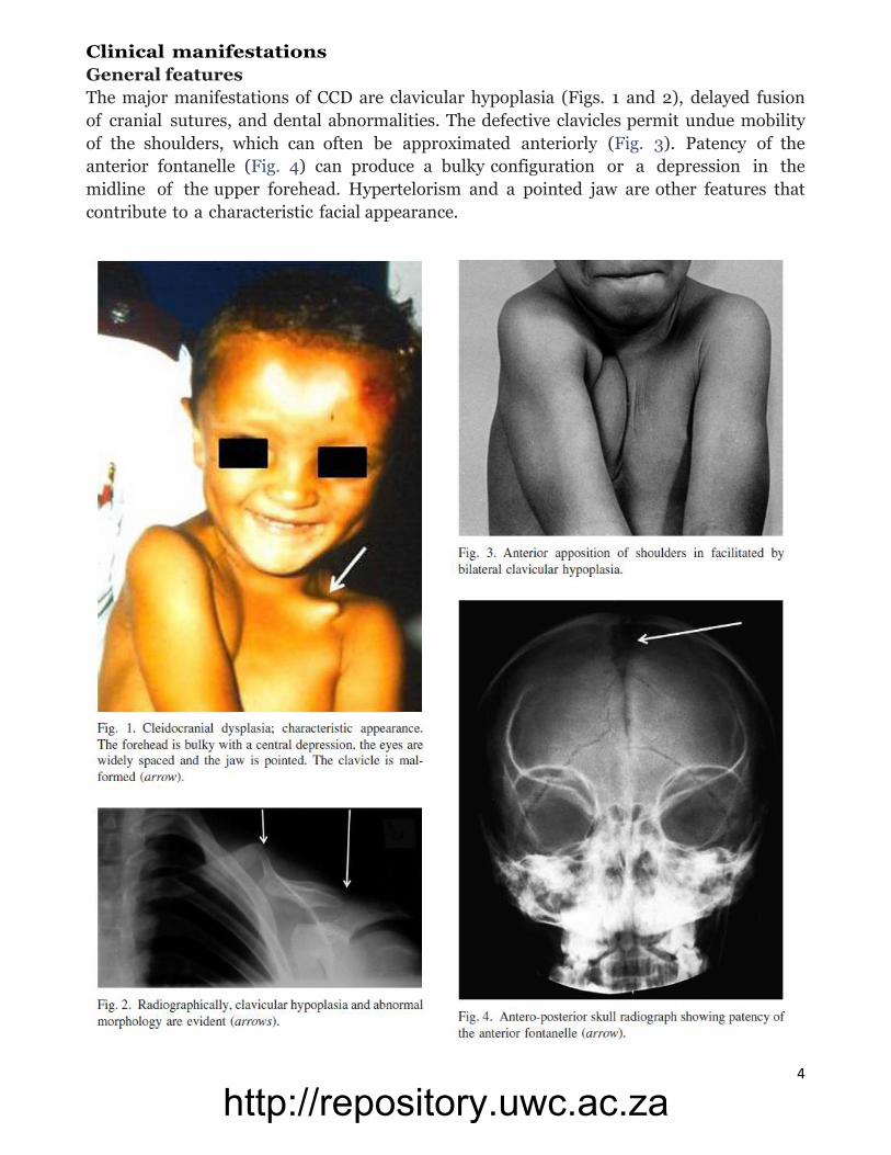
Clinical manifestations
General features
The major manifestations of CCD are clavicular hypoplasia (Figs. 1 and 2), delayed fusion
of cranial sutures, and dental abnormalities. The defective clavicles permit undue mobility
of the shoulders, which can often be approximated anteriorly (Fig. 3). Patency of the
anterior fontanelle (Fig. 4) can produce a bulky configuration or a depression in the
midline of the upper forehead. Hypertelorism and a pointed jaw are other features that
contribute to a characteristic facial appearance.
http://repository.uwc.ac.za

5
The number of teeth may be excessive (hyperdontia), and lead to dental crowding and
malalignment.23 Skeletal abnormalities may also occur, including slight stature, short
terminal phalanges, spinal malalignment, genu valgus (knock knees), and pes planus24 (flat
feet). Affected persons may experience recurrent infections of the upper respiratory tract
owing to maldevelopment of the sinuses, with a potential for hearing loss consequent upon
chronic otitis media.25 Despite these problems, CCD is very variable and often
comparatively mild; apart from dental complications, affected persons usually have little
disability.
Radiographically, Wormian bones may be evident in the cranial sutures (Fig. 5).26
hypoplastic or absent; these anomalies are usually bilateral but not symmetric. Other
skeletal abnormalities include a wide pubic symphysis, dysplastic scapulae, coxa vara, and
a variety of vertebral anomalies. These changes are variable and frequently clinically
insignificant.
http://repository.uwc.ac.za
6
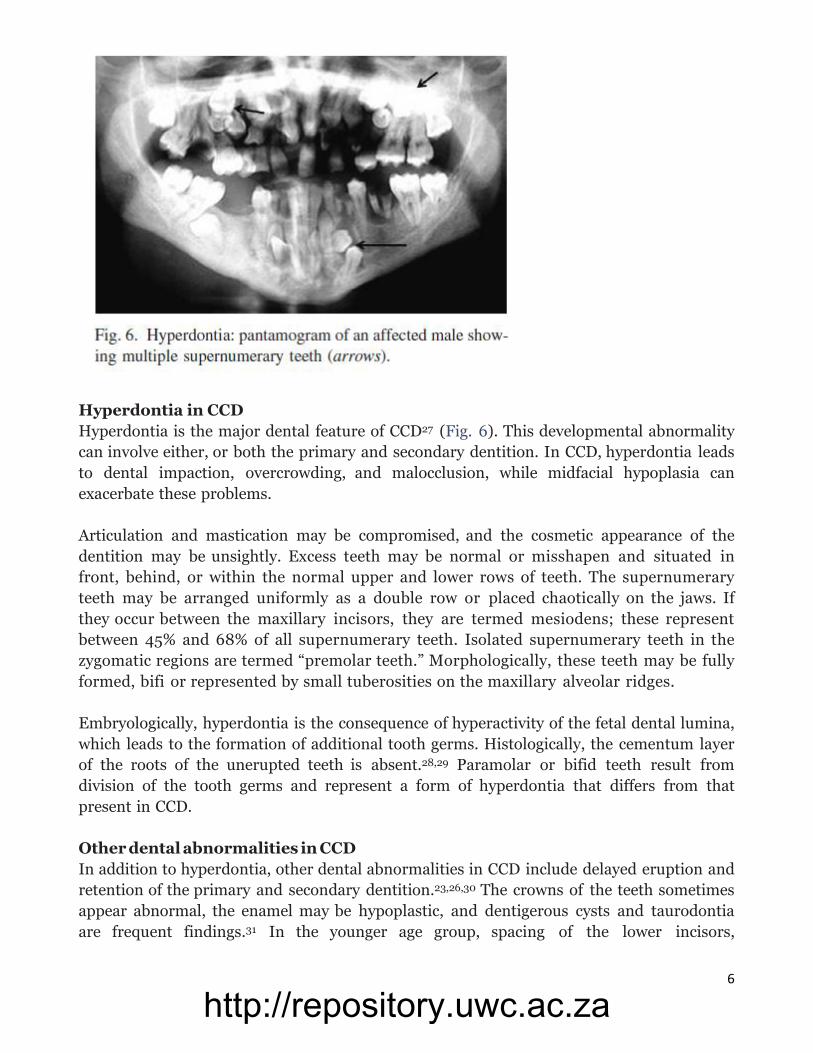
Hyperdontia in CCD
Hyperdontia is the major dental feature of CCD27 (Fig. 6). This developmental abnormality
can involve either, or both the primary and secondary dentition. In CCD, hyperdontia leads
to dental impaction, overcrowding, and malocclusion, while midfacial hypoplasia can
exacerbate these problems.
Articulation and mastication may be compromised, and the cosmetic appearance of the
dentition may be unsightly. Excess teeth may be normal or misshapen and situated in
front, behind, or within the normal upper and lower rows of teeth. The supernumerary
teeth may be arranged uniformly as a double row or placed chaotically on the jaws. If
they occur between the maxillary incisors, they are termed mesiodens; these represent
between 45% and 68% of all supernumerary teeth. Isolated supernumerary teeth in the
zygomatic regions are termed “premolar teeth.” Morphologically, these teeth may be fully
formed, bifi or represented by small tuberosities on the maxillary alveolar ridges.
Embryologically, hyperdontia is the consequence of hyperactivity of the fetal dental lumina,
which leads to the formation of additional tooth germs. Histologically, the cementum layer
of the roots of the unerupted teeth is absent.28,29 Paramolar or bifid teeth result from
division of the tooth germs and represent a form of hyperdontia that differs from that
present in CCD.
Other dental abnormalities in CCD
In addition to hyperdontia, other dental abnormalities in CCD include delayed eruption and
retention of the primary and secondary dentition.23,26,30 The crowns of the teeth sometimes
appear abnormal, the enamel may be hypoplastic, and dentigerous cysts and taurodontia
are frequent findings.31 In the younger age group, spacing of the lower incisors,
http://repository.uwc.ac.za
7
supernumerary tooth buds, and parallel-sided rami are consistent manifestations.
Radiographic manifestations in children include rounded gonion angles, kyphotic sphenoid
bones, and Wormian bones in the cranial sutures.26
Differential diagnosis of cleidocranial dysplasia and hyperdontia
As CCD is an autosomal dominant trait, recognition of occurrence of the disorder in family
members is important in the diagnostic process. The presence of clavicular hypoplasia is
strongly suggestive of CCD, but this anomaly can also occur as an isolated nonsyndromic
entity, which is usually unilateral. Complete absence of both clavicles is a manifestation of
the Yunisaron syndrome (OMIM 216340). In this rare genetic disorder, intellectual
dysfunction and anomalies of the hands and feet are associated with malformations in
other systems.32
Defective cranial ossification leading to patency of the anterior fontanelle and Wormian
bones in the sutures is an important feature of CCD. Similar manifestations occur in
osteogenesis imperfecta (frequent fractures), pycnodysostosis (skeletal density), and
congenital hypothyroidism (disturbed thyroid metabolism).
Hyperdontia is frequently the presenting feature in CCD, and awareness of this
diagnostic possibility is important in dental practice. Apart from CCD, supernumerary
teeth may be sporadic or familial.33 The familial form is inherited as an isolated autosomal
dominant trait, with reduced penetrance and variable phenotypic expression. Hyperdontia
may also be a component of specific genetic syndromes, including the Gardner syndrome
(OMIM 175100) (familial polyposis of the colon and osteomata), Hallerman-Streiff syndrome
(OMIM 234100) (narrow face, hypotrichosis, microphthalmia), and the orofaciodigital
syndrome type I (OMIM 311200). In these conditions, the hyperdontia is overshadowed by
other nondental syndromic manifestations that can have a significant impact on normal
development and health. In these circumstances, diagnostic precision facilitates
appropriate medical management and meaningful genetic counseling. Equally, in special
demographic circumstances, such as the high frequency of CCD in Cape Town, the presence
of hyperdontia raises a strong possibility that the affected person has CCD.
Genetic background of oral manifestations in CCD
Although CCD is comparatively uncommon, it has a wide geographic distribution. This
situation can be explained by the benign nature of the disorder and the ongoing random
occurrence of new mutations in the determinant gene. In this scenario, there is little
biological pressure against autosomal dominant transmission from generation to
generation and a chance mutation or a founder effect can be perpetuated in a particular
population. This situation is exemplified by the large extended CCD family in Cape Town, in
which numerous persons are affected.3
The molecular defect in CCD is situated at the chromosomal locus of 6p2116 and the
causative gene in the South African family is located at this site.21 The determinant gene,
RUNX2 codes for a core-binding transcription factor protein (CBFA1), which is involved in
http://repository.uwc.ac.za
8
the differentiation of osteoblasts and bone formation.1,17,18 RUNX2 plays an important
role in the epithelial-mesenchymal interactions that control progressive tooth
morphogenesis and histodifferentiation of the epithelial enamel organ.
The supernumerary teeth in CCD may result from the lack of inhibition or incomplete
resorption of tooth bud formation. Supernumerary teeth may also result from the presence
of remnants of dental laminae following dental extraction. These epithelial cell rests are
usually resorbed during the normal tooth morphogenesis.34
Experimental studies have revealed that mice lacking the RUNX2 gene fail to develop bone
and tooth structure, whereas mice with mutant RUNX2 genes show arrested tooth
development.35 The most common site of RUNX2 gene expression during odontogenesis is
the papillary mesenchyme; levels are highest before the development of the tooth crown
but taper after completion of crown formation.36 In mice, the RUNX2 gene is also expressed
in the mesenchyme of the dental follicle and periodontal ligament before tooth eruption. A
lack of both alleles of the RUNX2 gene results in absence of osteoblastic differentiation,
whereas haplo-insufficiency of RUNX2 in mice impairs the differentiation and recruitment
of osteoclasts together with reduction in the capacity of periodontal ligament cells to
induce active osteoclastic differentiation. These processes could, in part, account for
delayed tooth eruption patterns in humans with CCD.37-39
Bone is formed by 2 processes, namely, endochondral and intramembranous osteogenesis,
both of which require the presence of the RUNX2 protein. The formation and
development of both the cranium and clavicles occur by intramembranous ossification
Although the clavicles are the fi embryonic bones to ossify, the maturation process is
slow. In mice, clavicular defects result from the disruption of intramembranous bone
formation during embryogenesis. Low levels of functional RUNX2 protein are implicated
as the causative agent. Although this process begins during early embryonic development,
the effects are evident in adult mice. The mouse model offers a reasonable explanation
of the clavicle and cranial abnormalities occurring in CCD in humans. It also suggests
that the levels of normal RUNX2 proteins are critical for the successful
intramembranous ossification during embryogenesis.
There is considerable intragenic heterogeneity in CCD, and numerous different
mutations have been identifi within the RUNX2 gene.40,41 Evidence has been advanced
for genotype-phenotype correlation,42 including dental abnormalities.43 In a series of 24
Japanese persons with CCD, it was found that small stature and the number of
supernumerary teeth werepositively correlated.44 Disparity in hyperdontia in affected
siblings has been documented.45
In a further study of affected persons in Japan, mutational analysis revealed that a wide
range of supernumerary teeth can occur in the presence of identical RUNX2
mutations.46 These authors suggested that hyperdontia in CCD might be regulated by
environmental influence together with epigenetic factors and copy number variation.
http://repository.uwc.ac.za
9
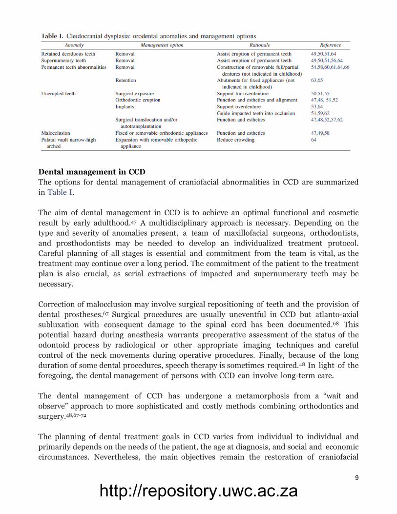
Dental management in CCD
The options for dental management of craniofacial abnormalities in CCD are summarized
in Table I.
The aim of dental management in CCD is to achieve an optimal functional and cosmetic
result by early adulthood.47 A multidisciplinary approach is necessary. Depending on the
type and severity of anomalies present, a team of maxillofacial surgeons, orthodontists,
and prosthodontists may be needed to develop an individualized treatment protocol.
Careful planning of all stages is essential and commitment from the team is vital, as the
treatment may continue over a long period. The commitment of the patient to the treatment
plan is also crucial, as serial extractions of impacted and supernumerary teeth may be
necessary.
Correction of malocclusion may involve surgical repositioning of teeth and the provision of
dental prostheses.67 Surgical procedures are usually uneventful in CCD but atlanto-axial
subluxation with consequent damage to the spinal cord has been documented.68 This
potential hazard during anesthesia warrants preoperative assessment of the status of the
odontoid process by radiological or other appropriate imaging techniques and careful
control of the neck movements during operative procedures. Finally, because of the long
duration of some dental procedures, speech therapy is sometimes required.48 In light of the
foregoing, the dental management of persons with CCD can involve long-term care.
The dental management of CCD has undergone a metamorphosis from a “wait and
observe” approach to more sophisticated and costly methods combining orthodontics and
surgery.48,67-72
The planning of dental treatment goals in CCD varies from individual to individual and
primarily depends on the needs of the patient, the age at diagnosis, and social and economic
circumstances. Nevertheless, the main objectives remain the restoration of craniofacial
http://repository.uwc.ac.za
10
and dental function together with esthetics.49 Although there are numerous options, there is
a general consensus that the best results are obtained if the condition is diagnosed and
treated at an early age.
The most popular orthodontic-surgical regimes are the Toronto-Melbourne, Belfast-
Hamburgh, and Jerusalem approaches (Table II). The Toronto-Melbourne approach is
based on timed, serial extraction of deciduous teeth and depends on the extent to which the
roots of the permanent teeth have developed. During each procedure, which is performed
under general anesthesia, supernumerary teeth are also removed together with the bone
covering the underlying permanent teeth. The rationale is to facilitate the spontaneous
eruption of the unerupted permanent teeth.69,70
The Belfast-Hamburg approach advocates a single surgical procedure under general
anesthesia to extract all retained deciduous and supernumerary teeth. In addition, all
unerupted permanent teeth are exposed and the surgical sites are allowed to heal. After
healing is complete, orthodontic appliances are placed on fully erupted teeth with traction
bands attached to partially erupted teeth so as to promote further eruption of the
latter.50,73 The advantage of this procedure is that the patient is exposed to only a single
surgical operation under general anesthesia.
The Jerusalem approach is based on at least 2 surgical interventions, the timing of which is
dependent on the root development of the permanent dentition. During the fi procedure, the
anterior deciduous teeth and supernumerary teeth are extracted and the permanent anterior
teeth are exposed. At the same time, orthodontic brackets and traction elastics are applied and
surgical flaps are closed. During the second component of the Jerusalem approach, which
takes place at approximately 13 years of age, the residual primary teeth are extracted,
unerupted canines and premolars are exposed, and the necessary orthodontic and surgical
processes are completed.
http://repository.uwc.ac.za

11
The Bronx approach uses 2, and at most 3, surgical interventions.58 As in the Toronto-
Melbourne and Jerusalem techniques, deciduous teeth and underlying supernumerary
teeth are removed under general anesthesia and surgical flaps are closed. Unlike the
previously documented techniques, this approach uses the placement of a removable
partial overdenture for esthetic and functional purposes. As with the Toronto-Melbourne
and Jerusalem techniques, the age at which the management commences depends on the
stage of root development of the underlying permanent teeth. If necessary, an intermediate
operation is undertaken so as to expose unerupted teeth and place orthodontic brackets
over fully erupted molars. A transpalatal arch appliance is welded to the brackets and these
are used in conjunction as a base for an artificial dentition. After the natural eruption of the
permanent teeth with sufficient posterior support, orthodontic appliances are used to bring
the teeth into occlusion. Finally, a Leforte I osteotomy-orthognathic procedure is
performed and dental implants are placed.
http://repository.uwc.ac.za
12
These procedures are all undertaken over a long period. It is relevant that patient
compliance is essential to a favorable outcome for any of these modalities. In addition, they
each have individual benefits and short- comings.
In South Africa, the dental and orthodontic approach to CCD has several constraints.
Extensive medical expertise is available, but access is limited and costly. Initially, most
persons with dental problems visit primary health care facilities, which are often
understaffed and overcrowded. Medical and dental professionals at these institutions may
not have adequate experience to diagnose conditions such as CCD. Once diagnosed,
however, patients are referred to specialized facilities that are located in the major cities
and often over-booked, making early intervention challenging. Surgical, orthodontic, and
prosthodontic procedures are expensive and, when presented with any of the management
strategies and the projected cost of treatment, many patients decide not to proceed. In these
instances, alternative, more cost-effective strategies can be offered to the patient with CCD.
These options may include removal of non-functional deciduous teeth, erupted
supernumerary teeth that are not in occlusion, or teeth that may eventually cause
complications. Edentulous areas can be managed with removable prostheses. Dentures could
be adjusted and/or replaced as the individual grows or as supernumerary teeth erupt.
The high prevalence of substance abuse in the general population of South Africa,
including individuals affected with CCD, has an influence on dental management at
different levels. For example, a young male patient with classic features of CCD was
recently referred to the Faculty of Dentistry, University of Western Cape, for dental
management. In addition to the orodental manifestations of CCD, he also had poor oral
hygiene and multiple carious teeth. He was initially regarded as having mild
intellectual disability but on further clinical investigation, it emerged that he was a
regular user of methamphetamine (or “tik” as it is termed in South Africa). Individuals
who are regular uses frequently have defective oral hygiene and multiple carious teeth.
His poor oral health status and diminished mental capacity was probably a reflection of
drug abuse and poor socioeconomic status. After extensive dental management
planning, the young man avoided further treatment. This situation often arises in South
Africa and compounds difficulties in the management of complicated orofacial disorders.
In addition to poverty, lack of education, and drug abuse, HIV infection is another
negative factor. Moreover, the cost of private dental treatment is unaffordable by the
average person. These problems reflect the situation in other developing countries, and in
these circumstances, it is evident that the provision of sophisticated facilities needs to be
balanced against the dental needs of the general population.
We are grateful to Greta for her efficient preparation and processing of the manuscript.
http://repository.uwc.ac.za
13
References
1. Mundlos S. Cleidocranial dysplasia: clinical and molecular genetics. J Med Genet
1999;36:177-82.
2. Cooper SC, Flaitz CM, Johnston DA, Lee B, Hecht JT. A natural history of
cleidocranial dysplasia. Am J Med Genet 2001; 104:1-6.
3. Jackson WP. Osteo-dental dysplasia (cleido-cranial dysostosis): the “Arnold head”.
Acta Med Scand 1951;139:292-303.
4. Greig DM. A neanderthaloid skull presenting features of cleido-cranial dysostosis and
other peculiarities. Edinb Med J 1933;40:497-557.
5. Bartsocas CS. Cleidocranial dysostosis in Homer. Archeia Ellin Pediátr Etair
1973;36:107.
6. Bartsocas CS. Stature of Greeks of the Pylos area during the second millennium BC.
Hippocrates (Athens) 1977;2:157.
7. Paultauf R. Demonstration eines Skelettes von ein Falle von dysostosis
cleidocranialis. Verh Dtsch Pathol Ges 1912:337-53.
8. Beighton P, Sujansky E, Patzak B, Portele KA. Genetic skeletal dysplasias in the
Museum of Pathological Anatomy, Vienna. Am J Med Genet 1993;47:843-7.
9. Meckel JF. Cited by Siggers CD. Cleidocranial dysostosis. Dev Med Child Neurol
1975;17:522-4.
10. Martin. Sur déplacement de la clavicule. J Med Chir Pharmacol 1765;23:456-460.
Cited by Gorlin RJ, Cohen MM, Hannekom RCM. Syndromes of the head and neck.
Oxford, UK: Oxford University Press, 2001. p. 310.
11. Scheuthauer G. Kombination rudimentärer Schlüsselbeine mit Anomalien des
Schädels beim Erwachsenen Menschen. Allg Wein Med Ztg 1871;16:293-295.
12. Marie P, Sainton P. Observation d’hydrocéphalie héréditaire (père et fils) par vice de
development du crane et du cerveau. Bull Soc Med Hop Paris 1897;14:706-12.
13. Marie P, Sainton P. On hereditary cleidocranial dysostosis. Rev Neurol 1898;6:835.
14. Hultkranz JW. U ber dysostosis cleidocranialis. Z Morphol Anthropol 1908;11:385-
528.
15. Jensen BL. Somatic development in cleidocranial dysplasia. Am J Med Genet
1990;35:69-74.
16. Mundlos S, Mulliken JB, Abramson DL, Warman ML, Knoll JH, Olsen BR. Genetic
mapping of cleidocranial dysplasia and evidence of a microdeletion Dental
abnormalities in cleidocranial dysplasia in one family. Hum Mol Genet 1995;4:71-5.
17. Mundlos S, Otto F, Mundlos C, Mulliken JB, Aylsworth AS, Albright S, et al.
Mutations involving the transcription factor CBFA1 cause cleidocranial dysplasia.
Cell 1997;89:773-9.
18. Li Y, Pan W, Xu W, He N, Chen X, Liu H, et al. RUNX2 mutations in Chinese
patients with cleidocranial dysplasia. Mutagenesis 2009;24:425-31.
19. Staz J. Proceedings of the 21st International Congress of the SA. Division of IADR –
Abstract. J Dent Res 1987;67:778.
20. Staz J. Cleidocranial dysplasia in the south western Cape: preliminary report. J Dent
Assoc S Afr 1988;43:547-51.
http://repository.uwc.ac.za
14
21. Ramesar RS, Greenberg J, Martin R, Goliath R, Bardien S, Mundlos S, Beighton P.
Mapping of the gene for cleidocranial dysplasia in the historical Cape Town (Arnold)
kindred and evidence for locus homogeneity. J Med Genet 1996;33:511-4.
22. Dawjee SM, Nkhumeleni F. Cleidocranial dysplasia—a case report. SADJ
2002;57:187-90.
23. Brooks JK, Nikitakis NG. Multiple unerupted teeth. Cleidocranial dysplasia. Gen
Dent 2008;56:395-6.
24. Garg RK, Agrawal P. Clinical spectrum of cleidocranial dysplasia: a case report. Cases
J 2008;1:377.
25. Segal N, Puterman M. Cleidocranial dysplasia—review with an emphasis on otological
and audiological manifestations. Int J Pediatr Otorhinolaryngol 2007;71:523-6.
26. Golan I, Baumert U, Hrala BP, Müssig D. Early craniofacial signs of cleidocranial
dysplasia. Int J Paediatr Dent 2004; 14:49-53.
27. Gorlin RJ, Cohen MM Jr, Hennekam RCM. Syndromes of the head and neck. 4th
ed. Oxford, UK: Oxford University Press; 2001. p. 306-10.
28. Rushton MA. An anomaly of cementum in cleidocranial dysostosis. Br Dent J
1956;100:81-3.
29. Manjunath K, Kavitha B, Saraswathi TR, Sivapathasundharam B, Manikandhan R.
Cementum analysis in cleidocranial dysostosis. Indian J Dent Res 2008;19:253-6.
30. Golan I, Baumert U, Hrala BP, Müssig D. Dentomaxillofacial variability of
cleidocranial dysplasia: clinicoradiological presentation and systematic review.
Dentomaxillofac Radiol 2003; 32:347-54.
31. McNamara CM, O’Riordan BC, Blake M, Sandy JR. Cleidocranial dysplasia:
radiological appearances on dental panoramic radiography. Dentomaxillofac Radiol
1999;28:89-97.
32. Corona-Rivera JR, Romo-Huerta CO, López-Marure E, Ramos FJ, Estrada-Padilla
SA, Zepeda-Romero LC. New ocular findings in two sisters with Yunis-Varon
syndrome and literature review. Eur J Med Genet 2011;54:76-81.
33. Díaz A, Orozco J, Fonseca M. Multiple hyperodontia: report of a case with 17
supernumerary teeth with nonsyndromic association. Med Oral Patol Oral Cir Bucal
2009;14:E229-231.
34. Lukinmaa PL, Jensen BL, Thesleff I, Andreasen JO, Kreiborg S. Histological
observations of teeth and peridental tissues in cleidocranial dysplasia imply
increased activity of odontogenic epithelium and abnormal bone remodeling. J
Craniofac Genet Dev Biol 1995;15:212-21.
35. Åberg T, Cavender A, Gaikwad JS, Bronckers AL, Wang X, Waltimo-Sirén J,
Thesleff I, et al. Phenotypic changes in dentition of RUNX2 homozygote-null mutant
mice. J Histochem Cytochem 2004;52:131-9.
36. D’Souza RN, Åberg T, Gaikwad J, Cavender A, Owen M, Karsenty G, Thesleff I. Cbfa1
is required for epithelial-mesenchymal interactions regulating tooth development in
mice. Development 1999;126:2911-20.
37. Kreiborg S, Jensen BL, Larsen P, Schleidt DT, Darvann T. Anomalies of craniofacial
skeleton and teeth in cleidocranial dysplasia. J Craniofac Genet Dev Biol
1999;19:75-9.
http://repository.uwc.ac.za
15
38. Yoda S, Suda N, Kitahara Y, Komori T, Ohyama K. Delayed tooth eruption and
suppressed osteoclast number in the eruption pathway of heterozygous
RUNX2/Cbfa1 knockout mice. Arch Oral Biol 2004;49:35-42.
39. Lossdörfer S, Abou Jamra B, Rath-Deschner B, Götz W, Abou Jamra R, Braumann B,
Jg er A. The role of periodontal ligament cells in delayed tooth eruption in patients
with cleidocranial dysostosis. J Orofac Orthop. 2009;70:495-510.
40. Zhang CY, Zheng SG, Wang YX, Zhu JX, Zhu X, Zhao YM, Ge LH. Novel RUNX2
mutations in Chinese individuals with cleidocranial dysplasia. J Dent Res
2009;88:861-6.
41. Ryoo HM, Kang HY, Lee SK, Lee KE, Kim JW. RUNX2 mutations in cleidocranial
dysplasia patients. Oral Dis 2009; 16:55-60.
42. Zhou G, Chen Y, Zhou L, Thirunavukkarasu K, Hecht J, Chitayat D, et al. CBFA1
mutation analysis and functional correlation with phenotypic variability in
cleidocranial dysplasia. Hum Mol Genet 1999;8:2311-6.
43. Quack I, Vonderstrass B, Stock M, Aylsworth AS, Becker A, Brueton L, et al.
Mutation analysis of core binding factor A1 in patients with cleidocranial dysplasia.
Am J Hum Genet 1999; 65:1268-78.
44. Yoshida T, Kanegane H, Osato M, Yanagida M, Miyawaki T, Ito Y, Shigesada K.
Functional analysis of RUNX2 mutations in Japanese patients with cleidocranial
dysplasia demonstrates novel genotype-phenotype correlations. Am J Hum Genet
2002;71:724-38.
45. Suda N, Hamada T, Hattori M, Torii C, Kosaki K, Moriyama K. Diversity of
supernumerary tooth formation in siblings with cleidocranial dysplasia having
identical mutation in RUNX2: possible involvement of non-genetic or epigenetic
regulation. Orthod Craniofac Res 2007;10:222-5.
46. Suda N, Hattori M, Kosaki K, Banshodani A, Kozai K, Tanimoto K, Moriyama K.
Correlation between genotype and supernumerary tooth formation in cleidocranial
dysplasia. Orthod Craniofac Res 2010;13:197-202.
47. Becker A, Lustman J, Shteyer A. Cleidocranial dysplasia: part 1—General principles
of the orthodontic and surgical treatment modality. Am J Orthod Dentofacial Orthop
1997;111:28-33.
48. Becker A, Shteyer A, Bimstein E, Lustmann J. Cleidocranial dysplasia: part 2—
treatment protocol for the orthodontic and surgical modality. Am J Orthod
Dentofacial Orthop 1997; 111:178-83.
49. D’Alessandro G, Tagariello T, Piana G. Craniofacial changes and treatment of the
stomatognathic system in subjects with cleidocranial dysplasia. Eur J Paediatr Dent
2010;11:39-43.
50. Richardson A, Swinson T. Combined orthodontic and surgical approach to
cleidocranial dysostosis. Trans Eur Orthod Soc 1987;63:23.
51. Angle AD, Rebellato J. Dental team management for a patient with cleidocranial
dysostosis. Am J Orthod Dentofacial Orthop 2005;128:110-1.
52. Becker A. Cleidocranial dysplasia. In: Becker A, editor. The orthodontic treatment
of impacted teeth. St Louis: Mosby; 1998. p. 199-229.
http://repository.uwc.ac.za
16
53. Becktor KB, Becktor JP, Keller EE. Growth analysis of a patient with ectodermal
dysplasia treated with endosseous implants: a case report. Int J Oral Maxillofac
Implants 2001;16:864-74.
54. Fardy MJ. Cleidocranial dysostosis: some problems in the dental management of
occlusion. Dent Update 1984;11:363-8.
55. Hitchin AD, Fairley JM. Dental management in cleido-cranial dysostosis. Br J Oral
Surg 1974;12:46-55.
56. Hemalatha R, Balasubramaniam MR. Cleidocranial dysplasia: a case report. J Indian
Soc Pedod Prevent Dent. 2008;26:40-43.
57. Jensen BL, Kreiborg S. Dental treatment strategies in cleidocranial dysplasia. Br
Dent J 1992;172:243-7.
58. Kelly E, Nakamoto RY. Cleidocranial dysostosis—a prosthodontic problem. J Prosthet
Dent 1974;31:518-26.
59. Lombardas P, Toothaker RW. Bone grafting and osseointegrated implants in the
treatment of cleidocranial dysplasia. Compend Contin Educ Dent 1997;18:509-14.
60. Magnus WW, Sands NR. Cleidocranial dysostosis—report of a case. Am J Orthod
1974;65:638-43.
61. Maw RB. Cleidocranial dysostosis: report of case. J Am Dent Assoc 1978;96:306-9.
62. Nordenram A. Autotransplantation of teeth in cleidocranial dysostosis. Odontol Revy
1971;22:363-70.
63. Pröbster L, Bachmann R, Weber H. Custom-made resin-bonded attachments
supporting a removable partial denture using the spark erosion technique: a case
report. Quintessence Int 1991; 22:349-54.
64. Suba Z, Balaton G, Gyulai-Gaal S, Balaton P, Barabas J, Tarjan Cleidocranial
dysplasia: diagnostic criteria and combined treatment. J Craniofac Surg 2005;6:1122-
6.
65. Trimble LD, West RA, McNeill RW. Cleidocranial dysplasia: comprehensive
treatment of the dentofacial abnormalities. J Am Dent Assoc 1982;105:661-6.
66. Winter GR. Dental conditions in cleidocranial dysostosis. Am J Orthod 1943;29:61-
89.
67. Kuroda S, Yanagita T, Kyung HM, Takano-Yamamoto T. Titanium screw anchorage
for traction of many impacted teeth in a patient with cleidocranial dysplasia. Am J
Orthod Dentofacial Orthop 2007;131:666-9.
68. Kobayashi S, Uchida K, Baba H, Takeno K, Yayama T, Nakajima H, et al.
Atlantoaxial subluxation-induced myelopathy in cleidocranial dysplasia. Case report.
J Neurosurg Spine 2007; 7:243-247.
69. Smylski PT, Woodside DG, Harnett BE. Surgical and orthodontic treatment of
cleidocranial dysostosis. Int J Oral Surg 1974;3:380-85.
70. Hall RK, Hyland AL. Combined surgical and orthodontic management of the oral
abnormalities in children with cleidocranial dysplasia. Int J Oral Surg 1978;7:267-73.
71. Daskalogiannakis J, Piedade L, Lindholm TC, Sándor GK, Carmichael RP.
Cleidocranial dysplasia: 2 generations of management. J Can Dent Assoc
2006;72:337-42.
http://repository.uwc.ac.za
17
72. Farronato G, Maspero C, Farronato D, Gioventù S. Orthodontic treatment in a
patient with cleidocranial dysostosis. Angle Orthod 2009;79:178-85.
73. Behlfelt K. Cleidocranial dysplasia: diagnosis and treatment concept. Trans Eur
Orthod Soc 1987;63:25.
74. Berg RW, Kurtz KS, Watanabe I, Lambrakos I. Interim prosthetic phase of
multidisciplinary managementof cleidocranial dysplasia: “the Bronx approach.” J
Prosthodont 2011;20:S20-5.
http://repository.uwc.ac.za