Embed Size (px)
Citation preview
1
To our limited intelligence, it would seem a simple task to divide a nucleus into equal parts. The cell, manifestly, entertains a very different opinion.�
E. B. Wilson, 1923
Cleavage
Rapid cell division without growth
Rate of cell division is unparallelled Examples: Early Drosophila embryo - mitosis occurs every 10 min for 2 hr (4096 nuclei); 50,000 cells in only 12 hr
Xenopus (frog) embryo - 37,000 cells /43 hr
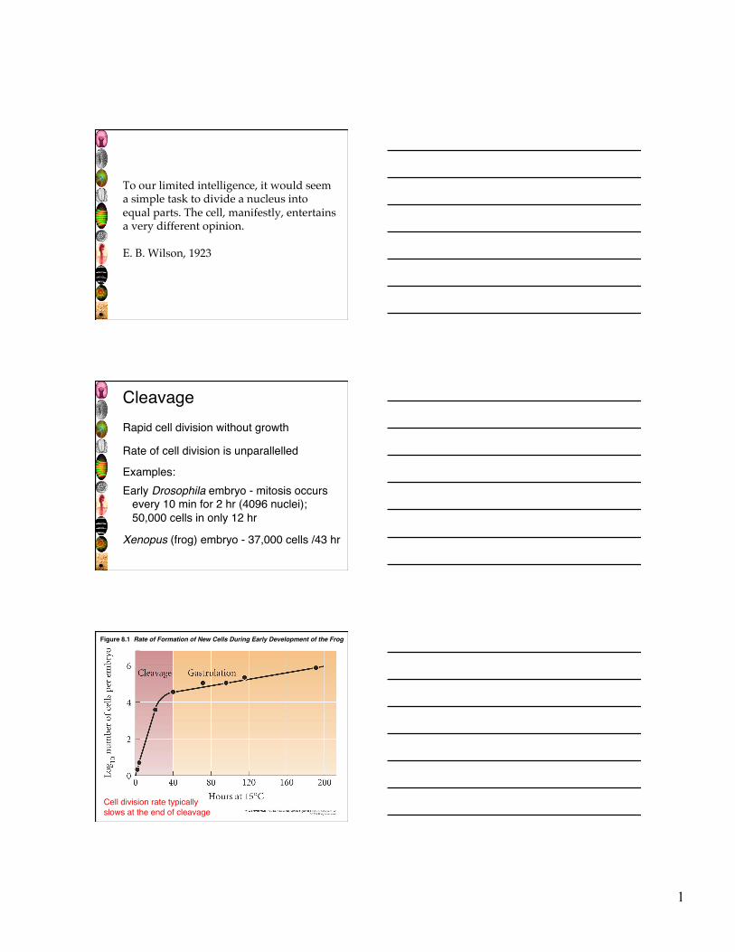
Figure 8.1 Rate of Formation of New Cells During Early Development of the Frog
Cell division rate typically slows at the end of cleavage
2
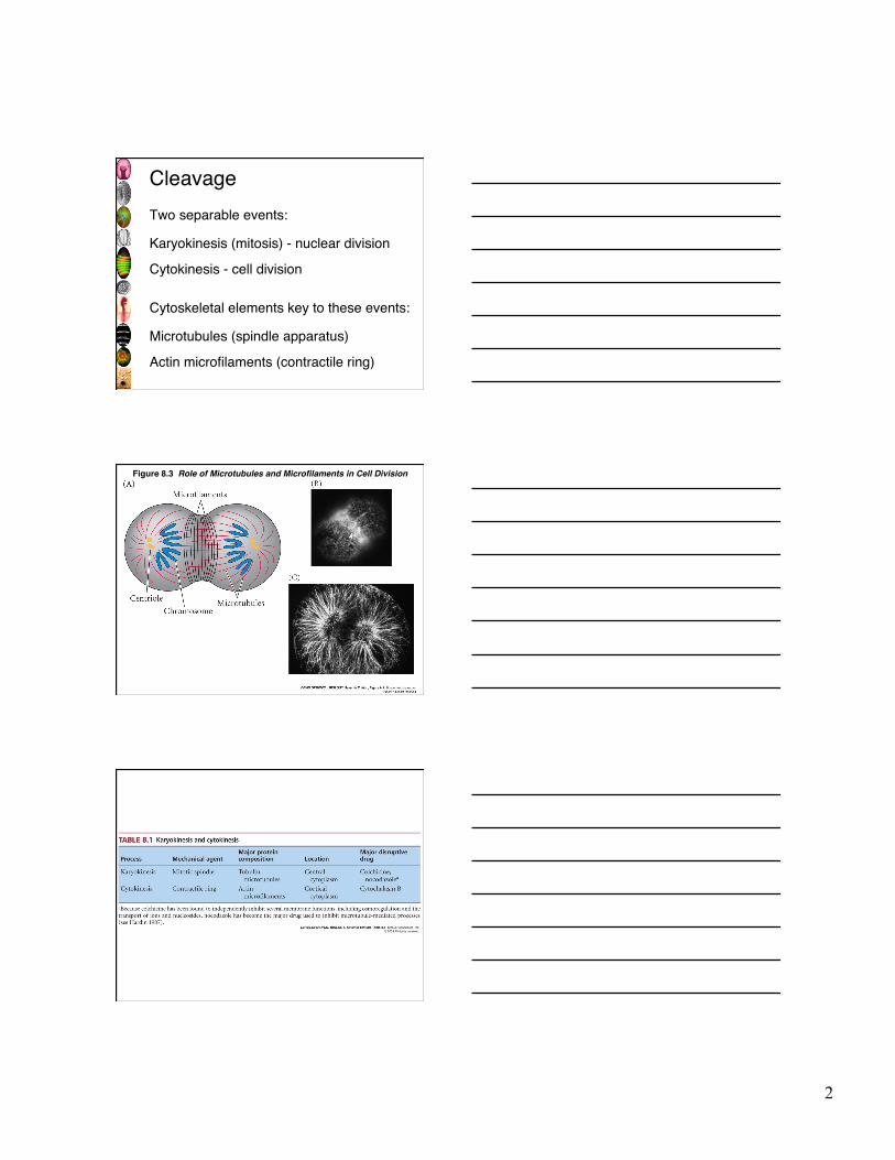
Cleavage
Two separable events:
Karyokinesis (mitosis) - nuclear division Cytokinesis - cell division
Cytoskeletal elements key to these events:
Microtubules (spindle apparatus) Actin microfilaments (contractile ring)
Figure 8.3 Role of Microtubules and Microfilaments in Cell Division
3
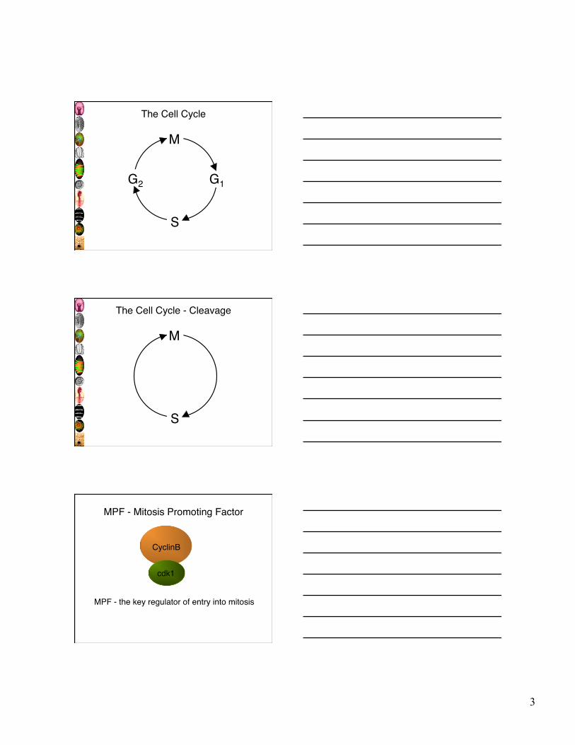
The Cell Cycle
M
S
G2 G1
The Cell Cycle - Cleavage
M
S
CyclinB
cdk1
MPF - Mitosis Promoting Factor
MPF - the key regulator of entry into mitosis
4
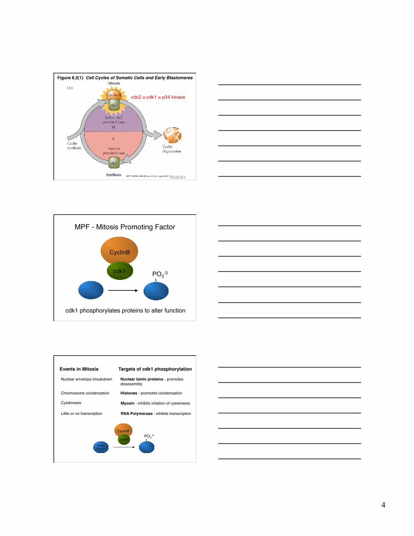
Figure 8.2(1) Cell Cycles of Somatic Cells and Early Blastomeres
cdc2 = cdk1 = p34 kinase
CyclinB
cdk1 PO3-3
MPF - Mitosis Promoting Factor
cdk1 phosphorylates proteins to alter function
Nuclear envelope breakdown Nuclear lamin proteins - promotes disassembly
Events in Mitosis Targets of cdk1 phosphorylation
CyclinB
cdk1 PO3
-3
Histones - promotes condensation Chromosome condensation
Cytokinesis Myosin - inhibits initation of cytokinesis
Little or no transcription RNA Polymerase - inhibits transcription
Target Protein
5
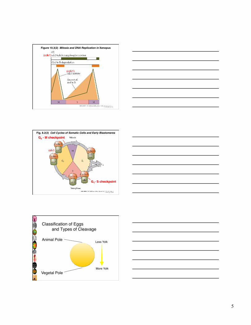
Figure 10.3(2) Mitosis and DNA Replication in Xenopus
(cdk1)
(cdk1)
Fig. 8.2(2) Cell Cycles of Somatic Cells and Early Blastomeres
G1- S checkpoint
G2 - M checkpoint
cdk1
Classification of Eggs and Types of Cleavage
Animal Pole
Vegetal Pole More Yolk
Less Yolk
6
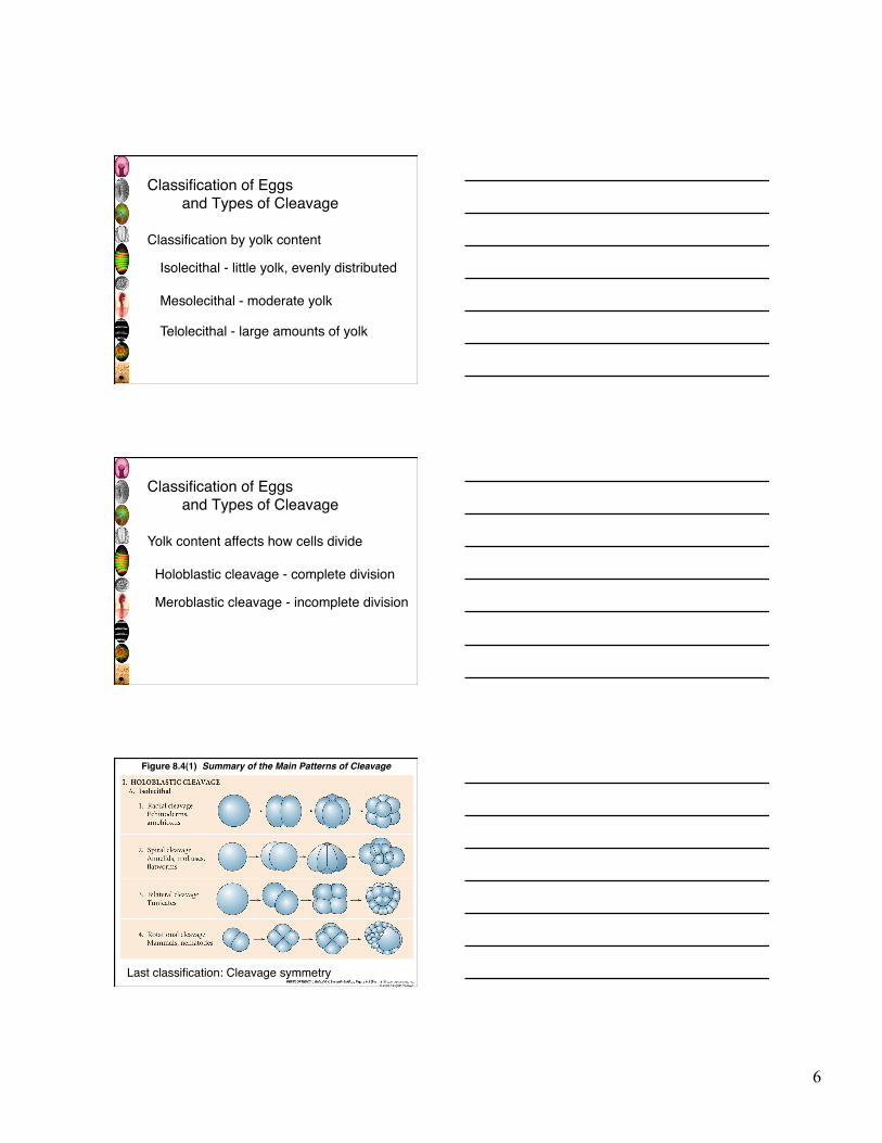
Classification of Eggs and Types of Cleavage
Classification by yolk content
Isolecithal - little yolk, evenly distributed
Mesolecithal - moderate yolk
Telolecithal - large amounts of yolk
Classification of Eggs and Types of Cleavage
Yolk content affects how cells divide
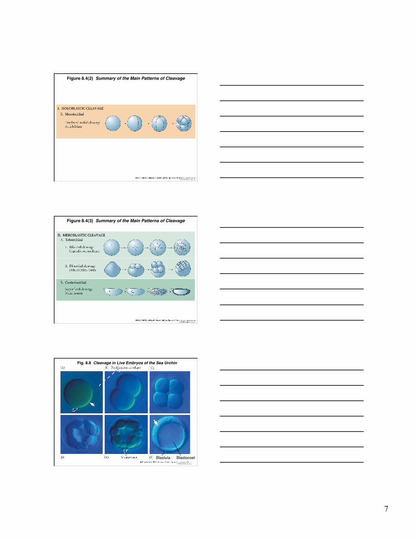
Holoblastic cleavage - complete division
Meroblastic cleavage - incomplete division
Figure 8.4(1) Summary of the Main Patterns of Cleavage
Last classification: Cleavage symmetry
7
Figure 8.4(2) Summary of the Main Patterns of Cleavage
Figure 8.4(3) Summary of the Main Patterns of Cleavage
Fig. 8.8 Cleavage in Live Embryos of the Sea Urchin
Blastula Blastocoel
8
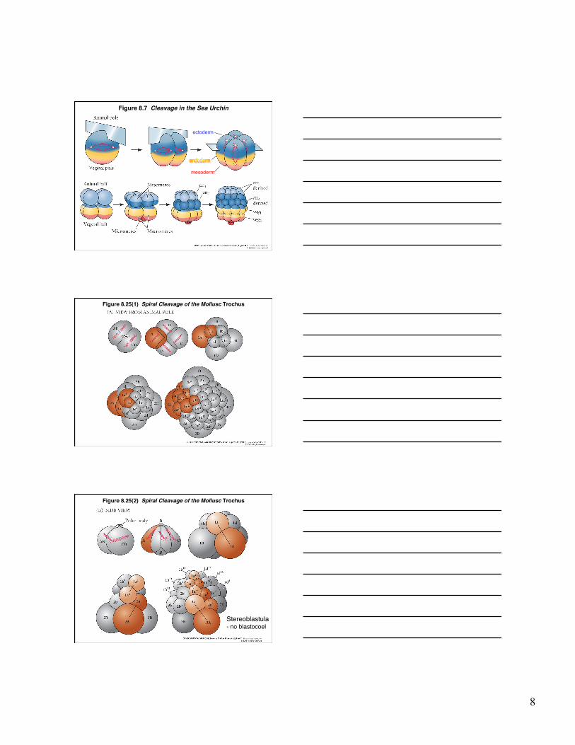
Figure 8.7 Cleavage in the Sea Urchin
mesoderm
ectoderm
Figure 8.25(1) Spiral Cleavage of the Mollusc Trochus
Figure 8.25(2) Spiral Cleavage of the Mollusc Trochus
Stereoblastula - no blastocoel
9
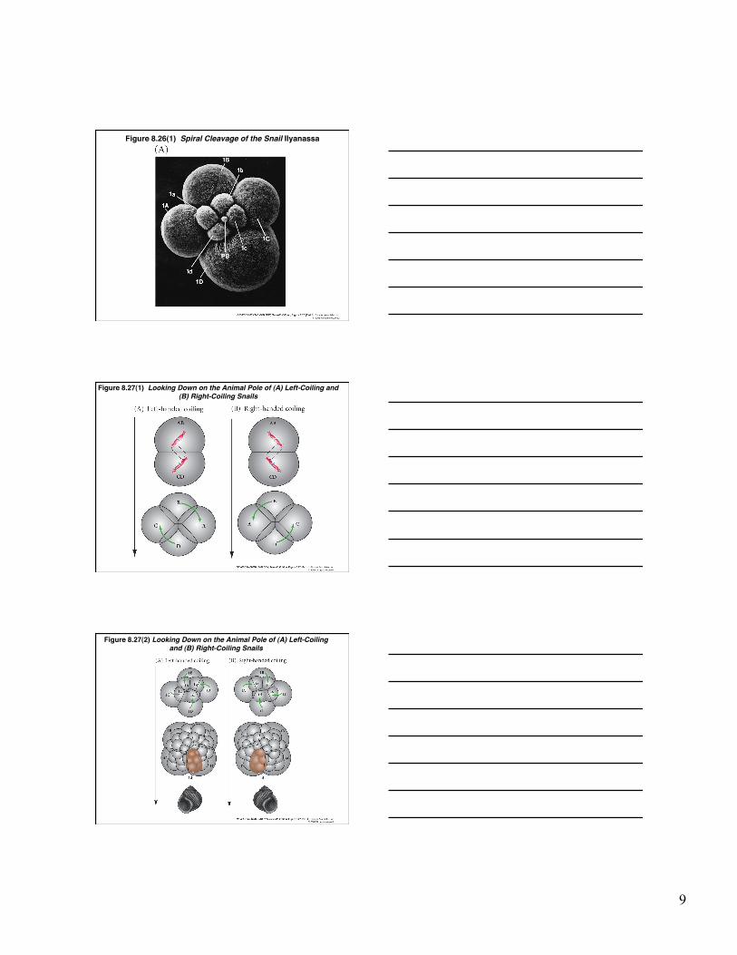
Figure 8.26(1) Spiral Cleavage of the Snail Ilyanassa
Figure 8.27(1) Looking Down on the Animal Pole of (A) Left-Coiling and (B) Right-Coiling Snails
Figure 8.27(2) Looking Down on the Animal Pole of (A) Left-Coiling and (B) Right-Coiling Snails
10
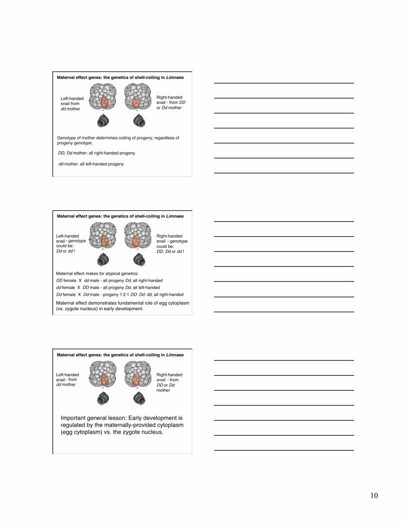
Maternal effect genes: the genetics of shell-coiling in Limnaea
Genotype of mother determines coiling of progeny, regardless of progeny genotype.
Right-handed snail - from DD or Dd mother
Left-handed snail from dd mother
DD, Dd mother: all right-handed progeny
dd mother: all left-handed progeny
Maternal effect genes: the genetics of shell-coiling in Limnaea
Maternal effect makes for atypical genetics:
Right-handed snail
Left-handed snail
DD female X dd male - all progeny Dd, all right-handed dd female X DD male - all progeny Dd, all left-handed Dd female X Dd male - progeny 1:2:1 DD: Dd: dd, all right-handed
Maternal effect demonstrates fundamental role of egg cytoplasm (vs. zygote nucleus) in early development.
- genotype could be: Dd or dd !
- genotype could be: DD, Dd or dd !
Maternal effect genes: the genetics of shell-coiling in Limnaea
Important general lesson: Early development is regulated by the maternally-provided cytoplasm (egg cytoplasm) vs. the zygote nucleus.
Right-handed snail
Left-handed snail - from dd mother
- from DD or Dd mother
11
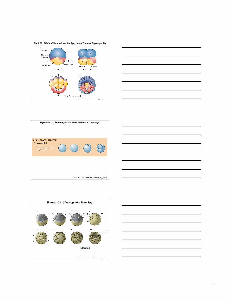
Fig. 8.36 Bilateral Symmetry in the Egg of the Tunicate Styela partita
Figure 8.4(2) Summary of the Main Patterns of Cleavage
Figure 10.1 Cleavage of a Frog Egg
Blastula
12
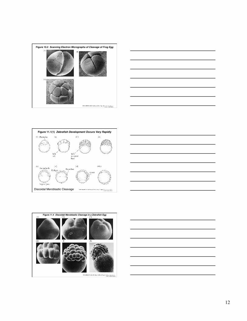
Figure 10.2 Scanning Electron Micrographs of Cleavage of Frog Egg
Figure 11.1(1) Zebrafish Development Occurs Very Rapidly
Discoidal Meroblastic Cleavage
Figure 11.4 Discoidal Meroblastic Cleavage in a Zebrafish Egg
13
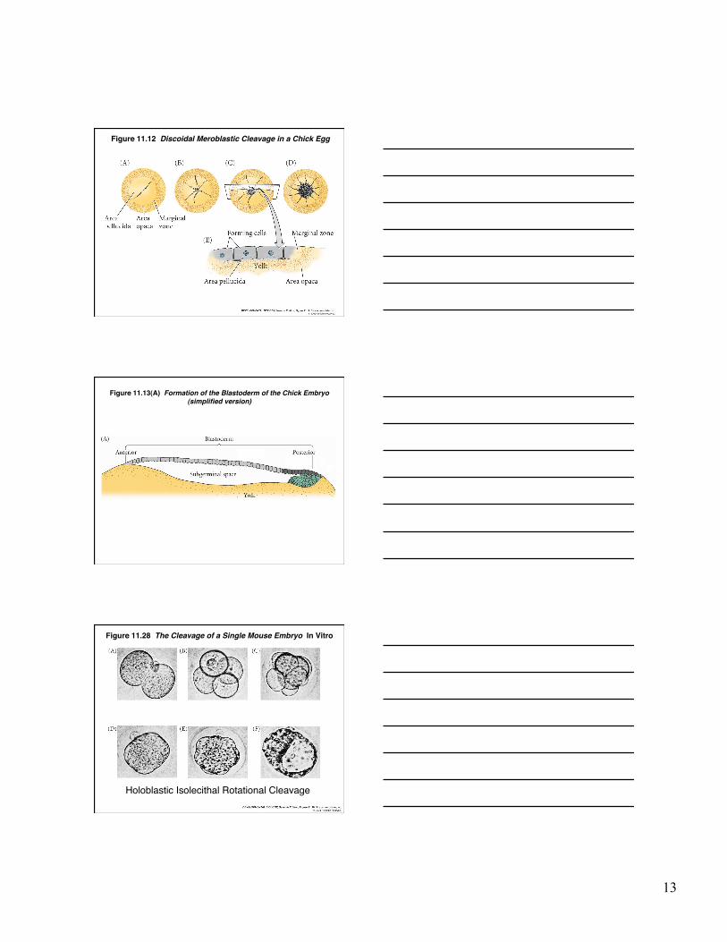
Figure 11.12 Discoidal Meroblastic Cleavage in a Chick Egg
Figure 11.13(A) Formation of the Blastoderm of the Chick Embryo(simplified version)
Figure 11.28 The Cleavage of a Single Mouse Embryo In Vitro
Holoblastic Isolecithal Rotational Cleavage
14
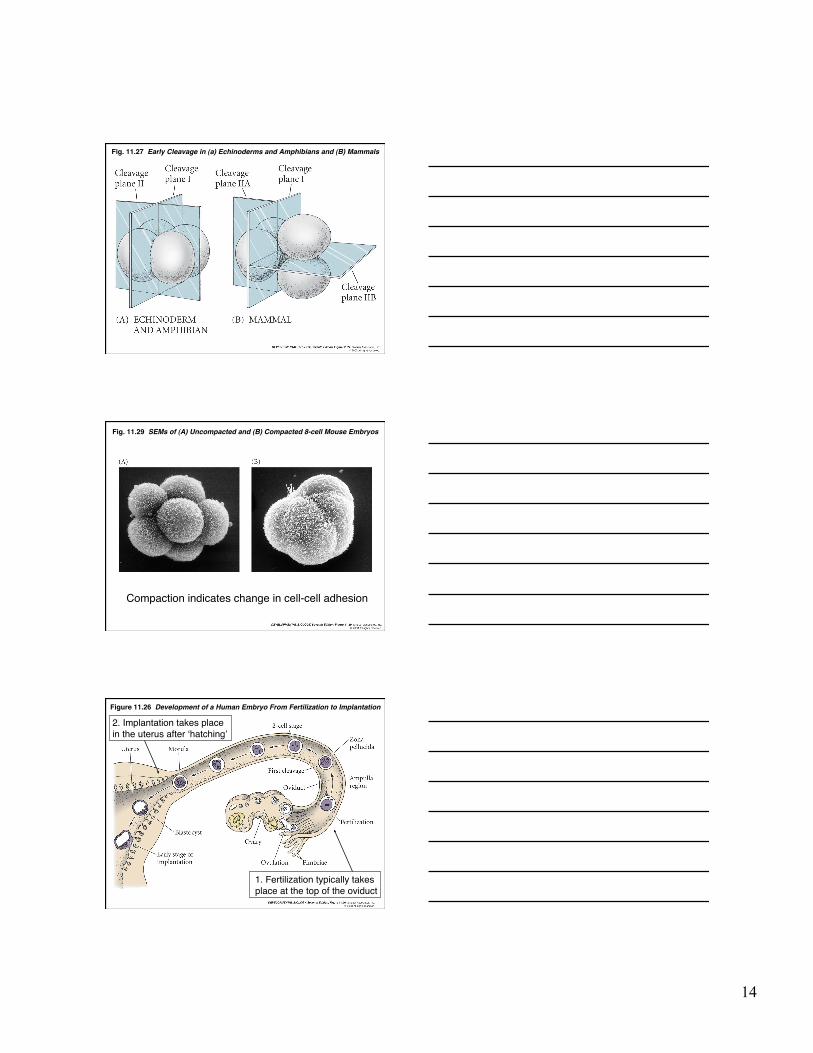
Fig. 11.27 Early Cleavage in (a) Echinoderms and Amphibians and (B) Mammals
Fig. 11.29 SEMs of (A) Uncompacted and (B) Compacted 8-cell Mouse Embryos
Compaction indicates change in cell-cell adhesion
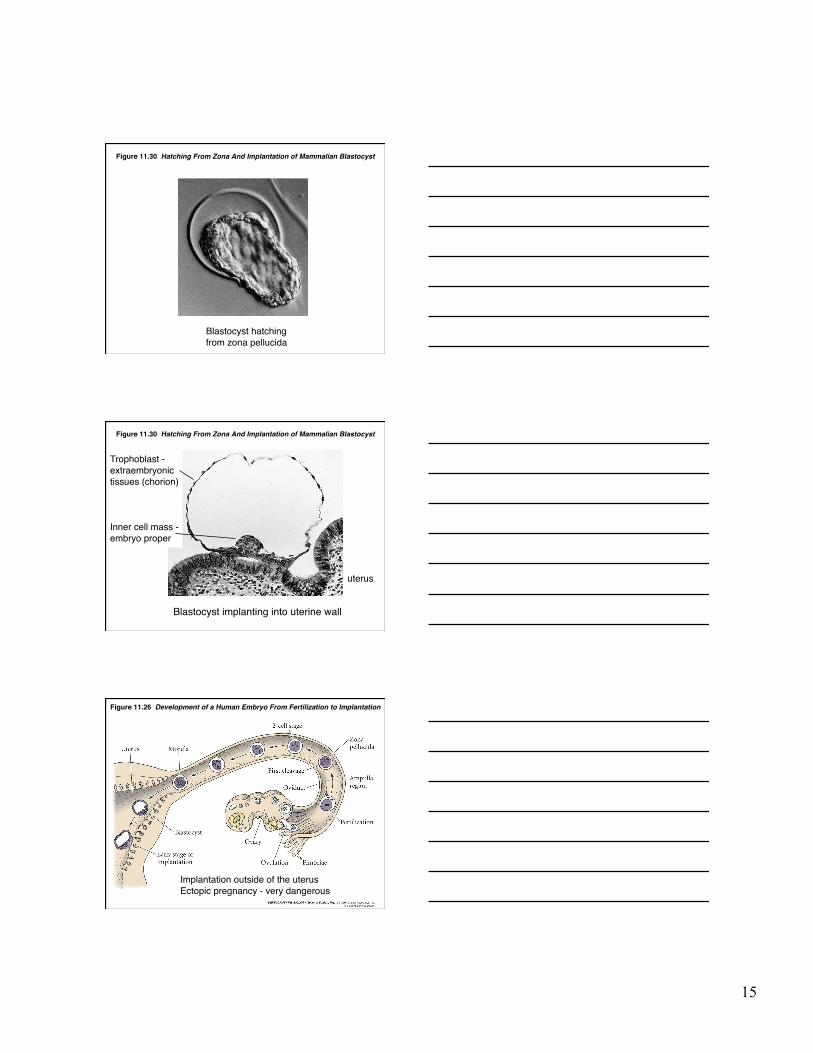
Figure 11.26 Development of a Human Embryo From Fertilization to Implantation
1. Fertilization typically takes place at the top of the oviduct
2. Implantation takes place in the uterus after ʻhatchingʼ
15
Figure 11.30 Hatching From Zona And Implantation of Mammalian Blastocyst
Blastocyst hatching from zona pellucida
Figure 11.30 Hatching From Zona And Implantation of Mammalian Blastocyst
Blastocyst implanting into uterine wall
uterus
Inner cell mass - embryo proper
Trophoblast - extraembryonic tissues (chorion)
Figure 11.26 Development of a Human Embryo From Fertilization to Implantation
Implantation outside of the uterus Ectopic pregnancy - very dangerous
16
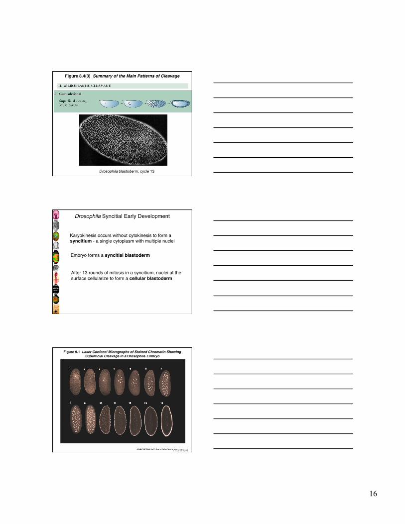
Figure 8.4(3) Summary of the Main Patterns of Cleavage
Drosophila blastoderm, cycle 13
Drosophila Syncitial Early Development
Karyokinesis occurs without cytokinesis to form a syncitium - a single cytoplasm with multiple nuclei
Embryo forms a syncitial blastoderm
After 13 rounds of mitosis in a syncitium, nuclei at the surface cellularize to form a cellular blastoderm
Figure 9.1 Laser Confocal Micrographs of Stained Chromatin ShowingSuperficial Cleavage in a Drosophila Embryo
17
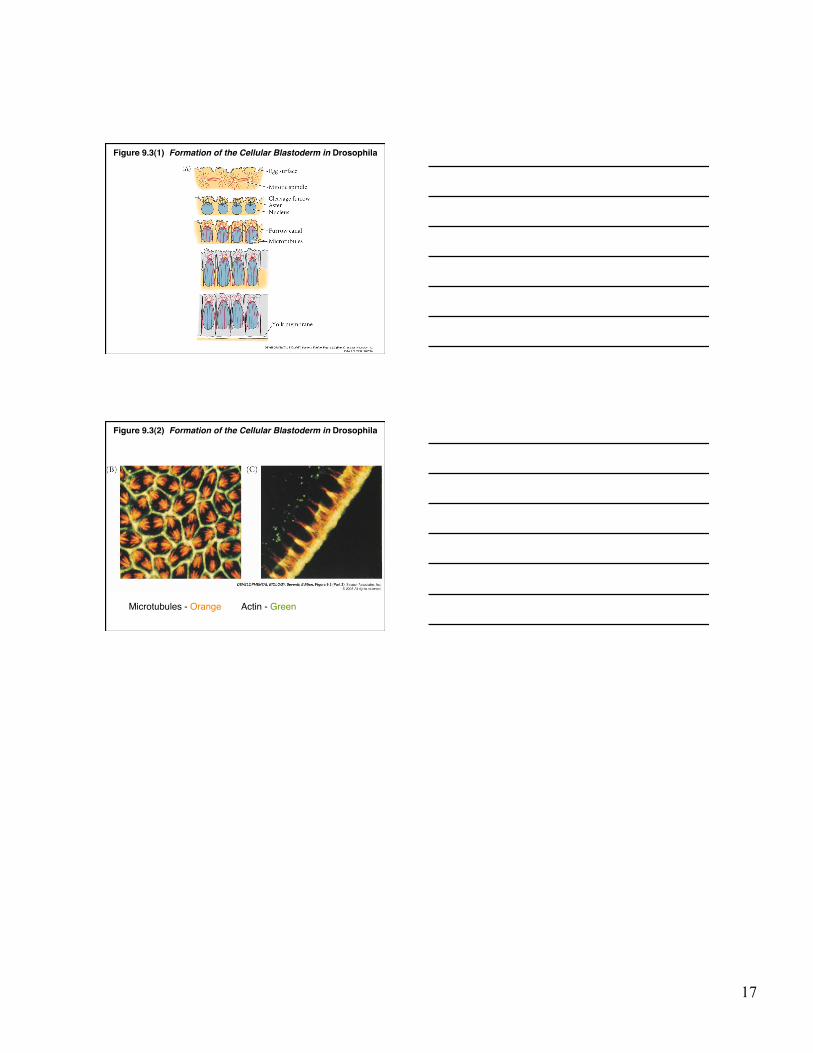
Figure 9.3(1) Formation of the Cellular Blastoderm in Drosophila
Figure 9.3(2) Formation of the Cellular Blastoderm in Drosophila
Microtubules - Orange Actin - Green


![) [111] cleavage plane](https://img.pdfslide.us/doc/110x75/61c7329341512e61f73ea613/-111-cleavage-plane.jpg)