Embed Size (px)
Citation preview
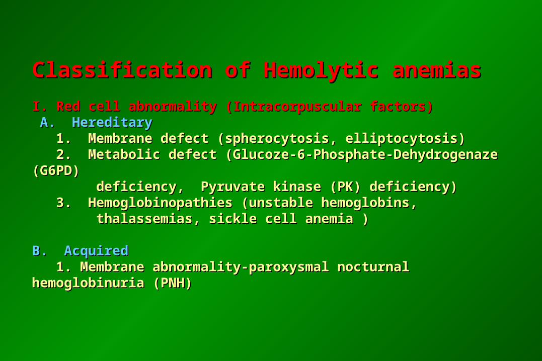
Classification of Hemolytic anemias Classification of Hemolytic anemias
I. Red cell abnormality (Intracorpuscular factors)I. Red cell abnormality (Intracorpuscular factors) A. Hereditary A. Hereditary 1. Membrane defect (spherocytosis, elliptocytosis) 1. Membrane defect (spherocytosis, elliptocytosis) 2. Metabolic defect (Glucoze-6-Phosphate-Dehydrogenaze (G6PD) 2. Metabolic defect (Glucoze-6-Phosphate-Dehydrogenaze (G6PD) deficiency, Pyruvate kinase (PK) deficiency) deficiency, Pyruvate kinase (PK) deficiency) 3. Hemoglobinopathies (unstable hemoglobins, 3. Hemoglobinopathies (unstable hemoglobins, thalassemias, sickle cell anemia ) thalassemias, sickle cell anemia )
B. AcquiredB. Acquired 1. Membrane abnormality-paroxysmal nocturnal hemoglobinuria (PNH) 1. Membrane abnormality-paroxysmal nocturnal hemoglobinuria (PNH)
HEMOLYTIC ANEMIASHEMOLYTIC ANEMIAS
Hemolytic anemiasHemolytic anemias = reduced red-cell life span = reduced red-cell life span
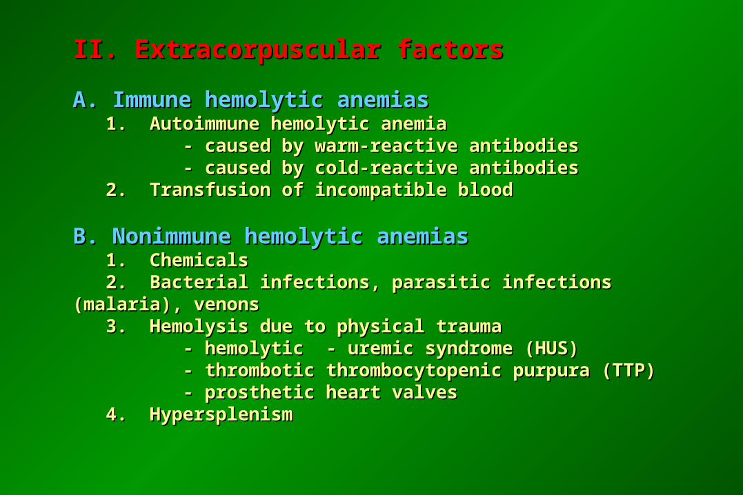
II. Extracorpuscular factorsII. Extracorpuscular factors
A. Immune hemolytic anemias A. Immune hemolytic anemias 1. Autoimmune hemolytic anemia 1. Autoimmune hemolytic anemia - caused by warm-reactive antibodies - caused by warm-reactive antibodies - caused by cold-reactive antibodies - caused by cold-reactive antibodies 2. Transfusion of incompatible blood 2. Transfusion of incompatible blood
B. Nonimmune hemolytic anemiasB. Nonimmune hemolytic anemias 1. Chemicals 1. Chemicals 2. Bacterial infections, parasitic infections (malaria), venons 2. Bacterial infections, parasitic infections (malaria), venons 3. Hemolysis due to physical trauma 3. Hemolysis due to physical trauma - hemolytic - uremic syndrome (HUS) - hemolytic - uremic syndrome (HUS) - thrombotic thrombocytopenic purpura (TTP) - thrombotic thrombocytopenic purpura (TTP) - prosthetic heart valves - prosthetic heart valves 4. Hypersplenism 4. Hypersplenism
Mechanisms of hemolysis:Mechanisms of hemolysis:
- intravascular- intravascular - extravascular - extravascular
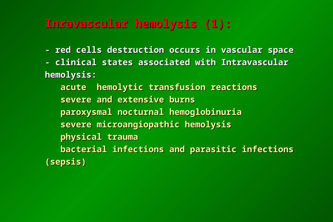
Inravascular hemolysis (1):Inravascular hemolysis (1):
- red cells destruction occurs in vascular space - red cells destruction occurs in vascular space
- clinical states associated with Intravascular hemolysis:- clinical states associated with Intravascular hemolysis:
acute hemolytic transfusion reactions acute hemolytic transfusion reactions
severe and extensive burns severe and extensive burns
paroxysmal nocturnal hemoglobinuria paroxysmal nocturnal hemoglobinuria
severe microangiopathic hemolysis severe microangiopathic hemolysis
physical trauma physical trauma
bacterial infections and parasitic infections (sepsis) bacterial infections and parasitic infections (sepsis)
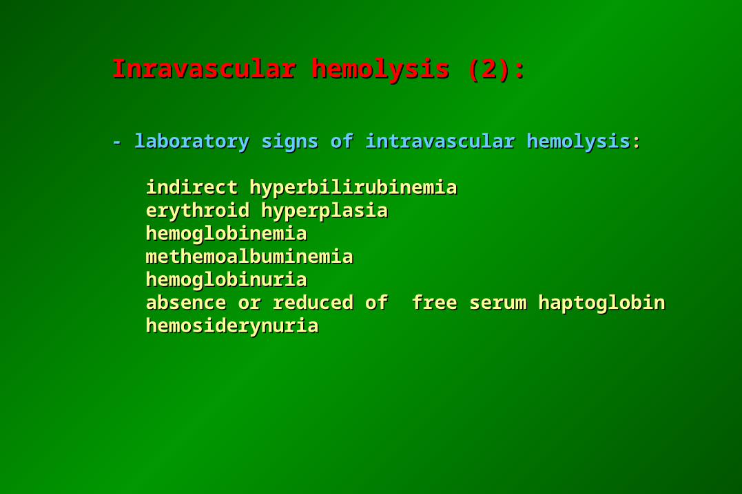
Inravascular hemolysis (2):Inravascular hemolysis (2):
- laboratory signs of intravascular hemolysis- laboratory signs of intravascular hemolysis:: indirect hyperbilirubinemia indirect hyperbilirubinemia erythroid hyperplasia erythroid hyperplasia hemoglobinemia hemoglobinemia methemoalbuminemia methemoalbuminemia hemoglobinuria hemoglobinuria absence or reduced of free serum haptoglobin absence or reduced of free serum haptoglobin hemosiderynuria hemosiderynuria
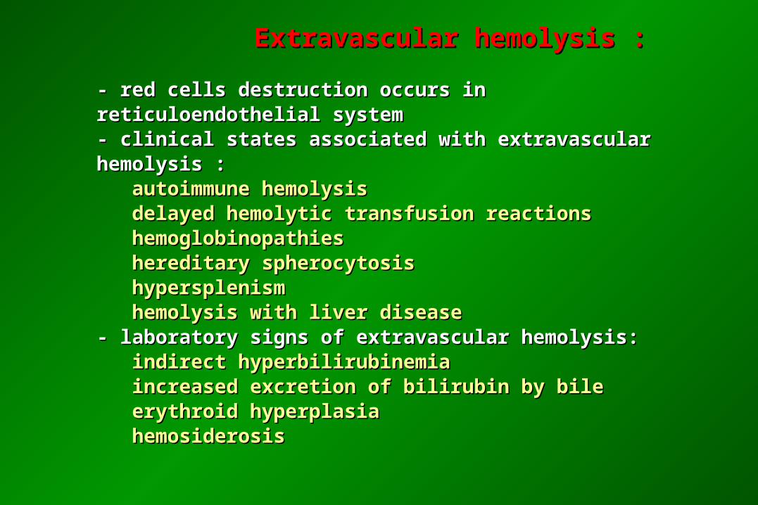
Extravascular hemolysis :Extravascular hemolysis :
- red cells destruction occurs in reticuloendothelial system - red cells destruction occurs in reticuloendothelial system - clinical states associated with extravascular hemolysis :- clinical states associated with extravascular hemolysis : autoimmune hemolysis autoimmune hemolysis delayed hemolytic transfusion reactions delayed hemolytic transfusion reactions hemoglobinopathies hemoglobinopathies hereditary spherocytosis hereditary spherocytosis hypersplenism hypersplenism hemolysis with liver disease hemolysis with liver disease- laboratory signs of extravascular hemolysis:- laboratory signs of extravascular hemolysis: indirect hyperbilirubinemia indirect hyperbilirubinemia increased excretion of bilirubin by bile increased excretion of bilirubin by bile erythroid hyperplasia erythroid hyperplasia hemosiderosis hemosiderosis
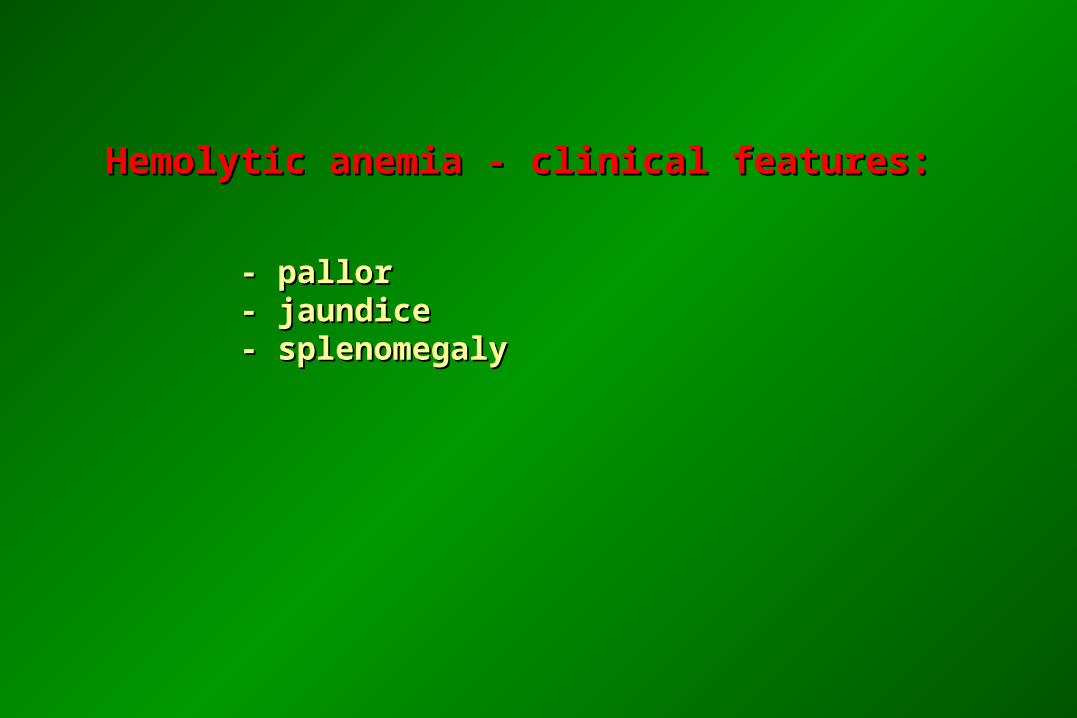
Hemolytic anemia - clinical features:Hemolytic anemia - clinical features:
- pallor - pallor - jaundice - jaundice - splenomegaly - splenomegaly
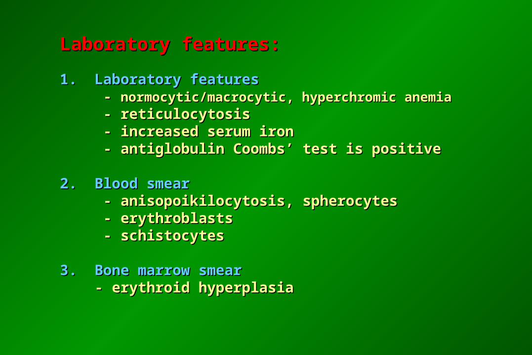
Laboratory features:Laboratory features:
1. Laboratory features1. Laboratory features - - normocytic/macrocytic, hyperchromic anemianormocytic/macrocytic, hyperchromic anemia - reticulocytosis - reticulocytosis - increased serum iron - increased serum iron - antiglobulin Coombs’ test is positive - antiglobulin Coombs’ test is positive
2. Blood smear 2. Blood smear - anisopoikilocytosis, spherocytes - anisopoikilocytosis, spherocytes - erythroblasts - erythroblasts - schistocytes - schistocytes
3. Bone marrow smear3. Bone marrow smear - erythroid hyperplasia - erythroid hyperplasia
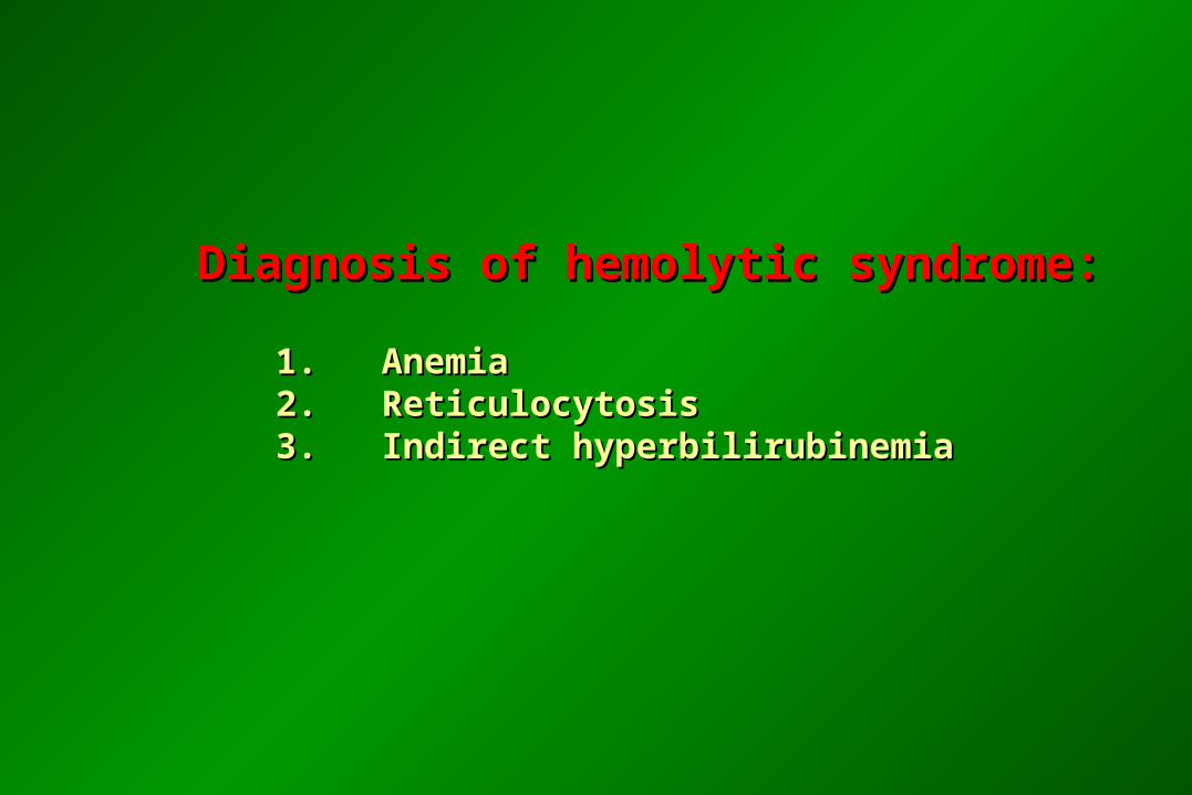
Diagnosis of hemolytic syndrome:Diagnosis of hemolytic syndrome:
1. Anemia1. Anemia 2. Reticulocytosis 2. Reticulocytosis 3. Indirect hyperbilirubinemia 3. Indirect hyperbilirubinemia
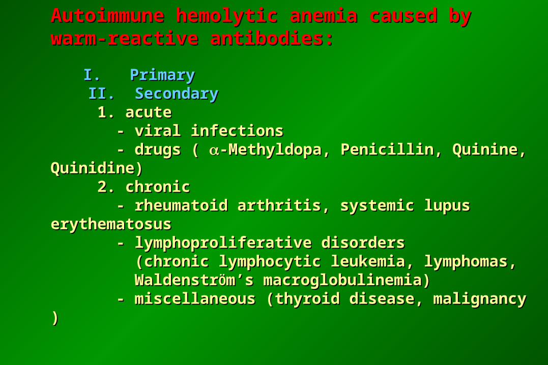
Autoimmune hemolytic anemia caused by warm-Autoimmune hemolytic anemia caused by warm-reactive antibodies:reactive antibodies:
I. PrimaryI. Primary II. Secondary II. Secondary 1. acute 1. acute - viral infections - viral infections - drugs ( - drugs ( -Methyldopa, Penicillin, Quinine, Quinidine)-Methyldopa, Penicillin, Quinine, Quinidine) 2. chronic 2. chronic - rheumatoid arthritis, systemic lupus erythematosus - rheumatoid arthritis, systemic lupus erythematosus - lymphoproliferative disorders - lymphoproliferative disorders (chronic lymphocytic leukemia, lymphomas, (chronic lymphocytic leukemia, lymphomas, Waldenstr WaldenstrÖÖm’s macroglobulinemia)m’s macroglobulinemia) - miscellaneous (thyroid disease, malignancy ) - miscellaneous (thyroid disease, malignancy )
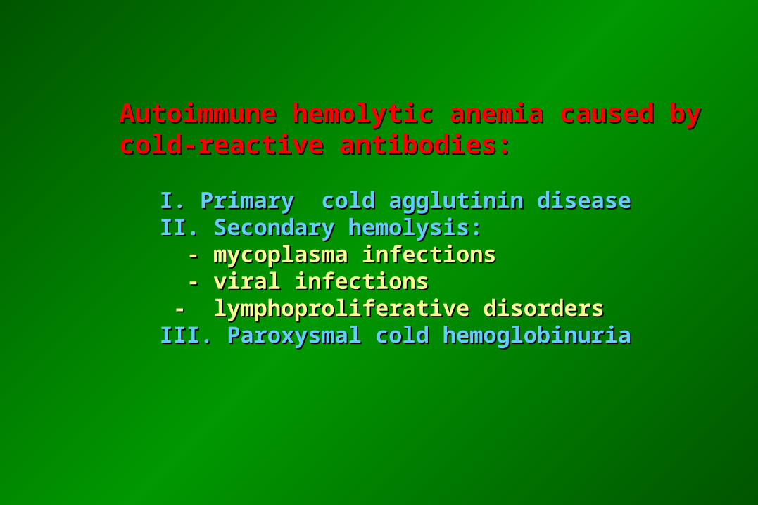
Autoimmune hemolytic anemia caused by cold-Autoimmune hemolytic anemia caused by cold-reactive antibodies:reactive antibodies:
I. Primary cold agglutinin diseaseI. Primary cold agglutinin disease II. Secondary hemolysis:II. Secondary hemolysis: - mycoplasma infections - mycoplasma infections - viral infections - viral infections - lymphoproliferative disorders - lymphoproliferative disorders III. Paroxysmal cold hemoglobinuriaIII. Paroxysmal cold hemoglobinuria
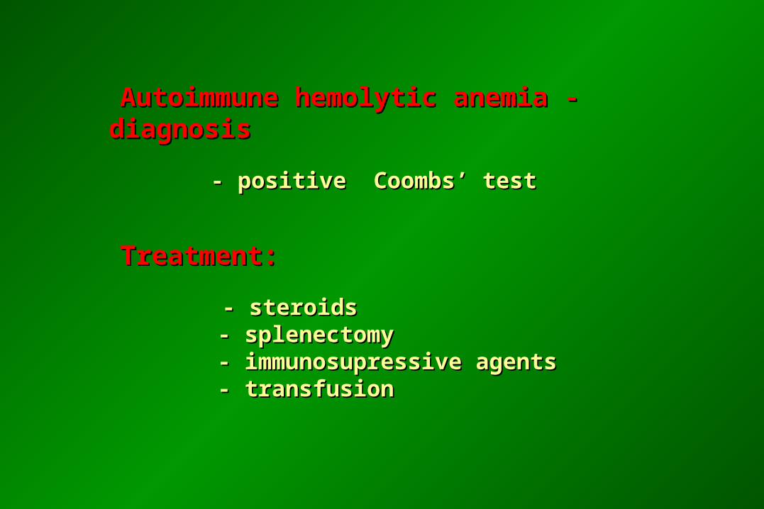
Autoimmune hemolytic anemia - diagnosis Autoimmune hemolytic anemia - diagnosis
- positive Coombs’ test- positive Coombs’ test
Treatment:Treatment:
- steroids- steroids - splenectomy - splenectomy - immunosupressive agents - immunosupressive agents - transfusion - transfusion
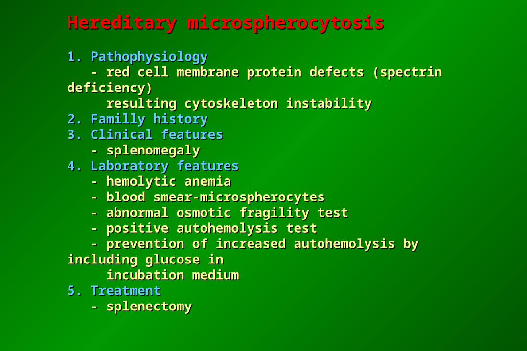
Hereditary microspherocytosisHereditary microspherocytosis
1. Pathophysiology1. Pathophysiology - red cell membrane protein defects (spectrin deficiency) - red cell membrane protein defects (spectrin deficiency) resulting cytoskeleton instability resulting cytoskeleton instability2. Familly history2. Familly history3. Clinical features3. Clinical features - splenomegaly - splenomegaly4. Laboratory features4. Laboratory features - hemolytic anemia - hemolytic anemia - blood smear-microspherocytes - blood smear-microspherocytes - abnormal osmotic fragility test - abnormal osmotic fragility test - positive autohemolysis test - positive autohemolysis test - prevention of increased autohemolysis by including glucose in - prevention of increased autohemolysis by including glucose in incubation medium incubation medium 5. Treatment 5. Treatment - splenectomy - splenectomy
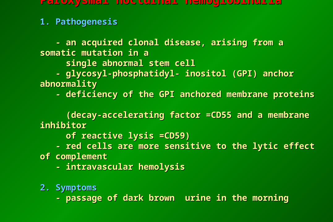
Paroxysmal nocturnal hemoglobinuriaParoxysmal nocturnal hemoglobinuria
1. Pathogenesis1. Pathogenesis - an acquired clonal disease, arising from a somatic mutation in a - an acquired clonal disease, arising from a somatic mutation in a
single abnormal stem cell single abnormal stem cell - glycosyl-phosphatidyl- inositol (GPI) anchor abnormality - glycosyl-phosphatidyl- inositol (GPI) anchor abnormality - deficiency of the GPI anchored membrane proteins - deficiency of the GPI anchored membrane proteins (decay-accelerating factor =CD55 and a membrane inhibitor (decay-accelerating factor =CD55 and a membrane inhibitor of reactive lysis =CD59) of reactive lysis =CD59) - red cells are more sensitive to the lytic effect of complement - red cells are more sensitive to the lytic effect of complement - intravascular hemolysis - intravascular hemolysis 2. Symptoms2. Symptoms - passage of dark brown urine in the morning - passage of dark brown urine in the morning
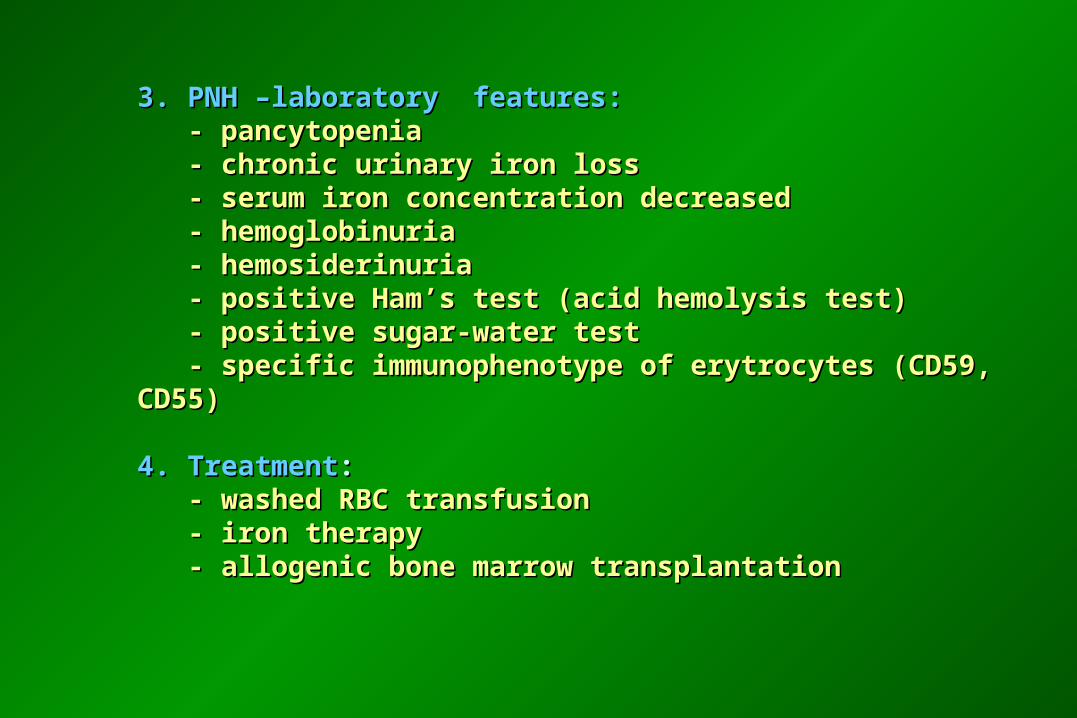
3. PNH –laboratory features:3. PNH –laboratory features: - pancytopenia - pancytopenia - chronic urinary iron loss - chronic urinary iron loss - serum iron concentration decreased - serum iron concentration decreased - hemoglobinuria - hemoglobinuria - hemosiderinuria - hemosiderinuria - positive Ham’s test (acid hemolysis test) - positive Ham’s test (acid hemolysis test) - positive sugar-water test - positive sugar-water test - specific immunophenotype of erytrocytes (CD59, CD55) - specific immunophenotype of erytrocytes (CD59, CD55)
4. Treatment4. Treatment:: - washed RBC transfusion - washed RBC transfusion - iron therapy - iron therapy - allogenic bone marrow transplantation - allogenic bone marrow transplantation
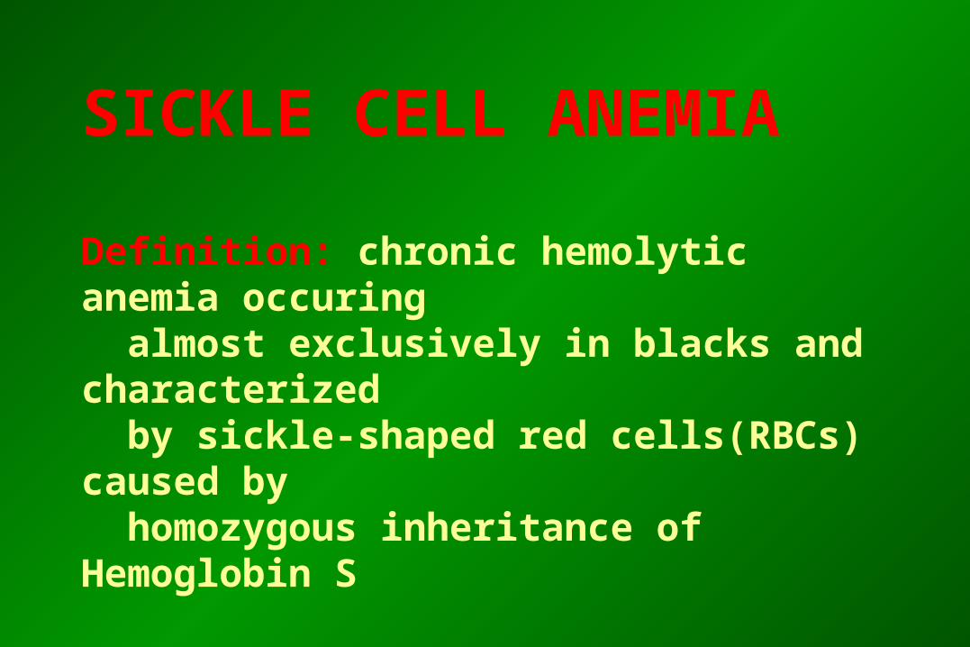
SICKLE CELL ANEMIA
Definition: chronic hemolytic anemia occuring almost exclusively in blacks and characterized by sickle-shaped red cells(RBCs) caused by homozygous inheritance of Hemoglobin S
SICKLE CELL ANEMIA-pathogenesis
- In Hb S, valine is substituted for glutamic acid in
the sixth amino acid of the ß chain.
- Deoxy-Hb S is much less soluble than deoxy Hb A;
it forms a gelatinous network of fibrous polymersthat cause RBCs to
sickle at sites of low pO2.
- Hemolysis-because sickle RBCs are too fragile to withstand the
mechanical trauma of circulation
- Occlusion in microvascular circulation caused by distorted, inflexible
RBCs adhering to vascular endothelium
SICKLE CELL ANEMIA-incidence
- Homozygous - about 0,3% of blacks in the USA
(have sickle cell anemia)
- Hetezygotes-8-13% of blacks, (are not anemic, but the sickling trait=sicklemia can be demonstrated in vitro)
SICKLE CELL ANEMIA-clinical featuresIN HOMOZYGOTES1. Clinical complications due to severe hemolytic anaemia
- slowed growth and development in children - bilirubins stones- aplastic crisis- congestive heart failure from chronic anemias and cardiac
overload compensation2. Consequences of vaso-occlusion of the microcirculations (tissue
ischemia and infarction)- infarction of spleen, brain, marrow, kidney, lung, aseptic
necrosis, central nervous system and ophtalmic vascular lesions
SICKLE CELL ANEMIA-laboratory findinges
1. Anemia-normocytic or slightly macrocytic
2. Leukocytosis(chronic neutrophilia)
3. Thrombocytosis-usually mild<1000G/l
4. Reticulocytosis
5. Peripheral smear: sickle shaped red cells, polychromatophilia, Howell-Jolly bodies
6. Hb -electrophoresis
SICKLE CELL ANEMIA-therapy
Preventive measures:
prevention or remedy of: infections(penicillin prophylaxis and pneumococcal vaccination), fever, dehydratation,acidosis, hypoxemia, cold exposure
Blood transfusions for very severe anemia
New approaches to therapy;
1. Activation of Hb F synthesis -5-azacytidine
2. Antisickling agents acting on hemoglobin or membrane
3. Bone marrow transplantation