Embed Size (px)
Citation preview

Washington University School of MedicineDigital Commons@Becker
Open Access Publications
7-1-2009
Classification and developmental biology ofcongenital anomalies of the hand and upperextremityPaul R. ManskeWashington University School of Medicine in St. Louis
Kerby C. ObergLoma Linda University
Follow this and additional works at: http://digitalcommons.wustl.edu/open_access_pubsPart of the Medicine and Health Sciences Commons
This Open Access Publication is brought to you for free and open access by Digital Commons@Becker. It has been accepted for inclusion in OpenAccess Publications by an authorized administrator of Digital Commons@Becker. For more information, please contact [email protected].
Recommended CitationManske, Paul R. and Oberg, Kerby C., ,"Classification and developmental biology of congenital anomalies of the hand and upperextremity." Journal of Bone and Joint Surgery.91,Supplement 4. 3-18. (2009).http://digitalcommons.wustl.edu/open_access_pubs/837

Classification and Developmental Biologyof Congenital Anomalies of the Hand
and Upper ExtremityBy Paul R.Manske, MD, and Kerby C. Oberg, MD, PhD
Congenital anomalies of the hand and upper extremityare classified according to appearance; thus, the myriadof disparate presentations are organized into groups that
share commonmorphologic features. The primary purpose of aclassification is to enhance communication about the specificfeatures of a condition by providing a descriptive framework forclinicians. Therefore, classification schemes should reflect thefull spectrum of morphologic abnormalities within a givencondition, and should be uncomplicated and easy for cliniciansto remember and use. While an ideal classification would alsoguide treatment, provide insight into prognosis, and incorpo-rate the etiologic mechanism of the condition, congenitalclassification systems often fall short of these goals.
The embryological development of the upper limb pro-ceeds along three axes: proximal-distal, anterior-posterior (re-ferred to postnatally as radial-ulnar by clinicians, because the fetalupper limb rotates during development), and dorsal-ventral1.The apical ectodermal ridge and the underlying mesodermcontrol proximal-distal development through a reciprocal loopof fibroblast growth factors and Wnt proteins2. The zone ofpolarizing activity located in the posterior (ulnar) limb meso-derm expands and posteriorizes the limb along the anterior-posterior (radial-ulnar) axis through a secreted morphogen,sonic hedgehog (SHH)3. The apical ectodermal ridge and thezone of polarizing activity are closely linked by a reciprocalfeedback loop that maintains SHH expression at the posterior(ulnar)-distal border of the apical ectodermal ridge duringprogressive outgrowth. Dorsal ectoderm controls limb dorsali-zation through the secretion of Wnt7a and the induction ofLmx1b in the underlying dorsal mesoderm4. A reciprocal feed-back loop between Wnt7a and SHH has also been demon-strated5. Thus, the integration of SHH into the pathways thatcontrol proximal-distal, anterior-posterior, and dorsal-ventralaxes ensures coordinated patterning during each phase of limboutgrowth and development (i.e., humerus, forearm, and hand).
Targeted disruption of these organizing tissues (apicalectodermal ridge, zone of polarizing activity, and dorsal ec-
toderm) or their associated molecular pathways has given usinsight into the etiologies of congenital limb malformations.For example, transverse deficiencies can be created in animalmodels by removal of the apical ectodermal ridge6 or disrup-tion of fibroblast growth-factor signaling7,8; ulnar longitudinaldeficiency can be produced by eliminating production of SHHfrom the zone of polarizing activity 9,10; and double palms occurin the absence of Wnt7a or Lmx1b4.
Although current classification schemes attempt to con-sider themorphologic features of the developing limb bud, theydo not convey the molecular mechanisms disrupted in theproduction of these deformities. Consequently, current classi-fication schemes have been criticized as being too simplistic.While it is possible that a more sophisticated molecular or ge-netic classification system could be devised, the complexities ofnature’s genetic ‘‘toolbox’’ that influence the development ofmultiple tissue and organ systems might result in a scheme toocomplex to be useful for practicing hand surgeons. Neverthe-less, insights from developmental biology may help refine themorphologic classification scheme to more precisely representunderlying mechanisms and related developmental pathways.
History
The history of the classification of congenital anomalies isrich, dating back to the mid-nineteenth century11-13. Saint-
Hilaire initially classified these ‘‘vices of conformation’’ asslight or severe in 182914. In 1831, Otto15 grouped these ‘‘vicesof organization’’ according to ten variations (number, size,form, position, connection, color, consistency, continuity,texture, and content). In 1832, Saint-Hilaire16 focused on thevariation in number and size of the limb-segment anomalies,coining the terms ectromelia, hemimelia, and phocomelia.Fort17 added the categories of axial deviation and adhesion (i.e.,syndactyly) in 1869. Leboucq18 noted the longitudinal ar-rangement of defects, and Polaillon19 applied the adjectivesradial, central, and ulnar in 1884. Potel20 distinguished betweenlongitudinal and transverse anomalies in 1914. O’Rahilly ex-
Disclosure: The authors did not receive any outside funding or grants in support of their research for or preparation of this work. Neither they nor amember of their immediate families received payments or other benefits or a commitment or agreement to provide such benefits from a commercialentity.
3
COPYRIGHT � 2009 BY THE JOURNAL OF BONE AND JOINT SURGERY, INCORPORATED
J Bone Joint Surg Am. 2009;91 Suppl 4:3-18 d doi:10.2106/JBJS.I.00008

panded the concept of intercalary deficiencies (i.e., phoco-melia) in 195121,22.
Two important contributions from the German litera-ture were made in the late nineteenth and early twentiethcenturies. Kummel in 189523 (subsequently modified by Nigstin 192724) divided congenital anomalies into three categories:(1) defect malformation (i.e., deficiencies), (2) syndactyly (i.e.,fusion of parts), and (3) polydactyly (i.e., excessive number ofparts). Muller in 1937 noted that malformations could presentas a continuum in varying degrees or in different stages ofdevelopment25. This concept of ‘‘teratological progression’’allowed anomalies to be graded according to morphologicseverity; it also allowed grouping of anomalies that might havedifferent morphologic appearances, thus simplifying classifi-cation schemes.
Several classification schemes evolved from these con-cepts, but none were universally adopted. They were not suf-ficiently inclusive of all congenital anomalies, and there weresubstantial terminologic differences, particularly between handsurgeons from the United States and Germany. The currentclassification scheme accepted by most hand surgeons was firstproposed by Swanson in 196426,27 and was based on the premisethat anomalies should be grouped according to parts of thelimb that have been primarily affected during development.There were six basic categories in the initial proposal: failure ofdifferentiation of parts, arrest of development, duplications,overgrowth, congenital circular constriction band syndrome,and generalized skeletal defects.
Following extensive deliberation by representatives of theAmerican Society for Surgery of the Hand (ASSH), the Inter-national Federation of Societies for Surgery of the Hand (IFSSH),and the International Society for Prosthetics and Orthotics(ISPO), the classification groupings were reordered and ex-panded to include a seventh basic category (undergrowth). Thisclassification was published in the orthotics and prosthetics lit-erature in 197412, and as the first scientific article in the inauguralissue of the American volume of the Journal of Hand Surgery28 in1976. Termed the IFSSH classification, this includes the sevenmajor categories as well as subcategories, subclassifications, andanatomic levels of anomalies and diagnoses (Table I).
Although this classification is comprehensive, it has beencriticized by authors who have found it difficult to classifycomplex cases29-33, particularly the complex spectrum of clefthand and symbrachydactyly 34. Knight and Kay presented amore detailed version in 200035, attempting to include allcongenital anomalies while still maintaining the seven basiccategories.
Recently, the Japanese Society for Surgery of the Hand36
suggested a modification, adding two additional groups: ab-normal induction of rays as group IV (thereby renumberingcategories V through VIII), and unclassifiable cases as groupIX. The proposed category of abnormal induction of rays at-tempts to incorporate the concept of causation as suggested byexperimental studies37,38; the proposed category includes syn-dactyly (both simple and complex), the central polydactyly-cleft hand-osseous syndactyly complex, and triphalangeal
thumb. These conditions would move from their currentcategories of failure of formation (transverse), failure of dif-ferentiation, and duplication on the premise that they representinduction abnormalities rather than formation abnormalities.This proposal has not been universally accepted; critics arguethat the differences between abnormal induction and abnor-mal formation are primarily semantic rather than actual39.
Tonkin recognized that problems arise when attemptingto incorporate our current understanding of causation into amorphology-based classification, and further contended thatgrouping according to such categories as failed formation,failed differentiation, or duplication may be inappropriate39.He proposed focusing the classification purely on descriptivefeatures, with the primary classification noting the location(i.e., arm, forearm, wrist, or hand) and subcategories listingthe tissue involved (bone or soft tissue) as well as the specificmorphologic features of the anomalies.
Nevertheless, the IFSSH classification, which has servedas the basis for scientific discussion and communication formore than forty years, continues to be accepted by most handsurgeons. There are specific classifications for twenty diag-nostic conditions within these seven categories, with eighteenof the twenty conditions included in the first three categories(seven conditions with regard to failure of formation; eightconditions with regard to failure of differentiation; and threeconditions with regard to duplication). We will review eachclassification scheme within the IFSSH classification, inte-grating modifications made subsequent to the original de-scriptions and assessing the value of the schemes to clinicians.Finally, we will present new information regarding the un-derlying etiology of the various conditions and propose amodification to the IFSSH classification that is based on cur-rent concepts of developmental biology.
Group I: Failure of Formation
Abnormalities regarding failure of formation have beensubdivided into longitudinal and transverse deficiencies.
The longitudinal deficiencies are grouped according to radial,central, and ulnar deficiencies, reflecting the location of thecellular abnormalities on the developing limb bud. Transversedeficiencies are grouped as terminal or intercalary (i.e., pho-
TABLE I The Classification System Developed by the
International Federation of Societies for Surgery
of the Hand (IFSSH) in Partnership with
Other Organizations28
I. Failure of formation
II. Failure of differentiation
III. Duplication
IV. Overgrowth (gigantism)
V. Undergrowth (hypoplasia)
VI. Congenital constriction band syndrome
VII. Generalized skeletal abnormalities
4
THE JOURNAL OF BONE & JOINT SURGERY d J B J S .ORG
VOLUME 91-A d SUPPLEMENT 4 d 2009CLASS I F ICAT ION AND DEVELOPMENTAL BIOLOGY OF CONGENITAL
ANOMAL IE S OF THE HAND AND UPPER EXTREMITY
comelia) deficiencies. Recent studies have suggested that in-tercalary deficiencies are difficult to explain according to de-velopmental biology concepts and may represent severe formsof longitudinal deficiency40,41.
Radial Longitudinal DeficiencyRadial longitudinal deficiency represents defects occurring atthe preaxial (radial) border of the limb bud. Although skeletaland soft-tissue structures of the entire radial aspect of theupper extremity are progressively affected, the classificationsystems are based on (1) thumb hypoplasia, and (2) defi-ciencies of the radius (Fig. 1).
The underlying etiology of radial longitudinal deficiencyis linked to decreased limb volume with intact SHH expres-sion; thus, despite a reduction in tissue and frequently a re-duction in overall limb length, posterior or ulnar elements aresomewhat preserved. In animal models, progressive reductionsin apical ectodermal ridge-associated fibroblast growth factorscause progressive reductions in the size of the developing limbbud and generate radial longitudinal deficiencies that closelycorrelate with what is seen clinically42,43. A number of molec-ular pathways impact cell growth or apoptosis, altering limbvolume during the development of the forelimb or hand9. Thissusceptibility is reflected in the association of radial longitudinaldeficiencies with a wide variety of syndromes and conditions44.
Thumb HypoplasiaMuller initially introduced four categories, expanded to five byBlauth in 196745. Type I was subsequently defined more spe-cifically by James et al.46 in 1996, and type III was expanded byManske et al.47 in 1995.
Type I. Blauth45 described this form of hypoplasticthumb as ‘‘minimal shortening and narrowing.’’ James et al.46
more clearly limited the deficiencies to aplasia or hypoplasia ofthe intrinsic muscles of opposition, that is, the abductor pol-licis brevis and the opponens pollicis.
Type II. There are three specific features of type-II hy-poplastic thumbs: (1) aplasia or hypoplasia of the intrinsicmuscles innervated by the median nerve, sparing the ulnar-innervated intrinsic muscles; (2) narrowing of the thumb-index web space; and (3) instability of the metacarpophalangealjoint due to ulnar collateral ligament insufficiency.
Type III. The three manifestations of type-II hypoplasiaare also present in type-III hypoplasia, frequently in moresevere form. The narrowed thumb-index web space may pres-ent as a more distal takeoff of the thumb, and metacarpo-phalangeal joint instability may be global, also involving theradial collateral ligament.
There are two additional deficiencies in type-IIIhypoplasia47:
� Extrinsic muscle-tendon abnormalities, includingabsent extensor pollicis longus, absent or aberrantflexor pollicis longus, tendon interconnections be-tween the extensor pollicis longus and flexor pollicislongus, and a variety of muscle-tendon intercon-nections at the volar aspect of the wrist and distalpart of the forearm as described by Graham andLouis48.
� Deficiencies at the base of the first metacarpal. In typeIII-A, the metacarpal base is hypoplastic but presentand stable; in type III-B, the metacarpal base is absentand unstable.
Type IV. This form of hypoplasia is the classic pouceflottant. The thumb is attached to the hand by a skin bridgeand neurovascular elements with no musculotendinous unitsor osseous structures stabilizing it to the hand.
Type V. The thumb is completely absent, although theremay be rudimentary thenar muscles, tendons, or neurovascularstructures along the radial border of the index metacarpal.
This classification system enhances communication,provides some prognostic information, and guides surgicaldecisions. Types I, II, and III-A deformities are treated withthumb reconstruction, while types III-B, IV, and V deformitiesare treated with thumb ablation and index-finger pollicization.
Deficiencies of the RadiusThe predominant features of this classification include pro-gressive skeletal deficiency of the radius along with radial an-gulation of the hand at the level of the wrist. Frequentlyreferred to as ‘‘radial club hand,’’ the condition is now moreacceptably referred to as radial deficiency (or dysplasia).
The abnormal features can include general hypoplasia ofthe upper extremity, absent active elbow flexion or elbowflexion contracture, shortening and/or bowing of the ulna, andaplasia or hypoplasia of the scaphoid and other carpal bones.The thumb is always affected, and the fingers frequently havelimited motion and function, progressing in severity from the
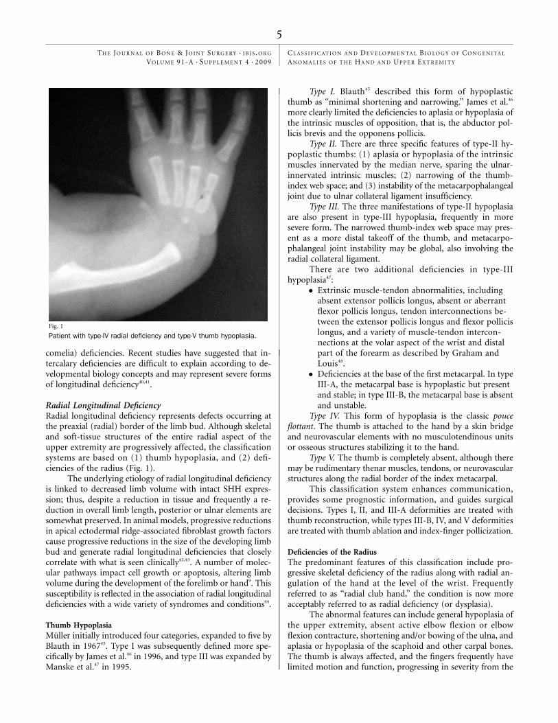
Fig. 1
Patient with type-IV radial deficiency and type-V thumb hypoplasia.
5
THE JOURNAL OF BONE & JOINT SURGERY d J B J S .ORG
VOLUME 91-A d SUPPLEMENT 4 d 2009CLASS I F ICAT ION AND DEVELOPMENTAL BIOLOGY OF CONGENITAL
ANOMAL IE S OF THE HAND AND UPPER EXTREMITY

radial to the ulnar side of the hand. Radial neurovascularstructures may be abnormal, and the radial musculotendinousstructures may be absent or abnormal, often forming a firmfibrous band that tethers the wrist and hand, thus contrib-uting to the radial angled position. The severity of these ad-ditional abnormal features parallels the severity of the radialdeficiency.
Classification of the condition was initially presented byBayne and Klug49 as types I through IV in 1987, based on theradiographic appearance of the radius. The classification wasexpanded by James et al.50 in 1999 to recognize radial-sideddeficiencies limited to the carpus (type 0) and thumb (type N).More recently, the classification was extended by Goldfarbet al.40 to include type V.
Type N. The radius and carpus are normal, with defi-ciencies limited to the thumb (see hypoplastic-thumb classi-fication above).
Type 0. The radius is normal in length. The scaphoid andother radial carpal bones are hypoplastic or absent, thus po-tentiating radial angulation of the hand and carpus. Not alltype-0 wrists demonstrate radial angulation; soft-tissue con-traction of the radial joint capsule and musculotendinousstructures are also necessary to produce the angulation. Thethumb is hypoplastic.
Type I. Initially described by Bayne and Klug49 as a ‘‘shortdistal radius,’’ the description was more specifically defined byJames et al.50 as a distal portion of the radius that is >2 mmshorter than the distal portion of the ulna. The proximalportion of the radius is usually normal, but may be charac-terized by radioulnar synostosis or congenital dislocation ofthe radial head.
Type II. The radius is hypoplastic in its entirety, referredto by Bayne and Klug as ‘‘radius in miniature’’ with proximaland distal physes. This hypoplastic radius is often associatedwith severe bowing of the ulna.
Type III. The distal part of the radius (including physis) isabsent.
Type IV. The radius is completely absent.Type V. Proximal radial longitudinal deficiency was for-
merly considered a form of phocomelia. Frantz and O’Rahilly 21
initially proposed it as a severe intercalary segmental defect ofthe osseous structures of the arm and forearm. Recent au-thors40,41 have challenged the concept of intercalary defects inthat such defects are difficult to explain according to devel-opmental biology concepts; rather, phocomelic upper limbsrepresent forms of longitudinal deficiency (radial, ulnar, or acombination of both)40. Extremities with type V radial longi-tudinal deficiency have the following characteristics:
� abnormal glenoid� absent proximal portion of the humerus� distal portion of the humerus articulates with ulna� radial-sided hand abnormalitiesThis classification enhances communication but does
not guide treatment decisions or provide prognostic infor-mation. It does attempt to incorporate concepts of abnormaldevelopmental mechanisms.
Central DeficiencyCentral deficiency, also descriptively referred to as ‘‘cleft hand,’’represents suppressed development of bone and associatedsoft-tissue structures of the central part of the hand (or foot)(Fig. 2). The presentation can vary from a simple soft-tissuecleft between the long and ring fingers without loss of digits51,52
to suppression of all osseous elements of the hand except forthe little finger ray53,54. The defect does not include deficienciesof the wrist, but associated carpal coalition and proximal ra-dioulnar synostosis have been reported51. Defects of the centralor medial apical ectodermal ridge are responsible for centralclefting55. Syndromic forms of this disorder exhibit mutationsin genes that participate in apical ectodermal ridge function55,56.
Central deficiency occurs in association with central poly-dactyly and syndactyly (soft-tissue and osseous)53,54,57. Ogino58
produced this spectrum of defects in littermates of pregnantrats, thus suggesting an etiologic relationship between theseconditions. In this animal model, there is diffuse cell death ofthe limb-bud ectoderm and mesoderm with reduced expres-sion of multiple limb organizing factors, along with apoptoticfactors and focal interruption of the apical ectodermal ridge59.These experimental observations are the basis for the proposedabnormal induction category35 noted previously.
Various classification systems have been proposed thatare based primarily on the characteristics of the central defect.Barsky 60 distinguished between typical cleft hand (deficienciesof the central rays with preservation of the marginal digits) andatypical cleft hand (central rays progressively reduced in size
Fig. 2
The hand of a patient with a type-II (narrowed web) central
deficiency.
6
THE JOURNAL OF BONE & JOINT SURGERY d J B J S .ORG
VOLUME 91-A d SUPPLEMENT 4 d 2009CLASS I F ICAT ION AND DEVELOPMENTAL BIOLOGY OF CONGENITAL
ANOMAL IE S OF THE HAND AND UPPER EXTREMITY

with shortened marginal rays). However, current opinion isthat the latter represents a form of transverse deficiency(symbrachydactyly)57,61,62 and therefore is not appropriatelyincluded in the central deficiency classification. Several authorshave classified the associated presence of central polydactylyand osseous syndactyly53,63 as ‘‘atypical.’’ Finally, several authorshave classified the hand according to the number of deficientcentral osseous elements51-54.
In hands with a central deficiency, the cleft is not func-tionally limiting but is aesthetically unsightly. Flatt64 describedthe cleft as ‘‘a functional triumph and a social disaster.’’ In ad-dition to the central cleft, there is an associated narrowing of thethumb web space, which is a notable functional deficiency65.Consequently, surgical treatment of the cleft is primarily foraesthetic reasons, while surgical treatment of the narrowedthumb web is functionally important. The progressive nar-rowing of the thumb web space parallels the progressive defi-ciencies of the central cleft. Consequently, Manske and Halikisproposed a classification of the central deficiency that is basedon the progressive narrowing of the thumb web space65.
� Type I. Normal web. The thumb web space shows nonarrowing.
� Type II. Narrowed web. The web space is narrowed,either mildly (type II-A) or severely (type II-B).
� Type III. Syndactylized web. The thumb and indexrays are syndactylized, with complete obliteration ofthe web space.
� Type IV. Merged web. The osseous elements of theindex ray are suppressed and the web space of thethumb is merged with the cleft, frequently in associ-ation with ulnar collateral ligament instability of thethumb metacarpophalangeal joint.
� Type V. Absent web. The thumb and radial digitalelements are completely suppressed; only the ulnarray(s) remain.
This classification system enhances communication andguides surgical reconstruction. In type I, the web space is nor-mal and does not need reconstruction; therefore, cleft closure isthe surgical focus. In types II-A, II-B, and III, the narrowedthumbweb is surgically widenedwith use of local flaps or dorsaland/or volar transposition flaps from the cleft. In type IV, noreconstruction of the web space is necessary; reduction of anexcessively wide web space and/or stabilization of the meta-carpophalangeal joint may be necessary. In type V, first meta-carpal lengthening or toe-to-hand transfer may be considered.
Ulnar Longitudinal DeficiencyUlnar longitudinal deficiency represents a spectrum of ab-normalities that affect the ulnar border of the upper limb.Although the deficiencies follow the ulnar longitudinal axis inthe forearm and upper arm, both the ulnar and radial sides ofthe hand can be affected (Fig. 3). Despite this apparentparadox, the abnormalities seen in ulnar longitudinal defi-ciency are best explained by disruption of the zone of polar-izing activity that is present in the posterior (postaxial) regionof the limb bud.
The zone of polarizing activity, via the secreted mor-phogen SHH, posteriorizes (ulnarizes) the developing limb.The spectrum of ulnar longitudinal deficiencies reflects varia-tion in the timing, degree, and duration of SHH disruption.Furthermore, loss of SHH function also accounts for the radialinvolvement seen in ulnar longitudinal deficiency. Recentexperiments in animal models demonstrate that, in additionto posteriorizing the developing limb, SHH also plays a role inlimb proliferation, expanding the distal portion of the limbduring development9,10. Thus, loss of SHH also reduces limbvolume. Depending on the timing and extent of SHH dis-ruption, the hand may also be involved. In mice, progressivedigit loss with declining SHH function follows an unex-pected pattern, with digit 3 being lost first, followed by digit 5,then digit 2, and finally digit 410. This sequence of digit lossmay help clarify carpal morphology and tendon attachmentsin the remaining digits in limbs with ulnar longitudinaldeficiency.
Classification schemes for ulnar longitudinal deficiencyare based on (1) forearm and elbow and (2) hand abnormalities.
Fig. 3
Radiograph of a patient with a type-II ulnar longitudinal deficiency
of the forearm and a type-C hand abnormality.
7
THE JOURNAL OF BONE & JOINT SURGERY d J B J S .ORG
VOLUME 91-A d SUPPLEMENT 4 d 2009CLASS I F ICAT ION AND DEVELOPMENTAL BIOLOGY OF CONGENITAL
ANOMAL IE S OF THE HAND AND UPPER EXTREMITY

Forearm and ElbowRecent clinical reviews have reported only 222 cases of ulnarlongitudinal deficiency66; nevertheless, six different classifica-tions based on the deformities of the forearm and elbow havebeen presented23,67-71. While all of the categories are similar, thefour-category classification of Bayne67 is generally preferred. Ofinterest, none of the forearm-and-elbow classifications con-siders hand abnormalities; to address this limitation, Bayne’sforearm and elbow classification has recently been modified72
to include Type 0. Additionally, as noted in the presentation ofradial deficiencies, Goldfarb et al.40 have added Type V to in-clude those phocomelic extremities with characteristics sug-gestive of ulnar longitudinal deficiency.
� Type 0. Normal-length ulna (i.e., distal aspect of theulna at the level of the distal aspect of the radius) withulnar-sided hand deficiencies
� Type I. Hypoplastic ulna with distal and proximalepiphyses present
� Type II. Distal ulnar aplasia� Type III. Complete ulnar aplasia� Type IV. Radial-humeral synostosis� Type V. Proximal ulnar longitudinal deficiency40:
hypoplastic glenoid; single arm and forearm bonethat is usually bifurcated distally, with proximalfeatures resembling a humerus and distal featurescharacteristic of a radius; absent elbow joint; andhand abnormalities typical of ulnar longitudinaldeficiency
While useful for facilitating communication, this clas-sification does not guide treatment or provide prognosticinformation.
HandSeveral authors have noted the high prevalence (68% to 100%)of hand abnormalities (particularly abnormalities of thethumb) in ulnar longitudinal deficiency38,73-77 as well as the factthat the majority of surgical procedures (55% to 75%) areperformed for hand and thumb abnormalities76 as comparedwith forearm abnormalities. Thus, the need for additionalclassification of hand abnormalities seems apparent. Oginoand Kato38 classified hand abnormalities according to thenumber of missing rays, proceeding sequentially from theulnar to the radial side of the hand, but did not include absenceor abnormalities of the thumb.
In view of the high prevalence of thumb abnormalitiesand the importance of the thumb to the function of the hand,Cole and Manske proposed a system of classification based onfeatures of the thumb76, adding alphabetic letters to the romannumerals of the forearm and elbow classifications.
� Type A. Normal first web and thumb� Type B. Mild first web and thumb deficiency� Type C. Moderate to severe first web deficiencies: loss
of opposition; malrotation of thumb into the plane ofthe fingers; thumb and index syndactyly; absentextrinsic tendon function
� Type D. Absent thumb
This classification enhances communication and pro-vides information about the likelihood of surgical inter-vention, as surgical reconstruction is more commonlyrecommended for types C and D hands compared with types Aand B hands.
Transverse DeficiencyTransverse deficiency includes both terminal and intercalary(phocomelic) deficiencies. There are two forms of terminalfailure of formation: symbrachydactyly and transverse arrest(also known as congenital amputation). Although standardtextbooks78-85 have separate chapters for each form and theIFSSH classification places the two in separate categories(transverse arrest in failure of formation; symbrachydactyly inundergrowth), symbrachydactyly is probably a more distal man-ifestation of transverse deficiency, and transverse arrest (congen-ital amputation) is a more proximal manifestation62,86-88. Thisconcept has received scientific support from Kallemeier et al.89,who noted that 93% of extremities with transverse arrest at thelevel of the forearm had rudimentary manifestations of digits(finger nubbins, nail remnants, or skin invagination) which areseen in symbrachydactyly.
Summerbell6 demonstrated a mechanism for transversearrest by removing the apical ectodermal ridge from devel-oping wing buds, correlating the timing of apical ectodermalridge removal to level of truncation90. Fibroblast growth factorsemanating from the apical ectodermal ridge promote meso-dermal proliferation and impede apoptosis. Consequently,abating apical ectodermal ridge-associated fibroblast growth-factor signaling can also yield terminal truncations7,8. Recently,Winkel et al. demonstrated a link between Wnt signaling andROR2, the receptor tyrosine kinase frequently mutated inbrachydactyly type B (another name for symbrachydactyly)91.Wnt signaling is critical for apical ectodermal ridge-relatedfibroblast growth-factor expression and function; thus, thisfinding links abnormal apical ectodermal ridge function tosymbrachydactyly and provides further evidence for inclusionas a transverse deficiency.
SymbrachydactylyThe four-category classification of symbrachydactyly (Fig. 4)was initially proposed by Pol92 and subsequently adopted byBlauth and Gekeler86 and others87
� Short finger type. This includes hands with short,underdeveloped, or absent middle phalanges.
� Oligodactylic type. Partial or complete absence ofthe central three fingers relative to the border (thumband little finger) digits. This type was previouslycalled ‘‘atypical’’ cleft hand (see above); it hasalso been referred to as the ‘‘cleft-hand type’’ ofsymbrachydactyly.
� Monodactylous type. Absence of all fingers, includingparts of the metacarpals. The thumb is present butis of varying size and stature.
� Peromelic type. Absence of all digital rays with onlydigital nubbins and nail remnants.
8
THE JOURNAL OF BONE & JOINT SURGERY d J B J S .ORG
VOLUME 91-A d SUPPLEMENT 4 d 2009CLASS I F ICAT ION AND DEVELOPMENTAL BIOLOGY OF CONGENITAL
ANOMAL IE S OF THE HAND AND UPPER EXTREMITY

Transverse ArrestMore proximal manifestations of transverse arrest (Fig. 5) arereferred to as:
� Acarpia. Congenital absence at the level of the carpalbones.
� Congenital forearm amputation. Congenital absenceat the forearm, also called congenital below-the-elbowdeficiency.
These descriptive classifications enhance communica-tion, but do not help with prognosis or treatment or incor-porate concepts of underlying etiology.
Intercalary Deficiency (Phocomelia)Intercalary deficiency has been considered a segmental trans-verse deficiency. It came into common usage in the early 1960sfollowing the report by Frantz and O’Rahilly 21, who classified itaccording to three types:
� Type I. Complete phocomelia. Complete segmentaldeficiency of the arm and forearm, with the handattached to the trunk.
� Type II. Proximal phocomelia. The humerus is absent,and the forearm and hand are attached directly to thetrunk.
� Type III. Distal phocomelia. The forearm is absent; thehand is attached to the humerus.
On close inspection, these extremities usually do notdemonstrate true segmental deficits; rather, the limb is ab-normal proximal and distal to the segmental defect, chal-lenging the existence of true intercalary deformities28,40,41,93.Furthermore, it is difficult to classify as much as 75% of ex-
tremities diagnosed as phocomelic with use of the features ofthe above classification. Instead, phocomelia most likely rep-resents the most severe form of longitudinal deficiencies40:
� Proximal radial longitudinal deficiency. Characteris-tics as noted above (type V radial longitudinaldeficiency).
� Proximal ulnar longitudinal deficiency. Characteristicsas noted above (type V ulnar longitudinal deficiency).
� Severe combined dysplasia, type A. A normal orhypoplastic shoulder with an underdeveloped glenoid;a normal or short humerus with normal distal flare;absence of both radius and ulna; abnormal hand withfeatures that do not correspond to either radial orulnar deficiency.
� Severe combined dysplasia, type B. Complete absenceof both the humerus and forearm segments; abnormalhand elements (unclassifiable according to radial orulnar dysplasia deficiency) attached to an abnormalshoulder.
While the reclassification of phocomelia does not assistwith determination of treatment or prognosis, it advancesunderstanding of these conditions by incorporating currentconcepts of underlying etiology.
Group II: Failure of Differentiation
Conditions categorized under failure of differentiationmore aptly represent abnormalities of development rather
than differentiation; yet, for the most part, they share a com-mon theme in the disruption of hand plate (or foot plate)development and involve carpals, metacarpals, digits, orinterdigital spaces. Several of these conditions have beenclassified.
SyndactylySyndactyly can be seen as an isolated condition or as a com-ponent of other conditions in the failure of formation andduplication categories. Programmed cell death or apoptosisbetween digits creates the interdigital space. This interdigitalapoptosis is under the control of bone morphogenetic proteins(BMPs) and the associated regression of fibroblast growth-factor signaling in the overlying apical ectodermal ridge. Inducks and bats, where webbing is prominent between digits,there is inhibition of interdigital BMP signaling and persis-tent fibroblast growth factor94,95. Furthermore, targeted dis-ruption in interdigital BMP signaling or abnormal fibroblastgrowth factor signaling leads to interdigital webbing orsyndactyly 59,96.
Syndactyly is descriptively grouped according to thedegree and complexity of the webbing97,98, facilitating com-munication but with limited usefulness for determiningtreatment and prognosis (Fig. 6).
� Simple. Web contains only skin and soft tissues; theweb extends either to the fingertip (complete) or to apoint between the base and tip (incomplete).
� Complex. Web includes osseous interconnectionsbetween adjacent digits.
Fig. 4
Patient with oligodactyly-type symbrachydactyly.
9
THE JOURNAL OF BONE & JOINT SURGERY d J B J S .ORG
VOLUME 91-A d SUPPLEMENT 4 d 2009CLASS I F ICAT ION AND DEVELOPMENTAL BIOLOGY OF CONGENITAL
ANOMAL IE S OF THE HAND AND UPPER EXTREMITY

� Complicated. Web includes osseous interconnectionsof more than two digits as well as neurovascularand musculotendinous interconnections.
Apert SyndromeThe acrosyndactyly seen in Apert syndrome is a severe defor-mity involving all five digits. There are two classificationschemes. Upton’s scheme considers the shape of the hand,specifically the involvement of the first web space and theconfiguration of the digital (finger) ‘‘mass.’’99
Type I. Spade Hand� Incomplete syndactyly of first web� Digital mass flat in palmar plane� Good digital metacarpophalangeal joints� Varying degrees of symphalangism
Type II. Mitten (Spoon) Hand� Complete simple syndactyly of first web� Digital mass forms palmar concavity� Proximal metacarpals splayed� Tight fusion of fingertips� Synonychia of central nails
Type III. Hoof (Rosebud) Hand� Complete complex syndactyly of first web� Thumb incorporated into mass� Hand tightly cupped� Skeletal abnormalities of index ray� Synonychia of central nailsWhile Upton’s classification is purely descriptive, Van
Heest et al. guides surgical planning in the staged recon-struction of the hand100.
Type I� No metacarpophalangeal angular deformity� Parallel alignment of metacarpals and phalanges� Surgical goal: four-fingered hand
Type II-A� Mild angular joint deformity at metacarpophalangeal
joints� Extensive complex distal osseous syndactyly� Surgical goal: three-fingered hand with third-ray
resection
Type II-B� Marked angular deformity at metacarpophalangeal
joints (apex radial of second ray, apex ulnar of fourthand fifth rays)
� Extensive complex distal osseous syndactyly� Second ray pronated, lies in plane of thumb� Surgical goal: three-fingered hand with second-ray
resection
Type II-C� Marked angular deformity at metacarpophalangeal
joints (apex radial of second and third rays, apex ulnarof fourth and fifth rays)
� Extensive complex distal osseous syndactyly� Fourth ray supinated, lies in plane of digit 5� Surgical goal: three-fingered hand with fourth-ray
resection
CamptodactylyCamptodactyly is a flexion contracture of the proximal inter-phalangeal joint, which can occur at birth or can present later
Fig. 5
Left hand of a patient with an acarpia form of transverse arrest.
10
THE JOURNAL OF BONE & JOINT SURGERY d J B J S .ORG
VOLUME 91-A d SUPPLEMENT 4 d 2009CLASS I F ICAT ION AND DEVELOPMENTAL BIOLOGY OF CONGENITAL
ANOMAL IE S OF THE HAND AND UPPER EXTREMITY

in childhood or adolescence (Fig. 7). The classification systemof Benson et al. is based on the time of presentation and theassociated conditions101, providing useful descriptive andprognostic information.
� Type I. Occurs in infancy; usually an isolatedfinding limited to little finger; affects boys and girlsequally.
� Type II. ‘‘Acquired,’’ occurring at seven to eleven yearsof age; affects girls more than boys.
� Type III. Associated with a variety of syndromes; moresevere, involving multiple digits; occurs bilaterallybut asymmetrically.
ClinodactylyClinodactyly presents as radial angulation of the little fingerdue to triangular or trapezoidal shape of the middle phalanx. Itusually occurs as an isolated condition, but on occasion canoccur in association with syndactyly, polydactyly, or macro-dactyly. Cooney’s classification is purely descriptive102.
� Simple. Osseous deformity, middle phalanx <45�� Simple complicated. Osseous deformity, middle pha-
lanx >45�� Complex osseous deformity. Middle phalanx <45�;
soft-tissue deformity (syndactyly)
� Complex complicated osseous deformity. Middlephalanx >45�; soft-tissue deformity (polydactyly,macrodactyly)
Clasped ThumbThe clasped thumb represents a spectrum of thumb abnor-malities, including (1) tightly adducted web at the first webspace and deficiency of web-space skin, (2) severe flexioncontracture of the metacarpophalangeal joint, often includingvolar subluxation, and (3) absent intrinsic muscles of oppo-sition. The clasped thumb may be associated with ulnar-deviated fingers (i.e., windblown hand) and is often seen inconjunction with arthrogryposis, Freeman-Sheldon syndrome,or other syndromic conditions. This congenital flexion-adduction deformity was first classified by Weckesser et al.103 in1968 into four groups:
� Group I. Absent or weak function of the thumbextensor
� Group II. Absent or weak extension of the thumb andflexion contracture of fingers
� Group III. More complex thumb abnormalities,including thenar muscle hypoplasia and joint laxity orcontracture
� Group IV. The above thumb abnormalities in additionto thumb duplication
Although this classification emphasized the spectrum ofabnormalities, it did not prove clinically useful. McCarroll104,105
presented a more practical two group classification in 1985,which was later extended by Mih in 1998106 to specificallyidentify its occurrence in conjunction with other conditions(Type III). This descriptive scheme facilitates communicationabout this condition but provides limited information abouttreatment and prognosis.
� Type I. Supple. Absent or weak extension of thethumb.
� Type II. Complex. Hypoplastic or aplastic extensortendons, fixed metacarpophalangeal joint flexioncontracture, ulnar collateral ligament laxity,adduction-extension contractures of thecarpometacarpal joint, absent or hypoplasticthenar muscles of opposition, or insufficientskin in the thumb-index web.
� Type III. Other conditions. The above-notedclasped-thumb abnormalities are associated witharthrogryposis, Freeman-Sheldon syndrome, orother syndromic conditions.
SynostosesThree osseous synostoses have been classified on the basis ofradiologic appearance; they have limited implications forsurgery or prognosis.
Proximal Radioulnar SynostosesMital107 presented a two-group classification system, which waslater expanded by Cleary and Omer108 to a four-group classi-fication system.
Fig. 6
Patient with complex syndactyly of the index, long, ring, and little
fingers.
11
THE JOURNAL OF BONE & JOINT SURGERY d J B J S .ORG
VOLUME 91-A d SUPPLEMENT 4 d 2009CLASS I F ICAT ION AND DEVELOPMENTAL BIOLOGY OF CONGENITAL
ANOMAL IE S OF THE HAND AND UPPER EXTREMITY
� Type I. Clinical evidence of proximal radioulnarfusion without radiographic evidence (i.e., radioulnarsyndesmosis)
� Type II. Osseous radioulnar synostosis, normal radialhead
� Type III. Osseous radioulnar synostosis; posteriorlydislocated hypoplastic radial head
� Type IV. Osseous radioulnar synostosis; anteriorlydislocated radial head
Lunotriquetral CoalitionCarpal coalitions can occur between several bones in theproximal or distal carpal rows. However, only the luno-triquetral coalition has been classified109.
� Type I. Incomplete fibrous or cartilaginous coalition� Type II. Proximal synostosis, deep distal cleft� Type III. Complete synostosis� Type IV. Complete synostosis with other intercarpal
fusions
Fourth and Fifth Metacarpal SynostosisWood’s classification of the synostosis of the fourth and fifthmetacarpals notes the skeletal abnormalities and the functionof the little finger110.
� Type I. Synostosis limited to the base of the fourth andfifth metacarpals; minimal functional limitations anddeformity
� Type II. Synostosis of the proximal one-half of thefourth and fifth metacarpals; little finger hypoplastic,ulnar deviated
� Type III. Synostosis affecting more than half of thefourth and fifth metacarpals; metacarpal heads areseparate, but fifth metacarpal is shortened and angledradially; little finger hypoplastic and abducted
Group III: Duplication
This category includes classifications for radial polydactyly(including triphalangeal thumb), ulnar polydactyly, and
ulnar dimelia (mirror hand). Central polydactyly has no de-tailed classifications; as noted above, its features are consideredin conjunction with the central deficiency classification. Tri-phalangeal thumb is now included in radial polydactyly;interestingly, it was omitted from the original IFSSH classifi-cation scheme.
The developmental etiology of duplications is fairly wellunderstood. In mice and chickens, ectopic anterior (radial)SHH increases limb volume and generates radial polydactyly,ranging from an extra digit to a complete mirror hand, andmay further ulnarize the radius, yielding ulnar dimelia111,112.Mutations in the limb-specific SHH regulatory region are re-sponsible for preaxial polydactyly or triphalangeal thumb113,114.Defects in Gli3, an antagonist of SHH, are linked to both radialand ulnar polydactyly, implicating overexpression of SHH inboth forms of polydactyly 115. However, polydactyly is likelymore complicated than simple overexpression of SHH sincemutations in a variety of other genes that appear unrelated tothe SHH pathway are also linked to polydactyly.
Radial PolydactylyThe long-standing classification of thumb polydactyly (Fig. 8)was initially described by Wassel116 in 1969, predating adoptionof the current IFSSH classification system:
� Type I. Bifurcation at level of distal phalanx� Type II. Bifurcation at level of interphalangeal joint� Type III. Bifurcation at level of proximal phalanx� Type IV. Bifurcation at level of metacarpophalangeal
joint� Type V. Bifurcation at level of metacarpal� Type VI. Bifurcation at level of carpometacarpal joint� Type VII. Triphalangeal thumb at level of metacar-
pophalangeal joint (variant of Type IV)Wood117 modified this classification to extend the de-
scription of the duplicated triphalangeal thumb. In contrast toWassel, he noted the triphalangeal thumb occurred not only atthe metacarpophalangeal joint (type IV), but also at the car-pometacarpal joint. He modified the Wassel type-IV classifi-
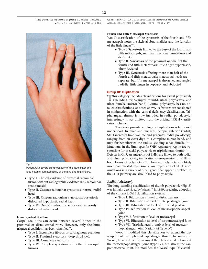
Fig. 7
Patient with severe camptodactyly of the little finger and
less notable camptodactyly of the long and ring fingers.
12
THE JOURNAL OF BONE & JOINT SURGERY d J B J S .ORG
VOLUME 91-A d SUPPLEMENT 4 d 2009CLASS I F ICAT ION AND DEVELOPMENTAL BIOLOGY OF CONGENITAL
ANOMAL IE S OF THE HAND AND UPPER EXTREMITY

cation to include the triphalangeal thumb as type IV-A andIV-B. Miura118 later added type IV-C.
� Type IV-A. Both thumb components are triphalangealat the metacarpophalangeal joint.
� Type IV-B. Only the radial component is triphalangealat the metacarpophalangeal joint.
� Type IV-C. Only the ulnar component is triphalangealat the metacarpophalangeal joint.
Wood also limited Type VII to triphalangeal thumbduplication at the carpometacarpal joint, which he furthersubclassified.
� Type VII-A. Radial component triphalangeal, ulnarcomponent biphalangeal at the carpometacarpaljoint
� Type VII-B. Both components triphalangeal at thecarpometacarpal joint
� Type VII-C. Ulnar component triphalangeal, radialcomponent biphalangeal at the carpometacarpal joint
� Type VII-D. Triple thumbs; central componenttriphalangeal, radial and ulnar components bipha-langeal at the carpometacarpal joint
The Wassel classification, including the modifications byWood and Miura, facilitates communication but does notconsider important features such as relative size of the com-ponent parts, angulation of the phalanges, and presence ofseparate or common nail plates, which affect function, aes-thetic appearance, and surgical decision-making.
Zuidam et al.119 recently attempted to address some ofthese concerns with a more extended classification. This classi-fication includes the carpal bones; type VII represents partialbifurcation at the level of the trapezium, and type VIII representscomplete bifurcation at the scaphotrapezial joint. Additionalfeatures include specific notations for triphalangeal (Tph), trip-lication (T), symphalangism (S), deviation (D), and hypoplasticor floating (H); the directional positions of the deformities areassigned abbreviations: u (ulnar), m (middle), and r (radial).This classification may prove to be too complex to be useful, anddoes not provide information about treatment or prognosis.
Ulnar PolydactylyUlnar polydactyly is manifested as a supernumerary digit onthe ulnar side of the hand. The anomaly is classified accordingto the development of the digits120.
� Type A. The supernumerary digit has well-developedlittle-finger soft-tissue and osseous structures, whicharticulate with the fifth metacarpal.
� Type B. The supernumerary digit is a rudimentary andpedunculated appendage attached to the ulnar side ofthe little finger or hand by a narrow skin bridge.
The classification provides both treatment guidelinesand prognostic information, as type A is associated with syn-dromes in Caucasian patients.
Ulnar Dimelia (Mirror Hand)This condition represents a spectrum of presentations ofduplication of the ulnar aspect of the embryonic limb bud.
The clinical features depend on the level of the duplication,ranging from duplication of the entire ulna at the elbow toduplication of the ulnar digits to form a six to eight-fingerhand with no thumb. It is a rare and bizarre manifestation ofduplication which is aesthetically unattractive and difficult tosurgically reconstruct. The descriptive classification is based onthe level of duplication121 and does not guide treatment or pro-vide prognosis.
� Type 1. Ulnar dimelia. Two ulnae with multiplefingers
� Type 1-A. Each ulna well formed� Type 1-B. Preaxial ulna hypoplastic� Type 2. Intermediate form. Two ulnae and a radius
with multiple fingers� Type 3. Intermediate form. One ulna and a radius with
multiple fingers� Type 3-A. Radius well formed� Type 3-B. Radius hypoplastic� Type 4. Syndromic form. Bilateral occurrence� Type 4-A. Two ulnae� Type 4-B. Ulna and radius� Type 5. Multiple hand. Complete duplication of the
hand, including the thumb; forearm normal
Group IV: Overgrowth (Gigantism)
Macrodactyly is characterized by enlargement of osseousand various soft-tissue structures of one or more
digits; extension of the enlargement into the wrist and entire
Fig. 8
Type-IV radial polydactyly.
13
THE JOURNAL OF BONE & JOINT SURGERY d J B J S .ORG
VOLUME 91-A d SUPPLEMENT 4 d 2009CLASS I F ICAT ION AND DEVELOPMENTAL BIOLOGY OF CONGENITAL
ANOMAL IE S OF THE HAND AND UPPER EXTREMITY

upper extremity may be noted (Fig. 9). The digits may beangled, with stiff joints. Macrodactyly may be associatedwith various syndromes, including Ollier disease, Maffuccisyndrome, Klippel-Trenaunay-Weber syndrome, and Proteussyndrome.
The classification is based on the underlying pathologicprocesses and does not reflect differences in appearance orguide surgical reconstruction122.
� Type 1. Gigantism and lipofibromatosis. Enlargedfatty infiltrated nerves within the digit, extendingproximally through the carpal tunnel. This is the mostcommon form.
� Type 2. Gigantism and neurofibromatosis. Occurs inconjunction with plexiform neurofibromatosis; oftenbilateral. Enlarged osseous skeleton may be associatedwith osteochondral masses.
� Type 3. Gigantism and digital hyperostosis. Osteo-chondral periarticular masses that develop in infancy;no significant nerve enlargement; digits nodular andstiff; rare.
� Type 4. Gigantism and hemihypertrophy. Macrodac-tyly is a component of hemihypertrophy of the entireupper extremity. Intrinsic muscles abnormal orhypertrophic; all digits may be involved, including thethumb.
Group V: Undergrowth (Hypoplasia)
See symbrachydactyly (central longitudinal deficiency-transverse) as noted above. There are no additional classi-
fications in this category.
Group VI: Congenital Constriction Band Syndrome
Ring constriction syndrome is characterized by bands thatencircle the limbs at various levels. The bands can cause
permanent indentation of the skin, local swelling, distaledema, syndactyly of adjacent and nonadjacent digits (usu-ally with a proximal sinus), and terminal amputation. Thesyndrome is classified according to the severity of thepresentation123.
� Type 1. Simple constriction rings� Type 2. Rings accompanied by distal soft-tissue de-
formity, with or without lymphedema� Type 3. Distal osseous syndactyly� Type 4. Terminal amputationThis classification is purely descriptive, and does not
guide treatment or provide prognosis.
Group VII: Generalized Skeletal Abnormalities
None of the congenital abnormalities in this category havebeen classified.
Proposed Classification Modification Based onMolecular Biology
Based on our current understanding of limb development,several modifications to the IFSSH classification could be
considered.Since most of the conditions under failure of formation
are deficiencies in axis formation or differentiation, it would bereasonable to group all of the following conditions involvingaxis disruption together in a Failure of Axis Formation and/orDifferentiation category.
Fig. 9
Patient with type-I macrodactyly of the index and long digits, extending proximally into
the hand.
14
THE JOURNAL OF BONE & JOINT SURGERY d J B J S .ORG
VOLUME 91-A d SUPPLEMENT 4 d 2009CLASS I F ICAT ION AND DEVELOPMENTAL BIOLOGY OF CONGENITAL
ANOMAL IE S OF THE HAND AND UPPER EXTREMITY

� Disruption of the radial-ulnar (anterior-posterior)axis either via loss of limb width, resulting in radialdeficiencies, or a loss in posterior (ulnar) patterning,leading to ulnar deficiencies
� Disruption of the proximal-distal axis, resulting intransverse deficiencies
� Disruption of the dorsal-ventral axis, causing nail-patella syndrome and palmar nail syndrome
In addition, synostoses involving the radius and ulnaseem to be more related to longitudinal deficiencies and thuswould necessitate a shift to the failure of axis formation and/ordifferentiation category. Furthermore, symbrachydactyly hasrecently been linked to a disruption in Wnt signaling and, assuch, is consistent with a form of transverse deficiency91; itshould be placed in the failure of axis formation and/or dif-ferentiation category, eliminating the undergrowth category.
Failure of differentiation is a somewhat vague term thatdoes not really reflect the conditions that have been groupedunder this category. A term that may better represent most ofthese entities is failure of hand-plate development and/ordifferentiation. This category would include developmentalmalformations targeting the hand. Therefore, shifting centraldeficiency to this category would be appropriate. Furthermore,since recent work indicates that similar misregulation or ec-topic expression of SHH induces both radial polydactyly andtriphalangeal thumb113,114, it would be reasonable to includetriphalangeal thumb in the radial polydactyly subcategory offailure of hand-plate development and/or differentiation.
The proposed modification is shown in Table II. It re-flects the basic concepts of the original IFSSH classificationbut includes the current understanding of limb development.We recommend it to congenital hand surgeons for theirconsideration.
Conclusions
The IFSSH classification has provided a useful frameworkfor congenital upper-extremity anomalies for over forty
years, accommodating specific classifications for twenty dif-ferent conditions within its seven categories. As detailed above,few of these classification schemes live up to the ideal, whichis to guide treatment, provide insight into prognosis, or in-corporate the etiologic mechanism of the condition. Recentdevelopments in molecular biology allow us to propose amodification to the IFSSH classification to incorporate ourenhanced understanding of the etiology of congenital mal-formations. The ideal classification scheme awaits furtherinvestigation. n
Paul R. Manske, MDWashington University School of Medicine,Campus Box 8233, 660 South Euclid Avenue,St. Louis, MO 63110. E-mail address:[email protected]
Kerby C. Oberg, MD, PhDDivisions of Pediatric Pathology and Human Anatomy,Department of Surgery, Loma Linda University,24785 Stewart Street, Room B09c,Loma Linda, CA 92350
References
1. Daluiski A, Yi SE, Lyons KM. The molecular control of upper extremity develop-ment: implications for congenital hand anomalies. J Hand Surg [Am]. 2001;26:8-22.
2. Kawakami Y, Capdevila J, Buscher D, Itoh T, Rodrıguez Esteban C, IzpisuaBelmonte JC. WNT signals control FGF-dependent limb initiation and AER inductionin the chick embryo. Cell. 2001;104:891-900.
3. Panman L, Zeller R. Patterning the limb before and after SHH signalling. J Anat.2003;202:3-12.
4. Chen H, Johnson RL. Interactions between dorsal-ventral patterning geneslmx1b, engrailed-1 and wnt-7a in the vertebrate limb. Int J Dev Biol. 2002;46:937-41.
5. Kawakami Y, Wada N, Nishimatsu S, Nohno T. Involvement of frizzled-10 inWnt-7a signaling during chick limb development. Dev Growth Differ. 2000;42:561-9.
6. Summerbell D. A quantitative analysis of the effect of excision of the AER fromthe chick limb-bud. J Embryol Exp Morphol. 1974;32:651-60.
7. Lu P, Yu Y, Perdue Y, Werb Z. The apical ectodermal ridge is a timer for gen-erating distal limb progenitors. Development. 2008;135:1395-405.
8. Yu K, Ornitz DM. FGF signaling regulates mesenchymal differentiation andskeletal patterning along the limb bud proximodistal axis. Development. 2008;135:483-91.
TABLE II Proposed Modifications to the IFSSH Classification
I. Failure of axis formation and/or differentiation
Radial longitudinal deficiency
Radial-ulnar synostosis
Ulnar longitudinal deficiency
Transverse deficiency (including symbrachydactyly)
Dorsal ventral deficiency
II. Failure of hand-plate formation and/or differentiation
Syndactyly
Apert syndrome
Central deficiency (cleft hand)
Camptodactyly
Clinodactyly
Clasped thumb
Hand-plate synostoses
Metacarpal synostosis
Carpal synostosis
III. Duplication
Radial polydactyly (including triphalangeal thumb)
Ulnar dimelia
Ulnar polydactyly
IV. Overgrowth
V. Amniotic band sequence
VI. Generalized skeletal abnormalities
15
THE JOURNAL OF BONE & JOINT SURGERY d J B J S .ORG
VOLUME 91-A d SUPPLEMENT 4 d 2009CLASS I F ICAT ION AND DEVELOPMENTAL BIOLOGY OF CONGENITAL
ANOMAL IE S OF THE HAND AND UPPER EXTREMITY

9. Towers M, Mahood R, Yin Y, Tickle C. Integration of growth and specification inchick wing digit-patterning. Nature. 2008;452:882-6.
10. Zhu J, Nakamura E, Nguyen MT, Bao X, Akiyama H, Mackem S. UncouplingSonic hedgehog control of pattern and expansion of the developing limb bud. DevCell. 2008;14:624-32.
11. Buck-Gramcko D. Congenital malformation in hand surgery. In: Nigst H, Buck-Gramcko D, Millesi H, Lister GD, editors. Hand surgery. New York: Thieme MedicalPublishers; 1988. p 12.1-12.110.
12. Kay H. A proposed international terminology for the classification of congenitallimb deficiencies. Orthotics Prosthetics. 1974;28:33-48.
13. Kelikian H. Congenital deformities of the hand and forearm. Philadelphia: WBSaunders; 1974. Classifications; p 51-88.
14. Saint-Hilaire IG. Propositions sur la monstruosite. Paris: Imp. Didot le Jeune;1829. p 18-9.
15. Otto AW. A compounding of human comparative pathological anatomy. Lon-don: B Fellows; 1831. p 1-41, 216-20.
16. Saint-Hilaire IG. Histoire generale et particuliere des anomalies de l’orga-nisation chez l’homme et les animaux. Paris: JB Bailliere; 1832. p 257-78,670-702.
17. Fort AJ. Des difformites congenitales et acquises des doigts, et des moyensd’y remedier. Paris: Adrien Delahaye; 1869. p 8-11.
18. Leboucq J. Recherches sur la morphologie du carpe chez les mammiferes.Arch Biol. 1884;5:35-102.
19. Polaillon JF. Doigt. In: Dechambre K, editor. Dictionnaire encyclopedique dessciences medicales. Paris: G Masson and P Asselin; 1884. p 115-353.
20. Potel G. Essai sur les malformations congenitales des membres. Leurclassification pathogenique. Rev Chir. 1914;49:293-326, 625-48, 822-58; 50:84-114.
21. Frantz C, O’Rahilly R. Congenital skeletal limb deficiencies. J Bone Joint SurgAm. 1961;43:1202-24.
22. O’Rahilly R. Morphological patterns in limb deficiencies and duplications. Am JAnat. 1951;89:135-94.
23. Kummel W. Die Missbildungen der Extremitaeten durch Defekt, Verwachsungund Ueberzahl. Cassel, Germany: TG Fischer; 1895. p 1-83.
24. Nigst PF. Uber kongenitale Missbildungen des menschlichen Extremitatenskeletts,mit Rontgenbildern. Schweiz Med Wschr. 1927;8:7-18, 53-61, 81-91, 97-106.
25. Muller W. Die Angeborenen Fehlbildungen der menschlichen Hand. Leipzig:Thieme; 1937.
26. Swanson AB. A classification for congenital malformations of the hand. BullAcad Med N J. 1964;10:166-9.
27. Swanson AB, Barsky AJ, Entin MA. Classification of limb malformations on thebasis of embryological failures. Surg Clin N Amer. 1968;48:1169-79.
28. Swanson AB. A classification for congenital limb malformations. J Hand Surg[Am]. 1976;1:8-22.
29. Baruch AD, Hecht OA. Treatment of monodactyly by the distraction-lengtheningprinciple: a case report. J Hand Surg [Am]. 1983;8(5 Pt 1):604-6.
30. Buckwalter JA, Flatt AE, Shurr DG, Dryer RF, Blair WF. The absent fifth meta-carpal. J Hand Surg [Am]. 1981;6:364-7.
31. Kato S, Ishii S, Ogino T, Shiono H. Anomalous hands with cleft formationbetween the fourth and fifth digits. J Hand Surg [Am]. 1983;8:909-13.
32. Light TR, Ogden JA. The longitudinal epiphyseal bracket: implications for sur-gical correction. J Pediatr Orthop. 1981;1:299-305.
33. Miura T. Cleft hand involving only the ring and small fingers. J Hand Surg [Am].1988;13:530-5.
34. Cheng JC, Chow SK, Leung PC. Classification of 578 cases of congenital upperlimb anomalies with the IFSSH system—a 10 years’ experience. J Hand Surg [Am].1987;12:1055-60.
35. Knight SL, Kay SPJ. Classification of congenital anomalies. In: Gupta A, KaySPJ, Scheker LR, editors. The growing hand: diagnosis and management of theupper extremity in children. London: Mosby; 2000. p 125-35.
36. Congenital Hand Committee of the JSSH. Modified IFSSH classification. J Ja-pan Soc Surg Hand. 2000;17:353-65.
37. Kato H, Ogino T, Minami A, Ohshio I. Experimental study of radial ray defi-ciency. J Hand Surg [Br]. 1990;15:470-6.
38. Ogino T, Kato H. Clinical and experimental studies on ulnar ray deficiency.Handchir Mikrochir Plast Chir. 1988;20:330-7.
39. Tonkin MA. Description of congenital hand anomalies: a personal view. J HandSurg [Br]. 2006;31:489-97.
40. Goldfarb CA, Manske PR, Busa R, Mills J, Carter P, Ezaki M. Upper-extremityphocomelia reexamined: a longitudinal dysplasia. J Bone Joint Surg Am. 2005;87:2639-48.
41. Tytherleigh-Strong G, Hooper G. The classification of phocomelia. J Hand Surg[Br]. 2003;28:215-7.
42. Mariani FV, Ahn CP, Martin GR. Genetic evidence that FGFs have an instructiverole in limb proximal-distal patterning. Nature. 2008;453:401-5.
43. Sun X, Mariani FV, Martin GR. Functions of FGF signalling from the apicalectodermal ridge in limb development. Nature. 2002;418:501-8.
44. Goldfarb CA, Wall L, Manske PR. Radial longitudinal deficiency: the incidenceof associated medical and musculoskeletal conditions. J Hand Surg [Am]. 2006;31:1176-82.
45. Blauth W. Der hypoplastische Daumen. Arch Orthop Unfallchir. 1967;62:225-46.
46. James MA, McCarroll HR Jr, Manske PR. Characteristics of patients with hy-poplastic thumbs. J Hand Surg [Am]. 1996;21:104-13.
47. Manske PR, McCarroll HR Jr, James M. Type III-A hypoplastic thumb. J HandSurg [Am]. 1995;20:46-53.
48. Graham TJ, Louis DS. A comprehensive approach to surgical management ofthe type IIIA hypoplastic thumb. J Hand Surg [Am]. 1998;23:3-13.
49. Bayne LG, Klug MS. Long-term review of the surgical treatment of radial defi-ciencies. J Hand Surg [Am]. 1987;12:169-79.
50. James MA, McCarroll HR Jr, Manske PR. The spectrum of radiallongitudinal deficiency: a modified classification. J Hand Surg [Am]. 1999;24:1145-55.
51. Nutt JN 3rd, Flatt AE. Congenital central hand deficit. J Hand Surg [Am].1981;6:48-60.
52. Ogino T. Cleft hand. Hand Clin. 1990;6:661-71.
53. Tada K, Yonenobu K, Swanson AB. Congenital central ray deficiency in thehand—a survey of 59 cases and subclassification. J Hand Surg [Am]. 1981;6:434-41.
54. Watari S, Tsuge K. A classification of cleft hands, based on clinical findings:theory of developmental mechanism. Plast Reconstr Surg. 1979;64:381-9.
55. Lo Iacono N, Mantero S, Chiarelli A, Garcia E, Mills AA, Morasso MI, CostanzoA, Levi G, Guerrini L, Merlo GR. Regulation of Dlx5 and Dlx6 gene expression byp63 is involved in EEC and SHFM congenital limb defects. Development. 2008;135:1377-88.
56. Duijf PH, van Bokhoven H, Brunner HG. Pathogenesis of split-hand/split-footmalformation. Hum Mol Genet. 2003;12 Spec No 1:R51-60.
57. Buck-Gramcko D. Cleft hands: classification and treatment. Hand Clin. 1985;1:467-73.
58. Ogino T. Teratogenic relationship between polydactyly, syndactyly and clefthand. J Hand Surg [Br]. 1990;15:201-9.
59. Naruse T, Takahara M, Takagi M, Oberg KC, Ogino T. Busulfan-inducedcentral polydactyly, syndactyly and cleft hand or foot: a common mechanism ofdisruption leads to divergent phenotypes. Dev Growth Differ. 2007;49:533-41.
60. Barsky AJ. Cleft hand: classification, incidence, and treatment. Review ofthe literature and report of nineteen cases. J Bone Joint Surg Am. 1964;46:1707-20.
61. Flatt AE. The care of congenital hand anomalies. St. Louis: Mosby; 1977.p 328-41.
62. Ogino T, Minami A, Kato H. Clinical features and roentgenograms of sym-brachydactyly. J Hand Surg [Br]. 1989;14:303-6.
63. Sandzen SC Jr. Classification and functional management of congenital cen-tral defect of the hand. Hand Clin. 1985;1:483-98.
64. Flatt AE. The care of congenital hand anomalies. St. Louis: Mosby; 1977.p 265-85.
65. Manske PR, Halikis MN. Surgical classification of central deficiency accordingto the thumb web. J Hand Surg [Am]. 1995;20:687-97.
16
THE JOURNAL OF BONE & JOINT SURGERY d J B J S .ORG
VOLUME 91-A d SUPPLEMENT 4 d 2009CLASS I F ICAT ION AND DEVELOPMENTAL BIOLOGY OF CONGENITAL
ANOMAL IE S OF THE HAND AND UPPER EXTREMITY

66. Sykes PJ, Eadie PA. Longitudinal ulnar deficiency in the hand. In: Gupta A, KaySPJ, Scheker LR, editors. The growing hand: diagnosis and management of theupper extremity in children. London: Mosby; 2000. p 189-95.
67. Bayne LG. Ulnar club hand (ulnar deficiencies). In: Green DP, editor. Operativehand surgery. New York: Churchill Livingstone; 1982. p 245-57.
68. Miller JK, Wenner SM, Kruger LM. Ulnar deficiency. J Hand Surg [Am].1986;11:822-9.
69. Ogden JA, Watson HK, Bohne W. Ulnar dysmelia. J Bone Joint Surg Am.1976;58:467-75.
70. Riordan DC. The upper limb. In: Lowell WW, Lovell RB, Winter RB, editors.Pediatric orthopaedics. Vol 2. Philadelphia: JB Lippincott; 1978. p 685-719.
71. Swanson AB, Tada K, Yonenobu K. Ulnar ray deficiency: its various manifes-tations. J Hand Surg [Am]. 1984;9:658-64.
72. Havenhill TG, Manske PR, Patel A, Goldfarb CA. Type 0 ulnar longitudinaldeficiency. J Hand Surg [Am]. 2005;30:1288-93.
73. Blair WF, Shurr DG, Buckwalter JA. Functional status in ulnar deficiency.J Pediatr Orthop. 1983;3:37-40.
74. Broudy AS, Smith RJ. Deformities of the hand and wrist with ulnar deficiency.J Hand Surg [Am]. 1979;4:304-15.
75. Carroll RE, Bowers WH. Congenital deficiency of the ulna. J Hand Surg [Am].1977;2:169-74.
76. Cole RJ, Manske PR. Classification of ulnar deficiency according to the thumband first web. J Hand Surg [Am]. 1997;22:479-88.
77. Johnson J, Omer GE Jr. Congenital ulnar deficiency. Natural history and ther-apeutic implications. Hand Clin. 1985;1:499-510.
78. Buchler U. Symbrachydactyly. In: Gupta A, Kay SPJ, Scheker LR, editors. Thegrowing hand: diagnosis and management of the upper extremity in children.London: Mosby; 2000. p 213-24.
79. Erhardt PR, Lindley SG, Functional development of the hand. In: Gupta A, KaySPJ, Scheker LR, editors. The growing hand: diagnosis and management of theupper extremity in children. London: Mosby; 2000. p 71-82.
80. James M, Bednar M. Deformities of the wrist and forearm. In: Green DP,Hotchkiss RN, Pederson WC, Wolfe SW, editors. Green’s operative hand surgery.5th ed. Philadelphia: Elsevier Churchill Livingstone; 2005. p 1489-91.
81. Kay SP. Congenital hand deformities: hypoplastic and absent digits. In: GreenDP, Hotchkiss RN, Pederson WC, editors. Green’s operative hand surgery. 4th ed,vol 1. New York: Churchill Livingstone; 1998. p 368-75.
82. Kay SP, McCombe D, Kozin SH. Deformities of the hand and fingers. In:Green DP, Hotchkiss RN, Pederson WC, Wolfe SW, editors. Green’s operativehand surgery. 5th ed. Philadelphia: Elsevier Churchill Livingstone; 2005. p 1417-30.
83. Light TR. Congenital anomalies: syndactyly, polydactyly, and cleft hand. In:Peimer CA, editor. Surgery of the hand and upper extremity. New York: McGraw-Hill;1996. p 2121-3.
84. Tonkin MA. Congenital hand deformities: major limb defects. In: Green DP,Hotchkiss RN, Pederson WC, editors. Green’s operative hand surgery. 4th ed,vol 1. New York: Churchill Livingstone; 1998. p 338-41.
85. Waters PM, Simmons BP. Congenital anomalies: elbow region. In: Peimer CA,editor. Surgery of the hand and upper extremity. New York: McGraw-Hill; 1996.p 2050-1.
86. Blauth W, Gekeler J. [Morphology and classification of symbrachydactylia].Handchirurgie. 1971;3:123-8. German.
87. Buck-Gramcko D. Symbrachydactyly: a clinical entity. Tech Hand Up ExtremSurg. 1999;3:242-58.
88. Yamauchi Y, Tanabu S. Symbrachydactyly. In: Buck-Gramcko D, editor. Con-genital malformations of the hand and forearm. Philadelphia: Churchill Livingstone;1998. p 149-58.
89. Kallemeier PM, Manske PR, Davis B, Goldfarb CA. An assessment of therelationship between congenital transverse deficiency of the forearm andsymbrachydactyly. J Hand Surg [Am]. 2007;2:1408-12.
90. Summerbell D, Lewis JH. Time, place and positional value in the chick limb-bud. J Embryol Exp Morphol. 1975;33:621-43.
91. Winkel A, Stricker S, Tylzanowski P, Seiffart V, Mundlos S, Gross G, HoffmannA. Wnt-ligand-dependent interaction of TAK1 (TGF-beta-activated kinase-1) with thereceptor tyrosine kinase Ror2 modulates canonical Wnt-signalling. Cell Signal.2008;20:2134-44.
92. Pol R. ‘Brachydaktylie’—‘Klinodaktylie’—Hyperphalangie und ihre Grundlagen:Form und Entstehung der meist unter dem Bild der Brachydaktylie auftretendenVarietaten, Anomalien und Mißbildungen der Hand und des Fußes. Virchows ArchPath Anat. 1921;229:388-530.
93. Kelikian H. Congenital deformities of the hand and forearm. Philadelphia: WBSaunders; 1974. Phocomelia; p 891-901.
94. Merino R, Rodriguez-Leon J, Macias D, Ganan Y, Economides AN, HurleJM. The BMP antagonist Gremlin regulates outgrowth, chondrogenesis andprogrammed cell death in the developing limb. Development. 1999;126:5515-22.
95. Weatherbee SD, Behringer RR, Rasweiler JJ 4th, Niswander LA. Inter-digital webbing retention in bat wings illustrates genetic changes underlyingamniote limb diversification. Proc Natl Acad Sci U S A. 2006;103:15103-7.
96. Pajni-Underwood S, Wilson CP, Elder C, Mishina Y, Lewandoski M. BMP sig-nals control limb bud interdigital programmed cell death by regulating FGF signal-ing. Development. 2007;134:2359-68.
97. Flatt AE. The care of congenital hand anomalies. St. Louis: Mosby; 1977.p 170-92.
98. Dobyns JH. Syndactyly. In: Green DP, Hotchkiss RN, Pederson WC, editors.Green’s operative hand surgery. 3rd ed. New York: Churchill Livingstone; 1993.p 346.
99. Upton J. Apert syndrome. Classification and pathologic anatomy of limbanomalies. Clin Plast Surg. 1991;18:321-55.
100. Van Heest AE, House JH, Reckling WC. Two-stage reconstruction of Apertacrosyndactyly. J Hand Surg [Am]. 1997;22:315-22.
101. Benson LS, Waters PM, Kamil NI, Simmons BP, Upton J 3rd. Camptodactyly:classification and results of nonoperative treatment. J Pediatr Orthop. 1994;14:814-9.
102. Cooney WP. Camptodactyly and clinodactyly. In: Carter PR, editor. Re-construction of the child’s hand. Philadelphia: Lea and Febiger; 1991.p 209-35.
103. Weckesser EC, Reed JR, Heipel KG. Congenital clasped thumb (congenitalflexion-adduction deformity of the thumb): a syndrome, not a specific entity. J BoneJoint Surg Am. 1968;50:1417-28.
104. McCarroll HR Jr. Congenital flexion deformities of the thumb. Hand Clin.1985;1:567-75.
105. McCarroll HR Jr, Manske PR. The windblown hand: correction of the complexclasped thumb deformity. Hand Clin. 1992;8:147-59.
106. Mih AD. Congenital clasped thumb. Hand Clin. 1998;14:77-84.
107. Mital MA. Congenital radioulnar synostosis and congenital dislocation of theradial head. Orthop Clin North Am. 1976;7:375-83.
108. Cleary JE, Omer GE Jr. Congenital proximal radio-ulnar synostosis.Natural history and functional assessment. J Bone Joint Surg Am. 1985;67:539-45.
109. Minnaar A. Congenital fusion between the lunate and the triquetral bones inthe South African Bantu. J Bone Joint Surg Br. 1952;34:45-8.
110. Wood VE. Metacarpal synostosis. In: Green DP, Hotchkiss RN, Pederson WC,editors. Green’s operative hand surgery. 3rd ed. New York: Churchill Livingstone;1993. p 326-38.
111. Charite J, de Graaff W, Shen S, Deschamps J. Ectopic expression of Hoxb-8causes duplication of the ZPA in the forelimb and homeotic transformation of axialstructures. Cell. 1994;78:589-601.
112. Yang Y, Drossopoulou G, Chuang PT, Duprez D, Marti E, Bumcrot D,Vargesson N, Clarke J, Niswander L, McMahon A, Tickle C. Relationship betweendose, distance and time in Sonic Hedgehog-mediated regulation of anteroposteriorpolarity in the chick limb. Development. 1997;124:4393-404.
113. Lettice LA, Heaney SJ, Purdie LA, Li L, de Beer P, Oostra BA, Goode D, ElgarG, Hill RE, de Graaff E. A long-range Shh enhancer regulates expression in thedeveloping limb and fin and is associated with preaxial polydactyly. Hum Mol Genet.2003;12:1725-35.
114. Gurnett CA, Bowcock AM, Dietz FR, Morcuende JA, Murray JC, Dobbs MB.Two novel point mutations in the long-range SHH enhancer in three families withtriphalangeal thumb and preaxial polydactyly. Am J Med Genet A. 2007;143:27-32.
115. Radhakrishna U, Bornholdt D, Scott HS, Patel UC, Rossier C, Engel H,Bottani A, Chandal D, Blouin JL, Solanki JV, Grzeschik KH, Antonarakis SE. Thephenotypic spectrum of GLI3 morphopathies includes autosomal dominant pre-
17
THE JOURNAL OF BONE & JOINT SURGERY d J B J S .ORG
VOLUME 91-A d SUPPLEMENT 4 d 2009CLASS I F ICAT ION AND DEVELOPMENTAL BIOLOGY OF CONGENITAL
ANOMAL IE S OF THE HAND AND UPPER EXTREMITY

axial polydactyly type-IV and postaxial polydactyly type-A/B; no phenotype pre-diction from the position of GLI3 mutations. Am J Hum Genet. 1999;65:645-55.
116. Wassel HD. The results of surgery for polydactyly of the thumb. Clin OrthopRelat Res. 1969;64:175-93.
117. Wood VE. Polydactyly and the triphalangeal thumb. J Hand Surg [Am].1978;3:436-44.
118. Miura T. Triphalangeal thumb. Plast Reconstr Surg. 1976;58:587-94.
119. Zuidam JM, Selles RW, Ananta M, Runia J, Hovius SE. A classification systemof radial polydactyly: inclusion of triphalangeal thumb and triplication. J Hand Surg[Am]. 2008;33:373-7.
120. Temtamy S, McKusick VA. Synopsis of hand malformations with particularemphasis on genetic factors. Birth Defects. 1969;5:125-84.
121. Al-Qattan MM, Al-Thunayan A, De Cordier M, Nandagopal N, Pitkanen J.Classification of the mirror hand-multiple hand spectrum. J Hand Surg [Br].1998;23:534-6.
122. Flatt AE. The care of congenital hand anomalies. 2nd ed. St. Louis, MO:Quality Medical Publishing; 1994. p 317-33.
123. Patterson TJ. Congenital ring-constrictions. Br J Plast Surg. 1961;14:1-31.
18
THE JOURNAL OF BONE & JOINT SURGERY d J B J S .ORG
VOLUME 91-A d SUPPLEMENT 4 d 2009CLASS I F ICAT ION AND DEVELOPMENTAL BIOLOGY OF CONGENITAL
ANOMAL IE S OF THE HAND AND UPPER EXTREMITY