Embed Size (px)
Citation preview
Postgrad MedJ 1998;74:459-467 c The Fellowship of Postgraduate Medicine, 1998
Classic diseases revisited
Abdominal tuberculosis
V K Kapoor
SummaryTuberculosis has staged a globalcomeback and forms a dangerouscombination with AIDS. The ab-domen is one of the common sitesof extrapulmonary involvement.Patients with abdominal tubercu-losis have a wide range and spec-trum of symptoms and signs; thedisease is therefore a great minmc.Diagnosis, mainly radiologicaland supported by endoscopy, isdifficult to make and laparotomyis required in a large number ofpatient. Management involves ju-dicious combination of antituber-cular therapy and surgery whichmay be required to treat compli-cations such as intestinal obstruc-tion and perforation. The disease,though potentially curable, car-ries a significant morbidity andmortality.
Keywords: tuberculosis
Department of SurgicalGastroenterology, Sanjay GandhiPostgraduate Institute ofMedicalSciences, Lucknow 226014, IndiaV K Kapoor
Accepted 10 March 1998
"The captain of all these men ofdeath that came against him to take him away was theconsumption;for it was that that brought him down to grave." John Bunyan, 1628-88'
Tuberculosis causes 3 million deaths every year, accounting for 6% of alldeaths. It is among the 10 leading causes of death and is one of the commonestcauses of death in the young.2
Incidence
Tuberculosis is the most important communicable disease world-wide. Despiteexpectations such as "tuberculosis should be virtually eradicated from mostdeveloping countries within 50 years"3 it has come back with a vengeance andhas recently been declared a global emergency by the World HealthOrganisation. It continues to be prevalent in the underdeveloped and developingThird world, and although it was on the verge of eradication in the developedworld, its prevalence is increasing there too, due to factors such as transglobalimmigration, ageing populations, alcoholism, socio-economic deprivation, andmore recently, acquired immunodeficiency syndrome (AIDS). It is estimatedthat half of the world's population is infected and about 10 million new casesoccur every year.The abdomen is involved in 11% of patients with extra-pulmonary
tuberculosis.4 In a recent series of 820 patients with tuberculosis reported fromSaudi Arabia,5 16% had abdominal involvement. Abdominal tuberculosiscontinues to be common in various parts of the world with large series beingreported from Chile,6 Egypt,7 India,8" Iraq,'4 Kuwait,'5 Nigeria,'6 Saudi Arabia'7and Sudan.'8
In the UK, pulmonary tuberculosis has declined in incidence butnon-pulmonary tuberculosis continues to be common.'9 In 1985, abdominaltuberculosis accounted for 5% of all cases of tuberculosis notified in a district inthe UK.20 The disease was largely under control in the 1950s but revived in the1960s and 70sQ2 and its renaissance in the 1980s and 90s22 is disturbing. In aDistrict General Hospital in London, Palmer et a!23 reported having seen 90patients with abdominal tuberculosis during the 10-year period up to 1984; overthe same period Crohn's disease was diagnosed in 102 patients. This may not betypical of all of the country but may reflect experience in areas with a large Asianpopulation.24 Other large series of abdominal tuberculosis have been reportedfrom areas in the UK with large immigrant populations.25-28 Abdominal tubercu-losis has also reappeared in the US29-34 and is likely to become more commonbecause of AIDS.35
Aetiopathogenesis
Abdominal tuberculosis probably occurs due to reactivation of a dormant focus.This primary gastrointestinal focus is established as a result of haematogenousspread from a pulmonary focus acquired during primary infection in childhood.It may also be caused by swallowed bacilli which pass through the Peyer's patchesof the intestinal mucosa and are transported by macrophages through the lym-phatics to the mesenteric lymph nodes, where they remain dormant.Suppression of host defences by conditions such as malnutrition, weight loss,alcoholism, diabetes, chronic renal failure, immunosuppression, AIDS, etc,increases the risk of such reactivation. Ingestion of bacilli from an active pulmo-nary focus, haematogenous spread from active tuberculosis in other organs, anddirect extension from adjacent organs are other possible mechanisms of involve-ment of the abdomen. Ingestion of infected milk is rarely a cause because of dis-appearance ofbovine tuberculosis and pasteurisation of milk in the West and thepractice of boiling milk before consumption in the developing countries. Mostbacilli isolated in patients with abdominal tuberculosis are Mycobacterium tuber-culosis and not Mycobacterium bovis."1 28 36
on 24 March 2019 by guest. P
rotected by copyright.http://pm
j.bmj.com
/P
ostgrad Med J: first published as 10.1136/pgm
j.74.874.459 on 1 August 1998. D
ownloaded from
460 Kapoor
Extrapulmonary disease is more common in patients with AIDS; 50% ofAIDS patients with tuberculosis have extrapulmonary involvement, compared toonly 10-15% of non-HIV tuberculosis patients."7 The diagnosis of tuberculosismay precede the diagnosis of AIDS by several months; tuberculosis frequentlydisseminates in AIDS patients, progresses rapidly and is associated with a highmortality."8 Treatment of tuberculosis in AIDS patients is the same as innon-HIV infected patients'9 but multi-drug-resistant tuberculosis is more com-mon in patients with AIDS.40
Pathology
Abdominal tuberculosis denotes involvement of the gastrointestinal tract,peritoneum, lymph nodes, and solid viscera, eg, liver, spleen, pancreas, etc. Thegastrointestinal tract is involved in 65%' to 78%12 of patients; associated perito-neal and lymph node involvement is common in these patients. The commonsites of involvement in the gastrointestinal tract are the ileum9 " and the ileocae-cal region,2' 41 followed by the colon and the jejunum. In 196 patients with gas-trointestinal tuberculosis,9 the ileum was involved in 102 and caecum in 100patients. In another series of 300 patients,'0 however, the ileocaecal region wasinvolved in 162 and the ileum in only 89 patients. Three types of intestinallesions are commonly seen - ulcerative, stricturous, and hypertrophic, cicatricialhealing of the ulcerative lesions resulting in strictures. Occlusive arterial changesmay produce ischaemia and contribute to development of strictures.42 Thesemorphological types can coexist, eg, ulcero-constrictive and ulcero-hypertrophiclesions. Small intestinal lesions are usually ulcerative or stricturous and largeintestinal lesions are ulcero-hypertrophic. Colonic lesions are usually associatedwith ileocaecal or ileal involvement but isolated segmental colonic tuberculosisdoes occur.4 44 Some patients have involvement ofperitoneum and lymph nodesalone without involvement of the gastrointestinal tract. Peritoneal involvementmay be of either an ascitic or adhesive (plastic) type. The lymph nodes in thesmall bowel mesentery and the retroperitoneum are commonly involved, andthese may caseate and calcify. Disseminated abdominal tuberculosis involvingthe gastrointestinal tract, peritoneum, lymph nodes and solid viscera has alsobeen described. Chen et al" reported disseminated involvement of the abdomenin 21 out of 60 patients with large bowel tuberculosis, while most of the 96patients with tuberculous hepatitis reported by Essop et al'6 had disseminateddisease. Multiple lesions are common. Bhansali9 reported that small intestinalstrictures were multiple in 71 out of 119 patients; as many as 12,47 1 6,"' and 1948strictures have been reported in a single patient.
Clinical features
Abdominal tuberculosis can occur at any age but is predominantly a disease ofyoung adults; two-thirds of patients are 21-40 years old9 11 23 49 and the mean ageof patients is 30-40 years.'2 17 23 41 The mean age of white patients is higher - 56years.20 Although some reports mention a higher incidence in females,'0 12 41 itseems that the disease affects both sexes equally.9 23 28 Abdominal tuberculosis isalso seen in children, where the spectrum of disease is different from that inadults; 90% of child patients have peritoneal and lymph node involvement,intestinal lesions being present in less than 10% of cases.50Abdominal tuberculosis is characterised by different modes of presentation,
viz, chronic, acute and acute-on-chronic, or it may be an incidental finding atlaparotomy for other diseases; incidental abdominal tuberculosis is usually peri-toneal and lymph nodal.'5 The clinical presentation depends upon the site andtype of involvement (table 1). Bhansali9 observed frank malabsorption in 21% ofpatients, while Tandon et arl reported biochemical evidence ofmalabsorption in75% of patients with intestinal obstruction and 40% of those without it. Thelump in patients with abdominal tuberculosis is firm, mobile and only slightlytender. Rectal bleeding has been reported in 4%49 to 6%"1 of patients; massivelower gastrointestinal bleeding is rare.52 Subacute intestinal obstruction isdescribed as colicky abdominal pain, distension, vomiting, gurgling, feeling of aball of wind moving in the abdomen, and visible loops and peristalsis; thesesymptoms are relieved spontaneously after passage of flatus. Ano-rectal tubercu-losis presents as stricture,5' fistula-in-ano,54 55 or fissure-in-ano.56 Tubercular fis-tulae are usually multiple; as many as 12 out of 15 multiple flstulae but only fourout of 61 single peni-anal flstulae were tubercular.57
Gastroduodenal tuberculosis may present as peptic ulcer with or without gas-tric outlet obstruction58 59 or perforation20 and may mimic carcinoma.60 Shortduration of history, early onset of obstruction, bizarre endoscopic findings, andnon-response to H2-receptor antagonists in a patient with a diagnosis of peptic
on 24 March 2019 by guest. P
rotected by copyright.http://pm
j.bmj.com
/P
ostgrad Med J: first published as 10.1136/pgm
j.74.874.459 on 1 August 1998. D
ownloaded from
Abdominal tuberculosis 461
Acute tubercular abdomen
* intestinal obstruction: acute oracute-on-chronic
* peritonitis: with or without perforation* acute mesenteric lymphadenitis* acute tubercular appendicitis
Box 1
Differential diagnosis
Intestinal lesions* ulcerative: coeliac disease, tropical
sprue, immunoproliferative smallintestinal disease, giardial infestation74
* strictures: Crohn's disease, malignancy(adenocarcinoma and lymphoma),ischaemic"6
* hypertrophic: carcinoma caecum,appendicular lump, amoeboma,actinomycosis
* perforations: typhoid"
Peritoneal* ascites: cardiac failure, malnutrition,
nephrotic syndrome, cirrhosis* tubercles: carcinomatosis76
Box 2
Table 1 Clinical features
Site Type Clinicalfeatures
Small intestine Ulcerative* Diarrhoea, malabsorptionStricturous Obstruction
Large intestine Ulcerative Rectal bleedingHypertrophic Lump, obstruction
Peritoneal Ascitic* Pain, distensionAdhesive Obstruction
Lymph nodes Lump, obstruction
*Systemic symptoms of tuberculous infection also present
ulcer should arouse the suspicion of gastroduodenal tuberculosis.6' Microscopicinvolvement of the liver is common in patients with abdominal tuberculosis butisolated focal lesions (tuberculoma) are rare.46 Tuberculosis at unusual sitesmimics more common diseases in those organs, eg, oesophagus - carcinoma,62pancreas - carcinoma,63 pancreatitis,64 and abscess.65
Varying grades of tenderness and guarding may be present in patients withascitic peritoneal tuberculosis but board-like rigidity or rebound tenderness asseen in pyogenic peritonitis is absent.66 Loculation of the ascitic fluid may resultin a soft cystic lump. Involvement of the mesenteric lymph nodes produces alump in the central abdomen. Enlarged lymph nodes at the root of the mesen-tery may cause obstruction to the third part of the duodenum.58 61 Portal hyper-tension due to portal vein compression67 and obstructive jaundice due to com-pression of the common bile duct due to tuberculous nodes, have beenreported.68
Systemic manifestations of tuberculous infection include low-grade fever withevening rise, lethargy, malaise, night sweats, anorexia and weight loss (failure tothrive in children). These are present in about one-third of patients withabdominal tuberculosis" and are more frequent in those with ulcerative intesti-nal lesions and ascitic peritoneal tuberculosis. Some patients, particularly thosewith miliary tuberculosis, may have tubercular toxaemia," with high fever,tachycardia, anaemia, and leucocytosis. Tuberculous involvement of otherorgans or systems has been reported in as many as one-third of patients.'2 Thecommonest sites of involvement are pulmonary and pleural. Genital tractinvolvement has been reported in 10% of women with abdominaltuberculosis.9 21 Peripheral lymph nodes (cervical or axillary) may be involved in3-10%1" 12 49 50 of patients. A family history of tuberculosis, reported in aboutone-third ofpatients in the UK,20 28 is rarely revealed by patients in India becauseof the social stigma still attached to the disease.
Tuberculosis is regarded as a disease with insidious onset and chronic presen-tation, most patients having symptoms for a few weeks to months, sometimesyears; Lambrianides et af2' even stated that tuberculosis is rarely an emergency.Between 15 and 40% of patients 1O 23 49 may, however, present with an acuteabdomen69 (box 1). Intestinal obstruction in tuberculosis is usually chronic/subacute but may be acute-on-chronic (episode of acute obstruction with historyof subacute obstruction) or acute (no previous history of obstruction). Perfora-tion has been reported in 8%9 to 12%7° 7' of patients; while 19 out of 123 bowelperforations in children were tubercular.72 Tuberculous perforations are usuallysingle and proximal to a stricture; a previous history of subacute intestinalobstruction and evidence of tuberculosis on chest X-ray suggest the diagnosis.73
Differential diagnosis
Because of its varied clinical presentations, abdominal tuberculosis is a greatmimic and figures in the list of differential diagnoses of a large number of medi-cal and surgical conditions (box 2).1 It should be included in the differentialdiagnosis of pyrexia of unknown origin,23 unexplained weight loss,77 andhepatosplenomegaly.46 Abdominal tuberculosis should be considered in anypatient with unexplained and chronic abdominal symptoms78 and should bethought of whenever a diagnosis of Crohn's disease or gastrointestinalmalignancy is being entertained.79
It is important to distinguish Crohn's disease from tuberculosis; while steroidsare the mainstay of treatment in the former they may be disastrous in the latter.79This can be done in the majority of patients on the basis of clinical features andradiological investigations but the distinction may not be clear in some caseswithout laparotomy and histological examination.80 Colonic tuberculosis mayrarely present as diffuse pancolitis and mimic ulcerative colitis.8' Ascites due toperitoneal tuberculosis should be differentiated from that due to cirrhosis, as
on 24 March 2019 by guest. P
rotected by copyright.http://pm
j.bmj.com
/P
ostgrad Med J: first published as 10.1136/pgm
j.74.874.459 on 1 August 1998. D
ownloaded from
462 Kapoor
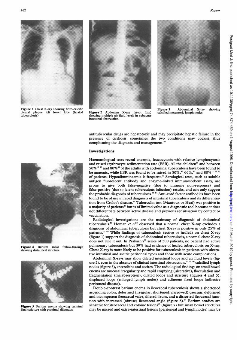
_!!F ~~~~~~~~~~~~~~~~~~~~~~~~~~~~~~~~~~.....,....Figure1Chest X-ray showing fibro-calcific~~~~~~~~~~:.pleuralplaque left lower lobe (healed~~~~~~~~~.:tuberculosis)~~~~~~~~~~~~~~~~~~~~~~~~~~~~~~~~~~~~~~
ww f s Li__i -._
't .,.m._ _E | l S^:-N-E | -_r
! 1_-| - | lill | t.1EE._|-| !! !! | p
-| W-a.S:: :. ..I 1111 l .,. BLkS,.... Bll..11 | t:. 5ssi
l llilkii'..::<...>EERIj
__ Ww'y_vg1;.:. r
........................_!!:_
.,| _::.
l,:_I___
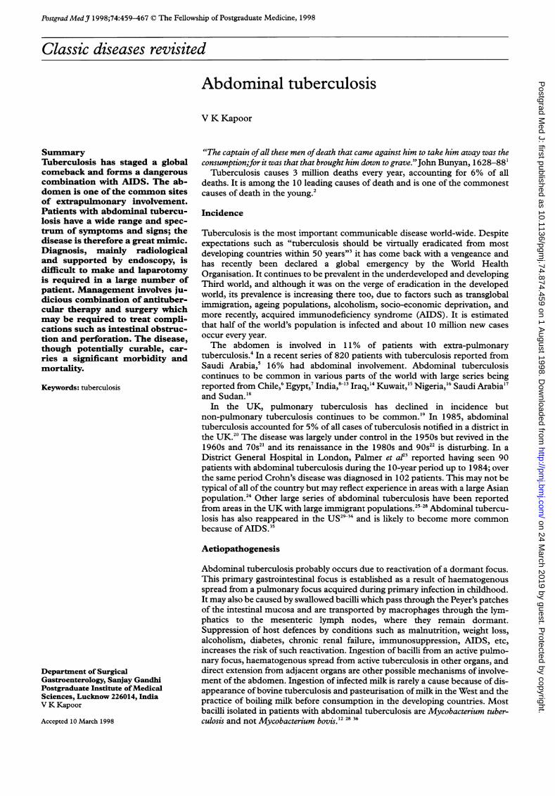
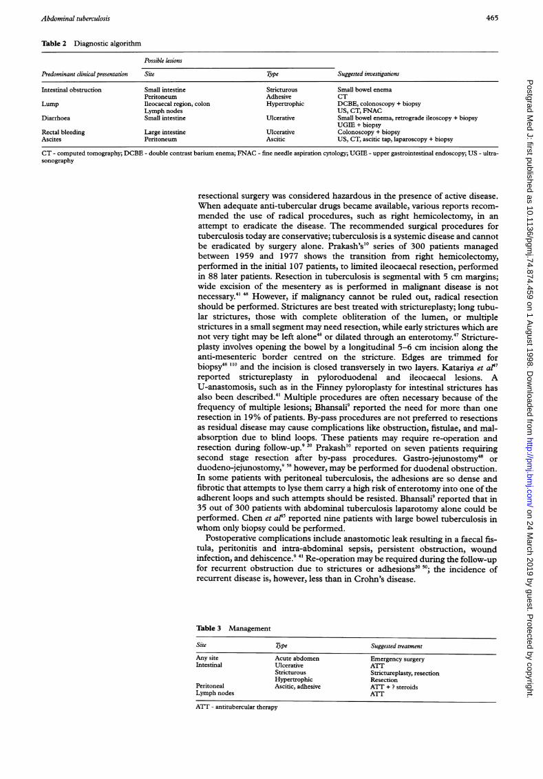
|__:Figure 4 Barium meal follow-throughshowing distal ileal stricture
X ::. 's ll- - -5 -<: -4 t - | ..4 -
ge>>eS>>>ei o _-Wn Sne :>' -
_1-i isl:: . l l le e e l l l
.Xs l l
L l| S |* | :: r-* llll L_
_ | g i __ i i _ __.... ..... ... ... .. 11111 I!.IIOJDDIINIlUlel:ps 1s1 llWl : ................. .... ..... :; :# _Figure S Barium enema showing terminalileal stricture with proximal dilatation
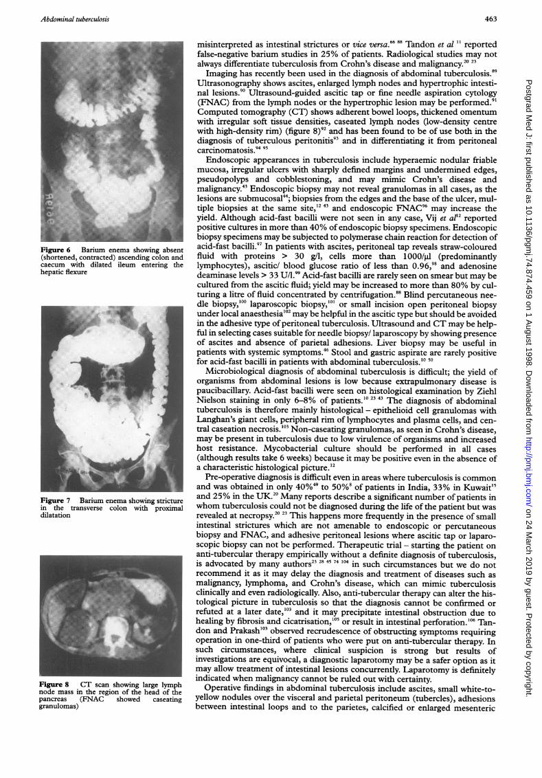
Figure 2 Abdomen X-ray (erect film)showing multiple air fluid levels in subacuteintestinal obstruction
.':'.'', 3E~~~~~~~~~~~~~~~~ .#.. .... ' ..*.'.'.'-,,~~~~~~~~~~~~~~~~~~~~~~~~~~~~~~~~~~~~~~~~~... .......R|
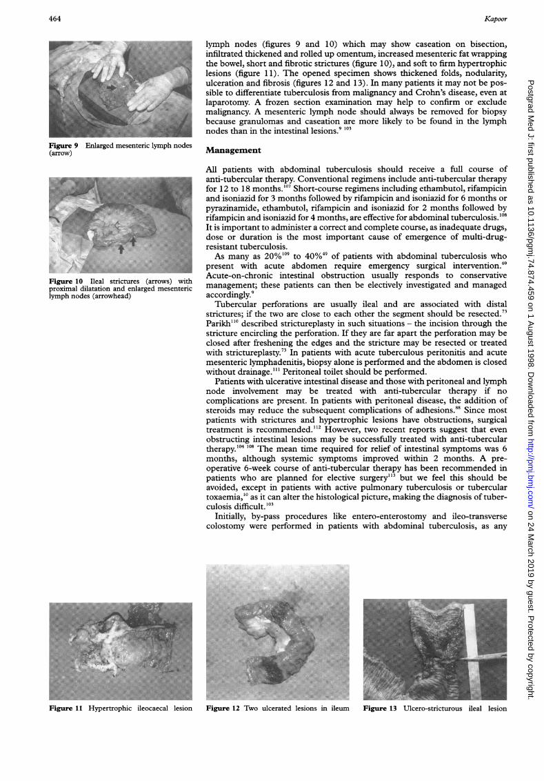
Figur 3 Aboia -a hwncalcified', meetei lyp nodes
antitubercular drugs are hepatotoxic and may precipitate hepatic failure in thepresence of cirrhosis; sometimes the two conditions may coexist, thuscomplicating the diagnosis and management.82
Investigations
Haematological tests reveal anaemia, leucocytosis with relative lymphocytosisand raised erythrocyte sedimentation rate (ESR). All the children50 and between50%1O '1 and 80%49 of the adults with abdominal tuberculosis have been found tobe anaemic, while ESR was found to be raised in 50%,49 66%,1o and 80%1" 23 43of patients. Hypoalbuminaemia is frequent.20 Serological tests, such as solubleantigen fluorescent antibody and enzyme-linked immunosorbent assay, areprone to give both false-negative (due to immune non-response) andfalse-positive (due to latent tuberculous infection) results, and can only suggestthe probable diagnosis of tuberculosis.83 84 Anti-cord factor antibodies have beenfound to be ofuse in rapid diagnosis of intestinal tuberculosis and its differentia-tion from Crohn's disease.85 Tuberculin test (Mantoux or Heaf) was positive ina majority of patients20 but is of limited value as a diagnostic tool because it doesnot differentiate between active disease and previous sensitisation by contact orvaccination.
Radiological investigations are the mainstay of diagnosis of abdominaltuberculosis.86 Homan et al° observed that a normal chest X-ray excludes adiagnosis of abdominal tuberculosis but chest X-ray is positive in only 25% ofpatients. l 49 While findings of tuberculosis (active or healed) on chest X-ray(figure 1) support the diagnosis of abdominal tuberculosis, a normal chest X-raydoes not rule it out. In Prakash's'0 series of 300 patients, no patient had activepulmonary tuberculosis but 39% had evidence of healed tuberculosis on X-ray.Chest X-ray is more likely to be positive for tuberculosis in patients with ulcera-tive intestinal and ascitic peritoneal types and those with acute complications.Abdominal X-rays may show dilated intestinal loops and air fluid levels (fig-
ure 2), even in the absence of clinical intestinal obstruction,'0 11 50 calcified lymphnodes (figure 3), enteroliths and ascites. The radiological findings on small bowelenema are mucosal irregularity and rapid emptying (ulcerative), flocculation andfragmentation (malabsorption), dilated loops and stricture (figures 4 and 5),displaced loops (enlarged lymph nodes) and adherent fixed loops (adhesiveperitoneal disease).
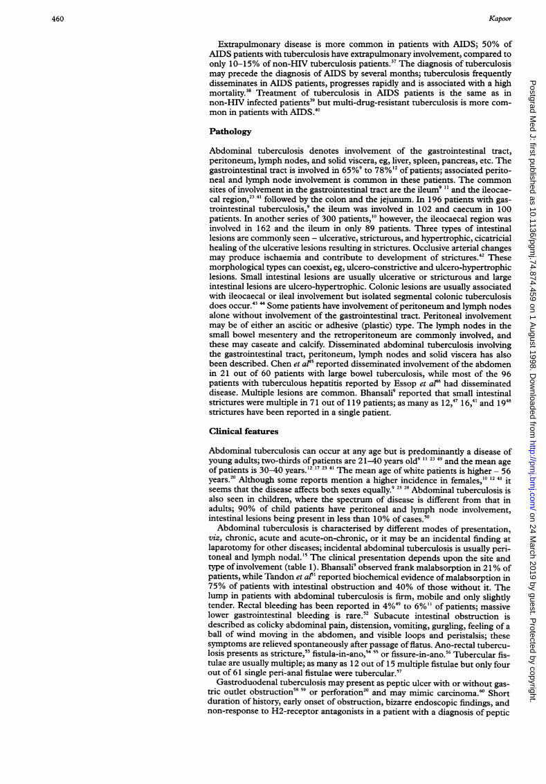
Double-contrast barium enema in ileocaecal tuberculosis shows a shortenedascending colon, deformed (irregular, shortened, narrowed) caecum, deformedand incompetent ileocaecal valve, dilated ileum, and a distorted ileocaecal junc-tion with increased (obtuse) ileocaecal angle (figure 6).87 Barium studies aresensitive for ileocaecal and colonic lesions49 (figure 7) but small bowel stricturesmay be missed and extra-intestinal lesions (peritoneal and lymph nodes) may be
on 24 March 2019 by guest. P
rotected by copyright.http://pm
j.bmj.com
/P
ostgrad Med J: first published as 10.1136/pgm
j.74.874.459 on 1 August 1998. D
ownloaded from
Abdominal tuberculosis 463
Figure 6 Barium enema showing absent(shortened, contracted) ascending colon andcaecum with dilated ileum entering thehepatic flexure
Figure 7 Barium enema showing smrcturein the transverse colon with proximaldilatation
Figure 8 CT sca shwn lag lymph
pancrea (6 A showedcnmhoigabseatnggshraenudlomas) d acndn clnn
misinterpreted as intestinal strictures or vice versa.66 88 Tandon et al " reportedfalse-negative barium studies in 25% of patients. Radiological studies may notalways differentiate tuberculosis from Crohn's disease and malignancy.20 23Imaging has recently been used in the diagnosis of abdominal tuberculosis.89
Ultrasonography shows ascites, enlarged lymph nodes and hypertrophic intesti-nal lesions.90 Ultrasound-guided ascitic tap or fine needle aspiration cytology(FNAC) from the lymph nodes or the hypertrophic lesion may be performed.9'Computed tomography (CT) shows adherent bowel loops, thickened omentumwith irregular soft tissue densities, caseated lymph nodes (low-density centrewith high-density rim) (figure 8)92 and has been found to be of use both in thediagnosis of tuberculous peritonitis9" and in differentiating it from peritonealcarcinomatosis.9495Endoscopic appearances in tuberculosis include hyperaemic nodular friable
mucosa, irregular ulcers with sharply defined margins and undermined edges,pseudopolyps and cobblestoning, and may mimic Crohn's disease andmalignancy.4' Endoscopic biopsy may not reveal granulomas in all cases, as thelesions are submucosal44; biopsies from the edges and the base of the ulcer, mul-tiple biopsies at the same site,'2 43 and endoscopic FNAC96 may increase theyield. Although acid-fast bacilli were not seen in any case, Vij et al'2 reportedpositive cultures in more than 40% of endoscopic biopsy specimens. Endoscopicbiopsy specimens may be subjected to polymerase chain reaction for detection ofacid-fast bacilli.97 In patients with ascites, peritoneal tap reveals straw-colouredfluid with proteins > 30 g/l, cells more than 1 000/,l (predominantlylymphocytes), ascitic/ blood glucose ratio of less than 0.96,98 and adenosinedeaminase levels > 33 UIA.99 Acid-fast bacilli are rarely seen on smear but may becultured from the ascitic fluid; yield may be increased to more than 80% by cul-turing a litre of fluid concentrated by centrifugation.88 Blind percutaneous nee-dle biopsy,'00 laparoscopic biopsy,'0' or small incision open peritoneal biopsyunder local anaesthesia'02 may be helpful in the ascitic type but should be avoidedin the adhesive type of peritoneal tuberculosis. Ultrasound and CT may be help-ful in selecting cases suitable for needle biopsy/ laparoscopy by showing presenceof ascites and absence of parietal adhesions. Liver biopsy may be useful inpatients with systemic symptoms.46 Stool and gastric aspirate are rarely positivefor acid-fast bacilli in patients with abdominal tuberculosis.'0 50
Microbiological diagnosis of abdominal tuberculosis is difficult; the yield oforganisms from abdominal lesions is low because extrapulmonary disease ispaucibacillary. Acid-fast bacilli were seen on histological examination by ZiehlNielson staining in only 6-8% of patients.'0 23 43 The diagnosis of abdominaltuberculosis is therefore mainly histological - epithelioid cell granulomas withLanghan's giant cells, peripheral rim of lymphocytes and plasma cells, and cen-tral caseation necrosis.'03 Non-caseating granulomas, as seen in Crohn's disease,may be present in tuberculosis due to low virulence of organisms and increasedhost resistance. Mycobacterial culture should be performed in all cases(although results take 6 weeks) because it may be positive even in the absence ofa characteristic histological picture.'2
Pre-operative diagnosis is difficult even in areas where tuberculosis is commonand was obtained in only 40%49 to 50%' of patients in India, 33% in Kuwait'5and 25% in the UK.20 Many reports describe a significant number of patients inwhom tuberculosis could not be diagnosed during the life of the patient but wasrevealed at necropsy.20 23 This happens more frequently in the presence of smallintestinal strictures which are not amenable to endoscopic or percutaneousbiopsy and FNAC, and adhesive peritoneal lesions where ascitic tap or laparo-scopic biopsy can not be performed. Therapeutic trial - starting the patient onanti-tubercular therapy empirically without a definite diagnosis of tuberculosis,is advocated by many authors23 28 45 74 104 in such circumstances but we do notrecommend it as it may delay the diagnosis and treatment of diseases such asmalignancy, lymphoma, and Crohn's disease, which can mimic tuberculosisclinically and even radiologically. Also, anti-tubercular therapy can alter the his-tological picture in tuberculosis so that the diagnosis cannot be confirmed orrefuted at a later date,'03 and it may precipitate intestinal obstruction due tohealing by fibrosis and cicatrisation,'05 or result in intestinal perforation.'06 Tan-don and Prakash'03 observed recrudescence of obstructing symptoms requiringoperation in one-third of patients who were put on anti-tubercular therapy. Insuch circumstances, where clinical suspicion is strong but results ofinvestigations are equivocal, a diagnostic laparotomy may be a safer option as itmay allow treatment of intestinal lesions concurrently. Laparotomy is definitelyindicated when malignancy cannot be ruled out with certainty.
Operative findings in abdominal tuberculosis include ascites, small white-to-yellow nodules over the visceral and parietal peritoneum (tubercles), adhesionsbetween intestinal loops and to the parietes, calcified or enlarged mesenteric
on 24 March 2019 by guest. P
rotected by copyright.http://pm
j.bmj.com
/P
ostgrad Med J: first published as 10.1136/pgm
j.74.874.459 on 1 August 1998. D
ownloaded from
464 Kapoor
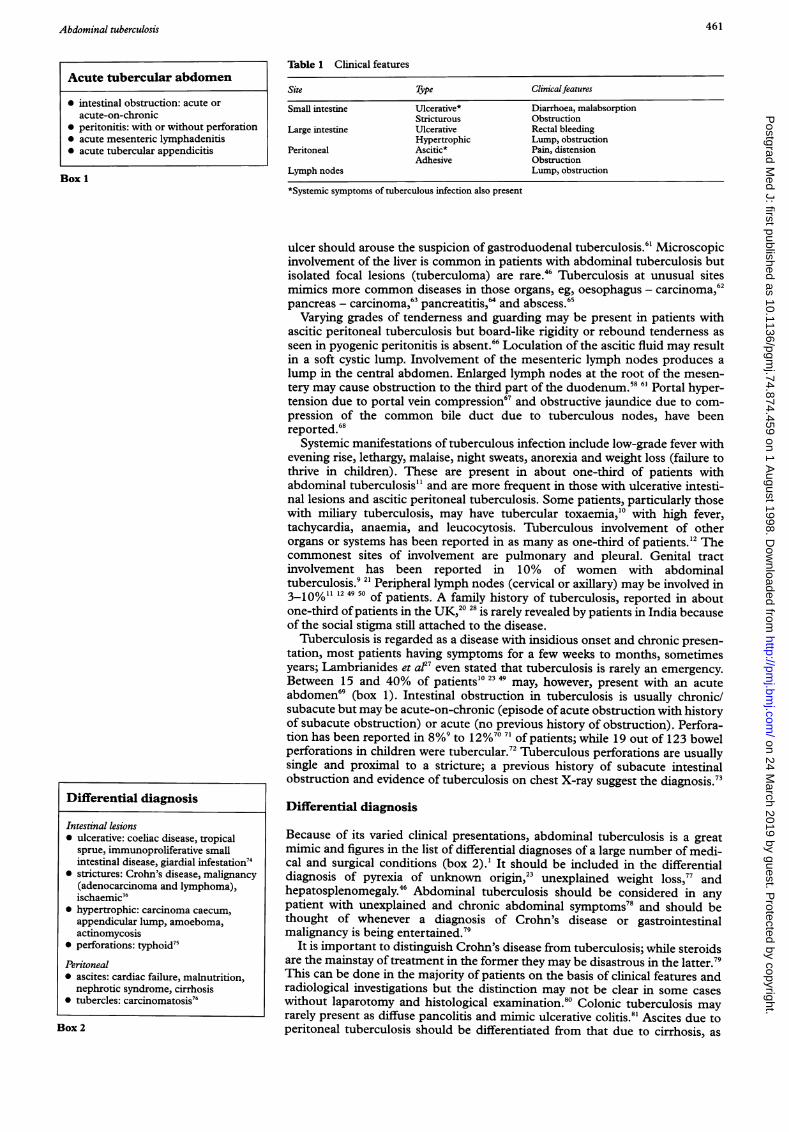
Figure 9 Enlarged mesentenc lymph nodes(arrow)
Figure 0 Ileal strictures (arrows) withproximal dilatation and enlarged mesentericlymph nodes (arrowhead)
Fiue11 Hyerophlic0 giloaea lesio
lymph nodes (figures 9 and 10) which may show caseation on bisection,infiltrated thickened and rolled up omentum, increased mesenteric fat wrappingthe bowel, short and fibrotic strictures (figure 10), and soft to firm hypertrophiclesions (figure 11). The opened specimen shows thickened folds, nodularity,ulceration and fibrosis (figures 12 and 13). In many patients it may not be pos-sible to differentiate tuberculosis from malignancy and Crohn's disease, even atlaparotomy. A frozen section examination may help to confirm or excludemalignancy. A mesenteric lymph node should always be removed for biopsybecause granulomas and caseation are more likely to be found in the lymphnodes than in the intestinal lesions.9 103
Management
All patients with abdominal tuberculosis should receive a full course ofanti-tubercular therapy. Conventional regimens include anti-tubercular therapyfor 12 to 18 months.'07 Short-course regimens including ethambutol, rifampicinand isoniazid for 3 months followed by rifampicin and isoniazid for 6 months orpyrazinamide, ethambutol, rifampicin and isoniazid for 2 months followed byrifampicin and isoniazid for 4 months, are effective for abdominal tuberculosis.'08It is important to administer a correct and complete course, as inadequate drugs,dose or duration is the most important cause of emergence of multi-drug-resistant tuberculosis.As many as 20%'19 to 40%49 of patients with abdominal tuberculosis who
present with acute abdomen require emergency surgical intervention.69Acute-on-chronic intestinal obstruction usually responds to conservativemanagement; these patients can then be electively investigated and managedaccordingly.9
Tubercular perforations are usually ileal and are associated with distalstrictures; if the two are close to each other the segment should be resected.7Parikh"' described strictureplasty in such situations - the incision through thestricture encircling the perforation. If they are far apart the perforation may beclosed after freshening the edges and the stricture may be resected or treatedwith strictureplasty.73 In patients with acute tuberculous peritonitis and acutemesenteric lymphadenitis, biopsy alone is performed and the abdomen is closedwithout drainage."' Peritoneal toilet should be performed.
Patients with ulcerative intestinal disease and those with peritoneal and lymphnode involvement may be treated with anti-tubercular therapy if nocomplications are present. In patients with peritoneal disease, the addition ofsteroids may reduce the subsequent complications of adhesions.88 Since mostpatients with strictures and hypertrophic lesions have obstructions, surgicaltreatment is recommended."2 However, two recent reports suggest that evenobstructing intestinal lesions may be successfully treated with anti-tuberculartherapy.'04 108 The mean time required for relief of intestinal symptoms was 6months, although systemic symptoms improved within 2 months. A pre-operative 6-week course of anti-tubercular therapy has been recommended inpatients who are planned for elective surgery"'3 but we feel this should beavoided, except in patients with active pulmonary tuberculosis or tuberculartoxaemia,'° as it can alter the histological picture, making the diagnosis of tuber-culosis difficult.'03
Initially, by-pass procedures like entero-enterostomy and ileo-transversecolostomy were performed in patients with abdominal tuberculosis, as any
.........
3R_~~~~~~~~~~~~~~~~~~~~~~~~~~~~~~~~~~~~~........Fiue1. w leae lein in #leu
Figure 13 Ulcero-stricturous ileal lesion
on 24 March 2019 by guest. P
rotected by copyright.http://pm
j.bmj.com
/P
ostgrad Med J: first published as 10.1136/pgm
j.74.874.459 on 1 August 1998. D
ownloaded from
Abdominal tuberculosis 465
Table 2 Diagnostic algorithm
Possible lesions
Predominant clinical presentation Site Type Suggested investigations
Intestinal obstruction Small intestine Stricturous Small bowel enemaPeritoneum Adhesive CT
Lump Ileocaecal region, colon Hypertrophic DCBE, colonoscopy + biopsyLymph nodes US, CT, FNAC
Diarrhoea Small intestine Ulcerative Small bowel enema, retrograde ileoscopy + biopsyUGIE + biopsy
Rectal bleeding Large intestine Ulcerative Colonoscopy + biopsyAscites Peritoneum Ascitic US, CT, ascitic tap, laparoscopy + biopsy
CT - computed tomography; DCBE - double contrast barium enema; FNAC - fine needle aspiration cytology; UGIE - upper gastrointestinal endoscopy; US - ultra-sonography
resectional surgery was considered hazardous in the presence of active disease.When adequate anti-tubercular drugs became available, various reports recom-mended the use of radical procedures, such as right hemicolectomy, in anattempt to eradicate the disease. The recommended surgical procedures fortuberculosis today are conservative; tuberculosis is a systemic disease and cannotbe eradicated by surgery alone. Prakash's's series of 300 patients managedbetween 1959 and 1977 shows the transition from right hemicolectomy,performed in the initial 107 patients, to limited ileocaecal resection, performedin 88 later patients. Resection in tuberculosis is segmental with 5 cm margins;wide excision of the mesentery as is performed in malignant disease is notnecessary.4' 48 However, if malignancy cannot be ruled out, radical resectionshould be performed. Strictures are best treated with strictureplasty; long tubu-lar strictures, those with complete obliteration of the lumen, or multiplestrictures in a small segment may need resection, while early strictures which arenot very tight may be left alone48 or dilated through an enterotomy.47 Stricture-plasty involves opening the bowel by a longitudinal 5-6 cm incision along theanti-mesenteric border centred on the stricture. Edges are trimmed forbiopsy48 l1o and the incision is closed transversely in two layers. Katariya et a7reported strictureplasty in pyloroduodenal and ileocaecal lesions. AU-anastomosis, such as in the Finney pyloroplasty for intestinal strictures hasalso been described.4' Multiple procedures are often necessary because of thefrequency of multiple lesions; Bhansali9 reported the need for more than oneresection in 19% of patients. By-pass procedures are not preferred to resectionsas residual disease may cause complications like obstruction, fistulae, and mal-absorption due to blind loops. These patients may require re-operation andresection during follow-up.9 20 Prakash'° reported on seven patients requiringsecond stage resection after by-pass procedures. Gastro-jejunostomy48 orduodeno-jejunostomy,9 5 however, may be performed for duodenal obstruction.In some patients with peritoneal tuberculosis, the adhesions are so dense andfibrotic that attempts to lyse them carry a high risk of enterotomy into one of theadherent loops and such attempts should be resisted. Bhansali9 reported that in35 out of 300 patients with abdominal tuberculosis laparotomy alone could beperformed. Chen et al'5 reported nine patients with large bowel tuberculosis inwhom only biopsy could be performed.
Postoperative complications include anastomotic leak resulting in a faecal fis-tula, peritonitis and intra-abdominal sepsis, persistent obstruction, woundinfection, and dehiscence.9 4" Re-operation may be required during the follow-upfor recurrent obstruction due to strictures or adhesions20 50; the incidence ofrecurrent disease is, however, less than in Crohn's disease.
Table 3 Management
Site Type Suggested treatment
Any site Acute abdomen Emergency surgeryIntestinal Ulcerative ATT
Stricturous Strictureplasty, resectionHypertrophic Resection
Peritoneal Ascitic, adhesive AT + ? steroidsLymph nodes ATT
ATT - antitubercular therapy
on 24 March 2019 by guest. P
rotected by copyright.http://pm
j.bmj.com
/P
ostgrad Med J: first published as 10.1136/pgm
j.74.874.459 on 1 August 1998. D
ownloaded from
466 Kapoor
Prognosis
Delayed diagnosis and injudicious treatment due either to limited experience orpoor understanding of the disease are principally responsible for the mortalityrate of 4-12%.'° 17 45 49 50 The high mortality is partly due to the associated mal-nutrition, anaemia and hypoalbuminaemia; mortality is even higher (12-25%) inthe presence of acute complications. 49 114 Timely diagnosis based on a highindex of suspicion in areas and in populations in which tuberculosis is common,an algorithmic diagnostic approach using radiology, imaging and endoscopy(table 2), and management with a judicious combination of anti-tuberculartherapy and conservative surgery (table 3), can reduce the mortality of this 'eas-ily curable yet potentially lethal' disease."15
"The first country to eliminate tuberculosis will be that which regards it as a seriousproblem right to the end." Prof Etienne Bernard, Paris3
Tuberculosis unfortunately still remains the world's most neglected healthcrisis.
The author reviewed the subject during a Visiting Professorship to the Academic Department of Surgery atthe King's College Hospital, London on a Commonwealth Fellowship.Acknowledgements are due to Ms Dympna Kelly, Lecturer, Academic Department of Surgery, King's
College Hospital, London for her comments on the manuscript.
1 Cook GC. Tuberculosis - certainly not a diseaseof the past! QJ Med 1985;56:519-21.
2 Anonymous. The challenge of tuberculosis.Lancet 1994;34:277-9.
3 Home NW. Problems of tuberculosis in decline.BMJ7 1984;288: 1249-51.
4 Anonymous. Management of non-respiratorytuberculosis. Lancet 1986;1: 1423-4.
5 Al-Karawi MA, Mohamed AE, Yasawy MI, et al.Protean manifestations of gastrointestinal tuber-culosis. J Clin Gastroenterol 1995;20:225-32.
6 Fica A, Belletti J, Cruzat C, Rojas D, MontalvaM. Intestinal tuberculosis: analysis of clinicalcases and autopsy. Rev Med Chile 1991;119:1153-9.
7 Hibbs RG, Kamal M, Farid Z. Abdominaltuberculosis in Cairo, Egypt. Trans R Soc TropMed Hyg 1994;88:317-8.
8 Das P, Shukla HS. Clinical diagnosis ofabdominal tuberculosis. BrJt Surg 1976;63:941-6.
9 Bhansali SK. Abdominal tuberculosis. Experi-ences with 300 cases. Am J Gastroenterol1977;67:324-37.
10 Prakash A. Ulcero-constrictive tuberculosis ofthe bowel. Int Surg 1978;63:23-9.
11 Tandon RK, Sarin SK, Bose SL, Berry M, Tan-don BN. A clinico-radiological reappraisal ofintestinal tuberculosis - changing profile? Gas-troenterolJpn 1986;21:17-22.
12 Vij JC, Malhotra V, Choudhary V, et al. Aclinicopathological study of abdominal tubercu-losis. Indian J Tubercul 1992;39:213-20.
13 Singh V, Jain AK, Agrawal AK, et al. Clinico-pathological profile of abdominal tuberculosis.BrJ Clin Pract 1995;49:22-4.
14 Al-Bahrani ZR, Al-Saleem T. Intestinal tuber-culosis in Iraq: a study of 50 cases. Int Surg1982;67:483-5.
15 Al-Hadeedi S, Walia HS, Al-Sayer HM. Ab-dominal tuberculosis. Can J Surg 1990;33:233-7.
16 Lewis EA, Kolawole TM. Tuberculosis ileo-colitis in Ibadan: a clinico-radiological review.Gut 1972;13:646-53.
17 Al-Quorain A, Satti MB, Al-Freihi HM, Al-Gindan YM, Al-Awad N. Abdominal tuberculo-sis in Saudi Arabia: a clinico-pathological studyof 65 cases. Am J Gastroenterol 1993;88:75-9.
18 El Masri SH, Boulos P, Malick MOA. Abdomi-nal tuberculosis in Sudanese patients. East AfrMedJ 1977;54:319-26.
19 Innes JA. Non-respiratory tuberculosis. J R CoilPhysicians Lond 1981;15:227-31.
20 Klimach OE, Ormerod LP. Gastrointestinaltuberculosis: a retrospective review of 109 casesin a district general hospital. Q J Med 1985;56:569-78.
21 Addison NV. Abdominal tuberculosis - a diseaserevived. Ann R Coil Surg Engl 1983;65:105-1 1.
22 Jayanthi V, Probert CS, Sher KS, Wicks AC,Mayberry JF. The renaissance of tuberculosis.Dig Dis 1993;11:36-44.
23 Palmer KR, Patil DH, Basran S, Riordan JF,Silk DBA. Abdominal tuberculosis in urbanBritain: a common disease. Gut 1985;26:1296-305.
24 Probert CS, Jayanthi V, Wicks AC, Carr LockeD, Garner P, Mayberry JF. Epidemiologicalstudy of abdominal tuberculosis among Indian
migrants and the indigenous population ofLeicester, 1972-89. Gut 1992;33:1085-9.
25 Mandal BK, Schofield PF. Abdominal tubercu-losis in Britain. Practitioner 1976;216:683-9.
26 Findlay JM, Addison NV, Stevenson DK, MirzaZA. Tuberculosis of the gastrointestinal tract inBradford, 1967-77. J R Soc Med 1979;72:587-90.
27 Lambrianides AL, Ackroyd N, Shorey BA.Abdominal tuberculosis. Br J7 Surg 1980;67:887-9.
28 Sharp JF, Goldman M. Abdominal tuberculosisin East Birmingham: a 16 year study. PostgradMedJ 1987;63:539-42.
29 Dineen P, Homan WP, Grafe WR. Tuberculousperitonitis: 43 years experience in diagnosis andtreatment. Ann Surg 1976;184:717-22.
30 Homan WP, Grafe WR, Dineen P. A 44-yearexperience with tuberculous enterocolitis. WorldJ Surg 1977;1:245-50.
31 Sherman S, Rohwedder JJ, Ravikrishnan KP, etal. Tuberculous enteritis and peritonitis: Reportof 36 general hospital cases. Arch Intern Med1980;140:506-8.
32 McGee GS, Williams LF, Potts J, Barnwell S,Sawyers JL. Gastrointestinal tuberculosis: resur-gence of an old pathogen.Am Surg 1989;55:16-20.
33 Guth AA, Kim U. The reappearance of abdomi-nal tuberculosis. Surg Gynecol Obstet 1991;172:432-6.
34 Ko CY, Schmit PJ, Petrie B, Thompson JE.Abdminal tuberculosis: the surgical perspective.Am Surg 1996;62:865-8.
35 Rosengart TK, Coppa GF. Abdominal myco-bacterial infections in immunocompromisedpatients. Am J Surg 1990;159:125-31.
36 Tandon HD. The pathology of intestinal tuber-culosis and distinction from other diseases caus-ing stricture. Trop Gastroenterol 1981;2:77-93.
37 Goldman KP. AIDS and tuberculosis. Tubercle1988; 69: 71-2.
38 Fee MJ, Oo MM, Gabayan AE, Radin DR,Barnes PF. Abdominal tuberculosis in patientsinfected with the human immunodeficiencyvirus. Clin Infect Dis 1995;20:938-44.
39 Watters DAK. Surgery for tuberculosis beforeand after HIV infection: a tropical perspective.BrJ Surg 1997;84:8-14.
40 Iseman MD. Treatment of multidrug resistanttuberculosis. N Engl JtMed 1993;329:784-9 1.
41 Joshi MJ. The surgical management of intestinaltuberculosis - a conservative approach. Indian JfSurg 1978;40:79-83.
42 Shah P, Ramakantan R. Role of vasculitis in thenatural history of abdominal tuberculosis -evaluation by mesenteric angiography. Indian JGastroenterol 1991;10:127-30.
43 Shah S, Thomas V, Mathan M, et al. Colono-scopic study of 50 patients with colonictuberculosis. Gut 1992;33:347-51.
44 Singh V, Kumar P, Kamal J, Prakash V, VaipheiK, Singh K. Clinico-colonoscopic profile ofcolonic tuberculosis. Am J Gastroenterol 1996;91:565-8.
45 Chen WS, Leu SY, Lin JK, Lin TC. Trend oflarge bowel tuberculosis and relation withpulmonary tuberculosis. Dis Colon Rectum 1992;35:189-92.
46 Essop AR, Posen JA, Hodkinson JH, Segal I.Tuberculous hepatitis: a clinical review of 96cases. QJIMed 1984;212:465-77.
47 Katariya RN, Sood S, Rao PG, Rao PLNG.Strictureplasty for tubercular strictures of thegastrointestinal tract. BrJ3 Surg 1977;64:496-8.
48 Pujari BD. Modified surgical procedures inintestinal tuberculosis. Br Jf Surg 1979;66: 180-1.
49 Dandapat MC, Mohan Rao V. Management ofabdominal tuberculosis. Indian J Tubercul 1985;32:126-9.
50 Sharma AK, Aggarwal LD, Sharma CS, SarinYK. Abdominal tuberculosis in children:experience over a decade. Indian Paediatr 1993;30:1149-53.
51 Tandon RK, Bansal R, Kapur BML, Shriniwas.A study of malabsorption in intestinaltuberculosis: stagnant loop syndrome. AmJ ClinNutr 1980;33:244-50.
52 Monkemuller KE, Lewis JB Jr. Massive rectalbleeding from colonic tuberculosis. Am JGastroenterol 1996;91: 1439-41.
53 Puri AS, Vij JC, Chaudhary A, et al. Diagnosisand outcome of isolated rectal tuberculosis. DisColon Rectum 1996;39:1126-9.
54 Shukla HS, Gupta SC, Singh G, Singh PA.Tubercular fistula in ano. BrJ'Surg 1988;75:38-9.
55 Alyoune M, Nadir S, Merzouk M, et al. Tuber-culous anal fistulas. Ann Gastroenterol Hepatol1994;30:9-1 1.
56 Myers SR. Tuberculous fissure-in-ano. 7 R SocMed 1994;87:46.
57 Dandapat MC, Mukherjee LM, Behera AN.Fistula-in-ano. Indian J Surg 1990;52:265-8.
58 Khanna AK, Kumar K, Mishra MK, Jain AK,Gupta JP. Duodenal tuberculosis: presentationin a surgical unit. Ann NadAcadMed Sci (India)1994;30:282-4.
59 DiPlacido R, Pietroletti R, Leardi S, Simi M.Primary gastroduodenal tuberculous infectionpresenting as pyloric outlet obstruction. Am JGastroenterol 1996;91:807-8.
60 Wells AD, Northover JMA, Howard ER. Ab-dominal tuberculosis: still a problem today. J RSoc Med 1986;79:149-53.
61 Ali W, Sikora SS, Bannerjee D, et al. Gastrodu-denal tuberculosis. Aust NZ Jf Surg 1993;63:466-7.
62 Monge E, Lozano R, Salazar S. Esophagealtuberculosis. Am J Gastroenterol 1992;87:156.
63 Hashimoto M, Koyama H, Noie T, ShibayamaK, Kitamura N. Abdominal tuberculosis mim-icking a pancratic neoplasm. Surg Today 1995;25:365-8.
64 Mokoena J, Bairagi N. Acute tuberculouspancreatitis associated with HIV infection. TropDoct 1994;24: 172-3.
65 Sozbilen M, Erhan Y, Koyuncu A. Tuberculosisof the pancreas. Br3' Surg 1992;79:802.
66 Bastani B, Shariatzadeh MR, Dehdashti F.Tuberculous peritonitis: report of 30 cases andreview of the literature. Q J Med 1985;56:549-57.
67 Diab S, Nema TA, Zidan FA. Portal hyperten-sion complicating abdominal tuberculosis. ActaChirurg Scand 1990;156:495-7.
68 Murphy TF, Gray GF. Biliary tract obstructiondue to tuberculous adenitis. AmJMed 1980; 68:452-4.
on 24 March 2019 by guest. P
rotected by copyright.http://pm
j.bmj.com
/P
ostgrad Med J: first published as 10.1136/pgm
j.74.874.459 on 1 August 1998. D
ownloaded from
Abdominal tuberculosis 467
69 Kapoor VK, Gupta S, Sikora SS, Chattopad-hyay TK, Sharma LK. Acute tubercular abdo-men. Indian JSurg 1991; 53: 71-5.
70 Eggleston FC, Deodhar MC, Kumar A. Tuber-culous perforation of the bowel - results in 21cases. Trop Gastroenterol 1983; 4: 164-7.
71 Kakkar A, Aranya RC, Nair SK. Acute perfora-tion of small intestine due to tuberculosis. AustNZJtSurg 1983;55:381-3.
72 Dhar A, Bagga D, Taneja SB. Perforated tuber-cular enteritis of childhood: a ten year study.Indian J Pediatr 1990;57:713-6.
73 Kapoor VK, Kriplani AK, Chattopadhyay TK,Sharma LK. Tuberculous perforations of thesmall intestine. Indian J Tubercul 1986;33:188-9.
74 Yachha SK, Misra S, Malik AK, Nai B, MehtaS. Spectrum of malabsorption syndrome innorth Indian children. Indian J Gastroenterol1993;12: 120-5.
75 Sharma LK, Gupta S, Soin AS, Sikora SS,Kapoor VK. Generalised peritonitis - the tropi-cal spectrum. Jpn Jf Surg 199 1;21:72-7.
76 Nistal de Paz F, Herrero Fernandez B, PerezSimon R, et al. Pelvic peritoneal tuberculosissimulating ovarian carcinoma. Am J Gastroen-terol 1996;91:1660-1.
77 Underwood MJ, Thompson MM, Sayers RD,Hall AW. Presentation of abdominal tuberculo-sis to general surgeons. BrJ Surg 1992;79: 1077-9.
78 Walia HS, Khafagy AR, al Sayer HM, et al.Unusual presentations of abdominal tuberculo-sis. Can JSurg 1994;37:300-6.
79 Anonymous. Abdominal tuberculosis in Britain.BMJ 1977;1:1557.
80 Kapoor VK. Koch's or Crohn's? Int3J Clin Pract1997;51:246-7.
81 Misra A, Khanduri A, Jain M, Gupta RK,Choudhuri G. Colonic tuberculosis presentingas diffuse pancolitis. IndianJy Gastroenterol 1996;15:105.
82 Aguado JM, Pons F, Casafont F, et al. Tubercu-lous peritonitis: a study comparing cirrhotic andnon-cirrhotic patients. J Clin Gastroenterol 1990;12:550-4.
83 Chawla TC, Sharma A, Kiran U, Bhargava DK,Shriniwas, Tandon BN. Serodiagnosis of intesti-nal tuberculosis by enzyme immunoassay andsoluble antigen fluorescent antibody tests usinga saline extracted antigen. Tubercle 1986;67:55-60.
84 Bhargava DK, Dasarathy S, Shriniwas,Kushwaha AKS, Duphare H, Kapur BML.Evaluation of enzyme-linked immunosorbentassay using mycobacterial saline-extracted anti
gen for the serodiagnosis of abdominal tubercu-losis. Am J Gastroenterol 1992;87: 105-8.
85 Kashima K, Oka S, Tabata A, et al. Detection ofanti-cord factor antibodies in intestinal tubercu-losis for its differentiatial diagnosis fromCrohn's disease and ulcerative colitis. Dig DisSci 1995;40:2630-4.
86 Lundstedt C, Nyman R, Brismar J, HugossonC, Kagevi I. Imaging of tuberculosis. II.Abdominal manifestations in 112 patients. ActaRadiol 1996;37:489-95.
87 Kapoor VK, Chattopadhyay TK, Sharma LK.Radiology of abdominal tuberculosis. AustralasRadiol 1988;32:365-7.
88 Singh MM, Bhargava AN, Jain KP. Tuberculousperitonitis. An evaluation of pathogeneticmechanisms, diagnostic procedures and thera-peutic measures. N EnglJ Med 1969;281: 1091-4.
89 Sheikh M, Abu Zidan F, Al Hilaly M, Behbe-hani A. Abdominal tuberculosis: comparison ofsonography and computed tomography. J ClinUltrasound 1995;23:413-7.
90 Kedar RP, Shah PP, Shirde RS, Malde HM.Sonographic findings in gastrointestinal andperitoneal tuberculosis. Clin Radiol 1994;49:24-9.
91 Liu KW, Chan YL, Tseng R. Childhoodabdominal tuberculosis. The role of echo-guided fine needle aspiration in its manage-ment. SurgEndosc 1994;8:326-8.
92 Bankier AA, Fleischmann D, Wiesmayr MN, etal. Update: abdominal tuberculosis - unusualfindings on CT. Clin Radiol 1995;50:223-8.
93 Lisehora GB, Peters CC, Lee YT, Barcia PJ.Tuberculous peritonitis - do not miss it. DisColon Rectum 1996;39:394-9.
94 Ha HK, Jung JI, Lee MS, et al. CT differentia-tion of tuberculous peritonitis and peritonealcarcinomatosis. AJ3R 1996;167:743-8.
95 Rodriguez E, Pombo F. Peritoneal tuberculosisversus peritoneal carcinomatosis: distinctionbased on CT findings. J Computed Assist Tomogr1996;20:269-72.
96 Kochhar R, Rajwanshi A, Goenka MK, et al.Colonoscopic fine needle aspiration cytology inthe diagnosis of ileocaecal tuberculosis. Am JGastroenterol 199 1;86: 102-4.
97 Anand BS, Schneider FE, El-Zaatari FA,Shawar RM, Clarridge JE, Graham DY. Diagno-sis of intestinal tuberculosis by polymerasechain reaction on endoscopic biopsy specimens.Am J Gastroenterol 1994;89:2248-9.
98 Wilkins EGL. Tuberculous peritonitis: diagnos-tic value of the ascitic/ blood glucose ratio.Tubercle 1984;65:47-52.
99 Dwivedi M, Misra SP, Misra V, Kumar R. Valueof adenosine deaminase estimation in thediagnosis of tuberculous ascites. Am J7 Gastroen-terol 1990;85:1123-5.
100 Levine H. Needle biopsy diagnosis of tubercu-lous peritonitis. Am Rev Respir Dis 1968;97:889-94.
101 Bhargava DK, Shriniwas, Chopra P, et al. Peri-toneal tuberculosis: laparoscopic patterns andits diagnostic accuracy. Am J Gastroenterol 1992;87: 109-11.
102 Shukla HS, Bhatia S, Naithani YP, Das P,Gupta SC. Peritoneal biopsy for diagnosis ofabdominal tuberculosis. Postgrad Med J 1982;58:226-8.
103 Tandon HD, Prakash A. Pathology of intestinaltuberculosis and its distinction from Crohn'sdisease. Gut 1972;13:260-9.
104 Anand BS, Nanda R, Sachdev K. Response oftuberculous strictures to antituberculous treat-ment. Gut 1988;29:62-9.
105 Schoffield PF. Abdominal tuberculosis. Gut1985;26: 1275-8.
106 Jayaswal R, Jena PK, Gopinathan VP, ChauhanMS. Recurrent acute tuberculous perforationsof the small intestine associated with miliarytuberculosis of lung. J Assoc Physicians India1986;34:817-8.
107 Cooke NJ. Treatment of tuberculosis. BMJ1985;291:497-8.
108 Balasubramanian R, Ramachandran R, JosephP, et al. Interim results of a clinical study ofabdominal tuberculosis. Indian J Tubercul 1989;36:117-21.
109 Haddad FS, Ghossain A, Sawaya E, NelsonAR. Abdominal tuberculosis. Dis Colon Rectum1987;30:724-35.
110 Parikh AJ. Conservative approach in manage-ment of intestinal tuberculosis. Indian 7 Surg1978;40:85-90.
111 Arunabh, Kapoor VK, Chattopadhyay TK,Sharma LK. Tuberculous peritonitis presentingas acute abdomen. Indian 7 Tubercul 1986;33:190-1.
112 Kapoor VK, Sharma LK. Abdominal tubercu-losis. BrJ Surg 1988;75:2-3.
113 Vanderpool DM, O'Leary JP. Primary tuber-culous enteritis. Surg Gynecol Obstet 1988;167:167-73.
114 Jain SK. Tubercular abdomen with acutemanifestation. Indian MedJ 1993;87:76-7.
115 Lingenfelser T, Zak J, Marks IN, Steyn E,Halkett J, Price SK. Abdominal tuberculosis:still a potentially lethal disease. Am Y Gastroen-terol 1993;88:744-50.
on 24 March 2019 by guest. P
rotected by copyright.http://pm
j.bmj.com
/P
ostgrad Med J: first published as 10.1136/pgm
j.74.874.459 on 1 August 1998. D
ownloaded from