Embed Size (px)
Citation preview
www.caymanchem.comCustomer Service 800.364.9897Technical Support 888.526.53511180 E. Ellsworth Rd · Ann Arbor, MI · USA
Citrullinated Histone H3 (Clone 11D3) ELISA Kit
Item No. 501620
3GENERAL INFORMATION
TABLE OF CONTENTS GENERAL INFORMATION 3 Materials Supplied
4 Safety Data
4 Precautions
4 If You Have Problems
5 Storage and Stability
5 Materials Needed but Not Supplied
INTRODUCTION 6 About This Assay
6 Description of Sandwich ELISAs
7 Definition of Key Terms
PRE-ASSAY PREPARATION 8 Buffer Preparation
9 Sample Preparation
10 Sample Matrix Properties
ASSAY PROTOCOL 14 Preparation of Assay-Specific Reagents
16 Plate Set Up
17 Performing the Assay
ANALYSIS 19 Calculations
20 Performance Characteristics
RESOURCES 23 Troubleshooting
25 Plate Template
26 References
27 Notes
27 Warranty and Limitation of Remedy
GENERAL INFORMATION
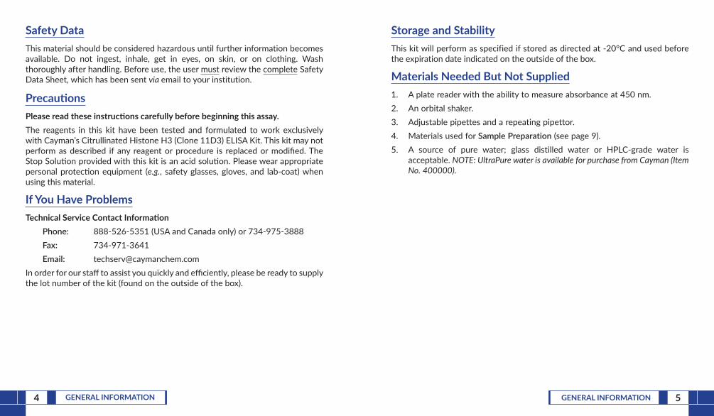
Materials Supplied
Item Number Item 96 wells Quantity/Size
401620 Anti-Histone H3 HRP Conjugate 1 vial/1.5 ml
401621 Anti-Citrullinated Histone H3 ELISA Strip Plate 1 plate
401444 Citrullinated Histone ELISA Standard 2 vials
400054 Immunoassay Buffer B Concentrate (10X) 1 vial/10 ml
400062 Wash Buffer Concentrate (400X) 1 vial/5 ml
400035 Polysorbate 20 1 vial/3 ml
400074 TMB Substrate Solution 1 vial/12 ml
10011355 HRP Stop Solution 1 vial/12 ml
400012 96-Well Cover Sheet 3 covers
If any of the items listed above are damaged or missing, please contact our Customer Service department at (800) 364-9897 or (734) 971-3335. We cannot accept any returns without prior authorization.
! WARNING: THIS PRODUCT IS FOR RESEARCH ONLY - NOT FORHUMAN OR VETERINARY DIAGNOSTIC OR THERAPEUTIC USE.
4 GENERAL INFORMATION 5GENERAL INFORMATION
Safety DataThis material should be considered hazardous until further information becomes available. Do not ingest, inhale, get in eyes, on skin, or on clothing. Wash thoroughly after handling. Before use, the user must review the complete Safety Data Sheet, which has been sent via email to your institution.
PrecautionsPlease read these instructions carefully before beginning this assay.The reagents in this kit have been tested and formulated to work exclusively with Cayman’s Citrullinated Histone H3 (Clone 11D3) ELISA Kit. This kit may not perform as described if any reagent or procedure is replaced or modified. The Stop Solution provided with this kit is an acid solution. Please wear appropriate personal protection equipment (e.g., safety glasses, gloves, and lab-coat) when using this material.
If You Have ProblemsTechnical Service Contact Information
Phone: 888-526-5351 (USA and Canada only) or 734-975-3888Fax: 734-971-3641Email: [email protected]
In order for our staff to assist you quickly and efficiently, please be ready to supply the lot number of the kit (found on the outside of the box).
Storage and StabilityThis kit will perform as specified if stored as directed at -20°C and used before the expiration date indicated on the outside of the box.
Materials Needed But Not Supplied1. A plate reader with the ability to measure absorbance at 450 nm.2. An orbital shaker.3. Adjustable pipettes and a repeating pipettor.4. Materials used for Sample Preparation (see page 9).5. A source of pure water; glass distilled water or HPLC-grade water is
acceptable. NOTE: UltraPure water is available for purchase from Cayman (Item No. 400000).
6 INTRODUCTION 7INTRODUCTION
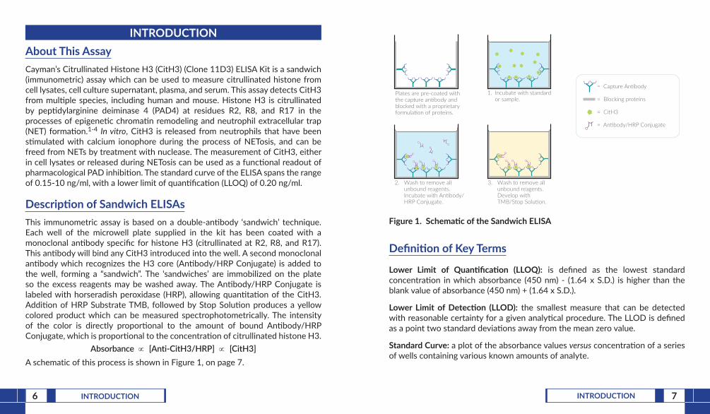
INTRODUCTIONAbout This AssayCayman’s Citrullinated Histone H3 (CitH3) (Clone 11D3) ELISA Kit is a sandwich (immunometric) assay which can be used to measure citrullinated histone from cell lysates, cell culture supernatant, plasma, and serum. This assay detects CitH3 from multiple species, including human and mouse. Histone H3 is citrullinated by peptidylarginine deiminase 4 (PAD4) at residues R2, R8, and R17 in the processes of epigenetic chromatin remodeling and neutrophil extracellular trap (NET) formation.1-4 In vitro, CitH3 is released from neutrophils that have been stimulated with calcium ionophore during the process of NETosis, and can be freed from NETs by treatment with nuclease. The measurement of CitH3, either in cell lysates or released during NETosis can be used as a functional readout of pharmacological PAD inhibition. The standard curve of the ELISA spans the range of 0.15-10 ng/ml, with a lower limit of quantification (LLOQ) of 0.20 ng/ml.
Description of Sandwich ELISAsThis immunometric assay is based on a double-antibody ‘sandwich’ technique. Each well of the microwell plate supplied in the kit has been coated with a monoclonal antibody specific for histone H3 (citrullinated at R2, R8, and R17). This antibody will bind any CitH3 introduced into the well. A second monoclonal antibody which recognizes the H3 core (Antibody/HRP Conjugate) is added to the well, forming a “sandwich”. The ‘sandwiches’ are immobilized on the plate so the excess reagents may be washed away. The Antibody/HRP Conjugate is labeled with horseradish peroxidase (HRP), allowing quantitation of the CitH3. Addition of HRP Substrate TMB, followed by Stop Solution produces a yellow colored product which can be measured spectrophotometrically. The intensity of the color is directly proportional to the amount of bound Antibody/HRP Conjugate, which is proportional to the concentration of citrullinated histone H3.
Absorbance ∝ [Anti-CitH3/HRP] ∝ [CitH3]A schematic of this process is shown in Figure 1, on page 7.
Plates are pre-coated withthe capture an body andblocked with a proprietaryformula on of proteins.
2. Wash to remove all unbound reagents. Incubate with An body/ HRP Conjugate.
3. Wash to remove all unbound reagents. Develop with TMB/Stop Solu on.
1. Incubate with standard or sample.
= Capture An body
= Blocking proteins
= CitH3
= An body/HRP Conjugate
Figure 1. Schematic of the Sandwich ELISA
Definition of Key Terms
Lower Limit of Quantification (LLOQ): is defined as the lowest standard concentration in which absorbance (450 nm) - (1.64 x S.D.) is higher than the blank value of absorbance (450 nm) + (1.64 x S.D.).
Lower Limit of Detection (LLOD): the smallest measure that can be detected with reasonable certainty for a given analytical procedure. The LLOD is defined as a point two standard deviations away from the mean zero value.
Standard Curve: a plot of the absorbance values versus concentration of a series of wells containing various known amounts of analyte.
8 PRE-ASSAY PREPARATION 9PRE-ASSAY PREPARATION
Sample PreparationSample Collection and StorageSerum and plasma contain nucleases and other factors that can interfere with the functioning of the assay. In general, serum or plasma (prepared using citrate as the anticoagulant) can be used without a purification step in the assay if they are first diluted a minimum of 1:2 in Assay Buffer. Plasma samples prepared with heparin or EDTA as the anticoagulant have been shown to cause a slight interference in the assay with higher than expected recoveries.Sample DilutionAll human plasma, human serum and cell culture supernatant or lysate samples MUST be diluted at least 1:2 with Assay Buffer prior to use in this assay. A minimum volume of 200 µl of each diluted sample is needed to run the samples in duplicate in the assay; for convenience, we recommend preparing 250 µl of each diluted sample.
PRE-ASSAY PREPARATION
Buffer PreparationStore all diluted buffers at 4°C; they should be stable for two months. NOTE: It is normal for the concentrated buffer to contain crystalline salts after thawing. These will completely dissolve upon dilution with water. Polysorbate 20 is a viscous liquid and cannot be measured by a regular pipette. A positive displacement pipette or a syringe should be used to deliver small quantities accurately. 1. Assay Buffer Preparation
Dilute the contents of one vial of Immunoassay Buffer B Concentrate (10X) (Item No. 400054) with 90 ml of water and add 100 µl of Polysorbate 20 (Item No. 400035). Be certain to rinse the vial to remove any salts that may have precipitated.
2. Wash Buffer Preparation
5 ml vial Wash Buffer Concentrate (400X) (Item No. 400062): Dilute to a total volume of 2 L with water and add 1 ml of Polysorbate 20.
10 PRE-ASSAY PREPARATION 11PRE-ASSAY PREPARATION
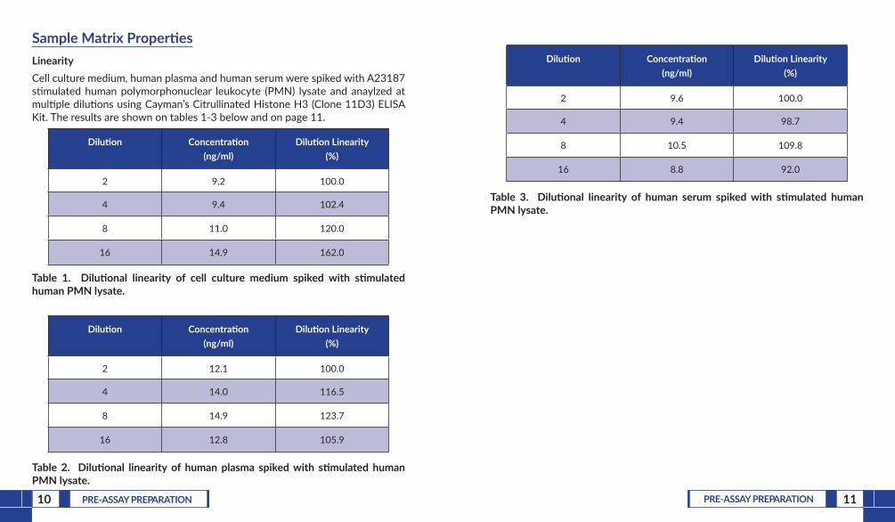
Sample Matrix PropertiesLinearityCell culture medium, human plasma and human serum were spiked with A23187 stimulated human polymorphonuclear leukocyte (PMN) lysate and anaylzed at multiple dilutions using Cayman’s Citrullinated Histone H3 (Clone 11D3) ELISA Kit. The results are shown on tables 1-3 below and on page 11.
Dilution Concentration (ng/ml)
Dilution Linearity (%)
2 9.2 100.0
4 9.4 102.4
8 11.0 120.0
16 14.9 162.0
Table 1. Dilutional linearity of cell culture medium spiked with stimulated human PMN lysate.
Dilution Concentration (ng/ml)
Dilution Linearity (%)
2 12.1 100.0
4 14.0 116.5
8 14.9 123.7
16 12.8 105.9
Table 2. Dilutional linearity of human plasma spiked with stimulated human PMN lysate.
Dilution Concentration (ng/ml)
Dilution Linearity (%)
2 9.6 100.0
4 9.4 98.7
8 10.5 109.8
16 8.8 92.0
Table 3. Dilutional linearity of human serum spiked with stimulated human PMN lysate.
12 PRE-ASSAY PREPARATION 13PRE-ASSAY PREPARATION
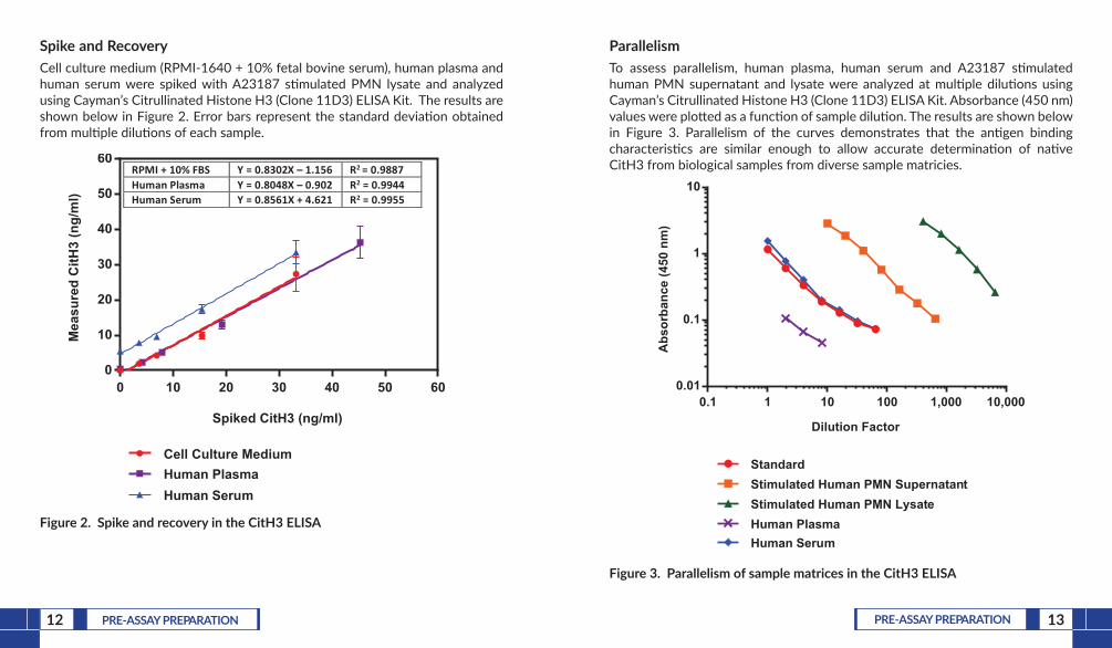
Spike and RecoveryCell culture medium (RPMI-1640 + 10% fetal bovine serum), human plasma and human serum were spiked with A23187 stimulated PMN lysate and analyzed using Cayman’s Citrullinated Histone H3 (Clone 11D3) ELISA Kit. The results are shown below in Figure 2. Error bars represent the standard deviation obtained from multiple dilutions of each sample.
0 10 20 30 40 50 600
10
20
30
40
50
60
Spiked CitH3 (ng/ml)
Mea
sure
d C
itH3
(ng/
ml)
Cell Culture MediumHuman PlasmaHuman Serum
RPMI + 10% FBS Y = 0.8302X – 1.156 R2 = 0.9887Human Plasma Y = 0.8048X – 0.902 R2 = 0.9944Human Serum Y = 0.8561X + 4.621 R2 = 0.9955
Figure 2. Spike and recovery in the CitH3 ELISA
ParallelismTo assess parallelism, human plasma, human serum and A23187 stimulated human PMN supernatant and lysate were analyzed at multiple dilutions using Cayman’s Citrullinated Histone H3 (Clone 11D3) ELISA Kit. Absorbance (450 nm) values were plotted as a function of sample dilution. The results are shown below in Figure 3. Parallelism of the curves demonstrates that the antigen binding characteristics are similar enough to allow accurate determination of native CitH3 from biological samples from diverse sample matricies.
0.1 1 10 100 1,000 10,0000.01
0.1
1
10
Dilution Factor
Abs
orba
nce
(450
nm
)
StandardStimulated Human PMN SupernatantStimulated Human PMN Lysate
Human SerumHuman Plasma
Figure 3. Parallelism of sample matrices in the CitH3 ELISA
14 ASSAY PROTOCOL 15ASSAY PROTOCOL
ASSAY PROTOCOL
Preparation of Assay-Specific Reagents
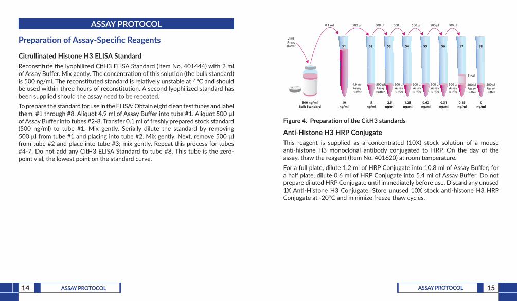
Citrullinated Histone H3 ELISA StandardReconstitute the lyophilized CitH3 ELISA Standard (Item No. 401444) with 2 ml of Assay Buffer. Mix gently. The concentration of this solution (the bulk standard) is 500 ng/ml. The reconstituted standard is relatively unstable at 4°C and should be used within three hours of reconstitution. A second lyophilized standard has been supplied should the assay need to be repeated.To prepare the standard for use in the ELISA: Obtain eight clean test tubes and label them, #1 through #8. Aliquot 4.9 ml of Assay Buffer into tube #1. Aliquot 500 µl of Assay Buffer into tubes #2-8. Transfer 0.1 ml of freshly prepared stock standard (500 ng/ml) to tube #1. Mix gently. Serially dilute the standard by removing 500 µl from tube #1 and placing into tube #2. Mix gently. Next, remove 500 µl from tube #2 and place into tube #3; mix gently. Repeat this process for tubes #4-7. Do not add any CitH3 ELISA Standard to tube #8. This tube is the zero-point vial, the lowest point on the standard curve.
500 ng/mlBulk Standard
0.1 ml 500 µl 500 µl 500 µl 500 µl 500 µl 500 µl
500 µlAssayBu�er
10ng/ml
S1 S2 S3 S4 S5 S6 S7 S8
5ng/ml
2.5ng/ml
1.25ng/ml
0.62ng/ml
0.31ng/ml
0.15ng/ml
0ng/ml
500 µlAssayBu�er
500 µlAssayBu�er
500 µlAssayBu�er
500 µlAssayBu�er
500 µlAssayBu�er
500 µlAssayBu�er
2 ml AssayBu�er
Final
4.9 mlAssayBu�er
Figure 4. Preparation of the CitH3 standards
Anti-Histone H3 HRP ConjugateThis reagent is supplied as a concentrated (10X) stock solution of a mouse anti-histone H3 monoclonal antibody conjugated to HRP. On the day of the assay, thaw the reagent (Item No. 401620) at room temperature.For a full plate, dilute 1.2 ml of HRP Conjugate into 10.8 ml of Assay Buffer; for a half plate, dilute 0.6 ml of HRP Conjugate into 5.4 ml of Assay Buffer. Do not prepare diluted HRP Conjugate until immediately before use. Discard any unused 1X Anti-Histone H3 Conjugate. Store unused 10X stock anti-histone H3 HRP Conjugate at -20°C and minimize freeze thaw cycles.
16 ASSAY PROTOCOL 17ASSAY PROTOCOL
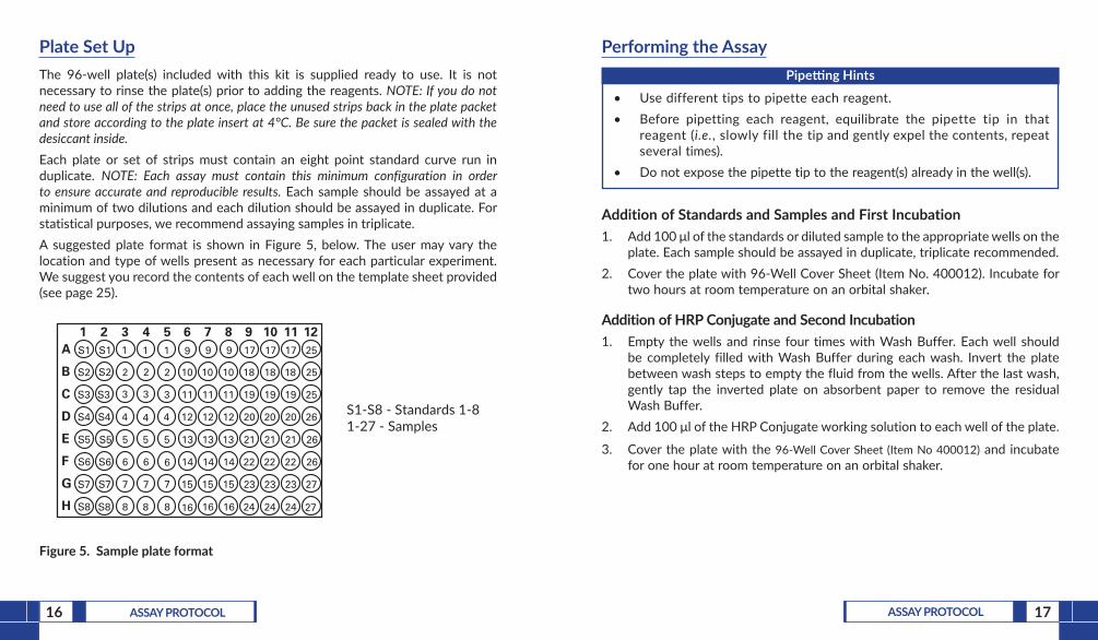
Plate Set UpThe 96-well plate(s) included with this kit is supplied ready to use. It is not necessary to rinse the plate(s) prior to adding the reagents. NOTE: If you do not need to use all of the strips at once, place the unused strips back in the plate packet and store according to the plate insert at 4°C. Be sure the packet is sealed with the desiccant inside. Each plate or set of strips must contain an eight point standard curve run in duplicate. NOTE: Each assay must contain this minimum configuration in order to ensure accurate and reproducible results. Each sample should be assayed at a minimum of two dilutions and each dilution should be assayed in duplicate. For statistical purposes, we recommend assaying samples in triplicate.A suggested plate format is shown in Figure 5, below. The user may vary the location and type of wells present as necessary for each particular experiment. We suggest you record the contents of each well on the template sheet provided (see page 25).
S1-S8 - Standards 1-81-27 - Samples
A
B
C
D
E
F
G
H
1 2 3 4 5 6 7 8 9 10 11 12S1
S2
S3
S4
S5
S6
S7
S8 8
7
6
5
4
3
2
1
8
7
6
5
4
3
2
1
8
7
6
5
4
3
2
1
16
15
14
13
12
11
10
9
16
15
14
13
12
11
10
9
16
15
14
13
12
11
10
9
24
23
22
21
20
19
18
17
24
23
22
21
20
19
18
17
24
23
22
21
20
19
18
17 25
27
27
26
26
26
25
25
S1
S2
S3
S4
S5
S6
S7
S8
Figure 5. Sample plate format
Performing the Assay Pipetting Hints
• Use different tips to pipette each reagent.• Before pipetting each reagent, equilibrate the pipette tip in that
reagent (i.e., slowly fill the tip and gently expel the contents, repeat several times).
• Do not expose the pipette tip to the reagent(s) already in the well(s).
Addition of Standards and Samples and First Incubation1. Add 100 μl of the standards or diluted sample to the appropriate wells on the
plate. Each sample should be assayed in duplicate, triplicate recommended. 2. Cover the plate with 96-Well Cover Sheet (Item No. 400012). Incubate for
two hours at room temperature on an orbital shaker.
Addition of HRP Conjugate and Second Incubation1. Empty the wells and rinse four times with Wash Buffer. Each well should
be completely filled with Wash Buffer during each wash. Invert the plate between wash steps to empty the fluid from the wells. After the last wash, gently tap the inverted plate on absorbent paper to remove the residual Wash Buffer.
2. Add 100 μl of the HRP Conjugate working solution to each well of the plate.
3. Cover the plate with the 96-Well Cover Sheet (Item No 400012) and incubate for one hour at room temperature on an orbital shaker.
19ANALYSIS18 ASSAY PROTOCOL
Development of the Plate1. Empty the wells and rinse four times with Wash Buffer. Each well should
be completely filled with Wash Buffer during each wash. Invert the plate between wash steps to empty the fluid from the wells. After the last wash, gently tap the inverted plate on absorbent paper to remove the residual Wash Buffer.
2. Add 100 μl of TMB Substrate Solution (Item No. 400074) to each well of the plate.
3. Cover the plate with the 96-Well Cover Sheet (Item No 400012) and incubate for 30 minutes at room temperature in the dark on an orbital shaker.
4. DO NOT WASH THE PLATE. Add 100 μl of HRP Stop Solution (Item No. 10011355) to each well of the plate. Blue wells should turn yellow and colorless wells should remain colorless. NOTE: The Stop Solution in this kit contains an acid. Wear appropriate protection and use caution when handling this solution.
Reading the Plate1. Wipe the bottom of the plate with a clean tissue to remove fingerprints,
dirt, etc.2. Read the plate at a wavelength of 450 nm.
ANALYSISMany plate readers come with data reduction software that plots data automatically. Alternatively a spreadsheet program can be used.
Calculations
Plotting the Standard Curve and Determining the Sample ConcentrationUsing computer reduction software, plot absorbance (linear y-axis) versus concentration (linear x-axis) for standards (S1-S7) and fit the data with a four-parameter logistic equation, or alternatively a linear curve fit.
20 ANALYSIS 21ANALYSIS
Performance Characteristics
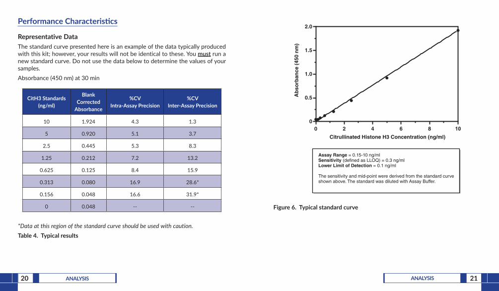
Representative DataThe standard curve presented here is an example of the data typically produced with this kit; however, your results will not be identical to these. You must run a new standard curve. Do not use the data below to determine the values of your samples. Absorbance (450 nm) at 30 min
CitH3 Standards (ng/ml)
Blank Corrected
Absorbance
%CV Intra-Assay Precision
%CV Inter-Assay Precision
10 1.924 4.3 1.3
5 0.920 5.1 3.7
2.5 0.445 5.3 8.3
1.25 0.212 7.2 13.2
0.625 0.125 8.4 15.9
0.313 0.080 16.9 28.6*
0.156 0.048 16.6 31.9*
0 0.048 -- --
*Data at this region of the standard curve should be used with caution.Table 4. Typical results
Citrullinated Histone H3 Concentration (ng/ml)
Assay Range = 0.15-10 ng/mlSensitivity (defined as LLOQ) = 0.3 ng/mlLower Limit of Detection = 0.1 ng/ml
The sensitivity and mid-point were derived from the standard curve shown above. The standard was diluted with Assay Buffer.
Ab
sorb
ance
(45
0 n
m)
0 2 4 6 8 100
0.5
1.0
1.5
2.0
Figure 6. Typical standard curve
23RESOURCES22 ANALYSIS
Precision:Intra-assay precision was determined by analyzing 24 replicates of three matrix controls (RPMI media spiked with analyte) in a single assay.
Matrix Control (ng/ml)
%CV
4.6 3.6
0.9 8.8
0.4 19.9
Table 5. Intra-assay precisionInter-assay precision was determined by analyzing replicates of three matrix controls (RPMI media spiked with analyte) in separate assays spanning across several days.
Matrix Control (ng/ml)
%CV
4.6 6.0
0.9 10.9
0.3 29.7
Table 6. Inter-assay precision
Cross Reactivity:Cross reactivity of the assay to unmodified recombinant histone H3 was tested at 2000X the Citrullinated Histone H3 High Standard concentration and found to be 0.05% cross-reactive. Unmodified core histones were also tested and found to be ≤0.05%.
RESOURCES
Troubleshooting
Problem Possible Causes Recommended Solutions
Poor development of test sample
CitH3 is a part of a complex extracellular trap and unavailable for binding the capture antibody
Treat the sample with DNAse to disrupt the extracellular trap, thereby releasing the free CitH3
Poor development (low signal) of standard curve
A. Standard was diluted incorrectly
B. The standard is degraded
24 RESOURCES 25RESOURCES
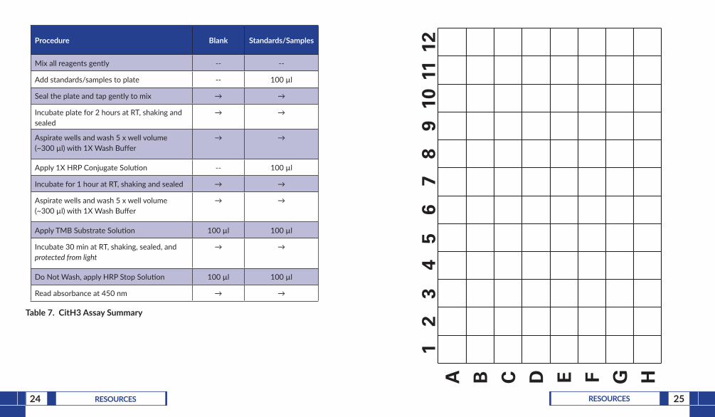
Procedure Blank Standards/Samples
Mix all reagents gently -- --
Add standards/samples to plate -- 100 µl
Seal the plate and tap gently to mix → →
Incubate plate for 2 hours at RT, shaking and sealed
→ →
Aspirate wells and wash 5 x well volume (~300 µl) with 1X Wash Buffer
→ →
Apply 1X HRP Conjugate Solution -- 100 µl
Incubate for 1 hour at RT, shaking and sealed → →
Aspirate wells and wash 5 x well volume (~300 µl) with 1X Wash Buffer
→ →
Apply TMB Substrate Solution 100 µl 100 µl
Incubate 30 min at RT, shaking, sealed, and protected from light
→ →
Do Not Wash, apply HRP Stop Solution 100 µl 100 µl
Read absorbance at 450 nm → →
Table 7. CitH3 Assay Summary
A B C D E F G H
12
34
56
78
910
1112
26 RESOURCES 27RESOURCES
References1. Cuthbert, G.L., Daujat, S., Snowden, A.W., et al. Cell 118(5), 545-553 (2004).2. Neeli, I., Khan, S.N., and Radic, M. J. Immunol. 180(3), 1895-1902 (2008).3. Darrah, E., Rosen, A., Giles, J.T., et al. Ann. Rheum. Dis. 71(1), 92-98 (2012).4. Li, Y., Liu, B., Fukudome, E.Y., et al. Surgery 150(3), 442-451 (2011).
NOTES
Warranty and Limitation of RemedyBuyer agrees to purchase the material subject to Cayman’s Terms and Conditions.Complete Terms and Conditions including Warranty and Limitation of Liability information can be found on our website.This document is copyrighted. All rights are reserved. This document may not, in whole or part, be copied, photocopied, reproduced, translated, or reduced to any electronic medium or machine-readable form without prior consent, in writing, from Cayman Chemical Company.©08/15/2018, Cayman Chemical Company, Ann Arbor, MI, All rights reserved. Printed in U.S.A.