Embed Size (px)
Citation preview
Vol. 11, No. 6MOLECULAR AND CELLULAR BIOLOGY, June 1991, p. 2971-29790270-7306/91/062971-09$02.00/0Copyright © 1991, American Society for Microbiology
cis-Acting Sequences Required for Expression of the DivergentlyTranscribed Drosophila melanogaster Sgs-7 and Sgs-8
Glue Protein GenesANNEMARIE HOFMANN,t MARK D. GARFINKEL,t AND ELLIOT M. MEYEROWITZ*Division ofBiology 156-29, California Institute of Technology, Pasadena, California 91125
Received 26 November 1990/Accepted 7 March 1991
The Sgs-7 and Sgs-8 glue genes at 68C are divergently transcribed and are separated by 475 bp. Fusion geneswith Adh or lacZ coding sequences were constructed, and the expression of these genes, with different amountsof upstream sequences present, was tested by a transient expression procedure and by germ line transforma-tion. A cis-acting element for both genes is located asymmetrically in the intergenic region between -211 and-43 bp relative to Sgs-7. It is required for correct expression of both genes. This element can confer the stage-and tissue-specific expression pattern of glue genes on a heterologous promoter. An 86-bp portion of theelement, from -133 to -48 bp relative to Sgs-7, is shown to be capable of enhancing the expression of atruncated and therefore weakly expressed Sgs-3 fusion gene. Recently described common sequence motifs ofglue gene regulatory elements (T. Todo, M. Roark, K. Vijay Raghavan, C. A. Mayeda, and E. M. Meyerowitz,Mol. Cell. Biol. 10:5991-6002, 1990) are located within this 86-bp region.
The salivary gland secretion (Sgs), or glue protein, genesexpressed in the salivary glands of Drosophila melanogasterthird-instar larvae represent an example of a coordinatelyexpressed set of genes. The set codes for proteins that aresynthesized in one tissue and at one time in development (1,25, 26). The proteins are secreted into the lumen of thesalivary gland and expelled just prior to pupariation to affixthe pupa to a surface during metamorphosis (12, 27). Sevengenes are known to code for components of the glue. TheSgs-3, Sgs-7, and Sgs-8 genes are clustered within a 5,000-bpsegment on the left arm of chromosome 3 at 68C, while theSgs-J, Sgs4, Sgs-5, and Sgs-6 genes each reside at adifferent location in the genome. All of the chromosomalsites with glue genes correspond to regions showing largeintermolt polytene chromosome puffs during the time of glueprotein synthesis.The 68C intermolt puff is interesting because of the
clustering of the three genes found there. The order of thegenes is telomere, Sgs-8, Sgs-7, Sgs-3, centromere withrespect to the left arm of the third chromosome. Transcrip-tion of Sgs-8 proceeds leftward, transcription of Sgs-7 pro-ceeds rightward, and transcription of Sgs-3 proceeds right-ward (16, 40). The 5' end of Sgs-8 is separated from the 5'end of Sgs-7 by 475 bp of nontranscribed intergenic se-quences (16), and the two genes are surrounded by two285-bp elements that form an inverted repeat. The 3' end ofSgs-7 is separated from the 5' end of Sgs-3 by 1,958 bp (16,40).Two trans-acting regulators of this gene set have been
described. The steroid hormone ecdysterone is required forboth activation and cessation ofRNA synthesis (11, 18), andthe function eliminated by the lethal(l)nonpupariating-1[l(J)npr-1] mutation is required for expression of the three68C genes (10).
* Corresponding author.t Present address: Institut fur Genetik der Freien Universitat
Berlin, 1000 Berlin 33, Germany.* Present address: Department of Molecular Genetics and Cell
Biology, The University of Chicago, Chicago, IL 60637.
Previous studies on the sequences required in cis forregulation of these genes have concentrated on those regu-lating the largest of the three genes, Sgs-3, and have shownthat there are at least three regulatory elements within thesequences upstream of the Sgs-3 gene: the proximal elementbetween -106 and -56 (34, 46, 57), the distal elementbetween -629 and -491 (5, 41, 45, 46), and multiple remoteregions from 1.5 to 2.5 kb upstream (17). The proximal anddistal elements are functionally equivalent and act synergis-tically to give high-level expression (46).The experiments described here comprise an analysis of
the Sgs-71Sgs-8 gene pair. As the genes are divergentlytranscribed, the general question is to determine the arrange-ment of cis-acting sequences required for each gene tofunction. Sequence analysis of the intergenic region hasrevealed conserved regions within the first 100 bp upstreamof Sgs-7 and the first 100 bp upstream of Sgs-8 (16). Theseelements, the right copy and the left copy, respectively,contain sequences that resemble the ecdysterone regulatoryelement identified by Mestril et al. (39) in their study of thehsp23 gene.We have identified the cis-acting regulatory sequences for
Sgs-7 and Sgs-8 by reintroducing in vitro-manipulated geneseither into somatic cells via the transient assay or somatictransformation procedure (35) or into the germ line of D.melanogaster via P-element-mediated transformation (47,54). We have used gene fusion constructs of regulatoryregions of Sgs-7 and Sgs-8 with the D. melanogaster Adh(alcohol dehydrogenase) gene or the Escherichia coli lacZ(,-galactosidase) gene as reporter genes. These reportergene choices were motivated by several considerations: first,to exploit sensitive histochemical reactions for the in situdetection of the protein products encoded by introducedfusion gene constructions; second, to exploit sensitive quan-titative enzyme activity assays for measurement of genefunction; third, to distinguish the gene products of newlyintroduced glue protein genes from the endogenous geneproducts, as there are no stocks that produce reducedamounts, or size variants, of Sgs-7 or Sgs-8 RNA or protein;and fourth, to enable us to test plasmid constructions simul-
2971

2972 HOFMANN ET AL.
taneously for Sgs-7 and Sgs-8 gene function. The experi-ments confirm that a single 86-bp region functioning bidirec-tionally, located closer to the Sgs-7 gene, is required forstage- and tissue-specific full expression of both genes. Itcontains sequences that have recently been identified asregulatory element consensus sequences for glue genes (55).
pGLAX1.0
P Adh 7 8 lacZ 8 rosy P
MATERIALS AND METHODS
Plasmid constructions. All plasmids were named accordingto the following convention: G = glue gene, A = Adh gene,L = lacZ gene, X = xanthine dehydrogenase gene, 0 =
synthetic oligonucleotide, S = Sgs4 gene, and C = cis-acting sequence.
Construction of the Sgs-7-Adh fusion gene. Plasmid pHAP(4) was used as the source of the promoterless D. melano-gaster Adh gene for fusion to Sgs-7 promoter sequences.The Adh gene in this plasmid is modified by the addition ofa HindlIl linker in the 5' untranslated region. A 1.8-kbHindIII-XbaI fragment encompassing the Adh gene wassubcloned into pUC18. An 824-bp XbaI fragment containingthe Sgs-7ISgs-8 intergenic region and portions of the Sgs-7and Sgs-8 transcription units was cloned into the HindIII siteafter partially filling the HindIlI and XbaI termini. Theplasmid was named pGAO-1, and the sequence of the jointbetween Sgs-7 and Adh was determined (36, 37). The junc-tion between the two genes is in their 5' untranslatedregions. The Sgs-7 contribution ends at +25 bp and the Adhcontribution begins at +13 bp relative to their respectivetranscripts. The Adh contribution starts at + 13 of thetranscript that is initiated from the proximal promoter (3).Five nucleotides from the synthetic HindIll linker are sand-wiched between the two Drosophila gene segments.
Construction of pGLAX1.0. The 2.2-kb fragment frompGAO-1 that contains the Sgs-7-Adh fusion gene was fusedto custom-made synthetic 14-mer linker/adaptors and clonedinto the EcoRI site of pOX4. Plasmid pOX4 (39a) containsSgs-8 sequences beginning in the 5'-flanking sequence andending at the BglII site at +245 bp joined to the E. coli lacZgene. The first 51 codons of the Sgs-8 protein-coding regionare joined to a synthetic-linker-derived codon, followed bythe E. coli 3-galactosidase-coding region beginning at theninth codon. At the 3' end of the lacZ gene, a 268-bpsegment of the Sgs-8 gene contributes a polyadenylationsite. The transformation vector pGLAX1.0 (Fig. 1) wasconstructed by cloning the 9.2-kb Sall fragment that con-tained the Sgs-7-Adh and Sgs-8-lacZ fusion genes intoCarnegie 20 (48).
Construction of the pGLX and the pGAX series. The 4.2-kbXbaI-KpnI fragment from pGLAX1.0 containing the entireSgs-8-lacZ fusion gene was subcloned into pUC18 to givethe starting clone for the pGLX series. The 2.2-kb EcoRIfragment from pGAZ-1 containing the Sgs-7-Adh gene wasrecloned into the vector fragment of pGAO-1 and served asthe starting clone for the pGAX series. To remove DNAupstream of the fusion genes, BAL 31 digestions were usedby setting up replica reactions that varied either the time ofreaction or the concentration of BAL 31. The amount ofDNA removed by BAL 31 was determined by digesting thereaction mixtures with the appropriate restriction enzymesand subjecting them to electrophoresis through a 2.5%agarose gel. The endpoints of the BAL 31 deletion moleculesused in this study were determined by standard proceduresfor dideoxy sequencing (49, 56). Germ line transformationplasmids (Fig. 1) were constructed by recloning the pro-moter deletion molecules into the SalI site of Carnegie 20.
pGAX Series
4-
1K tiltP Adh 7
4
rosy p
pGLX Series
P lacZ 8 rosy PFIG. 1. Schematic representation of plasmids pGLAX1.0,
pGAX, and pGLX. The origins of the various fragments are shown,as are the transcription directions of the different genes. P, Droso-phila P element; Adh, Drosophila alcohol dehydrogenase gene;lacZ, E. coli P-galactosidase gene; rosy, Drosophila xanthine dehy-drogenase gene; 7, transcribed sequences from Sgs-7; 8, transcribedand 3' sequences from Sgs-8. The thin line between 7 and 8represents the intergenic region of Sgs-7 and Sgs-8.
Construction of plasmids containing the cis-acting sequencesand reporter genes. Plasmids of the pGGL series wereconstructed by cloning sequences from the intergenic regionbetween Sgs-7 and Sgs-8 into the Sall site of pGLO-1 afterthey had been flanked with synthetic Sall linkers. pGLO-1 ispUC18 that contains the Sgs-3-lacZ fusion gene (57) with130 bp of Sgs-3 upstream sequence. The Sall site is at -130.The number in the plasmid name is the amount of cis-actingsequence present (in base pairs); the number in parenthesesgives the orientation of the sequence. For example, (7)means that the reporter gene is in the same orientation as theendogenous Sgs-7. Plasmid pCSA-400(8) was constructed bycloning the 194-bp cis-acting sequences from Sgs-7ISgs-8 viaSalI linkers into the Sall site of pSA-400. pSA-400 is pUC18containing an Sgs4-Adh transcriptional fusion gene and 400bp of 5'-flanking Sgs4 sequences. Plasmids of the pCAseries were constructed by ligating sequences from theintergenic region (in one case together with stuffer sequencesfrom pUC18) with attached Sall linkers to a synthetic Sallsite 56 bp upstream from the D. melanogaster Adh gene inplasmid pNS1186 (obtained from N. Shen, Rutgers Univer-sity). Germ line transformation vectors of the pCAX serieswere obtained by cutting out the relevant sequences of thepCA plasmid, filling in the ends with the Klenow fragment ofE. coli DNA polymerase I, and ligating the blunt ends to theunique HpaI site in the polylinker of Carnegie 20. Plasmidsof the pGGAX series were obtained by attaching Sall linkersto the 194-bp cis-acting region and cloning it into the Sall siteof pGOAX.130, which contains an Sgs-3-Adh fusion genewith 130 bp of Sgs-3 upstream sequence present (46). Threedifferent plasmids were obtained: pGGAX-7, in which thefusion gene is in the same orientation relative to the cis-acting sequence as is Sgs-7 in the endogenous situation;
MOL. CELL. BIOL.
TRANSCRIPTION OF DROSOPHILA Sgs-7 AND Sgs-8 GENES 2973
pGGAX-8, in which the fusion gene is in the same orienta-tion as is Sgs-8; and pGGAX-88, which contains a tandemduplication of the cis-acting sequences in the Sgs-8 orienta-tion.Drosophia strains and transformations. The host strain for
injections was Adhfn6 cn; y502 (obtained from J. W. Posa-kony, University of California, San Diego). Germ line trans-formation was done by the standard method (47, 54), as thetransposase-producing helper plasmid phsiT (54a) orp1T25.7wc (22) was used. Transformants were identified inthe Gl progeny of injected animals as ry' flies. Standardgenetic techniques using the balancer strain T(2;3)AtaICyO;TM3, Sb ryRK Ser (constructed by M. A. Crosby) wereapplied to map, balance, and make homozygous the autoso-mal insertion events. Markers and balancers are describedby Lindsley and Grell (28) and by Lindsley and Zimm(29-31). DNA gel blotting (53) was used to determine thateach transformed line analyzed contained only one insertion.In some cases, in situ hybridization to polytene chromo-somes was performed by using a slight modification of themethod described by Pardue and Gall (44). Transformantlines were named according to the following convention: Tf,to indicate a transformant line; the numeral in parenthesesindicates the chromosomal location of the insertion event;followed by the name of the plasmid construct transposedinto the chromosome. The transient expression assay wasoriginally developed by Martin et al. (35). The procedure asused in this work followed the technique described by Roarket al. (46). A positive result in this assay was histochemicalstaining only in the salivary gland cells of third-instar larvae.
Expression assays. For analysis of transient expression,histochemical staining assays on glands or dissected larvaewere used. P-Galactosidase staining was performed as de-scribed by Vijay Raghavan et al. (57). Staining of glands andtissues for ADH activity was performed as described byRoark et al. (46). The quantitative spectrophotometric assayof ADH enzyme activity was done by the method of Soferand Ursprung (52) with modifications and details describedby Roark et al. (46). The quantitative measurement ofP-galactosidase enzyme activity was performed as following.One animal equivalent of the same extract as prepared forthe ADH assay was added to 2 ml of Z buffer (42) adjusted topH 7.5 and supplemented with 0.5 mM 4-methylumbelliferyl-,-D-galactopyranoside, which was used as the fluorogenicsubstrate (21). The reaction mixtures were incubated at 37°Cwhile fluorescence of the reaction product 4MU (4-methyl-umbelliferone) was read every 30 s for 10 min. The fluores-cence was detected by using a Kontron SFM 25 spectroflu-orimeter; excitation wavelength was 365 nm, and emissionwavelength was 455 nm. A standard curve was constructedby using the above solutions containing four known concen-trations of 4MU and was used to convert the raw fluores-cence value into picomoles of 4MU. A laboratory softwareprogram (17a) running on a Commodore PC20 was used fordata evaluation and to calculate linear regression lines fromthe data. Total protein in each extract was measured by themethod of Lowry et al. (33). We define 1 U of hybridSgs-8-p-galactosidase enzyme as the amount of enzyme thatreleases 1 pmol of 4MU per min. Final data were compiledand averaged, and standard deviations were calculated byusing Microsoft's Excel program.
RESULTS
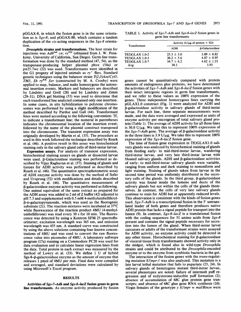
Activities of Sgs-7-Adh and Sgs-8-acZ fusion genes in germline transformants. As enzyme activity produced by fusion
TABLE 1. Activity of Sgs-7-Adh and Sgs-8-lacZ fusion genes ingerm line transformants
Activity (U/p.g of protein ± SD)Transformant
ADH 3-Galactosidase
Tf(2)GLAX 1.0-2 25.3 + 3.0 3.09 + 0.82Tf(3)GLAX 1.0-3 30.2 + 5.6 4.07 ± 0.95Tf(3)GLAX 1.0-7 34.7 ± 6.2 4.62 ± 1.23Avg 30.1 3.93
genes cannot be quantitatively compared with proteinamounts of endogenous glue proteins, we have determinedthe activities of Sgs-7-Adh and Sgs-8-lacZ fusion genes withtheir intact intergenic regions in germ line transformants,and we refer to these values as 100% expression of thegenes. Three independent homozygous lines carrying thepGLAX1.0 construct (Fig. 1) were analyzed for ADH and3-galactosidase activity in salivary glands of third-instar
larvae. For each line, three separate measurements weremade, and the data were averaged and expressed as units ofenzyme activity per microgram of total salivary gland pro-tein (Table 1). The average of ADH activity for all three linesis 30.0 U/,ug. We take this to represent 100% expression ofthe Sgs-7-Adh gene. The average of P-galactosidase activityin the three lines is 3.9 U/,ug. We take this to represent 100%expression of the Sgs-8-lacZ fusion gene.The time of fusion gene expression in Tf(GLAX1.0 sali-
vary glands was analyzed by histochemical staining of glandsfrom feeding early- to mid-third-instar larvae, wanderingthird-instar larvae, and very late third-instar larvae withbloated salivary glands. ADH and P-galactosidase activitiesof early- to mid-third-instar salivary glands were variable,ranging from uniform and dark staining to nonuniform andlight staining. Staining of glands taken from larvae in thesecond time period was uniformly distributed in the secre-tory cells of the glands. In the third group, P-galactosidaseactivity was found inside the lumens and ducts of thesalivary glands but not within the cells of the glands them-selves. In contrast, the cells of very late salivary glandscontinue to stain for ADH but at apparently reduced levels.This observation is consistent with the kinds of fusion genesused. Sgs-7-Adh is a transcriptional fusion in the 5' untrans-lated leader of both genes and therefore produces nativeADH protein that lacks a signal peptide for transport into thelumen (9). In contrast, Sgs-8-lacZ is a translational fusionwith the coding sequences for 51 amino acids from Sgs-8present and contains the signal peptide necessary for secre-tion into the lumen of the gland. When third-instar larvalcarcasses or adults of the transformant strains were assayedfor ADH activity, no enzyme activity could be detected inany other tissue. Histochemical staining for P-galactosidaseof visceral tissue from transformants showed activity only inthe midgut, which is found also in wild-type Drosophilastrains and could be attributed to the Drosophila-encodedenzyme or to the enzyme from symbiotic bacteria in the gut.The interaction of the fusion genes with the trans-regulat-
ing mutation l(J)npr-J was also analyzed. This mutation is alate larval lethal mutation that fails to pupariate (23, 24). Insalivary glands of hemizygous mutant third-instar larvae,several phenotypes are noted: failure of intermolt puff re-gression and of ecdysterone-inducible puff formation (2);absence of accumulation of 68C glue protein gene tran-scripts; and absence of 68C glue gene RNA synthesis (10).Virgin females of the genotype y l(J)npr w mallBinsn were
VOL. 11, 1991
2974 HOFMANN ET AL.
-236 s
- 21 1:
8-lac Z
4
4
8
Expression
Transient Germline
Activity In %
7-Adh
-92
100
100
0
-432 + 100
.415 _
-226 -
-125 n.d.
7
1 0
0
i- I.
-236 -43
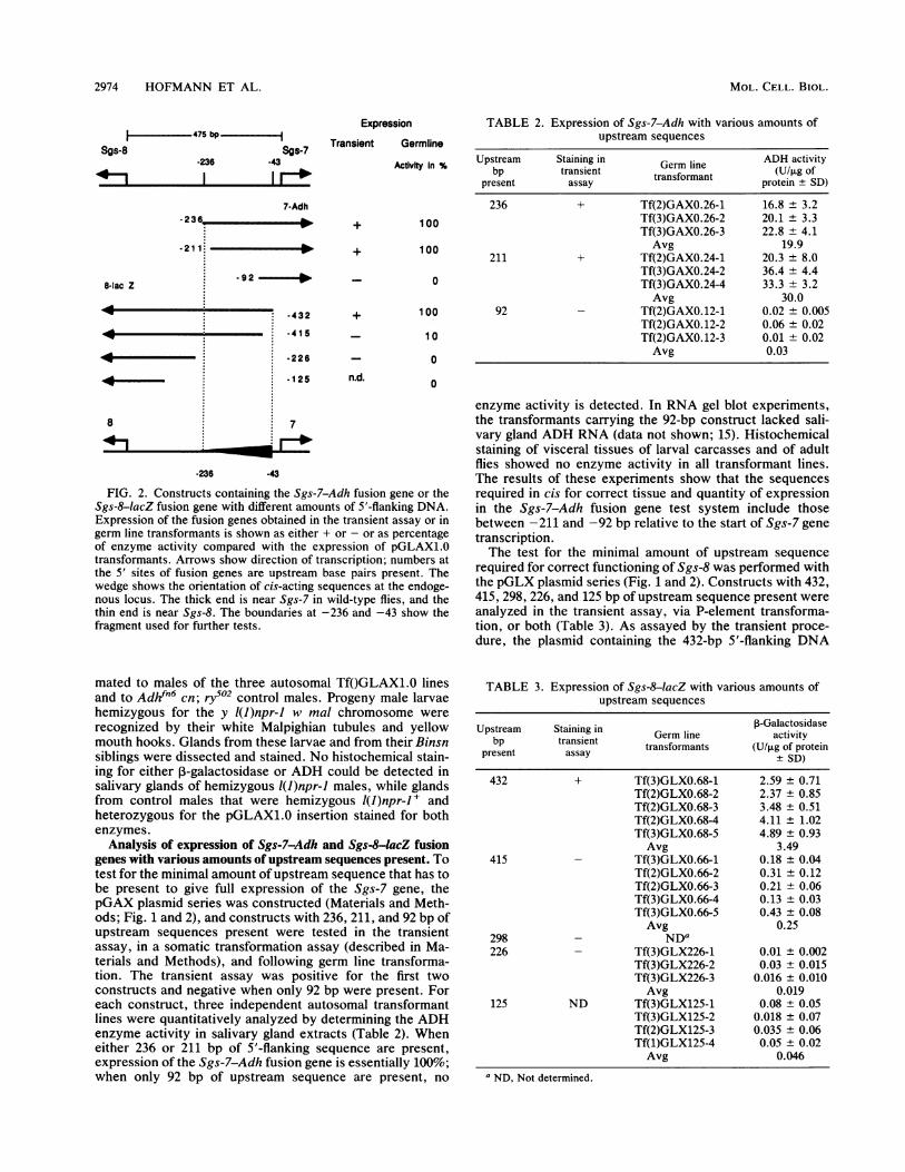
FIG. 2. Constructs containing the Sgs-7-Adh fusion gene or theSgs-8-lacZ fusion gene with different amounts of 5'-flanking DNA.Expression of the fusion genes obtained in the transient assay or ingerm line transformants is shown as either + or - or as percentageof enzyme activity compared with the expression of pGLAX1.0transformants. Arrows show direction of transcription; numbers atthe 5' sites of fusion genes are upstream base pairs present. Thewedge shows the orientation of cis-acting sequences at the endoge-nous locus. The thick end is near Sgs-7 in wild-type flies, and thethin end is near Sgs-8. The boundaries at -236 and -43 show thefragment used for further tests.
TABLE 2. Expression of Sgs-7-Adh with various amounts ofupstream sequences
Upstream Staining in Germ line ADH activitybp transient transformant (U/pg of
present assay protein ± SD)
236 + Tf(2)GAXO.26-1 16.8 ± 3.2Tf(3)GAXO.26-2 20.1 ± 3.3Tf(3)GAXO.26-3 22.8 ± 4.1Avg 19.9
211 + Tf(2)GAXO.24-1 20.3 ± 8.0Tf(3)GAXO.24-2 36.4 ± 4.4Tf(3)GAXO.24-4 33.3 ± 3.2Avg 30.0
92 - Tf(2)GAXO.12-1 0.02 ± 0.005Tf(2)GAXO.12-2 0.06 ± 0.02Tf(2)GAXO.12-3 0.01 ± 0.02Avg 0.03
0
enzyme activity is detected. In RNA gel blot experiments,the transformants carrying the 92-bp construct lacked sali-vary gland ADH RNA (data not shown; 15). Histochemicalstaining of visceral tissues of larval carcasses and of adultflies showed no enzyme activity in all transformant lines.The results of these experiments show that the sequencesrequired in cis for correct tissue and quantity of expressionin the Sgs-7-Adh fusion gene test system include thosebetween -211 and -92 bp relative to the start of Sgs-7 genetranscription.The test for the minimal amount of upstream sequence
required for correct functioning of Sgs-8 was performed withthe pGLX plasmid series (Fig. 1 and 2). Constructs with 432,415, 298, 226, and 125 bp of upstream sequence present wereanalyzed in the transient assay, via P-element transforma-tion, or both (Table 3). As assayed by the transient proce-dure, the plasmid containing the 432-bp 5'-flanking DNA
mated to males of the three autosomal Tf(GLAX1.0 linesand to Adhfn6 cn; ry502 control males. Progeny male larvaehemizygous for the y l(J)npr-J w mal chromosome wererecognized by their white Malpighian tubules and yellowmouth hooks. Glands from these larvae and from their Binsnsiblings were dissected and stained. No histochemical stain-ing for either 3-galactosidase or ADH could be detected insalivary glands of hemizygous l(J)npr-J males, while glandsfrom control males that were hemizygous l(J)npr-J + andheterozygous for the pGLAX1.0 insertion stained for bothenzymes.
Analysis of expression of Sgs-7-Adh and Sgs-8-lacZ fusiongenes with various amounts of upstream sequences present. Totest for the minimal amount of upstream sequence that has tobe present to give full expression of the Sgs-7 gene, thepGAX plasmid series was constructed (Materials and Meth-ods; Fig. 1 and 2), and constructs with 236, 211, and 92 bp ofupstream sequences present were tested in the transientassay, in a somatic transformation assay (described in Ma-terials and Methods), and following germ line transforma-tion. The transient assay was positive for the first twoconstructs and negative when only 92 bp were present. Foreach construct, three independent autosomal transformantlines were quantitatively analyzed by determining the ADHenzyme activity in salivary gland extracts (Table 2). Wheneither 236 or 211 bp of 5'-flanking sequence are present,expression of the Sgs-7-Adh fusion gene is essentially 100%;when only 92 bp of upstream sequence are present, no
TABLE 3. Expression of Sgs-8-lacZ with various amounts ofupstream sequences
Upstream Staining in .3-Galactosidasebp transient Germ line activity
transformants (U/pg of proteinpresent assay ± SD)
432 + Tf(3)GLXO.68-1 2.59 ± 0.71Tf(2)GLXO.68-2 2.37 ± 0.85Tf(2)GLXO.68-3 3.48 ± 0.51Tf(2)GLXO.68-4 4.11 ± 1.02Tf(3)GLXO.68-5 4.89 ± 0.93Avg 3.49
415 - Tf(3)GLXO.66-1 0.18 ± 0.04Tf(2)GLXO.66-2 0.31 ± 0.12Tf(2)GLXO.66-3 0.21 ± 0.06Tf(3)GLXO.66-4 0.13 + 0.03Tf(3)GLXO.66-5 0.43 ± 0.08Avg 0.25
298 - NDa226 - Tf(3)GLX226-1 0.01 ± 0.002
Tf(3)GLX226-2 0.03 ± 0.015Tf(3)GLX226-3 0.016 ± 0.010Avg 0.019
125 ND Tf(3)GLX125-1 0.08 ± 0.05Tf(3)GLX125-2 0.018 ± 0.07Tf(2)GLX125-3 0.035 ± 0.06Tf(1)GLX125-4 0.05 ± 0.02Avg 0.046
a ND, Not determined.
Sgs-8
4I
Sgs-7-236 .43
1 1
0 ... 0
MOL. CELL. BIOL.

TRANSCRIPTION OF DROSOPHILA Sgs-7 AND Sgs-8 GENES 2975
sequence gave staining; none of the other constructs exhib-ited any 3-galactosidase activity. Germ line transformantscarrying four of the five deletion constructs were estab-lished, and ,-galactosidase activities were measured in sal-ivary gland extracts of climbing third-instar larvae fromtransformation-homozygous lines. When 432 bp of upstreamsequences are present, the expression of Sgs-8-lacZ is100%, but expression drops to 10% when 415 bp are present.In transformants carrying Sgs-8-lacZ fusion genes with 226or 125 bp of 5'-flanking sequences, no enzyme activity couldbe detected. Again, staining of other tissues and stagesshowed no enzyme activity for all the transformed linesexcept for the background staining in the midgut. Theseresults show that sequences located asymmetrically in theintergenic region are required for correct expression of bothSgs-7 and Sgs-8, as outlined in Fig. 2. The overlappingsequences required for expression of both genes are referredto as the cis-acting element and were further tested for theirregulatory capacity.The cis-acting element can enhance the expression of other
glue genes. The putative cis-acting region was tested for itscapacity to restore normal quantity of expression of an Sgs-3or Sgs4 gene construction lacking its own quantitativecontrol sequences. The 194-bp region between -236 and-43 relative to Sgs-7 was linked to an Sgs-3-lacZ fusiongene with 130 bp of 5'-flanking sequence. This gene con-struction without the 194-bp region (pGLO-1) is negative inthe transient assay but gives faint histochemical staining insalivary glands of germ line transformant third-instar larvae(57). In our experiments, none of 14 third-instar larvaeshowed staining in the salivary glands after injection ofpGLO-1 into the embryos, whereas 6 of 9 larvae showedstaining in the salivary glands after injection of pGGL-194(7), in which the cis-acting element is placed in the Sgs-7orientation in front of the -130-bp Sgs-3-lacZ gene. Withplasmid pGGL-194(8), in which the element is placed in theSgs-8 orientation, we obtained 14 positives out of 36 larvaeanalyzed in the transient assay (Fig. 3). For testing thecis-acting element for an enhancing effect on expression ofSgs4, we linked the 194-bp region to a transcriptionalSgs4-Adh fusion gene with 400 bp of Sgs4 upstreamsequence present (pSA-400). An Sgs4 gene with 398 bp ofupstream sequence has been shown to be completely inac-tive in germ line transformants (38). In our transient assayswith pSA-400, none of 18 larvae showed histochemicalstaining in their salivary glands, whereas with pCSA-400(8),in which the cis-acting element is located at -400 bp of theSgs4-Adh fusion gene in Sgs-8 orientation, we obtained 3out of 26 positively reacting larvae (Fig. 3). These resultsshow that the cis-acting element can enhance expression ofother glue genes and can replace the enhancers ordinarilyfound upstream of these genes.
It is not possible to draw conclusions about quantitativeexpression from the transient assays. The number of indi-vidual larvae or the number of cells in the salivary glandsthat stain positively depends apparently on the uptake ofplasmid DNA by the salivary gland precursor cells in theinjected embryo and not on any quality of the promotertested. Therefore, germ line transformation via P-elementvectors was used to quantify the enhancing effect of thecis-acting element. The 194-bp region was cloned into theSall site at -130 of GOAX.130, a construct containing anSgs-3-Adh fusion gene with 130 bp of 5'-flanking sequence(46). Three different derivatives were tested in the germ line:pGGAX-7, in which the reporter gene is in the same orien-tation relative to the cis-acting element as is Sgs-7 in the
A
-130 Sg.4aCZ
Staining ofsalivary glands
pGLO-1
pGGL-1 94(7) +
pGGL-1 94(8) I
pSA-400
pSA-400(8)
B
.130 SOg-3-Adh
+
Adh activity insalivary glands
GOAX.13
,, 91=GGAX-7
GGAX-8
90
27
GGAX-88 46FIG. 3. Constructs used in the transient expression procedure
(A) and in germ line transformation analysis (B). Open bars, E. colilacZ coding sequences; striped bars, D. melanogaster Adh codingsequences; thin lines, upstream sequences of Sgs-3 and Sgs4(arrows indicate direction of transcription); wedge-shaped line,cis-acting sequences from -236 of Sgs-7 (thin end) to -43 of Sgs-7(thick end). For nomenclature of plasmids, see Materials andMethods.
endogenous situation; pGGAX-8, in which the reporter geneis in the Sgs-8 orientation; and pGGAX-88, a tandem inser-tion of the cis-acting element in Sgs-8 orientation (Fig. 3).ADH enzyme activities in extracts from salivary glands ofdifferent transformant lines were determined (Table 4) andcompared with the enzyme activity of the Sgs-3-Adh fusion
TABLE 4. ADH activity in GGAX germ line transformants
ADH activityTransformant line (U/p.g of
protein ± SD)
Tf(3)GOAX.13-1 ........................................ 0.082 ± 0.029aTf(3)GOAX.13-2 ........................................ 0.087 ± 0.044aTf(2)GOAX.13-3 ........................................ 0.138 ± 0.060aAvg .......... .............................. 0.102
Tf(3)GGAX-7-1 ........................................ 6.45 ± 0.98Tf(2)GGAX-7-2 ........................................ 13.71 ± 2.82Tf(3)GGAX-7-3 ........................................ 7.89 ± 2.22Tf(2)GGAX-7-4........................................ 7.90 ± 0.54Avg ........................................ 8.99
Tf(2)GGAX-8-1 ........................................ 3.77 ± 1.23Tf(3)GGAX-8-2........................................ 3.69 ± 0.98Tf(2)GGAX-8-3 ........................................ 1.89 ± 0.61Tf(3)GGAX-8-4........................................ 1.65 ± 0.42Avg ........................................ 2.75
Tf(2)GGAX-88-1 ........................................ 3.71 ± 0.14bTf(3)GGAX-88-2 ........................................ 7.34 ± 3.03Tf(3)GGAX-88-3 ........................................ 2.76 ± 0.42bAvg ........................................ 4.61
a Data from Roark et al. (46).b Values from homozygous lethals were doubled after it was ascertained
that the lethal effect is before third larval instar (20 larvae from each line weretested for heterozygosity in polytene chromosome squashes).
I
- l-U
VOL. 1 l, 1991
-W Sgs-4-Adh
9=;0.111

2976 HOFMANN ET AL.
pNS 1186
CA-194 (7)
CA-194 (8)
CA-194 (88) _
COA-194 (8)
FIG. 4. Constructs tested in the tramelanogaster Adh coding sequencessequences; thin line, 200 bp from pUCshaped line, cis-acting sequences fror-43 of Sgs-7 (thick end). For norMaterials and Methods.
gene without the cis-acting elemeGOAX.130 lines (data reportedlarvae transformed with the GGgland activity of ADH is increas(GOAX.130 lines. In the Tf(GGcis-acting element in the oppositeeffect is 27-fold. A tandem duielement in the Sgs-8 orientation aof the reporter gene relative to thsingle copy (Fig. 3).The cis-acting element shows a d
specific regulatory function. To tesits capacity to confer the Sgs gerheterologous promoter, plasmidrconstructed. The 194-bp region N
truncated at -56 bp upstream omoter. Three different clones w(with the Adh gene in the Sgs-7 oriithe Adh gene in the Sgs-8 orientatia tandem duplication of the cis-aorientation (Fig. 4). The three cothe transient assay, and only p(result, with 5 of 21 third-instar larin the salivary glands. pCA-194(negative, with none of 27 and ntively, showing staining. These194-bp region contains the regulattissue-specific expression. One piative results obtained with plasmicould be that the lower-level eyGGAX-8 lines is below the thrercan be monitored in the transientbility, we transferred the pCA-P-element vector Carnegie 20;transformation. Salivary glands fiTfOCAX-194(8) lines were testedassay, but no staining was obseifor the nonexpression in the Sgs-,the element in this orientation eplaced at the same distance fromSgs-8 in the endogenous situat
Staining of tested in the transient assay by injecting plasmid pCOA-saAhvar glads 194(8), which has a 190-bp stuffer sequence from pUC18,
between the cis-acting element and the start of the Adh gene.~- None of 38 third-instar larvae tested in the transient assay
showed staining in the salivary glands.Further delimitation of the cis-acting element. The 194-bp
+ region was split into two segments, a 86-bp segment from-48 to -133 (relative to Sgs-7) and a 99-bp segment from-138 to -236. Both fragments were fused to the Sgs-3-lacZgene with 130 bp of 5'-flanking sequences in pGLO-1, andplasmids pGGL-86(8) and pGGL-99(7) were obtained. OnlypGGL-86(8) gave a positive result when tested in the tran-sient assay, with 9 of 37 larvae showing staining in theirsalivary glands. With pGGL-99(7), none of 35 larvae showedstaining in their salivary glands. This result shows that atleast the enhancing or amplifying effect on glue gene expres-sion is mediated by the 86-bp portion of the Sgs-7ISgs-8
Lnsient assay. Striped bars, D. cis-acting sequences located at -48 to -133 relative tos; thick lines, Adh promoter Sgs-7.m18 (stuffer sequence); wedge- Sequence homologies at 68C. At least three regulatorymenclature of plasmids, see elements required for full expression of Sgs-3 have been
identified: the proximal element (at -56 to -98), the distalelement (at -491 to -629), and one or more remote up-stream elements (34, 46). Roark et al. (46) have shown thatthe proximal and distal elements are functionally equivalent,
nt from Sgs-7ISgs-8 in the each acting to direct correct tissue- and stage-specificby Roark et al. [46]). In expression at low levels. When combined, these elementsiAX-7 construct, salivary act synergistically to give high levels of expression. We havesed 90-fold relative to Tf- shown in this work that the Sgs-7ISgs-8 cis-acting element isrAX-8 larvae, carrying the capable of functioning as an enhancer when present with theorientation, the enhancing proximal element of Sgs-3. This finding suggests that it is)lication of the cis-acting also functionally equivalent to this element and may have atlmost doubles the activity similar regulatory sequence. Todo et al. (55) identified sin-te similar construct with a gle-base-pair substitutions at several sites in the proximal
element that inactivate it. Aligning the sequences of thelirectional tissue- and stage- proximal Sgs-3 element, the distal Sgs-3 element, and theit the cis-acting element for Sgs-7ISgs-8 cis-acting element reveals a similar sequence inne expression pattern on a each of the three elements. The relevant sequences ares of the CA series were shown in Fig. 5, with the common sequences boxed. Notewas fused to an Adh gene that within the Sgs-7ISgs-8 cis-acting element, there are twoIf the larval proximal pro- copies of the homology present, both facing Sgs-7, and theyere obtained: pCA-194(7), are located in the 86-bp portion of the cis-acting element thatentation; pCA-194(8), with was shown by our experiments to contain the regulatoryion; and pCA-194(88), with sequences.,cting element in the Sgs-8)nstructions were tested inCA-194(7) gave a positive*vae showing ADH activity(8) and pCA-194(88) werelone of 17 larvae, respec-results indicate that the
tory element for stage- andossible reason for the neg-ids of the Sgs-8 orientationrpression observed in theshold at which expressiont assay. To test this possi--194(8) construct into theand performed germ linerom larvae of the resultingin the ADH histochemicalrved. Another explanation8 orientation could be thatcan work only when it isthe Adh gene as it is fromion. This possibility was
DISCUSSION
Regulatory sequences for Sgs-7 and Sgs-8 are not separatefor each gene and are located closer to Sgs-7. The fact thatthere are homologous sequences present in the base pairsimmediately upstream of both the Sgs-7 and Sgs-8 genessuggests that each of these sequences is solely requiredseparately for correct expression of the gene close to it. Thisis the case for Sgs-7 but not for Sgs-8, as reported in thiswork. Sequences located closer to Sgs-7 are required forcorrect functioning of both Sgs-7 and Sgs-8 genes.The cis-acting element of the Sgs-71Sgs-8 intergenic region
as defined in this work regulates the correct expression ofSgs-7. It is also necessary for full expression of Sgs-8. Wecannot exclude that sequences located between the tran-scription start site of Sgs-8 and the cis-acting element areadditionally necessary for correct functioning of Sgs-8 fortwo reasons. First, the cis-acting element does not confer itsenhancing capability equivalently in both orientations. In theTfOGGAX transformant lines, the enhancing effect of thecis-acting element in the Sgs-8 orientation is only one-third
MOL. CELL. BIOL.

TRANSCRIPTION OF DROSOPHILA Sgs-7 AND Sgs-8 GENES 2977
Sgs-7/8AG|TG]GTGTGTTMAImrAMAATACAATT
-139
Sgs-3(proximal)
Sgs-3(distal)
-1 06
AGTTTTATGGGGCT TAMG
-637
CATAMTCGAAATACTG
TAAAGCTAAATTAAGCTAAATTTAA
AC TCCAT*-
- 43
TffAGA.57
CTG TCCTTAITATA
-578
FIG. 5. Comparison of the sequence of the cis-acting elements of Sgs-7/Sgs-8 with the sequences of the two functionally equivalentelements of Sgs-3. The elements show boxes of homology with an AT-rich spacer region between them. Nucleotides indicated by asteriskshave been shown to be important by single-base substitution mutation (55). Blank spaces between sequences were inserted where needed toallow alignment. Numbering is from the start site of Sgs-3 or Sgs-7.
of the effect found in the Sgs-7 orientation. A similarquantitative difference due to orientation has been observedfor the distal element of Sgs-3 (46). Second, the cis-actingelement cannot confer stage- and tissue-specific expressionon a heterologous promoter in the Sgs-8 orientation, al-though it can in the Sgs-7 orientation. We have excluded onepossible explanation for this phenomenon, that the elementin the Sgs-8 orientation can work only when it is placed at adistance to a reporter gene that reflects the original distancefrom Sgs-8. This possibility is ruled out by the negativeresults of the transient expression assay using a plasmid inwhich the cis-acting element was placed in about the samedistance to the Adh gene as it is to Sgs-8 in the endogenoussituation. Another explanation is that the sequences neces-sary for enhancing the activity of a stage- and tissue-specificelement are less than those necessary to actually be a stage-and tissue-specific element. One conclusion that might bedrawn from these experiments is that there may be se-quences near Sgs-8 (and also within the 400 bp 5' of Sgs4)that are not sufficient to confer any expression on theadjacent gene but in combination with a potent enhancer(and the proteins binding there) give rise to normal expres-sion. In the case of Sgs-8, a possible explanation could bethat the sequences nearer to Sgs-8 have changed to weakerbinding sites for salivary gland-specific trans-acting factors.Expression of the Sgs-8 gene continues because of theproximity of the Sgs-7 regulating sequences, in combinationwith the weak sites. If Sgs-7 regulatory sequences were notso close, then Sgs-8 might be a transcriptionally quiescentpseudogene.
Interaction with the mutation l(1)npr-1. The fact that theexpression of the two glue protein fusion genes in theTfOGLAX1.0 transformant lines is dependent on thel(J)npr-J+ gene product suggests three conclusions. First,the process that l(J)npr-l+ regulates is promoter specific,since the Adh gene, with its proximal promoter that func-tions in a variety of larval tissues, directs the accumulationof substantial amounts of ADH activity in the fat body,midgut, and gastric cecae of l(J)npr-l-mutant larvae (15).Second, the process of 68C glue protein gene expressioncontrolled (either directly or indirectly) by the l(J)npr-l+gene product is most likely to be gene transcription. Expres-sion of the Sgs-7-Adh fusion gene, which contains only 25nucleotides of the glue protein gene transcription unit,requires the I(J)npr-J + gene product. Any model for1(J)npr-J + function that requires a sequence-specific interac-tion with the body of the Sgs-7 glue protein gene or of itsprimary transcript (the intervening sequence, for example) isuntenable. Third, at 68C there are a minimum of two sites ofaction for l(l)npr- +, since the sequences required for cor-
rect stage- and tissue-specific expression of Sgs-3 also retainthe requirement for this genetic function (57).
Divergently transcribed gene pairs and shared enhancers.At least three different kinds of divergently transcribed genepairs are known. One is the type that has both genesexpressed in a single tissue at a single time during develop-ment, like the Sgs-7ISgs-8 gene pair. These depend on a
single relatively small region that functions bidirectionally tospecify tissue and stage of gene expression. The secondkind, for example, the yolk protein-1/yolk protein-2 (Yp-J/Yp-2) gene pair, has both genes expressed in two differenttissues at a single time during development. The third kind,like the Sgs4IPig-J gene pair, has both genes expressedwithin a single tissue but at different times during develop-ment (8, 20). The Yp-J and Yp-2 genes are expressed in twotissues of the adult female: the ovarian follicle cells and thefat body (6). They are divergently transcribed, with a
1,225-bp intergenic spacer between the 5' ends (13). Theintergenic region contains an enhancer conferring fat-body-specific expression located adjacent to Yp-J and two enhanc-ers for expression in ovarian follicle cells located within andadjacent to Yp-2 (13). These DNA sequences function in a
bidirectional manner when positioned near reporter genes(14, 32). One of the follicle cell specificity elements resideswithin the first exon of Yp-2 (32). The follicle cell specificityelement, located 43 to 343 bp upstream of Yp-2, in theintergenic region, has multiple components. These includeboth positive-acting and negative-acting sequences thatserve to regulate yolk protein gene transcription in spatiallydistinct subpopulations of follicle cells. The Sgs-7/Sgs-8gene pair is a simplified example of the regulatory sequencearrangement seen in the Yp-JlYp-2 gene pair: expression inone tissue and one bidirectional regulatory element.
Bidirectional control by a shared enhancer is reported forat least two additional gene pairs. Expression of chicken I-
and e-globin genes in erythroid cells is controlled by a sharedenhancer which is located about 1.6 kb 3' of the Pi promoterand about 1.5 kb 5' of the e promoter (43). The invertedorientation of this enhancer leads to 60 to 80% activity of anadjacent reporter gene. The murine al(IV) and ot2(IV) col-lagen chain genes are divergently transcribed and separatedby 130 bp. Transcription of these genes is regulated by a
bidirectional promoter located in the intergenic region and a
shared enhancer located in the first intron of the al(IV) chaingene (7). These genes are coordinately regulated by theirenhancers.Comparison with other glue genes. The upstream se-
quences of Sgs-3, Sgs4, Sgs-5, Sgs-7, and Sgs-8 have beendetermined (15, 20, 50). A search for common sequencemotifs within the regulatory regions of these genes revealed
GGGGAGAAGGCrTGIrG1T
VOL . 1 l, 1991
Il
2978 HOFMANN ET AL.
regions of homology between -90 and -76 bp of Sgs-3,between -137 and -123 bp of Sgs4, between -116 and-105 bp of Sgs-5, and in the conserved regions upstream ofSgs-7 (-93 to -48) and Sgs-8 (-91 to -44). In all cases, theregion of homology is located within sequences required forcorrect expression of the adjacent glue gene (19, 51, 57; thiswork). The most striking evidence for a regulatory functionof conserved sequences in the upstream region of glue genescomes from the work of Todo et al. (55); they first showwhich single base pairs in the proximal element of Sgs-3 areabsolutely required for correct function and then find thesebase pairs to be located among conserved sequence regions5' of all sequenced glue genes. Todo et al. (55) found that afunctional Sgs-3 regulatory element contains a TNTTTGhomology and a TCCAT(T/A) homology separated by acertain number of nucleotides. There are two of theseelements located in the 86-bp cis-acting region of Sgs-71Sgs-8described in this work in an interspersed array facing Sgs-7.We know that at least the one closer to Sgs-7 is necessary forcorrect expression of Sgs-8, because the deletion of 17 bpthat reduces Sgs-8 expression 10-fold removes the TCCATTbox of this element.
ACKNOWLEDGMENTS
We thank C. A. Mayeda for assisting with the microinjection ofDrosophila embryos and M. F. Yanofsky for the sequence determi-nation of BAL 31 deletion endpoints. We also thank B. A. Hamiltonand P. H. Mathers for critical reading of different drafts of themanuscript. We are grateful also to the following individuals: J. J.Bonner, V. Pirrotta, and G. M. Rubin for providing cloned DNAmolecules, in some cases prior to publication; and J. W. Posakonyand B. T. Wakimoto for fly strains.A.H. was supported by grant Ho1047/2-1 from the Deutsche
Forschungsgemeinschaft. At the outset of this work, M.D.G. wassupported by National Research Award 5 T32 GM07616, awardedby the National Institute of General Medical Sciences of theNational Institutes of Health to the California Institute of Technol-ogy. The work was funded by grant GM28075 (to E.M.M.) from theNational Institute of General Medical Sciences of the NationalInstitutes of Health.
REFERENCES
1. Beckendorf, S. K., and F. C. Kafatos. 1976. Differentiation in thesalivary glands of Drosophila melanogaster: characterization ofthe glue proteins and their developmental appearance. Cell9:365-373.
2. Belyaeva, E. S., I. E. Viassova, Z. M. Biyasheva, V. T. Kakpa-kov, G. Richards, and I. F. Zhimulev. 1981. Cytogenetic analysisof the 2B34-2B11 region of the X-chromosome of Drosophilamelanogaster. II. Changes in 20-OH ecdysone puffing caused bygenetic effects. Chromosoma 84:207-219.
3. Benyajati, C., N. Spoerel, H. Haymerle, and M. Ashburner.1983. The messenger RNA for alcohol dehydrogenase in Droso-phila melanogaster differs in its 5' end in different developmen-tal stages. Cell 33:125-133.
4. Bonner, J. J., C. Parks, J. Parker-Thornburg, M. A. Mortin,and H. R. B. Pelham. 1984. The use of promoter fusions inDrosophila genetics: isolation of mutations affecting the heatshock response. Cell 37:979-991.
5. Bourouis, M., and G. Richards. 1985. Remote regulatory se-quences of the Drosophila glue gene Sgs-3 as revealed byP-element transformation. Cell 40:349-357.
6. Brennan, M. D., A. J. Wiener, T. J. Goralski, and A. P.Mahowald. 1982. The follicle cells are a major site of vitelloge-nin synthesis in Drosophila melanogaster. Dev. Biol. 89:225-236.
7. Burbelo, P. D., G. R. Martin, and Y. Yamada. 1988. oxl(IV) and
a2(IV) collagen genes are regulated by a bidirectional promoterand a shared enhancer. Proc. Natl. Acad. Sci. USA 85:9679-9682.
8. Chen, C.-N., T. Malone, S. K. Beckendorf, and R. L. Davis.1987. At least two genes reside within a large intron of the duncegene of Drosophila. Nature (London) 329:721-724.
9. Crowley, T. E., M. W. Bond, and E. M. Meyerowitz. 1983. Thestructural genes for three Drosophila glue proteins reside at asingle polytene chromosome puff locus. Mol. Cell. Biol. 3:623-634.
10. Crowley, T. E., P. H. Mathers, and E. M. Meyerowitz. 1984. Atrans-acting regulatory product necessary for expression of theDrosophila melanogaster 68C glue gene cluster. Cell 39:149-156.
11. Crowley, T. E., and E. M. Meyerowitz. 1984. Steroid regulationof RNAs transcribed from the Drosophila 68C polytene chro-mosome puff. Dev. Biol. 102:110-121.
12. Fraenkel, G., and V. J. Brookes. 1953. The process by which thepuparia of many species of flies become fixed to the substrate.Biol. Bull. 105:442-449.
13. Garabedian, M. J., M.-C. Hung, and P. C. Wensink. 1985.Independent control elements that determine yolk protein geneexpression in alternative Drosophila tissues. Proc. Natl. Acad.Sci. USA 82:1396-1400.
14. Garabedian, M. J., B. M. Shepard, and P. C. Wensink. 1986. Atissue-specific transcription enhancer from the Drosophila yolkprotein 1 gene. Cell 45:859-867.
15. Garfinkel, M. D. 1988. Structural and functional studies of the68C glue protein gene cluster in Drosophila melanogaster.Ph.D. dissertation, California Institute of Technology, Pasa-dena.
16. Garfinkel, M. D., R. E. Pruitt, and E. M. Meyerowitz. 1983.DNA sequences, gene regulation and modular protein evolutionin the Drosophila 68C glue gene cluster. J. Mol. Biol. 168:765-789.
17. Giangrande, A., C. Mettling, and G. Richards. 1987. Sgs-3transcript levels are determined by multiple remote elements.EMBO J. 6:3079-3084.
17a.Grams, R. Unpublished data.18. Hansson, L., and A. Lambertsson. 1983. The role of su(f) gene
function and ecdysterone in transcription of glue polypeptidemRNAs in Drosophila melanogaster. Mol. Gen. Genet. 192:395-401.
19. Hofmann, A., A. Keinhorst, A. Krumm, and G. Korge. 1987.Regulatory sequences of the Sgs4 gene of Drosophila melano-gaster analysed by P-element mediated transformation. Chro-mosoma 96:8-17.
20. Hofmann, A., and G. Korge. 1987. Upstream sequences ofdosage-compensated and non-compensated alleles of the larvalsecretion protein gene Sgs4 in Drosophila. Chromosoma 96:1-7.
21. Jefferson, R. A., T. A. Kavanagh, and M. W. Bevan. 1987. GUSfusions: 3-glucuronidase as a sensitive and versatile gene fusionmarker in higher plants. EMBO J. 6:3901-3907.
22. Karess, R. E., and G. M. Rubin. 1984. Analysis of P transpos-able element functions in Drosophila. Cell 38:135-146.
23. Kiss, I., G. Bencze, E. Fekete, A. Fodor, J. Gausz, P. Maroy, J.Szabad, and J. Szidonya. 1976. Isolation and characterization ofX-linked lethal mutants affecting differentiation of the imaginaldiscs in Drosophila melanogaster. Theor. Appl. Genet. 48:217-226.
24. Kiss, I., J. Szabad, and J. Major. 1978. Genetic and develop-mental analysis of puparium formation in Drosophila. Mol. Gen.Genet. 164:77-83.
25. Korge, G. 1975. Chromosome puff activity and protein synthesisin larval salivary glands of Drosophila melanogaster. Proc.Natl. Acad. Sci. USA 72:4550-4554.
26. Korge, G. 1977. Larval saliva in Drosophila melanogaster:production, composition and relationship to chromosome puffs.Dev. Biol. 58:339-355.
27. Lane, N. J., Y. R. Carter, and M. Ashburner. 1972. Puffs andsalivary gland function: the fine structure of the larval andprepupal salivary glands of Drosophila melanogaster. Wilhelm
MOL. CELL. BIOL.
TRANSCRIPTION OF DROSOPHILA Sgs-7 AND Sgs-8 GENES 2979
Roux' Archiv. Entwicklungsmech. Org. 169:216-238.28. Lindsley, D. L., and E. H. Grell. 1968. The genetic variations of
Drosophila melanogaster. Publication 627. Carnegie Institutionof Washington, Baltimore.
29. Lindsley, D., and G. Zimm. 1985. In Drosophila InformationService 62.
30. Lindsley, D., and G. Zimm. 1985. In Drosophila InformationService 64.
31. Lindsley, D., and G. Zimm. 1985. In Drosophila InformationService 65.
32. Logan, S. K., and P. C. Wensink. 1990. Ovarian follicle cellenhancers from the Drosophila yolk protein genes: differentsegments of one enhancer have different cell-type specificitiesthat interact to give normal expression. Genes Dev. 4:613-623.
33. Lowry, 0. H., N. J. Rosebrough, A. L. Farr, and R. J. Randall.1951. Protein measurement with the Folin phenol reagent. J.Biol. Chem. 193:265-275.
34. Martin, M., A. Giangrande, C. Ruiz, and G. Richards. 1989.Induction and repression of the Drosophila Sgs-3 glue gene aremediated by distinct sequences in the proximal promoter.EMBO J. 8:561-568.
35. Martin, P., A. Martin, A. Osmani, and W. Sofer. 1986. Atransient expression assay for tissue-specific gene expression ofalcohol dehydrogenase in Drosophila. Dev. Biol. 117:574-580.
36. Maxam, A. M., and W. Gilbert. 1977. A new method forsequencing DNA. Proc. Natl. Acad. Sci. USA 74:560-564.
37. Maxam, A. M., and W. Gilbert. 1980. Sequencing end-labeledDNA with base-specific chemical cleavages. Methods Enzymol.65:499-560.
38. McNabb, S. L., and S. K. Beckendorf. 1986. Cis-acting se-quences which regulate expression of the Sgs-4 glue proteingene of Drosophila. EMBO J. 5:2331-2340.
39. Mestril, R., P. Schiller, J. Amin, H. Klapper, J. Ananthan, andR. Voellmy. 1986. Heat shock and ecdysterone activation of theDrosophila melanogaster hsp23 gene: a sequence element im-plied in developmental regulation. EMBO J. 5:1667-1673.
39a.Meyerowitz, E. M. Unpublished data.40. Meyerowitz, E. M., and D. S. Hogness. 1982. Molecular organi-
zation of a Drosophila puff site that responds to ecdysone. Cell28:165-176.
41. Meyerowitz, E. M., K. VUay Raghavan, P. H. Mathers, and M.Roark. 1987. How Drosophila larvae make glue: control ofSgs-3 gene expression. Trends Genet. 3:288-293.
42. Miller, J. H. 1972. Experiments in molecular genetics. ColdSpring Harbor Laboratory, Cold Spring Harbor, N.Y.
43. Nickol, J. M., and G. Felsenfeld. 1988. Bidirectional control of
the chicken 13- and r-globin genes by a shared enhancer. Proc.Natl. Acad. Sci. USA 85:2548-2552.
44. Pardue, M. L., and J. G. Gall. 1975. Nucleic acid hybridizationto the DNA of cytological preparations. Methods Cell Biol.10:1-16.
45. Ramain, P., A. Giangrande, G. Richards, and M. Bellard. 1988.Analysis of a DNaseI-hypersensitive site in transgenic Droso-phila reveals a key regulatory element of Sgs-3. Proc. Natl.Acad. Sci. USA 85:2718-2722.
46. Roark, M., K. VQay Raghavan, T. Todo, C. A. Mayeda, andE. M. Meyerowitz. 1990. Cooperative enhancement at theDrosophila Sgs-3 locus. Dev. Biol. 139:121-133.
47. Rubin, G. M., and A. C. Spradling. 1982. Genetic transforma-tion of Drosophila with transposable element vectors. Science218:348-353.
48. Rubin, G. M., and A. C. Spradling. 1983. Vectors for P elementmediated gene transfer in Drosophila. Nucleic Acids Res.11:6341-6351.
49. Sanger, F., S. Nicklen, and A. R. Coulson. 1977. DNA sequenc-ing with chain-terminating inhibitors. Proc. Natl. Acad. Sci.USA 74:5463-5467.
50. Shore, E. M., and G. M. Guild. 1986. Larval salivary glandsecretion proteins in Drosophila. Structural analysis of theSgs-5 gene. J. Mol. Biol. 190:149-158.
51. Shore, E. M., and G. M. Guild. 1987. Closely linked DNAelements control the expression of the Sgs-5 glue protein gene inDrosophila. Genes Dev. 1:829-839.
52. Sofer, W., and H. Ursprung. 1968. Drosophila alcohol dehydro-genase: purification and partial characterization. J. Biol. Chem.243:3110-3115.
53. Southern, E. M. 1975. Detection of specific sequences amongDNA fragments separated by gel electrophoresis. J. Mol. Biol.98:503-517.
54. Spradling, A. C., and G. M. Rubin. 1982. Transposition ofcloned P elements into Drosophila germline chromosomes.Science 218:341-347.
54a.Steller, H., and V. Pirrotta. Unpublished data.55. Todo, T., M. Roark, K. Vqay Raghavan, C. A. Mayeda, and
E. M. Meyerowitz. 1990. Fine structure mutational analysis of astage- and tissue-specific promoter element of the Drosophilaglue gene Sgs-3. Mol. Cell. Biol. 10:5991-6002.
56. Vieira, J., and J. Messing. 1987. Production of single-strandedplasmid DNA. Methods Enzymol. 153:3-11.
57. Vijay Raghavan, K., M. A. Crosby, P. H. Mathers, and E. M.Meyerowitz. 1986. Sequences sufficient for correct regulation ofSgs-3 lie close to or within the gene. EMBO J. 5:3321-3326.
VOL . 1 l, 1991


![Adagio e Cantabile. Transcribed for concert Organ solo ...€¦ · Adagio e Cantabile. Transcribed for concert Organ solo. [from Concerto "Il Piacere" RV 180] Transcribed for concert](https://img.pdfslide.us/doc/110x75/5eacc3c1cad0900a403344f1/adagio-e-cantabile-transcribed-for-concert-organ-solo-adagio-e-cantabile-transcribed.jpg)


![Queen - Best of [Transcribed Scores]](https://img.pdfslide.us/doc/110x75/55cf969f550346d0338cbe3f/queen-best-of-transcribed-scores.jpg)