Embed Size (px)
Citation preview
Cirrhosis Management – A Case Based Approach to Disease Management
Brenda Appolo PAC, MHSUniversity of Pennsylvania, Perelman School of Medicine
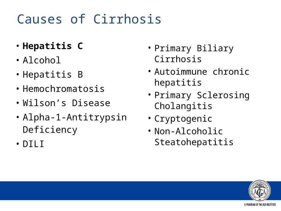
Causes of Cirrhosis
• Hepatitis C• Alcohol• Hepatitis B• Hemochromatosis• Wilson’s Disease• Alpha-1-Antitrypsin
Deficiency• DILI
• Primary Biliary Cirrhosis
• Autoimmune chronic hepatitis
• Primary Sclerosing Cholangitis
• Cryptogenic• Non-Alcoholic
Steatohepatitis
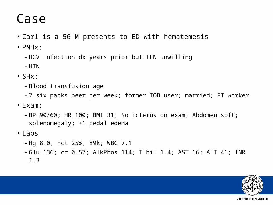
Case• Carl is a 56 M presents to ED with hematemesis• PMHx:
– HCV infection dx years prior but IFN unwilling– HTN
• SHx: – Blood transfusion age – 2 six packs beer per week; former TOB user; married; FT worker
• Exam: – BP 90/60; HR 100; BMI 31; No icterus on exam; Abdomen soft;
splenomegaly; +1 pedal edema
• Labs – Hg 8.0; Hct 25%; 89k; WBC 7.1– Glu 136; cr 0.57; AlkPhos 114; T bil 1.4; AST 66; ALT 46; INR 1.3

Portal Hypertensive Bleeding Esophgaeal Varices
Gastric VaricesPortal Hypertensive GastropathyColopathy / Rectal Varices
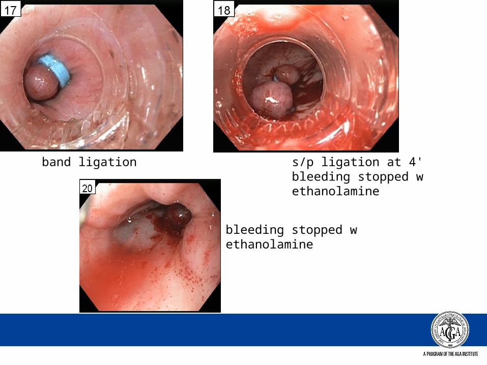
band ligation s/p ligation at 4' bleeding stopped w ethanolamine
bleeding stopped w ethanolamine
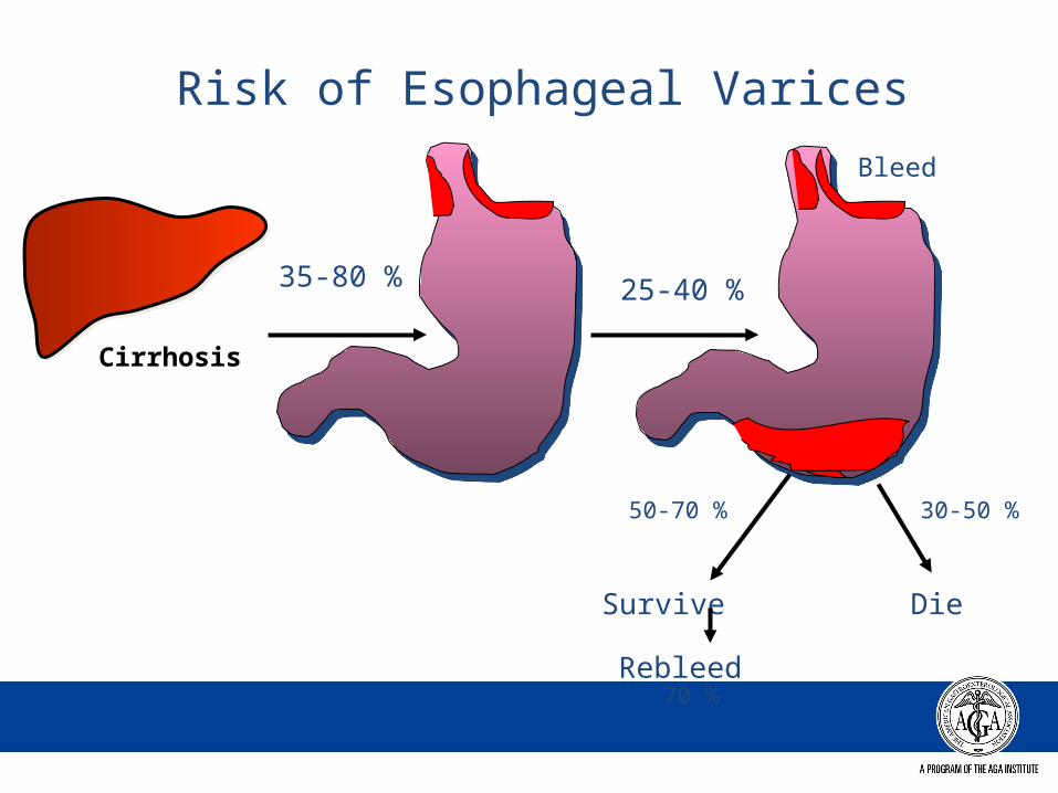
Cirrhosis
35-80 %
Risk of Esophageal Varices
25-40 %
Die
30-50 %50-70 %
Survive
Rebleed70 %
Bleed
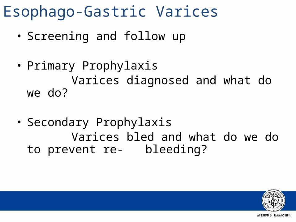
Esophago-Gastric Varices• Screening and follow up
• Primary Prophylaxis Varices diagnosed and what do we do?
• Secondary Prophylaxis Varices bled and what do we do to
prevent re- bleeding?
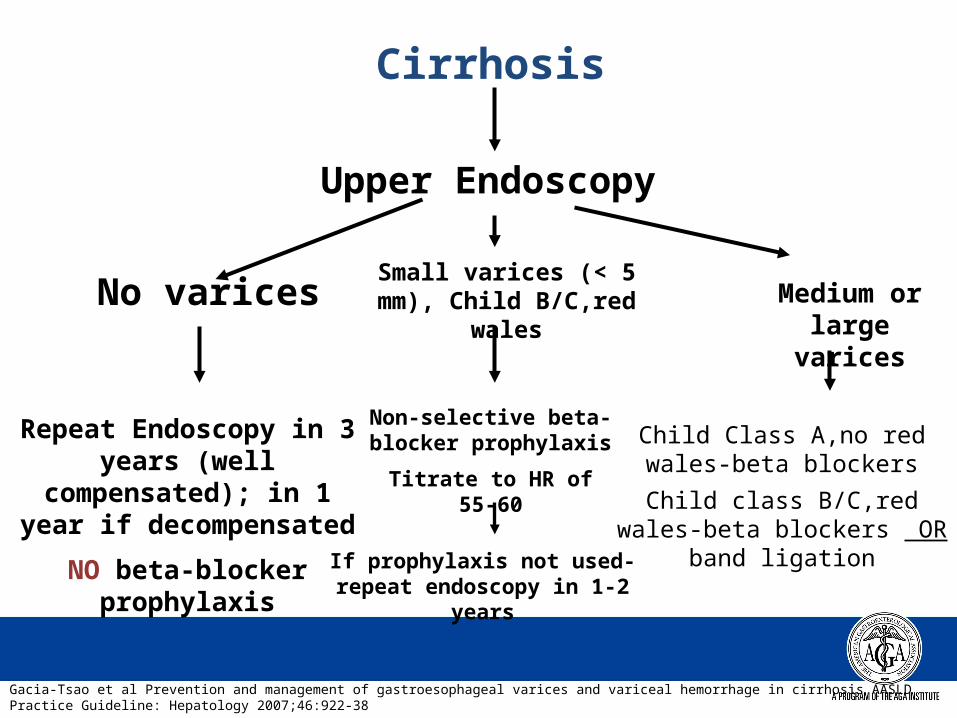
Cirrhosis
Upper Endoscopy
No varices
Repeat Endoscopy in 3 years (well compensated);
in 1 year if decompensated
NO beta-blocker prophylaxis
Small varices (< 5 mm), Child B/C,red wales
Non-selective beta-blocker prophylaxis
Titrate to HR of 55-60
Medium or large varices
Child Class A,no red wales-beta blockers
Child class B/C,red wales-beta blockers OR band ligation
Gacia-Tsao et al Prevention and management of gastroesophageal varices and variceal hemorrhage in cirrhosis AASLD Practice Guideline: Hepatology 2007;46:922-38
If prophylaxis not used-repeat endoscopy in 1-2 years
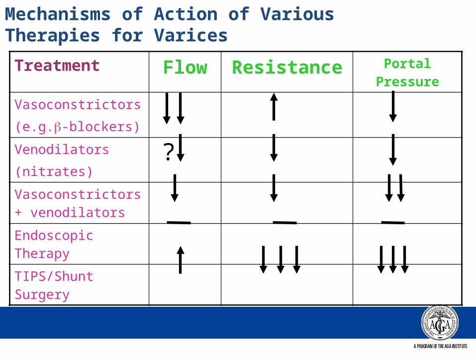
Mechanisms of Action of Various Therapies for Varices
Treatment Flow Resistance Portal Pressure
Vasoconstrictors
(e.g.-blockers)
Venodilators
(nitrates)
Vasoconstrictors + venodilators
Endoscopic Therapy
TIPS/Shunt Surgery
?
Case – continued (Carl)
• Carl undergoes serial EGD to obliterate varices; placed on beta blocker
• Remains IFN unwilling; stopped alcohol• 2 years later presents with increased abdominal
girth and dyspnea; wife is concerned about his fatigue and odd behavior this AM
• Presents to ED; no imaging on file x 1 yr• Exam –
– BP 90/60; HR 62; Weigh > 20lbs since last ED visit; + tense abd; alert and oriented
Ascites- SBP
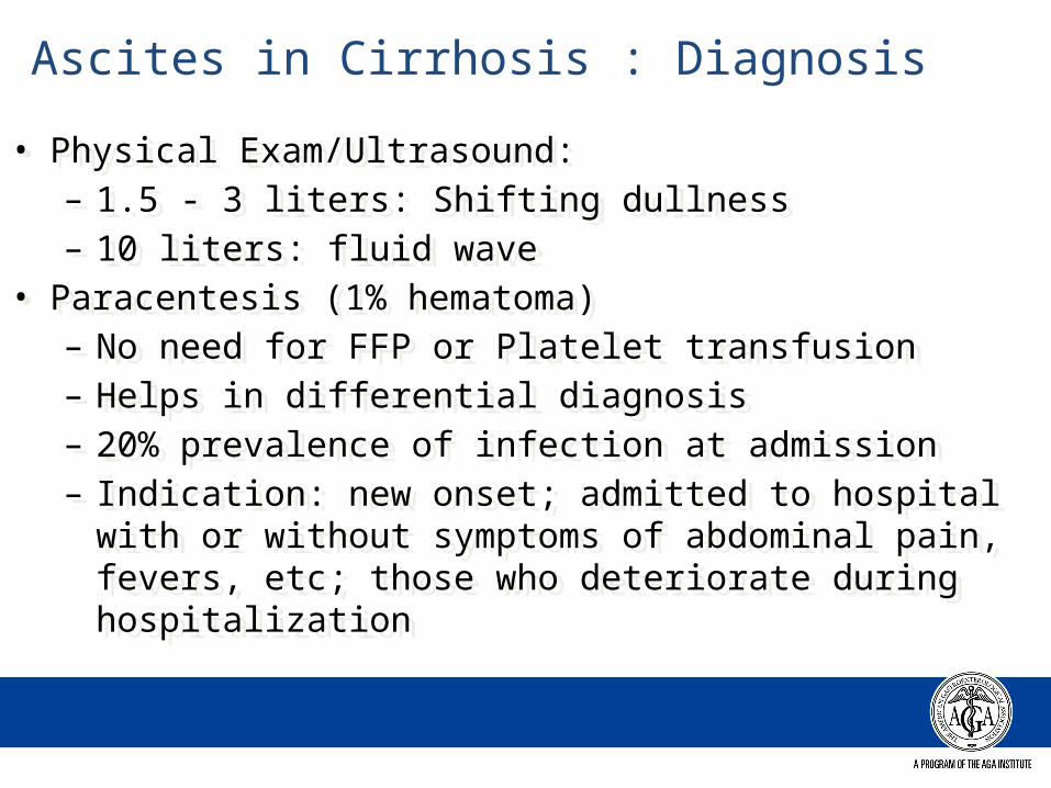
• Physical Exam/Ultrasound: – 1.5 - 3 liters: Shifting dullness– 10 liters: fluid wave
• Paracentesis (1% hematoma)– No need for FFP or Platelet transfusion– Helps in differential diagnosis– 20% prevalence of infection at admission– Indication: new onset; admitted to hospital with or without
symptoms of abdominal pain, fevers, etc; those who deteriorate during hospitalization
Ascites in Cirrhosis : Diagnosis
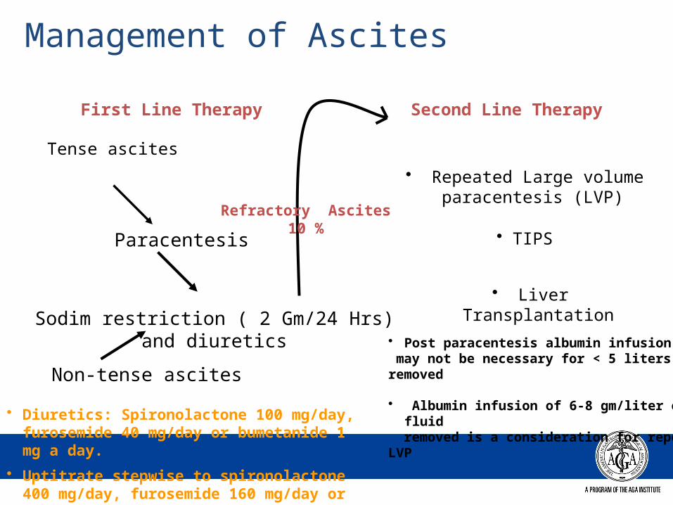
Management of Ascites
First Line Therapy Second Line Therapy
Tense ascites
Paracentesis
Sodim restriction ( 2 Gm/24 Hrs) and diuretics
Non-tense ascites
• Repeated Large volume paracentesis (LVP)
• TIPS
• Liver Transplantation
Refractory Ascites 10 %
• Diuretics: Spironolactone 100 mg/day, furosemide 40 mg/day or bumetanide 1 mg a day.
• Uptitrate stepwise to spironolactone 400 mg/day, furosemide 160 mg/day or bumetanide 4 mg/day as long as it is tolerated
• Post paracentesis albumin infusion may not be necessary for < 5 liters removed
• Albumin infusion of 6-8 gm/liter of fluid removed is a consideration for repeated LVP
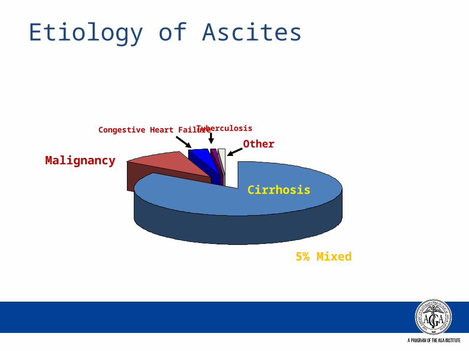
Etiology of Ascites
Cirrhosis
Malignancy
Congestive Heart Failure Tuberculosis
Other
5% Mixed
20
40
60
80
100
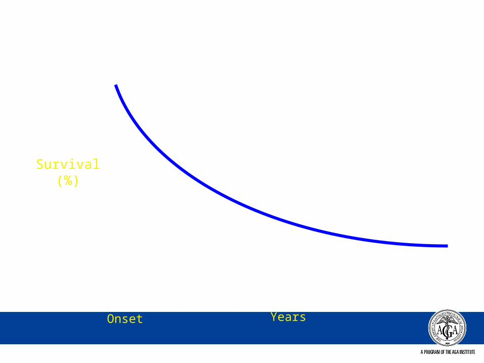
1 2 3 4 5 6YearsOnset
Survival(%)
20
40
60
80
100
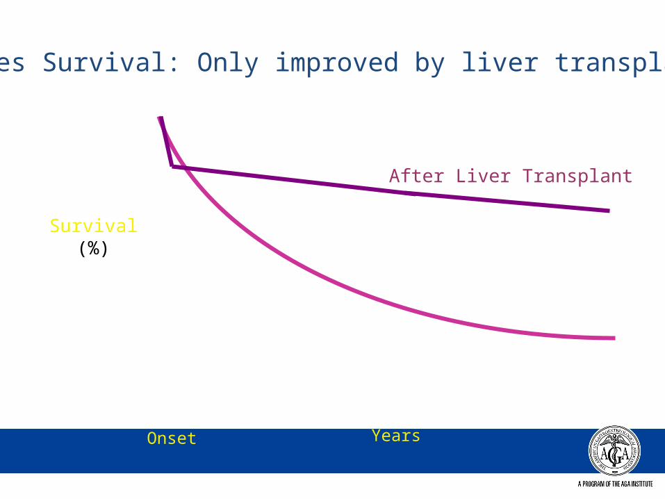
1 2 3 4 5 6YearsOnset
Survival(%)
Ascites Survival: Only improved by liver transplant
After Liver Transplant
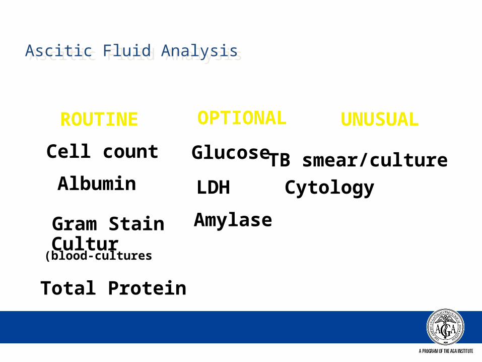
Ascitic Fluid AnalysisAscitic Fluid Analysis
ROUTINE OPTIONAL UNUSUAL
Cell count Glucose TB smear/cultureAlbumin LDH Cytology
Gram StainCulture(blood-cultures)
Amylase
Total Protein
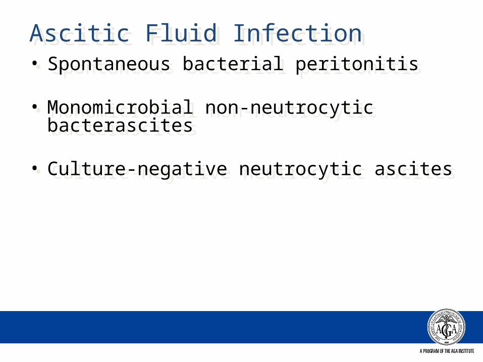
Ascitic Fluid InfectionAscitic Fluid Infection• Spontaneous bacterial peritonitis
• Monomicrobial non-neutrocytic bacterascites
• Culture-negative neutrocytic ascites
• Spontaneous bacterial peritonitis
• Monomicrobial non-neutrocytic bacterascites
• Culture-negative neutrocytic ascites
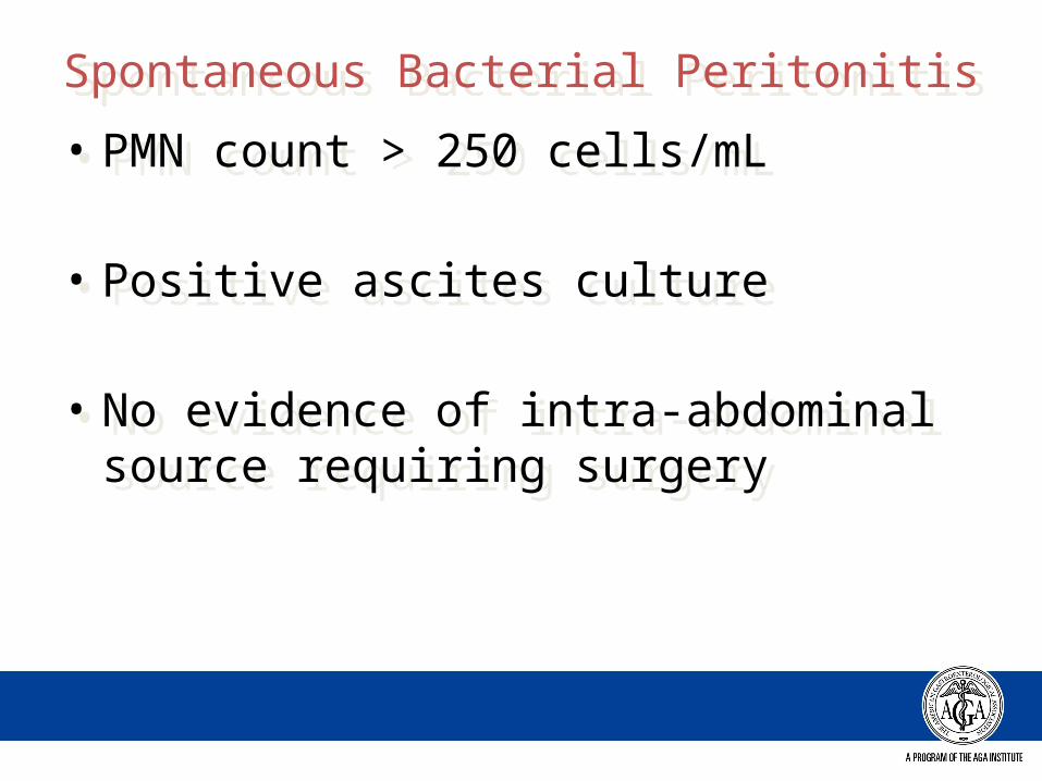
Spontaneous Bacterial PeritonitisSpontaneous Bacterial Peritonitis
• PMN count > 250 cells/mL
• Positive ascites culture
• No evidence of intra-abdominal source requiring surgery
• PMN count > 250 cells/mL
• Positive ascites culture
• No evidence of intra-abdominal source requiring surgery
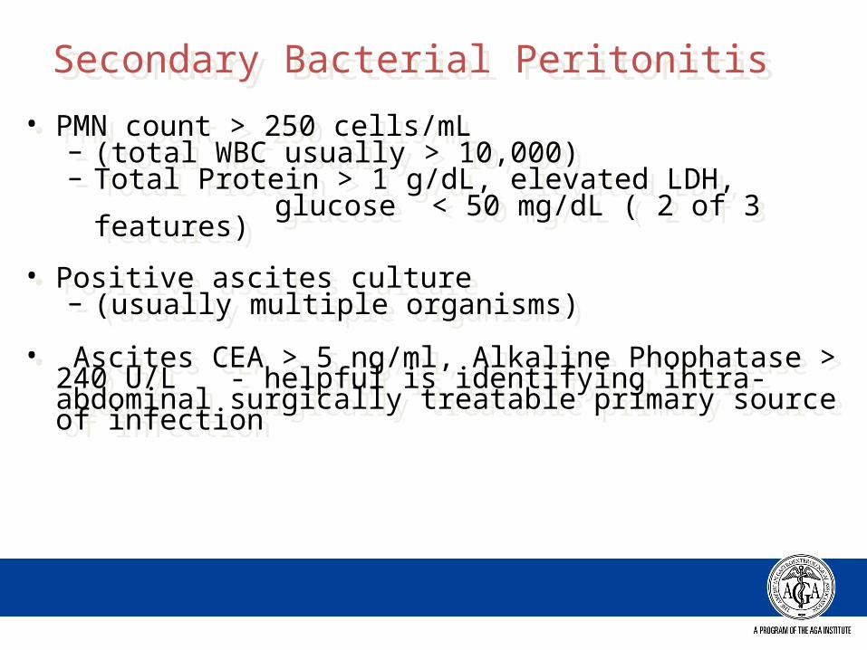
Secondary Bacterial PeritonitisSecondary Bacterial Peritonitis
• PMN count > 250 cells/mL – (total WBC usually > 10,000)– Total Protein > 1 g/dL, elevated LDH, glucose < 50 mg/dL ( 2 of 3 features)
• Positive ascites culture – (usually multiple organisms)
• Ascites CEA > 5 ng/ml, Alkaline Phophatase > 240 U/L - helpful is identifying intra-abdominal surgically treatable primary source of infection
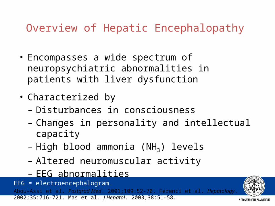
Overview of Hepatic Encephalopathy
• Encompasses a wide spectrum of neuropsychiatric abnormalities in patients with liver dysfunction
• Characterized by– Disturbances in consciousness– Changes in personality and intellectual capacity– High blood ammonia (NH3) levels
– Altered neuromuscular activity– EEG abnormalities
EEG = electroencephalogram.Abou-Assi et al. Postgrad Med. 2001;109:52-70. Ferenci et al. Hepatology. 2002;35:716-721. Mas et al. J Hepatol. 2003;38:51-58.
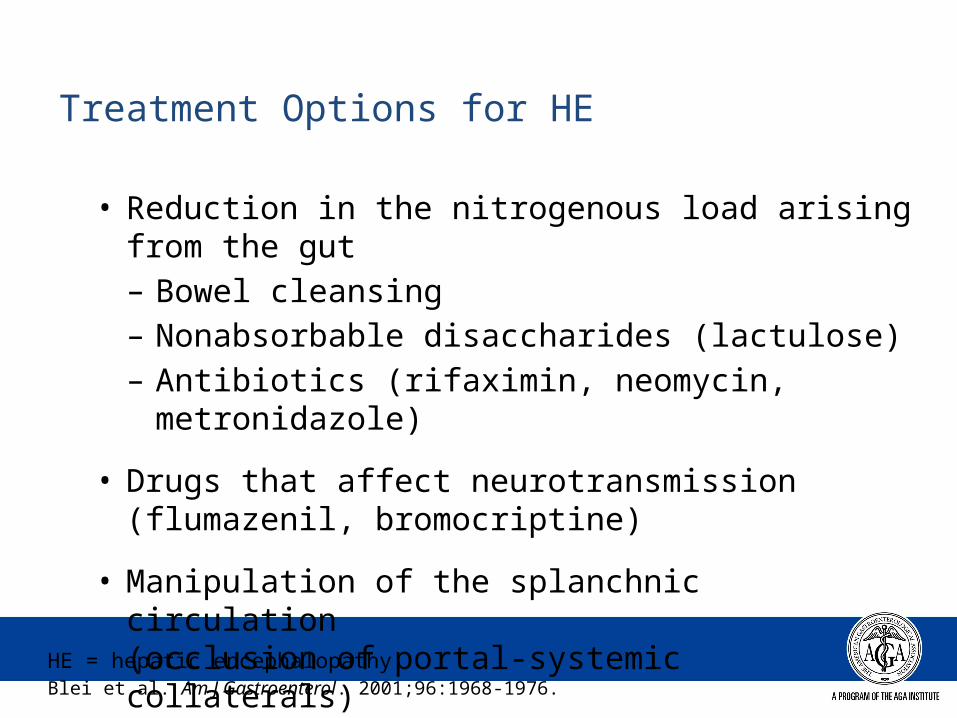
Treatment Options for HE
• Reduction in the nitrogenous load arising from the gut– Bowel cleansing– Nonabsorbable disaccharides (lactulose)– Antibiotics (rifaximin, neomycin, metronidazole)
• Drugs that affect neurotransmission (flumazenil, bromocriptine)
• Manipulation of the splanchnic circulation (occlusion of portal-systemic collaterals)
HE = hepatic encephalopathy. Blei et al. Am J Gastroenterol. 2001;96:1968-1976.

Transjugular Intrahepatic Portosystemic Shunt and Surgical Shunts
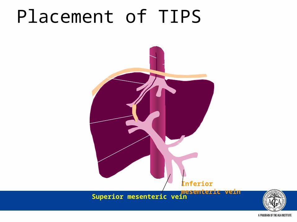
Placement of TIPS
Catheter
Hepatic veins
TIPS
Portal vein
Superior mesenteric vein
Inferior mesenteric vein
Left gastric vein
Inferior vena cava
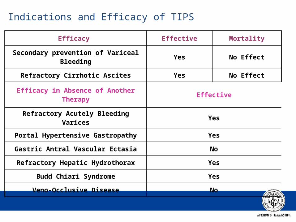
Indications and Efficacy of TIPS
Efficacy Effective Mortality
Secondary prevention of Variceal Bleeding Yes No Effect
Refractory Cirrhotic Ascites Yes No Effect
Efficacy in Absence of Another Therapy Effective
Refractory Acutely Bleeding Varices Yes
Portal Hypertensive Gastropathy Yes
Gastric Antral Vascular Ectasia No
Refractory Hepatic Hydrothorax Yes
Budd Chiari Syndrome Yes
Veno-Occlusive Disease No
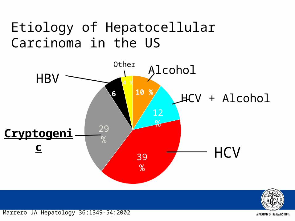
Etiology of Hepatocellular Carcinoma in the US
HCV39 %
29 %
Cryptogenic
HCV + Alcohol12 %
Alcohol
10 %
HBV6 %
4 %
Other
Marrero JA Hepatology 36;1349-54:2002
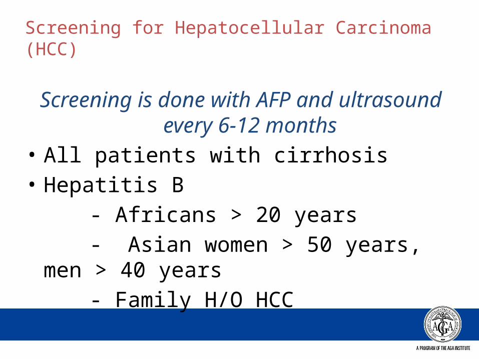
Screening for Hepatocellular Carcinoma (HCC)
Screening is done with AFP and ultrasound every 6-12 months
• All patients with cirrhosis• Hepatitis B - Africans > 20 years - Asian women > 50 years, men > 40 years - Family H/O HCC
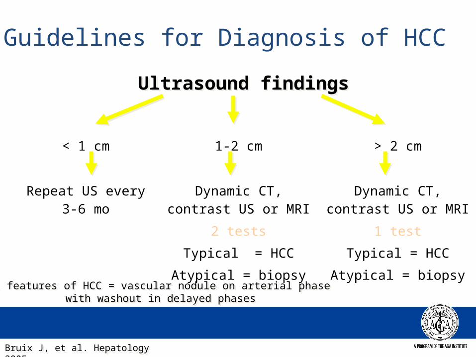
Guidelines for Diagnosis of HCC
< 1 cm 1-2 cm > 2 cm
Repeat US every 3-6 mo
Dynamic CT, contrast US or MRI
2 tests
Typical = HCC
Atypical = biopsy
Dynamic CT, contrast US or MRI
1 test
Typical = HCC
Atypical = biopsy
Typical features of HCC = vascular nodule on arterial phase with washout in delayed phases
Typical features of HCC = vascular nodule on arterial phase with washout in delayed phases
Ultrasound findingsUltrasound findings
Bruix J, et al. Hepatology 2005Bruix J, et al. Hepatology 2005
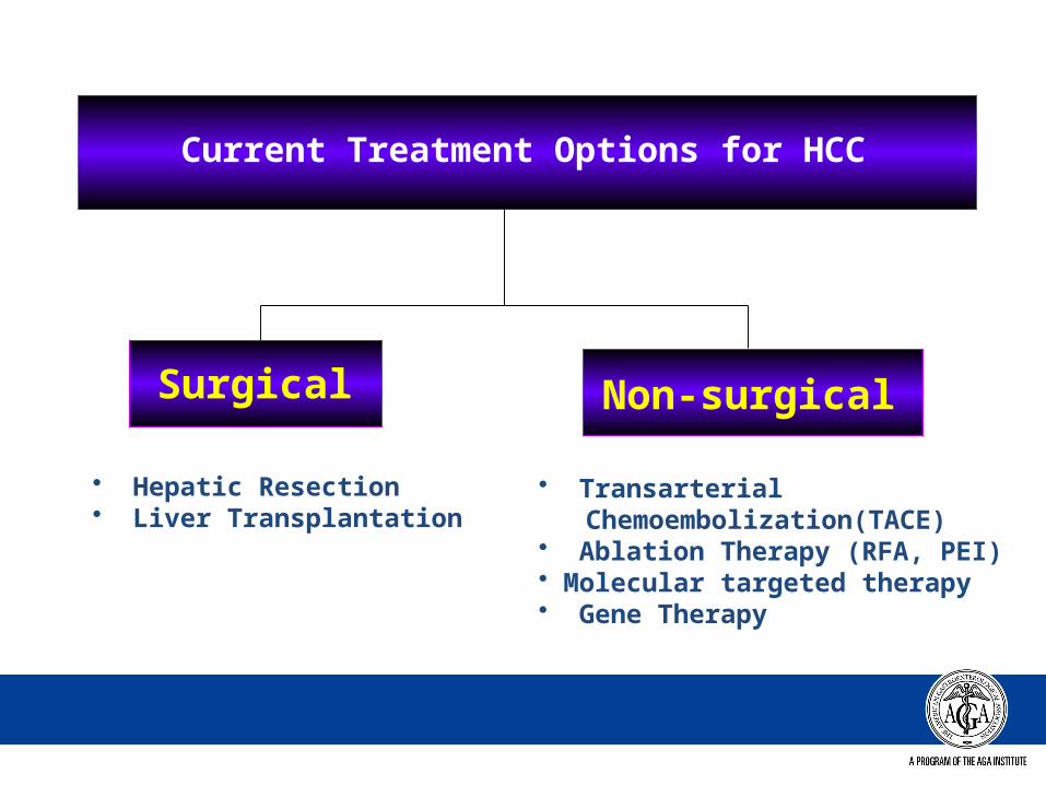
Current Treatment Options for HCC
Surgical Non-surgical
• Hepatic Resection• Liver Transplantation
• Transarterial Chemoembolization(TACE) • Ablation Therapy (RFA, PEI)• Molecular targeted therapy• Gene Therapy
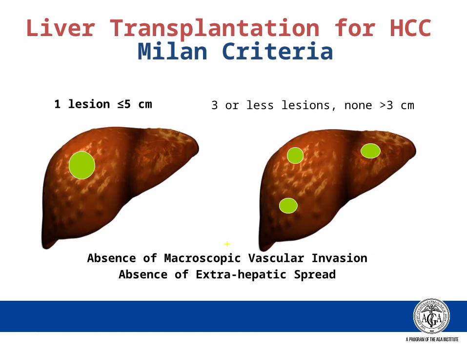
+Absence of Macroscopic Vascular Invasion
Absence of Extra-hepatic Spread
1 lesion ≤5 cm 3 or less lesions, none >3 cm
Liver Transplantation for HCC Milan Criteria
Mazzaferro V, et al. N Engl J Med 1996;334:693–699.
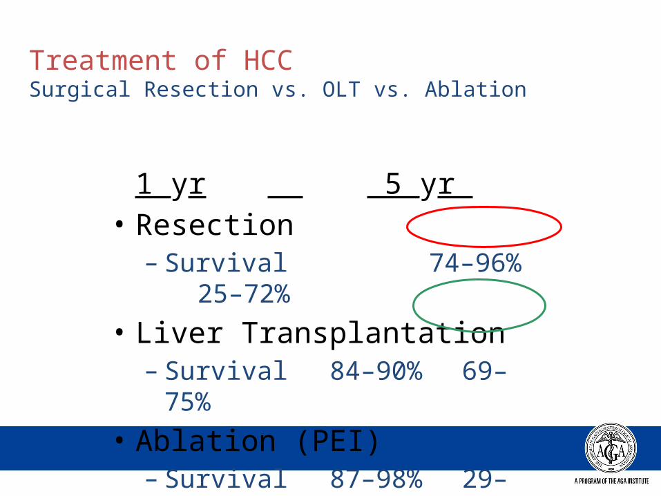
Treatment of HCC Surgical Resection vs. OLT vs. Ablation
1 yr 5 yr
• Resection– Survival 74–96% 25–
72%
• Liver Transplantation– Survival 84–90%
69–75%
• Ablation (PEI)– Survival 87–98%
29–54%
Summary: Complication of Cirrhosis
• Varices• Ascites / Refractory Ascites• Hepatic Encephalopathy• Hepatocellular Carcinoma• Synthetic Dysfunction (jaundice,
coagulopathy)