Embed Size (px)
Citation preview

CIRCULA
TORY
SYSTE
M
CO
MP
ON
EN
TS
OF B
L OO
D
ST
RU
CT
UR
E A
ND
FU
NC
TI O
N O
F TH
E H
EA
RT
,
AR
TE
RI E
S,
VE
I NS
AN
D C
AP
I LL A
RI E
S

COMPONENTS OF BLOOD
The three main parts of thee circulatory system are the heart, blood and blood vessels.
The three main functions of the blood are :
• Transportation of oxygen and nutrients to the tissues and removal of carbon dioxide and wastes.
• Protection of the body via the immune system and by clotting to prevent blood loss.
• Regulation of the body’s temperature and the fluid content of the body’s tissue.

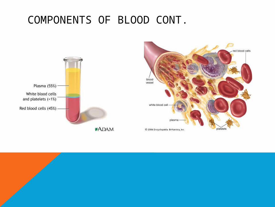
COMPONENTS OF BLOOD CONT.
Plasma
• Features: Plasma is a straw coloured liquid mainly consisting of water (about 90%).
• Function: It contains proteins, nutrients, hormones, minerals, salts and wastes which are in a dissolved state and are necessary for the nourishment and functioning of tissues.
Red Blood Cells
• Features: They are formed in the bone marrow and contain iron and haemoglobin for the transportation of oxygen and carbon dioxide around the body. They are a flat disc shaped cell that provides a large surface area for taking up oxygen. About two million red blood cells are destroyed and replaced every second, they only live for around four months.
• Function: To carry oxygen and carbon dioxide around the body.

COMPONENTS OF BLOOD CONT.
White Blood Cells:
• Features: They are formed in the bone marrow and lymph nodes. They can change shape and move against the flow of blood to the area of infection. The two most common types are called phagocytes and lymphocytes. Phagocytes engulf foreign material and harmful bacteria. Lymphocytes produce antibodies to fight disease.
• Function: To provide the body with a mobile protection system against disease.
Platelets:
• Features: tiny structures made from bone marrow cells that have no nucleus.
• Function: They help to produce clotting substances that are important in preventing blood loss when a blood vessel is damaged.
COMPONENTS OF BLOOD CONT.
READ THROUGH THE INFORMATION BELOW AND COMPLETE THE CLOZE PASSAGE ON PAGE 64 OF YOUR EXERCISE BOOK
Blood is an integral component of the cardiovascular system, the fluid that flows through the vessels, that is pumped by the heart. Blood accounts for about 8% of total body weight. Blood consists of 55% plasma. Plasma is mostly water (90%), but also includes dissolved nutrients, proteins, salts, glucose, hormones, gases and waste products. The function of plasma is to transport these substances as well as the blood cells and their contents around the body.
Blood cells make up 45% of blood volume, and 95% of these are red blood cells (RBC’s). The red blood cells main function is to carry oxygen and it does this through the presence of a protein called haemoglobin. Red blood cells are made in the marrow of bones.
White blood cells (WBC’s) or leukocytes come in five different forms, but they all have a similar function, which is to protect the body from disease. White blood cells are also made in the bone marrow.
Platelets are also formed in the bone marrow, and they are small cells that have the important function of clotting the blood. If a person cuts themself or gets a nose bleed, a protein in the plasma called fibrinogen, constructs long fibres that form a mesh across the hole. The platelets block this mesh and form a clot. It this clot is exposed to the air it will form a scab. This is the bodies’ way of protecting itself from loosing blood, but also preventing germs entering the body.

STRUCTURE AND FUNCTION OF THE HEART, ARTERIES, VEINS AND CAPILLARIES
Read through the information below and complete the cloze passage on page 64 of your exercise book
The heart is one of the major organs of the cardiorespiratory system. It is a hollow, fist sized muscle that lies just to the left of centre in the thorax.
Blood enters the right atrium through two large veins, the superior vena cava and the inferior vena cava. The blood is deoxygenated and is bluish in colour.
The right atrium contracts and pushes blood through to the right ventricle. This is turn contracts and pushes blood to the lungs through the pulmonary artery.
When the blood reaches the lungs it receives oxygen and turns bright red. This oxygenated blood then flows back to the heart and enters through the left atrium via the pulmonary veins. This chamber also contracts and pushes blood through to the left ventricle. When this chamber contracts blood is pushed out of the aorta to circulate around the body.
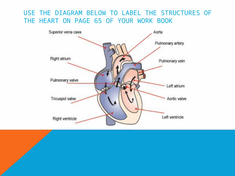
USE THE DIAGRAM BELOW TO LABEL THE STRUCTURES OF THE HEART ON PAGE 65 OF YOUR WORK BOOK

THE BLOOD VESSELSUse the information below to complete the cloze passages on pages 65 & 66 in
your work books.Arteries:
Arteries push blood away from the heart. Blood is pushed through the arteries by surges of pressure caused when the heart beats. The pressure lessens as the blood travels further from the heart. The speed of blood flow decreases as pressure decreases.
Veins
Veins have thinner walls than arteries. Because the pressure of the heartbeat is too low in the vein to push the blood back to the heart it is assisted in the following ways:
• muscles surrounding the veins expand and contract
• valves prevent the blood from moving back to where it came from
• the pumping action of the heart creates a sucking action in the veins close to the heart
• through hydrostatic pressure (there is an attraction between molecules of fluids moving in a particular direction)
Capillaries
Capillaries are:
• fed by arterioles (small arteries)
• microscopically thin
• semi-permeable
• found in clusters called capillary networks

PULMONARY AND SYSTEMIC CIRCULATIONPulmonary circulation is the flow of blood from the heart to the lungs
and back to the heart. Both sides of the heart work together like two pumps with overlapping circuits. The right side receives venous blood that is low in oxygen content (de-oxygenated) from all parts of the body and pumps it to the lungs.
The left side of the heart receives blood high in oxygen content (oxygenated) from the lungs and pumps it around the body. This circuit to and from the body is called systemic circulation. Therefore systemic circulation is the flow of blood from the heart to body tissue and back to the heart.
Please view the following slide to see a diagram of this in action.

PULMONARY AND SYSTEMIC CIRCULATION CONT.
View the following video clips:The Heart: http://www.youtube.com/watch?v=OqCzkg5Wz3kPulmonary and Systemic Circulation:http://www.youtube.com/watch?v=466zDaHIozUArteries: http://www.youtube.com/watch?v=GBf59Z8tgA0Blood Vessels: http://www.youtube.com/watch?v=CjNKbL_-cwA

REVISION QUESTION ANSWERS1. Functions:
• a support network for the attachment of muscles
• protection of internal organs
• allows movement to occur when muscles contract
• a storage house for minerals such as calcium
• production of blood cells from within the bone marrow
2. Fibrous bands of connective tissue that connect bone to bone e.g. femur and tibia. Their role is to help maintain joint stability by limiting excessive movement.
3. Synovial (freely moveable), cartilaginous (slightly moveable) and fibrous (immovable).
4. Produce movement, provide stabilisation and generate heat for the muscle to function correctly.
5. Slow twitch (red fibres) – contract slowly, produce less force, fatigue slowly and are suited to aerobic (distance) events or activities.
Fast twitch (white fibres) – contract quickly, produce a great deal of force, fatigue quickly and are suited to anaerobic (speed/power) events or activities.
6. These muscles work at a joint to help stabilise it. This allows other muscles to work more effectively.
7. A muscle develops tension and there is a change in the length of the muscle causing movement e.g. bicep curl.
8. Synovial fluid acts as a lubricant in a joint. It helps to keep moving surfaces apart and provides a cushioning or shock absorbing effect.