Embed Size (px)
Citation preview
838
In December 2011, a 39-year-old man resuscitated from out-of-hospital cardiac arrest caused by ventricular fibrillation
received the diagnosis of Brugada syndrome on the basis of a spontaneous Brugada type 1 ECG pattern (Figure 1A). Before implantation of a cardioverter-defibrillator, the patient, after providing written informed consent, underwent a 3-dimen-sional electroanatomic mapping of the right ventricle (RV) as part of a clinical research study approved by ethics committee of our institution. A localized low-voltage area with delayed and fragmented potentials was evident in both bipolar and unipolar voltage maps in the anterior RV outflow tract. The patient was discharged with no antiarrhythmic medication and was free of arrhythmic events at implantable cardioverter-defi-brillator interrogation for 1 year.
In January 2013, he was admitted at our institution again for arrhythmic storm with multiple consecutive appropriate implantable cardioverter-defibrillator shocks resulting from recurrent ventricular fibrillation.
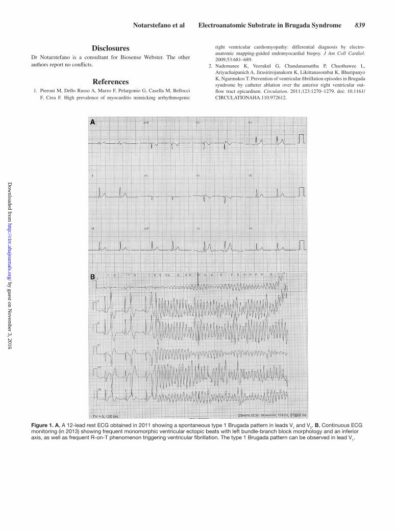
Continuous ECG monitoring documented frequent mono-morphic ventricular extrasystoles with left bundle-branch block morphology with an inferior axis and frequent R-on-T phenomenon triggering multiple episodes of ventricular fibril-lation (Figure 1B). He was treated with isoproterenol infu-sion, leading to electric storm suppression and cardiac rhythm normalization.
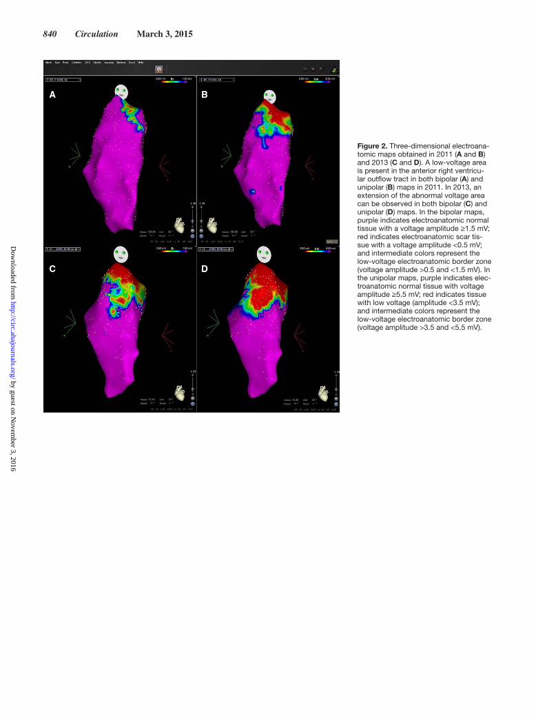
As indicated in the research study in the case of arrhyth-mic events, 3-dimensional RV electroanatomic mapping was repeated and documented an increase (from 1 to 14.2 cm2 and from 10.2 to 19.8 cm2 in the bipolar and unipolar maps, respectively) of the RV outflow tract low-voltage area previ-ously documented (Figure 2A–2D). To investigate the path-ological substrate of the low-voltage area and the possible mechanisms of disease progression and arrhythmias recur-rence, we also performed a CARTO-guided RV endomyo-cardial biopsy, drawing 3 myocardial samples from an area with both bipolar and unipolar low voltages.1 In addition, dur-ing the same procedure, we performed endocardial radiofre-quency ablation to abolish low-voltage fractionated potentials
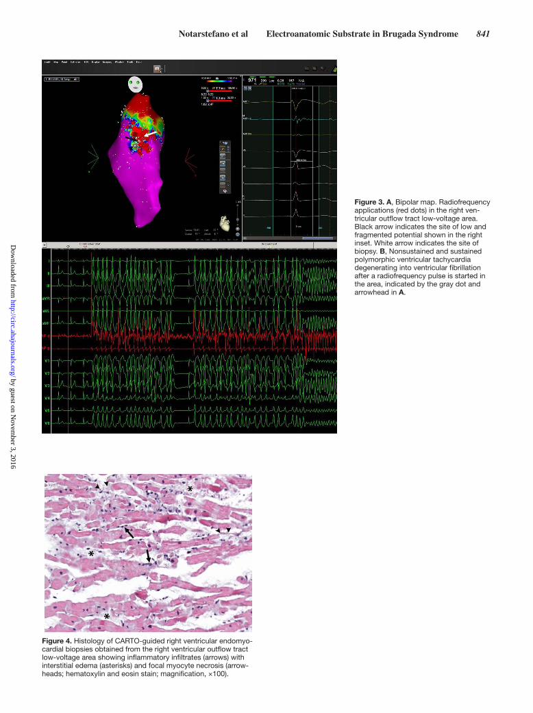
in the anterior RV outflow tract.2 Interestingly, radiofrequency applications in the inferior portion of the low-voltage area repeatedly evoked sustained polymorphic ventricular tachy-cardias self-terminating at radiofrequency pulse cessation, but in 3 cases degenerating into ventricular fibrillation and requir-ing external defibrillation (Figure 3). Histological analysis of endomyocardial biopsies showed the presence of myocardial inflammation with focal necrosis of adjacent myocytes and no evidence of fibrofatty substitution (Figure 4). The patient was discharged on therapy with quinidine 150 mg 3 times daily. After a follow-up of 18 months, he is asymptomatic and free of arrhythmic events at implantable cardioverter-defibrillator interrogation. Genetic analysis failed to identify mutations in the main genes so far associated with the syndrome.
To the best of our knowledge, this is the first description of electroanatomic substrate progression associated with the recurrence of ventricular fibrillation in a patient with Brugada syndrome. Our case suggests that electroanatomic and struc-tural abnormalities underlie the ECG and arrhythmic features of Brugada syndrome and that the progression of these abnor-malities can be associated with arrhythmia recurrence. Further studies are needed to define the prevalence of electroanatomic abnormalities in Brugada syndrome and to clarify the role of myocardial inflammation in electroanatomic substrate pro-gression and arrhythmogenesis. Similarly, the potential role of electroanatomic mapping in monitoring disease progression and in prognostic stratification in Brugada syndrome warrants further investigation.
AcknowledgmentsWe thank engineer Marina Mercurio (Biosense Webster, Italy) for the invaluable technical support.
Sources of Funding This study was supported by the Telethon grant GGP10186, “Identification of Genetic, Electroanatomical and Structural Predictors of Malignant Ventricular Arrhythmias in Patients With Brugada syndrome,” to Dr Pieroni.
(Circulation. 2015;131:838-841. 10.1161/CIRCULATIONAHA.114.013773.)© 2015 American Heart Association, Inc.
Circulation is available at http://circ.ahajournals.org DOI: 10.1161/CIRCULATIONAHA.114.013773
From Cardiovascular and Neurological Department, San Donato Hospital, Arezzo, Italy (P.N., M.P., R.G., T.R., S.G., A.F., L.B.); and Institute of Legal Medicine, Catholic University, Rome, Italy (A.O.).
Correspondence to Maurizio Pieroni, MD, PhD, Cardiovascular Department, San Donato Hospital, Via Pietro Nenni 20, 52100 Arezzo, Italy. E-mail [email protected]
Progression of Electroanatomic Substrate and Electric Storm Recurrence in a Patient With Brugada Syndrome
Pasquale Notarstefano, MD; Maurizio Pieroni, MD, PhD; Raffaele Guida, MD; Teresa Rio, MD; Antonio Oliva, MD; Simone Grotti, MD; Aureliano Fraticelli, MD;
Leonardo Bolognese, MD, FESC
Images in Cardiovascular Medicine
by guest on Novem
ber 3, 2016http://circ.ahajournals.org/
Dow
nloaded from
Notarstefano et al Electroanatomic Substrate in Brugada Syndrome 839
DisclosuresDr Notarstefano is a consultant for Biosense Webster. The other authors report no conflicts.
References 1. Pieroni M, Dello Russo A, Marzo F, Pelargonio G, Casella M, Bellocci
F, Crea F. High prevalence of myocarditis mimicking arrhythmogenic
right ventricular cardiomyopathy: differential diagnosis by electro-anatomic mapping-guided endomyocardial biopsy. J Am Coll Cardiol. 2009;53:681–689.
2. Nademanee K, Veerakul G, Chandanamattha P, Chaothawee L, Ariyachaipanich A, Jirasirirojanakorn K, Likittanasombat K, Bhuripanyo K, Ngarmukos T. Prevention of ventricular fibrillation episodes in Brugada syndrome by catheter ablation over the anterior right ventricular out-flow tract epicardium. Circulation. 2011;123:1270–1279. doi: 10.1161/CIRCULATIONAHA.110.972612.
Figure 1. A, A 12-lead rest ECG obtained in 2011 showing a spontaneous type 1 Brugada pattern in leads V1 and V2. B, Continuous ECG monitoring (in 2013) showing frequent monomorphic ventricular ectopic beats with left bundle-branch block morphology and an inferior axis, as well as frequent R-on-T phenomenon triggering ventricular fibrillation. The type 1 Brugada pattern can be observed in lead V1.
by guest on Novem
ber 3, 2016http://circ.ahajournals.org/
Dow
nloaded from
840 Circulation March 3, 2015
Figure 2. Three-dimensional electroana-tomic maps obtained in 2011 (A and B) and 2013 (C and D). A low-voltage area is present in the anterior right ventricu-lar outflow tract in both bipolar (A) and unipolar (B) maps in 2011. In 2013, an extension of the abnormal voltage area can be observed in both bipolar (C) and unipolar (D) maps. In the bipolar maps, purple indicates electroanatomic normal tissue with a voltage amplitude ≥1.5 mV; red indicates electroanatomic scar tis-sue with a voltage amplitude <0.5 mV; and intermediate colors represent the low-voltage electroanatomic border zone (voltage amplitude >0.5 and <1.5 mV). In the unipolar maps, purple indicates elec-troanatomic normal tissue with voltage amplitude ≥5.5 mV; red indicates tissue with low voltage (amplitude <3.5 mV); and intermediate colors represent the low-voltage electroanatomic border zone (voltage amplitude >3.5 and <5.5 mV).
by guest on Novem
ber 3, 2016http://circ.ahajournals.org/
Dow
nloaded from
Notarstefano et al Electroanatomic Substrate in Brugada Syndrome 841
Figure 4. Histology of CARTO-guided right ventricular endomyo-cardial biopsies obtained from the right ventricular outflow tract low-voltage area showing inflammatory infiltrates (arrows) with interstitial edema (asterisks) and focal myocyte necrosis (arrow-heads; hematoxylin and eosin stain; magnification, ×100).
Figure 3. A, Bipolar map. Radiofrequency applications (red dots) in the right ven-tricular outflow tract low-voltage area. Black arrow indicates the site of low and fragmented potential shown in the right inset. White arrow indicates the site of biopsy. B, Nonsustained and sustained polymorphic ventricular tachycardia degenerating into ventricular fibrillation after a radiofrequency pulse is started in the area, indicated by the gray dot and arrowhead in A.
by guest on Novem
ber 3, 2016http://circ.ahajournals.org/
Dow
nloaded from
Grotti, Aureliano Fraticelli and Leonardo BolognesePasquale Notarstefano, Maurizio Pieroni, Raffaele Guida, Teresa Rio, Antonio Oliva, Simone
With Brugada SyndromeProgression of Electroanatomic Substrate and Electric Storm Recurrence in a Patient
Print ISSN: 0009-7322. Online ISSN: 1524-4539 Copyright © 2015 American Heart Association, Inc. All rights reserved.
is published by the American Heart Association, 7272 Greenville Avenue, Dallas, TX 75231Circulation doi: 10.1161/CIRCULATIONAHA.114.013773
2015;131:838-841Circulation.
http://circ.ahajournals.org/content/131/9/838World Wide Web at:
The online version of this article, along with updated information and services, is located on the
http://circ.ahajournals.org//subscriptions/
is online at: Circulation Information about subscribing to Subscriptions:
http://www.lww.com/reprints Information about reprints can be found online at: Reprints:
document. Permissions and Rights Question and Answer this process is available in the
click Request Permissions in the middle column of the Web page under Services. Further information aboutOffice. Once the online version of the published article for which permission is being requested is located,
can be obtained via RightsLink, a service of the Copyright Clearance Center, not the EditorialCirculationin Requests for permissions to reproduce figures, tables, or portions of articles originally publishedPermissions:
by guest on Novem
ber 3, 2016http://circ.ahajournals.org/
Dow
nloaded from