Embed Size (px)
Citation preview
Circuit reconstruction tools todayStephen J Smith
To understand how a brain processes information, we must
understand the structure of its neural circuits—especially
circuit interconnection topologies and the cell and synapse
molecular architectures that determine circuit-signaling
dynamics. Our information on these key aspects of neural
circuit structure has remained incomplete and fragmentary,
however, because of limitations of the best available imaging
methods. Now, new transgenic tool mice and new image
acquisition tools appear poised to permit very significant
advances in our abilities to reconstruct circuit connection
topologies and molecular architectures.
Addresses
Department of Molecular and Cellular Physiology, Stanford University
School of Medicine, Stanford, CA 94305, United States
Corresponding author: Smith, Stephen J ([email protected])
Current Opinion in Neurobiology 2007, 17:601–608
This review comes from a themed issue on
New technologies
Edited by Mark Schnitzer and Atsushi Miyawaki
0959-4388/$ – see front matter
# 2007 Elsevier Ltd. All rights reserved.
DOI 10.1016/j.conb.2007.11.004
IntroductionThe modern understanding of brain function grew from
Ramon y Cajal’s beautiful and prescient india-ink recon-
structions of neural circuit architectures (e.g. [1��],Figure 1). These drawing were based on observations
using used two then-new imaging tools: Abbe’s apochro-
matic objective and Golgi’s silver impregnation stain.
Ramon y Cajal’s drawings and insights were possible
because the Golgi method could be titrated to stain a
small fraction of cells intensely and completely while
leaving the majority of adjacent cells unstained, allowing
complete forms of the rare stained neurons to be visual-
ized clearly by a well-corrected objective. Nearly, every
circuit reconstruction effort since has likewise relied upon
sparse staining methods to overcome the difficulties of
resolving the individual elements of very densely packed
neural circuit elements. Thus, the best reconstructions of
circuit connectivity available today still extrapolate from
isolated observations of individual neurons and still pro-
vide only fragmentary and qualitative information about
neural circuit architectures. Moreover, as our understand-
ing of the vast molecular diversity of neurons and
synapses has grown [2–5], it has become increasingly
www.sciencedirect.com
clear that reconstruction of neural circuits will require
molecular information about cells and synapses much
more detailed than any presently available.
Today, rapid advances in molecular, physical and com-
putational imaging tools are beginning to extend our sight
far beyond what was possible with Ramon y Cajal’s
apochromatic objective and Golgi stains and promising
to extend our abilities to reconstruct far beyond those
allowed by india-ink drawing. This commentary will
provide an overview of some of these new imaging tools,
focusing on (1) new genetic methods for neuroanatomical
staining, (2) new physical methods for the high-resolution
imaging of molecular architecture, (3) new strategies for
high-throughput volume electron microscopy, and (4)
new computational tools for the analysis of volume EM
data. For brevity, this review will focus on a single target:
the reconstruction of mammalian cerebral cortex. A sum-
mary section will consider the feasibility of a hypothetical
project at the edge of today’s envelope for reconstruction
technology.
Of tool mice and menThe mouse cerebral cortex stands out today as a uniquely
advantageous system for the study of cortical structure
and function. The mouse offers a unique abundance of
genetic information, transgenically labeled ‘tool mouse’
lines, and genetic models for human disease. Meanwhile,
the superficial location, relatively unfolded anatomy and
small dimensions of the mouse cortex adapt it particularly
well to physiological study by modern in vivo optical
methods. These advantages are all the more valuable
because of the strong similarities between mouse and
human cerebral cortex.
A rapidly growing cornucopia of XFP tool mice is begin-
ning to have an enormous impact on neuroscience. These
transgenic mouse lines express genetically encoded fluor-
escent protein (XFP) markers in distinct subsets of
neurons defined by intrinsic genetic control elements
(e.g. [6��,7–8]). In many cases, these subsets appear to
correspond to classical morphologically and physiologi-
cally defined cell types. Sparseness of labeled subsets
allows for Golgi-like optical resolution of individual
neurons in many of these lines, but these genetic XFP
labels offer enormous advantages over Golgi stains by
allowing tagged cells to be imaged in live as well as fixed
tissues and in being more predictable, repeatable and
informative in their cell specificity. These advantages are
being multiplied by cross breeding mouse lines carrying
spectrally distinct XFP tags, to produce brain specimens
exhibiting spectrally multiplexed labeling of distinct
Current Opinion in Neurobiology 2007, 17:601–608
602 New technologies
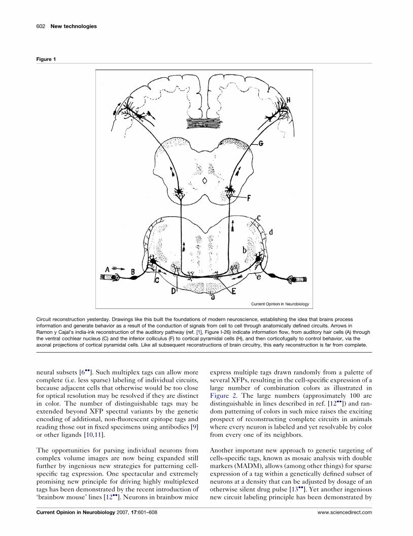
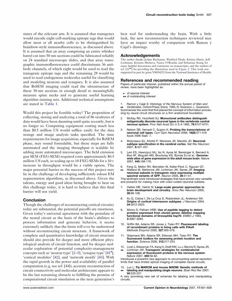
Figure 1
Circuit reconstruction yesterday. Drawings like this built the foundations of modern neuroscience, establishing the idea that brains process
information and generate behavior as a result of the conduction of signals from cell to cell through anatomically defined circuits. Arrows in
Ramon y Cajal’s india-ink reconstruction of the auditory pathway (ref. [1], Figure I-26) indicate information flow, from auditory hair cells (A) through
the ventral cochlear nucleus (C) and the inferior colliculus (F) to cortical pyramidal cells (H), and then corticofugally to control behavior, via the
axonal projections of cortical pyramidal cells. Like all subsequent reconstructions of brain circuitry, this early reconstruction is far from complete.
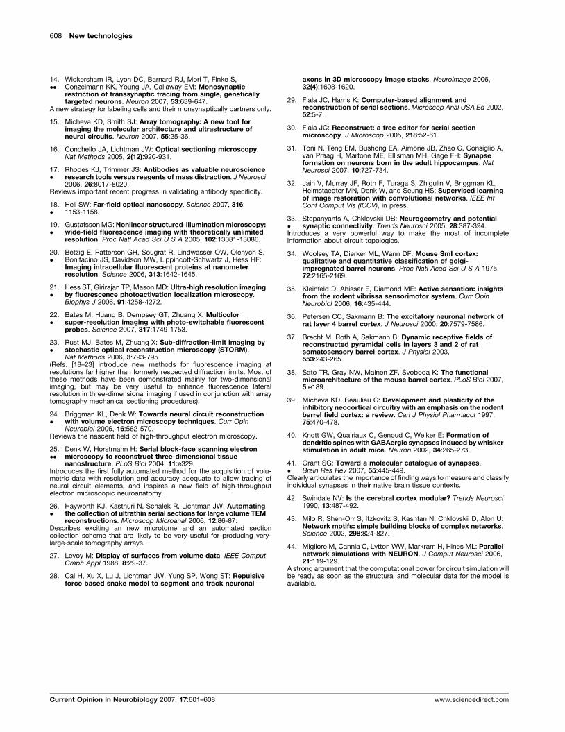
neural subsets [6��]. Such multiplex tags can allow more
complete (i.e. less sparse) labeling of individual circuits,
because adjacent cells that otherwise would be too close
for optical resolution may be resolved if they are distinct
in color. The number of distinguishable tags may be
extended beyond XFP spectral variants by the genetic
encoding of additional, non-fluorescent epitope tags and
reading those out in fixed specimens using antibodies [9]
or other ligands [10,11].
The opportunities for parsing individual neurons from
complex volume images are now being expanded still
further by ingenious new strategies for patterning cell-
specific tag expression. One spectacular and extremely
promising new principle for driving highly multiplexed
tags has been demonstrated by the recent introduction of
‘brainbow mouse’ lines [12��]. Neurons in brainbow mice
Current Opinion in Neurobiology 2007, 17:601–608
express multiple tags drawn randomly from a palette of
several XFPs, resulting in the cell-specific expression of a
large number of combination colors as illustrated in
Figure 2. The large numbers (approximately 100 are
distinguishable in lines described in ref. [12��]) and ran-
dom patterning of colors in such mice raises the exciting
prospect of reconstructing complete circuits in animals
where every neuron is labeled and yet resolvable by color
from every one of its neighbors.
Another important new approach to genetic targeting of
cells-specific tags, known as mosaic analysis with double
markers (MADM), allows (among other things) for sparse
expression of a tag within a genetically defined subset of
neurons at a density that can be adjusted by dosage of an
otherwise silent drug pulse [13��]. Yet another ingenious
new circuit labeling principle has been demonstrated by
www.sciencedirect.com
Circuit reconstruction tools today Smith 603
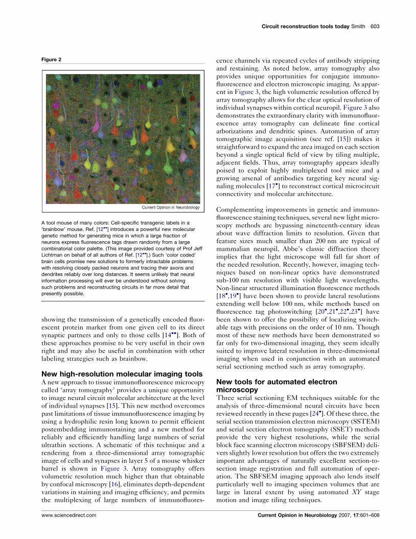
Figure 2
A tool mouse of many colors: Cell-specific transgenic labels in a
‘brainbow’ mouse. Ref. [12��] introduces a powerful new molecular
genetic method for generating mice in which a large fraction of
neurons express fluorescence tags drawn randomly from a large
combinatorial color palette. (This image provided courtesy of Prof Jeff
Lichtman on behalf of all authors of Ref. [12��].) Such ‘color coded’
brain cells promise new solutions to formerly intractable problems
with resolving closely packed neurons and tracing their axons and
dendrites reliably over long distances. It seems unlikely that neural
information processing will ever be understood without solving
such problems and reconstructing circuits in far more detail that
presently possible.
showing the transmission of a genetically encoded fluor-
escent protein marker from one given cell to its direct
synaptic partners and only to those cells [14��]. Both of
these approaches promise to be very useful in their own
right and may also be useful in combination with other
labeling strategies such as brainbow.
New high-resolution molecular imaging toolsA new approach to tissue immunofluorescence microcopy
called ‘array tomography’ provides a unique opportunity
to image neural circuit molecular architecture at the level
of individual synapses [15]. This new method overcomes
past limitations of tissue immunofluorescence imaging by
using a hydrophilic resin long known to permit efficient
postembedding immunostaining and a new method for
reliably and efficiently handling large numbers of serial
ultrathin sections. A schematic of this technique and a
rendering from a three-dimensional array tomographic
image of cells and synapses in layer 5 of a mouse whisker
barrel is shown in Figure 3. Array tomography offers
volumetric resolution much higher than that obtainable
by confocal microscopy [16], eliminates depth-dependent
variations in staining and imaging efficiency, and permits
the multiplexing of large numbers of immunofluores-
www.sciencedirect.com
cence channels via repeated cycles of antibody stripping
and restaining. As noted below, array tomography also
provides unique opportunities for conjugate immuno-
fluorescence and electron microscopic imaging. As appar-
ent in Figure 3, the high volumetric resolution offered by
array tomography allows for the clear optical resolution of
individual synapses within cortical neuropil. Figure 3 also
demonstrates the extraordinary clarity with immunofluor-
escence array tomography can delineate fine cortical
arborizations and dendritic spines. Automation of array
tomographic image acquisition (see ref. [15]) makes it
straightforward to expand the area imaged on each section
beyond a single optical field of view by tiling multiple,
adjacent fields. Thus, array tomography appears ideally
poised to exploit highly multiplexed tool mice and a
growing arsenal of antibodies targeting key neural sig-
naling molecules [17�] to reconstruct cortical microcircuit
connectivity and molecular architecture.
Complementing improvements in genetic and immuno-
fluorescence staining techniques, several new light micro-
scopy methods are bypassing nineteenth-century ideas
about wave diffraction limits to resolution. Given that
feature sizes much smaller than 200 nm are typical of
mammalian neuropil, Abbe’s classic diffraction theory
implies that the light microscope will fall far short of
the needed resolution. Recently, however, imaging tech-
niques based on non-linear optics have demonstrated
sub-100 nm resolution with visible light wavelengths.
Non-linear structured illumination fluorescence methods
[18�,19�] have been shown to provide lateral resolutions
extending well below 100 nm, while methods based on
fluorescence tag photoswitching [20�,21�,22�,23�] have
been shown to offer the possibility of localizing switch-
able tags with precisions on the order of 10 nm. Though
most of these new methods have been demonstrated so
far only for two-dimensional imaging, they seem ideally
suited to improve lateral resolution in three-dimensional
imaging when used in conjunction with an automated
serial sectioning method such as array tomography.
New tools for automated electronmicroscopyThree serial sectioning EM techniques suitable for the
analysis of three-dimensional neural circuits have been
reviewed recently in these pages [24�]. Of these three, the
serial section transmission electron microscopy (SSTEM)
and serial section electron tomography (SSET) methods
provide the very highest resolutions, while the serial
block face scanning electron microscopy (SBFSEM) deli-
vers slightly lower resolution but offers the two extremely
important advantages of naturally excellent section-to-
section image registration and full automation of oper-
ation. The SBFSEM imaging approach also lends itself
particularly well to imaging specimen volumes that are
large in lateral extent by using automated XY stage
motion and image tiling techniques.
Current Opinion in Neurobiology 2007, 17:601–608
604 New technologies
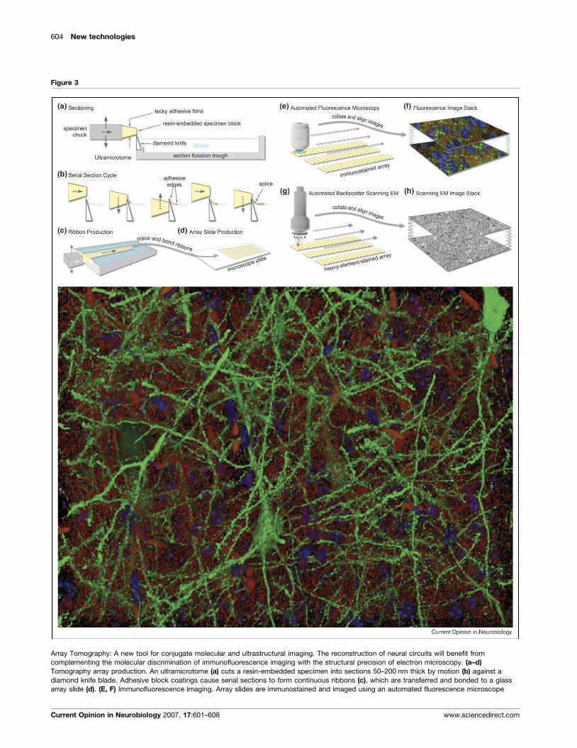
Figure 3
Array Tomography: A new tool for conjugate molecular and ultrastructural imaging. The reconstruction of neural circuits will benefit from
complementing the molecular discrimination of immunofluorescence imaging with the structural precision of electron microscopy. (a–d)
Tomography array production. An ultramicrotome (a) cuts a resin-embedded specimen into sections 50–200 nm thick by motion (b) against a
diamond knife blade. Adhesive block coatings cause serial sections to form continuous ribbons (c), which are transferred and bonded to a glass
array slide (d). (E, F) Immunofluorescence imaging. Array slides are immunostained and imaged using an automated fluorescence microscope
Current Opinion in Neurobiology 2007, 17:601–608 www.sciencedirect.com
Circuit reconstruction tools today Smith 605
Additional new serial section EM methods introduced
this year make use of the backscattered-electron scanning
electron microscopy (BsSEM) imaging modality that was
introduced to neural circuit analysis by Denk and Horts-
mann [25��]. One is based on the use of a focused ion
beam scanning electron microscope (FIBSEM). As in
SBFSEM, BsSEM is used to image the specimen block
face rather than a thin section, but successive ultrathin
sections are removed in the FIBSEM by an ablative
focused ion beam instead of the diamond knife used in
the SBFSEM. This method has produced extremely
high-quality results from small circuit volumes (G. Knott,
D. Wall, H. Marchman, B. Lich, Soc Neurosci Abstr 2007,
534.5), but it remains to be seen how it may be scaled to
larger circuit volumes. The other new method is based on
placing arrays of ultrathin sections on solid substrates,
staining with heavy elements, and then imaging the
sections themselves by BsSEM. Lichtman and colleagues
(Soc Neurosci Abstr 2007, 534.11) have demonstrated
excellent serial EM results from epoxy-embedded sec-
tions that were arrayed onto silicon wafer substrates using
an automated tape-collecting lathe ultramicrotome [26�].Micheva and Smith [15] have meanwhile shown that good
backscattered electron SEM images can be obtained also
from the same acrylic sections used for immunofluores-
cence array tomography.
Segmentation and reconstruction toolsSeveral of the new high-resolution volume imaging
methods discussed above have the potential to capture
automatically the trillions of voxels of image data necessary
to begin defining complete cortical circuit structures, but
with such large data sets come enormous challenges of
reliably abstracting biologically meaningful information
about circuit connectivity and molecular architecture. For-
tunately, the analysis of immunofluorescence data is sim-
plified by the magic of antibody specificity. The antibodies
themselves do the ‘heavy lifting’ of discriminating
neurons, arbors and synapses and the basis of distinctive
antigens, making it relatively easy for known volume image
analysis and visualization tools (e.g. [27,28]; B. Busse, Soc
Neurosci Abstr 2007, 534.1) to extract biologically mean-
ingful information about these specific circuit elements. It
is likely, however, that even with every ‘brainbow’ and
non-linear optical resolution-enhancement trick now
known, the resolution obtainable by fluorescence imaging
will not suffice to trace all arbors reliably from fluorescence
data. It also seems certain that the resolution of electron
microscopy will be necessary to measure many important
(Figure 3 Legend Continued ) (e). Resulting two-dimensional images are th
cycles of immuno-staining, imaging and antibody elution allow multiplexing
Electron-microscopy. After immunofluorescence imaging, arrays can be re-s
complementary strengths of immuno-fluorescence and electron microscopic
immunofluorescence image of a subset of layer 5 pyramidal cells (green) an
180 mm � 140 mm � 30 mm volume of mouse whisker barrel. Blue objects a
objects are erythrocytes within capillaries. The specimen is from a Line H T
immunofluorescence and SEM array tomography.
www.sciencedirect.com
details of circuit structure accurately (e.g. dendritic spine
necks can be less than 50 nm wide, and spine signaling
depends very strongly on spine width).
The abstraction of circuit information from EM data has
proven difficult. Even before the introduction of high-
throughput methods for the collection of volumetric EM
images, the overwhelming ‘bottleneck’ to the reconstruc-
tion of neural circuit features via serial EM was not the
acquisition but instead the interpretation and segmenta-
tion steps [29]. EM images of cortical neuropil exhibit an
enormous density of detail, but automated discrimination
of neurobiologically meaningful objects, such as axons,
dendrites, and synapses, and reliable automated tracking
of long processes has yet to be demonstrated. Definitive
results so far have been achieved only by manual tracing,
performed by human hand and eye. Even with the help of
the latest hardware and software for handling and tracing
images (e.g. [30]), however, this processes is agonizingly
slow—on the order of tens of person-hours per cubic
micrometer. Considering that the newer automated serial
EM approaches can acquire data at rates more like one
cubic micron per second, and that even very small circuits
extend through volumes of many millions of cubic
micrometers, it is clear that progress in EM-based circuit
analysis will depend heavily of the development of
schemes for robust and efficient automated segmentation.
Work is now under way to address this goal, including
efforts to optimize EM staining specifically to ease seg-
mentation (e.g. [31]; K. Briggman, Soc Neurosci Abstr
2007, 534.8; J. Buchanan, Soc Neurosci Abstr 2007, 534.4)
as well as work on the segmentation algorithms them-
selves.
One promising new approach to EM segmentation uses
‘machine learning’ algorithms, where a program automati-
cally optimizes its own operation based on ‘training sets’
pairing raw EM images and corresponding manual seg-
mentation results [32]. After the assimilation of a suffi-
cient quantity of sufficiently accurate training data, the
learning algorithm should be able to automatically and
reliably segment any new image data that is generally
similar to that presented by the training sets. One poten-
tial limitation, however, lies in the difficulty of producing
training sets of sufficient size and accuracy to train a
sufficiently robust and reliable learning algorithm.
The generation of conjugate, voxel-registered immuno-
fluorescence (IF) and electron microscopic (EM) volume
en aligned to form a three-dimensional image stack (f). Repeated
of very large numbers of immunofluorescence channels [15]. (g, h)
tained for imaging by SEM (g), providing unique opportunities to tap
volume imaging (h). (i) Volume rendering of an array tomographic
d putative synapses (anti-synapsin-I puncta, red) in a
re DAPI stained nuclei of otherwise unstained cells. The larger red
hy-1-YFP mouse [6��]. See ref. [15] for examples of conjugate
Current Opinion in Neurobiology 2007, 17:601–608
606 New technologies
images (e.g. by array tomography) may help to solve EM
segmentation problems, merging the molecular discrimi-
nation strengths of IF imaging with the high resolution
strengths of EM. For instance, array tomographic IF
image data could be used to pinpoint all synapses and
to discriminate cell-specific axonal and dendritic tags in a
tool mouse specimen and thus pass helpful ‘prior’ infor-
mation to an EM segmentation algorithm. One charac-
teristic EM segmentation error that is very difficult to
avoid and most devastating to the accurate abstraction of
circuit topology analysis is a skip from one fine axon to the
next when attempting to track densely packed axons over
distance. Here, the availability low-resolution but cell-
specific optical information (e.g. from a brainbow mouse)
may prove crucial to the successful detection and avoid-
ance skipping errors. Large and accurate EM segmenta-
tions training sets derived from conjugate IF/EM tool
mouse data sets also might be used as large and highly
accurate training sets to refine segmentation algorithms
that might learn eventually to segment and interpret data
sets comprising EM data alone. That is, such algorithms
might lead eventually to an ability to reconstruct a circuit
using a fully automated EM technique, such as SBFSEM,
and to reconstruction of human cortical circuits, where
tool-mouse genetic tricks are not available.
Tooling up for a whisker barrelComplete reconstruction of mammalian cortical circuit
structure is an obvious and necessary goal for neuroscience,
but one that still lies very far off. Were could we start today?
How far could we go with presently available tools? To
address such questions, the feasibility of one hypothetical,
very ambitious project will be considered. That project
would be to reconstruct one rodent ‘whisker barrel’, defin-
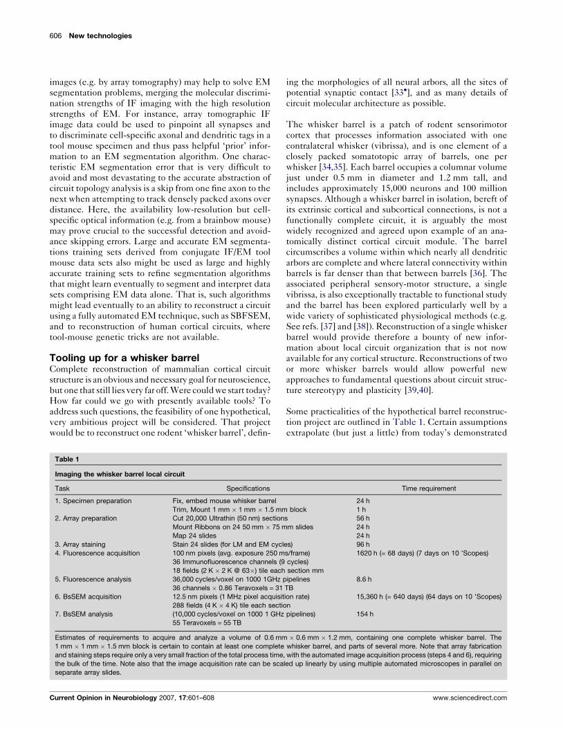
Table 1
Imaging the whisker barrel local circuit
Task Specifications
1. Specimen preparation Fix, embed mouse whisker barrel
Trim, Mount 1 mm � 1 mm � 1.5 mm
2. Array preparation Cut 20,000 Ultrathin (50 nm) section
Mount Ribbons on 24 50 mm � 75 m
Map 24 slides
3. Array staining Stain 24 slides (for LM and EM cycl
4. Fluorescence acquisition 100 nm pixels (avg. exposure 250 m
36 Immunofluorescence channels (9
18 fields (2 K � 2 K @ 63�) tile each
5. Fluorescence analysis 36,000 cycles/voxel on 1000 1GHz p
36 channels � 0.86 Teravoxels = 31
6. BsSEM acquisition 12.5 nm pixels (1 MHz pixel acquisit
288 fields (4 K � 4 K) tile each sectio
7. BsSEM analysis (10,000 cycles/voxel on 1000 1 GHz
55 Teravoxels = 55 TB
Estimates of requirements to acquire and analyze a volume of 0.6 mm
1 mm � 1 mm � 1.5 mm block is certain to contain at least one complete
and staining steps require only a very small fraction of the total process time,
the bulk of the time. Note also that the image acquisition rate can be sca
separate array slides.
Current Opinion in Neurobiology 2007, 17:601–608
ing the morphologies of all neural arbors, all the sites of
potential synaptic contact [33�], and as many details of
circuit molecular architecture as possible.
The whisker barrel is a patch of rodent sensorimotor
cortex that processes information associated with one
contralateral whisker (vibrissa), and is one element of a
closely packed somatotopic array of barrels, one per
whisker [34,35]. Each barrel occupies a columnar volume
just under 0.5 mm in diameter and 1.2 mm tall, and
includes approximately 15,000 neurons and 100 million
synapses. Although a whisker barrel in isolation, bereft of
its extrinsic cortical and subcortical connections, is not a
functionally complete circuit, it is arguably the most
widely recognized and agreed upon example of an ana-
tomically distinct cortical circuit module. The barrel
circumscribes a volume within which nearly all dendritic
arbors are complete and where lateral connectivity within
barrels is far denser than that between barrels [36]. The
associated peripheral sensory-motor structure, a single
vibrissa, is also exceptionally tractable to functional study
and the barrel has been explored particularly well by a
wide variety of sophisticated physiological methods (e.g.
See refs. [37] and [38]). Reconstruction of a single whisker
barrel would provide therefore a bounty of new infor-
mation about local circuit organization that is not now
available for any cortical structure. Reconstructions of two
or more whisker barrels would allow powerful new
approaches to fundamental questions about circuit struc-
ture stereotypy and plasticity [39,40].
Some practicalities of the hypothetical barrel reconstruc-
tion project are outlined in Table 1. Certain assumptions
extrapolate (but just a little) from today’s demonstrated
Time requirement
24 h
block 1 h
s 56 h
m slides 24 h
24 h
es) 96 h
s/frame) 1620 h (= 68 days) (7 days on 10 ’Scopes)
cycles)
section mm
ipelines 8.6 h
TB
ion rate) 15,360 h (= 640 days) (64 days on 10 ’Scopes)
n
pipelines) 154 h
� 0.6 mm � 1.2 mm, containing one complete whisker barrel. The
whisker barrel, and parts of several more. Note that array fabrication
with the automated image acquisition process (steps 4 and 6), requiring
led up linearly by using multiple automated microscopes in parallel on
www.sciencedirect.com
Circuit reconstruction tools today Smith 607
states of the relevant arts. It is assumed that transgenes
would encode eight cell-marking epitope tags that would
allow most or all nearby cells to be distinguished by
brainbow-style immunofluorescence, as discussed above.
It is assumed that an array comprising an entire whisker
barrel cut into 50 nm sections could be fabricated reliably
on 24 standard microscope slides, and that array tomo-
graphic immunofluorescence could discriminate 36 anti-
body channels, of which eight would be used to read the
transgenic epitope tags and the remaining 28 would be
used to read endogenous molecules useful for classifying
and modeling neurons and synapses. It is also assumed
that BsSEM imaging could read the ultrastructure of
these 50 nm sections in enough detail to meaningfully
measure spine necks and to generate useful learning
algorithm training sets. Additional technical assumptions
are stated in Table 1.
Would this project be feasible today? The proposition of
collecting, storing and analyzing a total of 86 terabytes of
data would have been daunting until quite recently, but is
no longer so. Computing equipment costing much less
than $0.5 million US would suffice easily for the data
storage and image analysis tasks specified. The time
requirements for image acquisition, especially for BsSEM
phase, may sound formidable, but these steps are fully
automated and the imaging throughput is scalable by
adding more automated microscopes. The field emission
gun SEM (FEG-SEM) required costs approximately $0.5
million US each, so scaling up to 10 FEG-SEMs for a 10�increase in throughput would be a viable option. The
major potential barrier to the success of this project may
lie in the challenge of developing sufficiently robust EM
segmentation algorithms, as discussed above. Given the
talents, energy and good ideas being brought to bear on
this challenge today, it is hard to believe that this final
barrier will not yield.
ConclusionThough the challenges of reconstructing cortical circuitry
today are substantial, the potential payoffs are enormous.
Given today’s universal agreement with the postulate of
the neural circuit as the basis of the brain’s abilities to
process information and generate behavior, it seems
extremely unlikely that the brain will ever be understood
without reconstructing circuit structure. A framework of
complete and quantitative knowledge of circuit structure
should also provide for deeper and more efficient phys-
iological analysis of circuit function, and for deeper mol-
ecular exploration of potential complexity-management
concepts such as ‘neuron type’ [2–5], ‘synapse type’ [41�],‘cortical modules’ [42], and ‘network motifs’ [43]. With
the rapid growth in the power and availability of parallel
computation (e.g. see ref. [44]), accurate reconstruction of
circuit connectivity and molecular architecture appears to
be the last remaining obstacle to fulfilling the promise of
computational circuit simulation as the next generation’s
www.sciencedirect.com
best tool for understanding the brain. With a little
luck, the new reconstruction techniques reviewed may
have an impact worthy of comparison with Ramon y
Cajal’s drawings.
AcknowledgementsThe author thanks JoAnn Buchanan, Winfried Denk, Kristen Harris, JeffLichtman, Kristina Micheva, Nancy O’Rourke and Sebastian Seung formany helpful discussions and comments on manuscripts, and the authors ofref. [12��] for providing the illustration used in Figure 2. This work wassupported in part by grant NS054252 from the National Institutes of Health.
References and recommended readingPapers of particular interest, published within the annual period ofreview, have been highlighted as:
� of special interest�� of outstanding interest
1.��
Ramon y Cajal S: Histology of the Nervous System of Man andVertebrates. Oxford Press; (trans. 1995, N. Swanson, L. Swanson).
Summarizes the work that first placed the concept of information proces-sing by neural circuit structures on a firm anatomical foundation.
2. McKay RD, Hockfield SJ: Monoclonal antibodies distinguishantigenically discrete neuronal types in the vertebrate centralnervous system. Proc Natl Acad Sci U S A 1982, 79:6747-6751.
3. Nelson SB, Hempel C, Sugino K: Probing the transcriptome ofneuronal cell types. Curr Opin Neurobiol 2006, 16(5):571-576Epub 2006 Sept. 7.
4. Molyneaux BJ, Arlotta P, Menezes JR, Macklis JD: Neuronalsubtype specification in the cerebral cortex. Nat Rev Neurosci2007, 6:427-437.
5. Lein ES, Hawrylycz MJ, Ao N, Ayres M, Bensinger A, Bernard A,Boe AF, Boguski MS, Brockway KS, Byrnes EJ et al.: Genome-wide atlas of gene expression in the adult mouse brain. Nature2007, 445:168-176.
6.��
Feng G, Mellor RH, Bernstein M, Keller-Peck C, Nguyen QT,Wallace M, Nerbonne JM, Lichtman JW, Sanes JR: Imagingneuronal subsets in transgenic mice expressing multiplespectral variants of GFP. Neuron 2000, 28:41-51.
This landmark work introduced strategies that have proven very versatileand powerful for making mice with color-coded neuronal subsets.
7. Hatten ME, Heintz N: Large-scale genomic approaches tobrain development and circuitry. Annu Rev Neurosci 2005,28:89-108.
8. Xu Q, Cobos I, De La Cruz E, Rubenstein JL, Anderson SA:Origins of cortical interneuron subtypes. J Neurosci 2004,24:2612-2622.
9. Monro S, Pelham HRB: Use of peptide tagging to detectproteins expressed from cloned genes: deletion mappingfunctional domains of Drosophila hsp70. EMBO J 1984,3:3087-3093.
10. Griffin BA, Adams SR, Jones J, Tsien RY: Fluorescent labelingof recombinant proteins in living cells with FlAsH.Methods Enzymol 2000, 327:565-578.
11. Giepmans BN, Adams SR, Ellisman MH, Tsien RY: Thefluorescent toolbox for assessing protein location andfunction. Science 2006, 312:217-224.
12.��
Livet J, Weissman TA, Kang H, Draft RW, Lu J, Bennis R, Sanes JR,Lichtman JW: Transgenic strategies for combinatorialexpression of fluorescent proteins in the nervous system.Nature 2007, 450:56-62.
Introduces a powerful new approach to circumventing optical resolutionlimits that have limited optical imaging of complete neural circuits.
13.��
Luo L: Fly MARCM and mouse MADM: Genetic methods oflabeling and manipulating single neurons. Brain Res Rev 2007,55:220-227.
A very promising new set of schemes for labeling and manipulatingcircuits.
Current Opinion in Neurobiology 2007, 17:601–608
608 New technologies
14.��
Wickersham IR, Lyon DC, Barnard RJ, Mori T, Finke S,Conzelmann KK, Young JA, Callaway EM: Monosynapticrestriction of transsynaptic tracing from single, geneticallytargeted neurons. Neuron 2007, 53:639-647.
A new strategy for labeling cells and their monsynaptically partners only.
15. Micheva KD, Smith SJ: Array tomography: A new tool forimaging the molecular architecture and ultrastructure ofneural circuits. Neuron 2007, 55:25-36.
16. Conchello JA, Lichtman JW: Optical sectioning microscopy.Nat Methods 2005, 2(12):920-931.
17.�
Rhodes KJ, Trimmer JS: Antibodies as valuable neuroscienceresearch tools versus reagents of mass distraction. J Neurosci2006, 26:8017-8020.
Reviews important recent progress in validating antibody specificity.
18.�
Hell SW: Far-field optical nanoscopy. Science 2007, 316:1153-1158.
19.�
Gustafsson MG: Nonlinear structured-illumination microscopy:wide-field fluorescence imaging with theoretically unlimitedresolution. Proc Natl Acad Sci U S A 2005, 102:13081-13086.
20.�
Betzig E, Patterson GH, Sougrat R, Lindwasser OW, Olenych S,Bonifacino JS, Davidson MW, Lippincott-Schwartz J, Hess HF:Imaging intracellular fluorescent proteins at nanometerresolution. Science 2006, 313:1642-1645.
21.�
Hess ST, Girirajan TP, Mason MD: Ultra-high resolution imagingby fluorescence photoactivation localization microscopy.Biophys J 2006, 91:4258-4272.
22.�
Bates M, Huang B, Dempsey GT, Zhuang X: Multicolorsuper-resolution imaging with photo-switchable fluorescentprobes. Science 2007, 317:1749-1753.
23.�
Rust MJ, Bates M, Zhuang X: Sub-diffraction-limit imaging bystochastic optical reconstruction microscopy (STORM).Nat Methods 2006, 3:793-795.
(Refs. [18–23] introduce new methods for fluorescence imaging atresolutions far higher than formerly respected diffraction limits. Most ofthese methods have been demonstrated mainly for two-dimensionalimaging, but may be very useful to enhance fluorescence lateralresolution in three-dimensional imaging if used in conjunction with arraytomography mechanical sectioning procedures).
24.�
Briggman KL, Denk W: Towards neural circuit reconstructionwith volume electron microscopy techniques. Curr OpinNeurobiol 2006, 16:562-570.
Reviews the nascent field of high-throughput electron microscopy.
25.��
Denk W, Horstmann H: Serial block-face scanning electronmicroscopy to reconstruct three-dimensional tissuenanostructure. PLoS Biol 2004, 11:e329.
Introduces the first fully automated method for the acquisition of volu-metric data with resolution and accuracy adequate to allow tracing ofneural circuit elements, and inspires a new field of high-throughputelectron microscopic neuroanatomy.
26.�
Hayworth KJ, Kasthuri N, Schalek R, Lichtman JW: Automatingthe collection of ultrathin serial sections for large volume TEMreconstructions. Microscop Microanal 2006, 12:86-87.
Describes exciting an new microtome and an automated sectioncollection scheme that are likely to be very useful for producing very-large-scale tomography arrays.
27. Levoy M: Display of surfaces from volume data. IEEE ComputGraph Appl 1988, 8:29-37.
28. Cai H, Xu X, Lu J, Lichtman JW, Yung SP, Wong ST: Repulsiveforce based snake model to segment and track neuronal
Current Opinion in Neurobiology 2007, 17:601–608
axons in 3D microscopy image stacks. Neuroimage 2006,32(4):1608-1620.
29. Fiala JC, Harris K: Computer-based alignment andreconstruction of serial sections. Microscop Anal USA Ed 2002,52:5-7.
30. Fiala JC: Reconstruct: a free editor for serial sectionmicroscopy. J Microscop 2005, 218:52-61.
31. Toni N, Teng EM, Bushong EA, Aimone JB, Zhao C, Consiglio A,van Praag H, Martone ME, Ellisman MH, Gage FH: Synapseformation on neurons born in the adult hippocampus. NatNeurosci 2007, 10:727-734.
32. Jain V, Murray JF, Roth F, Turaga S, Zhigulin V, Briggman KL,Helmstaedter MN, Denk W, and Seung HS: Supervised learningof image restoration with convolutional networks. IEEE IntConf Comput Vis (ICCV), in press.
33.�
Stepanyants A, Chklovskii DB: Neurogeometry and potentialsynaptic connectivity. Trends Neurosci 2005, 28:387-394.
Introduces a very powerful way to make the most of incompleteinformation about circuit topologies.
34. Woolsey TA, Dierker ML, Wann DF: Mouse SmI cortex:qualitative and quantitative classification of golgi-impregnated barrel neurons. Proc Natl Acad Sci U S A 1975,72:2165-2169.
35. Kleinfeld D, Ahissar E, Diamond ME: Active sensation: insightsfrom the rodent vibrissa sensorimotor system. Curr OpinNeurobiol 2006, 16:435-444.
36. Petersen CC, Sakmann B: The excitatory neuronal network ofrat layer 4 barrel cortex. J Neurosci 2000, 20:7579-7586.
37. Brecht M, Roth A, Sakmann B: Dynamic receptive fields ofreconstructed pyramidal cells in layers 3 and 2 of ratsomatosensory barrel cortex. J Physiol 2003,553:243-265.
38. Sato TR, Gray NW, Mainen ZF, Svoboda K: The functionalmicroarchitecture of the mouse barrel cortex. PLoS Biol 2007,5:e189.
39. Micheva KD, Beaulieu C: Development and plasticity of theinhibitory neocortical circuitry with an emphasis on the rodentbarrel field cortex: a review. Can J Physiol Pharmacol 1997,75:470-478.
40. Knott GW, Quairiaux C, Genoud C, Welker E: Formation ofdendritic spines with GABAergic synapses induced by whiskerstimulation in adult mice. Neuron 2002, 34:265-273.
41.�
Grant SG: Toward a molecular catalogue of synapses.Brain Res Rev 2007, 55:445-449.
Clearly articulates the importance of finding ways to measure and classifyindividual synapses in their native brain tissue contexts.
42. Swindale NV: Is the cerebral cortex modular? Trends Neurosci1990, 13:487-492.
43. Milo R, Shen-Orr S, Itzkovitz S, Kashtan N, Chklovskii D, Alon U:Network motifs: simple building blocks of complex networks.Science 2002, 298:824-827.
44. Migliore M, Cannia C, Lytton WW, Markram H, Hines ML: Parallelnetwork simulations with NEURON. J Comput Neurosci 2006,21:119-129.
A strong argument that the computational power for circuit simulation willbe ready as soon as the structural and molecular data for the model isavailable.
www.sciencedirect.com