Embed Size (px)
Citation preview
Lab on a Chip
Publ
ishe
d on
05
June
201
4. D
ownl
oade
d by
Que
en M
ary,
Uni
vers
ity o
f L
ondo
n on
21/
06/2
014
13:3
9:11
.
PAPER View Article OnlineView Journal
This journal is © The Royal Society of Chemistry 2014
a Science for Life Laboratory, Department of Immunology, Genetics and Pathology,
Uppsala University, Box 815, 751 08 Uppsala, SwedenbKU Leuven – University of Leuven, BIOSYST-MeBioS, Willem de Croylaan 42,
Leuven, Belgium. E-mail: [email protected]; Tel: +32 16 32 14 59c Science for Life Laboratory, Department of Biochemistry and Biophysics,
Stockholm University, Box 1031, Se-171 21 Solna, Sweden.
E-mail: [email protected]; Tel: +46 (0)762 756 161
† Electronic supplementary information (ESI) available. See DOI: 10.1039/c4lc00348a
Cite this: DOI: 10.1039/c4lc00348a
Received 20th March 2014,Accepted 30th May 2014
DOI: 10.1039/c4lc00348a
www.rsc.org/loc
Circle-to-circle amplification on a digitalmicrofluidic chip for amplified singlemolecule detection†
Malte Kühnemund,a Daan Witters,b Mats Nilsson*c and Jeroen Lammertyn*b
We demonstrate a novel digital microfluidic nucleic acid amplification concept which is based on
padlock probe mediated DNA detection and isothermal circle-to-circle amplification (C2CA). This assay
platform combines two digital approaches. First, digital microfluidic manipulation of droplets which serve
as micro-reaction chambers and shuttling magnetic particles between these droplets facilitates the
integration of complex solid phase multistep assays. We demonstrate an optimized novel particle
extraction and transfer protocol for superparamagnetic particles on a digital microfluidic chip that allows
for nearly 100% extraction efficiencies securing high assay performance. Second, the compartmentalization
required for digital single molecule detection is solved by simple molecular biological means,
circumventing the need for complex microfabrication procedures necessary for most, if not all, other
digital nucleic acid detection methods. For that purpose, padlock probes are circularized in a strictly
target dependent ligation reaction and amplified through two rounds of rolling circle amplification,
including an intermediate digestion step. The reaction results in hundreds of 500 nm sized individually
countable DNA nanospheres per detected target molecule. We demonstrate that integrated miniaturized
digital microfluidic C2CA results in equally high numbers of C2CA products μL−1 as off-chip tube control
experiments indicating high assay performance without signal loss. As low as 1 aM synthetic Pseudomonas
aeruginosa DNA was detected with a linear dynamic range over 4 orders of magnitude up to 10 fM
proving excellent suitability for infectious disease diagnostics.
Introduction
DNA analysis plays an important role in molecular diagnos-tics. Traditional laboratory analysis methods, however,require time-consuming manual handling and sophisticatedinstrumentation. Recently, integrated microfluidic lab-on-chip devices emerged that can overcome these limitations byminiaturization and the automation of laboratory methods,1–4
making them attractive for research and point-of-care (POC)diagnostic applications. One elegant way of microfluidic liquidhandling is the transportation of nano- to microliter sizeddroplets by electrowetting-on-dielectric (EWOD) on digital
microfluidic (DMF) chips.5–7 On DMF chips, each dropletserves as a discrete reaction compartment that can beaddressed individually and independently.8 This allows fluiddroplets to be controlled with maximum flexibility and pro-grammability, an important asset for bio-assays requiringmany complex handling steps to be executed simultaneouslyon one single device. DMF chips have shown to be capable ofmanipulating magnetic particles with unprecedented preci-sion, speed and flexibility.9–11 As such, magnetic particles canbe separated, washed, mixed and incubated with new reagentsin a highly automated and miniaturized way allowing for theperformance of multistep solid-phase bio-assays.9,10,12 Efficientand rapid mixing is promoted by a droplet internal vortex thatis created by the transportation of droplets by EWOD.6,7
Recently, a magnetic extraction protocol for 3 μm sized ferro-magnetic particles on a digital microfluidic chip was describedthat allowed for the performance of a solid-phase immuno-assay in presence of a spatially fixed permanent magnet.13 Herewe report on 2 critical particle extraction parameters that havenot been addressed before and illustrate a novel highly effi-cient extraction and transfer protocol for 1 μm sized super-paramagnetic particles on a DMF chip using a spatially
Lab Chip
Lab on a ChipPaper
Publ
ishe
d on
05
June
201
4. D
ownl
oade
d by
Que
en M
ary,
Uni
vers
ity o
f L
ondo
n on
21/
06/2
014
13:3
9:11
. View Article Online
movable magnet allowing to shuttle particles between dropletswith high simplicity and perform particle-based enzymaticreactions that require a homogeneous particle suspension.
One of the critical issues for automated DNA detection andanalysis has been the difficulty of integrating DNA amplifica-tion assays on microfluidic platforms. The gold standard forDNA amplification, the polymerase chain reaction (PCR),requires precise cycling of temperatures which is challengingto achieve in integrated micro-systems. Nevertheless, Changet al. succeeded in integrating a PCR assay on a DMF chip mak-ing use of the capability to shuttle droplets between heatingzones to achieve cycling.14 Besides PCR, several isothermalamplification methods have emerged that are promising alter-natives to PCR. Amongst them are helicase dependent amplifi-cation (HDA),15 strand displacement amplification (SDA),16
loop mediated isothermal amplification (LAMP)17 and rollingcircle amplification (RCA).18 LAMP assays have successfullybeen integrated into micro-total analysis systems with highspeed and sensitivity.19 However, LAMP assays are poorlymultiplexable and the target dependent design of primer setsrather complicated. Hyper branched RCA is a simple and rapidmethod20 but the branching primer pairs often lead tounspecific background amplification.
High specificity and sensitivity are major requirements forDNA detection in diagnostics. Specificity can be achieved bythe requirement of two or more hybridization events and aDNA ligation event that generates amplifiable DNA reportermolecules.21 Padlock probes are linear oligonucleotides thatare ligated in a strictly target sequence-dependent manner toform a circle that can subsequently be amplified.22 Circulari-zation by ligation requires that both target complementaryprobe arms hybridize to the target sequence with perfectmatch. The high discrimination rate of thermophilic DNAligases enables single nucleotide mismatch detection.23
Amplification of padlock probes by RCA is exclusive to circu-larized probes which adds another step of specificity.24,25 TheRCA product, a concatemer of hundreds of padlock probereplicates, collapses into a distinct 500 nm sized spheric DNAcoil that, when labeled with fluorescent oligonucleotides, canbe digitally counted after deposition on a microscope slide26
or while flowed through a microfluidic channel.27 As thesereadout methods only detect a fraction of the generated RCAproducts an additional amplification step is needed whenhigh sensitivity is required. Circle-to-circle amplification(C2CA) generates hundreds of RCA products from one initialRCA product by monomerization of the concatemer, re-circularization of monomers and amplification of the newlygenerated circles in a second RCA.28 C2CA is an isothermalamplification method with low amplification bias and varia-tion coefficient facilitating high multiplexed analysis.28 Theuse of magnetic particles as a solid support significantlyincreases assay sensitivity and speed by separation of circlesfrom unreacted padlock probes and sample remains.29,30
Furthermore, in contrast to most digital single molecule detec-tion methods, RCA and C2CA do not require compartmentali-zation for multiplexed or digital analysis. Single molecules are
Lab Chip
amplified to individually countable objects which enables digi-tal read out.26,27 However, besides high analytical performanceand isothermal reaction conditions, C2CA requires multiplesteps which make its application in diagnostics less favorable.Therefore the integration and automation of C2CA into micro-fluidic lab-on-chip devices is of utmost importance in order tobring its advantages to diagnostic application.
Only a few studies have been reported in which micro-fluidic chips are used for the detection of RCA amplifiedmolecules.27,31,32 Sato et al. (2010) have described an on chipmicro-bead based RCA assay achieving low femtomolar detec-tion limits. The counting of RCA products, however, requiredtedious confocal microscopy in order to detect RCA productsdistributed around the micro-beads in the channel.32 Inanother study C2CA was performed in the sample-well of amicrofluidic chip for gel-electrophoretic separation of theC2CA products.33 Despite the detection of C2CA products onmicrofluidic systems the assay has, until now, not beenperformed integrated on a microfluidic chip.
Here we report, for the first time, the performanceof C2CA on a DMF chip with all assay steps integratedon-chip, except for heating. We demonstrate how DMF'sreconfigurable and automated droplet actuation principle,combined with an improved protocol for shuttling magneticparticles between droplets that serve as discrete reactionmicrochambers, offers a unique and novel way of performingminiaturized particle-based C2CA. C2CA performed on aDMF chip proved as efficient as compared to reactiontube conditions, a major achievement rendered by DMF'sexcellent droplet mixing capabilities and the new particlemanipulation protocol described in this paper. We appliedDMF C2CA for the detection of ultra-low amounts of syn-thetic P. aeruginosa DNA illustrating the suitability for infec-tious disease diagnostics.
Results and discussionDigital microfluidic C2CA assay layout
The integration of C2CA on the DMF chip is illustrated inFig. 1. The droplets containing the DNA sample, ligationreaction mixture, magnetic particles and washing buffer wereinitially placed on the chip before the assay was started. TheRCA, digestion and ligation & 2nd RCA droplets were consec-utively introduced during the assay, as described in theexperimental section.
In brief, a droplet containing ligase, target specific pad-lock probes and biotinylated capture oligonucleotides ismerged with a DNA sample (Fig. 1a and b step 1). After5 min hybridization and ligation the capture oligonucleotideis annealed and the padlock probe ligated on the DNA strand(Fig. 1d). Thereupon, streptavidin coated magnetic particlesare merged with the sample and mixed by droplet transporta-tion promoting the capture of the ligation complex by biotin–straptavidin bond (Fig. 1a, droplet movement is indicated bygreen arrows; Fig. 1b step 2). The particles are magnetically
This journal is © The Royal Society of Chemistry 2014
Fig. 1 Circle-to-circle amplification protocol on a DMF chip. a) DMF chip and assay layout. Positions of droplets with reaction mixes are depicted.Mixing of two droplets is illustrated with green arrows with double arrowheads. Mixing of single droplets is illustrated by green arrows with singlearrowhead. Magnetic particle transfer between droplets is indicated with red arrows. b) Flow chart illustrating magnetic particle extraction &transfer and droplet mixing; droplet color code as in 1a. c) Fluorescence imaging of individual RCA products that randomly align on a microscopeslide promoting digital read-out. d) C2CA: (1) a ligation mix containing target specific padlock probes and biotinylated capture oligonucleotides ismixed with a DNA sample. (2) After hybridization and ligation, streptavidin coated magnetic particles are mixed with the ligation complex to bindthe ligation complex to magnetic particles via the biotinylated capture oligonucleotide. (3) The particles are magnetically separated, transferred tothe washing droplet and washed. (4) The particles are separated again and mixed into RCA reaction mix. (5) After RCA the particles with coupledRCA products are extracted and transferred into the digestion mix where the RCA products are monomerized. (6) The droplet with monomers ismixed with a droplet for ligation + the 2nd RCA (for more details see Dahl et al., 2004 ref. 28).
Lab on a Chip Paper
Publ
ishe
d on
05
June
201
4. D
ownl
oade
d by
Que
en M
ary,
Uni
vers
ity o
f L
ondo
n on
21/
06/2
014
13:3
9:11
. View Article Online
extracted and transferred to the washing droplet by movingthe particles along with the magnet to the washing droplet(illustrated in Fig. 1a by red arrows and in Fig. S1†). Next, theparticles are dispersed and, hence, washed by droplet trans-portation along the electrode row (Fig. 1a, green arrows;Fig. 1b step 3). The particles are then separated and trans-ferred to and mixed in the RCA reaction mixture (Fig. 1b step 4).After 20 min RCA and 2 min heat inactivation the particleswith coupled RCA products are transferred into the digestionmix (Fig. 1b step 5) and the RCA products are enzymaticallymonomerized during 2 min incubation and 2 min heatinactivation. After particle extraction and disposal the dropletwith monomers is mixed with a second ligation and RCA mix(Fig. 1b step 6) where the monomers are re-circularizedand amplified in a second RCA step for 25 min. The totalassay time is ~60 min. The C2CA products are labeled
This journal is © The Royal Society of Chemistry 2014
with fluorescent oligonucleotides, imaged on a microscopeslide and counted digitally (Fig. 1c).
Highly efficient extraction of 1 μm sized superparamagneticparticles on a DMF chip
Magnetic particles play an important role in the automationof C2CA by supplying a solid phase for molecule capture andfor washing away sample remains and unreacted padlockprobes. In order to achieve a high performance with the assaylayout described above the first objective of this study was todevelop an extraction and transfer protocol for 1 μm sizedsuperparamagnetic particles on a DMF chip (Fig. S1†).Ng et al. (2012)10 have implemented a particle extraction andwashing protocol with a spatially removable magnet on aDMF chip, however, no information about the particles that
Lab Chip
Lab on a ChipPaper
Publ
ishe
d on
05
June
201
4. D
ownl
oade
d by
Que
en M
ary,
Uni
vers
ity o
f L
ondo
n on
21/
06/2
014
13:3
9:11
. View Article Online
were used in the study was provided. Vergauwe et al. havedeveloped an extraction protocol for ferromagnetic particleson a DMF chip with a spatially fixed permanent magnet.13
They investigated the effect of the capillary force of the mov-ing droplet on the extraction efficiency for different ferromag-netic particle concentrations. The automation of the C2CAprotocol, however, requires the use of superparamagnetic parti-cles because the particles need to be homogenously dispersedand incubated in enzyme reaction mixtures in absence of amagnet. In contrast to ferromagnetic particles, which after mag-netic attraction stay magnetized and which are hardly dispersible,superparamagnetic particles can easily be resuspended. Drop-let transportation in DMF creates a recirculating flow insidethe droplet which leads to very efficient mixing7 and super-paramagnetic particles can, after extraction from a droplet,be rapidly resuspended in a new droplet.9,10
In addition to the parameters tested by Ng et al. (2012)10
and Vergauwe et al.13 we investigated the influence of (i) thedistance between the magnet and the particles on the particleextraction efficiency by placing the magnet on top plates withdifferent thicknesses; and (ii) the position of the magnetedge above the electrodes.
The distance between the magnet and the particles has astrong effect on the particle extraction efficiency. When using a1.1 mm thick top plate, the extraction of 1 × 10⁶ particles μL−1
(the concentration usually used in C2CA) was insufficient as ahigh amount of particles remained in the droplet when the drop-let was transported away from the magnet (Fig. 2c; movie S3†).The incomplete extraction of low particle concentrationsis likely due to insufficient magnetic force on the particles asan increased distance between the magnet and the magneticparticles decreases the magnetic force on the particles.10,12 Atthe point of extraction the droplet meniscus starts necking andthe particle pellet eventually pinches off when the neck closes
Lab Chip
Fig. 2 Video clip captures of magnetic particle extraction from a droplet(a and b) or 1.1 mm thick top plate (c). The magnet is positioned in eithe1) Video clip capture at the point of extraction: magnetic particles are pelthe right. 2) The same droplets a few milliseconds after extraction. The red2) indicate the meniscus of the droplet which is enlarged and illustrated in 3
behind the extracted pellet.34 However, if the magnetic force isinsufficient the capillary force of the closing droplet breaks theparticle pellet and pulls a fraction of the particles back into themoving droplet. The loss of such large amounts of particleswould drastically decrease the assay efficiency.
The position of the magnet above the electrode wasfound to play an important role in the particle extractionprocess, as well. The extraction of low particle concentra-tions (105–106 particles μL−1) was successful only when themagnet edge was placed on the border between two electrodes(Fig. 2b and movie S4†) but insufficient when positioned in themiddle of the electrode. In the latter case a high amount of par-ticles was left inside the droplet when the droplet moved awayfrom the magnet (Fig. 2a; movie S5†). This effect can beexplained in relation to the length of the droplet meniscusstretch during the particle extraction process. With the magnetedge positioned on the border between 2 electrodes, the mag-netic particles are concentrated at the edge of the droplet.When the droplet moves to the neighboring electrode, thedroplet creates a longer stretched meniscus to the particlepellet than when the magnet edge is positioned in the middleof the electrode (compare point of extraction (1) and afterextraction (2) in Fig. 2a and b). The longer meniscus stretchforms a narrower neck and facilitates the pinching-off to agreater extent than a shorter stretch. Consequently, the parti-cles get extracted.
With the magnet positioned on the border betweentwo electrodes and a 0.7 mm thick top plate, 1 μm sizedsuperparamagnetic particles with a concentration as low as1.6 × 105 particles μL−1 were extracted successfully. Usingthese parameters, extraction efficiencies were also deter-mined for 1 μm sized T1 dynabeads and 2.8 μm sized M280beads. T1 dynabeads could be extracted in the same concen-tration (1.6 × 10⁵ particles μL−1) as C1 dynabeads and M280
This journal is © The Royal Society of Chemistry 2014
moving on the DMF chip under a magnet placed on top of a 0.7 mmr the middle (a) or on the border between two electrodes (b and c).leted at the edge of the magnet as the droplets move from the left toarrow in c-2) indicates remaining particles in the droplet. Red boxes in). The parameters were tested with 3 repeated extraction attempts.
Lab on a Chip Paper
Publ
ishe
d on
05
June
201
4. D
ownl
oade
d by
Que
en M
ary,
Uni
vers
ity o
f L
ondo
n on
21/
06/2
014
13:3
9:11
. View Article Online
beads could be extracted in 3 × 10⁴ particles μL−1 concentra-tion (Fig. S2†).
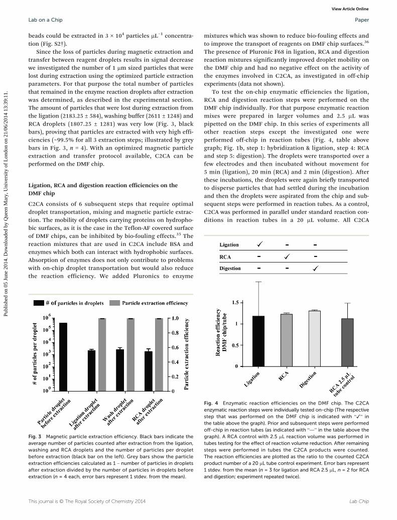
Since the loss of particles during magnetic extraction andtransfer between reagent droplets results in signal decreasewe investigated the number of 1 μm sized particles that werelost during extraction using the optimized particle extractionparameters. For that purpose the total number of particlesthat remained in the enzyme reaction droplets after extractionwas determined, as described in the experimental section.The amount of particles that were lost during extraction fromthe ligation (2183.25 ± 584), washing buffer (2611 ± 1248) andRCA droplets (1807.25 ± 1281) was very low (Fig. 3, blackbars), proving that particles are extracted with very high effi-ciencies (~99.5% for all 3 extraction steps; illustrated by greybars in Fig. 3, n = 4). With an optimized magnetic particleextraction and transfer protocol available, C2CA can beperformed on the DMF chip.
Ligation, RCA and digestion reaction efficiencies on theDMF chip
C2CA consists of 6 subsequent steps that require optimaldroplet transportation, mixing and magnetic particle extrac-tion. The mobility of droplets carrying proteins on hydropho-bic surfaces, as it is the case in the Teflon-AF covered surfaceof DMF chips, can be inhibited by bio-fouling effects.35 Thereaction mixtures that are used in C2CA include BSA andenzymes which both can interact with hydrophobic surfaces.Absorption of enzymes does not only contribute to problemswith on-chip droplet transportation but would also reducethe reaction efficiency. We added Pluronics to enzyme
This journal is © The Royal Society of Chemistry 2014
Fig. 3 Magnetic particle extraction efficiency. Black bars indicate theaverage number of particles counted after extraction from the ligation,washing and RCA droplets and the number of particles per dropletbefore extraction (black bar on the left). Grey bars show the particleextraction efficiencies calculated as 1 – number of particles in dropletsafter extraction divided by the number of particles in droplets beforeextraction (n = 4 each, error bars represent 1 stdev. from the mean).
mixtures which was shown to reduce bio-fouling effects andto improve the transport of reagents on DMF chip surfaces.36
The presence of Pluronic F68 in ligation, RCA and digestionreaction mixtures significantly improved droplet mobility onthe DMF chip and had no negative effect on the activity ofthe enzymes involved in C2CA, as investigated in off-chipexperiments (data not shown).
To test the on-chip enzymatic efficiencies the ligation,RCA and digestion reaction steps were performed on theDMF chip individually. For that purpose enzymatic reactionmixes were prepared in larger volumes and 2.5 μL waspipetted on the DMF chip. In this series of experiments allother reaction steps except the investigated one wereperformed off-chip in reaction tubes (Fig. 4, table abovegraph; Fig. 1b, step 1: hybridization & ligation, step 4: RCAand step 5: digestion). The droplets were transported over afew electrodes and then incubated without movement for5 min (ligation), 20 min (RCA) and 2 min (digestion). Afterthese incubations, the droplets were again briefly transportedto disperse particles that had settled during the incubationand then the droplets were aspirated from the chip and sub-sequent steps were performed in reaction tubes. As a control,C2CA was performed in parallel under standard reaction con-ditions in reaction tubes in a 20 μL volume. All C2CA
Lab Chip
Fig. 4 Enzymatic reaction efficiencies on the DMF chip. The C2CAenzymatic reaction steps were individually tested on-chip (The respectivestep that was performed on the DMF chip is indicated with “✓” inthe table above the graph). Prior and subsequent steps were performedoff-chip in reaction tubes (as indicated with “—” in the table above thegraph). A RCA control with 2.5 μL reaction volume was performed intubes testing for the effect of reaction volume reduction. After remainingsteps were performed in tubes the C2CA products were counted.The reaction efficiencies are plotted as the ratio to the counted C2CAproduct number of a 20 μL tube control experiment. Error bars represent1 stdev. from the mean (n = 3 for ligation and RCA 2.5 μL, n = 2 for RCAand digestion; experiment repeated twice).
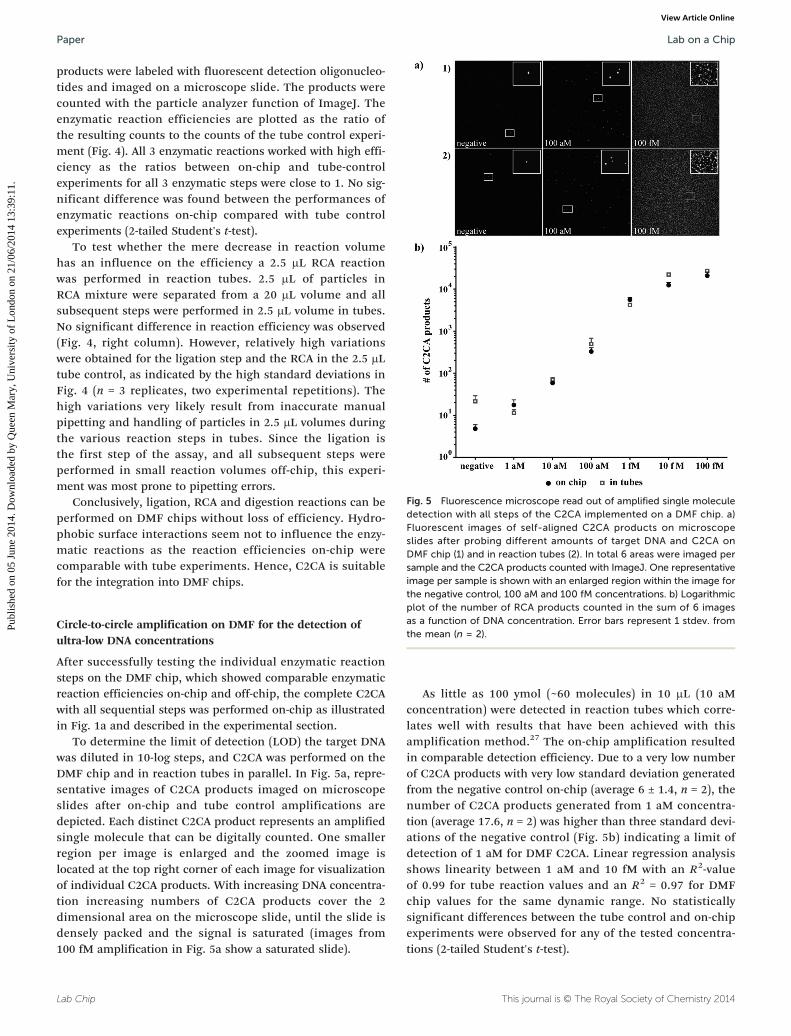
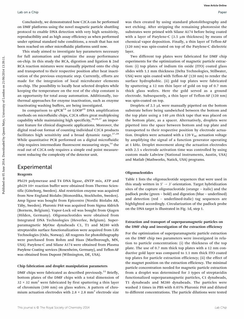
Fig. 5 Fluorescence microscope read out of amplified single moleculedetection with all steps of the C2CA implemented on a DMF chip. a)Fluorescent images of self-aligned C2CA products on microscopeslides after probing different amounts of target DNA and C2CA onDMF chip (1) and in reaction tubes (2). In total 6 areas were imaged persample and the C2CA products counted with ImageJ. One representativeimage per sample is shown with an enlarged region within the image for
Lab on a ChipPaper
Publ
ishe
d on
05
June
201
4. D
ownl
oade
d by
Que
en M
ary,
Uni
vers
ity o
f L
ondo
n on
21/
06/2
014
13:3
9:11
. View Article Online
products were labeled with fluorescent detection oligonucleo-tides and imaged on a microscope slide. The products werecounted with the particle analyzer function of ImageJ. Theenzymatic reaction efficiencies are plotted as the ratio ofthe resulting counts to the counts of the tube control experi-ment (Fig. 4). All 3 enzymatic reactions worked with high effi-ciency as the ratios between on-chip and tube-controlexperiments for all 3 enzymatic steps were close to 1. No sig-nificant difference was found between the performances ofenzymatic reactions on-chip compared with tube controlexperiments (2-tailed Student's t-test).
To test whether the mere decrease in reaction volumehas an influence on the efficiency a 2.5 μL RCA reactionwas performed in reaction tubes. 2.5 μL of particles inRCA mixture were separated from a 20 μL volume and allsubsequent steps were performed in 2.5 μL volume in tubes.No significant difference in reaction efficiency was observed(Fig. 4, right column). However, relatively high variationswere obtained for the ligation step and the RCA in the 2.5 μLtube control, as indicated by the high standard deviations inFig. 4 (n = 3 replicates, two experimental repetitions). Thehigh variations very likely result from inaccurate manualpipetting and handling of particles in 2.5 μL volumes duringthe various reaction steps in tubes. Since the ligation isthe first step of the assay, and all subsequent steps wereperformed in small reaction volumes off-chip, this experi-ment was most prone to pipetting errors.
Conclusively, ligation, RCA and digestion reactions can beperformed on DMF chips without loss of efficiency. Hydro-phobic surface interactions seem not to influence the enzy-matic reactions as the reaction efficiencies on-chip werecomparable with tube experiments. Hence, C2CA is suitablefor the integration into DMF chips.
the negative control, 100 aM and 100 fM concentrations. b) Logarithmicplot of the number of RCA products counted in the sum of 6 imagesas a function of DNA concentration. Error bars represent 1 stdev. fromthe mean (n = 2).
Circle-to-circle amplification on DMF for the detection ofultra-low DNA concentrations
After successfully testing the individual enzymatic reactionsteps on the DMF chip, which showed comparable enzymaticreaction efficiencies on-chip and off-chip, the complete C2CAwith all sequential steps was performed on-chip as illustratedin Fig. 1a and described in the experimental section.
To determine the limit of detection (LOD) the target DNAwas diluted in 10-log steps, and C2CA was performed on theDMF chip and in reaction tubes in parallel. In Fig. 5a, repre-sentative images of C2CA products imaged on microscopeslides after on-chip and tube control amplifications aredepicted. Each distinct C2CA product represents an amplifiedsingle molecule that can be digitally counted. One smallerregion per image is enlarged and the zoomed image islocated at the top right corner of each image for visualizationof individual C2CA products. With increasing DNA concentra-tion increasing numbers of C2CA products cover the 2dimensional area on the microscope slide, until the slide isdensely packed and the signal is saturated (images from100 fM amplification in Fig. 5a show a saturated slide).
Lab Chip
As little as 100 ymol (~60 molecules) in 10 μL (10 aMconcentration) were detected in reaction tubes which corre-lates well with results that have been achieved with thisamplification method.27 The on-chip amplification resultedin comparable detection efficiency. Due to a very low numberof C2CA products with very low standard deviation generatedfrom the negative control on-chip (average 6 ± 1.4, n = 2), thenumber of C2CA products generated from 1 aM concentra-tion (average 17.6, n = 2) was higher than three standard devi-ations of the negative control (Fig. 5b) indicating a limit ofdetection of 1 aM for DMF C2CA. Linear regression analysisshows linearity between 1 aM and 10 fM with an R2-valueof 0.99 for tube reaction values and an R2 = 0.97 for DMFchip values for the same dynamic range. No statisticallysignificant differences between the tube control and on-chipexperiments were observed for any of the tested concentra-tions (2-tailed Student's t-test).
This journal is © The Royal Society of Chemistry 2014
Lab on a Chip Paper
Publ
ishe
d on
05
June
201
4. D
ownl
oade
d by
Que
en M
ary,
Uni
vers
ity o
f L
ondo
n on
21/
06/2
014
13:3
9:11
. View Article Online
Conclusively, we demonstrated how C2CA can be performedon DMF platforms using the novel magnetic particle shuttlingprotocol to enable DNA detection with very high sensitivity,reproducibility and as high assay efficiency as when performedunder optimal standard tube conditions, a result that has notbeen reached on other microfluidic platforms until now.
This study aimed to investigate key parameters necessaryfor full automation and optimize the assay performanceon-chip. In this study the RCA, digestion and ligation & 2ndRCA reaction mixtures were manually pipetted onto the chipand transported to their respective position after heat inacti-vation of the previous enzymatic step. Currently, efforts aremade for the integration of local microheater elementson-chip. The possibility to locally heat selected droplets whilekeeping the temperature on the rest of the chip constant isan opportunity for total assay automation. Additionally, iso-thermal approaches for enzyme inactivation, such as enzymeinactivating washing buffers, are being investigated.
In comparison to qPCR14 or LOOP19 DNA amplificationmethods on microfluidic chips, C2CA offers great multiplexingcapability while maintaining high specificity,26,28,37 an impor-tant feature for clinical diagnostic applications. Moreover, thedigital read-out format of counting individual C2CA productsfacilitates high sensitivity and a broad dynamic range.27,28
While quantitative PCR performed on a digital microfluidicchip requires intermediate fluorescent measuring steps,14 theread out of C2CA only requires a simple end point measure-ment reducing the complexity of the detector unit.
ExperimentalReagents
Phi29 polymerase and T4 DNA ligase, dNTP mix, ATP andphi29 10× reaction buffer were obtained from Thermo Scien-tific (Göteborg, Sweden). AluI restriction enzyme was acquiredfrom New England Biolabs (Bionordika, Stockholm, Sweden).Amp ligase was bought from Epicentre (Nordic Biolabs AB,Täby, Sweden). Pluronic F68 was acquired from Sigma Aldrich(Bornem, Belgium). Vapor-Lock oil was bought from Qiagen(Hilden, Germany). Oligonucleotides were obtained fromIntegrated DNA Technologies (Heverlee, Belgium). Super-paramagnetic MyOne dynabeads C1, T1 and M280 withstreptavidin surface functionalization were acquired from LifeTechnologies (Oslo, Norway). All reagents for photolithographywere purchased from Rohm and Haas (Marlborough, MN,USA). Parylene-C and Silane A174 were obtained from PlasmaParylene Coating services (Rosenheim, Germany), and Teflon-AFwas obtained from Dupont (Wilmington, DE, USA).
Chip fabrication and droplet manipulation parameters
DMF chips were fabricated as described previously.11 Briefly,bottom plates of the DMF chips with a total dimension of32 × 32 mm2 were fabricated by first sputtering a thin layerof chromium (100 nm) on glass wafers. A pattern of chro-mium actuation electrodes with 2.8 × 2.8 mm2 electrode size
This journal is © The Royal Society of Chemistry 2014
was then created by using standard photolithography andwet etching. After stripping the remaining photoresist thesubstrates were primed with Silane A174 before being coatedwith a layer of Parylene-C (3.5 μm thickness) by means ofchemical vapor deposition. Finally, a thin layer of Teflon-AF(120 nm) was spin-coated on top of the Parylene-C dielectriclayer.
Two different top plates were fabricated for DMF chipexperiments for the optimization of magnetic particle extrac-tion: (i) top plates of indium tin oxide (ITO) coated glassslides with 1.1 mm thickness (Delta Technologies, Stillwater,USA) were spin coated with Teflon-AF (120 nm) to render thesurface hydrophobic. (ii) gold top plates were fabricatedby sputtering a 12 nm thin layer of gold on top of 0.7 mmthick glass wafers. Here the gold served as a groundelectrode. Subsequently, a thin layer of Teflon-AF (120 nm)was spin-coated on top.
Droplets of 2.5 μL were manually pipetted on the bottomsubstrate before being sandwiched between the bottom andthe top plate using a 140 μm thick tape that was placed onthe bottom plate, as a spacer. Alternatively, droplets werepipetted into the space between bottom and top plate andtransported to their respective position by electrode actua-tion. Droplets were actuated with a 120 VAC actuation voltageby amplifying the signal of a function generator operatingat 1 kHz. Droplet movement along the actuation electrodeswith 2.5 s electrode activation time was controlled by usingcustom made Labview (National instruments, Austin, USA)and Matlab (Mathworks, Natick, USA) programs.
Oligonucleotides
Table 1 lists the oligonucleotide sequences that were used inthis study written in 5′ → 3′ orientation. Target hybridizationsites of the capture oligonucleotide (orange – italic) and thepadlock probe (green – bold) and digestion (blue – underlined)and detection (red – underlined-italic) tag sequences arehighlighted accordingly. Circularization of the padlock probeon the DNA target is illustrated in Fig. 1d, step 1.
Extraction and transport of superparamagnetic particles onthe DMF chip and investigation of the extraction efficiency
For the optimization of superparamagnetic particle extractionon the DMF chip two parameters were investigated in rela-tion to particle concentration: (i) the thickness of the topplate. The use of 0.7 mm thick top plates with a 12 nm con-ductive gold layer was compared to 1.1 mm thick ITO coatedtop plates for particle extraction efficiency; (ii) the effect ofthe magnet position on the extraction efficiency. The minimalparticle concentration needed for magnetic particle extractionfrom a droplet was determined for 3 types of streptavidinfunctionalized superparamagnetic particles, C1 dynabeads,T1 dynabeads and M280 dynabeads. The particles werewashed 3 times in PBS with 0.05% Pluronic F68 and dilutedto different concentrations. The particle dilutions were tested
Lab Chip
Table 1 Oligonucleotide sequences used in C2CA. Oligonucleotide sequences with highlighted internal tag sequences are listed. Modifications ofoligonucleotides are listed where necessary
Lab on a ChipPaper
Publ
ishe
d on
05
June
201
4. D
ownl
oade
d by
Que
en M
ary,
Uni
vers
ity o
f L
ondo
n on
21/
06/2
014
13:3
9:11
. View Article Online
for particle extraction on-chip and the minimal concentrationrequired for successful extraction was determined.
For the particle extraction and transfer between droplets amagnet (NdFeB, 6 mm diameter, 12.7 N, Supermagnete,Gottmadingen, Germany) was placed on top of the top platein proximity to the particle containing droplet (see Fig. 1 and S1†).Extraction of the magnetic particles occurs when the dropletmoves away from the magnet. The extracted magnetic parti-cles are surrounded by a thin shell of liquid and oil. Next,the extracted particle pellet was moved to another droplet bymoving the magnet. Finally the particles were released in anew droplet and resuspended again by droplet transportation(movie S1†). During the C2CA assay the particles were, afterextraction, transported one row forward to the next droplet bymoving the magnet, as illustrated by the red arrows in Fig. 1a.
The performance of the abovementioned extraction proto-col was investigated by counting the total number of particlesthat remained in the droplets after extraction. For that pur-pose, the droplets were aspirated from the chip and appliedto a nanoliter well microscope slide chip (Picovitro, Stock-holm, Sweden). Each droplet was spread out into 4 separate500 nl wells and the wells were sealed with a rubber gasket.Additionally, a positive control with a 1/10 dilution of theparticle concentration (1 × 10⁶ particles μL−1) used in theassay was applied. After 30 min of sedimentation to the bot-tom of the wells the particles were imaged with bright fieldmicroscopy. The particles were counted using the particleanalyzer function of ImageJ analysis software (NiH, USA).
DNA detection with padlock probes and circle-to-circle ampli-fication on the digital microfluidic chip
The assay layout for DMF C2CA is depicted in Fig. 1a. More-over, a flow chart illustrating the magnetic particle extraction& transfer and droplet mixing scheme is provided in Fig. 1b.The chip was placed on the temperature controllable heatingplate of a thermocycler (Biometra, Göttingen, Germany) toapply the required temperatures on-chip.
Dynabeads T1 magnetic particles were washed 3 times inPBS with 0.05% Pluronic F68 and were diluted to 1 × 10⁶
particles μL−1. 2.5 μL of particles and 2.5 μL washing buffer(PBS with 0.05% Pluronic F68) were initially positioned on-
Lab Chip
chip as illustrated in Fig. 1a. Then, 1.25 μL DNA sample and1.25 μL padlock probe ligation mix, containing 50 nM bio-tinylated capture oligonucleotide, 50 nM padlock probe,20 mM Tris–HCl (pH 8.3), 25 mM KCl, 10 mM MgCl2,0.5 mM NAD, 0.2 μg μL−1 BSA, 0.01% Triton® X-100, 0.05%Pluronic F68 and 625 mU amp ligase, were positioned andmixed on-chip and incubated at 50 °C for 5 min. Afterincubation, the particles and the sample-ligation mix weremerged and mixed by transporting the droplet over theneighboring electrodes on the first electrode row of the DMFchip for 5 min (see Fig. 1a, green arrows). The particles wereextracted with the magnet and moved to the washing bufferdroplet (see red arrows in Fig. 1a) and washed by 30 s droplettransportation between the 5 electrodes on the secondelectrode row (see grey droplet and green arrows indicatingdirection of droplet transportation in Fig. 1a and b).Afterwards, the RCA reaction droplet (phi29 buffer (33 mMTris–acetate, 10 mM Mg–acetate, 66 mM K–acetate, 0.1%Tween 20, 1 mM DTT), 0.2 μg μL−1 BSA, 0.05% Pluronic F68,125 μM dNTP mix and 500 mU phi29 polymerase) waspipetted into the space between the bottom and top plate,dragged under the top plate and transported to its positionby electrode actuation (as already demonstrated by Ng et al.201210 and herein illustrated in movie S2†). The magneticparticles were then extracted from the washing droplet andtransferred to and mixed in the RCA droplet. After incubationat 37 °C for 20 min and 65 °C for 2 min, the droplet wasbriefly mixed to re-dissolve settled particles. The digestiondroplet (phi29 buffer, 0.2 μg μL−1 BSA, 0.05% Pluronic F68,120 nM digestion oligonucleotide and 300 mU AluI restric-tion digest enzyme) was then inserted like the RCA dropletand the particles were extracted and transferred from theRCA into the digestion droplet. The chip was incubated at37 °C for 2 min and 65 °C for 2 min. The particles wereextracted and the particle pellet left behind. The droplet forligation and 2nd RCA (phi29 buffer, 0.2 μg μL−1 BSA, 0.05%Pluronic F68, 125 μM dNTP mix, 0.67 mM ATP, 35 mU T4DNA ligase and 300 mU phi29 polymerase) was inserted andmixed with the digestion droplet and incubated at 37 °C for25 min and at 65 °C for 2 min. This sequential insertion ofRCA, digestion and ligation & 2nd RCA enzyme mixes byon-chip pipetting was required because the heat inactivation
This journal is © The Royal Society of Chemistry 2014
Lab on a Chip Paper
Publ
ishe
d on
05
June
201
4. D
ownl
oade
d by
Que
en M
ary,
Uni
vers
ity o
f L
ondo
n on
21/
06/2
014
13:3
9:11
. View Article Online
between the enzymatic steps did not allow for all droplets tobe placed on-chip from the beginning.
Positive and negative controls were run in reaction tubes:10 μL target DNA was mixed with 10 μL padlock probe liga-tion mix. 20 μL magnetic particles in 1 × 10⁶ particles μL−1
concentration was added after ligation and incubated for5 min with slow tilt rotation. The particles were then washedin 80 μL washing buffer and incubated in 20 μL RCA reactionmix. Subsequently, 5 μL digestion mix was added. After diges-tion, the monomers were transferred to new reaction tubesand mixed with 25 μL ligation & 2nd RCA reaction mix.
Digital quantification of C2CA products
After the 2nd RCA the 5 μL droplet was aspirated from thechip, mixed with 5 μL labeling mix (1 M NaCl, 20 mM EDTA,20 mM Tris–HCl, 0.01% Tween-20 and 5 nM Cy3-labeleddetection oligonucleotide) and incubated at 75 °C for 2 minand 55 °C for 10 min. 5 μL of the fluorescently labeled C2CAproducts were applied on Superfrost slides (VWR, Belgium)and covered with a 2.25 cm2 cover slip. The C2CA productsare negatively charged which prevents them from interferingwith each other and to bind to the positively charged micro-scope slides aligning themselves in a random pattern as illus-trated in Fig. 1c. Six randomly chosen areas were imagedwith a 20× objective and the pictures analyzed with ImageJsoftware. Representative images are shown in Fig. 5a. C2CAproducts were counted with the ImageJ particle analyzerfunction. For more information regarding C2CA productcounting on the 2 dimensional slide surface see ESI.†
Conclusions
Despite several advantages over other amplification methods,C2CA consists of many manual steps making the integrationinto microfluidic platforms a complicated task. Until now, allreported efforts of microfluidic C2CA have suffered high effi-ciency loss.33,38 Hence, the automation on a microfluidic chipthat facilitates high assay efficiencies is of utmost importancein order to bring the advantages of C2CA to diagnostic appli-cation. We demonstrated in this paper a microfluidic plat-form that is highly suitable for performing this assay withoutefficiency loss.
We showed the effect of 2 critical particle extractionparameters for 1 μm sized superparamagnetic particles thathave not been addressed in previously reported particle extrac-tion studies.10,12,13 We described a protocol for extractingand transferring these particles in concentrations as low as1.6 × 105 particles μL−1 with nearly 100% extraction efficiencyusing a new simple tool for extraction efficiency quantifica-tion. The ultra-high particle recovery of this protocol, togetherwith the strong droplet mixing capabilities of DMF, facilitatethe performance of complex multistep assays, as hereindemonstrated with C2CA, with near 100% assay efficiencies.
Moreover, we showed that enzymatic ligation, RCA anddigestion reactions can be performed on a DMF chip with
This journal is © The Royal Society of Chemistry 2014
equally high efficiency as in reaction tubes, indicating no orlittle unspecific absorption of enzymes on the hydrophobicchip surface. We illustrated how the automated liquidhandling of small reaction volumes together with the highlyefficient particle extraction protocol on the DMF facilitatesthe miniaturization of the C2CA protocol with low variationas opposed to the manual handling of small reaction volumeswhich results in high variations.
With these prerequisites, the complete C2CA was success-fully performed on the DMF chip with high reproducibility,broad dynamic range and outstanding sensitivity in an over-all assay time of ~60 min. A 1 aM concentration of syntheticP. aeruginosa DNA was detected above background, with alinear dynamic range over 4 orders of magnitude up to 10 fM.In contrast to other microfluidic C2CA approaches, in whichthe on-chip assay efficiency is significantly lower than in reac-tion tubes,33 the assay efficiency on the DMF chip in this studywas equally high as in tube controls proving the unique advan-tages of our herein presented DMF particle manipulation pro-tocol for performing multistep magnetic particle-based assays.
The use of either an integrated local heating element oran isothermal enzyme inactivation procedure is required fortotal automation. However, the necessary parameters for totalautomation and the integration concept were presented inthis paper. We believe that this report can have a majorimpact on the implementation of RCA and C2CA assays forlaboratories and diagnostic settings, as the manual perfor-mance of these assays is otherwise cumbersome.
This study presents, to our knowledge, the first integrateddigital single molecule nucleic acid amplification on a DMFchip. The need for compartmentalization for digital read-outis solved by employing an amplification method that resultsin individually countable amplification products rather thanby using microfabrication procedures for compartmentaliza-tion. Exploiting the great multiplexing capability of padlockprobes the DMF C2CA platform offers a great tool for infec-tious disease diagnostics. However, the range of other possi-ble applications goes beyond diagnostics as this amplificationmethod is unique in creating precise copies of single strandedDNA which is of importance in DNA aptamer synthesis orDNA origami technologies.
Acknowledgements
The research leading to the reported results has receivedfunding from the European Commission's Seventh FrameworkProgramme (FP7/2007-2013) under the grant agreementBIOMAX (project no. 264737) (MK), the Fund for ScientificResearch Flanders (project G.0997.11 and G.0861.14), theSwedish Research Council, Science for Life Laboratory, andKU Leuven (OT 13/058 and IDO 10/012).
References
1 A. Manz, N. Graber and H. M. Widmer, Sens. Actuators, B,
1990, 1, 244–248.Lab Chip
Lab on a ChipPaper
Publ
ishe
d on
05
June
201
4. D
ownl
oade
d by
Que
en M
ary,
Uni
vers
ity o
f L
ondo
n on
21/
06/2
014
13:3
9:11
. View Article Online
2 A. Manz, E. Verpoorte, D. E. Raymond, C. S. Effenhauser,
N. Burggraf and H. M. Widmer, Micro Total AnalysisSystems, Mesa Monographs, 1995, pp. 5–27.3 S. Shoji and M. Esashi, J. Micromech. Microeng., 1994, 4,
157–171.4 T. Thorsen, S. J. Maerkl and S. R. Quake, Science, 2002, 298,
580–584.5 A. R. Wheeler, Science, 2008, 322, 539–540.
6 M. G. Pollack, R. B. Fair and A. D. Shenderov, Appl. Phys.Lett., 2000, 77, 1725–1726.7 M. G. Pollack, A. D. Shenderov and R. B. Fair, Lab Chip,
2002, 2, 96–101.8 K. Choi, A. H. Ng, R. Fobel and A. R. Wheeler, Annu. Rev.
Anal. Chem., 2012, 5, 413–440.9 Y. Fouillet, D. Jary, C. Chabrol, P. Claustre and C. Peponnet,
Microfluid. Nanofluid., 2008, 4, 159–165.10 A. H. Ng, K. Choi, R. P. Luoma, J. M. Robinson and
A. R. Wheeler, Anal. Chem., 2012, 84, 8805–8812.11 D. Witters, K. Knez, F. Ceyssens, R. Puers and J. Lammertyn,
Lab Chip, 2013, 13, 2047–2054.12 N. Vergauwe, D. Witters, F. Ceyssens, S. Vermeir,
B. Verbruggen, R. Puers and J. Lammertyn, J. Micromech.Microeng., 2011, 21, 054026.
13 N. Vergauwe, S. Vermeir, J. B. Wacker, F. Ceyssens,
M. Cornaglia, R. Puers, M. A. M. Gijs, J. Lammertyn andD. Witters, Sens. Actuators, B, 2014, 196, 282–291.14 Y. H. Chang, G. B. Lee, F. C. Huang, Y. Y. Chen and
J. L. Lin, Biomed. Microdevices, 2006, 8, 215–225.15 M. Vincent, Y. Xu and H. Kong, EMBO Rep., 2004, 5, 795–800.
16 G. T. Walker, M. C. Little, J. G. Nadeau and D. D. Shank,Proc. Natl. Acad. Sci. U. S. A., 1992, 89, 392–396.17 T. Notomi, H. Okayama, H. Masubuchi, T. Yonekawa,
K. Watanabe, N. Amino and T. Hase, Nucleic Acids Res.,2000, 28, E63.
18 A. Fire and S. Q. Xu, Proc. Natl. Acad. Sci. U. S. A., 1995, 92,
4641–4645.19 Y. Hataoka, L. Zhang, Y. Mori, N. Tomita, T. Notomi and
Y. Baba, Anal. Chem., 2004, 76, 3689–3693.20 P. M. Lizardi, X. Huang, Z. Zhu, P. Bray-Ward, D. C. Thomas
and D. C. Ward, Nat. Genet., 1998, 19, 225–232.21 T. Conze, A. Shetye, Y. Tanaka, J. Gu, C. Larsson,
J. Goransson, G. Tavoosidana, O. Soderberg, M. Nilsson andU. Landegren, Annu. Rev. Anal. Chem., 2009, 2, 215–239.Lab Chip
22 M. Nilsson, H. Malmgren, M. Samiotaki, M. Kwiatkowski,
B. P. Chowdhary and U. Landegren, Science, 1994, 265,2085–2088.23 M. Nilsson, K. Krejci, J. Koch, M. Kwiatkowski, P. Gustavsson
and U. Landegren, Nat. Genet., 1997, 16, 252–255.24 J. Baner, M. Nilsson, M. Mendel-Hartvig and U. Landegren,
Nucleic Acids Res., 1998, 26, 5073–5078.25 M. Nilsson, Histochem. Cell Biol., 2006, 126, 159–164.
26 J. Goransson, C. Wahlby, M. Isaksson, W. M. Howell,J. Jarvius and M. Nilsson, Nucleic Acids Res., 2009, 37, e7.27 J. Jarvius, J. Melin, J. Goransson, J. Stenberg, S. Fredriksson,
C. Gonzalez-Rey, S. Bertilsson and M. Nilsson, Nat. Methods,2006, 3, 725–727.
28 F. Dahl, J. Baner, M. Gullberg, M. Mendel-Hartvig,
U. Landegren and M. Nilsson, Proc. Natl. Acad. Sci. U. S. A.,2004, 101, 4548–4553.29 J. Goransson, R. Ke, R. Y. Nong, W. M. Howell, A. Karman,
J. Grawe, J. Stenberg, M. Granberg, M. Elgh, D. Herthnek,P. Wikstrom, J. Jarvius and M. Nilsson, PLoS One, 2012, 7,e31068.30 R. Q. Ke, A. Zorzet, J. Goransson, G. Lindegren, B. Sharifi-Mood,
S. Chinikar, M. Mardani, A. Mirazimi and M. Nilsson,J. Clin. Microbiol., 2011, 49, 4279–4285.31 J. Melin, H. Johansson, O. Soderberg, F. Nikolajeff,
U. Landegren, M. Nilsson and J. Jarvius, Anal. Chem.,2005, 77, 7122–7130.32 K. Sato, A. Tachihara, B. Renberg, K. Mawatari, Y. Tanaka,
J. Jarvius, M. Nilsson and T. Kitamori, Lab Chip, 2010, 10,1262–1266.33 L. Mahmoudian, N. Kaji, M. Tokeshi, M. Nilsson and
Y. Baba, Anal. Chem., 2008, 80, 2483–2490.34 R. C. den Dulk, K. A. Schmidt, G. Sabatte, S. Liebana and
M. W. Prins, Lab Chip, 2013, 13, 106–118.35 V. N. Luk, G. Mo and A. R. Wheeler, Langmuir, 2008, 24,
6382–6389.36 S. H. Au, P. Kumar and A. R. Wheeler, Langmuir, 2011, 27,
8586–8594.37 O. Ericsson, J. Jarvius, E. Schallmeiner, M. Howell,
R. Y. Nong, H. Reuter, M. Hahn, J. Stenberg, M. Nilsson andU. Landegren, Nucleic Acids Res., 2008, 36, e45.38 L. Mahmoudian, J. Melin, M. R. Mohamadi, K. Yamada,
M. Ohta, N. Kaji, M. Tokeshi, M. Nilsson and Y. Baba,Anal. Sci., 2008, 24, 327–332.This journal is © The Royal Society of Chemistry 2014