Embed Size (px)
Citation preview

CIN and mimics
Dr Michael CouttsConsultant Gynaecological Pathologist
West Kent Gynae Oncology Centre, Maidstone, UKand Centre Hospitalier Universitaire, Nice, France
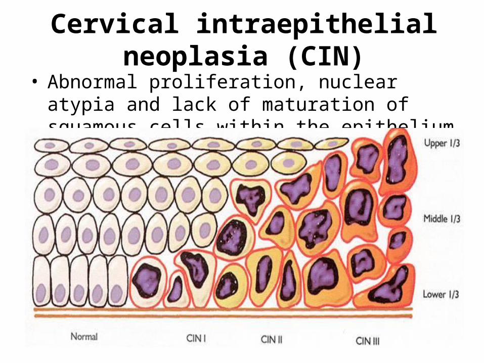
Cervical intraepithelial neoplasia (CIN)• Abnormal proliferation, nuclear atypia and lack of
maturation of squamous cells within the epithelium

Natural history of CIN
• Conflicting data from various studies, however:• 90% of CIN 1 regresses in 2 years• 50% of CIN 2 regresses in 2 years• Approx 10% of CIN 1 and 20% of CIN 2 will progress
to CIN 3• Approx 30% of CIN 3 will regress but roughly 20%
will progress to invasive squamous ca• Mean time for progression from CIN to invasive
cancer approx 10 – 20 years

Human papillomavirus (HPV)• Present in 99.7% of cervical cancer • A causative agent for virtually all cervical cancer (and
CIN and CGIN/ACIS)• 55nm non-enveloped DNA virus

Human papillomavirus• Over 100 sub-types according to gene sequence of L1
capsid protein• Highest risk 16, 18, 31, 45• HPV 16 and 18 together cause 70% of cervical cancer• HPV infection starts at TZ (squamocolumnar junction) • Most HPV infection is cleared by the immune system but
a minority progresses to CIN or invasive carcinoma • Factors influencing progression include HPV type, viral
load, host immunity, parity, smoking, oral contraceptives

Asymptomatic infection
>90%Infection with HPV
15-25 yrs
Koilocytosis and CIN 1
Most
? Appx 30% regression of CIN2/3 Effective immune system
20%
CIN 3
Invasive carcinoma
<10%
Lesions
Elimination of virus
Regression of lesions
Normal cervix
20% progression If untreated
CIN 2
20%

CIN 1 (LSIL)
• Nuclear atypia with mitoses and a high N:C ratio confined to the lower 1/3
• Koilocytosis most obvious in the upper levels of epithelium
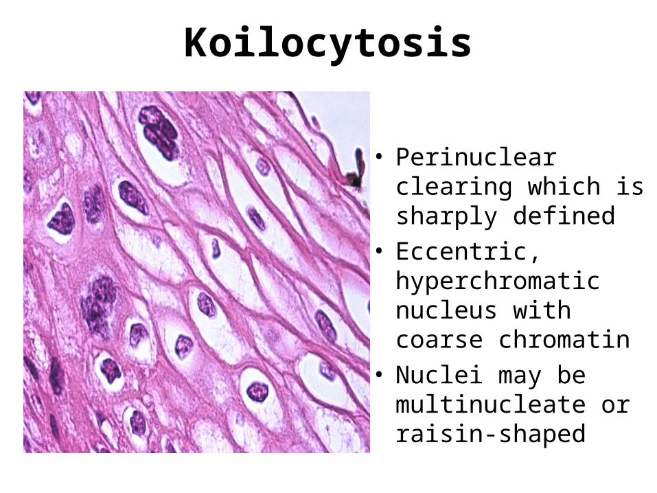
Koilocytosis
• Perinuclear clearing which is sharply defined
• Eccentric, hyperchromatic nucleus with coarse chromatin
• Nuclei may be multinucleate or raisin-shaped
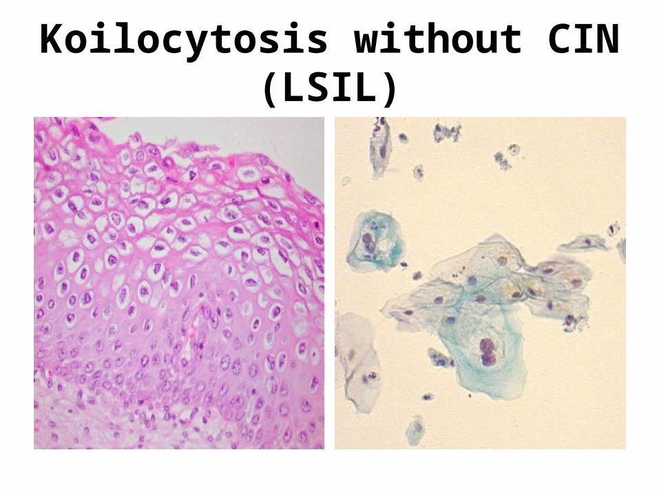
Koilocytosis without CIN (LSIL)

Condyloma acuminatum• Benign papilloma caused
by HPV• Usually low risk 6, 11• More common on vulva• Cauliflower-like growth of
fibrovascular cores lined by squamous mucosa with koilocytosis, parakeratosis
• Essentially LSIL, but occasionally high grade CIN may occur in a condyloma

CIN 2 (HSIL)• Nuclear
atypia with failure of maturation and mitoses in lower 2/3
• Abnormal mitoses may be seen
• Koilocytosis in upper layers

CIN 3 (HSIL)• Nuclear atypia
with failure of maturation in upper 1/3
• Abnormal mitoses
• Koilocytes hard to find
• No invasion

CIN 3

CIN 3

CIN 3 in endocervical crypt

Keratinising CIN 3
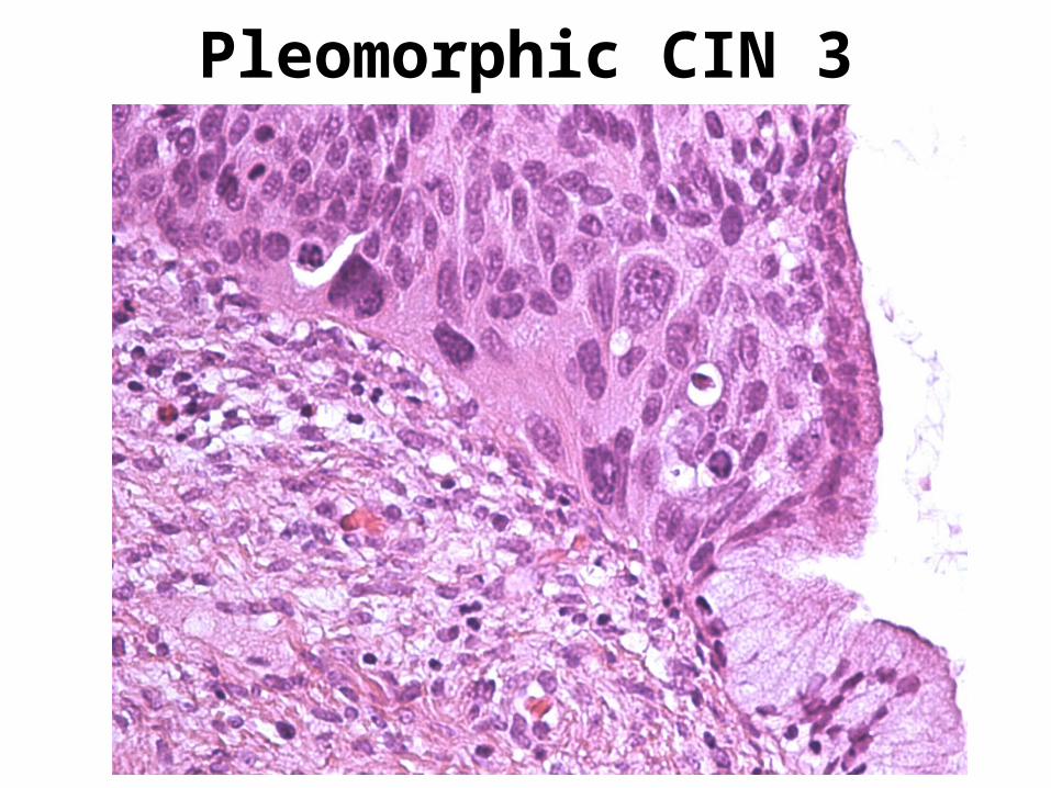
Pleomorphic CIN 3

Immunohistochemistry in CIN 3Ki67 p16

Mimics of CIN: Immature metaplasia
• Stratified layers of cells with little maturation (a high N:C ratio) which may extend into crypts
But:• Nuclei evenly spaced, without much crowding• Regular nuclear outline, without coarse chromatin• Low mitotic rate, without abnormal forms• Layer of residual endocervical epithelium may be present
on the surface (rare over high grade CIN)

Mimics of CIN:Immature squamous metaplasia

Mimics of CIN: Immature metaplasia

Immunohistochemistry of immature squamous metaplasia
Ki67 p16

Mimics of CIN: Atrophy• Stratified layer of cells with little maturation
(ie with a high N:C ratio) But:
• Epithelium is thin• Nuclei are evenly spaced, without crowding• Nuclear pleomorphism and mitoses are absent• Nuclei may be ovoid with longitudinal grooves (so-
called ‘transitional metaplasia’, probably a variant of atrophy)

Mimics of CIN: Atrophy

Mimics of CIN:Atrophy and inflammation

Mimics of CIN: Reactive, inflammatory changes
• Inflammation can cause nuclear enlargement, nucleolar prominence and scattered mitotic figures But:
• Pleomorphism is mild and N:C ratio generally normal• Mitoses are few and only in lower layers• Nucleolar prominence is not a typical feature of CIN• Acute and chronic inflammatory cells are present in the
epithelium together with intercellular oedema• Mild perinuclear clearing of squamous cells may be seen
as an inflammatory phenomenon

Mimics of CIN: Inflammation(with atrophy and tangential sectioning)

Mimics of CIN: inflammation

Inflamed CIN 3

Mimics of CIN: diathermy artefact
• Artefact in surface epithelium adjacent to resection margins due to heat from the loop
• Nuclear hyperchromasia and elongation• Changes most marked in the basal layers with the
overlying surface epithelium often normal• Mitoses not identified• Adjacent epithelium often normal• Low immunohistochemical staining with Ki67

Mimics of CIN: Diathermy artefact

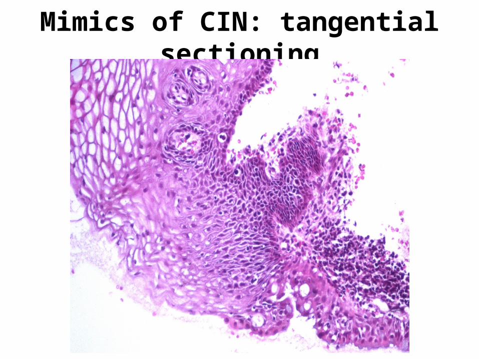
Mimics of CIN: tangential sectioning
• Poor orientation of the biopsy during paraffin embedding so that the histological section is not 90° to the epithelial surface
• The basal layer is sectioned obliquely so that it appears thicker than normal and gives a false impression of lack of maturation
• However, cellular spacing is generally even, without crowding
• Deeper levels cut from the block may clarify the overall architecture of the lesion

Mimics of CIN: tangential sectioning
Mimics of CIN: tangential sectioning

Tangential sectioning (deeper levels)

Conclusions
• HPV infection is common but only a minority progresses to CIN or invasive carcinoma
• This progression is slow and so CIN can be detected by screening before invasion occurs
• CIN is graded 1 to 3 • Mimics of CIN include immature squamous
metaplasia, atrophy, inflammation, diathermy artefact, tangential sectioning