Embed Size (px)
Citation preview

CIBA Special Publication Series No. 74
training Manual on
HealtH management practices for finfisH and sHellfisH of brackisHwater
environment
Coordinated byDr. K. P. Jithendran
Principal Scientist and Scientist In-charge
ConvenorsDr. P. Ezhil Praveena, ScientistDr. T. Bhuvaneswari, Scientist
Edited byDr. Subhendu Kumar Otta, Senior Scientist
Dr. M. Poornima, Senior ScientistDr. P. K. Patil, Senior Scientist
19 - 23 August 2014
AQUATIC ANIMAL HEALTH AND ENVIRONMENT DIVISION
CENTRAL INSTITUTE OF BRACKISHWATER AQUACULTURE 75, Santhome High Road, R.A.Puram, Chennai-600 028


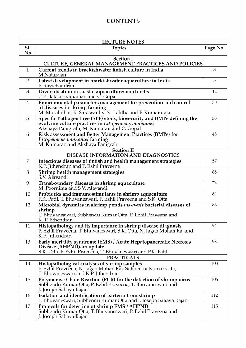
CONTENTS
LECTURE NOTESSl. No
Topics Page No.
Section ICULTURE, GENERAL MANAGEMENT PRACTICES AND POLICIES
1 Current trends in brackishwater finfish culture in IndiaM.Natarajan
3
2 Latest development in brackishwater aquaculture in IndiaP. Ravichandran
5
3 Diversification in coastal aquaculture: mud crabsC.P. Balasubramanian and C. Gopal
12
4 Environmental parameters management for prevention and control of diseases in shrimp farmingM. Muralidhar, R. Saraswathy, N. Lalitha and P. Kumararaja
30
5 Specific Pathogen Free (SPF) stock, biosecurity and BMPs defining the evolving culture practices in Litopenaeus vannameiAkshaya Panigrahi, M. Kumaran and C. Gopal
38
6 Risk assessment and Better Management Practices (BMPs) for Litopenaeus vannamei farmingM. Kumaran and Akshaya Panigrahi
48
Section IIDISEASE INFORMATION AND DIAGNOSTICS
7 Infectious diseases of finfish and health management strategiesK.P. Jithendran and P. Ezhil Praveena
57
8 Shrimp health management strategies S.V. Alavandi
68
9 Transboundary diseases in shrimp aquaculture M. Poornima and S.V. Alavandi
74
10 Probiotics and immunostimulants in shrimp aquacultureP.K. Patil, T. Bhuvaneswari, P. Ezhil Praveena and S.K. Otta
81
12 Microbial dynamics in shrimp ponds vis-a-vis bacterial diseases of shrimpT. Bhuvaneswari, Subhendu Kumar Otta, P. Ezhil Praveena and K. P. Jithendran
86
11 Histopathology and its importance in shrimp disease diagnosisP. Ezhil Praveena, T. Bhuvaneswari, S.K. Otta, N. Jagan Mohan Raj and K.P. Jithendran
91
13 Early mortality syndrome (EMS) / Acute Hepatopancreatic Necrosis Disease (AHPND)-an updateS.K. Otta, P. Ezhil Praveena, T. Bhuvaneswari and P.K. Patil
98
PRACTICALS14 Histopathological analysis of shrimp samples
P. Ezhil Praveena, N. Jagan Mohan Raj, Subhendu Kumar Otta, T. Bhuvaneswari and K.P. Jithendran
103
15 Polymerase Chain Reaction (PCR) for the detection of shrimp virusSubhendu Kumar Otta, P. Ezhil Praveena, T. Bhuvaneswari and J. Joseph Sahaya Rajan
106
16 Isolation and identification of bacteria from shrimpT. Bhuvaneswari, Subhendu Kumar Otta and J. Joseph Sahaya Rajan
112
17 Protocols for detection of shrimp EMS / AHPNDSubhendu Kumar Otta, T. Bhuvaneswari, P. Ezhil Praveena and J. Joseph Sahaya Rajan
115


Section ICulture, General ManaGeMent
praCtiCes and poliCies


Training Manual on “Health management practices for finfish and shellfish of brackishwater environment” 19-23 August 2014 3
CURRENT TRENDS IN BRACKISHWATER FINFISH CULTURE IN INDIA
M. NatarajanFinfish Culture Division
Brackishwater aquaculture has been traditionally practiced in India, mainly in the ‘bheries’ of west Bengal and ‘Pokkali’ fields along the coasts of Kerala state. India has a vast coastline of 8129 km (including A&N and Lakshadweep islands) with many estuaries, creeks, backwaters, lagoons and bays. The total brackishwater area available in the country is estimated to be around 1.2 million hectares, 14% of which has the potential for aquaculture use. About 0.15 million hectares is utilised for coastal aquaculture with an production of 0.5 to 1.0 million tonnes, which is mainly shrimps.
Cultivation of finfishes in brackishwater habitats is practiced mostly as extensive system of culture with low inputs in the form of seed, feed, fertiliser, water exchange etc. Fishes enter the culture system through ‘auto stocking’ during high tide, wherein they are retained for varying periods of time till harvest. In more advanced semi-intensive or intensive systems, selective stocking with desirable species, supplementary or complete feeding, manuring or fertilisation, frequent water exchange and other management measures are undertaken which results in considerably increased yields and profits.
Several species of finfishes are suitable for cultivation in brackishwater environments. Notable among them are the milkfish (Chanos chanos), Grey Mullet (Mugil cephalus, Liza parsia, L. tade, L. cunnesius, L. waigiensis, Valamugil seheli), Pearlspot (Etroplus suratensis), Sand Whiting (Sillago sihama), Rabbit fish (Siganus javus, S. canaliculatus), Seabass (Lates calcarifer), Grouper (Ephinephalus tauvina, E. malabaricus), Rawas (Polynemus sp.), Red Snapper (Lutjanus spp), and Sea Bream (Lethrenus sp.).
Hatchery produced stocking material (fry and fingerlings) of Asian seabass and pearlspot is currently available in the country with the development of successful breeding and seed production techniques at CIBA. Seabass breeding technology has been transferred to RGCA and they are also supplying seabass seed and fingerlings to interested farmers. The other varieties are generally collected from the wild during their respective breeding seasons, by fishermen and sold for farming. Concerted efforts are underway in organisations such as CIBA and CMFRI to artificially breed and produce stocking materials of other potential species from the hatchery.
Each of the species have specific biological requirements and accordingly the methods and systems suitable for their cultivation varies. Carnivores such as Asian seabass, groupers, snappers etc. are cultured in ‘monoculture’ mode, while omnivores or herbivores such as mullets, milkfish, pearlspot etc. are amenable for cultivation in ‘polyculture’ mode in varying ratios. However, monoculture in ponds and pens is the most preferred and profitable method practiced in many south east Asian countries. Bamboo pens or enclosures are erected in natural brackishwater bodies(Chilka), but in India, this practice is limited because of various reasons. Pond culture of mixed varieties of finfishes is more prevalent in states of Kerala, Andhra or West Bengal.
Training Manual on “Health management practices for finfish and shellfish of brackishwater environment” 19-23 August 20144
Cultivation in cages is more desirable for carnivores such as groupers and seabass because of their feed requirement with trash fish and aggressiveness. Frequent sorting and segregation of larger sized ‘shooters’ is an essential step in rearing of these predators. Various viable designs are available for construction of low cost cages which are suitable for erection in shallow coastal waters’.
Farming techniques adopted in India for brackishwater fish is different from those used in other countries and also differ from region to region within the country. Interest in commercial cultivation of Asian seabass as a profitable venture has increased mainly because of the availability of hatchery produced seed. CIBA and RGCA are presently doing this. Hatchery bred pearlspot seed of uniform sizes are also being regularly supplied, particularly to Kerala farmers. Simple but effective tank based and hapa based methods of seed production of pearlspot have been developed and are available for dissemination. Pond culture of milkfish in the state of Gujarat is picking up fast with the efforts of CIBA by supplying nursery reared milkfish fingerlings, for culture demonstrations. Establishment of broodstock banks and seed propagation centres in strategically located sites along the Indian coast would enhance the scientific cultivation of brackishwater fishes in India.

Training Manual on “Health management practices for finfish and shellfish of brackishwater environment” 19-23 August 2014 5
LATEST DEVELOPMENT IN BRACKISHWATER AQUACULTURE IN INDIA
P. Ravichandran Crustacean Culture Division
1. INTRODUCTION
Brackishwater aquaculture has been one of the important foreign exchange earning sectors for the country. In India, presently shrimps are the major constituent of coastal aquaculture production. Finfish production is negligible and they form a part of the production from traditional polyculture systems and a limited level of carnivorous fishes like seabass and cobia. In the financial year 2013-14, the export of marine products reached an all-time high of above Rs. 30,000 crores. Shrimp alone contributed about Rs. 19,000 crores, of which 73% was from cultured shrimps . The present article discusses the coastal resources available in the country, the development of aquaculture, the status of the technologies available in the country along with recent developments in the sector, and the future perspectives.
2.1 LAND RESOURCES
‘Brackishwater’ is defined as ‘mixing of seawater with fresh water’, as in estuaries and backwaters where the salinity of the water is more than freshwater but less than sea water’. Indian coastal area has 9 states, 2 island territories and 2 Union territories with a total coastline of 7516.6 km with mainland contributing about 5422 km and the Island territories about 2094 km. India by virtue of its extensive geographical stretch and varied terrain and climate supports coastal wetlands of about 43230 km2. It has 97 major estuaries and 34 major lagoons. There are 31 mangrove areas with the total extent of 6740 km² (57% East coast, 23% west coast, 20% Andaman &Nicobar Islands) .
India has a variety of natural coastal ecosystems. The mainland coastal area comprises of 43% of sandy beach, 11% of rocky coast, 36% of muddy flats and 10% of marshy coast. The islands of Lakshadeep are composed of atolls while the Andaman and Nicobar Islands are volcanic in origin, arising from a submerged mountain chain.
The coastal areas are productive and rich in natural resources. India has 14 major river systems, which has led to the formation of wide network of creeks and estuaries in the coastal areas of the country thus facilitating the coastal aquaculture. The Ministry of Environment and Forests, Government of India estimated that India has total estuarine area of 3.9 million ha and backwaters of 3.5 million ha. Among these coastal salt affected lands 1.2 million has been identified to be potentially suitable for shrimp farming. West Bengal and Gujarat are the two States which have the majority of the potential area because of the high tidal amplitude. The state-wise details of the potential area for brackishwater aquaculture is presented in Fig. 1

Training Manual on “Health management practices for finfish and shellfish of brackishwater environment” 19-23 August 20146
2.2 BIOLOGICAL RESOURCES
Potentially India has a vast resource of crustaceans, molluscs and finfishes that could be cultivated in the brackishwater/ marine environment. They are listed below:
Shrimps : Penaeus monodon, Fenneropenaeus indicus, F. merguiensis, F.penicillatus, Marsupenaeus japonicus, P. semisulcatus Metapenaeus monoceros, M. dobsoni and M. kutchensis.
Crabs : Scylla serrata, S. tranquebarica Fin-fishes
Sea bass : Lates calcarifer
Mullets : Mugil cephalus, Liza macrolepis, L. tade and L. parsia.
Milk fish : Chanos chanos
Snapper : Lutjanus spp.
Grouper : Epinephelus spp.
Pearlspot : Etroplus suratensis
Sea weeds : Gracilaria edulis and G. acerosa Kappaphycus alvarezii
3. Area under culture and production
Out of the total potential area available hardly 16% has been developed into shrimp farming which includes 4% of traditional farming in West Bengal, Kerala, Goa and Karnataka. The shrimp aquaculture production showed a phenomenal increase between 1990 to 1995 and thereafter there was stagnation during 1996 to 2000. From 2000 onwards there was a gradual increase in production which reached a maximum of 1, 40, 000 mt in 2006-07. But in 2007-08 and 2008-09, the production levels reduced drastically and reached the pre-1995 level of 75,000 mt. Introduction of SPF Litopenaeus vannamei in the country resulted in reviving the shrimp culture with production levels reaching nearly 300,000 tonnes in 2013-2014. (MPEDA, 2014) The details are presented in Fig. 2.

Training Manual on “Health management practices for finfish and shellfish of brackishwater environment” 19-23 August 2014 7
4 Introduction of SPF L. vannamei
Government permitted introduction of SPF L. vannamei in 2003 as a pilot programme for two hatcheries. Later in 2007, constituted a committee with CIBA and NBFGR to conduct risk analysis study for the introduction of L. vannamei in large scale in the country and as per the findings of the study, the remedial measures for the identified risks were indicated and the following guidelines were formulated.
Risks and Guidelines to mitigate the risks
In 2009, L. vannamei imports in large scale was permitted through centralized aquatic quarantine facility. Since then the shrimp production levels have increased from 1 lakh tonnes to 3 lakh tonnes in 2013 with L. vannamei contributing nearly 50%. With increased global demand and prices due to reduced supply, shrimp farming sector in the country is well placed. But still there are heavy losses due to shrimp diseases like WSSV, IHHNV and other unidentified

Training Manual on “Health management practices for finfish and shellfish of brackishwater environment” 19-23 August 20148
diseases. Two important aspects that require additional consideration are Biosecurity in farming areas and Assured supply of quality SPF seed. Though the regulation stipulates it, it can only be achieved fully through awareness creation among the stakeholders.
5. New Systems of farming
5.1. Organic farming
Organic farming is another way of adding value to the produce. This is also another form of certification scheme where the standards specify use of organically produced inputs and maintenance of welfare of the animals with minimal disturbance to the ecological conditions of the farming area. Though organic agricultural products are already being produced and marketed, it is still in a very formative stage in aquaculture. The major issues in the organic standards for shrimp especially tiger shrimp are
�� Induction of maturity is through eyestalk ablation which involved mutilation
�� Non-availability of domesticated SPF brood stock
�� The upper limit of production level specified is too low
�� The fish meal component in feed should be from a sustainable source
In India under the initiative of MPEDA, organic certification for scampi, Machrobrachium rosenbergii, culture as per the standards of Naturland has already been done as a group activity which fetches a premium of 20% on the sale price for the producers. Traditional shrimp farming in West Bengal and Kerala, which do not use any input could be easily taken up for organic certification. CIBA has successfully developed an organic shrimp feed and the feed mill has been certified for the production of organic shrimp feed. In collaboration with a private entrepreneur demonstrated the organic shrimp production in Kerala. Agriculture Products Export Development Authority (APEDA) has developed guidelines for certifying organic shrimp and its approval by EU countries will help reducing the cost of certification.
5.2 Biofloc based farming
Biofloc farming system become popular among the shrimp farmers because of its following advantages
�� Limited or zero water exchange
�� Increased bio-security
�� Environmentally friendly system - limited water exchange in the system reduces the nutrient-rich effluent discharge that often occurs in semi-intensive or intensive shrimp farms
�� Super-intensive biomass density leading to a significant increase in productivity
�� Decreased feed costs as shrimp can eat the bio-flocs (bio-flocs have good nutrition quality)
Training Manual on “Health management practices for finfish and shellfish of brackishwater environment” 19-23 August 2014 9
All of these traits of the bio-floc shrimp farm system contribute to a much more productive and cost-effective system than traditional semi-intensive or intensive shrimp farms. The capital costs are most likely higher, but it will pay off in spades with the increased biomass yields with each harvest.
5.3 Intensive farming in Recirculation Systems
A recirculating aquaculture system (RAS) is an enclosed system where the only water replacement is the water lost to evaporation and cleaning. The RAS has the following advantages:
a) lower water requirements,
b) lower land requirements,
c) reduced labor requirements,
d) increased control over water quality parameters,
e) lower risk of negative impact from adverse weather conditions,
f) lower risk of creating adverse environmental impacts, and
g) increased biosecurity
These systems are being deployed in developed countries where coastal land costs and labor costs are very high. RAS systems have become very popular for fish culture especially for intensive cultures. Shrimp farming, which is plagued with disease outbreaks , also look forward for introducing RAS systems both in the maturation systems in hatcheries and in land based tank cultures.
6. Diversification into other species
6.1 Other species of Shrimps
Indian white shrimp, Fenneropenaeus indicus
F. indicus was one of the mostly studied shrimp species in the country. CIBA had developed a very simplified small-scale hatchery technology for F. indicus. Technology for improving the traditional culture in Pokkali fields of Kerala was developed and tested after a detailed monitoring of the system of farming practiced. A nutritional requirement of F. indicus was studied and indigenous low-cost feed was developed for the species. F. indicus was domesticated and pond-grown broodstock was successfully matured and bred under captivity.
During the initial phase of development of shrimp farming in the country, semi-intensive culture of F. indicus has been taken up by many entrepreneurs. But later, it was discontinued because of the low price it fetched in comparison to tiger shrimp. Development of SPF broodstock of native F. indicus is being suggested instead of importing the exotic White-legged shrimp, L. vannamei. A flagship programme on the genetic improvement and domestication of F. indicus is being taken up by CIBA.
Kuruma shrimp, Marsupenaeus japonicus
Live M. japonicus has a niche market in Japan where it fetches a high price of about $200 per kilogram. M. japonicus tolerates low temperatures of up to 10oC and requires sandy bottom and

Training Manual on “Health management practices for finfish and shellfish of brackishwater environment” 19-23 August 201410
high salinity for growth. It is the most suitable species for sea based farming systems, where sandy bottom is available. It will be also suitable for North-western coastal areas where low temperature prevails during winter. CIBA has successfully domesticated the shrimp and rose upto F6 generation. Preliminary studies have indicated that the domesticated stock, through inadvertent selection, has acquired resistance to WSSV. To assess the extent of resistance to WSSV and its heritability, quantitative genetic studies have been initiated. Experiments have indicated the growth and production of the species is comparatively low with higher protein requirement. But live export of the species to Japanese market will fetch a very high profit.
Banana shrimp, Fenneropenaeus merguiensis
F. merguiensis has attracted the attention of shrimp culturists because it tolerates wide range of temperature and salinity and also low water quality. It matures and breeds easily in captivity. Studies on the maturation, breeding and culture of F. merguiensis has been undertaken by CIBA to provide an alternate species to P. monodon in the West coast especially in Gujarat, Maharashtra and Goa, where this species is naturally distributed. Seed production of the species from captive broodstock has been achieved. Culture technology has been successfully demonstrated in Gujarat. This species is now suggested for winter culture in northern coastal states like Gujarat and Maharashtra.
Mud crab, Scylla serrata and S. tranquebarica
Mud crabs have high export potential and is one of the important candidate species for brackishwater aquaculture. CIBA has standardized the techniques for development of captive broodstock, induced maturation, production of berried females and larval rearing for both the species. Similarly, RGCA has also developed the larval rearing techniques for both the species of mudcrabs.
Technologies for fattening, grow-out culture in ponds and in cages have been developed for both the species of mud-crabs. The major issue in crab culture is the development of nutritionally adequate palletized feed. CIBA has developed a balanced diet for fattening of mudcrabs which has given promising results when tested in cage crab fattening trials carried out by SHGs. Nursery rearing techniques for the crab megalopa has been developed and demonstrated in ponds as well as in tanks. Culture of S. serrata has been successfully demonstrated in West Bengal in farmers’ ponds.
FINFISHES
Asian sea bass, Lates calcarifer
Sea bass is one of the most preferred fish. CIBA has successfully bred the species for the first time in the country in 1997. Seed production technology has been perfected and year-round breeding of the species has been achieved. The technology for nursery rearing for the production of fingerlings have been developed and demonstrated which has been taken up by small farmers and WSHGs.
Technology for the culture of sea bass in ponds has been standardized with slow sinking feed developed by CIBA . The culture is followed in three stages – Nursery, Pre-grow-out and Grow-out. The technology has been demonstrated in six coastal states in farmers’ ponds and

Training Manual on “Health management practices for finfish and shellfish of brackishwater environment” 19-23 August 2014 11
there is a wide spread demand for the seed and feed. The major issue in the adoption of sea bass culture is the low profit margin in comparison to shrimp culture.
Grey mullets, Mugil cephalus, Liza spp
Grey mullets form an important component in the traditional farming systems. Among grey mullets, M. cephalus is the largest and fastest growing species and is suitable for monoculture. Other species are suitable as components of polyculture. Technologies have been developed for both the systems of farming. The profitability of the culture of mullets is low since they fetch a low price in the market compared to sea bass. Though successful induced breeding of mullets was achieved, the larval rearing was successful only in the case of L. macrolepis and L. parsia. Larval rearing of M. cephalus is yet to be standardized. Studies have been conducted on the hormonal influence on male maturity, cryopreservation of milt, influence of photoperiod on maturation and nutritional requirements of broodstock.
Pearl spot, Etroplus suratensis
E. suratensis is one of the candidate species which has regional preference in Kerala. This has also got an ornamental value. Technologies for the breeding of the species in ponds using salinity manipulation and breeding in cement tanks have been standardized. Culture technology using pelleted feed is being tested. The growth is comparatively low in the species and commercially it will be viable only when marketed in Kerala.
Conclusion
Brackishwater aquaculture in the country is presently anonymous with shrimp culture and with the increasing production levels of L. vannamei, it is most certain that P. monodon culture may be restricted only in traditional systems of farming. For sustainability of brackishwater aquaculture, it is necessary that we diversify into other crustaceans and fishes in commercial scale. We can sustain aquaculture only when it is regulated properly and integrated with the Coastal Zone Development Plans of all the maritime states and UTs.

Training Manual on “Health management practices for finfish and shellfish of brackishwater environment” 19-23 August 201412
DIVERSIFICATION IN COASTAL AQUACULTURE: MUD CRABS
C.P. Balasubramanian and C. GopalCrustacean Culture Division
Mud crabs are one of the most traded aquaculture crops. The pond based farming of mud crab has been practicing in China for at least 100 years and more than 30 years in Asian countries (Balalio, 2004). However, the mud crab was an incidental or secondary crop in shrimp and milk fish farming until recently. Owing to the high market value and profitability, the aquaculture of mud crab has received impetus in early 1990s. Considerable efforts have been made in the last few years to develop effective grow out technology for mud crab. Since 1970s, steady interest has been shown in culture of Scylla spp in many tropical Asian countries. Farming of mud crab is considered to be an important and valuable industry, offering advantages from a number of aspects: 1) uncomplicated technology, 2) abandoned shrimp ponds can be converted, 3) international markets, 4) native species to many tropical Asian countries, 5) easy transportation, potential for rural as well as industrialized aquaculture, 6) individual animals are valued in contrast to penaeid shrimps and 7) resilience of resources. Any aquaculture industry is composed of three sequential phases: seed production, nursery and grow out. The first part of this write up covers the current status of technical know-how on various sub sectors of mud crab farming, such as seed production, nursery and grow out. In the subsequent section, technical gaps and researchable issues are highlighted. Suggested package of practices, present status of adoption and issues related to promotion and commercialization are summarized in the later sections.
Current status of technology developed
Biology of Mud crabs
Aquaculture is essentially, a life-science based research, and basic knowledge on the aquacultured species is the essential prerequisite for the development and management of aquaculture
Taxonomy
Taxonomy of genus Scylla has been considerably confused. Estampador (1949) recognized three species and one variety. His classification was mainly based on coloration, morphological characters and behavior. Although many authors accepted Estampodaor’s classification, Stephenson and Campel (1960) concluded that there was insufficient evidence for separation of species beyond mono-specific term Scylla serrata. Recently, the taxonomy of genus Scylla was revised and confirmed the existence of four species (Figure 1) based on morphometric analysis, allozyme electrophoresis and mitochondrial sequences. Further, Indian mud crabs of genus Scylla was revised recently using DNA markers, and it is demonstrated that mud crabs of India are composed of two species: S. serrata and S. olivacea, which have long been misidentified as S. tranquebarica and S. serrata respectively.
Life History
Mud crab’s natural history can be considered as catadromous: adult spawn in the open ocean but young migrate inshore. The various stages of development are shown in Fig 2.

Training Manual on “Health management practices for finfish and shellfish of brackishwater environment” 19-23 August 2014 13
Scylla serrata Scylla olivacea
Fig.1 Identification of mud crab species: Scylla serrata two spines on the wrist, where as S. olivacea has only one spine.
Figure 2 Generalized life cycle of Scylla spp.

Training Manual on “Health management practices for finfish and shellfish of brackishwater environment” 19-23 August 201414
Biology of Crab Reproduction: The sex of crabs can easily be determined by external features. Male crabs are characterized by inverted ‘T’ Shaped abdomen, whereas in females the abdomen is semicircular. In addition the male has relatively larger chelate, and a general trimness for body contour than females. Males have two pleopods that modified as copulatory organs on the first and second abdominal segments. In the case of females first four abdominal segments carry pleopods, which are biramous and possess setae for attachments of eggs for brooding.
The female reproductive system comprises a pair of ovaries, a pair of spermatheca (= seminal receptacle) and a pair of vagina. The ovary is ‘H’ shaped and located dorsally just beneath the carapace. The horns of ovary extends anterolaterally from either side of the gastric mill and dorsal to the hepatopancreas. Two posterior horns, which lie ventral to the heart, extend posteriorly on either side of the intestine on either side of the intestine. The seminal receptacle arises from mid lateral border of the posterior horns. Each antennal receptacle leads into a narrow vaginal tube which further open outside through small circular gonopore situated ventrally. Eggs are produced in the paired ovaries. Sperms produced in the testes opens into coiled tubes (vas deferens) that package mature sperms into gelatinous bundles (spermatophore) for transfer to females. In natural conditions mud crabs attain sexual maturity at between 18 and 24 months.
Mating takes place in the estuarine environment, after which female crabs migrate to the sea where spawning takes place (Arriola, 1940; Ong, 1966). Berried S. serrata females have been caught in trawl nets up to 80 km from the shore in Australia (Poovichiranon, 1992; Hill, 1994). Spawning appears to occur throughout the year with some seasonal peaks (Heasman et al., 1985; Quinn and Kojis, 1987). These peaks seem to be related to seasonal rainfall for tropical populations, while in temperate regions reproduction is more strongly related to temperature, with a peak in spawning activity in the summer months (Heasman et al., 1985). S. serrata is highly fecund with up to 8.36 million eggs per female (Mann et al., 1999). The zoeal larvae develop and remain in the open ocean until they reach the megalopa stage, after which they migrate back into the estuarine environment (Keenan, 1999). Little is known about the oceanic phases of the life cycle.
Mating: The mature female releases a chemical attractant (pheromone) into the water, which attract males. The successful male picks up the female and carries her around for several days until she molts. Copulation can occur only when females are in soft shell condition. Male deposits Spermatophore inside the female storage sac (spermatheca) by using male first pleopods. The Spermatophore can remain viable, until fertilization takes place, for weeks or even months. When the eggs complete vitellogenesis they are passed down and fertilized by stored sperms, and extruded onto pleopods. The eggs adhere to the pleopod hairs and female is said to be berry or ovigerous.
Ovarian development: The classification of ovarian maturation provides a guide for broodstock management in hatchery facilities. For hatchery operation, animals with ripe or late maturing ovaries should be selected to minimize the use of resources such as time and money. The colour, size and texture of ovary of mud crabs are closely related to its cellular development. Based on external morphology and light microscopy, Quinitio et al, (2007) classified the ovarian development stages of S. serrata into five stages: immature (ovary thread like, sometimes difficult to discernible and transparent/transluscent), early maturing (ovary is yellow), later maturing (ovary is massive with apparent lobules and slight orange), fully mature (ovary occupies most of the internal cavity, orange to dark orange) and spent (ovary is similar to early maturing).
Training Manual on “Health management practices for finfish and shellfish of brackishwater environment” 19-23 August 2014 15
Incubation: Mud crabs brood their eggs, as all other pleocemata. During the incubation period, females stop feeding and therefore animals generally avoid ‘baited lift nets and ‘traps’. Egg incubation period generally varies from 7 to 14 days, but the duration of incubation is greatly influenced by the rearing water temperature. Egg incubation period is tested at different temperature (20 to 30ºC) and found that incubation period decreased exponentially with increasing temperature.
Larval stages: There are five zoeal stages passing through five molts to reach the megalopa stage. At a salinity of 31 ppt development from zoea 1 to megalopa requires 16-18 days; each zoeal stage takes minimum period of 3-4 days before it molts in to the next stage. The megalopa takes 11-12 days before it molts into the first crab stage; at lower salinity in the range of 21-27 ppt this period is reduced to 7-8 days. The faster rate of megalopa in lower salinity indicates that the megalopa in nature move shoreward into brackish water.
Description of Zoea (From Ong, 1966 ): The zoea are of typical brachyuran type with long rostral and dorsal spines. The abdomen in all stages have has lateral knobs on second and third pleomeres. Identifying characteristics of different zoeal stage of S. tranquebarica is given tin the Table 1
First Zoea: Body length 1.15 mm; eyes sessile. Antenna unsegmented and bears short setae apically. Mandible is broad with two large teeth and serrated edges. Maxilla with two segmented and unsegmented endopodite; the first and second maxillipeds bears four natatory setae. The abdomen is made up of five pleomeres. The telson bears a pair of long dorsolateral spines.
Second Zoea: Body length 1.51 mm; eyes stocked; Exopodite of both maxilliped bear six natatory setae. Telson has a pair of small setae at the inner margin of furca.
Third Zoea: Body length 1.9; Larger antennule than second zoea; antenna has developed a small bud. Exopodite of second maxilliped with 9 setae.
Fourth Zoea: Body length 2.4 mm; Antennule bears aesthets in a terminal group and a subterminal group; Flagellum of antenna elongated; first maxilliped bears 10 natatory setae; second maxilliped bears 10 natatory setae and one or two short setae. Rudiments of third maxilliped appear. Abdomen has bud on pleormeres 2-6. The telson grows additional setae between the innermost pair.
Fifth Zoea: Body length 3.43 mm; first maxilliped bears 11 long setae; second maxilliped has 12 setae. All the pereiopods are elongated and shows the signs of segmentation. Pleopod buds are well developed. Five pairs of setae are on the telson furca.
Megalopa: Single megalopa stage similar to other portunids; carapace length 2.18 mm; carapace width 1.52 abdominal length 1.87. The abdomen has five pair of pleopods.

Training Manual on “Health management practices for finfish and shellfish of brackishwater environment” 19-23 August 201416
Table 1: Summary of different zoeal characteristics of Scylla tranquebarica (Ong 1967)
Stages Size (mm)
Eyes Setae (2 nd max-illiped)
Append-ages (tho-racic)
Setae (middle furca)
Zoea 1 1.1 Sessile 4 Nil 3 pairsZoea 2 1.5 Stalked 6 Nil 4 pairsZoea 3 1.9 Stalked 9 Starts de-
veloping4 pairs
Zoea 4 2.4 Stalked 10 Large 4 pairs+middle one
Zoea 5 3.4 Stalked 12 Large 5 pairs
Hatchery production of mud crabs: This section is dealt with three subsections: Facility, Broodstock management and Larval rearing. For the development of mud crab hatchery, most of the shrimp hatchery can be converted into crab hatchery.
Facilities
Broodstock and larval rearing tanks: Tanks may be made of concrete, fibreglass, or wood lined with rubberized canvas. These can be circular, oval, or rectangular. However, rounded corners are preferable due to more effective water circulation. Tank capacity may vary from 1-10 mt for broodstock and 1-5 mt for larval rearing tanks.
Algal culture tanks: The green phytoplankton, Chlorella is needed for rotifer, Brachionus. Algal tanks must be shallow to allow enough light penetration. Barchionus are cultured in 5-10 mt tanks.
Spawning tanks: It is advantageous to have smaller round tanks with volumes ranging from 300 to 500 L tanks where berried tanks are held and allowed to hatch their eggs.
Artemia hatching tanks: Nauplii of Artemia or brine shrimp are protein rich organisms given to larvae starting second or third zoea. Tank capacity of Artemia varies from 30 to 50 L.
Reservoir: Storage tanks are necessary for chlorination and holding of filtered and treated water for daily use. An elevated storage tank that can distribute seawater to other tanks by gravity is advantages.
Seawater system: Seawater may be pumped from the sea or sump pit. Water is passed through sand filter, which is usually elevated prior to storage.
Other equipments and accessories: Other equipments and accessory such as refrigerator, weighing balances, Refractometer, pH meter and drainers etc are equally important in hatchery operations.
Broodstock management: Females of S. serrata can be obtained form fishers or landing centres. Male crabs are not required for hatchery operations as almost all matured crabs in wild would have mated. Animals range in size above 300 g (S. serrata ) and should be selected for larval

Training Manual on “Health management practices for finfish and shellfish of brackishwater environment” 19-23 August 2014 17
production. Further, females were identified as being matured by their wide, dark, U- shaped abdomen fringed with setae. Immature females were typically characterized by having an abdomen resembling that of male with slightly convex side and without setae. Maturity can be assessed by observing through gap at the junction of carapace and abdomen (Fig.3)
Fig. 3. In vivo evaluation of ovarian stage of Scylla tanquebarica
Transport: Crabs for transport are tied with twine to render the claws immobile. They can be kept out of water in cardboard cartons for two days. The bottom and sides of containers are lined with damp mangrove leaves, wooden shavings, or damp sackings. As dehydration affects survival of crabs, it should not be subjected to drying winds during transport. Likewise exposure of direct sunlight for long period could lower survival.
Disinfection: Under culture conditions, the ability to control disease is vital because the potential for pathogen proliferation increases with the density of cultured animals. Formalin has extensively been used in crustacean culture for disinfecting and disease prevention. Therefore, newly caught animals should be disinfected with formalin to reduce the number of symbionts and parasites. Formalin doses and exposure time varies widely between studies with doses ranging from 100 ppm for 1 h to 50 ppm for 20 min.
Eyestalk ablation: As the occurrence of berried crabs in nature is rare, it is essential to develop ovigerous crabs in captivity. Eyestalks are the sites of gonad inhibiting hormones and therefore the removal of eyestalks accelerate the gonad development and spawning. One of the eyestalk is removed. Intact animals can also be used as broodstock, however, the time to get ovigerous crab extended according to the ovarian stages of the animal. Reproductive performance of intact animals is significantly greater (Table 2) in intact animals (Millanema and Quiniito, 2000).
A sandy substratum should be provided in the spawning tank. Female crabs kept in a tank that has bare floor may often drop their eggs during spawning because eggs fail to remain securely attached to their pleopods. Half of the broodstock tank can be provided with 10 cm sand layer and another half can be bare floor for feeding purpose (Fig 4). Alternatively sand filled trays can be provided (Fig 5). Crabs can be stocked at the rate of one animal per 1 m or one per sq.m.

Training Manual on “Health management practices for finfish and shellfish of brackishwater environment” 19-23 August 201418
Fig. 4. Broodstock tank for Scylla spp. Half of the portion is provided with a sandy substratum
Fig 5: Broodstock tank of Scylla spp. Sand filled basin are provided for spawning
Table 2 .Reproductive performance of ablated and intact Scylla serrata (=S. tranquebarica) (Millamena and Quinitio, 2000)
Variables Ablated Intact
No viable spawning 8(40%) 12(60%)Mean eggs per BW 4437 5124Mean eggs fertilization rate (%) 58 80Total No of Zoea (million) 15.67 20.49Broodstock survival (%) 42 83

Training Manual on “Health management practices for finfish and shellfish of brackishwater environment” 19-23 August 2014 19
Feeds and Feeding: Broodstocks are fed with natural feeds such as molluscs, polychaete at the rate of 10% body weight. Uneaten feeds should be removed daily by siphoning the tank prior cleaning
Water management: Seawater with 28-35 ppt is used for crab broodstock, and water in the tank is changed daily. Sand substrate should be cleaned twice week.
Spawning and hatching: Crabs should be checked daily to know whether spawning has occurred or not. Once crabs spawned they are placed in the basin containing 150 ppm formalin for 30 min., and stocked individually in 300-500 L spawning tanks for subsequent spawning.
Larval rearing operations
Preparation of larval tanks for stocking: The tanks should be disinfected with 200 ppm chlorine water for 8-10 h, and scrubbed with a mixture of 200 ppm Chlorine and 5% detergent by using sponge pads. Then the tanks are thoroughly rinsed with fresh water and drained at least for 24 h. Just before filling the tanks it should be rinsed with fresh water. Before stocking zoea, algae (Chlorella) should be added at the rate of 50, 000 cells/ml. The tank water should be aerated mildly. Micro algae do not provide any nutritional benefits; it may enhance the water quality.
Acclimation and stocking: The larvae are estimated in the spawning tank directly. Aeration should be taken out before collecting the larvae, and the waste products settled down at the bottom should be siphoned out. Only active larvae are stoked in the larval rearing tank at 10 – 50 individual per litre. Active larvae are photo tactic; hence they swim up to the surface. The zoea received from the hatching tanks should be acclimatized by adding the larval tank water to the acclimatization basin. The acclimatized zoea can be released slowly in to the tank in small quantities.
Feeding: The most critical component of the mass larval rearing of aqua cultured species is the standardization of feeding regime. The feeding regime for mass rearing of Scylla has yet to be standardized. Nutrition has been suggested as a possible cause of mass mortalities experienced during the mud crab larval rearing. Absence of an optimal feeding regime may be the fore most reason for the failure of hatchery production of mud crab larvae. Many experiments were conducted in India and elsewhere using a variety of live feed organisms such as veliger of oysters, copepods, rotifers, artemia nauplii and micro algae. Trials were conducted using these live feed organisms individually or in combination. Heasman and Fielder (1983) reported their highest survival of 26% form zoea to first crab instar when larvae fed solely on artemia nauplii whereas Marichamy and Rajapakyam (1991) reported a maximum survival from first zoea to first crab instar when they used a combination of rotifer and artemia nauplii. In India Anil and Suseelan (1999) conducted experiments on feeding of S. tranquebarica (as S. oceanica). They used 3 feed combinations: 1) frozen artemia nauplii, rotifer and micro encapsulated feed 2) Frozen artemia nauplii, rotifer and chlorella and 3) artemia nauplii in suspension in addition to the fresh artemia nauplii (15 -20 individual/ ml), rotifer (Brachionus 20 individual/ ml) with antibacterial compound prefuran. The best survival obtained for the third combination (23%). Although artemia alone can be used and successful larval production is achieved by some authors, most of the authors reported with convincing evidence that rotifer is an indispensable component of mud crab larval rearing. Rotifers are significantly smaller (0.5 µg and 45 -200 µm) than artemia
Training Manual on “Health management practices for finfish and shellfish of brackishwater environment” 19-23 August 201420
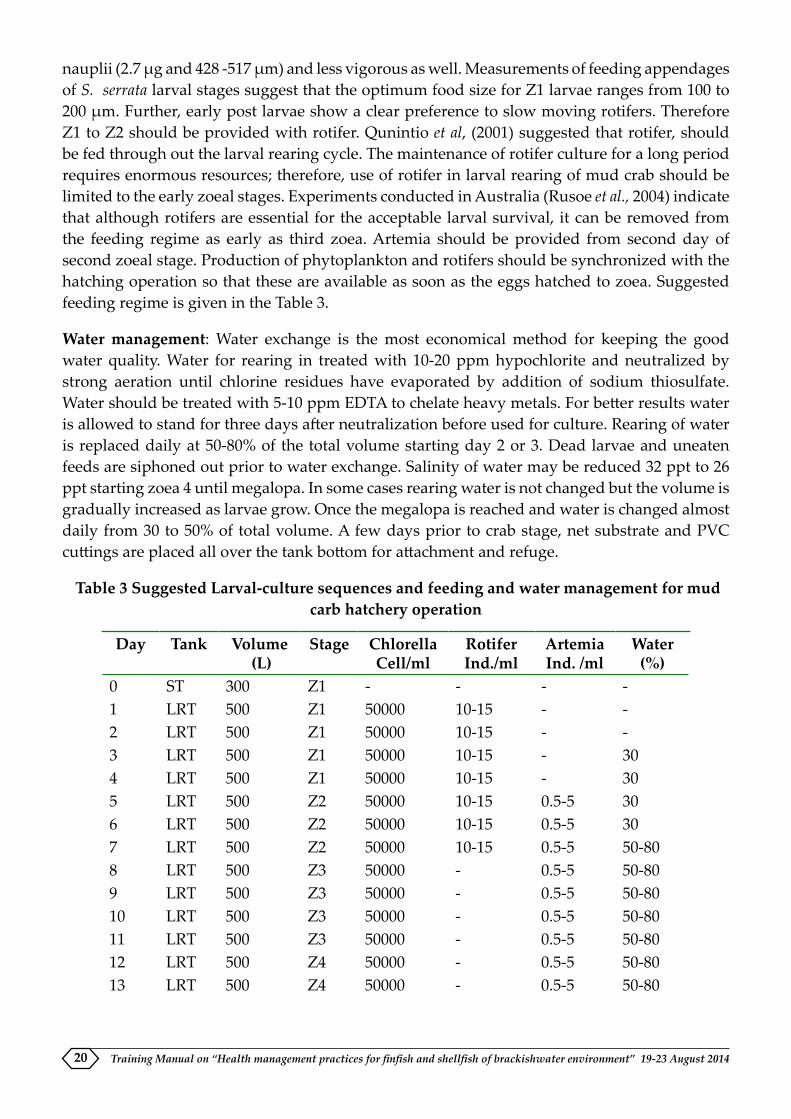
nauplii (2.7 µg and 428 -517 µm) and less vigorous as well. Measurements of feeding appendages of S. serrata larval stages suggest that the optimum food size for Z1 larvae ranges from 100 to 200 µm. Further, early post larvae show a clear preference to slow moving rotifers. Therefore Z1 to Z2 should be provided with rotifer. Qunintio et al, (2001) suggested that rotifer, should be fed through out the larval rearing cycle. The maintenance of rotifer culture for a long period requires enormous resources; therefore, use of rotifer in larval rearing of mud crab should be limited to the early zoeal stages. Experiments conducted in Australia (Rusoe et al., 2004) indicate that although rotifers are essential for the acceptable larval survival, it can be removed from the feeding regime as early as third zoea. Artemia should be provided from second day of second zoeal stage. Production of phytoplankton and rotifers should be synchronized with the hatching operation so that these are available as soon as the eggs hatched to zoea. Suggested feeding regime is given in the Table 3.
Water management: Water exchange is the most economical method for keeping the good water quality. Water for rearing in treated with 10-20 ppm hypochlorite and neutralized by strong aeration until chlorine residues have evaporated by addition of sodium thiosulfate. Water should be treated with 5-10 ppm EDTA to chelate heavy metals. For better results water is allowed to stand for three days after neutralization before used for culture. Rearing of water is replaced daily at 50-80% of the total volume starting day 2 or 3. Dead larvae and uneaten feeds are siphoned out prior to water exchange. Salinity of water may be reduced 32 ppt to 26 ppt starting zoea 4 until megalopa. In some cases rearing water is not changed but the volume is gradually increased as larvae grow. Once the megalopa is reached and water is changed almost daily from 30 to 50% of total volume. A few days prior to crab stage, net substrate and PVC cuttings are placed all over the tank bottom for attachment and refuge.
Table 3 Suggested Larval-culture sequences and feeding and water management for mud carb hatchery operation
Day Tank Volume (L)
Stage ChlorellaCell/ml
RotiferInd./ml
ArtemiaInd. /ml
Water(%)
0 ST 300 Z1 - - - -1 LRT 500 Z1 50000 10-15 - -2 LRT 500 Z1 50000 10-15 - -3 LRT 500 Z1 50000 10-15 - 304 LRT 500 Z1 50000 10-15 - 305 LRT 500 Z2 50000 10-15 0.5-5 306 LRT 500 Z2 50000 10-15 0.5-5 307 LRT 500 Z2 50000 10-15 0.5-5 50-808 LRT 500 Z3 50000 - 0.5-5 50-809 LRT 500 Z3 50000 - 0.5-5 50-8010 LRT 500 Z3 50000 - 0.5-5 50-8011 LRT 500 Z3 50000 - 0.5-5 50-8012 LRT 500 Z4 50000 - 0.5-5 50-8013 LRT 500 Z4 50000 - 0.5-5 50-80

Training Manual on “Health management practices for finfish and shellfish of brackishwater environment” 19-23 August 2014 21
14 LRT 500 Z4 50000 - 0.5-5 50-8015 LRT 500 Z4 50000 - 0.5-5 50-8016 LRT 500 Z5 50000 - 0.5-5 50-8017 LRT 500 Z5 50000 - 0.5-5 50-8018 LRT 500 Z5 50000 - 0.5-5 50-8019 LRT 500 M 50000 - 0.5-5 50-8020 LRT 500 M 50000 - 0.5-5 50-8021 LRT 500 M 50000 - 0.5-5 50-80
ST: Spawning tank, LRT: larval rearing tank, Z: Zoea, M: megalopa
Nursery
Nursery rearing is an essential component of mud crab aquaculture. At the end of the hatchery phase the megalopa should be weaned and reared out door or in door facility. Out door nursery may either be a pond or an open water ecosystem. Experiments conducted by CIBA showed that megalopa reached up to 10.8 g sized crab juveniles when they were reared in the out door ponds Possibilities of using open ecosystem was also explored, and found that growth in the open water hapas would be significantly less than the pond ecosystem, although there is no significant difference in survival between the two ecosystem. Megalopa needs substratum for the development, since the availability of seaweeds and survival of sea weeds in the low saline nursery system are unpredictable, we tested the possibility of using artificial substratum. It is found that there was no difference between natural and artificial substratum with regard to growth and survival (Fig. 6)
Fig 6: Harvest size and survival of megalopa of Scylla tranquebarica after one month nursery rearing in two different ecosystem with two different substratum (Balasubramanian un
published)

Training Manual on “Health management practices for finfish and shellfish of brackishwater environment” 19-23 August 201422
Grow out: Rearing of juvenile crabs
Farm design: Rectangular ponds with a size ranging from 250 m2 to 10,000 m2 (1 ha) area is suitable for mud crab pond construction. Essentially, any shrimp farm can be modified into mud crab farm. Although mud crabs are found to be tolerate wide range of salinity from 0- - 40 ppt, salinity above 34 ppt and below 10 ppt are found to be less suitable for pond culture. If there is a probability to enhance salinity above the optimum level in summer months, it is recommended to reduce the salinity by diluting with fresh water (Balalio, 2005). However according to regulations of Coastal Aquaculture Authority rules it is not acceptable.
The crab ponds should have a minimum water depth of 1 m and further, each pond should have ~12 earthen mounts (~ 5 m3). The top surface of these mounts should be above the water surface (Fig. 2). These mounts are breathing space for crabs when dissolved oxygen level of ponds drops below the optimum level. The ponds must be fenced with nylon netting to prevent the escape of crabs, and it should be extending minimum 50 cm above the water line. Further, a strip of plastic should be installed over the fence (about 30 cm width, Fig 3). The lower side of the netting is embedded 10 cm below the base of enclosure.
Pond preparation: Pond preparation strategies generally employed in shrimp/ prawn aquaculture can also be adopted in mud crab aquaculture. However, it is generally believed that meticulous and stringent pond preparation is not required. The installations like net fencing, earthen mounts should be considered. Pond should be drained and keep it for 1 week. If it is not drainable pond, the pest should be eradicated by applying tea seed cake or powder (15 to 30 ppm).
The procedure adopted by farmers for pond preparation is not available as in the case of shrimp aquaculture. Here we provide a protocol used by SEAFDEC researchers in their experimental culture (Trino et al., 2004). It can be modified according to the site and location of the farm. Liming and fertilization is the best way to increase the natural productivity of pond. Liming enhance the general health of the pond ecosystem. There are several types of liming material, and most common being agricultural lime stone, burnt lime and hydrated lime. Of these agricultural lime is found to be best, and it can be applied at the rate of 1 mt per ha. Inorganic fertilizers are applied to increase the phytoplankton productivity in shrimp aquaculture ponds; however, the utility of fertilization in crab aquaculture is not evaluated. It is however essential when crab aquaculture is integrated with seaweed culture. Fertilization with urea at the rate of 25 kg/ha and ammonium phosphate at the rate of 50 kg/ha is recommended.
Transportation and stocking: Farmers of mud crab rely on small crabs or juveniles (25-50 g) sourced from inter tidal flats, estuaries and mangrove to stock grow-out ponds. Handling, packing and transport activities are stress to animals. Nevertheless, crab juveniles are relatively easy to transport by using cane basket, carton lined with moist sea weeds or mangrove leaves (Fig 3). Chelae are tied to prevent fighting among crabs. In air, mud crabs have a life span of 2-18 days when packed with moist marine algae, cotton or wood shavings (Vasudeo, and Kewalramani,. 1960.). Stocking should be done with seeds having intact appendages, and without injury, and further seeds should be at uniform size. Differential size leads to cannibalism. Seeds should be stoked when water temperature is low; early morning or late evening preferably night. Stocking density in mud crab culture is generally far less than the shrimp farming. The stocking density

Training Manual on “Health management practices for finfish and shellfish of brackishwater environment” 19-23 August 2014 23
has a major effect on crab growth, survival and production, and it is generally ranged between 0.5 and 3 crabs/ m2. Several experiments were carried out to assess the optimum stocking density in mud crab aquaculture. Trino et al., (1999) from Philippines compared the effect of three levels of stocking density (0.5, 1.5, and 3. 0 crabs/ m2) on the growth performance of mixed species of mud crabs, Scylla serrata and Scylla tranquebarica (larger forms). Although there was no significant difference in the growth rate among different stocking density groups, highest harvest size, survival and efficient FCR were significantly higher at the lowest stocking density, and they concluded that mud crab culture at 0.5 and 1.5 crabs/ m2 is economically viable.
Nutrition and feeding: Despite the growing interest of mud crab aquaculture, formulated diets for grow-out mud crabs have yet to be available, although research institutes like CIBA and CMFRI are at the various stages of commercialization of formulated crab feed. Management of feed is the most crucial element for successful aquaculture as feed is the major input of crustacean aquaculture. Feed accounts for 40-50% total operating cost (Trino et al., 1999).
Natural diet of mud crab mainly includes crustacea and mollusks, whereas fin fish remnants are found to be very scarce. This is mainly due to the inefficiency of crabs to prey upon the fast moving preys. In the grow out culture management, locally available cheap protein sources (trash fish, mollusks ) at the rate of 8-10% of biomass can be given. The crabs can be fed a mixed diet of 25% fish bycatch (trash fish) and 75% fresh flesh of mollusca or crustacea. Crab biomass can be estimated as the product of mean body weight of stocks in the enclosure and percentage survival. Linear decrease of 5% at every 15 days can be used as an assumed survival (Rodriguez et al., 2003). An example for feed calculation is given in the Table (4). Rodriguez et al, (2003) further report that better growth for mud crabs obtained when fed with molluscan meat than trash fish, although results are not significant. While comparing the production performance of mud crabs using three different feed treatments, crustaceans, trash fish and without feed, Christensen et al, (2004) found no significant difference among the treatments. They concluded that endogenous biota of culture system contributes a significant level of nutrition to crab as their data does not show any significant difference fed and unfed pond ponds. They also assumed that feed input may deteriorate the pond conditions of fed pond and it may be the reason for low survival of crabs in these ponds.
Table 4. Example of feed calculation at two month old mud crab farm after stocking 1 ha pond with 5000 crabs
Weight of the crab after two months (g) : 150 Estimated survival (%) : 80Thus total number of crabs in the pond : 5000 X 80%=4000Total biomass : 4000 X 150=600000g or 600 kgFeeding rate (%) : 90Thus total quantity of feed to be given : 600 X 90% = 54 kg
Water quality characteristics: The water depth should be maintained at 80-100 cm level. The water should be replenished regularly, Rodrigueiz et al, (2003) and Trino et al., (1999) suggest that water should be exchanged three consecutive days during the spring tide. Generally water

Training Manual on “Health management practices for finfish and shellfish of brackishwater environment” 19-23 August 201424
should be refreshed at the rate of 40% during the first months, 50% during the second month and 60% during the third month. Water quality characteristics should be monitored regularly. The acceptable optimum level of water quality characteristics are given in the Table 5. If water quality remains within the optimum level, the water exchange is not required.
Table 5. The acceptable optimum water quality levels in mud crab grow out ponds
Variables RangeTemperature (oC) 23 – 33Transparency (cm) 25 – 45pH 7.5 – 8.5Dissolved oxygen (ppm) >3 Salinity (ppt) 10 – 35Total alkalinity (ppm) 200Dissolved inorganic phos-phate
0.1 – 0.2
Nitrate – N (ppm) <0.03Nitrite – N (ppm) <0.01Ammonia – N (ppm) <0.01Cadmium (ppm) <0.01Chromium (ppm) <0.1Copper (ppm) <0.025Lead (ppm) <0.1Mercury (ppm) <0.0001Zinc (ppm) <0.1
Harvest and post harvest: Culture period is generally 3 to 6 months and is determined mainly by the size at stocking and the preference and demand, existing in the market. Culture period may be restricted to 60 days, if the crabs having a size of about 250 gm are preferred in the market. Culture duration will be 150 days for S. tranquebarica from an initial size of 25 g to a harvestable size of 350-450 g, if the stocking density is 1 crab per m2. To obtain a harvestable size of 800-1000 g the culture has to be extended further up to 7 months. For Scylla serrata, culture duration will be 120 days with an initial size of 25 g and harvestable size of 200-300 g if the stocking density will be 1 crab per m2. To obtain larger sizes (400-500 g), culture period can be extended to further 3 months. Harvest of crabs can be effectively done in a tide-fed pond by letting in water through the sluice gate into the pond during incoming tide. As the water flushes in, mud crabs tend to swim against the incoming water and congregate near the sluice gate from where they can be caught with the help of a scoop net. Partial harvest can be made with baited lift nets and bamboo cages/traps. To have a total and complete harvest, crabs are to be hand-picked after completely draining the culture pond. Crabs should be tied immediately after their capture in order to curb their movement and to avoid the fighting among themselves and thereby losing their legs. Tying is a process in which a nylon/jute thread is placed in between the frontal portion of the body and the chelipeds and is coiled around their fingers after keeping the chelipeds in folding posture and subsequently both ends of the thread is put into a double knot at the rear end of the crab. The “water crabs” encountered in the final harvest can be utilized for fattening purpose.

Training Manual on “Health management practices for finfish and shellfish of brackishwater environment” 19-23 August 2014 25
The tied-up crabs are to be initially washed with fresh sea water and subsequently sent for local marketing after packing them in bamboo baskets, in which, they are kept in layers alternatively with materials such as wet seaweeds or moist wood shavings or cotton soaked with sea water to keep the crabs in cool and moist condition. Those crabs exported in live condition, are given a fresh sea water dip and packed in perforated thermocol boxes for air shipment. The expected survival rate during culture would be around 70 to 80%. Mud crabs are generally sold in live condition for both local consumption and live crab export trade. For the purpose of marketing, the mud crabs are graded as “extra large” (1 kg and above), “large” (500 g to less than 1 kg), “medium” (300 g to less than 500 g) and “small” (200 g to less than 300 g). The female crabs with fully developed ovary are usually sold for a higher price. Live and meaty mud crabs weighing above 300 g are considered for export, while the undersized live crabs (less than 300 g) and those live crabs which have lost their legs are sold in local markets. While marketing, about 20 % mortality is observed when the transport is by sea whereas transport by air reduces the mortality to about 5 to 10 %. Packing in ventilated and insulated containers instead of cardboard boxes, with 95 % relative humidity and 16 – 200 C temperature, will reduce the mortality of the mud crabs during transit up to 7 days and thereby reduce the mortality during transport.
Grow out: Fattening of mud crab
There are controversies to include crab fattening as a form of aquaculture (Pillai et al., 2004). However, historically mud crab aquaculture probably started as crab fattening. It is a way to improve the value of catch by holding them for a short period to improve the marketability (Overton and Macintosh, 1997). Grow out culture of mud crab in many cases merely fattening of wild crabs in ponds or cages as little as 20 to 30 days. The terminology of fattening has received a confused meaning among public. Fattening is only intended to allow crabs to develop firm flesh and hardened shells. In some cases to produce egg crabs; here female crabs that show early signs of gonad development are held until the gonad get matured. Essentially fattening improves the quality of crab meat and in turn the marketability of the products.
Description of farming: General farming practices are identical to the grow-out based on juvenile crabs except in the culture duration and size characteristics of the stocking material. Recently molted crabs that are unacceptable to the export market are used as ‘seed’ for stocking. The pond enclosures are smaller than the juvenile rearing ponds (100-200 m2). However pond netting and fencing are essentially identical to juvenile based grow-out system. The animals are fed with molluscan or fish by catch at the rate of 5-10% of biomass. Water is replenished once in 15 days depending on the availability of water source. Selective harvesting is carried out, and thus, fattening program is continuous through out the year. Performance of mud crab reared for one month in Chilka lagoon is given in the Table 6.
Table 6. Summary of the experimental fattening of mud crab conducted in Chilka lagoon Orissa
Parameter ValueNo of crabs stoked 61No recovered 52Mean initial weight (g) 519Mean final weight (g) 529Mean percent weight gain 2

Training Manual on “Health management practices for finfish and shellfish of brackishwater environment” 19-23 August 201426
Pond fattening is found to be economically viable aquaculture form through out the regions where it is being operated. After pond fattening, the market price of the crab increases to at least Rs 100-110 per kg. Taking an average price of 110 and 230 Rs per Kg for water and fattened crabs, respectively, indicates the gross profit per kg of crabs harvested is about110%.
Economics
There are number of research reports on the economic performance of mud crab aquaculture, although most of them are based on the grow-out of juvenile crabs collected from the wild. Hatchery production of mud crab seed is relatively recent, and it becomes available in only in few countries. Mud crab culture is proved to be a viable venture in all the countries where it is being practiced. In India Kathirvel et al, (2003) reported the economics of mud crab, and they reported a capital investment of ` 35 000/ and Operational cost of ` 64, 200 and gross profit of ` 49,200/ for one crop of 4 month period (Table 7). Although fattening of mud crab has several limitations such as constraints in obtaining water crabs, it is found to be more economically viable than the juvenile crabs rearing. Kathirvel et al, (2003) realized a net profit of ` 86 600 (Table 3)
Table 7. Grow out pond culture of mud crab, Scylla tranquebarica in 0.2 ha pond (Kathirvel et al 2003)
A Fixed cost `
Pond lease amount for one year 10000Pond development 5000Sluice gate, screens and fencing materials 15000Watchman shed 3000Miscellaneous 2000Total 35000
B Operational cost for 1 crop of four monthsSeed crabs 2000 numbers (80-100 g); total stocked biomass: 180 kg 10000Feed: Trash fish; feeding rate (5-10%) of stocked biomass; total quantity required for 120 days of culture: 2888 kg (Rs 15 per kg)
43200
Labor: 2 labors for 4 months 8000Pond maintenance 1000Miscellaneous 2000Total 64200
C IncomeProduction at 70% survival; 1400 crabs; average size: 450 g; 530 kg; ` 180 per kg 113400D Gross profit for one crop (C-B) 49200E Gross profit for two crops per year 98400F Net profit (after allowing 20% interest on capital cost) 84400

Training Manual on “Health management practices for finfish and shellfish of brackishwater environment” 19-23 August 2014 27
Conclusion
Aquaculture is generally equated with the intensive salmon culture in developing countries and penaeid shrimp aquaculture in developing countries. These culture practices are generally technology driven practices, however there are aquaculture systems which can support the poverty alleviation program and can popularize through participatory approach. The mud crab aquaculture is one of the best forms of rural aquaculture which has the potential for improving the rural villages of the tropics. Presently crab aquaculture is predominated by raising wild caught juveniles to marketable size. Although there are several disadvantages for this form of aquaculture, for example, variability in number of animals to be utilized for grow out, no scope for further sophistication and potential effects on ecosystem stemming from mortality of bycatch and removal of prey from the food chain, mud crab farming is relevant and useful at least as a transient link between small scale aquaculture and industrialized aquaculture. The advantages of mud crab farming based on wild caught juveniles are manifold: availability of seed stock, which is naturally selected, less occurrence of disease and further broader economic benefits including the opportunities for coastal dwellers in developing countries. In addition, responsible capture and culture of wild juveniles improves the fishery of target species by circumventing the high rate of natural mortality associated wit settlement of post larvae.
Fig 6 Diagrammatic representation of two forms of grow out culture (A) rearing from juvenile to marketable size and B) fattening of adult crab; note that size variation is not
occurred in this form of rearing
Fig 7. Mud crab grow-out system showing earthen mounts and hide outs (arrows)
A

Training Manual on “Health management practices for finfish and shellfish of brackishwater environment” 19-23 August 201428
Figure 8 Production characteristics of mixed species culture of mud crab (Scylla serrata and Scylla tranquebarica) at different stocking density
References
Anil, M. K. and Suseelan C. 1999. Laboratory larval rearing and seed production of the mud crab Scylla oceanica (Dana). Journal of Marine Biological Association of India. 41 (1 &2): 38-45
Arriola, F. J. 1940. A preliminary study of the life history of Scylla serrata Forskal. Philippine Journal of Science 73: 437-454
Christensen et al 2005. Pond production of mud crabs Scylla paramamosain and S. olivacea (Herbst) in the Mekong Delta, Vietnam, using two different supplementary diets. Aquaculture Research 35: 1013-1024
Churchil G. J. 2003. An investigation into the captive spawning, egg characteristics and egg quality of the mud crab (Scylla serrata) in south Africa. MS thesis, Rhodes univesity
Estampador, E. P. 1949. Scylla (Crustacea: Portunidae). 1. Revision of the genus. Philippine Journal of Science 78: 95-108
Heasman M. P. and Fielder, D. R. 1983. Laboratory spawning and mass rearing of the mangrove crab, Scylla serrata (Forskal) from first zoea to first crab stage. Aquaculture 34: 303-316
Heasman M. P., Fielder and Shepherd, R. K. 1985. Mating and spawning in the mud crab Scylla serrata (Forskal). Australian Journal of Marine and Freshwater Research 36: 773-783.
Hill, B. J. 1994. Offshore spawning by the portunid crab Scylla serrata. Marine Biology. 120: 379-384.

Training Manual on “Health management practices for finfish and shellfish of brackishwater environment” 19-23 August 2014 29
Kathirvel M. and Srinivasagam S. 1992. Taxonomy of the mud crab, Scylla serrata (Forskal) form India. In: Report of the seminar on mud crab culture and trade. (ed. C. A. Angell) Bay of Bengal Programme. Madras, BOBP/REP/51: 84-94
Keenan, C. P, Davie, P. J. F. and Mann D. L. 1998. A revision of genus Scylla de Han. 1833 (Crustacea: Decapoda: Brachyura: Portunidae). Raffles Bulletin of Zoology 46: 217-245
Keenan, CP and Blackshaw, A. 1999. Mud crab aquaculture and biology. Proceedings of an international scientific forum held in Darwin, Australia. ACIAR proceedings, pp. 216
Millamena, O. M. and Quinitio, E. T. 2000 The effect of diet on reproductive performance of eyestalk ablated and intact mud crab Scylla serrate. Aquaculture 181: 81-90
Ong, K. S. 1966. Observations on the post larval life history of Scylla serrata (Forskal), reared in the laboratory. Malaysian Agriculture Journal 45: 429-443
Pooviciranon S. 1992. Biological studies of the mud crab Scylla serrata (Forskal) of mangrove ecosystem in the Andaman Sea. In: Report of the seminar on mud crab culture and trade (ed. C. A. Angell) Bay of Bengal Programme. Madras, BOBP/REP/51: 49-59
Quinitio E. T, Parado-Estepa, F. D., Millamena, O. M., Rodriguez, E. Bolongan, E. 2001. Seed production of Mud crab Scylla serrata juveniles. Asian Fisheris Science 14: 161-174
Quinitio,, E. T. Pedro, J. de and Estepa, F. D. P. 2007. Ovarian maturation stages of mud crab Scylla serrata. Aquaculture Research 38: 1434-1441.
Quinn, N. J. and Kojis, B. L. 1987. Reproductive biology of Scylla spp. (Crustacea: Portunidae) from the Labu Estuary in Papua New Guinea. Bulletin of Marine Science 41: 234-241
Ruscoe, I. M., Williams, G. R., and Shelley, C. C. 2004. Limiting the use of rotifers to the first zoeal stage in mud crab (Scylla serrata Forskal) larval rearing. Aquaculture 231: 517-527
Shelley, C. 2008 Capture based aquaculture of mud crabs (Scylla spp.). In A. Lovatelli and P. F. Holthus (eds). Capture based aquaculture. Global overview, FAO Fisheries Technical paper. No 508, Rome, FAO. Pp 255-269.
Shen, Y., and Lai Q. 1994. Present status of mangrove crab (Scylla serrata)(Forskal) culture in China. Naga. The ICLARM quarterly January 1994. 28-29.

Training Manual on “Health management practices for finfish and shellfish of brackishwater environment” 19-23 August 201430
ENVIRONMENTAL PARAMETERS MANAGEMENT FOR PREVENTION AND CONTROL OF DISEASES IN SHRIMP FARMING
M. Muralidhar, R. Saraswathy, N. Lalitha and P. KumararajaAquatic Animal Health and Environment Division
The water and soil quality variables affecting shrimp survival and growth are determining factors for disease outbreaks. Disease is an expression of a complex interaction between host (shrimp), pathogen (bacteria/virus) and environment (pond soil and water quality). Severe alterations in the culture environment deviated from the optimum pose stress on the system leading to reduced immune status of the shrimp to fight infections. Darkish hepatopancreas in shrimp is disastrous as consumers will not accept and it is more significant in White legged pacific shrimp, Litopenaeus vannamei where the contrast between the dark areas and the pink body of the shrimp is more noticeable. This problem occurs when shrimp are farmed under poor pond conditions and stressful harvest procedures. Generally disease will not occur when the culture environment (water and soil parameters) is maintained at optimum and balanced condition. Bacterial and fungal diseases can be usually controlled by good management. Adverse water quality conditions compromise management and increase shrimp stress level thus, making them more susceptible to diseases.
Shrimp farming under varying source waters
Shrimp species P.monodon and L.vannamei are being cultured by farmers in sea, brackish and fresh waters. Though high salinity and clear water with less plankton always causes shrimp stunt, but this high salinity water affects shrimp only at juvenile stage when they mainly consume zooplankton. Bacterial infection and pond bottom deterioration generally caused by over blooming of phytoplankton as in brackishwater ponds are not observed in seawater based culture ponds. Culture in freshwater requires closed system to avoid viral diseases as virus carriers grow very fast in fresh water. Groundwater may differ significantly in terms of its relative ionic composition compared to seawater. Most saline groundwater is deficient in potassium although other key ions such as sodium, chloride, calcium and magnesium can also vary considerably depending on the aquifer. Low salinity water can also react with bottom soils, significantly affecting the ionic composition of water held in open ponds. Major ion deficiencies can have serious physiological consequences ranging from stunted or poor growth through to asphyxiation, oedema and death. Potassium has an essential role in regulating sodium and therefore fluid balance within the haemolymph. Hence there is a need to supplement potassium as and when required.
Environmental parameters and stress
The maintenance of good water quality in ponds is essential in providing a low stress rearing environment for shrimps. Shrimp under stress due to various environmental factors show higher levels of biogenic amines including noradrenaline and dopamine, which are immune suppressive in nature increasing susceptibility to pathogen infections. Pond environmental parameters like temperature, salinity, dissolved oxygen, pH, ammonia, nitrite, hydrogen sulphide and heavy metals have greater impact on the immune functions of shrimp. Extreme ranges of these parameters have proven to have adverse effect on cellular components of shrimp immune system. The important water and soil stress parameters that requires management are detailed below.

Training Manual on “Health management practices for finfish and shellfish of brackishwater environment” 19-23 August 2014 31
Water parameters
Salinity
Optimal salinity range of 10 to 35 ppt is considered optimum for growth and proper metabolic processes of tiger shrimp, P. monodon though it can tolerate wider range of salinity from 1 to 57 ppt. If the salinity in the shrimp body fluids is higher than the environment, the water in the environment will enter into the shrimp body so that the cell will swell. On the contrary, if the environmental salinity is higher than the salinity of shrimp body fluids, the water in the shrimp body will come out so that the shrimp become thin. At iso-osmotic salinity levels shrimp exhibit higher resistance against pathogen infection due to competent immune system. Researchers indicated that a population of L.vannamei juveniles infected by IHHNV (Infectious Hypodermal and Hematopoietic Necrosis Virus) grew at a slower rate when reared in a high salinity (49 ppt) than in lower salinities (5-15 or 25 ppt).
Temperature
Temperature is one factor controlling the speed of biochemical reactions and regulating the activities of cultured animals. The temperature below and above the optimum range (28 to 32oC) is known to weaken the immune status of the shrimp making it more susceptible to diseases due to Vibrio. In brackishwater shallow ponds, where regular exchange between the tidal water and the pond water is not maintained during the hot dry months, the temperature of pond water may shoot up beyond the tolerance limit causing mortality of reared shrimps. The variation in temperature is known to lower the levels of total haemocyte counts, phenol oxidase and respiratory burst in addition to reduction in the activity of superoxide dismutase (SOD) responsible for scavenging superoxide anion. It is common to expect outbreak of diseases when environmental temperatures go beyond the optimum range of culture shrimp species. If shrimp are infected, either as PL or older shrimp, they can survive reasonably well as long as the temperature remains above 300 C. However, if the temperature drops below around 270C, mortality rates increase. Studies show that that the rate of mortality in shrimp infected with some virus diseases such as WSSV and TSV is affected by water temperature and had total crop failures unlike those who stocked later when the temperature was high and stable. The high rate of evaporation will also occur increasing the water salinity beyond the tolerance level. Similarly, during the winter season, the low temperature will have a chilling effect reducing metabolic and growth rates of cultured shrimps.
pH
The shrimp should not experience stress in adjusting pH of the body to its environment. In intensive aquaculture ponds pH fluctuate between 6.6 and 10.2 due to consumption of carbon dioxide by plants for photosynthesis during day time and release of carbon dioxide from both plants and animals during the night. Below and above this pH range, there will be reduction in total haemocyte counts, granulocyte counts, respiratory burst, SOD activity, phagocytic activities and clearance efficiencies leading to increase susceptibility to infections especially vibriosis. Hence the pH stress could trigger the disease outbreaks by reducing the immune defence mechanisms of the host. pH in pond waters should be maintained in the range of 7.5-8.5. The influence of pH is harmful to the shrimp are usually caused by the mechanism of increasing the concentration of toxic or poisonous substances, such as an increase in anionic

Training Manual on “Health management practices for finfish and shellfish of brackishwater environment” 19-23 August 201432
ammonia (NH3) at pH above 7. Whereas in waters with low pH will cause an increase in the fraction of anionic sulphide (H2S) and the toxicity of nitrite, as well as physiological disorders in shrimp.
DO (Dissolved Oxygen)
In intensive aquaculture practices, dissolved oxygen (DO) is a major limiting factor especially in the bottom layers of shrimp culture ponds. Decomposition of accumulated feed and the animal faeces lead to hypoxic and sometime anoxic conditions particularly at night time. DO levels were above 4 mg/L during day time with aeration, whereas the levels may go less than 2 to 3 mg/L during night time/early morning. DO less than 2.8 mg/l is considered hypoxic condition and it is known to influence growth, survival, feeding, moulting, behaviour, osmoregulatory capacity and immune response of Penaeid shrimps. Though lethal DO levels vary from species to species, generally DO of 0.2 to 1.27 mg/l for P. monodon while 1 mg/l for L. vannamei is considered lethal after one hour of exposure. The DO availability is affected by parameters like temperature, salinity, TAN and free CO2 concentrations. DO content in the morning should be above 4 ppm and above 6 ppm during the day. Concentration of DO in the pond waters affects the physiology of the shrimp. Shrimp growth will be slow with decrease in DO concentration as the rate of feed consumption decreases. At the optimum salinity of 15-25 ppt tiger shrimp have better resistance to low DO toxicity than the in the higher or lower salinity ranges. Hence, factors like, body weight, temperature, salinity, pH, and feeding condition have significant effect on ability of shrimp to resist different lethal DO levels.
Transparency and water colour
It reflects the type and density of plankton. The more intense colour of water signifies the more number of existing plankton. Too high plankton density may affect fluctuations in dissolved oxygen and pH in the pond. On a sunny day, the amount of dissolved oxygen will be very high and the pH tends to lower, while the evening will be very high pH and DO can decrease to less than 2 ppm. Transparency must be maintained at a level of 30-40 cm. Flocculation and turning water milkfish colour with little or no primary productivity and excessive amount of foam are the causes for slow shrimp growth.
Alkalinity
Alkalinity is the amount of carbonate, bicarbonate, and hydroxide contained in the water. Alkalinity is important because of its ability to sustain the pH, because the addition of acid without lowering the pH value. Alkalinity should not be below 80 ppm.
Metabolites
Unfortunately a single metabolite may not be responsible for retarded growth or mortality of shrimp in ponds. It is essential to study at what level of toxicity shrimp can tolerate under combinations of two or more metabolites (ammonia, nitrite, sulphide).
TAN (Total Ammonia Nitrogen)
The concentration of total ammonia nitrogen (TAN) in intensive grow-out ponds increases as culture progress and levels of more than 1.0 ppm are toxic. Ammonia is present in water in

Training Manual on “Health management practices for finfish and shellfish of brackishwater environment” 19-23 August 2014 33
two forms, a toxic un-ionized ammonia (NH4+) form and a non-toxic ionized ammonia (NH3)
form, The relative amounts of these are dependent on the pH of water and to a lesser extent on water temperature. The percentage of the toxic form increases as pH and temperature rise during the day and can reach critical levels. Hence, it is necessary to know the pH of the pond water and use conversion tables to estimate the level of un-ionised ammonia in the pond. In addition to immune response, elevated concentration of TAN affects the growth, moulting, oxygen consumption and ammonia excretion. Continuous exposure of shrimp to ammonia leads to reduced phenol oxidase activity without affecting the number of circulating haemocytes. Increased concentration of TAN decreases the activity of superoxide dismutase responsible for the scavenging of reactive oxygen species (ROS) leading to increase in superoxide anion. Reduced phagocytic activity and clearance efficiency leads to increased susceptibility to vibrio bacterial infections. Shrimp growth and survival can be reduced with long-term exposure to un-ionised ammonia at 0.1ppm and short term exposure to as low as 0.4 ppm. Level of ammonia excretion by shrimp is altered by environmental factors like temperature, salinity and dissolved oxygen.
Nitrite
Nitrite (NO2) is the intermediate product of bacteria mediated conversion of ammonia to nitrate. Imbalance in levels of denitrifying and nitrifying bacteria leads to accumulation of nitrite. Among the metabolic toxicants nitrite is considered most dangerous as it can accumulate in haemolymph up to 10 fold higher than in water via active chloride uptake mechanism and passive entry. Increased concentration of nitrite in haemolymph leads to reduced levels of oxyhaemocyanin and increased deoxyhemocyanin. In addition to extracellular fluids, nitrite accumulates in gill, liver, brain and muscle tissue. The higher concentration of the nitrite is known to decrease the levels of total haemocyte counts to the reduced Prophenoloxidase and phagocytositic activities. Further, there is a reduction in superoxide dismutase activity consequently increasing the levels of cytotoxic superoxide anions. Shrimps when exposed to higher concentration of nitrite, increase oxygen consumption and ammonia excretion indicating increase of energy and protein catabolism and ultimately has adverse impact on growth and moulting. Nitrite is more toxic in low saline conditions compared to brackish and seawater based culture ponds.
Hydrogen sulphide
Under anaerobic condition, certain heterotrophic bacteria can use sulphate and other oxidized sulphur compounds as terminal electron acceptors in metabolism and excrete sulphide. Sulphide is an ionization product of hydrogen sulphide and pH regulates the distribution of total sulphide among its forms (H2S, HS- and S2-). Un-ionized hydrogen sulphide is toxic to aquatic organisms. Concentration of 0.01 to 0.05 mg/l of H2S may be lethal to aquatic organisms and any detectable concentration is undesirable. Presence of sulphide affects the immune parameters like total haemocyte count, hyaline cells, phenol oxidase activity, phagocytic activity and clearance efficiency thereby making the shrimp more susceptible to pathogenic infections like, vibriosis.

Training Manual on “Health management practices for finfish and shellfish of brackishwater environment” 19-23 August 201434
Heavy metals
Generally heavy metals in the pond water are well below the toxic levels and their concentration may exceed the permissible limit due to excessive addition, and one such example is copper. Excess growth of blue-green algae releases geosmin in low saline ponds. Shrimp cultured in such waters will have unpleasant flavour. Farmers often apply excess amount of copper sulphate due to lack of information to eradicate the filamentous and blue-green algae. Dose of copper sulphate application varies from 0.1 to 0.2 ppm and it depends mainly on the total alkalinity of pond water. Environmental deterioration due to Cu accumulation in the pond sediments poses serious concern to shrimp health and growth. Excess copper concentration is known to decrease the haemocyte count, phenol oxidase activity, phagocytic activity and respiratory burst in cultured shrimp. Exposure of copper sulphate, as low as 5 mg/l for 24h leads to cytotoxic levels of superoxide anion. This immune suppression is correlated with the increased susceptibility to Vibrio challenge. In addition to immune suppression, Cu-exposure causes oxidative stress leading to structural damages in the gills and hepatopancreas. Cu2+ disturbs the cell calcium homeostasis leading to altered mechanism of apoptosis. Mechanism of cupper toxicity has been attributed to generation of reactive oxygen species, over production of these cause oxidative damage to tissue macromolecules including DNA, proteins and lipids
Soil parameters
Redox potential
Oxygen is required for the decomposition of organic waste settling at the pond bottom during culture operations. The quantity of organic load increases with the progress of the culture. When an input of organic waste exceeds the supply of oxygen, anaerobic condition develops. This reducing condition can be measured by redox meter. Redox-potential is represented as Eh, which indicates whether the bottom soil is in reduced or oxidized condition. Reduced or anaerobic sediments may occur at the pond bottom of heavily stocked pond with heavy organic load and poor water circulation. Under anaerobic condition of the pond bottom, reduced substances such as H
2S, NH
3 which are toxic to benthic organisms are liberated
and diffused into water phase.
Soil pH
This is one of the most important soil quality parameters since it affects the pond condition. Generally, soil pH ranging between 6.5 and 7.5 is the best suited where availability of nitrogen, phosphorus, potassium, calcium and magnesium is maximum. The micronutrient whose requirements are very small is also available in this pH range. The low pH of bottom sediment indicates unhygienic condition and needs regular check-up.
Organic matter
Unutilized feed, carbonaceous matter, dissolved solids, dead plankton etc. settle at the pond bottom and results in the accumulation of organic loads. The change in the bottom in terms of increasing organic matter load should be recorded regularly for the management of the pond bottom.

Training Manual on “Health management practices for finfish and shellfish of brackishwater environment” 19-23 August 2014 35
Managing environmental parameters and stress
In view of the observed effects of environmental stress on immune system of cultured shrimp, the management strategies should include, maintaining optimum condition of pond environmental parameters. Good pond management is critical as the water quality can deteriorate quickly due to the accumulation of organic matter from uneaten feed, faeces, dead shrimp and algal bloom crashes. Shrimp pond water quality is influenced by both environmental and management factors. Better control of water quality within the ponds became vital when farms reported incidences of shrimp coming up to the surface and problems of shrimp mortality. Regular monitoring of water and bottom soil in culture ponds for pH, DO, ammonia, nitrite and H2S is the key in protecting the losses due to diseases. Since, it is not possible to go for water exchange in zero water exchange system maintaining the parameters within the permissible ranges is a challenge to farmers/farm managers. Water management for the production of L. vannamei is to focus attention on measures to maintain colour changes (plankton density) and increase DO concentration, use of chemical and biological technologies to improve water quality and sediment, and fed high-quality food to reduce water pollution.
Intake water treatment
Polluted or self-polluted source water through aquaculture causes slow growth, disease outbreak and accelerated mortalities in shrimp. Reservoir has to be integral component and should be attached to grow-out ponds for sedimentation to settle organic loads and silt and chlorination treatment. Adding treated water from reservoir (approximately 30%) throughout the crop is essential to prevent excess salinity which may gradually increase through evaporation.
Water exchange
Traditionally the management of water quality is through water exchange to reduce organic and to flush excess nutrients and plankton (cyanobacteria) out of the pond. Periodic partial removal of cyanobacteria and algal blooms by flushing or scooping out the scum facilitates optimum density and prevents sudden die-off of the bloom. However, due to increasing farm density, deteriorating intake water quality and rise in viral diseases, the use of water exchange as a method of pond water quality management is questionable. This practice increases the operating costs due to high water and energy consumption, and the lower retention time of nutrients within the culture systems, which would otherwise be available for biogeochemical recycling by bacteria and phytoplankton, thereby increasing the availability of natural food. Minimisation of water exchange will prevent viruses and carriers/bacterial pathogens from entering the ponds and reduce the possibility of disease transmission into shrimp ponds. This also led to the reduction of wastewater discharges and only the wastewater during harvest needs to be treated. But the reduction of water exchange requires closer control of water quality parameters such as pH and ammonia, effective sediment management, careful control of feeding and reduction of stocking density. However, improperly managed closed system increases the risk of stressful rearing conditions, bad water quality and diseases in ponds. Hence, the best water management option available to farmers is limited water exchange from treated reservoir, which enables good water quality conditions in ponds, while reducing the potential of disease introduction to the farms through intake water. The potential of zero water exchange system will be greater if the nutrients generated within the system and further accumulated in the sediment could be removed.

Training Manual on “Health management practices for finfish and shellfish of brackishwater environment” 19-23 August 201436
Aeration
In a typical black tiger shrimp pond, low rpm (revolution per minute) aerators may suffice but those with high rpm are required for L. vannamei culture. Paddle wheel aerators are commonly used and the newer ones such as the long arm aerators and spiral aerators can circulate oxygen to the pond bottom and apply more efficient aeration. In general, aeration to achieve more than 4 ppm of DO is related to production targets, stocking density, feed usage and salinity. Manage the concentration of DO in pond waters are very closely related to the amount and type of phytoplankton, the number and condition of the existing aerator, shrimp biomass, total organic matter content in the pond, and bacterial activity. Generally, one horsepower is suggested for 500 kg production and 50 PL/m. The placement of aerators is important to prevent localized deposition of sludge. Maintaining sufficient level of DO facilitates oxidation of ammonia to harmless nitrate by nitrifying bacteria.
Feed management
The practice of providing food for the shrimp is trade-off between food source and water quality in the pond. It has been estimated that as much as 0.4 ppm ammonia can be added to the system for each 100 kg of feed used. Overfeeding, even in one feed can lead to sudden increases in ammonia, sometimes called ammonia spikes, a few hours later. These spikes can often be missed during daily or weekly sampling of water for ammonia levels. Thus, it is a prudent management strategy to reduce ammonia in ponds, even at lower pH. Feeding quantity should be strictly controlled, according to the weather, water quality, containing shrimp density and the actual flexibility to adjust food intake and other factors, so that smaller meals and scientific feeding.
Pond bottom management
Pond bottom management is very important because most of the shrimp activities performed in the pond bottom. Pond bottom is a feeding area which is also where the accumulation of dirt as a result of the culture process. Keeping the pond bottom clean will indirectly protect water quality and shrimp health. Ponds with soft sludge give poorer yields. However, earthen pond bottoms can be improved with oxygenation by the tilling of the pond bottom and followed by sufficient drying and oxidation at least once a year. The accumulated materials on the pond bottom have combined effect on the pond environment. Water circulation by water exchange, wind or aeration helps to move water across mud surface and prevent the development of reduced condition. Bottom should be smoothened and sloped to facilitate draining of organic waste and toxic substances. Central drainage canal in the pond may also help in the removal of organic waste periodically. Negative (-) redox value shows reducing condition, whereas positive (+) value shows aerobic condition of the pond bottom mud. Eh of pond mud should not exceed -200 mV.
Use of chemicals, disinfectants and probiotics
Various chemicals have been recommended for reducing the load of harmful bacteria in the pond. There is very little evidence for the efficiency of these compounds. Most of the recommended substances are broad-spectrum disinfectants including quaternary ammonium compounds (Benzalkonium chloride), buffered iodophores and calcium hypochlorite. External
Training Manual on “Health management practices for finfish and shellfish of brackishwater environment” 19-23 August 2014 37
fouling is usually associated with deterioration in the pond bottom or the water quality. Chemical treatment should be resorted only if the environment has been improved but the shrimp have not moulted. Effective use of scientifically proven products helps in maintaining the optimum pond environment.
If the pH in pond waters are under the range of standardized, it must be enhanced by the provision of lime. Zeolites, although widely used, have been shown in several studies to be ineffective in reducing ammonia at salinities above 1 ppt due to competition with other ions in salt water such as sodium, potassium, magnesium and calcium. Formalin (37 to 40% formaldehyde) @ 25 to 30 ppm is recommended to treat external fouling in shrimp ponds. The aerators should be allowed to run to help disperse formalin and maintain good amount of dissolved oxygen levels. Formalin is a reducing agent, which removes DO from the water, therefore if it is applied at night, DO levels must be very carefully monitored. Application of gas adsorbents or probiotics to adsorb or reduce ammonia and H2S are being practiced. However, application of probiotics can give inconsistent results due to wide differences between bacteria counts and strains, differences in the environmental conditions in which they are used, and the slow growth of many probiotic bacteria strains in ponds.
Wastewater management
Coastal Aquaculture Authority has made wastewater (effluent) treatment system as mandatory for L.vannamei farming irrespective of the size of the farm. Shrimp farm wastewater after harvesting has to be treated and disinfected by chlorine before discharge to open water sources. The wastewater from the pond may be allowed into a settlement pond before letting it into the environment so that suspended solids may settle at the bottom and the sludge has to be removed periodically. Shrimp farm wastewater is rich in nutrients such as nitrogen and phosphorus and can be utilised by integration with other aquaculture production systems. Culture of finfish, molluscs and seaweeds in the wastewater from shrimp ponds can remove nutrients and particulate organic matter. To reuse the water, reservoir is required to ensure that water treated along the treatment system is within the standards acceptable for culture.
Conclusion
Sustainability of aquaculture depends on the maintenance of a good environment. The two-pronged approach of combining pond management and health monitoring is the key for successful shrimp production. It is important to know how much shrimp can be supported by the pond environment (carrying capacity of pond). Although the ideal carrying capacity can be low, higher production volumes can be achieved by partial harvesting more than once. The promotion of growth of natural planktonic or benthic microbial and microalgae communities (bioflocs and periphyton, respectively) present in the pond environment helps in the utilization of nutrients through autotrophic and heterotrophic processes accelerating the removal of organic and inorganic wastes, thus improving water quality. Regular monitoring of environmental parameters and timely mitigation using appropriate biological agents is the key to protect potential losses due stress and opportunistic bacterial infections. The understanding on ecological process occurring in shrimp culture ponds through regular monitoring will help to solve some of the disease issues faced by shrimp farms.

Training Manual on “Health management practices for finfish and shellfish of brackishwater environment” 19-23 August 201438
SPECIFIC PATHOGEN FREE (SPF) STOCK, BIOSECURITY AND BMPs DEFINING THE EVOLVING CULTURE PRACTICES IN
Litopenaeus vannamei
Akshaya Panigrahi, M. Kumaran and C. GopalCrustacean Culture Division
Introduction
Following its introduction in Indian aquaculture through SPF broodstocks, the pacific white leg shrimp Litopenaeus vannamei has well been adopted to several parts of India with phenomenal success touching an export of 1,75,071 tonnes thus thrusting record revenue of Rs. 30,213.26 crores from seafood export in 2013-14. This species is amenable for high stock densities, a higher productivity can be obtained. It has got greater culture potentials as high growth rate, crowding tolerance, low temperature, salinity tolerance, and lower protein requirements are some of the characteristics in its favour. The most important attribute of L. vannamei is successful captive breeding which enable to produce genetically superior and high health seed. However, worldwide shrimp farming activity is plagued by diseases and unsustainable practices which sometimes affecting the coastal ecosystem and livelihood. The SPF/SPR lines of broodstock and culturing it with total biosecurity (set of procedures undertaken to prevent, control and eradicate infectious diseases in organisms) are the panacea for the aquaculture of shrimp industry. Further, there is a paradigm shift from conventional intensive system of shrimp farming to eco-based sustainable culture practice through best management practices and the setting up of aquaculture authority to look after the environmental concerns. System specific and cost-effective, better management practices (BMPs) is being developed for sustainable shrimp farming
I. SPF and SPR Broodstock
These specific pathogen free (SPF), Specific pathogen resistant (SPR) or high health stock is one of the best way to recruit the stock in hatchery. In recent years, the high risk of introducing viral pathogens with wild broodstock has changed the preference of wild broodstock. SPF broodstock are sourced by approved hatchery operators from approved international suppliers for seed production and supply to farmers. SPF stocks are generally maintained in highly biosecure facilities and their offspring (designated “high health” rather than SPF) are supplied to the industry. SPR shrimp are those that are not susceptible to infection by one or several specific pathogens, and Specific Pathogen Tolerant (SPT) shrimp are those that are intentionally bred to develop resistance to the disease caused by one or several specific pathogens.
Domestication and selection program
Pacific White Shrimp Broodstock is selectively bred for good maturation performance, fast growth, resistance to diseases and high survival to the hatcheries and generation after generation adding to the vigour. Now, the Specific Pathogen Free (SPF) or resistant (SPR) lines with superior and high health seeds are available in Hawaii and other places. Domestication and genetic selection programmes provides more consistent supplies of high quality, disease free or resistant PL, which were cultured and reproduced. The selective breeding program in Hawaii and other places from 1989 resulted in the production of SPF and SPR lines, leading

Training Manual on “Health management practices for finfish and shellfish of brackishwater environment” 19-23 August 2014 39
to the industry in the United States of America and Asia. Specific advantages of genetically improved variety include rapid growth rate, tolerance of high stocking density, tolerance of low salinities and temperatures, lower protein requirements (and therefore production costs), certain disease resistance (if SPR stocks are used), and high survival during larval rearing. This is accomplished by challenging sub-lots of shrimp families to a particular pathogen (or combination of pathogens) and then selecting the most resistant families as broodstock for the next generation. The negative correlation between growth and disease resistance must therefore be taken into account.
Depending only on the imported broodstocks may not be enough and economical. Therefore, India should have its own selective breeding program and multiplication centre to produce SPF seeds. Recently, Rajiv Gandhi Centre for Aquaculture (RGCA) has tied up with M/s. Oceanic Institute, Hawaii pioneers had initiated domestication of L. vannamei at TASPARC facility of MPEDA at Vishakhapatnam in Andhra Pradesh.
Quarantine protocols
SPF L. vannamei brood stock is to be imported into India only with a valid sanitary import permit through the designated port of entry and following strictly the guidelines for operation of aquatic quarantine facilities for the import of SPF L. vannamei. Before passing to the production system, the broodstock must be screened for pathogens (i.e. via dot blot, polymerase chain reaction (PCR), immunoblot etc.). For imported stocks aquaculture quarantine centres (AQC) is established in Chennai to check all required virus before allowing the brood-stock L. vannamei in the country. Hatchery unit should comply with the set standards as propagated by CIBA and set by CAA.
II. Biosecurity principle and protocols
The role of biosecurity measures is to diminish the risks arising from the entry, establishment or spread of pathogens within the system to a manageable level. Biosecurity has been defined as “sets of practices that will reduce the probability of a pathogen introduction and its subsequent spread from one place to another” (Lotz, 1997). Biosecurity protocols are advocated to be followed in all coastal aquaculture units to minimize the disease risk (Lightner, 2011)and become very important when viral /bacterial threat is imminent and moreover when specific pathogen free or specific pathogen resistant stocks are used (as in the case of L. vannamei).
The basic elements of a biosecurity programme in a shrimp hatchery include the physical, chemical and biological methods necessary to protect the hatchery from the consequences of all diseases that represent a high risk. Various levels and strategies for biosecurity may be employed depending on the hatchery facility, the diseases of concern and the level of perceived risk. There are two common ways by which disease transmission occurs:
�� Vertical transmission - from mother shrimp to the post larvae in hatchery systems
�� Horizontal transmission - from one affected shrimp to the other in farming systems.
The components of biosecurity in an aquaculture facility comprises
i) prevention- protection of the cultured/managed organisms (especially pathogens) and the protection of humans and ecosystems from the adverse effects of the introduced culture system, and its targeted and non-targeted organisms,
Training Manual on “Health management practices for finfish and shellfish of brackishwater environment” 19-23 August 201440
ii) control- control of the culture system, the movement of organisms, risk related activities, and monitoring and recording of actions taken and
iii) contingency planning- planning for all possible eventualities. The biosecurity issues in shrimp hatcheries may be either internal concerning the introduction and transfer of pathogens within the facility or external concerning the introduction and transfer of pathogens from outside sources to the facility or vice versa.
In case of disease outbreak within the aquaculture facilities the options available are either treatment- by application of methods that reduce the effects of the diseases, containment- by restriction of the disease from spreading to other tanks/facilities, or elimination- of the diseases from the vicinity. Implementation of a biosecurity programme for a shrimp hatchery should include the following elements:
Total biosecurity both in hatchery and farm is a must as stipulated in the guideline of this exotic species and this is the most important pre-requisite to get permission. The physical separation or isolation of the different production facilities is a feature of good hatchery design and should be incorporated into the construction of new hatcheries.
Stocking of pathogen free post larvae alone will not guarantee a disease free culture since the pathogens could still enter the culture environment horizontally and infect shrimps during the culture. Viral pathogens still enter the culture through:
�� By persisting in the soil
�� Intake water
�� Aquatic vectors introduced through intake water, by crabs and other animals
�� Contaminated land animals and birds
�� Contaminated farm inputs
�� Contaminated farm implements
Crabs are one of the carriers of viral pathogens and providing crab fencing in shrimp farms is considered as one of the important bio-secured measure. Birds such as crow/ water crow pick up the dead and moribund shrimps affected with viral disease from ponds and may drop in unaffected ponds, there by transmitting the virus mechanically. This could be avoided by using bird scares and bird fencing over the pond. Feed ingredients of aquatic origin and wet/ moist feeds could be potential source of pathogens. Pond to pond transmission could occur through the use of farm implements and farm workers. Providing an independent set of implements for each ponds and its routine disinfection before use should be mandatory. Minimum movement of workers from pond to pond and also personal disinfection of workers may also be resorted.Bio security requirements for L vannamei farming are
�� Farm to be fenced (Bird &crab fencing)
�� Water intake through reservoirs
�� Installation of bird scarer/ bird netting
�� Separate implements for each pond

Training Manual on “Health management practices for finfish and shellfish of brackishwater environment” 19-23 August 2014 41
�� Effluent Treatment System (ETS) in position
�� Only tested and certified seeds produced in hatcheries authorized by CAA
�� Pelleted feed manufactured by reputed companies to be used for minimizing feed wastage and degradation of ecosystem
�� Farmers are required to maintain proper records regarding seed procurement, source, quantity , stocking density as well as the sale details
Elimination of viral particles from soil through drying and disinfection, Using quality water treated and filtered, Elimination and prevention of vectors entry, Prevention of viral pathogens in feed, Prevention of viral pathogens through birds and animals, Following strict sanitary protocols should be followed by all farm workers and disinfection of the feet and hands of the farm workers is mandatory before entering any pond, Disinfection of all the pond implements before use in any pond, Avoidance of fresh feed or farm made semi moist feed, Following proper disinfection protocol in case of disease outbreak or emergency harvest.
III. Best Management Practices
Best management practices (BMPs) are innovative, dynamic, and improved farming practices applied to shrimp farming and production systems to help ensure that sustainable development is achieved in an environmentally responsible manner. BMPs protect wildlife and coastal ecosystem as it primarily works to develop vitally needed quality shrimp production lowering the risk of disease outbreak and assuring sustainability, food security and safety.
The Origin and adoption of BMPs: As a part of the technical collaboration between NACA and MPEDA and on shrimp disease control in India, village demonstration programmes were conducted during the year of 2003, 2004 and 2005. These programme were successful in organizing small-scale farmers into self-help groups (Aquaclubs) for adoption of “BMPs”. With the introduction and naturalization of L. vannamei these BMPs have to be redefined to produce better quality of shrimps reducing disease outbreak in socially acceptable, environmentally sound and economically viable manner.
Guidelines and BMP: In principle, the BMP reflect the guidelines framed by CAA after the risk assessment by CIBA for L. vannamei farming. Restricted or no use of artificial chemical fertilizers and pesticides, chemotherapeutic medicines including antibiotics is encouraged thus giving emphasis on utilization of natural nutrients, probiotics and bioremedial measures.
The BMP developed over the past eight years can be strengthened and amended with regard to different farming systems.
What are Best Management Practices?
The continued support and use of BMPs will help to ensure a sustainable shrimp production program that is conducted in a manner that minimizes harm to the environment while serving the sustenance of coastal aquaculture industry.

Training Manual on “Health management practices for finfish and shellfish of brackishwater environment” 19-23 August 201442
Evolving culture practices in L. vannamei and BMPs
The traditional, extensive, semi-intensive and intensive farming systems are the major systems of shrimp farming. However, evolving culture practices like zero water exchange system, probiotic based farming, biofloc/ periphyton based system and organic farming are in place time to time to cope with the situations. It is evident that the manner in which the BMPs influence the performance varies depending on the farming system, its state, level of adoption.
The farm management in L. vannamei required more care because of stocking SPF seed, also stocking SPF seed alone don’t give any guarantee of successful culture, so it depends upon how we are maintaining farm and biosecurity measures are taken care in culture ponds. In any earthen pond culture system, the bottom soil plays a major role in pond yield. High organic matter content in neutral soil often promotes higher primary productivity and hence higher shrimp yield. Shrimp yield in pond can also be affected by the presence of predators, deteriorating water quality and improper pond management. Hence, from pond preparation to post harvesting method BMP have a greater role towards ensuring a better production.
The ability of L. vannamei to tolerate a wide range of salinities (0.5-40 ppt) has made it a popular species for low salinity culture. Despite the relative success of some farmers culturing it in inland low salinity waters, several problems still arise including that of the deficiencies in the ionic profiles of pond waters as certain minerals calcium (Ca+) and Magnesium (Mg2+), has been demonstrated to limit growth and survival of shrimp. It is essential to address these issues thus including its BMP significance.
Probiotic based farming system
Antibiotic and other chemotherapeutics agents and also pesticides were commonly used in fish farms either as a feed additives or immersion baths to achieve either prophylaxis or therapy, Nowadays, antimicrobial resistance is a growing public health threat and has been designated by the WHO as an emerging public health problem. Probiotic the natural beneficial bacteria are now well accepted and widely used in shrimp aquaculture. Antibiotics are used under the mistaken notion that they give better yield. Some hatcheries use banned antibiotics and other therapeutics causing environmental and health problems. Different probiotic strains have been
Training Manual on “Health management practices for finfish and shellfish of brackishwater environment” 19-23 August 2014 43
shown in the literature, to be capable of modulating the immune system in several ways. These probiotics were widely used in culture practices of L. vannamei in India, but a proper mechanism for their screening and efficacy testing should be in place.
Biofloc/ periphyton based farming
Biofloc is the conglomeration of heterotrophic bacteria, algae (dinoflagellates & diatoms), fungi, ciliates, flagellates, rotifers, nematodes, metazoans & detritus. Biofloc technology (BFT) is based on the concept of the retention of waste and its conversion to biofloc as a natural food within the culture system. This is a relatively new biotechnological means to maintain the water quality in aquaculture units, to reduce the water exchange and reutilize the feed and reduce the production cost. As the fish/shrimp ponds are rich in microbial community, the inorganic nitrogen added through the feed can be assimilated by these microorganisms and converted in to microbial protein through an adjustment of C: N ratio. There is a need for constant aeration and agitation of the water column and addition of carbon sources as organic matter substrate to allow aerobic decomposition and maintain high levels of microbial floc in suspension in feed and/or fertilized ponds. In a typical brackishwater pond, 20–25% of fed protein is retained in the fish/shrimp, rest is wasted as ammonia and other metabolites, Organic N in faeces and feed residue. Increased C:N ratio through carbon addition enhances conversion of toxic inorganic nitrogen species to microbial biomass available as food for culture animals. Microbial flocs were produced in sequencing batch reactors (SBRs) using fish farm effluent and sugar as a growth media. A C:N ratios above 10:1 is optimal for optimizing biofloc production while minimizing ammonia regeneration. Recent studies at CIBA showed that high-density nursery and grow-out of Pacific white shrimp are feasible, with excellent survival and yield. Even with high stocking of L. vannamei production and survival, weekly growth rate is at the rate of 1.5 to 2.0 g, depending on the stocking density. CIBA have successfully demonstrated the biofloc and periphyton based nursery technology for this shrimp in tank system where a very high survival of 98-99% was achieved in the treatments compared to 91-92% in the conventional system. The grow-out culture was also demonstrated under biofloc based rearing in tank system with final weight of 36-40 g and a production record of above 40 tons per ha.
Adoption in BMPs in L. vannamei farming will have overall impact including Environment, Social, Reduced costs and improved Profits, Reduced risk to small-scale farmers, Increased co-operation and harmony among farmers , Better organized farmer groups, Reduced disease incidence, Reduced FCR and increased efficiency of resource use (feed, seed, energy, finance in particular), Reduced pollution , Reduced chemical and antibiotic use.
With our current understanding of biosecurity, the risk of crop losses, was unacceptably high, thus BMP adoption have to play a bigger role through farmer’s group approach. A number of non-negotiable BMPs, including rigorous filtration of intake water and the use of barriers, use of healthy seeds from SPF brooders, total biosecurity etc are to be followed. Regular (e.g. weekly) monitoring of the WSSV status of the vannamei and recommending an emergency harvest if a significant positive result comes out. To maintain quality and hence value, vannamei crops need to be harvested and iced very quickly (within hours if possible).

Training Manual on “Health management practices for finfish and shellfish of brackishwater environment” 19-23 August 201444
Redefining BMPs followed in shrimp farming
Farm biosecurity: SPF status of the broodstock and disease free seeds are to be procured and for this reason the biosecurity of the farm is the most important aspects for the success of L. vannamei farming. Cross transfer equipments between the ponds should be prevented.
Good Pond Preparation: Good pond prepara tion is key to reducing disease risks and improving shrimp production. All the evolving culture practices endure less accumulation of black soil, however, it can be removed from the pond with machines after drying, amendment for pond bottom through appropriate level of liming depending on the pH after repeated ploughing (if required), proper screening by two layers of fine nets (60 mesh per inch) in the inlet, fertilization with organic/ inorganic fertilizers. Proper water depth even >120 cm to be maintained all time
Quality seed selection: Organized stocking within specific time period in the same locality and same batch in adjacent ponds; proximity of hatchery, uniform size seed and PL 10-12, healthy fry swim straight against the current-should not concentrate in the bottom when stirred; Salinity stress test; Microscopic test; (muscle:gut thickness 4:1); negative for WSSV, IHHNV, the transporta tion bags (2 liters of water) 2000-3000 (PL12) maximum. Transport during morn-ing or evening time, eliminating weak PL; 100 ppm formalin (50ml/ 500litres) and ideal stocking in nursery should be 100 PL/cu m. Con tract hatchery seed production system 45-60 days in advance of the planned stocking date help ensure better quality seed and bargaining capacity.
Water Quality Management: Water quality has a great influence on the efficiency of shrimp production. Fertilization with organic (10-30 kg./ha.) and inorganic fertilizers (1-3 kg./ha.) to get bloom during first 6 weeks to help maintain the natural productivity, growth of benthic algae can be avoided by maintaining more water depth; Water exchange, if necessary should be met from the reservoir prior use; no water exchange till next tide if nearby disease affected pond-; If water colour is too dark- feeding is to be stopped during this time, agrilime for reducing the fluctuation also after heavy rain/ exchange, applying quick lime (CaO) to avoid acid soil or orange water.
Aeration: White legged shrimp L. vannamei is very sensitive to oxygen stress and since a higher stocking density is maintained all along, aeration is very critical aspects in L. vannamei farming. As a general practice, rule for every 300-400 kg of biomass there should be at least 1 HP aerator. Aeration should be there from the beginning of farming and to be operated for effective circulation and to remove the stratification in the pond and maintain an uniform temperature
Better Feed management: Cost of feed accounts for about 40% to 50% of the total production cost. BMP for feed includes starter feed with sprinkle water is fed 2 to 4 meter area from the edge, A mix of two feed pellet sizes for at least 7-10 days if there is any size variation; Active swimming of shrimp around the edge of the pond during daylight indicate feed shortage; checking fullness of the gut 1-2 hours after feeding, If not increase the feeding rate; tray monitoring and demand feeding 20 DOC onwards; Shifting feeding area at least once in 7 to10 days depending on the bottom condition along feeding area. This allows shrimps to feed in a clean area. Feeding in pond corners and areas where it is dirty (black) must be avoided. Feed in the areas cleaned by the water movement by aeration. It is preferable to switch off the aerators just before feeding until the feed trays are checked (1-3 hrs). Reduce feeding during periods of low DO, plankton

Training Manual on “Health management practices for finfish and shellfish of brackishwater environment” 19-23 August 2014 45
crash, rain fall, extremes of temperature never over feed. Slightly under feeding is better than over feeding, which saves money and reduce disease risks and during disease outbreaks.
Pond Bottom Management: As the crop progresses, the bottom condition deteriorates depend-ing on the stocking density and feeding practice. Soil colour is black and smells bad, try to spread the feed further away from the dike (middle feeding), black soil occurs should be mildly and carefully agitated to dislodge the soil from the pond bottom during water exchange; benthic algae can be prevented for better pond bottom; chain dragging in one fourth of the pond; Pond bottom should be cleaned and should be without any dirty area, and exchange is done in case of heavy bloom. Probiotic based bottom managements, periodic sludge removal and design and infrastructure for this purpose should be incorporated.
Shrimp Health Management: The most success ful strategies for controlling diseases in shrimp ponds are based on a combination of prevention by exclusion, and Better Management Practices that focus on creating a healthy, non-stressful en vironment for the shrimp. SPF status of the broodstock and disease free seeds are to be procured and for this reason the biosecurity of the farm should be intact at all point of time. The gut content colour is a good indicator of the probable health status and corrective action to be taken. Black / brown / green gut content implies under feeding whereas a red or pink gut showed disease manifestation whereas a pale whitish gut showed gut infection. A normal gut will have a light or golden brown colour. Adding lime to the water (100-200 kg CaO/ha) and spread lime on pond dikes if after rain shrimps distress immediately not feeding shrimp with crustaceans (crabs or shrimp) or by catch waste, following BMP not feed shrimp with crustaceans (crabs or shrimp) or by catch waste. Testing periodically for any disease prevalence like for WSSV and IHHNV during the culture is required to understand and act as per the infection presence. In case of any sick or dead shrimps- gills, gut content of the shrimp, water quality and pond bottom condition can be checked, In case of WSD informing neighbours; and if mortality is increasing over 2 days don’t letting the water to change, in case. If >50% of the shrimp are not feeding, har vesting can be considered without draining the pond.
Better Practices for emergency harvesting: Emergency harvest prevents spread of the disease to neighbouring ponds and preserves the freshness and quality of the harvested shrimp; In L. vannamei farming, because there are reports of DMS (daily mortality syndrome), when the standing biomass is critical and /or because of some critical environmental factors ( not known yet), preparedness for emergency harvesting is more important. If daily mortality remains low (<5) or sub sides no harvest & water exchange is required but informing to neighbours is mandatory; Separate any dead, discoloured shrimp; Chill killing; Closely monitor neighboring ponds for shrimp health
Harvesting and post-harvest handling: Avoid harvesting during moulting. Newly moulted shrimps are >10%, delay the har vest by a day or two, Three to four days before harvest applying Agri. lime (100-200 kg/ha); 6 hours prior to har vestingno feeding; completing harvesting process in 6-8 hrs, harvesting between 6 PM to 6 AM to avoid hot time ; avoiding using cast nets for harvesting; dip the harvested shrimps in slurry of ice for not less than 15 minutes are some important considerations, transport crates with crushed ice at 1:1 ratio for better preservation; cleanliness all time.

Training Manual on “Health management practices for finfish and shellfish of brackishwater environment” 19-23 August 201446
ETS: The main environmental concerns in the shrimp farming sector are about the increased levels of nutrients including nitrogen and phosphorus, suspended solids and particulate organic matter that are released along with the waste water. Though the nutrients and organic wastes present in the farm effluents are biodegradable, the soluble nutrients such as nitrogen and phosphorus, beyond a reasonable limit can result in nutrient enrichment or eutrophication in the open waters where the wastes are released. So by having proper ETS system in the farm will lead to pollute free environment.
Mangrove plantation and conservation: Mangrove trees are the best buff ers against winds and waves, Mangrove trees (root, leaf and stem extracts of Rhizophora) have many medicinal properties. They are found to inhibit human pathogenic organisms; Mangrove saplings could be easily grown in the nurseries with the locally available seeds/ wildlings. No mangrove deforestation for shrimp pond con struction and conserving the existing mangroves are for the BMPs.
Conclusion
Complete bio-security measures have to be put in place in hatcheries and farms and misconception like “SPF seed never get affected by virus” should be removed. However, keeping the requirements and future projection in mind, it is worthwhile to mention here that India should start an effective domestication program to produce genetically improved L. vannamei through multiplication centre. The most successful strategies for controlling diseases in shrimp ponds are based on a combination of prevention by exclusion, and Better Management Practices that focus on creating a healthy, non-stressful environment for the shrimp.System specific and cost-effective, better management practices (BMPs) incorporating principles of eco-based management including biosecurity should be developed, demonstrated and validated further to make the shrimp farming sustainable. More and more resources will be used in increasing quantities and sometimes with exotic species, hence drive towards enhanced regulation and better Governance.
SUGGESTED READING
Boyd, C.E., J.A. Hargreaves and J.W. Clay (2002). “Codes of Practice and Conduct for Marine Shrimp Aquaculture” Report prepared under the World Bank, NACA, WWF and FAO
Briggs, M., Funge-Smith, S., Subasinghe, R. &and Phillips, M. (2004). Introductions and movement of Penaeus vannamei and Penaeus stylirostris in Asia and the Pacific. FAO Regional Office for Asia and the Pacific. RAP Publication 2004/10:1–12.
FAO, (2003). Health management and biosecurity maintenance in white shrimp (Penaeus vannamei) hatcheries in Latin America. FAO Fish. Tech. Pap. No. 450. Publ. FAO, Rome, Italy, 62 pp.
FAO, NACA, UNEP, WB, WWF (2006). International Principles for Responsible Shrimp Farming.
FAO, (2003). Health management and biosecurity maintenance in white shrimp (Penaeus vannamei) hatcheries in Latin America. FAO Fish Tech. Pap. No. 450. Publ. Rome, Italy, 62 pp.

Training Manual on “Health management practices for finfish and shellfish of brackishwater environment” 19-23 August 2014 47
FAO,(2005). Improving Penaeus monodon hatchery biosecurity: an Indian experience. FAO Fisheries Technical Paper XXX.128 pp.
Lightner D.V. et al., (2012). Early mortality syndrome affects shrimps in Asia. Global Aquaculture advocate.Jan/Feb 2012:40.
MPEDA/NACA. (2003). Shrimp Health Management Extension Manual.
Tran L, Lighner D.V. (2013). Determination of the infectious nature of the agent of AHPNS affecting penaeid shrimps. Diseases of Aquatic organism, 105: 45-55.

Training Manual on “Health management practices for finfish and shellfish of brackishwater environment” 19-23 August 201448
RISK ASSESSMENT AND BETTER MANAGEMENT PRACTICES (BMPs) FOR LITOPENAEUS VANNAMEI FARMING
M. Kumaran and A. PanigrahiSocial Sciences Division
Introduction
In India, the pacific white shrimp Litopenaeus vannamei was introduced for farming in 2009 owing to its Specific Pathogen Free Status. Since its introduction the vannamei farming area and production have been showing a steep increase (Fig-1) and it is being cultured in about 52000 ha with an all-time high production of 2.51 lakh tonnes (MPEDA, 2014). Being a euryhaline species vannamei shrimp is being farmed in different salinities with different stocking densities with or without adequate farm infrastructure. In fact vannamei has paved way for the revival of many abandoned shrimp farms, development of new farms and especially its scale of adoption in fresh to low saline areas rather unexpected. Around half of its production is from the low saline culture. Approved shrimp hatcheries import SPF vannamei brood stocks from the Coastal Aquaculture Authority (CAA) approved suppliers and routed through a centralized quarantine facility, the stock reaches to the hatchery. The farmers also need to avail exclusive permission for farming L. vannamei from the CAA as per the established procedure. The hatcheries should sell SPF seeds only to the registered farmers and vice versa. Similarly the approved hatcheries and farms should not practice other species of shrimp at their premises and both should send periodical reports to the CAA as per the guidelines. In contrast to tiger shrimp (P. monodon) vannamei has better survival in seed production, tolerate low temperature up to 150C, requires low protein (30-35%) and growth is relatively slow after 20 gm.
Fig-1. L.vannamei Area and Production in India (MPEDA, 2014)
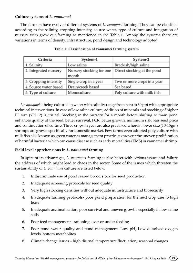
Training Manual on “Health management practices for finfish and shellfish of brackishwater environment” 19-23 August 2014 49
Culture systems of L. vannamei
The farmers have evolved different systems of L. vannamei farming. They can be classified according to the salinity, cropping intensity, source water, type of culture and integration of nursery with grow out farming as mentioned in the Table-1. Among the systems there are variations in terms of density; infrastructure, pond design and technology adopted.
Table 1: Classification of vannamei farming system
L. vannamei is being cultured in water with salinity range from zero to 60 ppt with appropriate technical interventions. In case of low saline culture, addition of minerals and stocking of higher PL size (>PL12) is critical. Stocking in the nursery for a month before shifting to main pond enhances quality of the seed, better survival, FCR, better growth, minimum risk, less seed price and continuation of culture. Three crops in year are also practised wherein lower size (10-15gm) shrimps are grown specifically for domestic market. Few farms even adopted poly culture with milk fish also known as green water as management practice to prevent the uneven proliferation of harmful bacteria which can cause disease such as early mortalities (EMS) in vannamei shrimp.
Field level apprehensions in L. vannamei farming
In spite of its advantages, L. vannamei farming is also beset with serious issues and failure the address of which might lead to chaos in the sector. Some of the issues which threaten the sustainability of L. vannamei culture are listed below.
1. Indiscriminate use of pond reared brood stock for seed production
2. Inadequate screening protocols for seed quality
3. Very high stocking densities without adequate infrastructure and biosecurity
4. Inadequate farming protocols- poor pond preparation for the next crop due to high lease
5. Inadequate acclimatization, poor survival and uneven growth especially in low saline soils
6. Poor feed management –rationing, over or under feeding
7. Poor pond water quality and pond management- Low pH, Low dissolved oxygen levels, bottom metabolites
8. Climate change issues – high diurnal temperature fluctuation, seasonal changes

Training Manual on “Health management practices for finfish and shellfish of brackishwater environment” 19-23 August 201450
9. Outbreak of diseases-WSSV, IHHNV, RMS/CIM, Gill choke, Vibriosis, White muscle
10. Market issues - sudden fall of sale price and middlemen
11. Inadequate processing capacity, cold storage and very low local consumption
12. Increasing fuel prices with poor electricity supply,
13. Food safety and social issues –labour, sanitary issues
14. Lack of regulation in the low saline areas
15. Unregistered shrimp farms
16. Lack of waste water treatment system
17. Indiscriminate use of chemical inputs and lack of awareness among the farmers
The above issues need to be adequately addressed to prevent large scale damages. Therefore, it is high time that adequate safe guards in the form of better management practices need to be developed and implemented to tackle these issues. A detailed risk analysis for identification and assessment of risk factors associated with L. vannamei farming and develop suitable measures for risk prevention, reduction, management and mitigation could be pragmatic approach.
Risk assessment for development of BMPs
Risk is a known danger where the probabilities are known unlike uncertainty though both terms are often used interchangeably. Risks in shrimp farming are of two kinds, “individualistic’’ wherein a particular farm alone is affected and the other is “compound” wherein the risk affect nearby farms too and transferable from one to another. The risks in shrimp farming are of compounded nature where the mistake of one farm might affect nearby farms. Identifying the potential risk factors, their probability of occurrences, symptoms, causes and their likely impact on the production is essential to develop an integrated approach for risk prevention and management. Based on the field investigations and interactions with stakeholders, nine major risk factors viz., diseases, poor water quality parameters, operational, climate change, environment, policy/institutional food safety, market and social issues have been identified. Extent of exposure to these risk factors, perceived economic losses due to them and allocation of resources for preventing the same determine the severity of that risk factor. The major risk factors observed and expressed are presented in the Table-2 below.
Table - 2. Major risks affecting L. vannamei shrimp farming
Major categories of risk Risk factors
Quality seed Non-availability of quality seed, low survivalDiseases WSSV, IHHNV, RMS/CIM, Gill choke, Vibriosis, White
muscleWater quality pH fluctuations, Low dissolved oxygen levels, algal blooms,
nutrient deficiency, bottom metabolites, poor feed manage-ment
Operational risks Lack of electricity, use of pond reared stocks, poor bio-se-curity and infrastructure

Training Manual on “Health management practices for finfish and shellfish of brackishwater environment” 19-23 August 2014 51
Climate change Seasonal changes, diurnal temperature variations, flood, cyclone
Environmental Lack of ETP, indiscriminate usage of drugsFood safety Presence of antibiotic residues, biological pathogens, anti-
oxidants Market High input cost, low sale price, middlemen, lack of institu-
tional credit, lack of domestic consumptionSocial Inadequate skilled manpower, competition for water, lack
of regulation, accessPolicy/institutional risks Un-registered farms, lack of regulation in the low saline
area, lack of institutional credit and insurance for aquacul-ture.
These risks are potentially harmful and hence, they need to be addressed on a priority basis at the farmer (individually), farm cluster (collectively) and policy (third party) levels to minimise the negative impacts. The exposure and impact of disease risks are respectively high and extremely negative to disastrous to the tune of 50 to 100% loss in the production or in economics. The exposure to climate change related risks, water quality issues and operational risks are likely to affect L. vannamei culture and impact moderately negative with 20-30% of additional expenses or similar level of loss in production. However, if not addressed they would lead to extremely negative impacts in the form of diseases or mortality. The food safety, institutional, environmental and social risks are perceived to be of low risk category since they seldom affect the production or incur considerable loss. The overall risk assessment is given in the following matrix. The perceptions of the farmers on the risk factors associated with L. vannamei farming have shown that the emerging diseases and water quality are the major threats to shrimp farming (Table-3).
Table-3. Matrix of risks and their probable impactsPROBABILITY
Likelihood/ Impact
1 2 3 4 5
IMPACT
54 Diseases Risks3 Water quality
Risks2 Climate/
Weather RisksMarket Risks
1 Food Safety Risks
Social Risks
Index
Extremely low riskLow riskModerate riskHigh riskExtremely high / Catastrophic

Training Manual on “Health management practices for finfish and shellfish of brackishwater environment” 19-23 August 201452
Development of risk specific Better Management Practices
An in-depth investigation on the risks identified, their primary causes, routes of entry and management measures for their prevention and management is very essential for the sustainability of L. vannamei farming.The risk management measures are either preventive form intended to eliminate the risks or management nature deals with coping mechanisms at the individual, group and state level to minimise impacts. The risk factors identified in the L. vannamei farming and the preventive and management measures suggested are indicated in the Table-4.
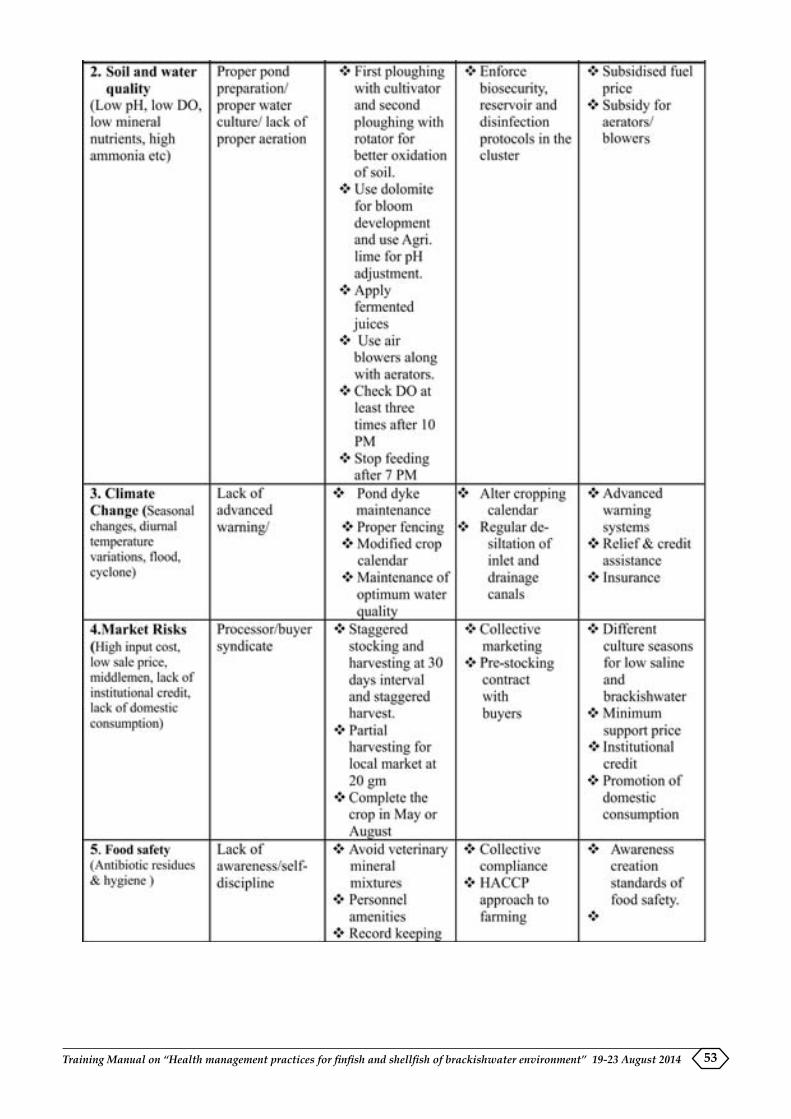
Training Manual on “Health management practices for finfish and shellfish of brackishwater environment” 19-23 August 2014 53

Training Manual on “Health management practices for finfish and shellfish of brackishwater environment” 19-23 August 201454
Table - 4. Better Management Practices (BMPs) for L. vannamei farming
Majority of L.vannamei shrimp farms are of small scale in nature and being operated with inadequate infrastructure and bio-security. Small scale farmers with very limited infrastructure and capacity should restrict their stocking density and other operations. Therefore, two distinct farming models and protocols respectively for farms with and without adequate infrastructure as well as for low and high saline cultures need to be developed and given to them for adoption. Similarly adjustments in culture seasons for low and high saline systems need to be worked out and implemented to tackle farming and market related issues. Further, farmers need to be advised to adopt batch stocking with 30 days interval to minimise market related problems. Nursery rearing to ensure the availability of seed in time and partial harvesting of 20-30% biomass at an appropriate stage may also be strategies to minimise the risks. Therefore, Hazard Analysis and Critical Control Point (HACCP) approach with appropriate protocols need to be developed system wise and popularise among the farming community for the sustainable L. vannamei shrimp farming.
Conclusion
The introduction of L. vannamei has given impetus to shrimp farming with a record production. However, serious risks and apprehensions have been observed and expressed which are threatening to its sustainability. Better management practices for risk reduction, management and mitigation need to be adopted at different levels to tackle the issues and ensure sustainability. At the same time, culture systems and farming protocols are to be fine-tuned according to the salinity regimes and infrastructure available. The stakeholders need to be adequately sensitized about the risks so that adequate efforts are to be implemented at each level to facilitate the sustainable L. vannamei farming in the country.

Section IIdisease inforMation and diaGnostiCs


Training Manual on “Health management practices for finfish and shellfish of brackishwater environment” 19-23 August 2014 57
INFECTIOUS DISEASES OF FINFISH AND HEALTH MANAGEMENT STRATEGIES
K.P. Jithendran and P. Ezhil PraveenaAquatic Animal Health and Environment Division
Introduction
Brackishwater aquaculture plays a vital role in national food security, employment generation, significant seafood export earnings and proper utilization of coastal waste lands. The commercially important species for brackishwater aquaculture are penaeid shrimps (Penaeus monodon and Litopenaeus vannamei), crabs (Scylla spp.) and finfishes such as Asian seabass (Lates calcarifer), grey mullet (Mugil cephalus), milk fish (Chanos chanos) and pearl spot (Etroplus suratensis). In India, presently shrimps are the major constituent of coastal aquaculture production reaching an export level of 3, 01,435 MT (MPEDA, 2014) while marine or brackishwater finfish production is still in its infancy with limited species under mono and polyculture systems. Seed production still remains a bottleneck in expanding mariculture activities to industrial level. However, limited culture of marine fish has taken up after the successful of breeding and hatchery technology for seed production of Asian seabass (Lates calcarifer) for the first time in India in 1997 (Thirunavukkarasu et al., 2001). This was followed by the improved hatchery technology by Central Institute of Brackishwater Aquaculture (Chennai, India) for seabass and Rajiv Gandhi Centre for Aquaculture, Sirkazhi (India) for multiple species like seabass, grouper etc. which enabled large scale fry production under controlled conditions.
Recent impetus on coastal aquaculture of marine/brackishwater fish species by National Fisheries Development Board (Hyderabad, India) for augmenting production by demonstration and promotion of open water sea cage culture by Central Marine Fisheries Research Institute (Kochi, India) along the coastline of India started yielding results with total production of 18,000-20,000 MT. Thus Asian seabass has been proven as ideal species for cage culture due to the availability of hatchery produced seed, market demand and fast growth. Other species with great potential are cobia (Rachycentron canadum), an excellent species for open sea cage culture and silver pompano (Trachinotus blochii) reported to be highly suitable for pond farming in vast low saline waters besides sea cage farming. For both fish species hatchery technology has been initiated (Gopakumar et al., 2012 a,b); and on-farm trails are demonstrated in land and cage based culture system at different locations in the country (CMFRI, 2012).
As in any animal production systems, infectious diseases are recognized as one of the major constraints for sustainable aquaculture. Broadly, infectious diseases of finfish are caused by virus, bacteria, fungi and parasites. Environmental stresses, genetic factors and nutritional deficiencies further make the animals more susceptible to these pathogens. A brief description of finfish diseases has been presented here with technical guidelines for health management.
Viral diseases in fish
Viral nervous necrosis
Introduction: Viral nervous necrosis (VNN) is a devastating disease of marine fish species cultured worldwide. It affects more than 50 wild and cultured marine fish species, especially

Training Manual on “Health management practices for finfish and shellfish of brackishwater environment” 19-23 August 201458
the larval and juvenile stages which records high mortality. Based on characteristic lesions, VNN is also known as viral encephalopathy and retinopathy (VER).
Etiology: The disease is caused by piscine nodavirus of the genus betanodavirus, family Nodaviridae. The virus contains single stranded, bipartite positive sense RNA genome.
Affected species: In India, betanodavirus infection has been observed in cultured and wild population of brackishwater/marine fish species such as Lates calcarifer, Mugil cephalus, Chanos chanos and Epinephelus tauvina etc.
Mode of transmission: Nodaviruses are regarded as pathogens of marine fishes. However, natural development of disease has also been reported from low saline and freshwater environments. Betanodaviruses are quite resistant to environmental conditions, which make it possible to get translocated by commercial activities via influent water, juvenile fish, utensils, vehicles, etc. Translocation of species for stocking purpose from one location to another may be another way but it is yet to be validated. Latent infection among wild fishes also serves as source of infection. Apart from all these means of horizontal transmission, vertical transmission is highly suspected to take place from infected spawners to fry.
Symptoms and lesions: The major clinical signs of VNN are common behavioral changes such as lack of appetite, erratic, spiral or belly-up swimming and dark coloration of body. Clinically, the affected animals show spiral or looping swim pattern, swim bladder hyperinflation and later wasting. Vacuolation is seen in the grey matter of brain and retina of eye. Necrosis is observed in the spinal cord, brain and retina while intracytoplasmic inclusion in nervous cells. The severity of disease is more in juveniles with equal higher rate of mortality. In seabass, the earliest onset of clinical signs of the disease is during 16-21 day post hatch. Recently, the disease has been noticed in fishes irrespective of their age. Instances of asymptomatic/ sub-clinical infection among wild fishes may possibly act as potential carriers.
Diagnosis: The first step in combating an infectious disease is detection and identification of the causative agent using a reliable diagnostic method followed by better surveillance techniques. Viral nervous necrosis can be diagnosed by demonstrating characteristic lesions in the brain and/or retina by light microscopy. Detection of virions by electron microscopy, viral antigens or antibodies by serological methods such as indirect fluorescent antibody test, and enzyme linked immunosorbent assay or detection of viral nucleotides by molecular techniques such as RT-PCR and nested PCR and by tissue culture are different ways for pathogen detection.
The RT-PCR is the most sensitive test and has become the main diagnostic method for fish nodaviruses. The capsid protein gene is the target sequence for this test. More recently, nested PCR was reported to be 10-100 folds more sensitive than the RT- PCR and permitted diagnosis by using blood, sperm, as well as nervous and ovarian tissues.
Prevention and control: Against VNN, no vaccine or treatment method is available. Broodstock fish acts as the most important source of the virus to their larvae by vertical transmission. Therefore, after RT-PCR screening, virus-carrying broodstock should be eliminated. Fertilized eggs should be disinfected with disinfectants such as iodine or ozone. Strict hygiene within the hatchery should also be maintained by regularly disinfecting the hatchery facility and farm materials with chlorine. Separate rearing facility of larvae /juveniles from brooder should be

Training Manual on “Health management practices for finfish and shellfish of brackishwater environment” 19-23 August 2014 59
maintained with each batch of larvae/ juveniles in the separate tanks supplied with sterilized (UV or ozone) seawater.
Iridovirus infection
Introduction: Iridovirus infection is a significant cause of mortality in farmed red sea bream (Pagrus major) and more than 30 other species of cultured marine fish belonging mainly to the orders Perciformes and Pleuronectiformes. The infection has also been detected in Asian seabass. It affects all the stages of fish but the susceptibility of juveniles is generally higher than adults.
Etiology: The disease is caused by double stranded DNA virus of genera Lymphocystivirus and Ranavirus. Ranaviruses causes systemic disease in infected fish and are associated with high morbidity and mortality.
Mode of transmission: The principal mode of transmission of Iridovirus infection is horizontal via water. The vertical mode of transmission has not yet been established.
Diagnosis: Outbreaks of the disease have been mostly reported in the summer season at water temperatures of 25°C and above. Depending on host fish species, fish age, water temperature, and other culture conditions, mortality rates ranges between 0 and 100%. Affected fish become lethargic, exhibit severe anaemia, petechiae of the gills, and enlargement of the spleen.
The sample should be collected from moribund fish. Gill and visceral organs such as spleen, heart, kidney, liver and intestine should be collected. However, the spleen and/or kidney tissues are the most appropriate organ for pathogen detection by IFAT. The fish sample should be stored at 4°C for use within 24 hours or -80°C for longer periods.
The disease is characterised by the appearance of abnormally enlarged cells stained deeply with Giemsa solution in the histopathological observations of the spleen, heart, kidney, intestine and gill of infected fish. These enlarged cells react to an anti -RSIV MAb by the antibody based antigen detection (Indirect fluorescent antibody test-IFAT) test. Electron microscopy confirms the presence of virions (200-240 nm in diameter) in these cells.
PCR is able to detect Iridovirus infection with high degree of sensitivity in short time. Recently real time PCR has been developed which shown improved rapidity, sensitivity, reproducibility, and the reduced risk of carry over contamination over normal PCR. An antibody based enzyme-linked immunosorbent assay (ELISA) has also been developed to detect iridovirus infection.
Prevention and control: An effective and commercially available formalin-killed vaccine is available against Red Sea Bream Iridovirus infection (RSIVD). A number of general husbandry practices are used to reduce RSIVD associated losses. These includes introducing pathogen free fish, implementing hygienic practices on farms and avoiding practices that can decrease water quality and/or increase stress, such as overcrowding and overfeeding.
Bacterial diseases in fish
Bacterial diseases occur as a result of the complex interactions between pathogen, fish and environmental stress. Environmental stresses can affect the homeostatic mechanism of fish
Training Manual on “Health management practices for finfish and shellfish of brackishwater environment” 19-23 August 201460
thus reducing their resistance to disease-causing organisms. Fish reared in intensive culture conditions are exposed to extreme environmental fluctuations and they may be more sensitive to disease than that in wild populations.
Aeromonas sp.
Aeromonas sp. is a common water-borne bacterium normally present in fishes. Whenever fishes are exposed to environmental stress or injury, it causes serious outbreaks of haemorrhagic disease with high mortality. Grossly, hemorrhages on the fin and tail and erosion of tail and fin can be clearly seen in severe cases. Temperature, pH, high CO2, O2 depletion, decomposition of products, free NH3, over-crowding and low salinity are considered as predisposing causes for Aeromonas infection.
Collumnaris disease
Collumnaris disease caused by Flexibacter columnaris, gram negative, aerobic bacillus, is one of the juvenile seabass diseases of low saline water. Clinically, the condition may be chronic, acute or peracute. Grossly, saddle-shaped lesions in the mid-body position about the dorsal fin of the fish and bilaterally symmetrical lesion as a fuzzy, pale yellow white plaque, with dark margins, often erode in the epidermis.
Vibrio sp.
Members of the genus Vibrio and other related genera are the causative agents of vibriosis, a deadly haemorrhagic septicemia disease in a diverse range of marine, estuarine and freshwater fish species. The typical symptom of vibrio disease includes extensive cutaneous and systemic hemorrhages and localized ulcerations in mandible, eyes, isthmus, bases, rays of dorsal, pectoral, pelvic, anal and caudal fins and ecchymosis and petechiae on the body surface and frequently, ulceration of the skin and muscle tissue are noted. The affected fish are anorectic. Internally, there is congestion and haemorrhages in the liver, spleen and kidney, frequently accompanied by the presence of necrotic lesions. The gut and particularly the rectum may be distended and filled with a clear viscous fluid. The body is completely covered by a thick layer of mucus. Microscopically, extensive lamellar hyperplasty, honey-comb vacuolization and fatty degeneration in hepatocytes, and aggregation of melano-macrophage centres (MMC) in kidneys and spleen are observed. The degree of the lesions varies with the severity of the disease. Fingerlings die more rapidly than adults.
Photobacteriosis
Photobacteriosis, also described as pasteurellosis, is caused by the halophilic bacterium Photobacterium damselae subsp. piscicida (formerly called as Pasteurella piscicida). The disease is characterized by the presence of white nodules in the internal viscera, particularly, spleen and kidney, so called as pseudo-tuberculosis. Severe mortalities occur usually when water temperatures are above 18-20°C. Below this temperature, fish can harbour the pathogen as subclinical infection for long time periods.
The presumptive identification of the pathogen is based on standard biochemical tests. Several commercial vaccines against Ph. damselae sub sp. piscicida are available on the market,

Training Manual on “Health management practices for finfish and shellfish of brackishwater environment” 19-23 August 2014 61
but their efficacy is dependent on the fish species, fish size, vaccine formulation and use of immunostimulants. As the majority of the pasteurellosis outbreaks occur from larval stages to fingerlings of 10-30 g, a vaccination programme which comprises a first dip immunization at the larval stage and a booster vaccination when fish reach a size of about 1-2 g is recommended to avoid high economic losses caused by this disease.
Treatment
It is advisable to know the quality of the water including pH and temperature which greatly affect treatment results. A few fishes are first treated and checked for how they react before treating the entire group. The total volume of water in the pond must be known to treat it accurately. To determine the volume of a pond, the surface unit area of water is multiplied by the average depth of the pond. Then, the approved and calculated therapeutics is evenly administered all over the pond.
Bacterial diseases are treated best by injection or use of food additives or antibiotics. Terramycin 2.5-3.0 g per 100 lb body weight (BW) per day for 10 to 12 days with a withdrawal period of 21 days, Sulfamerazine, Nitrofurans and Furazin 109 per 100 lb BW per day for 10 days, Erythromycin 4.5 g per 100 lb BW per day for two weeks are used as per the requirements. Potassium permanganate (KMn04) is used at the rate of 2-3 ppm as a wide-spectrum treatment in ponds against bacterial infections.
Fungal diseases in fish
Epizootic ulcerative syndrome
The disease is reported in freshwater as well as brackishwater fishes throughout India. It is caused by non septate fungus Aphanomyces invadans and is invariably associated with secondary infection by gram negative bacteria and rhabdoviruses. The transmission of disease occurs through zoospores which are transmitted through water, direct contact between fishes, and transport of infected fishes into new area.
The disease is diagnosed by sudden high mortality rate in wild and farmed fish with different types of fishes gets affected and show abnormal behaviour. Hemorrhagic ulcers are seen on head which often extend to skull leading to exposure of brain. Confirmatory diagnosis of disease is done by detection of non-septate, branching fungal hyphae in the periphery of the lesion. Invasive fungus extends through muscle tissue to spinal cord, kidney, and peritoneum. No vaccine is available for the control of disease. Controlled feeding regime should be operated during the outbreak of disease as a means of preventive measures. The affected fish should be destroyed and all farm appliances should be disinfected. The affected pond should be dried followed by liming application before restocking.
Parasitic diseases of fish
The major groups of parasites in brackishwater fishes are (i) protozoans and (ii) metazoans. Based on the location on the host fish it can be also categorised either ecto or endoparasites.

Training Manual on “Health management practices for finfish and shellfish of brackishwater environment” 19-23 August 201462
Protozoan parasites
Protozoans are single celled microscopic organism with specialized structures for movement, food gathering and attachment. They are either external or internal parasites. The following are the major protozoan parasites in fishes.
Dinoflagellates
Amyloodinium is a common dinoflagellate found in fishes particularly young fishes. These are microscopic external parasites with flagella for movement. It is usually attached with gill filaments or body surface of the affected fish. Predisposing factors are high level of organic matter in water and higher stocking density of fish.
Symptoms and lesions: The gill and skin of affected fish shows signs of necrosis and destruction. Darkening of body surface is observed. Affected fish often found on the surface or near the source of aeration. Mass mortality of fish is observed in severely affected pond, if not treated in time.
Treatment: Short bath treatment with 200 ppm formalin for 1 hr. and copper sulphate bath with 0.5 ppm for 3-5 days with aeration and daily water replenishment.
Ciliates
Ciliates are microscopic external parasites with cilia for movement. The most common ciliates are Cryptocaryon and Trichodina.
Cryptocaryon
Cryptocaryon is a pear shaped (0.3-0.5 mm in size) parasite. The parasites are found on the external body surface of fish. High stocking density and low water temperature serve as predisposing factor for infection.
Symptoms and lesions: It affects mainly gills and body surface. The affected part has increased mucus production. The white spots are observed over the body surface. Wound often has the complication of secondary bacterial infection. Affected fish are found to rub body against submerged objects. Respiratory distress with mass mortality is seen in untreated cases.
Treatment: Copper sulphate bath with 25 ppm or/and formalin bath with 0.5 ppm for 5-7 days with aeration and daily replenishment of water.
Trichodina
Trichodina has a circular body with cilia around the perimeter. It infects mainly gills, body surface and fins. High level of organic matter in water or poor water exchange serves as predisposing factors.
Symptoms and lesions: Affected fish has pale gills with irritation on body and hence rubs body against objects. Excessive mucus production is observed on gills and body surface. This leads to respiratory distress by clogging of gills by mucus.

Training Manual on “Health management practices for finfish and shellfish of brackishwater environment” 19-23 August 2014 63
Treatment: Short bath treatment with 200 ppm formalin for 30-60 minutes with strong aeration or extended bath treatment with 25 ppm formalin for 1-2 days with good aeration and daily replacement of water.
Myxozoans
Myxozoans are microscopic internal parasites. The parasites either infect tissue of various organs or live freely in cavities like gall bladder, abdominal cavity, etc. Poor water quality, high stocking density, feeding with infected trash fish and lack of quarantine measures facilitate infection. Common examples of myxozoans are Myxobolus, Myxidium, Kudoa, Ceratomyxa etc.
Symptoms and lesions: Clinical symptoms are not apparently visible. However, white or black cysts may be seen on body surface, gills, fins and internal organs. The parasites invade all major organs and forms cysts or freely floating mass called pansporoblast. They destroy gills and all major target organs of the fish.
Treatment: No effective treatment is available. Preventive steps should be taken by having efficient water exchange, avoiding feeding of trash fish and by quarantine measures.
Microsporidian
Microsporidians are intracellular internal parasites. It forms spore and resembles myxozoans. Poor water quality and poor nutrition is the predisposing factor.
Symptoms and lesions: No visible symptoms of infection are observed. Cysts are observed in various internal organs like intestinal wall, ovary, fat tissue etc. These cysts are brown or black in color and are of various size and shape called xenoma.
Treatment: No treatment is available. The disease can be prevented by good water exchange.
Metazoan parasites
Metazoans parasites of fishes include mainly helminths of different classes, arthropods dominated by parasitic crustaceans and some annelids such as leeches. Among helminths, trematodes or flatworms are of significance in cultured fishes though nematodes and cestodes are found rarely as massive infections. Trematodes constitute mainly external parasites with adhesive structures for attachment to the host. These are two types: 1. Monogeneans and 2. Digeneans.
Monogeneans
Monogeneans are mostly external parasites with specialized posterior attachment organs. It needs single host for completing its life cycle and multiply very fast in confined water bodies. They include skin and gill flukes.
Skin flukes
Skin flukes are 2-6 mm long and the most common forms are Benedinea sp., Dactylogyrus sp. The infection sets in when there is high stocking density, poor water exchange and overlapping of cultured fish.

Training Manual on “Health management practices for finfish and shellfish of brackishwater environment” 19-23 August 201464
Symptoms and lesions: The disease mainly affects body surface, fins, eyes, and sometimes gills. Mass mortality is observed among affected fish due to rapid multiplication of the parasites. The parasites damage the host tissue leading to secondary bacterial infection. Fish becomes lethargic with excessive mucus production on gills and body surface with opaque eyes and skin lesions. Affected fish rubs body against submerged objects. Eye infection sometimes leads to blindness.
Treatment: Short bath with 100 ppm formalin or freshwater for 10-30 minutes or 150 ppm hydrogen peroxide for 10-30 minutes with strong aeration.
Gill flukes
Gill flukes are ectoparasites. The common gill fluke are Diplectanum sp., Gyrodactylus sp. High density and poor sanitation serve as predisposing factors.
Symptoms and lesions: Mass mortality with respiratory problems is observed in severely affected fish. Other symptoms are pale gills, low consumption of feed, erratic swimming behaviour and mucus production on gills.
Treatment: Short bath treatment with freshwater for 10-30 minutes or 200 ppm hydrogen peroxide for 60 minutes or 100-200 ppm formalin with strong aeration are recommended.
Digeneans
Digeneans as a group are mostly intestinal parasites and involves more than one host for completing their life cycle. These are of variable size based on the species. No treatment method is suggested, although anthelmintic drugs are used. Digeneans complete their life cycle in a molluscan host; therefore, elimination of molluscs from the culture facility should stop the transmission cycle of the parasite.
Nematodes
Nematodes or roundworms are large intestinal parasites, with un-segmented body and mostly 1-2 cm length. Infection is prevented by maintaining hygienic conditions.
Crustaceans
Parasitic crustaceans include mainly copepods such as Caligus sp., Ergasilus sp., Lernanthropus sp. etc. and are commonly found on the gills, skin and fins. Caligus sp. is oval shaped parasite with about 3 mm length and l.6 mm width. The parasite has four pair of legs and a pair of suckers over the frontal edge of the body. Poor water quality serves as predisposing factors.
Symptoms and lesions: Affected fish had sluggish behaviour, shows anorexia and become weak due to heavy infestation. Gills and skin have erosions and secondary bacterial infection leading to high mortality, if not treated.
Treatment: Short bath treatment with 100 ppm hydrogen peroxide for 30 minutes or 200-250 ppm formalin for 1 hr. with good aeration. Mild infection can be controlled by simple freshwater bath for 10-15 minutes.
Training Manual on “Health management practices for finfish and shellfish of brackishwater environment” 19-23 August 2014 65
Lernanthropus sp., Ergasilus sp. and parasitic isopods such as Cymothoa sp. are relatively larger than Caligus and commonly found on the gills and fins. Dichlorvos @ 1ppm is effective against these parasites.
Health management strategies
Many brackishwater fishes can be cultured in a variety of aquaculture systems (open, semi-closed and closed) in freshwater, brackish and marine environments each of which present its own unique disease challenges. However, most of the losses due to microbial pathogens and parasites in aquaculture system can be prevented by adopting appropriate biosecurity protocols. These include routine health inspection, quarantine and treatment of wild-caught broodstock, egg disinfection, strict equipment sanitation, human traffic control, intake water treatment, discharge water treatment, clean feeds, restricting movement of stock, appropriate disposal of dead fishes and limiting interactions between wild and farmed fishes. Difficulties can arise once a disease is established and spread within a facility because eradication may be nearly impossible.
Technical Guidelines
Animal source
�� Animals should be obtained from disease free farms/sources after appropriate health testing and disease free certification; disinfect eggs or larvae where appropriate.
�� Quarantine all animal introductions from rest of the farm for an appropriate period of time. The duration depends on species of aquatic animal and potential diseases.
�� Seek advice on preventive treatments to all introduced animals while still in quarantine.
Operational management
�� Disinfection of eggs/larvae prior to use
�� Remove sick and dying animals as soon as possible and treat in quarantine section or dispose suitably so that there is no contamination of facility.
Record keeping
�� Record all introductions and disposal of aquatic animals; source /destinations
�� Record all outbreaks of diseases and death
�� Measure and record growth rates
Chemical use
�� Ensure the personnel are aware of the hazards of the chemicals used and trained on storage, use and disposal methods
�� Comply with regulations on the use of chemicals in aquaculture
Diseases
Any unusual mortality with any of the symptoms:
Training Manual on “Health management practices for finfish and shellfish of brackishwater environment” 19-23 August 201466
�� Animals coming to the edge or water surface
�� Unusual swimming patterns
�� Reduced feeding and failure to thrive
�� Unusual changes in physical appearance of the animal (redness, pale or black colouration, ulcers/mouldy growth on skin, pop-eye, erosions of fins and tails, fouling of gills etc.
Disease Management
�� Isolate any animals showing signs of any diseases or ectoparasitic infestation
�� Prevent movement of animal/vehicle/human from spreading disease
�� Personnel should work on healthy animals first before they have contact with sick ones.
�� Collect sick animals and attempt for diagnosis by laboratory examination.
�� Advise destruction, disposal and disinfection methods for diseased and dead animals.
�� Chemicals and therapeutics usage on advice
Disease emergencies
Prepare a contingency plan for any serious and infectious diseases like:
�� A plan and ability to completely isolate each pond/tank to prevent transfer of diseases by water
�� A capacity to disinfect an entire pond/tank/system
�� A capacity and appropriate method to destroy, collect and dispose of large number of aquatic animals as per expert advice.
�� Keeping records to allow tracing the origin and disposal of any affected group of animals.
Conclusions
Fish health management is a combination of prevention of onset of disease and measures to reduce losses from disease when it occurs. Health of the ecosystem that support the fish supplies are critical. Effective management focuses on reducing stress, hygienic husbandry practices, environmental management, preventing introduction of pathogens by efficient biosecurity measures, and use of effective drugs and vaccines, if available. In view of continued reliance on wild brood stock and/or stocking enables vertical and horizontal pathogen transmission. Understanding the source and transmission of infectious pathogens can enable proactive management strategies. A fish health surveillance programme is a vital asset to keep the farm disease free and maximize efficacy of disease control strategies in the event of an outbreak. Thus it is again crucial to further our knowledge of the diseases in the context of host, pathogen and environment interactions as a basis for farm management strategies and prevention of diseases.

Training Manual on “Health management practices for finfish and shellfish of brackishwater environment” 19-23 August 2014 67
Further reading
CMFRI (2012). Cadalmin, CMFRI News letter, No. 132 (January-March), p. 28.
Gopakumar, G., Abdul Nazar, A.K., Jayakumar, R., Tamilmani, G., Kalidas, C., Sakthivel, M., Ramesh Kumar, P., Hanumanta Rao, G., PremJothi, R., Balamurugan, V., Ramkumar, B., Jayasingh, M., Syda Rao, G. (2012a). Broodstock development through regulation of photoperiod and controlled breeding of silver pompano, Trachinotus blochii (Lacepede, 1801) in India. Indian Journal of Fisheries, 59: 53-57.
Gopakumar, G. , Abdul Nazar, A.K., Jayakumar, R., Tamilmani, G., Sakthivel, M., Kalidas, C., Ramamoorthy, S., Palanichamy, S., Ashok Maharshi, V., Srinivasa Rao, K., Syda Rao, G. (2012b). First experience in the larviculture of cobia, Rachycentron canadum (Linnaeus, 1752) in India, Indian Journal of Fisheries, 59: 59-63.
Jithendran,K.P. and Sujeet Kumar (2013). Handbook on Aquafarming - Diseases in Brackishwater Aquaculture. The Marine Products Export Development Authority, Kochi. p. 128.
Karunasagar, I., Karunasagar, I., Otta, S.K. (2003). Disease problems affecting fish in tropical environments. Journal of Applied Aquaculture, 13: 231-249.
Thirunavukkarasu, A.R., Kailasam, M., Kishore Chandra, P., Shiranee, P., Mathew Abraham, Charles, A.V.K. and Subburaj, R. (2001). Captive broodstock development and breeding of seabass Lates calcarifer (Bloch) in India. In: Perspectives in Mariculture in Asia (eds. N.G. Menon and P.P.Pillai), p. 111-124.
Toranzot, A.E., Magarinos, B., Romalde, J.L. (2005). A review of the main bacterial fish diseases in mariculture systems. Aquaculture, 246: 37-61.www.mpeda.com June 2014.

Training Manual on “Health management practices for finfish and shellfish of brackishwater environment” 19-23 August 201468
SHRIMP HEALTH MANAGEMENT STRATEGIES
S.V. Alavandi Aquatic Animal Health and Environment Division
Aquaculture is the fastest growing enterprise within the agricultural and animal rearing systems. According to FAO estimates, by 2030, over half of the fish consumed by the world’s people will be produced by aquaculture. Indian aquaculture too is no exception and has been significantly contributing to the people’s livelihood, food security, poverty alleviation, income generation, employment and trade. Five decades back, the Indian fisheries sector produced only six lakh tons of fish, and in 2012, it has produces over 14.6 lakh tones, including 2.4 million tons from aquaculture.
The aquaculture sector in the country is multifaceted with reference to the ecosystem (marine, brackishwater, freshwater and cold water), culture practices (traditional and scientific) and species (finfish, shrimps, crabs, lobsters, prawns, oysters and mussels). As wild catches of fish has been stagnating for the last several years, intensification of aquaculture was seen as an option, catered by hatchery produced seeds, formulated feeds and pond and water management methods. However, this increased production effort has been facing challenges due to disease problems, affecting productions.
Globally diseases have affected farmed aquaculture; Atlantic salmon in Chile, oysters in Europe, and marine shrimp farming in several countries of Asia, South America and Africa, resulting in partial to total loss of production. The most devastating diseases that impact economics of aquaculture include white spot disease and early mortality syndrome of shrimp, and infectious salmon anaemia. In 2010, aquaculture in China suffered production losses of 1.7 million tonnes (worth US$3.3 billion) caused by natural disasters and over 295 000 tones due to diseases. Disease outbreaks virtually wiped out marine shrimp farming production in Mozambique in 2011. Thailand’s production fell to 1.2 million tonnes in 2011 and 2012 owing to flood damage and shrimp disease. In 2013, Thailand experienced a decline in its exports (to US$7.0 billion, down more than13 percent on 2012), as disease problems reduced farmed shrimp production (FAO 2014). Current estimates predict that up to 40% of tropical shrimp production (>$3bn) is lost annually, mainly due to viral pathogens (Stentiford et al, 2012). World farmed shrimp production volumes decreased in 2012 and particularly in 2013, primarily due to the disease-related problems, such as early mortality syndrome in Asia and Latin America, reducing their production by 35 percent in 2013. As Asia accounts for 90 percent of global shrimp aquaculture, global supply would contract by 15 percent in 2015 (FAO 2014).
According to an earlier estimate, the viral epizootic caused by white spot syndrome (WSSV) alone has been causing an annual loss to the tune of Rs.300 crores, since 1994 to the Indian shrimp farming sector. Studies conducted during 2005-07 revealed that the national loss of shrimp production was estimated to be 48,717 metric tons of product and the economic loss due to diseases was estimated to the tune of Rs. 1022.13 crore per year (CIBA Annual Report, 2009-2010). Disease problems and related crop losses have emerged as the major limiting factor in the sustainability and economic viability of aquaculture. Therefore formulating proper disease management strategies are of paramount importance to ensure effective and sustainable development and profitability of aqua farming enterprise of the country.

Training Manual on “Health management practices for finfish and shellfish of brackishwater environment” 19-23 August 2014 69
Aquatic Animal Disease Management Strategies
Aquaculture disease management strategies have to be tackled at two levels, one at the International and National level, where regulatory aspects of aqua-farming, trans-boundary movements of aquatic animals, import risk analysis, quarantine and certification aspects have to be addressed. The second would be at the farm level, wherein, better management practices (BMPs) such as use of SPF stocks, use of only approved inputs such as probiotics, immunostimulants and medicines are effectively implemented taking care of the environment (water bodies) to achieve sustained productions.
Regulatory Aspects
Policies and legislature governing resources (soil and water) allocation and quality assurance in aquaculture, related to the physicochemical components and biological components should be in place and will facilitate aquaculture development. Functioning of a national level body with necessary responsibility and mandate to implement a ‘national aquatic animal health management strategy’ or ‘aquatic animal health management regulation’ on the basis of existing international standards, guidelines or recommendation from FAO, OIE and NACA and WTO will go a long way towards sustainability of aquaculture.
Regulations with respect to land and water usage, environmental protective measures, inputs that go into the aquaculture systems, farm-wise and region-wise must be put in place by the Government for disease management of aquatic animals and sustainable development of aquaculture at large. In addition, research, training programs, extension, and information exchange would be more effective. The FAO’s Code of Conduct for Responsible Fisheries would provide a good base for the national and international cooperation in harmonizing aquatic animal health management activities.
Specific pathogen free broodstock
Coastal aquaculture in general, and shrimp farming in particular, heavily relies upon wild broodstock (especially with regard to tiger shrimp) for seed production. The asymptomatic

Training Manual on “Health management practices for finfish and shellfish of brackishwater environment” 19-23 August 201470
wild broostock population plays a major role in the vertical transmission of the pathogen. There is a need to evaluate the health status of different stocks and to develop means of controlling the entry of the pathogen into the breeding and farmed populations. While specific pathogen free (SPF) broodstock of Pacific white shrimp are available and being imported, development of SPF stock of our native shrimp species is a must to produce disease free seeds. However these facilities need to be created and implemented adopting internationally accepted norms and with proper scientific evaluation.
Quarantine and Health Certification System
These are crucial component of an effective health management programme. Quarantine does not only mean that exotic species should be subjected to rigorous checks to avoid introduction of pathogens into a country or state, but it is also imperative that the broodstock/spawners/seeds arriving at a culture facility are screened for the presence of pathogens prior to their introduction to the system. Establishing effective quarantine guidelines and health certification procedures could help minimize the risk of introduction of harmful pathogens. Hence to provide a mechanism to facilitate trade in aquatic species, a proper health management mechanism such as quarantine and health certification is necessary for the trans-boundary movement of aquatic animals on the pre-border (exporter), border and post-border (importer), to minimize the risk of pathogen transfer and associated risk of disease outbreaks.
Surveillance Techniques, Contingency Planning and Import Risk Analysis (IRA)
New generation approaches such as Surveillance techniques, Contingency planning and Import risk analysis (IRA) are gaining importance as critical tools in the health management strategies of aquatic animals for quick and effective response to new disease outbreaks. Disease Surveillance means continuous investigation of a given population to detect the occurrence of disease for control purposes, which may involve testing of a part of the population. Monitoring constitutes on-going programmes directed at the detection of changes in the prevalence of disease in a given population and in its environment. Active surveillance differs from a passive reporting system in that it uses surveys of a relatively small, representative sample of the population to gather specific information about that population. The key advantages of active surveillance are that the quality of information collected is usually better, the information reflects the true situation in the entire population, and it is often faster and cheaper to collect than with passive methods.
Import Risk Analysis (IRA) is the process by which importing authorities determine whether live aquatic animal imports or their products pose a threat to the aquatic resources of their country. This is usually undertaken by the Competent Authority of the importing country, but risk analyses apply equally to the individual who wants to import live aquatic animals onto their farm or site. An import risk analysis involves the steps of hazard identification and characterization, risk assessment, risk management, and risk communication.
Extension System
Trained manpower and capacity building are the important steps towards an effective extension system. A working extension system for awareness building and effective

Training Manual on “Health management practices for finfish and shellfish of brackishwater environment” 19-23 August 2014 71
communication among farmers / aquaculturists, Govt. agencies and planners is pivotal for the successful implementation of any aquatic health management programme.
Aquatic Animal Health Information System
One of the most important factors dealing with a disease outbreak is information. Correct information is the key element in deciding upon the best means of dealing with a disease. To meet this objective a scientific and functional disease reporting system applicable at local (farm level), national and regional level and Aquatic animal health information system at national and regional level is necessary.Diagnostics
Timely and accurate diagnosis of the diseases using the right diagnostic tools is one of the most important components in the aquatic health management. Post-mortem necropsy and histopathology have been the primary methods for the diagnosis of fish and shellfish diseases. Direct culture of pathogens is also widely used, especially when bacterial etiology is suspected. However, these methods are time-consuming and costly, and, for shrimp and other crustaceans, cell lines suitable for virus culture are not been available, if one needs to diagnose diseases using culture viral pathogens. Efforts to overcome these problems have led to the development of immunoassay and DNA-based diagnostic methods including fluorescent antibody tests (FAT), enzyme-linked immunosorbent assays (ELISA), in situ hybridization (ISH), dot-blot hybridization (DBH) and polymerase chain reaction (PCR) amplification techniques. These techniques are highly sensitive and specific. During recent times, diagnostics kits are moving rapidly from development in specialized laboratories to routine application in less sophisticated laboratories and at farm level. DNA probes are expected to find increasing use in routine disease monitoring and epidemiology programs in aquaculture, and in prevention of spread of trans-boundary pathogens (national quarantine and certification programs). DNA-based detection methods for detection of most penaeid shrimp viruses are now used routinely in a number of laboratories around the world.
The most important disease management strategies in aquaculture is ‘prevention’ like the age old adage “prevention is better than cure”, considering the fact that to date no effective treatment protocols are available. Diagnostic tools play a very crucial role in prevention of diseases in aquaculture, in other words, termed as ‘biosecurity’. Biosecurity to shrimp aquaculture may be defined as the practice of exclusion of specific pathogens from cultured aquatic stocks in broodstock facilities, hatcheries, and farms, or from entire regions or countries for the purpose of disease prevention. Standard operating procedures can be adapted to minimize the risks of disease introduction and spread within a facility through such concepts as screening out / pretreatment of all inputs, stocking with Specific Pathogen Free or SPF and or Specific Disease Resistant varieties (SPR).
Aquaculture Inputs: Probiotics, Immunostimulants, Vaccines, Drugs and chemicals
Use of probiotics has been common in human health for a long time and this approach has been applied to veterinary animal health and recently for aquatic animals also. A number of reviews have been published on the use of probiotics in aquaculture. Several definitions have been proposed and the one proposed by Verschuere et al (2000) which is defined as “A live
Training Manual on “Health management practices for finfish and shellfish of brackishwater environment” 19-23 August 201472
microbial adjunct which has a beneficial effect on the host by modifying the host-associated or ambient microbial community, by ensuring improved use of the feed or enhancing its nutritional value, by enhancing the host response towards disease, or by improving the quality of its ambient environment” is more comprehensive and is largely based on the mode of action in aquaculture. It is suggested that in the case of preemptive colonization of rearing environments, a single addition of a ‘probiotic’ culture may suffice to achieve colonization and persistence in the host and or in its ambient environment, provided the cultures are well adapted to the prevailing environmental conditions. Such amendment of exogenous microbes is also termed as ‘bioaugmentation’ and such measures have been reported to significantly increase the rates of natural degradation, with negligible environmental impact. Shrimp farmers often resort to use of various probiotic products in the market out of desperation to improve production and sometimes save their standing crop. Efficacy of commercial bioaugmentation products has been often questioned by the farming community because of their inconclusive and inconsistent results.
The application of immunostimulants in shrimp aquaculture is increasingly gaining interest as an environmentally safe alternative to antibiotics and chemotherapeutants. Shrimp possess an innate immune system, consisting of cellular and humoral elements. Diets containing immunostimulants are used in aquaculture in order to increase resistance to stress and diseases of cultured fishes and invertebrates by alerting the immune system. The cell walls of microorganisms such as Gram-negative bacteria (lipopolysaccharides), Gram-positive bacteria (peptidoglycans), and fungi (β-1, 3-glucans) elicit nonspecific immune response and are usually employed as immunostimulants. Several methods of stimulation like immersion, injection, feeding etc have been examined and the most practical method of stimulation is by incorporating the immunostimulating substances into the feed.
Environment management
The ultimate goal of most aquaculture operations is to produce maximum possible biomass per culture unit area in a sustainable manner, regardless of the type of operation and the species cultured. However, the production depends upon a number of factors including environmental conditions, availability of good quality water, nutrition and disease and mortality of cultured stock. Incidence and severity of infectious disease outbreaks very often depend on the quality of environment. Hence the foremost important step in aquaculture health management is to provide the best quality environment within the culture unit. Incidence and severity of infectious diseases are dependent on the quality of aquatic environment in which the fishes live, quality of the feed they consume and the ease (without any disease) with which they are maintained. Hence health management in aquaculture requires a holistic approach, addressing all aspects that contribute to the development of disease. Disease outbreak is an end result of negative interaction between pathogen, host and the environment. Hence, management of disease problems must be aimed towards broader ecosystem management with a view to control farm-level environmental deterioration and to take preventative measures against the introduction of pathogens into the aquaculture system. The emphasis should be on better management for prevention, which is likely to be more cost effective than treatment, involving both on-farm management and the management of the environment. Steps must be initiated towards reducing the use of chemicals and drugs.

Training Manual on “Health management practices for finfish and shellfish of brackishwater environment” 19-23 August 2014 73
Conclusion
One of the main tasks in the development of aquaculture is to reconcile the disease risks of aquaculture and sustainable development. To achieve sustainable development, it is very important for the aquaculture operators to have common agenda based on mutual shared interests. Efforts of individual operators acting on their own agenda would fail because of the common geo-physical environment by of the farming areas, pathogens, candidate species, production and markets. According to FAO, sustainable development should be environmentally friendly, technically appropriate, economically viable, and socially acceptable. An understanding about the environment, biota and biology of the target species along with the in depth knowledge of the disease, pathogen, disease development, diagnostics, epidemiology and control measures are essential factors in management of a disease problem.

Training Manual on “Health management practices for finfish and shellfish of brackishwater environment” 19-23 August 201474
TRANSBOUNDARY DISEASES IN SHRIMP AQUACULTURE
M. Poornima and S.V. AlavandiAquatic Animal Health and Environment Division
More than 20 viruses have been identified that are known to infect penaeid shrimp. The OIE now lists five viral diseases and one bacterial disease (necrotising hepatopancreatitis) of penaeid shrimp in the Aquatic Animal Health Code Office International des Epizooties (OIE, 2010) which are considered to be transmissible and of significant socio-economic importance. These viral diseases include white spot disease (WSD), Yellow Head Disease (YHD), Taura Syndrome (TS), Infectious Myonecrosis (IMN) and Infectious Hypodermal and Haematopoietic necrosis (IHHN) (OIE, 2010). All OIE member countries are obliged to report these diseases so that disease spread can be monitored and legislation instituted to prevent disease spread. IMN and TS are so far not reported from India. Although YHV has been reported from India in one instance in farmed black tiger shrimp, using histopathological techniques, its economic impact was negligible.
Aquaculture of Litopenaeus vannamei has been recently permitted in India. This is undertaken using seed produced from Specific Pathogen Free (SPF) brood stock of the shrimp imported from selected companies overseas after they underwent require quarantine and disease screening process that determines them to be free from specified pathogens of concern to aquaculturists. SPF shrimp are expected to be free from the viral pathogens which are known to cause major losses to shrimp aquaculture, for various reasons including pathogenic viruses such as WSSV, YHV, TSV, IHHNV, BPV and HPV. SPF refers only to the present pathogen status for specific pathogens and not to pathogen resistance or future pathogen status. SPF means that these animals will not suffer from diseases caused by specified diseases for which the animal is declared ‘free’ when cultured under ‘strict’ biosecurity. However, it does not guarantee against these shrimp getting infected with unknown pathogens or known pathogens which are not screened. Further, the SPF shrimp are not resistant to pathogens and these shrimp can become infected by any pathogen that they encounter during culture.
In this article, a brief outline on these important diseases, the causative agents, symptoms, mode of transmission, possible methods of prevention and control are provided for the information of the shrimp farmers who have already taken up or are likely to take up L. vannamei culture in India.
I. OIE listed viral diseases
1. WHITE SPOT DISEASE (WSD)
This virus is a serious threat faced by the shrimp farming industry world wide. WSSV was first reported in farmed P. japonicus from Japan in 1992-93, but was thought to have been imported with live infected PL from Mainland China. WSD has been identified from crustaceans in China, Japan, Korea, South-East Asia, South Asia, the Indian sub -continent, the Mediterranean, the Middle East, and the Americas.

Training Manual on “Health management practices for finfish and shellfish of brackishwater environment” 19-23 August 2014 75
Causative agent: WSD is caused by a double-stranded DNA virus of 120-150 x 270-290 nm size, assigned to a new virus family, Whispoviridae. WSSV can infect a wide range of aquatic crustaceans including marine, brackish and freshwater penaeids, crabs and crayfish. All decapod crustaceans from marine and brackish or freshwater sources are susceptible host species. Penaeid shrimp species are highly susceptible to infection, often resulting in high mortality. Crabs, crayfish, freshwater prawns, spiny lobsters and clawed lobsters are susceptible to infection, but morbidity and mortality as a consequence of infection is highly variable. Prevalence of WSSV is reported as highly variable, from <1% in infected wild populations to up to 100% in captive populations.
Symptoms: The virus also severely damages the stomach, gills, antennal gland, heart and eyes. The virus affects organs of ectodermal and mesodermal origin and in later stages of infection, the organs are destroyed and many cells are lysed. The shrimp then show reddish discolouration of the hepatopancreas and characteristic 1-2 mm diameter white spots on their carapace, appendages and inside surfaces of the body. Affected shrimp show lethargic behavior. Cumulative mortality typically reaches 100 percent within two to seven days of infection.
Diagnosis: It can be visually diagnosed through the presence of the characteristic white spots, which can be seen in advanced stage of infection. However, white spots may not be always present in early stages of infection in shrimp. WSSV can be detected by using Polymerase Chain Reaction (PCR), or with probes for dot-blot and in situ hybridisation (ISH) tests. Efficency of PCR detection can be increased by exposure to stressful conditions (e.g. eye-stalk ablation, spawning, moulting, changes in salinity, temperature or pH, and during plankton blooms). WSSV can be confirmed histologically (particularly in asymptomatic carriers) by the presence of large numbers of Cowdrey A-type nuclear inclusions and hypertrophied nuclei in H&E-stained tissue sections, or simply by rapid fixation and staining of gill tissue.
Transmission: Dead and moribund animals can be a source of disease transmission. International transmission of WSD is believed to be through exports of live PL and infected broodstock. The infection can be transmitted horizontally by cannibalism, predation, and by water-borne routes. Some studies have shown that disinfection of water supplies and the washing and/or disinfection of the eggs and nauplius is reported to be successful in preventing the vertical transmission from positive broodstock to larvae. It is generally believed that the virus sticks to the outside of the egg, and if it gains entry to the egg, it is rendered infertile and will not hatch. Using proper testing and disinfection protocols, vertical transmission can be prevented in the hatchery. The presence of WSSV in a pond does not always lead to outbreaks. However, outbreaks are usually triggered from latent carriers by environmental stresses. Several biotic and abiotic stresses during the shrimp culture are known to precipitate the virus infection
Prevention/ control: Best way to prevent the disease occurrence is to use PCR screened broodstock for breeding. Every batch of PLs is also to be screened by PCR. Washing and disinfection of eggs and nauplii has also been shown to prevent vertical transmission of WSSV from infected broodstock to larval stages. Since crab and other crustaceans are known to act as carriers, feeding broodstock with these live feeds should be avoided.
In grow-out cultures, polyculture techniques with mildly carnivorous fish species (such as Tilapia sp.) has proven effective at limiting the virulence of WSSV in ponds, as the fish can eat

Training Manual on “Health management practices for finfish and shellfish of brackishwater environment” 19-23 August 201476
infected carriers before they become available to the live shrimp. Based on the recent information, the white spot virus only remains viable in water for a long period so specific disinfection protocols are necessary to be effective in preventing transmission. Formalin treatment (70 ppm) has been shown to prevent transmission and not cause any harm to shrimp. In addition, all effluent from farms or processing plants should be disinfected with formalin or chlorine prior to discharge.
2. INFECTIOUS HYPODERMAL AND HAEMATOPOIETIC NECROSIS (IHHN)
IHHN was first reported in L. vannamei and P. stylirostris in the Americas in the year 1981. However, it was thought to have been introduced along with live P. monodon from Asia. IHHNV has probably existed for some time in Asia without detection due to its insignificant effects on P. monodon, the major cultured species in Asia during that time. Recent studies have revealed geographic variations in IHHNV isolates and suggested that the Philippines was the source of the original infection in Hawaii and subsequently in most shrimp farming areas of Latin America. Large-scale epizootics were responsible for multi-million dollar losses in L. vannamei culture in the Americas during 1990s.
Causative agent: IHHNV is caused by a small (20-22 nm) single-stranded DNA-containing parvovirus.
Symptoms: Gross signs of disease are not specific to IHHN, but may include reduced feeding, elevated morbidity and mortality rates, fouling by epicommensals and bluish body coloration. Larvae, PL and broodstock rarely show symptoms. In L. vannamei, IHHNV can cause runt deformity syndrome (RDS), which typically results in cuticular deformities (particularly bent rostrums), slow growth, poor food conversion ratio (FCR) and a greater size variation at harvest, contributing substantially to reduction in profits. These effects are typically more pronounced when the shrimp are infected at larval stages. Hence strict hatchery biosecurity including checking of broodstock by PCR, or the use of SPF broodstock, washing and disinfecting of eggs and nauplii is essential in combating this disease. IHHNV typically causes no problems for P. monodon since they have developed a tolerance to it over a long period of time, but they may suffer with RDS. F. merguiensis and F. indicus appear refractory to the IHHNV. However, these species may be life-long carriers of the virus and so could easily pass it onto L. vannamei, which typically suffers from RDS when exposed to IHHNV.
Diagnosis: IHHNV can be diagnosed using methods such as DNA probes in dot blot, ISH and PCR techniques. Histological analysis of H&E-stained sections showing intracellular, Cowdrey type A inclusion bodies in ectodermal and mesodermal tissues such as cuticular epithelium, gills, foregut, hind gut, lymphoid organ and connective tissues are considered indicative of IHHN.
Transmission : Transmission of IHHN is known to occur rapidly by cannibalism of weak or moribund shrimp. It can also be transmitted through waterborne route and cohabitation. Vertical transmission from broodstock to larvae is common and has been shown to originate from the ovaries of infected females (whilst sperm from infected males was generally virus-free). IHHNV may be also transmitted through vectors such as insects and birds have been shown to act as mechanical carriers for the virus.
Training Manual on “Health management practices for finfish and shellfish of brackishwater environment” 19-23 August 2014 77
Prevention/ control IHHNV is reported to be highly resistant to all the common methods of disinfection including chlorine, lime and formalin. One of the big problems with IHHNV is its eradication in infected facilities. Complete eradication of all stocks, disinfection of the culture facility and avoidance of restocking with IHHNV-positive animals may bring down incidences of IHHNV infections.
3. TAURA SYNDROME (TS)
Taura Syndrome was first identified from farms around the Taura River in Ecuador in 1992 and the disease spread rapidly to the whole of Latin and North America within three years. Subsequently, TS was also reported from Asia including Mainland China and Taiwan (from 1999), and in late 2003 in Thailand, probably through the regional and international transfer of live PL and broodstock of L. vannamei.
Causative agent : Initial work suggested that TS was caused by a toxic pesticide. However, it is now known that a single or perhaps several very closely related mutant strains of the Taura syndrome virus (TSV) are responsible for the TS. TSV is a single stranded RNA virus of 32 nm size, non-enveloped icosahedrons and more prone to mutations causing more concern.
Symptoms: TSV infections occur in juvenile shrimp (0.1-1.5 g body weight) within two to four weeks of stocking and occur largely within the period of a single moult cycle. In the acute phase of the disease, during pre-moult stage, the shrimp are weak, soft-shelled, have empty gut and diffuse expanded chromatophores that appear red, particularly in the tail (hence the common name - red tail disease). Such animals will usually die during moulting (5-95 percent). Adult shrimp are known to be more resistant than juveniles. Shrimp that survive infection show signs of recovery and enter the chronic phase of the disease. Such shrimp show multiple, randomly distributed, irregular, pitted, melanised lesions of the cuticle. These gross lesions will persist, but may be lost during moulting, and the shrimp thereafter appear normal.
Diagnosis: TS can be diagnosed using standard histological and molecular methods of detection. Specific DNA probes applied to ISH assays with paraffin sections provide the confirmatory diagnosis. Reverse Transcriptase Polymerase Chain Reaction (RT-PCR) assay is commonly used for larger sample sizes and non-lethal sampling from broodstock. Histological lesions suggestive of TS are demonstration of enlarged lymphoid organs (LO) with multiple LO spheroids and multifocal areas of necrosis in the cuticular epithelium of the general body surface, appendages, gills, hindgut, and foregut (the oesophagus, anterior and posterior chambers of the stomach).
Transmission: The mechanism of transmission of TSV can be through infected PL and broodstock. Recently it has been shown that mechanical transfer through insect and avian vectors may be a more likely route of infection. Shrimp-eating seagulls can transmit TSV through their feces.
Prevention/control: Infected stocks must be totally destroyed and the culture facility must be disinfected. The disease can be prevented by avoidance of reintroduction of the virus from nearby facilities, wild shrimp and carriers and stocking with TSV-free PL produced from TSV-free broodstock. Switching of culture to refractory species such as P. stylirostris has been suggested. Other methods suggested for controlling the virus include the following best management practices (BMPs) and maintenance of optimal environmental conditions throughout the culture period.

Training Manual on “Health management practices for finfish and shellfish of brackishwater environment” 19-23 August 201478
4. YELLOW HEAD DISEASE (YHD)
Yellow head disease was the first major viral disease that caused extensive losses to shrimp farms in Thailand during 1990-91. YHD has been reported in China, Taipei, India, Indonesia, Malaysia, the Philippines, Sri Lanka, Thailand and Vietnam. Outbreaks of YHD with heavy mortalities have been reported in farmed black tiger shrimp and Pacific white shrimp. It is reported to be highly prevalent (>50%) in farmed and wild populations in Australia, Asia, East Africa and Mexico.
Causative agent: YHD is caused by yellow head virus (YHV), and its related gill-associated virus (GAV). YHV is rod-shaped enveloped virus of 40-60 nm by 150-200 nm size, containing single stranded RNA.
Symptoms: YHV affects tissues of ectodermal and mesodermal origin such as lymphoid organ, haemocytes, haematopoietic tissue, gill lamellae and spongy connective tissue of the subcutis, gut, antennal gland, gonads, nerve tracts and ganglia. YHV principally affects pond reared shrimp stages of 5 -15 g. Affected shrimp typically feed voraciously for two to three days and then stop feeding abruptly and are seen swimming near the periphery of the pond. YHV infections can cause swollen and light yellow colored hepatopancreas in infected shrimp and general pale appearance before dying within few hours. YHD can cause up to 100% mortality in infected P. monodon ponds within 3-5 days of the first appearance of clinical signs.
Diagnosis: Yellow head virus can be detected by RT-PCR or with a probe designed for dot-blot and ISH tests. It can also be diagnosed histologically in moribund shrimp by the presence of intensely basophilic inclusions, most easily in H&E stained sections of stomach or gill tissue, or simply by rapid fixation and staining of gill tissue and microscopic examination.
Transmission: The primary mechanism of spread of YHV in pond culture appears to be through water and mechanical means or from infected crustacean carriers. YHV is reported to remains viable in aerated seawater for up to three days. However, the most serious threat is latent or asymptomatic carriers, from which the virus can spread either by ingestion or cohabitation. Other shrimp such as F. merguiensis, F. indicus, Metapenaeus ensis, Palaemon styliferus and Acetes spp. may become infected and act as carriers having latent infections, while others such as Euphausia superba may die upon infection. Other crustaceans, such as Macrobrachium rosenbergii and many crab species and Artemia appear to be refractive to YHV. Infected broodstock can pass on the virus to larvae in the maturation/hatchery facilities if thorough disinfection protocols are not strictly adhered to.
Prevention/control:Although YHD is not causing much loss at present, methods of YHV eradication in ponds are much the same as for other viruses and involve BMPs that include pond preparation by disinfection and elimination of carriers, chlorination (30 ppm active ingredient) of reservoir water, filtering inlet water with fine screens, avoidance of live feeds, maintenance of stable environmental conditions, disinfection of YHV infected ponds before discharge, and monitoring (by PCR) and production of virus free broodstock and PL for pond stocking.

Training Manual on “Health management practices for finfish and shellfish of brackishwater environment” 19-23 August 2014 79
5. INFECTIOUS MYONECROSIS (IMN)
Infectious myonecrosis is an emerging L. vannamei disease, first detected in Brazil during 2004, and then in Indonesia in 2006. To date, IMN has been detected in East Java, Bali, and West Nusa Tenggara provinces. The principal host species in which IMNV is known to cause significant disease outbreaks and mortalities is L. vannamei.
Causative agent: IMN is caused by a putative totivirus. IMNV particles are icosahedral in shape and 40 nm in diameter.
Symptoms: Juveniles and sub-adults of L. vannamei, farmed in marine or low salinity brackish water, appear to be the most severely affected by IMN disease. The principal target tissues for IMNV include the striated muscles (skeletal and less often cardiac), connective tissues, haemocytes, and the lymphoid organ parenchymal cells. IMN disease causes significant mortality in grow out ponds and is characterized by acute onset of gross signs including focal to extensive whitish necrotic areas in the striated muscle, especially of the distal abdominal segments and the tail fan, which may become necrotic and reddened similar to the colour of cooked shrimp. Severely affected shrimp become moribund and mortalities can be instantaneously high and continue for several days. Mortalities from IMN range from 40 to 70% in cultivated L. vannamei, and FCR of infected populations increase from normal values of ~ 1.5 to 4.0 or higher.
Diagnosis: IMN can be confirmed by histopathology, using routine haematoxylin–eosin (H&E) stained paraffin sections and demonstrating characteristic coagulative necrosis of striated skeletal muscle fibers, often with marked oedema among affected muscle fibers. IMN may be also rapidly diagnosed using a nested RT-PCR method which provides a rapid, sensitive and specific test to detect IMNV in penaeid shrimp. Published methods available are ISH and nested RT-PCR and real-time RT-PCR for the molecular detection of IMNV.
Transmission: IMNV has been demonstrated to be transmitted through cannibalism. Transmission via water and vertical transmission from broodstock (transovarium or by contamination of the spawn eggs) to progeny is also likely to occur. IMNV may also be transmitted among farms by faeces of seabirds or shrimp carcasses. Outbreaks of IMN with sudden high mortalities may follow stressful events such as capture by cast-net, feeding, sudden changes in salinity or temperature, etc., in early juvenile, juvenile, or adult L. vannamei in regions where IMNV is enzootic.
Prevention/control: Stocking only pre-screened broodstock and/or their spawned eggs/nauplii and discarding those that test positive for the IMN virus by RT-PCR is the best possible way for prevention and control of IMN.Restocking of affected farms or entire culture regions with IMNV-free stocks of L. vannamei may help in preventing its recurrence.
II. OIE listed bacterial diseases 1. NECROTIZING HEPATOPANCREATITIS (NHP)
This disease is also known as Texas necrotizing hepatopancreatitis (TNHP), Texas pond mortality syndrome (TPMS) and Peru necrotizing hepatopancreatitis (PNHP). NHP has been reported as an important disease since its first diagnosis in 1985. It has been reported to cause mass mortalities to the tune of 20-90 percent of L. vannamei in highly saline commercial grow-out ponds nearly every year since then. By 1993, NHP spread to Ecuador, Guatemala, Honduras, Mexicoand Peru and by 1995, coincided with warm waters with high salinity associated with

Training Manual on “Health management practices for finfish and shellfish of brackishwater environment” 19-23 August 201480
El Nino, and caused severe mortalities (60-80 percent mortality) of L. vannamei and L. stylirostris throughout Ecuador. NHP has not yet been reported in Asia, but could cause significant damage were it to be transferred here with untested shrimp introduction.
Causative agent: Necrotizing hepatopancreatitis is caused by obligate intracellular Rickettsia-like bacterium, a member of the order -Proteobacteria. It is Gram-negative, pleomorphic, rod-shaped or helical-shaped bacterium.
Symptoms: Affected shrimp are lethargic, anorexic with empty gut and show epibiotic fouling. Exoskeleton becomes soft and show abdominal muscle atrophy. Affected ponds have increased FCR and growth of affected shrimp is retarded. The hepatopancreas becomes watery with white or black streaks. Mortality rates reach up to 90% within 30 days of the appearance of clinical signs.
Diagnosis: NHP can be diagnosed by demonstration of lipid droplets and melanisation of hepatopancreas by microscopic examination of wet mount preparations. It may be confirmed by histopathological examination showing atrophy and the presence of granulomata in the hepatopancreas and haemocyte aggregations around the hepatopancreatic tubules. Intra-cytoplasmic Rickettsia-like bacteria may be prominently seen in the cytoplasm. Molecular diagnostic tools such as ISH, dot blot hybridisation, and PCR for specific -Proteobacterial DNA are also available.
Transmission: NHP could be transmitted horizontally with infected shrimp.
Prevention/control: Stocking only pre-screened broodstock and/or their spawned eggs/nauplii and discarding those that test positive. Maintaining optimal environmental parameters using BMPs will be useful in preventing NHP.
Further reading
BONDAD-REANTASO, M.G., MCGLADDERY, S.E., EAST, I., AND SUBASINGHE, R.P. (eds.) Asia Diagnostic Guide to Aquatic Animal Diseases. FAO Fisheries Technical Paper No. 402, Supplement 2. Rome, FAO. 2001. p. 240
BRIGGS M, FUNGE-SMITH S, SUBASINGHE R AND PHILLIPS M. (2004). Introductions and movement of Penaeus vannamei and Penaeus stylirostris in Asia and the Pacific. RAP Publication 2004/10.
BONNIE T. POULOS, KATHY F. J. TANG, CARLOS R. PANTOJA JEAN ROBERT BONAMI and DONALD V. LIGHTNER (2006). Purification and characterization of infectious myonecrosis virus of penaeid shrimp. Journal of General Virology 87: 987–996
D.V. LIGHTNER 2007 A Review of White Spot Disease of Shrimp & Other Decapod Crustaceans http://www.usaha.org/committees/fe/2007Presentations/FED-9- White Spot Disease - DonLightner.pdf
Jan Landsberg and Yasu Kiryu Shrimp Disease http://research.myfwc.com/features/view_article.asp?id=25055
OIE (2010) Manual of Diagnostic Tests for Aquatic Animals 2010, http://www.oie.int/eng/normes/fmanual/A_summry.htm .

Training Manual on “Health management practices for finfish and shellfish of brackishwater environment” 19-23 August 2014 81
PROBIOTICS AND IMMUNOSTIMULANTS IN SHRIMP AQUACULTURE
P.K. Patil, T. Bhuvaneswari, P. Ezhil Praveena and S.K. OttaAquatic Animal Health and Environment Division
In last three decades the shrimp aquaculture has grown from small livelihood option to a mega industry with international trade of billion dollars. Major importing countries in the world are USA, EU and Japan, while major exporting countries in addition to India are Thailand, China, Indonesia and Vietnam. India exported 3,01,435 MT of shrimp worth US $ 3210.94 million during the year 2013-14. Competition among the shrimp growing countries to capture the international export market has put pressure on Indian shrimp industry to enhance the productivity. Encouraged by the international demand shrimp farmers are adapting unscientific culture practices without sufficiently visualizing the adverse effects on the environment and long term economic viability of the industry. Several research organizations including Central Institute of Brackishwater Aquaculture (CIBA) have initiated the work on development of improved culture practices for healthy and economical shrimp production. Following the ban on application of antibiotics in aquaculture, two major group of products gained importance in shrimp culture operations; probiotics and immunostimulants.
PROBIOTICS IN SHRIMP AQUACULTURE
The term “probiotic” has been defined as “a mono- or mixed culture of live microorganisms that, applied to animal or man, affect beneficially the host by improving the properties of the indigenous microflora”.
Mechanisms of probiotics action in shrimp culture
Probiotics are known to act in more than one way in aquaculture. Though scientific community is sceptical about the benefits, it is generally believed by the farming community and industry that probiotics often helps in improving the quality of pond environment and shrimp growth leading to higher production. Scientific research has suggested the possible mode of action for probiotics as (a) production of inhibitory compounds (b) competition for chemicals or available energy (c) competition for adhesion sites (d) enhancement of the immune response (e) improvement of water quality (f) interaction with phytoplankton (g) source of macro-and micronutrients and (h) enzymatic contribution to digestion. Different types of probiotic microbes exhibit either one or more than one of the above mentioned actions.
General principles of probiotic application
Based on the site of action, probiotics applied in aquaculture can be classified into (a) gut acting (b) water probiotics and (c) soil probiotics. Specific bacterial consortia in each of the products have defined functions and hence act accordingly in culture environments.
Gut probiotics generally colonize in the shrimp gut and competitively inhibit the pathogenic bacteria in addition to possible release of some bactericidal molecules. Hence, these type of probiotics need to be applied on regular basis throughout the culture period including at larval stages in hatcheries.

Training Manual on “Health management practices for finfish and shellfish of brackishwater environment” 19-23 August 201482
Water probiotic generally contains bacteria those are associated with nitrogen recycling and photosynthetic bacteria getsestablished in the pond environment. Normally applied during the second half of the grow-out period when accumulation of nitrogenous waste increase in the pond bottom leading to the build-up of nitrogenous toxicants.
Soil probiotics which generally contain bacteria associated with sulphur recycling gets established in the pond bottom and help in converting toxic hydrogen sulphide into non-toxic sulphur and sulphur related compounds. Excess accumulation of feed material in the pond bottom and creation of anaerobic conditions are generally expected to occur during the latter period in the grow-out culture. Hence, probiotic products with sulphur recycling bacteria consortia are beneficial during the latter stages of culture.
Additionally, fermented products developed recently are available in the market that produce the native microbes via fermentation and thereby improve the water quality parameters in the culture ponds. Traditionally, shrimp farmers apply the indigenous preparation of fermentation filtrate into the culture ponds for better pond environment and shrimp growth though the scientific information on underlying mechanism is limited.
IMMUNOSTIMULANTS IN SHRIMP AQUACULTURE
The stress imposed due to intensive aquaculture practices by the high-density, supress the immune system of animals. Immune compromised animals are susceptible to pathogens ultimately leading to retarded growth, mortality and economic loss. Diseases are the main limitations threatening the economic sustainability of the aquaculture industry. Environmental hazards of using chemicals in aquaculture have prompted the scientists to search for alternative strategies that improve the immune competence of aquatic animals. For development of effective immune stimulatory compounds, it is essential to understand the basic mechanisms of host physiology and the defence system.
Unlike, higher vertebrates, decapod crustaceans have open circulatory systems which function for circulation of blood cell, called haemocytes, nutrients and hormone throughout the body. The circulating cells are classified morphologically as granular and agranular cells. These haemocytes in addition to the defence system also participate in wound and shell repair, nutrient transport, digestion and excretion processes.
Fig. 1.Shrimp circulatory systems of shrimp. H: Heart, HP: hepatopancreas, LO: lymphoid organ HDL: hematopoietic dorsal lobules, HVL: hematopoietic ventral lobules, HAL:
hematopoietic antennal lobules, VS: vascular system (Bachere et al. 2004).
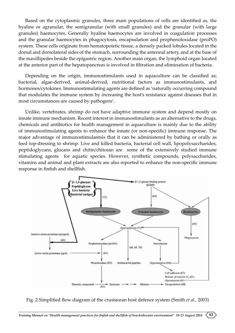
Training Manual on “Health management practices for finfish and shellfish of brackishwater environment” 19-23 August 2014 83
Based on the cytoplasmic granules, three main populations of cells are identified as, the hyaline or agranular, the semigranular (with small granules) and the granular (with large granules) haemocytes. Generally hyaline haemocytes are involved in coagulation processes and the granular haemocytes in phagocytosis, encapsulation and prophenoloxidase (proPO) system. These cells originate from hematopoietic tissue, a densely packed lobules located in the dorsal and dorsolateral sides of the stomach, surrounding the antennal artery, and at the base of the maxillipedes beside the epigastric region. Another main organ, the lymphoid organ located at the anterior part of the hepatopancreas is involved in filtration and elimination of bacteria.
Depending on the origin, immunostimulants used in aquaculture can be classified as; bacterial, algae-derived, animal-derived, nutritional factors as immunostimulants, and hormones/cytokines. Immunostimulating agents are defined as ‘naturally occurring compound that modulates the immune system by increasing the host’s resistance against diseases that in most circumstances are caused by pathogens’.
Unlike, vertebrates, shrimp do not have adaptive immune system and depend mostly on innate immune mechanism. Recent interest in immunostimulants as an alternative to the drugs, chemicals and antibiotics for health management in aquaculture is mainly due to the ability of immunostimulating agents to enhance the innate (or non-specific) immune response. The major advantage of immunostimulantsis that it can be administered by bathing or orally as feed top-dressing to shrimp. Live and killed bacteria, bacterial cell wall, lipopolysaccharides, peptidoglycans, glucans and chitin/chitosan are some of the extensively studied immune stimulating agents for aquatic species. However, synthetic compounds, polysaccharides, vitamins and animal and plant extracts are also reported to enhance the non-specific immune response in finfish and shellfish.
Fig. 2.Simplified flow diagram of the crustacean host defence system (Smith et al., 2003)

Training Manual on “Health management practices for finfish and shellfish of brackishwater environment” 19-23 August 201484
Parameters of immune stimulation
Generally the parameters to evaluate the immune stimulation in vertebrates are the estimation of antibody levels. In the absence of humoral immunity in invertebrates, several cellular functions are considered as immune parameters. Total haemocyte counts, granulocyte counts, phagocytic activity, bacterial clearance activity, respiratory burst and ultimately the challenge protection are some of the commonly evaluated factors in shrimp immunology research.
Herbal products as immunostimulants
Herbal drugs are being commonly used as immunomodulating agents in human and veterinary medicine for long time now. However, recently interest is growing in aquaculture for use of plant based products as growth promoting substances, antimicrobial agents, nutrients and to enhance the disease resistance. Asian countries especially India has rich heritage of medicinal plant usage in human medicine for centuries. With the background information from traditional literature, scientists are working to develop safe, effective and economical drugs for use in aquaculture.
Active ingredients of therapeutic effect from the herbal plants are classified as polysaccharides, alkaloids and flavonoids. Various therapeutic effects of these components have been studied in detail in higher vertebrate models.
Commonly used herbal immunomodulating drugs in aquaculture
Ayurvedic medicines Chinese medicines Sea weeds Ocimum sanctum Astragalus membranaceus Gracilaria fisheriSolanum trilobatum Scutelaria baicalensis Gracilaria corticataPhyllanthus emblica Ganoderma lucidium Cladosiphon okamuranusAzadirachta indica Lonicera japonica Sargassum polycystumWithania somnifera Rheum officinale Sargassum fusiformeCyanodon dactylon, Andrographis paniculataAegle marmelos Isatis indigoticaTinospora cordifoliaPicrorhiza kurooaEclipta alba
Application of herbal drugs has a great potential in aquaculture due to their easy availability, cost effectiveness and broad spectrum activity. Since, most of these drugs are administered orally which is the most desirable mode of application in aquaculture. Additionally, there is no toxic accumulation of the chemicals and moreover, biodegradability of these drugs makes them environmentally safe for extensive application.
Probiotics and Prebiotics as immunostimulants
Immune stimulation is one of the mechanisms by which the probiotic bacteria elicit beneficial effects in the host. Ability of probiotic products to stimulate different arms of immune system has been well documented. Recently prebiotics have also been reported to be acting as immune stimulants in shellfish and finfishes. Prebiotics are indigestible fibres that increase beneficial gut commensal bacteria resulting in general improvement in health of the host. It was believed that a

Training Manual on “Health management practices for finfish and shellfish of brackishwater environment” 19-23 August 2014 85
beneficial effect of prebiotics was due to the by-products generated following the fermentation of the indigestible fibres. However, recently, some of the prebiotics such as, fructooligosaccharide, mannanoligosaccharide, inulin and β–glucan which stimulate the immune system are called as immunosacchardies.
Synthetic oligodeoxynucleotides as immune stimulants and adjuvants
CpGoligodeoxynucleotides (CpG ODNs) with unmethylatedCpGdinucleotides are reported to activate immune response. These CpG motifs are considered as pathogen-associated molecular patterns (PAMPs) due to their abundance in microbial genomes and rarity in host genomes. Since CpG ODNs can induce apparent immune protection against various bacteria and virus, they are widely used as functional immunostimulant or vaccine adjuvant in mammals. In some of the recent studies in shrimp, CpG ODNs have been reported to activate the prophenoloxidase (proPO) system and enhance the respiratory burst level in hemocytes and reduce the WSSV copy numbers following infection. Hence, it is possible that in near future, CpG ODNs could be used as immunostimulant to trigger shrimp immune system, and protect shrimps against viral infection. One recent study using pUC57-CpG reported the activation of apoptosis and regeneration of circulating hemocytes, enhanced phagocytosis and ROS production, which might contribute to the boosted immunity against the infection of pathogens.
Conclusion
Several probiotic and immunostimulant products are known to have beneficial effect in shrimp aquaculture. For effective use of these products and to harness the maximum benefit, there is an urgent need for rigorous validation of the mechanism of action of these agents at molecular level. Further, type of agents, dose, schedule and route of application are to be standardized for the culture conditions and species of shrimp farmed. Combined utilization of probiotic and immunostimulant products could be helpful in prevention of disease in Indian aquaculture.
Training Manual on “Health management practices for finfish and shellfish of brackishwater environment” 19-23 August 201486
MICROBIAL DYNAMICS IN SHRIMP PONDS VIS A VIS BACTERIAL DISEASES OF SHRIMP
T. Bhuvaneswari, Subhendu Kumar Otta, P. Ezhil Praveena and K. P. JithendranAquatic Animal Health and Environment Division
Introduction
Shrimp production in aquaculture sector is substantial in yielding good revenue to our country. Shrimp is a wonderful commodity starting from producer to end user who is relishing the taste, but the risk involved in farming steadily increased with production due to changing aquaculture management practices and environment. The production without loss is the highest challenge put forth to the researchers of this field, understanding the ecology with regard shrimp growth is more important for sustainable and to control any loss in farming.
Microbial dynamics in shrimp ponds
In aquatic habitats, the entire food web is based on microbial primary producers. Theterm microbial ecology came into existence only in early 1960’s. Microorganisms occupy key positions in the global ecosystem by virtue of their metabolic abilities to transform organic and inorganic substances. They are crucial in solving some of our problems through dispersing various liquid and solid wastes in a safe and effective manner. Some of the important applications include recovering metals from low-grade ores (transformation of elements), production of food, feed and fuel from by-products and waste materials,relieving nitrogen fertilizer shortage and biological control of pest. Though microorganisms are mostly primary producers or decomposers in the ecosystem they can also be pathogens since they can obtain nutrients from live primary producers.
Classification of microorganism in pond ecosystem as, Autochthonous: Microbes that are indigenous and always present in the given ecosystem and their presence is based on the more or less constant supply of nutrients that are typical for the ecosystem. Their numbers are always constant in ecosystem. Zymogenous: Those are dependent on an occasional increase in concentration of certain nutrients or on the specific presence of certain nutrients. Theyalways found in very low numbers in any ecosystem and show an increase only when a particular substrate is present in the ecosystem. Allochthonous: They are total strangers to the ecosystem. Their presence is purely transitional and they are not permanent residents.
Microbiota that lived in association with aquatic animal may enhance host growth and survival by producing some digestive enzymes (Sugita et al., 1995; Seeto et al., 1996; Izvekova 2006), impeding pathogenic bacteria, and providing vital compound important for host metabolism. Rapid growth of shrimp occurred in unfiltered pond water, which contained organic particle including bacteria (Moss and Pruder, 1995).
In the shrimp culture pond, the major source of nitrogen waste come from protein in artificial feed pellets. In fact, only 20-30 % of the nitrogen found in feed is converted into shrimp biomass, while the rest is collected in high organic-content sediment at the bottom of the pond. In nature, ammonia is subsequently converted into nitrite and nitrate by the nitrification process, and the nitrate is thereafter eliminated from the pond by the denitrification process in the sediment. The

Training Manual on “Health management practices for finfish and shellfish of brackishwater environment” 19-23 August 2014 87
nitrification and denitrification processes are linked with specific groups of bacteria naturally found in ponds. Generally, β- and γ-proteobacteria are found in a higher percentage of bacterial communities from polluted waters, whereas cyanobacteria are used as feed for increasing the growth rate and size of shrimp or for nutrient removal in aquaculture systems.
A DGGE profile of the microbial community over the course of a single shrimp growing period of 3 months is shown with the majority of the 14 bacteria found were identified as β-proteobacteria (57.14%), which consists of several different groups of bacteria involved in degradation, nitrogen fixation and ammonia oxidation (Rubin &Leff, 2007). These bacteria included Nitrosomonas, Flavobacterium, Exiguobacterium, Burkholderia and Nitrosospira. The remaining bacteria identified in this study belonged to the following groups: α-proteobacteria (7.14%), γ-proteobacteria (14.28%), cyanobacteria (14.28%) and Cytophaga–Flavobacterium–Bacteroides (CFB; 7.14%).
Nitrosomonas is nitrifying bacterium found in shrimp grow-out ponds that remove ammonia from the water. Ammonia oxidizing bacteria are classified into 5 genera: Nitrosomonas, Nitrosovibrio, Nitrosococcus, Nitrosolobus and Nitrospira, while nitrite oxidizing bacteria are classified into three genera: Nitrobacter, Nitrococcus and Nitrospira. Nitrosomonas and Nitrobacter are commonly used commercially in aquaculture as bioremediators. Flavobacterium, which is a cellulose digesting bacterium involved in shrimp shell degradation.
Exiguobacterium spp. has been found in a variety of environments including alkaline, low temperature, or in aquaculture systems. In an aquaculture system, these bacteria have the potential to improve the survival rate and development of Artemia sp. which is used as feed in shrimp nurseries. Moreover, these bacteria, much like Bacillus sp. can produce polypeptide antibiotics, such as bacitracin, gramicidin S, polymyxin, and tyrotricidin, which are active against a wide range of bacteria that might be harmful to shrimp. The presence of cyanobacteria was already suggested by the increase in chlorophyll a content in the shrimp water that was observed during the shrimp growing period. Increased levels of chlorphyll were due to the growth of phytoplankton from nitrogen uptake or nitrogen-fixing in the shrimp pond. Based on traditional culturing studies, the Pseudomonadaceae family of bacteria member of γ-proteobacteriais usually considered to be a predominant bacterial population in mariculture environments (Deng et al., 2009).
Bacterial abundance in shrimp ponds
Total bacterial count in shrimp ponds ranges between 105-6 cells ml-1. The culturable bacterial abundance in the pond followed the following order: heterotrophs >nitrifiers and denitrifiers>sulfate reducers. The heterotrophic population in ponds were generally stable, gradual increase from 103 CFU ml-1 at the start of the culture period will reach 104CFU ml-1. The heterotrophs were found to negatively influence the variation in phosphate concentration. Among the bacteria capable of inorganic nitrogen cycling, the nitrifiers were generally higher than the denitrifiers and were most abundant in the aerated pond water towards the end of the observation period where they reached a higher level 0.76x10³ CFU ml-1. In the non-aerated pond, the nitrifiers were abundant during the initial days of culture at 0.89x10³ CFU ml-1 and a steady decrease in their abundance followed thereafter. A negative relationship of heterotrophs with nitrifiers in non-aerated conditions was also observed. In contrast to nitrifiers, abundance

Training Manual on “Health management practices for finfish and shellfish of brackishwater environment” 19-23 August 201488
of denitrifiers in the ponds decreased over time ranging between 0.005-0.12 x10³ CFU ml-1 and 0.001- 0.067 x10³ CFU ml-1 in the aerated and non-aerated pond. A decrease in the population of sulfate reducers over time was observed in the non-aerated pond. The SRB abundance remained low and hydrogen sulphide content in the aerated pond increased towards the end of the culture period.
Bacterial Diseases of Shrimp
In the farm affected by shrimp disease, exponential phytoplankton growth showed rapid and intense pulses, and their sudden collapses suggested low ecosystem stability and loss of homeostatic mechanisms. The shift from pico- to nanophytoplankton was observed in relation to shrimp mortalities (Lemonnier et al., 2006). Strong phytoplankton oscillations could favour the proliferation of Vibrio sp. in the water column. These were linked to an imbalance of N:P ratio in the water column, which could depend on nutrient flux intensity from sediment to the water column. The combination of anaerobic and acidic conditions in the sediments and low ecosystem stability are also potential stress factors that could have weakened the shrimps reared in the farm affected by the disease. These conditions could create an environment favouring the proliferation of the pathogen.
1. Vibriosis
Vibriosis is a major problem in shrimp hatcheries. The main cause of the disease is Gram-negative bacteria belonging to the genus Vibrio. The possible four main species are V. parahemolyticus, V. vulnificus, V. alginolyticus and V. harveyi. However, V. harveyi, a luminescent species is considered as the most devastating that causes extreme losses in both the hatcheries and shrimp-rearing farms. All larval substages of L. vannamei were reported to be susceptible to V. harveyi and V. parahaemolyticus with high mortality rates. This bacterial disease is mainly caused by pathogenic vibrio species affecting various larval stages of shrimps resulting in about 80% mortality. Further, the developmental stage from naupli to protozoea-3 exhibited greater susceptibility to these pathogens when compared to mysis to postlarval stages. Gross signs of vibriosis are light or dark brown focal lesions and necrosis of appendage tips. The change of color is the result of melanin produces by host hemocytes involved in the inflammatory process. Large number of motile bacteria is visible in the hemocoel and hepatopancreas of moribund shrimp. Affected shrimp exhibit decreased appetite. Some also had a darker, larger or shrunken hepatopancreas.
2. Bacterial necrosis in larval stages
This disease l appears as localized necrosis or discoloration on any appendage, causing high mortality of zoea and mysis stages, but also affects post larvae to a lesser extent. In zoea, it often starts with a liquefaction of the gut contents. If the necrosis starts at zoea-1 stage, the entire tank is collapsed by zoea-2 or zoea-3 within a matter of 24 hours. However, if necrosis starts at zoea-3 stage, only part of them will moult to mysis and mortality prolongs for a few days.
3. Bolitasnegricans
Robertson et al., (1998) reported that infection of L. vannamei larvae with V. harveyi at 105
cfu /ml produced a larval disease called Bolitas negricans and bioluminescence. The highest
Training Manual on “Health management practices for finfish and shellfish of brackishwater environment” 19-23 August 2014 89
mortalities were recorded during the transition from zoea-3 to mysis. “Bolitas” is the Spanish name given to a syndrome involving the detachment of epithelial cells from the intestine and hepatopancreas, which appear as small spheres within the digestive tract. Development of bioluminescence reduced feeding and retarded development, sluggish swimming, reduced escape mechanisms, degeneration and formation of bundles of necrotic tissues within the hepatopancreas leading to death of infected larvae.
4. Red spot syndrome
The disease is more common in Zoea-1 to post larval stages of L. vannamei where the affected larvae exhibit acute necrosis of the entire body and lysis of all the tissue cells. The level of mortality observed in this disease can go up to 100%. Further, in this syndrome the hatchery water of the tanks stocked with the infected larvae turns red to pink colour. This is most commonly seen in L. vannamei hatcheries. It was reported to be caused by the bacterium belonging to the genus Pseudomonas which was identical to P. mesophilica and P. anguilliseptica (Soltani et al., 2010).
5. Luminescent Disease
Vibrio harveyi is a gram-negative, luminescent, marine bacterium isolated both in a free-living state and as a commensal organism in the enteric contents of marine animals is recognized as a primary pathogen of many commercially cultured invertebrate species, such as the black tigerprawn (P. monodon), V. harveyi can cause up to 100% mortality of larvae in the hatchery stage of penaeid culture.
6. Early Mortality Syndrome (EMS)
Also referred as acute hepatopancreatic necrosis disease (AHPND) causes acute progressive degeneration of hepatopancreas (HP) from medial to distal with dysfunction of all HP cells, prominent necrosis and sloughing of tubular epithelial cells. The terminal stage is marked inter- and intra-tubular hemocytic inflammation with massive secondary bacterial infections that seen in association with necrotic and sloughed HP tubule cells. Aetiological agent is identified as a member of the V. harveyi clade, a strain most closely related to V. parahemolyticus.Susceptible host species are L. vannamei and P. monodon and other species not reported. The susceptible stages of the host are 20 to 30 days of stocking ponds with postlarvae. Target organs are hepatopancreas and gut tissue. Clinical signs of the disease may become apparent as early as 10 days of post stocking in a pond which includes slow growth, corkscrew swimming, loose shells, with abnormal shrunken, small, swollen or discoloured (whitish/pale/yellowish) hepatopancreas. Besides this, an empty gastrointestinal tract, whitish, ‘milky’ appearance of the stomach, whitish, atrophied hepatopancreas, lethargy, and soft shells were also seen. Moribund shrimp sink to bottom.
7. White gut syndrome
The etiology of whitegut syndrome cannot be pinpointed to a single agent in provocating symptoms like stunted growth with reduced feed intake. The gut was found to be empty and appeared opaque. Gut filled with gas and feed intermittently. Signs are more visible when the shrimp reaches 3 gms and above. If the environmental conditions are favourable to infectious agent causing white gut, the shrimp will stop feeding and die with the development of loose

Training Manual on “Health management practices for finfish and shellfish of brackishwater environment” 19-23 August 201490
shell. The white gut in many occasions is correlated with decrease in pathogenic vibrios in the cultured pond. In some instances the mortality has been recorded with the symptoms of stunted growth and opaque white gut visible through the transparent cuticle as a white streak. The feed consumed was released as white fluid material. Large number of motile bacteria is visible in the hemolymph and hepatopancreas of moribund shrimp. The possible main vibrio species are V. parahemolyticus, V. vulnificus, V. alginolyticus and V. harveyi. However, V. harveyi is highly pathogenic luminescent species that causes extreme losses in all stages shrimp farming. It has been reported that white gut affected samples had 0.2 x 10⁵cfu/mL of vibrio load in hemolymph (Jayashreeet.al. 2006).
8. Filamentous bacterial Infection
Leucothrixmucor, Thiothrix spp., Flexibacter spp., lavobacterium, Cytophaga spp., are filamentous bacteriacause infection in penaeid shrimplarvae. Discolouration of gills, low growth and feeding,increased mortality and lethargy are common signs of the disease. The disease is associated with poorwater quality. Higher degree of infection may lead to necrosis in gill tissue.
9. Necrotising Hepatopancreatitis (NHP)
NHP is caused by a bacterium that is relatively small, highly pleomorphic, Gram negative, and an apparent obligate intracellular pathogen. The NHP bacterium has two morphologically different forms: one is as mall pleomorphic rod and lacks flagella; while the other is a longer helical rod possessing eight flagella on the basal apex of the bacterrium, and an additional flagellum (or possibly two) on the crest of the helix. The NHP bacterium occupies a new genus in the alpha Proteobacteria, and is closely related to other bacterial endosymbionts of protozoans (Lightner, 1996). NHP is also known as Texas necrotizing hepatopancreatitis (TNHP), Texas Pond Mortality Syndrome (TPMS) and Peru necrotizing hepatopancreatitis (PNHP).
10. Bacterial White Spot Syndrome
The bacterial white spot is similar to gross clinical signs of White spot disease (WSD) but PCR test for WSSV will be negative. This syndrome was reported in cultured Penaeus monodon. Dull white spots are seen on the carapace and all over the body. The white spots are rounded and not as dense as those seen in WSD. The wet mount microscopy shows the spots as opaque brownish lichen-like lesions with a crenelated margin. The spot center is often eroded and even perforated. During the early stage of infection, shrimp are still active, feeding and able to moult–at which point the white spots may be lost. However, delayed moulting, reduced growth and low mortalities have been reported in severely infected shrimp, Bacillus subtilis has been suggested as the causative agent of this syndrome (Wang et al., 2000).Vibrio Cholera was also often isolated in some bacterial white a spot case in Thailand has been reported.

Training Manual on “Health management practices for finfish and shellfish of brackishwater environment” 19-23 August 2014 91
HISTOPATHOLOGY AND ITS IMPORTANCE IN SHRIMP DISEASE DIAGNOSIS
P. Ezhil Praveena, T. Bhuvaneswari, S.K. Otta, N. Jagan Mohan Raj and K.P. Jithendran Aquatic Animal Health and Environment Division
Introduction
Histopathology is the branch of pathology which deals with the study of pathological abnormalities in the form of tissue changes. The tissue undergoes a series of changes during the course of the disease. This branch of science helps in appreciating the changes that take place at tissue level and with the help of additional scientific information such as gene expression it aids one to understand the disease progression and thereby arrive at an accurate diagnosis. Though molecular method of diagnosis gained more importance in shrimp culture for the past several years with the increase in incidence of White Spot Diseases this method of diagnosis remains a Gold standard for many diseases.
Diagnosis of viral diseases by histopathology
The common viral diseases encountered in shrimp are White spot disease (WSD), Infectious Hypodermal and Hematopoietic Necrosis (IHHN), Yellow Head Disease (YHD), Taura Syndrome (TS), Infectious Myonecrosis (IMN), Penaeus vannamei Nodavirus (PvNV), Monodon BaculoVirus (MBV) and Hepatopancreatic Parvovirus Disease (HPV).
White Spot Disease (WSD)
This disease is considered as the single most serious shrimp pathogen worldwide and was first reported from farmed Penaeus japonicus in Japan in 1993. The virus can infect a wide range of aquatic crustaceans. All life stages are potentially susceptible. The best life stages for disease diagnosis are late PL stages, juveniles and adults. The major target organ for WSSV infection are tissues of ectodermal and mesodermal origin, especially the cuticular epithelium and subcuticular connective tissues, and therefore samples from the pleopods, gills, haemolymph, stomach or abdominal muscle are recommended for disease diagnosis. Based on the clinical manifestations, WSD outbreaks in penaeid shrimps are divided into three types (Type I, II and III). WSSV-infected shrimp always has a delayed (or sometimes completely absent) clotting reaction. The histopathological lesion is the presence of intranuclear inclusion bodies as prominent eosinophilic (in the early stages they are Cowdry type A) to large basophilic intranuclear inclusions with variable multifocal necrosis. These tissues include gills, haemocytes and haematopoietic tissue, lymphoid organ, connective tissues, subcuticular epidermis, stomach, foregut and hindgut epithelium, heart, striated muscle, midgut and ovary walls, antennal gland and the nervous tissues. Besides this affected tissues cells will exhibit severe nuclear hypertrophy, chromatin margination.

Training Manual on “Health management practices for finfish and shellfish of brackishwater environment” 19-23 August 201492
Photomicrographs of Gill tissue showing WSSV inclusion bodies (arrow) –100X H&E
Yellow Head Disease (YHD)
The causative agent of this disease is yellow head virus (YHV), a corona-like RNA virus in the genus Okavirus, family Ronaviridae and order Nidovirales. YHV can infect cultured shrimp from late postlarval stages onwards, but mass mortality usually takes place in early to late juvenile stages. Yellow head virus (genotype 1) is one of six known genotypes in the yellow head complex of viruses and is the only known agent of YHD. The target tissues of this disease are from ectodermal and mesodermal origin including lymphoid organ, haemocytes, haematopoietic tissue, gill lamellae and spongy connective tissue of the subcutis, gut, antennal gland, gonads, nerve tracts and ganglia. The most appropriate tissues for diagnosing this disease are lymphoid organ and gills. YHV can induce up to 100% mortality in infected shrimps within 3 days of the first appearance of clinical signs like white, yellow or brown gills, yellowing of the cephalothorax caused by the underlying yellow hepatopancreas and general bleaching of body (from which the disease got its name), yellow and swollen digestive gland which makes head appear yellow. The major histopathological changes noticed are the presence of moderate to large numbers of deeply basophilic, evenly stained, spherical, cytoplasmic inclusions approximately 2µm in diameter or smaller in tissues of ectodermal and mesodermal origin. The lymphoid organ, haemocytes, haemopoietic tissue, gill, heart, cuticular epithelium, midgut and connective tissues are the primary target tissues and organs for YHV infection. Systemic necrosis of ectodermal and mesodermal tissues with prominent nuclear pyknosis and karyorrhexis is also another feature of this disease. Cellular changes in early infections may include nuclear hypertrophy, chromatin diminution and margination, and lateral displacement of the nucleolus. Loss of tissue structure within lymphoid organ, stromal matrix cells that comprise tubules become infected leading to loss of tubular structure, tubules appear degenerate. Lymphoid organ spheroids (LOS) develop during infection, ectopic spheroids may lodge in constricted areas of the haemocoel (heart, gills, subcuticular connective tissues etc). Necrosis of the lymphoid organ can be used as a characteristic feature to distinguish YHD from acute Taura Syndrome (TS) in penaeid shrimp.

Training Manual on “Health management practices for finfish and shellfish of brackishwater environment” 19-23 August 2014 93
Taura Syndrome (TS)
This disease is seen in many shrimp species but the infection found to be very severe in L. vannamei farms. TS is best known as a disease of nursery or grow-out phase L. vannamei that occurs within ~14-40 days of stocking postlarvae (PL) into grow-out ponds or tanks. Larger shrimp may also be affected, especially if they are not exposed to the virus until they are larger juveniles or adults. It was first described in the year 1952 in Ecuador. The principal target tissue in the acute phase of infection is the cuticular epithelium while in chronic infections it is the lymphoid organ (LO). Suitable specimens for diagnosis of disease include PL, juveniles and adults. The clinical phase of the disease occurs in three distinct phase i.e. acute, transition and chronic. Gross signs are obvious in the acute and transition phases. Acute phase of the disease is characterized by multifocal areas of necrosis in the cuticular epithelium of the general body surface, appendages, gills, hindgut, and foregut oesophagus, anterior and posterior chambers of the stomach). Cells of the subcuticular connective tissues and adjacent striated muscle fibres are occasionally affected. In some severe cases the antennal gland tubular epithelium is also destroyed. Cytoplasmic remnants of necrotic cells are often extremely abundant and these are seen as spherical bodies (1-20µm in diameter) which are eosinophilic to pale basophilic. These structures, along with pyknotic and karyorrhectic nuclei, give a characteristic ‘peppered’ or ‘buckshot-riddled’ appearance, which is considered to be pathognomonic for TS disease and there is no concurrent necrosis of the parenchymal cells of the LO tubules which distinguishes it from acute phase of yellow head disease in which similar lesion is also seen. There is absence of haemocytic infiltration or other host-inflammatory response which distinguishes it from the transitional phase of the disease. In the transitional phase typical acute-phase cuticular lesions decline in abundance and are replaced by conspicuous infiltration and accumulation of haemocytes at the sites of necrosis. The masses of haemocytes may become melanised giving rise to irregular black spots that characterize the transition phase of the disease. In chronic phase there is no gross signs of infection, but the prominent histopathological lesion is the presence of an enlarged LO with numerous LOS, which may remain associated with the main body of the paired LO, or may be seen detach and become ectopic LOS bodies that lodge in constricted areas of the haemocoel (i.e. heart, gills, in the subcuticular connective tissues, etc.).
Infectious Hypodermal and Hematopoietic Necrosis (IHHN)
IHHN was first discovered in blue shrimp Penaeus stylirostris and white shrimp L. vannamei in the Americas in the early 1980’s. Three distinct genotypes have been identified and they are Type 1, Type 2 and Type 3. Type 3 is again subdivided as 3A and 3B. The first two genotypes are infectious to L. vannamei and P. monodon, while the latter two are not infectious to these species. Most penaeid species can be infected with IHHNV, including P. monodon; it has been reported to cause acute epizootics and mass mortality in P. stylirostris. By contrast, it does not cause mortality in L. vannamei, but rather cause reduced and irregular growth and cuticular deformities, gross signs collectively referred to as “Runt-Deformity Syndrome” (RDS). IHHNV infects and shown to replicate in tissues of ectodermal and mesodermal origin from the embryo. Thus, the principal target organs includes the gills, cuticular epithelium (or hypodermis), all connective tissues, the haematopoietic tissues, haemocytes, the lymphoid organ, antennal gland, and the ventral nerve cord, its branches and ganglia. Hence, whole shrimp (e.g. larvae or PLs) or tissue samples containing the aforementioned target tissues are suitable for most

Training Manual on “Health management practices for finfish and shellfish of brackishwater environment” 19-23 August 201494
tests using molecular methods. Haemolymph or excised pleopods may be collected and used for testing when non-lethal testing of valuable broodstock is necessary. RDS, a chronic form of IHHN disease, occurs in L. vannamei. RDS has also been reported in cultured stocks of P. stylirostris and P. monodon. Juvenile shrimp with RDS may display a bent (45° to 90° bend to left or right) or otherwise deformed rostrum, a deformed sixth abdominal segment, wrinkled antennal flagella, cuticular roughness, ‘bubble-heads’, other cuticular deformities and disparate growth with a wide variation in sizes of shrimp many smaller than expected (‘runted’). The characteristic IHHN inclusion bodies are eosinophilic and often haloed, intranuclear inclusion bodies within chromatin-marginated, hypertrophied nuclei of cells in tissues of ectodermal (epidermis, hypodermal epithelium of fore- and hindgut, nerve cord and nerve ganglia) and mesodermal origin (haematopoietic organs, antennal gland, gonads, lymphoid organ, and connective tissue). The inclusion bodies caused by this infection may be easily confused with inclusion bodies seen in WSSV infection.
Presence of prominent intranuclear, Cowdry type A inclusion bodies in the gill tissue of IHHNV infected shrimps (H & E 100X)
Infectious Myonecrosis (IMN)
Infectious myonecrosis (IMN) is a recently identified disease in cultured L. vannamei. IMN causes significant disease and mortalities in juvenile and subadult L. vannamei. IMNV infects tissues of mesodermal origin. The principal target tissues for IMN include the striated muscles (skeletal and less often cardiac), connective tissues, haemocytes, and the lymphoid organ parenchymal cells. In chronic infections, the lymphoid organ may be the principal target tissue. Haemolymph or excised pleopods may be collected and used when non-lethal testing of valuable broodstock is necessary. Affected shrimp present extensive white necrotic areas in the striated muscle, especially in the distal abdominal segments and tail fan which may become necrotic and reddened in some individual shrimp. In acute phase the lesions noticed are coagulative necrosis of muscle, often with edema. In chronic phase liquefactive necrosis of the muscle is seen which is accompanied by haemocytic infiltration and fibrosis. Significant LOS formation is seen and ectopic LOS is often found in the haemocoel and loose connective tissues, especially in the heart lumen and adjacent to antennal gland tubules. In some cases, perinuclear pale basophilic to darkly basophilic inclusion bodies are evident in muscle cells, connective tissue cells, haemocytes, and in cells that comprise LOS.
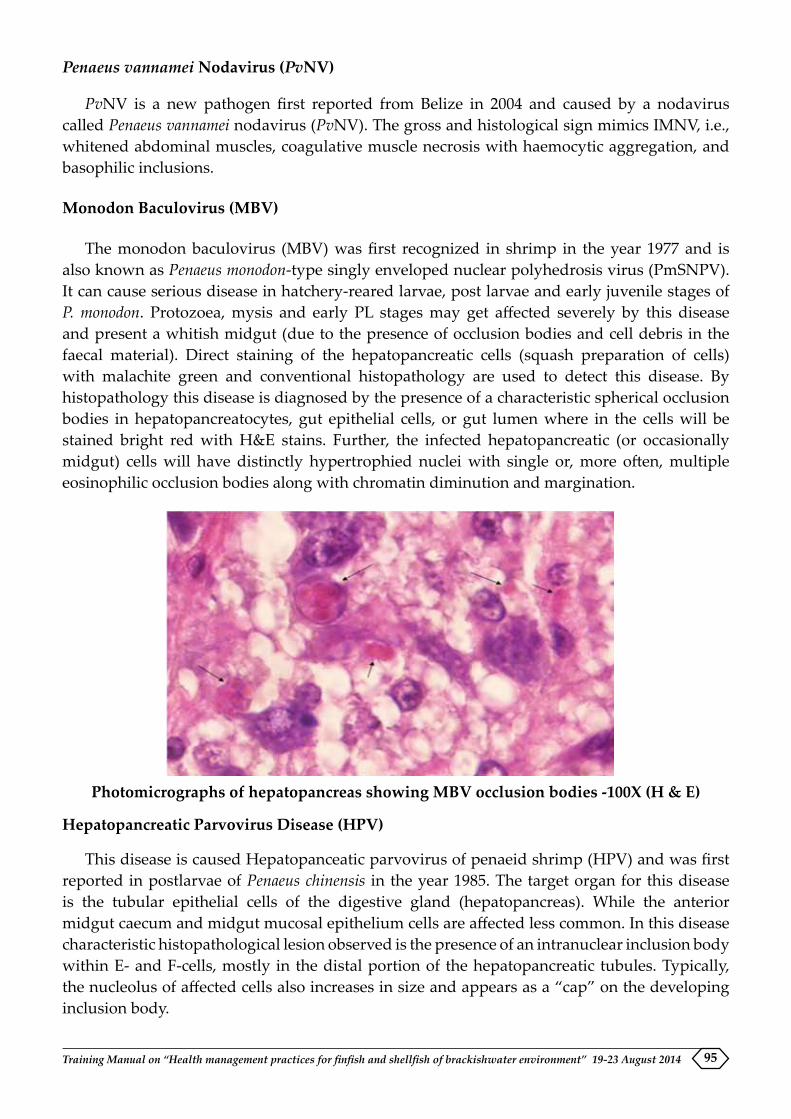
Training Manual on “Health management practices for finfish and shellfish of brackishwater environment” 19-23 August 2014 95
Penaeus vannamei Nodavirus (PvNV)
PvNV is a new pathogen first reported from Belize in 2004 and caused by a nodavirus called Penaeus vannamei nodavirus (PvNV). The gross and histological sign mimics IMNV, i.e., whitened abdominal muscles, coagulative muscle necrosis with haemocytic aggregation, and basophilic inclusions.
Monodon Baculovirus (MBV)
The monodon baculovirus (MBV) was first recognized in shrimp in the year 1977 and is also known as Penaeus monodon-type singly enveloped nuclear polyhedrosis virus (PmSNPV). It can cause serious disease in hatchery-reared larvae, post larvae and early juvenile stages of P. monodon. Protozoea, mysis and early PL stages may get affected severely by this disease and present a whitish midgut (due to the presence of occlusion bodies and cell debris in the faecal material). Direct staining of the hepatopancreatic cells (squash preparation of cells) with malachite green and conventional histopathology are used to detect this disease. By histopathology this disease is diagnosed by the presence of a characteristic spherical occlusion bodies in hepatopancreatocytes, gut epithelial cells, or gut lumen where in the cells will be stained bright red with H&E stains. Further, the infected hepatopancreatic (or occasionally midgut) cells will have distinctly hypertrophied nuclei with single or, more often, multiple eosinophilic occlusion bodies along with chromatin diminution and margination.
Photomicrographs of hepatopancreas showing MBV occlusion bodies -100X (H & E)
Hepatopancreatic Parvovirus Disease (HPV)
This disease is caused Hepatopanceatic parvovirus of penaeid shrimp (HPV) and was first reported in postlarvae of Penaeus chinensis in the year 1985. The target organ for this disease is the tubular epithelial cells of the digestive gland (hepatopancreas). While the anterior midgut caecum and midgut mucosal epithelium cells are affected less common. In this disease characteristic histopathological lesion observed is the presence of an intranuclear inclusion body within E- and F-cells, mostly in the distal portion of the hepatopancreatic tubules. Typically, the nucleolus of affected cells also increases in size and appears as a “cap” on the developing inclusion body.

Training Manual on “Health management practices for finfish and shellfish of brackishwater environment” 19-23 August 201496
Photomicrographs of hepatopancreas showing HPV like inclusion bodies-100X (H & E)
Diagnosis of bacterial diseases by histopathology
The common bacterial diseases seen in shrimps are mainly vibriosis caused by various Vibrio species.
Vibriosis
Vibriosis is a bacterial disease caused by gram-negative, motile, facultative anaerobic bacteria of the family Vibrionaceae. It is pervasive throughout the world and all marine species including shrimp, are vulnerable when their natural defense mechanisms are suppressed. This disease is caused by a number of species like Vibrio harveyi, V. vulnificus, V. parahaemolyticus, V. alginolyticus and V. penaeicida and are commonly referred as black shell disease, tail rot, septic hepatopancreatic necrosis, brown gill disease, swollen hindgut syndrome and luminous bacterial disease. The commonly observed clinical signs are lethargy, loss of appetite, discoloured and necrotic hepatopancreas with red discolouration of the body, yellowing of the gill tissue and white patches in the abdominal muscle, melanisation, granulomatous encapsulation, necrosis and inflammation of organs (lymphoid organ, gills, heart etc.) and luminescence. Adult shrimps suffering from vibriosis may appear hypoxic, red colouration of the body with red to brown gills, reduced feed intake, lethargic swimming behaviour and seen at the edges and surface of ponds. Vibrio spp. also causes red-leg disease, characterized by red colouration of the pleopods, periopods and gills. Infected post larvae may show symptoms like empty guts with reduced motility and phototaxis. In shrimps with lesions of bacterial shell disease the body cuticle, appendages and the gills will appear brown or black. While the post larvae may display cloudy hepatopancreas, gills appear brown in colour. Septic hepatopancreatitis is characterized by atrophy of the hepatopancreas with multifocal necrosis and haemocytic inflammation. Systemic vibriosis the changes noticed are formation of septic haemocytic nodules in the lymphoid organ, heart and connective tissues of the gills, hepatopancreas, antennal gland, nerve cord, telson and muscle. Infected hepatopancreocytes may appear poorly vacuolated, indicating low lipid and glycogen reserves. In external vibriosis the lesions observed are heavy cuticular bacterial colonization. In enteric vibriosis, the changes seen are colonization in the internal cuticle i.e. in the oral region, esophagus and stomach. Sloughing, necrosis, inflammation and melanisation are the changes observed in the hepatopancreas or midgut region.

Training Manual on “Health management practices for finfish and shellfish of brackishwater environment” 19-23 August 2014 97
Early Mortality Syndrome
Recently, a new emerging disease commonly referred to as ‘‘early mortality syndrome’’ (EMS) or more technically known as ‘‘acute hepatopancreatic necrosis disease’’ (AHPND) was reported. The disease was first reported in China in 2009 and subsequently in Vietnam, Thailand, and Malaysia. It affects shrimp post larvae within 20-30 days after stocking and frequently causes 100% mortality. The species which are commonly affected by this disease are L. vannamei and P. monodon. The causative agent has been reported to be a bacterium presumably V. parahaemolyticus. The clinical signs and mortality can be observed as early as 10 days of post stocking. The affected shrimps exhibit lesions like pale to white hepatopancreas with significant atrophy, soft shells and partially full to empty guts, black spots or streaks are visible within the hepatopancreas. Hepatopancreas (HP) does not squash easily between thumb and finger. Moribund shrimp will sink to bottom. The major lesions noticed in the hepatopancreas with acute progressive degeneration of the hepatopancreas with initial decrease of R, B and F-cells followed by a marked reduction of mitotic activity in E-cells. The development of lesion is noticed from proximal to distal with dysfunction of R, B, F, and lastly E-cells, with affected HP tubule mucosal cells presenting prominent karyomegaly (enlarged nuclei), and rounding and sloughing into the HP tubules. The sloughed HP cells provide a substrate for bacterial growth, resulting in massive secondary bacterial infection (putative Vibrio spp.) and complete destruction of HP at the terminal phase of the disease. Inter tubular haemocytic aggregation and haemocytes encapsulation of necrotic HP tubules and melanisation of the more proximal portions of HP tubules is seen in some shrimp.
Necrotizing Hepatopancreatitis (NHP)
The NHP bacterium is a gram-negative, dimorphic, intracellular rickettsial like organism that occurs free within the cytoplasm of infected hepatopancreatic cells. This disease is seen in many penaeid species and mainly affects late larval stage, juvenile and adult stages of the animal. Mortality of about 90% is seen within 30 days of the outbreak of the disease. The affected shrimps are nonspecific in nature and characterized by lethargy, emaciation, soft shells, heavy fouling from external parasites, black gills, reduced growth and an atrophied hepatopancreas. Infected shrimp display empty midgut with increased superficial epicommensal cuticular fouling and/or opportunistic infections (i.e. black spots) also present. Digestive gland (hepatopancreas) appears pale to white. The hepatopancreas is the target organ for this disease. Microscopic examination of unstained tissue squashes from suspected hepatopancreas may show reduced lipid and dark melanised necrotic tubules. Histologically, infected tubular epithelial cells will appear initially hypertrophied with a generalized basophilic intra-cytoplasmic granularity due to the presence of numerous pleomorphic rickettsial-like organisms. Three stages of infection have been described in histologic studies. In early NHP infection (stage I), intra-cytoplasmic rickettsial-like organisms can be detected free within the cytoplasm of resorptive, fibrillar B cells of scattered tubules with increasing levels of tubular epithelial cell hypertrophy or desquamation. Tubular necrosis, interstitial haemocytic infiltration and lipid depletion occurs in stages II and III.
Conclusions
Though many techniques have come in diagnosing various shrimp diseases, histopathology technique remains a gold standard for recently emerged AHPND. Further, this technique is cheaper when compared to other techniques. Shrimp disease diagnosing laboratories can arrive at a definite diagnosis by employing this technique in combination with other molecular pathological tools like in situ hybridization.

Training Manual on “Health management practices for finfish and shellfish of brackishwater environment” 19-23 August 201498
EARLY MORTALITY SYNDROME (EMS) / ACUTE HEPATOPANCREATIC NECROSIS DISEASE (AHPND)-AN UPDATE
S.K. Otta, P. Ezhil Praveena, T. Bhuvaneswari and P.K. Patil Aquatic Animal Health and Environment Division
Aquaculture plays a vital role in terms of its contribution to the global food supply. From the ancient traditional form, the industry has evolved to the present developed form through several scientific interventions and thereby has been able to constantly increase its share in terms of both production and value. Almost 50% of the total fish food for the world population is generated from aquaculture practice. The trend is likely to continue and with the depletion of natural stock, aquaculture is expected to overtake as a major fish food supply industry in near future. The situation in India is also having a similar trend where the progress is at a rapid pace. During 2012-13, India’s marine product export reached all-time high production of 928215 tons with a value near to 19000 crores. More than 50% of this share was from frozen shrimp and thereby signifying the importance of shrimp aquaculture. However, the production in the country had been undulating since the first reported disease outbreak in 1993-94. Thus, disease problems have often caused concern to the sustainability of shrimp aquaculture. However, most of disease have been diagnosed but since 2009, a new disease, the early mortality syndrome (EMS) or specifically known as acute hepatopancreatic necrosis disease (AHPND) of farmed shrimp has emerged in the South East Asian countries and has adversely affected the shrimp production.
About the disease
Based on the onset of mortality during early days of culture, the name of the disease was coined accordingly as EMS. Mortality of shrimps starts as early as 10 days of post stocking in a pond and mass mortality may occur within 30-35 days of stocking. Subsequently after the scientific intervention and due to typical pathological changes in hepatopancreas, the disease was called as acute hepatopancreatic necrosis syndrome (AHPNS). However, when the causative agent was identified, the nomenclature was changed to acute hepatopancreatic necrosis disease (AHPND).
Spread pattern
The disease is assumed to have originated from the Hainan area in China in 2009 in the name of “covert mortality disease”. By 2010, the disease further spread to several other areas in China and EMS/AHPND was confirmed to be present. Towards the end of 2010, some of the coastal provinces of Vietnam also reported similar problems. In Malaysia, EMS/AHPND became more prominent in 2011 when it started spreading to Pahang, Penang, Perak, Sabah and Sarawak after the initial report from Johar. Thailand was the next country to get this disease, where suspected cases were reported in the Eastern Gulf during the late 2011 and the disease spread to other parts, assuming severity by 2012. The most recent country which has been confirmed to be infected with EMS/AHPND is Mexico. In 2013, EMS was detected from Sinaloa and Nayarit areas of Mexico. As per the recent information, this disease has not yet been reported from any other countries. Presence of EMS/AHPND has caused widespread loss in all the affected

Training Manual on “Health management practices for finfish and shellfish of brackishwater environment” 19-23 August 2014 99
countries. Many of the farmers from EMS/AHPND affected areas have reported losses to be as high as 80%.
Clinical signs of the disease
As a part of general observation, the hepatopancreas (HP) of shrimp has been found to be the main target organ in EMS/AHPND. Clinical signs of AHPND include pale discoloration and atrophy (size reduction) of HP, which appears granular when pressed between the fingers, with occasional black streaks. Other clinical signs include pale and empty stomach, empty gut, reduced growth, loose shell and black discoloration. In addition to the typical clinical signs, some of the behavioural changes such as lethargy, swimming sluggishly along the dikes, corkscrew swimming and reduced preening and feeding are also observed in EMS/AHPND.
Species affected
Based on the earlier observation, it was reported that three of the major penaeid shrimps such as tiger shrimp, P. monodon, Pacific white shrimp, L. vannamei and Chinese shrimp, Penaeus chinensis are the main species affected by this disease. Further research has indicated that the tiger shrimp are more resistant to EMS/AHPND compared to others especially L. vannamei has been considered to be the most susceptible shrimp species.
Causative agent
It took nearly three years of systematic investigation by leading aquaculture pathologists from the University of Arizona, before the discovery of the actual causative organism for this disease. Through Koch’s postulate, it was confirmed that EMS/AHPND was caused by a specific strain of V. parahaemolyticus. Recent sequencing information indicates that EMS/AHPND strains have conserved sequences located in the plasmid.
Disease diagnosis
Preliminary diagnosis could be made based on the onset of typical clinical signs and behavioural changes. As a follow up to this, different laboratory investigations can be employed for accurate diagnosis. Since HP is the main target organ of this disease, histopathological analysis of this organ is essential to confirm diagnosis. Necrosis of B, F and R cells of HP, round off and then sloughing of HP cells to lumen, hemocytic infiltration and loss of mitotic E cells are some of the typical pathologies diagnostic of EMS/AHPND confirmation. As a general pattern, the degradation of HP progresses from the proximal to the distal part. During the terminal phase, the HP is completely degraded by the secondary infection due to other bacteria.
Metagenomics and direct sequencing of the pathogen revealed conserved regions based on which primers have been recently designed for identification by PCR. Commercial kits are also now available for detection of the causative agent of AHPND.
When the disease appears for the 1st time in a country, it is essential to study both with the histopathology and PCR methods. Additionally, it is also necessary to isolate the specific strain of V. parahaemolyticus from the diseased animal and prove Koch’s postulates.

Training Manual on “Health management practices for finfish and shellfish of brackishwater environment” 19-23 August 2014100
Disease mechanism
Laboratory investigations have shown that the pathogen has to reach specific numbers in order to produce the toxin and induce the specific pathology. It is hypothesized that the pathogen enters through the oral route, colonizes in the stomach and after reaching the specific numbers it produces the toxin by quorum sensing mechanism. The toxin is then transmitted to HP to cause necessary pathological changes and cause mortality.
Risk to humans
Specific strain of V. parahaemolyticus that cause EMS/AHPND does not contain the necessary genetic material that can also cause gastroenteritis which is otherwise caused by the human pathogenic V. parahaemolyticus. The EMS/AHPND strain of V. parahaemolyticus has been reported to be sensitive to cold temperature and does not survive during the transportation through the cold chain. Therefore, it is unlikely that the specific strain of V. parahaemolyticus will pose any risk when consumed by human beings.
Suggested reading:
FAO Fisheries and Aquaculture Report No. 1053 FIRA/R1053, 25-27 June 2013
Infofish International, 5/2013 (September/October): 56-59.
Joshi J, Srisala J, Truong VH, Chen I-T, Nuangsaeng B, Suthienkul O, Lo CF, Flegel TW, Sritunyalucksana K, Thitamadee S (2014). Variation in Vibrio parahaemolyticus isolates from a single Thai shrimp farm experiencing an outbreak of acute hepatopancreatic necrosis disease (AHPND). Aquaculture (428–429): 297-302.
Kondo H, Tinwongger S, Proespraiwong P, Mavichak R, Unajak S, Nozaki R, Hirono I, (2014). Draft Genome Sequences of Six Strains of Vibrio parahaemolyticus Isolated from Early Mortality Syndrome/Acute Hepatopancreatic Necrosis Disease Shrimp in Thailand. Genome A 2(2): e00221-1
Leaño EM, Mohan VC (2012) Early mortality syndrome threatens Asia’s shrimp farms. Global Aquaculture Advocate, July/August 2012, pp 38-39.
Lightner DV, Redman RM, Pantoja CR, Noble BL, Tran L (2012) Early Mortality Syndrome affects shrimp in Asia. Global Aquaculture Advocate, January/February 2012 p 40.
NACA (2012). Report of the Asia Pacific emergency regional consultation on the emerging shrimp disease: Early mortality syndrome (EMS)/ acute hepatopancreatic necrosis syndrome (AHPNS), 9-10 Aug 2012. Published by the Network of Aquaculture Centres in Asia-Pacific, Bangkok, Thailand. August 2012.
Tran L, Nunan L, Redman RM, Lightner DV, Fitzsimmons K. (2013). EMS/AHPNS: Infectious disease caused by bacteria. Global Aquaculture Advocate, July/August 2013 pp 18-20.
Tran L, Nunan L, Redman RM, Mohney LL, Pantoja CR, Fitzsimmons K, Lightner DV (2013). Determination of the infectious nature of the agent of acute hepatopancreatic necrosis syndrome affecting penaeid shrimp. Diseases of Aquatic Organisms, 106: 45-55, 2013.

Section IIIpraCtiCals


Training Manual on “Health management practices for finfish and shellfish of brackishwater environment” 19-23 August 2014 103
HISTOPATHOLOGICAL ANALYSIS OF SHRIMP SAMPLES
P. Ezhil Praveena, N. Jagan Mohan Raj, Subhendu Kumar Otta, T. Bhuvaneswari, and K.P. Jithendran
Aquatic Animal Health and Environment Division
Histopathology plays an important role in disease diagnosis. This technique involves collection of suspected tissue samples from necropsy, fixation, preparation of sections, staining and finally microscopic interpretation. The various steps are
A. Collection of Materials
Collect sufficient number of moribund and normal shrimp separately in containers and label it with all details. It is advisable to collect three to five shrimp showing morbid changes along with equal number of normal shrimps.
B. Fixation
It is a process by which the cells and tissue constituents are fixed in a physical and partly also in a chemical state so that they will withstand subsequent treatment with various reagents with minimum loss of tissue architecture. This is attained by exposing the tissue to various chemical compounds, called fixatives. The shrimp samples should remain in fixative at room temperature for 48-72, which mainly depends on the size of shrimps. Then they can be transferred to 50% alcohol which can be kept until further processing. If the shrimp is of larger size (>12 g) they should be transversely slit open at the abdomen/cephalothorax and then immerse it in the fixative and can be kept in the fixative for longer time. The volume of the fixative added should be 10 times more than the volume of the tissues. Initially, the shrimp with hepatopancreas as 1st organ should get fixed by injecting the fixative. The amount of this fixative should be 1/10 of the weight of the shrimp. Thin pieces of various organs of shrimps of 3-5 mm thickness are dissected out from it and are processed.
Common fixatives used for collection of shrimp are
1. Davidson Fixative
Ethyl alcohol 95% 330 ml
Formalin 220 ml
Glacial acetic acid 115 ml
Tap/Distilled water 335 ml
2. Formal Saline
Formalin 100 ml
Sodium Chloride 8.5g
Tap/Distilled water 900 ml

Training Manual on “Health management practices for finfish and shellfish of brackishwater environment” 19-23 August 2014104
C. Processing
1. Dehydration
This is the process by which the water is removed from the tissues. This is done to prevent undue shrinkage to the tissues. The steps involved in this process are
Ethyl alcohol 70% - 1 hour
Ethyl alcohol 90% - 1 hour
Absolute alcohol I - 1 hour
Absolute alcohol II - 1 hour
2. Clearing
It is process of removal of alcohol from the tissues and prepares it for paraffin penetration for embedding and the steps involved are
Xylene I - 1 hour
Xylene II - 1 hour
3. Embedding
This is the process by which impregnating the tissues completely with paraffin (54- 56°C). The steps involved are two changes of paraffin one hour each.
4. Blocking
Melted paraffin is poured into the moulds and the tissues are oriented in such a position that the cutting surface of the tissue faces down. The blocks are removed from the moulds and they are ready for sectioning.
5. Section cutting
The blocks are trimmed off the excess paraffin and the section is cut using a microtome. Then the sections are transferred from the microtome to a tissue flotation bath having warm water. Sections spread out uniformly are then taken on to a clean glass slides coated with Meyer’s albumin-glycerin mixture.
6. Staining of sections
Haematoxylin and eosin method of staining (H&E) is the routinely used stain for tissue sections. The steps involved are
1. Deparaffinise the section in Xylene for 5-10 minutes, two changes.
2. Removal of xylene by treating with absolute alcohol for 5-10 minute, two changes.
3. Treat the sections in 90%, 70% and 50% alcohol each about 5-10 minutes and then wash it in tap water.
4. Stain the tissues with Haematoxylin for 4-8 minutes and wash it in running tap water for 5-10 minutes.

Training Manual on “Health management practices for finfish and shellfish of brackishwater environment” 19-23 August 2014 105
5. Blue the sections by treating with ammonia water (0.5% Ammonium hydroxide)
6. Wash in tap water.
7. Counter stain with eosin 0.5% until the section appears light pink (15-30 seconds)
8. Wash in tap water.
9. Blot it dry
10. Dehydrate in alcohol
11. Clear in xylene.
12. Mount in DPX mount, keep slides dry and remove air bubbles, if any.
13. Allow to dry at least for 24 hours
D. Observation and interpretation
The processed slides are ready for examination under microscope. Each organ should be carefully observed for any pathological changes and compared with control samples.

Training Manual on “Health management practices for finfish and shellfish of brackishwater environment” 19-23 August 2014106
POLYMERASE CHAIN REACTION (PCR) FOR THE DETECTION OF SHRIMP VIRUS
Subhendu Kumar Otta, P. Ezhil Praveena, T. Bhuvaneswari and J. Joseph Sahaya RajanAquatic Animal Health and Environment Division
General Precautions
�� While in the lab, always use lab coat
�� Handle the samples and chemicals with gloves
�� Use separate pipettes for: a). sample preparation, b). master mix for PCR and c). Agarose gel loading. This step is mainly necessary to avoid contamination and false positive reaction
�� Prefer to use filter tips for master mix preparation
�� While using corrosive solvents for DNA/RNA extraction or any other purpose, use gloves and face mask. At the same time, protect your eyes also.
�� Use separate gloves for each sample preparation
�� Take extra care to label the tubes for sample preparation and PCR reaction
Different steps for PCR set up
�� Sampling
�� Extraction of the nucleic acid
�� Preparation of cDNA in case of RNA pathogens
�� PCR mixture consisting of DNAse / RNAse free water, Buffer with required concentration of magnesium chloride, pair of primers (Forward and Reverse), dNTPs, DNA polymerase and the template (DNA or cDNA)
�� Run the PCR programme in a Thermocycler consisting of several cycles containing denaturation, primer annealing and extension
�� Separation of PCR product in a agarose gel
�� Documentation
Detailed PCR protocol for detection of shrimp virus
Sampling
Both lethal and non-lethal sampling can be carried out based on the requirement. For valuable stocks like broodstocks and adult samples in culture condition, non-lethal sampling is done where only the top part of pleopod is cut and used for PCR and not necessary to sacrifice the whole animal. However, for lethal sampling, entire animal (in case of larvae) or part of tissue material (juveniles or adult) can be collected based on the type of viral pathogen required for detection by sacrificing the animal and dissecting out the required part . Different parts those can be used for nucleic acid extraction include hemolymph, gill, muscle, pleopod, lymphoid organ, hepatopancreas, eye stalk and faecal matter. While moribund shrimps are usually preferred to

Training Manual on “Health management practices for finfish and shellfish of brackishwater environment” 19-23 August 2014 107
detect the actual disease status, samples can also be collected from healthy shrimps to find out whether a particular virus is present or not. For PCR detection of virus, freshly dead animals can also be used.
Table 1. List of important shrimp viruses, their nucleic acid and required samples
Virus DNA/RNA
Sample
White Spot Syndrome Virus (WSSV)
DNA Larvae, Hemolymph, Gill, Lymphoid organ (LO), Pleopod, other ecto/meso-dermal tissues
Infectious Hypodermal Hemat-opoietic necrosis virus (IHHNV)
DNA Larvae, Hemolymph, Gill, Lymphoid organ (LO), Pleopod, other ecto/meso-dermal tissues
Monodon Baculovirus (MBV) DNA Larvae, Hepatopancreas, gut, Faecal matter
Hepatopancreatic Parvovirus (HPV)
DNA Larvae, Hepatopancreas, gut, Faecal matter
Yellow Head Virus/Gill Associ-ated Virus (YHV/GAV)
RNA Larvae, Gill, LO, Pleopod
Taura Syndrome Virus (TSV) RNA Larvae, Gill, LO, PleopodInfectious Myonecrosis Virus (IMNV)
RNA Larvae, Telson, Pleopod, Gill, Muscle, LO
Laem-Singh virus (LSNV) RNA Larvae, Eye stalk, Pleopod, Gill, Muscle, LO
Panaeus vannamei noda virus (PvNV)
RNA Larvae, Muscle, Pleopod, LO, Gill
Tissue samples indicated in bold letters are preferred for PCR
Extraction of nucleic acid
DNA
While using any kit, strictly follow the instructions provided by the manufacturer. Several published methods are also available by which DNA can be extracted.
Method I
50 mg tissue - Add 500 µl of buffer (6M Guanidinium Hydrochloride, 10mM Tris-Hcl pH 8.0, 0.1 M EDTA pH 8, 0.1 M Sodium acetate) - Homogenise - 30 minutes incubation at room temperature (RT) - Centrifuge 5000 rpm 5 mins at 4o C - Take 300 µl supernatant - Add 300 µl of ice cold ethanol - Vertex well - Centrifuge at 14000 rpm for 10 mins at 4⁰ C - Wash the pellet with 95% ethanol (10000 rpm for 3 mins) - Wash with 70% ethanol (8000 rpm 5 mins) - Air dry the pellet - Dissolve with 100 µl PCR grade water
Method II (OIE protocol)
To 100–200 mg shrimp tissue in a 1.5 ml microfuge tube, add 600 µl lysis solution (100 mM NaCl, 10 mM Tris/HCl, pH 8, 25 mM EDTA [ethylene diamine tetra-acetic acid], 0.5%
Training Manual on “Health management practices for finfish and shellfish of brackishwater environment” 19-23 August 2014108
SLS [sodium N-laurylsarcosinate] or 2% SDS [sodium dodecyl sulphate], and 0.5 mg ml–1 proteinase K added just before use). Using a disposable stick, homogenise the tissue in the tube thoroughly and incubate at 65°C for 1 hour. Add 5 M NaCl to a final concentration of 0.7 M. Next, slowly add 1/10 volume of N-cetyl N,N,Ntrimethylammonium bromide (CTAB)/NaCl solution (10% CTAB in 0.7 M NaCl) and mix thoroughly. Incubate at 65°C for 10 minutes, and then, at room temperature, add an equal volume of chloroform/isoamyl alcohol (24/1) and mix gently. Centrifuge at 13,000 g for 5 minutes and then transfer the aqueous solution (upper layer) to a fresh 1.5 ml tube and add an equal volume of phenol. Mix gently and centrifuge at 13,000 g for 5 minutes. Collect the upper layer solution and repeat the phenol extraction process once or twice. Transfer the final upper layer to a new tube, mix gently with two volumes of chloroform/isoamyl alcohol (24/1) and centrifuge at 13,000 g for 5 minutes. Transfer the upper layer to a new tube and precipitate DNA by adding two volumes of 95% or absolute ethanol followed by standing at –20°C for 30 minutes or –80° C for 15 minutes. Centrifuge at 13,000 g for 30 minutes and discard the ethanol. Wash the DNA pellet with 70% ethanol, dry and resuspend in 100 µl sterilised double-distilled water at 65°C for 15 minutes. Use 1 µl of this DNA solution for one PCR.
RNA
Kits can be used to extract RNA following manufacturer’s instruction.
A general method for the extraction of RNA is given below:
Homogenise 50mg tissue with 1ml Trizol reagent - Centrifuge at 12000 g for 10 mins - Transfer the supernatant to a new tube and incubate at RT for 5 mins- Add 0.2 ml chloroform for 1 ml Trizol, shake vigorously for 15 secs - Incubate at RT for 2 to 3 mins - Centriguge at 12000 g for 15 mins at 4⁰C - Take the aquous phase to new tube- Add 0.5 ml of isopropanol - Incubate at RT for 10 mins - Centrifuge at 12000g for 10 mins - Wash the pellet with 1 ml 75% ethanol (7500 g 5 mins) - Air dry pellet - Dissolve the pellet with 30 µl RNAse free water
Synthesis of cDNA:
1x buffer, Reverse transcriptase 1 µl, Nuclease free water 10 µl, RNA 5 µl - 25⁰ C for 5 mins, 42⁰C 30 mins, 85⁰ C 5 mins
PCR reaction set up
The reagents and enzymes can be added one by one to each PCR tube. The amounts are calculated based on the total reaction volume (25, 50 or 100 µl). PCR enzymes and reagents are extremely temperature sensitive and therefore, care should be taken to keep it in ice or cooling box throughout the period.
Generally PCR reactions involved with more than one reaction tube. Even for a single sample, 3 tubes are required (to include a positive and a negative control). It is preferable to prepare master mixes if several samples are there to analyze at the same time. For each PCR set up, a positive control and a negative control are included.
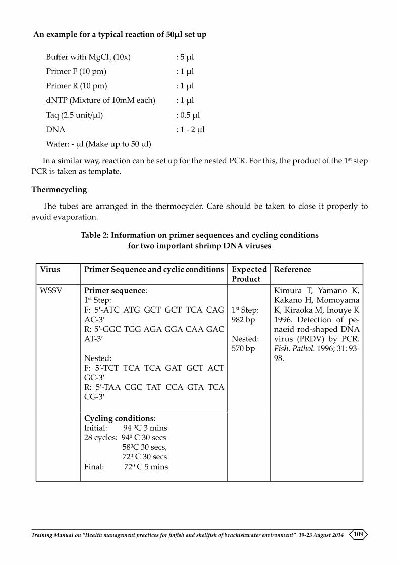
Training Manual on “Health management practices for finfish and shellfish of brackishwater environment” 19-23 August 2014 109
An example for a typical reaction of 50µl set up
Buffer with MgCl2 (10x) : 5 µl
Primer F (10 pm) : 1 µl
Primer R (10 pm) : 1 µl
dNTP (Mixture of 10mM each) : 1 µl
Taq (2.5 unit/µl) : 0.5 µl
DNA : 1 - 2 µl
Water: - µl (Make up to 50 µl)
In a similar way, reaction can be set up for the nested PCR. For this, the product of the 1st step PCR is taken as template.
Thermocycling
The tubes are arranged in the thermocycler. Care should be taken to close it properly to avoid evaporation.
Table 2: Information on primer sequences and cycling conditions for two important shrimp DNA viruses
Virus Primer Sequence and cyclic conditions Expected Product
Reference
WSSV Primer sequence:1st Step:F: 5′-ATC ATG GCT GCT TCA CAG AC-3′R: 5′-GGC TGG AGA GGA CAA GAC AT-3′
Nested:F: 5′-TCT TCA TCA GAT GCT ACT GC-3′R: 5′-TAA CGC TAT CCA GTA TCA CG-3′
1st Step: 982 bp
Nested: 570 bp
Kimura T, Yamano K, Kakano H, Momoyama K, Kiraoka M, Inouye K 1996. Detection of pe-naeid rod-shaped DNA virus (PRDV) by PCR. Fish. Pathol. 1996; 31: 93-98.
Cycling conditions:Initial: 94 ⁰C 3 mins 28 cycles: 94⁰ C 30 secs
58⁰C 30 secs, 72⁰ C 30 secs
Final: 72⁰ C 5 mins

Training Manual on “Health management practices for finfish and shellfish of brackishwater environment” 19-23 August 2014110
IHHNV Primer sequencesF: 5’-GGG CGA ACC AGA ATC ACT TA-3’R: 5’-ATC CGG AGG AAT CTG ATG TG-3’
F: 5’-ATC GGT GCA CTA CTC GGA-3’R: 5’-TCG TAC TGG CTG TTC ATC-3’
392 bp
356 bp
OIE. Aquatic Animal Health Code, 11th edn. World Organization for Animal Health, Paris, 2010
Cycling conditionsInitial: 95°C 5 mins35 cycles: 95°C 30 secs 55°C 30 secs 72°C 1 minsFinal: 72⁰C 5 mins
Gel separation of PCR products
Based on the size of the amplified product, 0.8 to 2% agarose gels are prepared either in 1x Tris-Acetate-EDTA buffer (1 litre 50x TAE – 242 g Tris base, 55 ml Glacial acetic acid and 37.2 g EDTA, pH 8) or 0.5 x Tris Boric acid EDTA buffer (1 litre 50x TBE – Tris base 540 g, Boric acid 275 g and EDTA 18.5g, pH 8.0). Ethidium bromide is added to the molten agarose (0.5 µg/ml final concentration) and then poured into the base. Once the gels are solidified, it is submerged in the tank with the same buffer. The amplified products are then mixed with 6x gel loading dye (For 100 ml – 30mg Bromo Phenol Blue, 30 mg Xylene cyanol, 12 ml of 0.5M EDTA pH8, 1ml of 1M Tris-Hcl pH8, 27 ml of distilled water and 60 ml of sterile glycerol). A total volume of 10 to 20 µl is added to each well. A molecular weight marker is also loaded to the gel to verify the size of the amplified product. After loading, the tank is connected to a power pack and electrophoresis is carried out. This starts with an initial voltage of 80 which is then increased to 120. Based on the gel size and voltage set, it may take 45 mins to 1 hour for the gel to complete the separation.
Observation and documentation
The gel is finally put in a gel-doc for complete analysis or on a UV-transilluminator for visualization. The positive result is read in the form of a band at the right position in the gel. Absence of band indicates negative reaction or absence of virus. Presence of band in the positive control and absence of band in the negative control indicates absence of technical error or contamination.

Training Manual on “Health management practices for finfish and shellfish of brackishwater environment” 19-23 August 2014 111
This is an example of a successive PCR reaction for WSSV in shrimp
1: Molecular Weight Marker 2: Sample 1-1st step 3: Sample 2 – 1st step 4: Sample 3 -1st step 5: Negative control-1st step 6: Positive control-1st step7: Sample 1-nested 8: Sample 2 – nested 9: Sample 3- nested10: Negative control – nested 11: Positive control - nested
Record maintenances
It is necessary to maintain a record regarding the results of each sampling. This will help to interpret the overall situation over a period of time.

Training Manual on “Health management practices for finfish and shellfish of brackishwater environment” 19-23 August 2014112
ISOLATION AND IDENTIFICATION OF BACTERIA FROM SHRIMP
T. Bhuvaneswari, Subhendu Kumar Otta, and J. Joseph Sahaya RajanAquatic Animal Health and Environment Division
Sampling
�� Hemolymph – Draw aseptically from the heart or from the base of the periopod
�� From infected parts with necrosis/blisters – wash the area with sterile saline to get rid of external contamination. With the help of a sterile swab, collect sample from the infected area
�� From internal organs – Dissect out shrimp carefully to avoid any contamination from nearby organs. Sampling can be done from Hepatopancreas with an inoculating loop or swab. Similarly, other organs can be taken aseptically, homogenized with 1% TSB and proceed for culture
Culture
�� Sample can be put either on TCBS or ZMA plates (1 or 2 drops of hemolymph and spread, streak the swab on plates, inoculate 100 µl from the homogenized part of internal organ and then spread)
�� Incubate at 30 ⁰C for 18-24 hours for colonies to develop
Identification by Biochemical characterization of bacterial isolates:
Identification of bacterial isolates is done based on the colony characteristics, physiological and biochemical characteristics.
Amino Acid Decarboxylase Test
�� Prepare Moeller’s decarboxylase broth base with 1% arginine, Moeller’s decarboxylase broth base with 1% ornithine and Moeller’s decarboxylase broth base with 1% lysine and dispense in tubes before sterilizing.
�� Inoculate freshly prepared bacterial culture into the above broth using sterile inoculation loop. Overlay with sterile mineral (paraffin) oil after inoculation.
�� Incubate at 37oC and observe for four days.
�� Development of purple colour is positive, yellow colour represents negative result.
Salt Tolerance
�� Prepare Tryptone water broth by adding 0.5 gm of Tryptone, 0.2 gm of yeast extract with 0%, 3%, 8% and 11% of NaCl to 100 ml of distilled water and transfer the prepared contents to each test tube and keep for sterilization.
�� Pick single colony from TCBS plate and inoculate in sterile ZMA broth and incubate at 30 ºC for 24 hrs.
�� Innoculatefreshly prepared bacterial culture into test tubes with different salt concentration andincubate at 37oC for 24 hrs and observe the medium for turbidity.

Training Manual on “Health management practices for finfish and shellfish of brackishwater environment” 19-23 August 2014 113
Indole Production Test
�� This test detects production of indole from tryptophan.
�� Inoculate the tryptone water with bacterial culture and incubate for 24 hours.
�� After incubation add 0.5 ml of Kovac’s reagent.
�� In positive cases a pink colour ring appears is positive, yellow ring indicate negative result.
Methyl Red
�� This test is performed to identify bacteria which produce lactic, acetic or formic acid from glucose via mixed acid fermentation pathways.
�� PrepareMRVP broth and inoculate with freshly prepared broth culture and incubate for 48-72 hrs
�� Add a few drops of methyl red indicator solution.
�� Development of strong red colour indicates positive reaction.
Voges Proskauer’s Test (MRVP)
�� This test detects the production of acetyl methyl carbinol as chief end product of glucose metabolism.
�� PrepareMRVP broth and inoculate with freshly prepared broth culture and incubate for 48-72 hrs
�� After incubation, add drops of alpha-naphthol and 40% sodium hydroxide along the walls of the test tubes carefully.
�� Shake the test tubes at intervals to ensure maximum aeration. Appearance of red color indicates positive result for VP, whereas yellow color indicates negative result for VP.
�� To above broth culture add Methyl red indicator
Fermentative Utilisation of Carbohydrates
�� Prepare Phenol red broth base supplemented with 1% Salt and 1% Lactose, similarly prepare 1% Salicin, 1% Sucrose and 1% Mannitol, 1%Maltose and 1% Starch and dispense into tubes and sterilize.
�� Inoculate the broth with freshly prepared bacterial culture and incubate at 37oC and observe for colour change at 24, 48 and 72 hr. intervals.
�� Appearance of pink colour indicates positive result and yellow colour indicate negative result.
Oxidase Test
�� Place an oxidase disc in a clean glass slide and moisten with distilled water.
�� Pick single colony and place on moistened surface of oxidase disc.
�� Observe for colour change immediately, purple colour as positive and yellow colour as negative.

Training Manual on “Health management practices for finfish and shellfish of brackishwater environment” 19-23 August 2014114
Citrate Test
�� Prepare Simmon Citrate Agar slants and inoculate by streaking the slants using sterile inoculation loop with freshly prepared bacterial culture and incubate at 30 ºC for 24 to 48 hr and observe for growth and colour change.
�� Colour change of slants to blue is positive and green colour slantrepresents negative.
Antibiotic sensitivity test
�� The organisms were grown for 24 hrs in ZMA broth and 0.1 ml of culture was uniformly spreaded on the ZMA plates.
�� The antibiotic discs were gently pressed and placed at equal distance and the plates were incubated at 37 ºC for 24 hrs.
�� The following nine antibiotic discs namely Ampicillin, Streptomycin, Neomycin, Tetracycline, Oxytetracycline, Ciprofloxacin, Chloromphenicol, were used and observed for the zone of inhibition.
Nitrate Reduction Test
�� Nitrate broth is used to determine the ability of an organism to reduce nitrate (NO3) to nitrite (NO2) using the enzyme nitrate reductase. It also tests the ability of organisms to perform nitrification on nitrate and nitrite to produce molecular nitrogen.
�� Prepare Nitrate broth and incubate after inoculation with bacterial culture
�� After incubating the nitrate broth, add a dropper ful of sulfanilic acid and a-naphthylamine. If the organism has reduced nitrate to nitrite, the nitrites in the medium will form a red-colored compound. Therefore, if the medium turns red after the addition of the nitrate reagents, it is considered a positive result for nitrate reduction. If the medium does not turn red after the addition of the reagents, it can mean that the organism was unable to reduce the nitrate. Therefore, another step is needed in the test. If the medium does not turn red after the addition of the nitrate reagents, add a small amount of powdered zinc. If the tube turns red after the addition of the zinc, it means that unreduced nitrate was present. Therefore, a red color on the second step is a negative result. If the medium does not turn red after the addition of the zinc powder, then the result is called a positive complete.
A typical chart is presented below for the identification. Several of such charts can be found in the published literature based on which, the bacterial can be identified.
Training Manual on “Health management practices for finfish and shellfish of brackishwater environment” 19-23 August 2014 115
PROTOCOLS FOR DETECTION OF SHRIMP EMS/AHPND
Subhendu Kumar Otta, T. Bhuvaneswari, P. Ezhil Praveena and J. Joseph Sayhaya RajanAquatic Animal Health and Environment Division
Early Mortality Syndrome (EMS) / Acute Hepatopancreatic Necrosis Disease (AHPND) is caused by the bacteria, Vibrio parahaemolyticus. Bacterial isolation and identification, PCR and histopathology protocols are available for the detection of this disease.
Samples
Shrimps with typical clinical sign for EMS/AHPND or healthy shrimps from cultured ponds can be used as samples. Healthy post larvae or faecal strings of brood stocks can also be used for PCR detection
Step I: Bacterial isolation
While confirming the disease in a country for the 1st time, it is necessary to isolate the causative bacterial agent of the disease.
Samples with typical clinical signs or from the suspected ones, aseptically dissect out stomach and HP and put in a 1.5 ml microfuge tube containing 500 µl 1% Tryptic Soy Broth (TSB) . TSB is available commercially. The composition is given below:
Enzymatic Digest of Casein 17.0 g Enzymatic Digest of Soybean Meal 3.0 g Sodium Chloride 10.0 g Dipotassium Phosphate 2.5 g Dextrose 2.5 g Final pH: 7.3 ± 0.2 at 25°C)
Homogenize with a pestle. From this, add 100 µl to a fresh 2 ml TSB. Take a loop full and streak on TCBS plate.
Direct touch from the HP can also be taken and streaked on TCBS plates.
Incubate the plates and the broth at 30 ⁰C for 18 – 24 hours. Bacteria grown on the broth is further purified on TCBS plates.
The green colonies grown can be taken for identification by biochemical reaction or for gene specific PCR for V. parahaemolyticus. Part of this colony can directly be taken for DNA extraction and PCR reaction (Adopted from Dr. T. W. Flegel, Thailand).
PCR primers for the detection of EMS/AHPND specific V. parahaemolyticus strainAP1F, 5’- CCT TGG GTG TGC TTA GAG GAT G -3’; AP1R, 5’- GCA AAC TAT CGC GCA GAA CAC C -3’; AP2F: 5’- TCA CCC GAA TGC TCG CTT GTG G -3’; AP2R: 5’- CGT CGC TAC TGT CTA GCT GAA G -3’.AP3F: 5’-ATGAGTAACAATATAAAACATGAAAC-3’AP3R: 5’-GTGGTAATAGATTGTACAGAA-3’

Training Manual on “Health management practices for finfish and shellfish of brackishwater environment” 19-23 August 2014116
PCR conditions
AP1 and AP2
Pre-heat: 94°C, 5min
30 cycles: Denaturation: 94°C, 30 sec, Annealing: 53°C, 30 sec, Extension: 72°C, 40 sec
Final extension: 72°C, 10 min
When separated in an agarose gel, both the primer pairs yield products of ~ 700 bp.
AP3
Pre-heat: 94°C, 5min
30 cycles: Denaturation: 94°C, 30 sec, Annealing: 60°C, 30 sec, Extension: 72°C, 60 sec
Final extension: 72°C, 10 min
When separated in an agarose gel, this primer pairs yield products of ~ 336 bp.
While AP1 and AP2 give false positive to some extent, AP3 primers have been verified to be 100% accurate.
Rapid method for detection of EMS/AHPND V. parahaemolyticus strain
Fecal string from broodstock
Stomach/HP/Gut from juveniles
Whole macerated larvae (15-20 no.)
Incubate in a Vibrio enrichment media (Ex. 1% Alkaline Peptone Water (APW) at 1:9 ration) for 4-6 hours at 30 ⁰C
Select green colonies from TCBS streak, extract DNA and do PCR
Confirm the PCR positive isolates by infection experiment
Histopathology
The target organ here is hepatopancreas. Fix only moribund samples for histopathology. Process the HP sample for histopathology following the protocol described in this manual. Observe for typical pathology.
Bioassay
Grow bacteria in 1% TSB for 12-18 hours. Take healthy juvenile shrimps in a container. Based on the quantity of water, add the bacterial inoculum (Estimate the count in the broth either by plating method or by measuring the OD in a spectrophotometer) to have final bacterial count of 108/ml. With aeration, keep the shrimps for 1 hour. Thereafter release the shrimps to the original tanks with aeration. Observe for mortality. Confirm the mortality by PCR and histopathology.

Training Manual on “Health management practices for finfish and shellfish of brackishwater environment” 19-23 August 2014 117
Information on Resource personnel
Sl. No. Name Designation & Division
1 Dr.P.Ravichandran Principal Scientist & OIC, PMECrustacean Culture Division
2 Dr.M.Natarajan Principal Scientist & Scientist In-chargeFinfish Culture Division
3 Dr.K.P.Jithendran Principal Scientist & Scientist In-charge Aquatic Animal Health and Environment Division
4 Dr.S.V.Alavandi Principal ScientistAquatic Animal Health and Environment Division
5 Dr.M.Muralidhar Principal Scientist, Environment GroupAquatic Animal Health and Environment Division
6 Dr.C.P.Balasubramanian Principal ScientistCrustacean Culture Division
7 Dr. M. Kailasam Principal Scientist & Scientist In-chargeMuthukadu Experimental Station
8 Dr. K. Ambasankar Principal Scientist, Nutrition GroupNutrition, Genetics and Biotechnology Division
9 Dr. Akshaya Panigrahi Principal ScientistCrustacean Culture Division
10 Dr. M. Kumaran Principal ScientistSocial Science Division
11 Dr. M. Poornima Senior ScientistAquatic Animal Health and Environment Division
12 Dr. P.K.Patil Senior ScientistAquatic Animal Health and Environment Division
13 Dr. S.K. Otta Senior ScientistAquatic Animal Health and Environment Division
14 Dr. P. Ezhil Praveena Scientist,Aquatic Animal Health and Environment Division
15 Dr. T. Bhuvaneswari ScientistAquatic Animal Health and Environment Division
16 Dr. R. Vidya ScientistAquatic Animal Health and Environment Division
17 Mr. T. Sathish Kumar ScientistAquatic Animal Health and Environment Division
18 Mr. J. Joseph Sahaya Rajan
Senior Technical OfficerAquatic Animal Health and Environment Division
19 Mr. N. Jagan Mohan Raj Senior Technical AssistantAquatic Animal Health and Environment Division

Training Manual on “Health management practices for finfish and shellfish of brackishwater environment” 19-23 August 2014118
Information on participants
Sl. No.
Name Designation and Address Tel. No &E-mail
1 Sri. Suhair Assistant Director of Fisheries,O/o Regional Shrimp Hatchery, Azhikode
9387221531
2 Smt. Jayalekshmi.M. K Deputy Director of Fisheries,O/o Deputy Director of Fisheries, Kadungallur
3 Sri. MuhammedAshrudeen
Sub Inspector of Fisheries,O/o Deputy Director ofFisheries,Kasaragod
4 Sri. Sreejan Sub Inspector of Fisheries,O/o Deputy Director of Fisheries, Ponnani
5 Sri. Vishnu Sub Inspector of Fisheries,Package Scheme, Thanur
6 Sri. ArunShuri Sub Inspector of FisheriesO/o Assistant Director of Fisheries, Fisheries station,Vypin
7 Sri. Anwar Ali Aquaculture Expert,O/o Deputy Director of Fisheries, Eranakulam
8 Sri. Aneesh Aquaculture Expert,O/o Deputy Director of Fisheries, Thrissur
9 Smt. Chaithanya Sub Inspector of Fisheries,O/o Inspector of Fisheries, Chettuva
10 Sri. Benny William Inspector of Fisheries,O/o Deputy Director of Fisheries, Alapuzha