Embed Size (px)
Citation preview
J Toxicol Pathol 2008; 21: 199–205
Review
Chronic Progressive Nephropathy (CPN) in the Rat: Review of Pathology and Relationship to Renal Tumorigenesis
John Curtis Seely1 and Gordon C. Hard2
1Experimental Pathology Laboratories, Inc., PO Box 12766, Research Triangle Park, NC 27709, USA2Private Consultant, Tairua, New Zealand
Abstract: Chronic progressive nephropathy (CPN) is a rodent-specific, age-related renal disease, particularly of malerats, characterized by a spectrum of distinct histological changes which may begin early in the animal’s life and progressto end-stage renal disease in certain rat strains. Although CPN-related pathology is well known to most toxicologicalpathologists other features of CPN such as pathogenesis, modulating factors, proliferative nature, response to chemicalexposure and relationship to tumorigenesis are less clearly acknowledged. CPN is generally regarded as a degenerativeto atrophic disease with compensatory regenerative hyperplasia. The proliferative nature of CPN often becomesproblematic in advanced to end-stage renal disease. At this stage, a number of tubule profiles may be mistaken foratypical tubule hyperplasia, the reported precursor lesion of tubule adenoma. CPN associated proliferative tubuleprofiles must be carefully separated from atypical tubule hyperplasia particularly in studies where chemical exposure hasexacerbated CPN. Over the past several years increasing evidence has supported the hypothesis that CPN may beregarded as a type of “mode of action” during renal carcinogenesis in rodent bioassay studies. Retrospective studies ofcontrol and treated animals have consistently shown a relationship between the increased severity of CPN and thepresence of atypical tubule hyperplasia and small, incipient renal adenomas. Understanding CPN-related tumorigenesisis important for human risk assessment interpretation. Since CPN is a rodent specific disease with no apparent similarhuman kidney disease condition, evidence that renal tumors may arise from an interaction with CPN could assistregulatory agencies in interpreting data from studies with exacerbated CPN. (J Toxicol Pathol 2008; 21: 199–205)
Key words: rat, kidney, chronic progressive nephropathy, pathology, tumorigenesis, risk assessment
Introduction and Overview
Chronic progressive nephropathy (CPN) is one of themost widely recognized disease entities in rodent preclinicalstudies. CPN is a common spontaneous age-related diseaseof rodent kidneys, particularly in rats1–3. Because of thekidney’s central role in maintaining the normal physiologicbalance of the body’s internal environment and itsimportance in drug metabolism and excretion, understandingthe basic mechanisms and pathology of CPN is critical. Theetiology of CPN remains unknown. In some models ofnephropathy, speculation regarding hemodynamicalterations leading to hyperperfusion and hyperfiltration ofmacromolecules within the glomerulus resulting inmesangial overload and glomerulosclerosis has beenpostulated4. However, hemodynamic alterations do notseem to be associated with CPN5. There are strain, sex, and
Received: 16 April 2008, Accepted: 29 August 2008Mailing address: John Curtis Seely, Experimental Pathology Laboratories, Inc., PO Box 12766, Research Triangle Park, NC, 27709, USATEL: 1-919-313-0630 FAX: 1-919-998-9607E-mail: [email protected]
age differences with respect to the incidence and severity ofCPN associated lesions6–10. Sprague-Dawley and F344 ratsgenerally have an earlier onset and higher incidence andseverity than the Wistar, Brown Norway and Long-Evans ratstrains. Although the term CPN has undergone a number ofname changes over the years , cur ren t ly CPN or“nephropathy” are synonymous terms and recommended bystandardized nomenclature classifications.
Pathologists beginning their career in toxicologicalpathology are quickly exposed to the spectrum of CPNassociated lesions such as regenerative (basophilic) tubulessurrounded by thickened basement membranes, hyalineproteinaceous casts, glomerulosclerosis, interstitial fibrosisand infiltration of mononuclear inflammatory cells whichcan begin as early as 2–3 months of age and progresscontinually through the life-span of the animal. Histologicalchanges associated with CPN also result in a number offunctional changes that result in increased proteinuria anddecreased urine concentration ability11,12.
Most pathologists agree that the earliest CPN lesion, bylight microscopy, is represented by small cross sections ofbasophilic tubules surrounded by a thickened basementmembrane in the renal cortex. Only later does another oneof the hallmarks of CPN, glomerulosclerosis, become
200 Chronic Progressive Nephropathy in the Rat
evident. By the end of 2-year carcinogenic studies, theadvanced stage of CPN may result in end-stage kidney anddeath of the animal due to chronic renal failure. In fact,mortality associated with CPN is an issue with seriousimplications in the conduct of some carcinogenic studies13.CPN may be modulated by a number of factors includingdiet , administrat ion of hormones and many otherexperimental manipulations13–17. Recently, the type of dietparticularly with regard to the protein content and/or caloricrestriction have been investigated as ways to reduce theseverity or limit the progression of CPN, thereby, extendingthe life of the animal14,18.
CPN has been described as a degenerative to atrophicdisease with compensatory hypertrophy and hyperplasia.The proliferative rate of CPN has been demonstrated by celllabeling studies, which have shown cell proliferative rateswithin CPN affected tubules increased over that of normaltubules19–21. In advanced stages of nephropathy, a number ofproliferative tubule profiles may be recognized as having anincreased number of lining epithelial cells. These tubuleprofiles are problematic for many pathologists as they maybe mistaken for atypical tubule hyperplasia (ATH), theprecursor lesion of renal adenoma. Hard and Seely22 recentlypublished recommendations to assist in the differentiation ofCPN tubules from actual ATH and early renal tubuleadenomas. An association between CPN and the presence ofATH and renal tubule tumors (RTT) has been observed fromsevera l ca rc inogen ic s tud ies in which chemica ladministration exacerbated the severity of CPN23–25.Retrospective studies of control and treated animals haveconsistently shown a relationship between the increasedseverity of CPN and the presence of ATH and RTT. Theunderlying factors associated with this relationship are notknown but, most likely, are multi-factorial and complex.
The interpretation of a renal tumorigenic responserelated to CPN is important for risk assessment analysis sinceapparently there is no human renal disease which is similar toCPN. Therefore, it has recently been postulated that RTTlinked to CPN has no relevance for extrapolation to humanrisk assessment because CPN is a rodent- specific disease3.
This review was written primarily for toxicologicpathologists, especially study pathologists, who would like aconcise yet instructive review of CPN with emphasis onpathology and pathology-related issues. Furthermore, thisreview is not intended to be a comprehensive overview of allaspects of CPN. Current investigations of CPN associatedtumorigenesis are also presented. These studies have beenthe interest of the authors for some time. This postulatedcarcinogenic “Mode of Action” is unique and undoubtedlywill have to undergo further examination and review by bothresearchers and by federal agencies responsible for chemicaland drug regulation.
Histological Spectrum of CPN Pathology
As noted previously, CPN is characterized by aspectrum of histological changes including regenerative
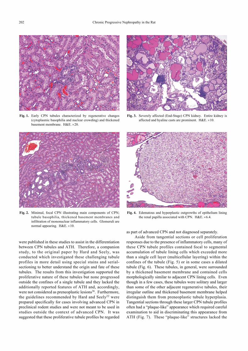
tubules (basophilic tubules) lined by a thickened basement,the presence of eosinophilic hyaline proteinacious tubulecasts, glomerulosclerosis, interstitial fibrosis and infiltrationof a variable mononuclear cell infiltrate1–3,8,17,26,27. It hasbeen reported that the earliest change by light microscopy ofH&E stained sections is the presence of one or more corticalsolitary basophilic tubule cross sections surrounded by athickened basement membrane (Fig. 1). The thickenedbasement membrane is one of the key components of CPNthroughout the course of the disease. The presence of thischange is helpful in distinguishing early CPN andregeneration due to toxic insult. If one examines CPN -affected kidneys in detail, degenerative to apoptotic cells, aswell as mitotic figures may be evident and responsible forthe regenerative appearance. However, in most instances,these changes are not fully appreciated unless thoroughexamination under higher power is conducted. In someinstances, affected tubules may appear as slightly dilated.Therefore, the tubule appearance is due to the presence ofincreased numbers of lining epithelial cells with basophiliccytoplasm and crowded, slightly enlarged and vesicularnuclei, all responsible for the overall basophilia noted onH&E staining. As the number of affected tubules increase, aminimal infiltration of mononuclear cells becomes evidentwithin the interstitium. At this time, a few eosinophilichyaline tubule casts may be noted in Henle’s loops withinthe medulla. When hyaline casts are observed in the absenceof basophilic tubules, early CPN is still usually suspectedbecause affected CPN tubules in the cortex are most likelyout of the plane of section. Shortly thereafter, changeswithin affected glomeruli may be noted. These changesreflect the early segmental and basement membranethickening of the glomerular tuft and Bowman’s capsule.Occasionally, a lightly eosinophilic staining and amorphousmaterial may be present in Bowman’s space. Variably-sized, eosinophilic protein droplets in sporadic tubules mayalso be observed. Proteinuria or the loss of urinary protein inthe urine, associated with CPN, is best determined bymeasuring urinary albumin excretion28. By electronmicroscopy, it has been reported that glomerular injury maybe observed29. However, there is no clear evidence that thesechanges precede the earliest tubule changes.
Among pathologists, discussion regarding the earliestlesion, namely the basophilic tubules, often causes debate onthe most correct terminology pathologists should use todescribe this change. Some pathologists refer to all changesassociated with CPN as “CPN or nephropathy” while othersd iagnose e i ther “basophi l i c tubu les” o r “ tubuleregeneration”. Because “basophilic tubules” may resultfrom a number of differing pathogenic mechanisms, theauthors do not recommend the use of “basophilic tubules” asa descriptor for the earliest lesion. However, if “basophilictubule” is used then it is suggested that this term be definedin the narrative portion of the pathology report with regard toits association with CPN. Furthermore, it is highlyrecommended not to separate and grade each component ofCPN individually. This effort requires considerable
Seely and Hard 201
diagnostic consistency and time and often results in lengthyand confusing incidence tables which add little to the overallinterpretation of a chemical effect. All of the components ofCPN should be grouped together as CPN or alternately, asnephropathy.
As the animal ages, the extent within the kidney and theseverity of CPN-related areas increase noticeably. Minimalchanges usually result in multiple foci of CPN-affected areasrandomly found throughout the cortex (Fig. 2). As thedisease progresses, hyaline casts become more prominentand are seen extending down through the outer to innermedulla within Henle’s loop. With increasing severity, fociof CPN-affected areas begin to merge and become confluentwith each other resulting in a more diffuse change.Continued progression of CPN results in striking tubule,glomerular and interstitial changes. Foci of basophilictubules are remarkable due to increased amounts ofthickened basement membrane material. An occasionalcortical tubule becomes hypertrophic with increasedamounts of pale eosinophilic staining cytoplasm. Glomerulimay either appear hypertrophic or atrophic due toglomerulosclerosis and the presence of adhesions betweenthe glomerular tuft and Bowman’s capsule. The presence ofinterstitial fibrosis often results in tubule dilatation and/orcyst formation. This phenomenon appears to be the result ofinteractions between fibrogenic growth factors produced bytubule epithelial cells, macrophages and myofibroblasts30,31.In “End-Stage” CPN hyaline casts may also be seen incollecting ducts. In advanced to end-stage CPN, littlenormal renal cortical tissue remains (Fig. 3). Grossly, thesekidneys are enlarged, pale and with a pitted surface.Animals that have died from end-stage renal disease oftenhave diffuse mineralization of tubule basement membranes.Secondary hyperparathyroidism and mineralization of othertissues may be noted.
Additionally, in more advanced cases of CPN smallirregularly-shaped, edematous and hyperplastic outgrowthsof epithelium are often noted on the surface of the renalpapilla (Fig. 4). These projections seemingly have littlepathological significance. Vascular or hemorrhagic lesionsare not a prominent part of the overall histological spectrumof CPN. On rare occasion only, the authors have notedperivascular inflammatory cell infiltrates and even somevasculitis and thrombosis. A light golden-brown pigment isoften noted in CPN tubules and interstitial tissue. Thispigment seems to be predominantly iron-positive andpresumably hemosiderin32.
Grading the severity of CPN is mainly a subjectiveevaluation often based on the pathologist’s training andexperience. There is little published guidance on how tograde CPN13. Most CPN grading schemes grade CPN on a3, 4 or 5 grade scale, representing severities from minimal tomarkedly-severe (End-Stage). Although all of these gradingschemes are appropriate, if used consistently, grades 1–4 or1–5 are more likely to help in the determination of anexacerbated chemical effect and are used more regularly ona global basis. Depending on the length of the study, study
pathologists may also modify their grading scheme for moreacute studies versus carcinogenicity studies. Furthermore, inany event of a potential chemical effect involving CPN,pathologists should provide a detailed description of thegrading criteria for CPN in the narrative report. Thisprovides information for the regulatory agencies to use ininterpreting and comparing data from these studies andassists the peer review pathologist in determining theaccuracy and consistency of the study pathologist when peerreview is conducted.
CPN Associated Cell Proliferation and Proliferative Lesions
As previously noted, CPN has been described as adegenerative to atrophic disease with compensatoryhypertrophy and hyperplasia. The prominent regenerativetubules noted with CPN are part of the compensatoryhyperplastic response. The term “simple tubule hyperplasia”was introduced to denote a tubule appearance consisting of asingle cell layer of increased numbers of lining epithelialcells and to help differentiate CPN tubules from ATH or,alternately diagnosed as, “renal tubule hyperplasia”33.Although pathologists differ in their diagnostic approach tothe ear ly changes associa ted wi th CPN, “ tubuleregeneration” or “basophilic tubules” are terms used morefrequently. Other pathologists simply include this earlychange within the overall diagnosis of CPN when they arereasonably comfortable with their diagnosis. The termsimple tubule hyperplasia has not been routinely used todiagnose the early tubule change34. However, the crowdednature of the lining cells of CPN-affected tubules implies thepresence of simple tubule hyperplasia.
Although it was easy for pathologists to recognize theincreased number of lining epithelial cells within theconfines of a single tubule by routine H&E light microscopy,it was not until sometime later when investigators began toapply specialized techniques such as tritiated thymidineautoradiography to demonstrate the cell labeling index (LI)of cells undergoing DNA synthesis within areas of CPN19–21.These studies have reported increased LI of up to 10 timesgreater in CPN affected areas than normal areas. It appearsthat the proliferation rate of affected tubules is the same formale and female rats; however, the number of affectedtubules is greater in males17. Proliferating cell nuclearantigen (PCNA) may also be used to measure CPN cellproliferating activity35.
In examining kidneys from rats with advanced(moderate to marked severity) to end-stage CPN, it iscommon to observe tubules containing proliferative changeswhich exceed the normal expectation for tubule regenerationor simple tubule hyperplasia. A number of these tubulephenotypes have been problematic for pathologists oftenleading to inappropriate diagnoses and interpretation.However, recent guidelines have been published which offerrecommendations in the interpretation of these proliferativechanges associated with CPN22. In addition, detailed criteria
202 Chronic Progressive Nephropathy in the Rat
were published in these studies to assist in the differentiationbetween CPN tubules and ATH. Therefore, a companionstudy, to the original paper by Hard and Seely, wasconducted which investigated these challenging tubuleprofiles in more detail using special stains and serial-sectioning to better understand the origin and fate of thesetubules. The results from this investigation supported theproliferative nature of these tubules but none progressedoutside the confines of a single tubule and they lacked theadditionally reported features of ATH and, accordingly,were not considered as preneoplastic lesions36. Furthermore,the guidelines recommended by Hard and Seely22 wereprepared specifically for cases involving advanced CPN inpreclinical rodent studies and were not meant to be used instudies outside the context of advanced CPN. It wassuggested that these proliferative tubule profiles be regarded
Fig. 1. Early CPN tubules characterized by regenerative changes(cytoplasmic basophilia and nuclear crowding) and thickenedbasement membrane. H&E. ×20.
Fig. 2. Minimal, focal CPN illustrating main components of CPN;tubule basophilia, thickened basement membranes andinfiltration of mononuclear inflammatory cells. Glomeruli arenormal appearing. H&E. ×10.
as part of advanced CPN and not diagnosed separately.Aside from tangential sections or cell proliferation
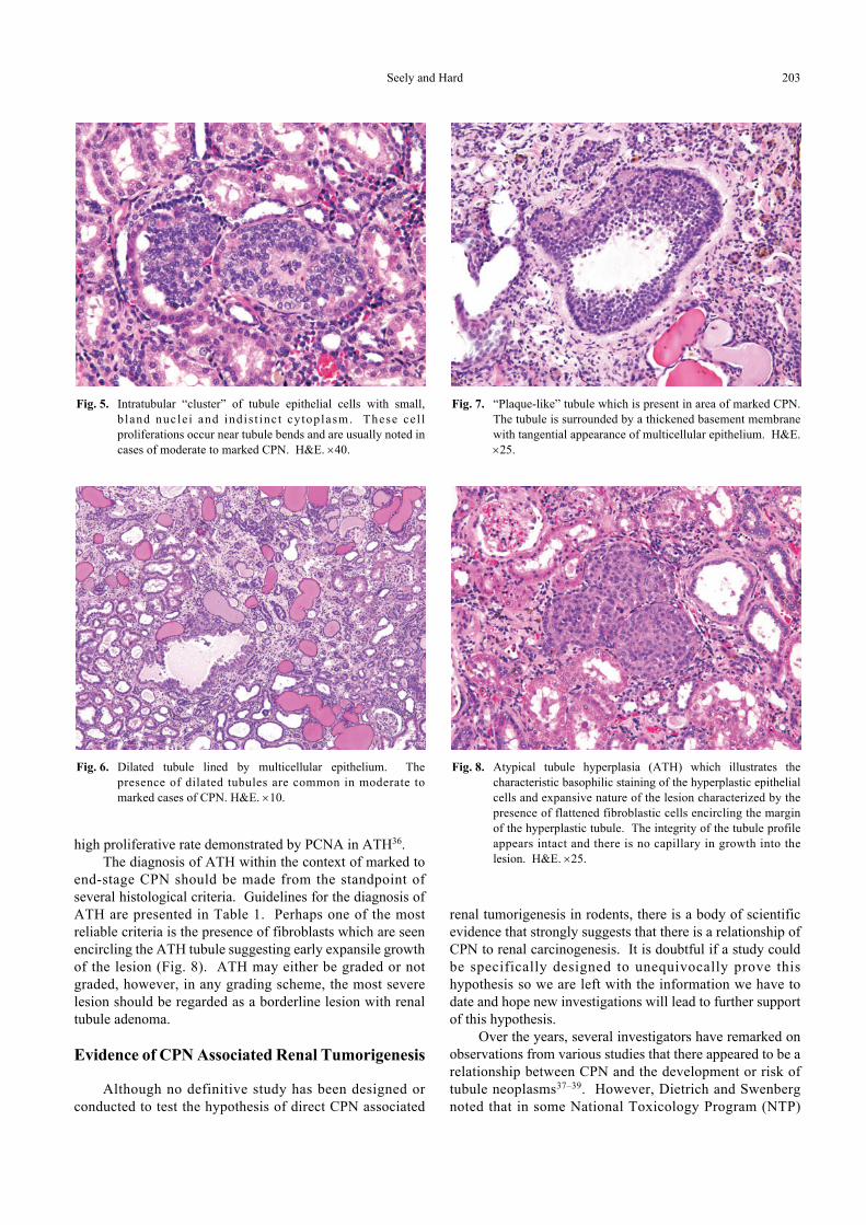
responses due to the presence of inflammatory cells, many ofthese CPN tubule profiles contained focal to segmentalaccumulation of tubule lining cells which exceeded morethan a single cell layer (multicellular layering) within theconfines of the tubule (Fig. 5) or in some cases a dilatedtubule (Fig. 6). These tubules, in general, were surroundedby a thickened basement membrane and contained cellsmorphologically similar to adjacent CPN lining cells. Eventhough in a few cases, these tubules were solitary and largerthan some of the other adjacent regenerative tubules, theirirregular outline and thickened basement membrane helpeddistinguish them from preneoplastic tubule hyperplasia.Tangential sections through these larger CPN tubule profilesoften had a “plaque-like” appearance which required carefulexamination to aid in discriminating this appearance fromATH (Fig. 7). These “plaque-like” structures lacked the
Fig. 3. Severely affected (End-Stage) CPN kidney. Entire kidney isaffected and hyaline casts are prominent. H&E. ×10.
Fig. 4. Edematous and hyperplastic outgrowths of epithelium liningthe renal papilla associated with CPN. H&E. ×6.4.
Seely and Hard 203
high proliferative rate demonstrated by PCNA in ATH36.The diagnosis of ATH within the context of marked to
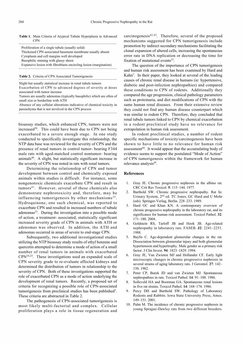
end-stage CPN should be made from the standpoint ofseveral histological criteria. Guidelines for the diagnosis ofATH are presented in Table 1. Perhaps one of the mostreliable criteria is the presence of fibroblasts which are seenencircling the ATH tubule suggesting early expansile growthof the lesion (Fig. 8). ATH may either be graded or notgraded, however, in any grading scheme, the most severelesion should be regarded as a borderline lesion with renaltubule adenoma.
Evidence of CPN Associated Renal Tumorigenesis
Although no definitive study has been designed orconducted to test the hypothesis of direct CPN associated
Fig. 5. Intratubular “cluster” of tubule epithelial cells with small,b land nuclei and indis t inct cytoplasm. These cel lproliferations occur near tubule bends and are usually noted incases of moderate to marked CPN. H&E. ×40.
Fig. 6. Dilated tubule lined by multicellular epithelium. Thepresence of dilated tubules are common in moderate tomarked cases of CPN. H&E. ×10.
renal tumorigenesis in rodents, there is a body of scientificevidence that strongly suggests that there is a relationship ofCPN to renal carcinogenesis. It is doubtful if a study couldbe specifically designed to unequivocally prove thishypothesis so we are left with the information we have todate and hope new investigations will lead to further supportof this hypothesis.
Over the years, several investigators have remarked onobservations from various studies that there appeared to be arelationship between CPN and the development or risk oftubule neoplasms37–39. However, Dietrich and Swenbergnoted that in some National Toxicology Program (NTP)
Fig. 7. “Plaque-like” tubule which is present in area of marked CPN.The tubule is surrounded by a thickened basement membranewith tangential appearance of multicellular epithelium. H&E.×25.
Fig. 8. Atypical tubule hyperplasia (ATH) which illustrates thecharacteristic basophilic staining of the hyperplastic epithelialcells and expansive nature of the lesion characterized by thepresence of flattened fibroblastic cells encircling the marginof the hyperplastic tubule. The integrity of the tubule profileappears intact and there is no capillary in growth into thelesion. H&E. ×25.
204 Chronic Progressive Nephropathy in the Rat
bioassay studies, which enhanced CPN, tumors were notincreased38. This could have been due to CPN not beingexacerbated to a severe enough stage. In one studyconducted to specifically investigate this relationship, theNTP data base was reviewed for the severity of CPN and thepresence of renal tumors in control tumor- bearing F344male rats with aged-matched control nontumor- bearinganimals40. A slight, but statistically significant increase inthe severity of CPN was noted in rats with renal tumors.
Determining the relationship of CPN and tumordevelopment between control and chemically exposedanimals within studies is difficult. For instance, somenongenotoxic chemicals exacerbate CPN and result intumors41. However, several of these chemicals alsodemonstrate nephrotoxicity and, therefore, may beinfluencing tumorigenesis by other mechanisms35.Hydroquinone, one such chemical, was reported toexacerbate CPN and resulted in increased numbers of tubuleadenomas23. During the investigation into a possible modeof action, a treatment- associated, statistically significantincreased severity grade of CPN in animals with ATH oradenomas was observed. In addition, the ATH andadenomas occurred in areas of severe to end-stage CPN.
Subsequently, two additional investigational studiesutilizing the NTP bioassay study results of ethyl benzene andquercetin attempted to determine a mode of action of a smallnumber of renal tumors in animals with exacerbatedCPN24,25. These investigations used an expanded scale ofCPN severity grade to re-evaluate affected kidneys anddetermined the distribution of tumors in relationship to theseverity of CPN. Both of these investigations supported therole of exacerbated CPN as a mode of action underlying thedevelopment of renal tumors. Recently, a proposed set ofcriteria for recognizing a possible role of CPN-associatedtumorigenesis from preclinical studies has been published3.These criteria are abstracted in Table 2.
The pathogenesis of CPN-associated tumorigenesis ismost likely multi-factorial and complex. Cellularproliferation plays a role in tissue regeneration and
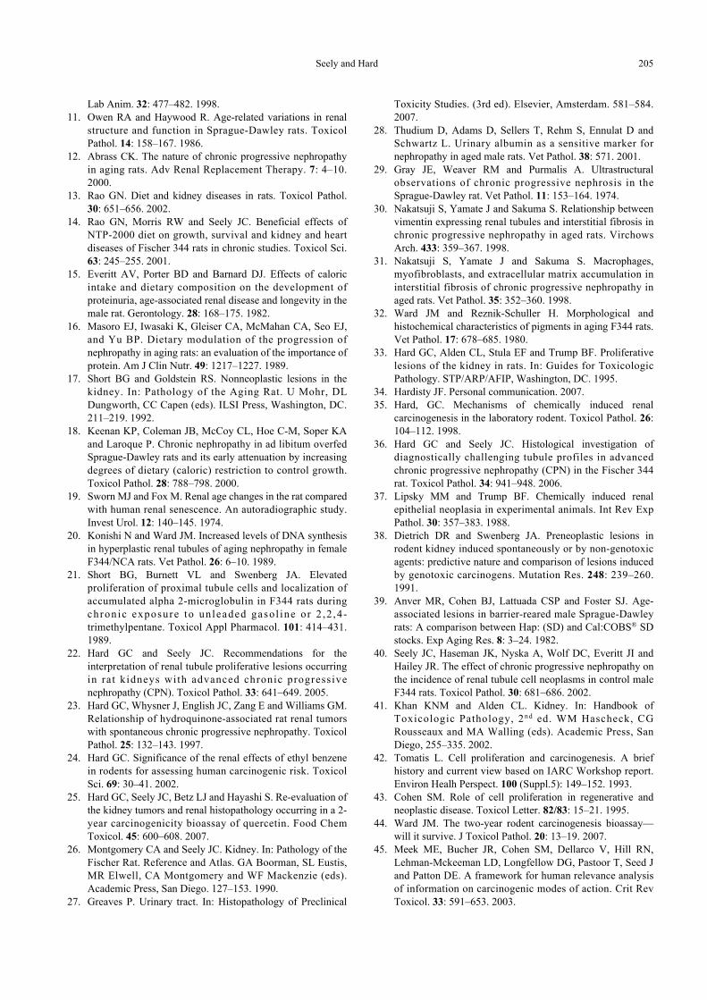
Table 2. Criteria of CPN Associated Tumorigenesis
Slight but usually statistical increase in renal tubule tumors Exacerbation of CPN to advanced degrees of severity at dosesassociated with tumor increaseTumors are usually adenomas (typically basophilic) which are often ofsmall size or borderline with ATHAbsence of any cellular alterations indicative of chemical toxicity inparenchyma that is not involved in the CPN process
Table 1. Main Criteria of Atypical Tubule Hyperplasia in AdvancedCPN
Proliferation of a single tubule (usually solid)Thickened CPN-associated basement membrane usually absentCytoplasm and cell margins well developed Basophilic staining with glassy sheenExpansive lesion with fibroblasts encircling lesion (margination)
carcinogenesis42,43. Therefore, several of the proposedmechanisms suggested for CPN tumorigenesis includepromotion by indirect secondary mechanisms facilitating theclonal expansion of altered cells, increasing the spontaneouserror rate in DNA replication or decreasing the time forfixation of mutational events23.
The question of the importance of CPN tumorigenesisand human risk assessment has been examined by Hard andKahn3. In their paper, they looked at several of the leadingcauses of chronic renal disease in humans (ie: hypertensive,diabetic and post-infection nephropathies) and comparedthese conditions to CPN of rodents. Additionally theycompared the age progression, clinical pathology parameterssuch as proteinuria, and diet modifications of CPN with thesame human renal diseases. From their extensive reviewthey could not find any human disease counterpart whichwas similar to rodent CPN. Therefore, they concluded thatrenal tubule tumors linked to CPN by chemical exacerbationin a rodent preclinical study have no relevance forextrapolation in human risk assessment.
In rodent preclinical studies, a number of rodentspecific mechanisms of toxicity/carcinogenesis have beenshown to have little to no relevance for human riskassessment44. It would appear that the accumulating body ofevidence seems to support the postulated “Mode of Action”of CPN tumorigenesis within the framework for humanrelevance analysis45.
References
1. Gray JE. Chronic progressive nephrosis in the albino rat.CRC Crit Rev Toxicol. 5: 115–144. 1977.
2. Barthold SW. Chronic progressive nephropathy. Rat In:Urinary System, 2nd ed. TC Jones, GC Hard and U Mohr(eds). Springer-Verlag, Berlin. 228–233. 1989.
3. Hard GC and Khan KN. A contemporary overview ofchronic progressive nephropathy in the laboratory rat, and itssignificance for human risk assessment. Toxicol Pathol. 32:171–180. 2004.
4. Goldstein RS, Tarloff JB and Hook JB. Age-relatednephropathy in laboratory rats. FASEB. J2: 2241–2251.1988.
5. Baylis C. Age-dependent glomerular changes in the rat.Dissociation between glomerular injury and both glomerularhypertension and hypertrophy. Male gender as a primary riskfactor. J Clin Invest. 94: 1823–1829. 1994.
6. Gray JE, Van Zwieten MJ and Hollander CF. Early lightmicroscopic changes in chronic progressive nephrosis inseveral strains of aging laboratory rats. J Gerontol. 27: 142–150. 1982.
7. Peter CP, Burek JD and van Zwieten MJ. Spontaneousnephropathies in rats. Toxicol Pathol. 14: 91–100. 1986.
8. Solleveld HA and Boorman GA. Spontaneous renal lesionsin five rat strains. Toxicol Pathol. 14: 168–174. 1986.
9. Percy DH and Barthold SW. Pathology of LaboratoryRodents and Rabbits. Iowa State University Press, Ames.149–151. 2001.
10. Palm M. The incidence of chronic progressive nephrosis inyoung Sprague-Dawley rats from two different breeders.
Seely and Hard 205
Lab Anim. 32: 477–482. 1998.11. Owen RA and Haywood R. Age-related variations in renal
structure and function in Sprague-Dawley rats. ToxicolPathol. 14: 158–167. 1986.
12. Abrass CK. The nature of chronic progressive nephropathyin aging rats. Adv Renal Replacement Therapy. 7: 4–10.2000.
13. Rao GN. Diet and kidney diseases in rats. Toxicol Pathol.30: 651–656. 2002.
14. Rao GN, Morris RW and Seely JC. Beneficial effects ofNTP-2000 diet on growth, survival and kidney and heartdiseases of Fischer 344 rats in chronic studies. Toxicol Sci.63: 245–255. 2001.
15. Everitt AV, Porter BD and Barnard DJ. Effects of caloricintake and dietary composition on the development ofproteinuria, age-associated renal disease and longevity in themale rat. Gerontology. 28: 168–175. 1982.
16. Masoro EJ, Iwasaki K, Gleiser CA, McMahan CA, Seo EJ,and Yu BP. Dietary modulation of the progression ofnephropathy in aging rats: an evaluation of the importance ofprotein. Am J Clin Nutr. 49: 1217–1227. 1989.
17. Short BG and Goldstein RS. Nonneoplastic lesions in thekidney. In: Pathology of the Aging Rat. U Mohr, DLDungworth, CC Capen (eds). ILSI Press, Washington, DC.211–219. 1992.
18. Keenan KP, Coleman JB, McCoy CL, Hoe C-M, Soper KAand Laroque P. Chronic nephropathy in ad libitum overfedSprague-Dawley rats and its early attenuation by increasingdegrees of dietary (caloric) restriction to control growth.Toxicol Pathol. 28: 788–798. 2000.
19. Sworn MJ and Fox M. Renal age changes in the rat comparedwith human renal senescence. An autoradiographic study.Invest Urol. 12: 140–145. 1974.
20. Konishi N and Ward JM. Increased levels of DNA synthesisin hyperplastic renal tubules of aging nephropathy in femaleF344/NCA rats. Vet Pathol. 26: 6–10. 1989.
21. Short BG, Burnett VL and Swenberg JA. Elevatedproliferation of proximal tubule cells and localization ofaccumulated alpha 2-microglobulin in F344 rats duringchronic exposure to unleaded gasol ine or 2 ,2 ,4-trimethylpentane. Toxicol Appl Pharmacol. 101: 414–431.1989.
22. Hard GC and Seely JC. Recommendations for theinterpretation of renal tubule proliferative lesions occurringin rat kidneys with advanced chronic progressivenephropathy (CPN). Toxicol Pathol. 33: 641–649. 2005.
23. Hard GC, Whysner J, English JC, Zang E and Williams GM.Relationship of hydroquinone-associated rat renal tumorswith spontaneous chronic progressive nephropathy. ToxicolPathol. 25: 132–143. 1997.
24. Hard GC. Significance of the renal effects of ethyl benzenein rodents for assessing human carcinogenic risk. ToxicolSci. 69: 30–41. 2002.
25. Hard GC, Seely JC, Betz LJ and Hayashi S. Re-evaluation ofthe kidney tumors and renal histopathology occurring in a 2-year carcinogenicity bioassay of quercetin. Food ChemToxicol. 45: 600–608. 2007.
26. Montgomery CA and Seely JC. Kidney. In: Pathology of theFischer Rat. Reference and Atlas. GA Boorman, SL Eustis,MR Elwell, CA Montgomery and WF Mackenzie (eds).Academic Press, San Diego. 127–153. 1990.
27. Greaves P. Urinary tract. In: Histopathology of Preclinical
Toxicity Studies. (3rd ed). Elsevier, Amsterdam. 581–584.2007.
28. Thudium D, Adams D, Sellers T, Rehm S, Ennulat D andSchwartz L. Urinary albumin as a sensitive marker fornephropathy in aged male rats. Vet Pathol. 38: 571. 2001.
29. Gray JE, Weaver RM and Purmalis A. Ultrastructuralobservations of chronic progressive nephrosis in theSprague-Dawley rat. Vet Pathol. 11: 153–164. 1974.
30. Nakatsuji S, Yamate J and Sakuma S. Relationship betweenvimentin expressing renal tubules and interstitial fibrosis inchronic progressive nephropathy in aged rats. VirchowsArch. 433: 359–367. 1998.
31. Nakatsuji S, Yamate J and Sakuma S. Macrophages,myofibroblasts, and extracellular matrix accumulation ininterstitial fibrosis of chronic progressive nephropathy inaged rats. Vet Pathol. 35: 352–360. 1998.
32. Ward JM and Reznik-Schuller H. Morphological andhistochemical characteristics of pigments in aging F344 rats.Vet Pathol. 17: 678–685. 1980.
33. Hard GC, Alden CL, Stula EF and Trump BF. Proliferativelesions of the kidney in rats. In: Guides for ToxicologicPathology. STP/ARP/AFIP, Washington, DC. 1995.
34. Hardisty JF. Personal communication. 2007.35. Hard, GC. Mechanisms of chemically induced renal
carcinogenesis in the laboratory rodent. Toxicol Pathol. 26:104–112. 1998.
36. Hard GC and Seely JC. Histological investigation ofdiagnostically challenging tubule profiles in advancedchronic progressive nephropathy (CPN) in the Fischer 344rat. Toxicol Pathol. 34: 941–948. 2006.
37. Lipsky MM and Trump BF. Chemically induced renalepithelial neoplasia in experimental animals. Int Rev ExpPathol. 30: 357–383. 1988.
38. Dietrich DR and Swenberg JA. Preneoplastic lesions inrodent kidney induced spontaneously or by non-genotoxicagents: predictive nature and comparison of lesions inducedby genotoxic carcinogens. Mutation Res. 248: 239–260.1991.
39. Anver MR, Cohen BJ, Lattuada CSP and Foster SJ. Age-associated lesions in barrier-reared male Sprague-Dawleyrats: A comparison between Hap: (SD) and Cal:COBS® SDstocks. Exp Aging Res. 8: 3–24. 1982.
40. Seely JC, Haseman JK, Nyska A, Wolf DC, Everitt JI andHailey JR. The effect of chronic progressive nephropathy onthe incidence of renal tubule cell neoplasms in control maleF344 rats. Toxicol Pathol. 30: 681–686. 2002.
41. Khan KNM and Alden CL. Kidney. In: Handbook ofToxicologic Pathology, 2n d ed. WM Hascheck, CGRousseaux and MA Walling (eds). Academic Press, SanDiego, 255–335. 2002.
42. Tomatis L. Cell proliferation and carcinogenesis. A briefhistory and current view based on IARC Workshop report.Environ Healh Perspect. 100 (Suppl.5): 149–152. 1993.
43. Cohen SM. Role of cell proliferation in regenerative andneoplastic disease. Toxicol Letter. 82/83: 15–21. 1995.
44. Ward JM. The two-year rodent carcinogenesis bioassay—will it survive. J Toxicol Pathol. 20: 13–19. 2007.
45. Meek ME, Bucher JR, Cohen SM, Dellarco V, Hill RN,Lehman-Mckeeman LD, Longfellow DG, Pastoor T, Seed Jand Patton DE. A framework for human relevance analysisof information on carcinogenic modes of action. Crit RevToxicol. 33: 591–653. 2003.