Embed Size (px)
DESCRIPTION
Arquivo detalhando fisiologia da dor
Citation preview
Neurobiology of Disease
Chronic Pain: Lost Inhibition?
Luke A. Henderson,1 Chris C. Peck,2,5 Esben T. Petersen,3 Caroline D. Rae,4 Andrew M. Youssef,1 Jenna M. Reeves,1
Sophie L. Wilcox,1 Rahena Akhter,2 Greg M. Murray,2 and Sylvia M. Gustin1
1Department of Anatomy and Histology and 2Faculty of Dentistry, University of Sydney, Sydney 2006, New South Wales, Australia, 3Departments ofRadiology and Radiotherapy, University Medical Center Utrecht, 3584 CX Utrecht, The Netherlands, 4Neuroscience Research Australia, Randwick 2031,New South Wales, Australia, and 5School of Dentistry and Health Sciences, Charles Sturt University, Orange, New South Wales 2800, Australia
Human brain imaging has revealed that acute pain results from activation of a network of brain regions, including the somatosensory,insular, prefrontal, and cingulate cortices. In contrast, many investigations report little or no alteration in brain activity associated withchronic pain, particularly neuropathic pain. It has been hypothesized that neuropathic pain results from misinterpretation of thalamo-cortical activity, and recent evidence has revealed altered thalamocortical rhythm in individuals with neuropathic pain. Indeed, it wassuggested nearly four decades ago that neuropathic pain may be maintained by a discrete central generator, possibly within the thalamus.In this investigation, we used multiple brain imaging techniques to explore central changes in subjects with neuropathic pain of thetrigeminal nerve resulting in most cases (20 of 23) from a surgical event. Individuals with chronic neuropathic pain displayed significantsomatosensory thalamus volume loss (voxel-based morphometry) which was associated with decreased thalamic reticular nucleus andprimary somatosensory cortex activity (quantitative arterial spin labeling). Furthermore, thalamic inhibitory neurotransmitter contentwas significantly reduced (magnetic resonance spectroscopy), which was significantly correlated to the degree of functional connectivitybetween the somatosensory thalamus and cortical regions including the primary and secondary somatosensory cortices, anterior insula,and cerebellar cortex. These data suggest that chronic neuropathic pain is associated with altered thalamic anatomy and activity, whichmay result in disturbed thalamocortical circuits. This disturbed thalamocortical activity may result in the constant perception of pain.
IntroductionHuman brain imaging investigations have led to the idea thatpain results from the coherent activation in multiple brain re-gions (Melzack, 1999). Although this appears true for acute pain,similar activation patterns do not occur in individuals withchronic pain, i.e., �3 months duration (Di Piero et al., 1991;Hsieh et al., 1995; Iadarola et al., 1995). Instead, pain resultingfrom nervous system damage (neuropathic pain) is consistentlyassociated with ongoing thalamic activity decreases on the sidecontralateral to the ongoing pain (Moisset and Bouhassira, 2007)and altered thalamocortical rhythm (Gucer et al., 1978; Llinas etal., 1999; Sarnthein et al., 2006; Stern et al., 2006; Walton andLlinas, 2010), which raises the idea that chronic pain may resultfrom the misinterpretation of altered thalamocortical coherence(Yezierski, 1996).
Consistent with this idea, it has been hypothesized that neu-ropathic pain is maintained by altered activity within a discretebrain region, i.e., a central generator (Head and Holmes, 1911;Melzack and Loeser, 1978; Latremoliere and Woolf, 2009; von
Hehn et al., 2012). Indeed, multiple lines of evidence suggest acritical role for the thalamus in chronic pain processing. In hu-mans, thalamic activation can evoke painful sensations (Lenz etal., 1993; Davis et al., 1996); lesions encompassing the somato-sensory thalamus [ventroposterior nucleus (VP)] can result inpersistent neuropathic pain (Kim et al., 2007; Klit et al., 2009;Hong et al., 2010); and individuals with neuropathic pain havereduced thalamic volumes and biochemical changes indicative ofneuronal loss (Pattany et al., 2002; Apkarian et al., 2004; Gustin etal., 2011) and display more frequent bursting activity of VP tha-lamic neurons (Hirayama et al., 1989; Lenz et al., 1989, 1998;Gerke et al., 2003). These thalamic alterations may result in thedevelopment of thalamocortical dysrhythmia.
Thalamocortical rhythm involves the processing of recurrentthalamic circuits of which the thalamic reticular nucleus (TRN)plays a significant role (Pinault, 2004). Cortically projecting VPthalamic neurons send collaterals to the TRN which in turn sendsinhibitory GABA inputs back to the VP thalamus (Lam and Sher-man, 2011). If neuropathic pain is associated with altered VP–cortical outputs, a reduction in TRN activity may develop,resulting in reduced GABAergic input to VP and ultimately al-tered thalamocortical rhythm, which may result in the persistentperception of pain (Yezierski, 1996).
In this investigation, we used magnetic resonance imaging toexplore thalamic function in subjects with painful trigeminalneuropathy. Although many previous studies have reported vol-umetric changes in chronic pain populations (Apkarian et al.,2004; Schmidt-Wilcke et al., 2007; Schweinhardt et al., 2008;Younger et al., 2010; Moayedi et al., 2011), few have explored
Received Jan. 14, 2013; revised March 15, 2013; accepted March 18, 2013.Author contributions: L.A.H., C.C.P., G.M.M., and S.M.G. designed research; L.A.H., C.C.P., S.L.W., R.A., G.M.M.,
and S.M.G. performed research; E.T.P. and C.D.R. contributed unpublished reagents/analytic tools; L.A.H., C.D.R.,A.M.Y., J.M.R., S.L.W., and S.M.G. analyzed data; L.A.H., C.C.P., G.M.M., and S.M.G. wrote the paper.
This work was supported by funding from the Australian National Health and Medical Research Council. We aregrateful to Richard A.E. Edden for provision of the software patch for MEGA-PRESS, enabling measurement of GABA.
The authors declare no competing financial interests.Correspondence should be addressed to Dr. Luke A. Henderson, Department of Anatomy and Histology, F13,
University of Sydney, Sydney 2006, NSW, Australia. E-mail: [email protected]:10.1523/JNEUROSCI.0174-13.2013
Copyright © 2013 the authors 0270-6474/13/337574-09$15.00/0
7574 • The Journal of Neuroscience, April 24, 2013 • 33(17):7574 –7582
individuals with trigeminal neuropathic pain. In this study,voxel-based morphometry (VBM), quantitative arterial spin la-beling, magnetic resonance spectroscopy, and resting andstimulus-evoked functional MRI (fMRI) were used to explorethalamic anatomy, biochemistry, activity, and connectivity in in-dividuals with neuropathic pain. We hypothesize that neuro-pathic pain will be associated with VP thalamus volume loss,decreased TRN blood flow, decreased thalamic GABAergic con-tent, and altered thalamocortical connectivity.
Materials and MethodsSubjectsTwenty-three subjects with painful trigeminal neuropathy [(PTN); 19females, mean age 49.8 � 1.7 years (�SEM)] and 43 pain-free controls(31 females, mean age 49.8 � 2.3 years) were recruited for the study.There was no significant difference in age (t test; p � 0.05) or gendercomposition (� 2 test, p � 0.05) between the two subject groups. PTNsubjects were diagnosed using the Liverpool criteria (Nurmikko and El-dridge, 2001). During the 7 d before the MRI session, each subject kept apain diary, recording three times a day the intensity of their ongoing pain.Subjects rated the intensity of their pain using a 10 cm horizontal visualanalog scale (VAS) with zero indicating “no pain” and 10 indicating “themost intense imaginable pain.” These pain intensity scores were thenaveraged over the 7 d period to create a mean “diary” pain intensity score.On the day of the MRI scanning, subjects also rated their pain on a 10 cmVAS, i.e., “scan” pain. Diary pain was used to determine relationshipsbetween pain and the long-term brain measure of gray-matter volume.Scan pain was used to determine relationships between pain and short-term measures, such as ongoing regional neural activity, magnetic reso-nance spectroscopy, and resting-state fMRI. During each scanningsession, each subject also drew the distribution of their ongoing pain andcompleted a McGill pain questionnaire. Informed written consent wasobtained for all procedures according to the Declaration of Helsinki andthe study was approved by our local Institutional Human Research EthicsCommittees. Some of the subjects used in this study were also used inprevious investigations (Gustin et al., 2011, 2012).
MRI acquisitionAll 23 PTN and 43 control subjects lay supine on the bed of a PhilipsAcheiva 3T MRI scanner (Philips Medical Systems) with their head im-mobilized in a tight-fitting head coil. In each subject, three high-resolution 3D T1-weighted anatomical image sets, covering the entirebrain, were collected (turbo-field echo; echo time, 2.5 ms; repetitiontime, 5600 ms; flip angle, 8° voxel size, 0.8 � 0.8 � 0.8 mm). Threeacquisitions were acquired to improve signal-to-noise ratios. In 18 of 23PTN and 29 of 43 control subjects (PTN: 14 females, mean age 49.4 � 2.0years; controls: 17 females, mean age 50.1 � 2.9, no significant differencein age or gender), a quantitative arterial spin labeling (QASL) series,encompassing the entire brain, was then collected [TR/TE/DTI/TI1 �4000/23/300/40 ms, 64 � 64 matrix, 14 slices; FOV, 240 � 240; flip angle,35/11.7°; SENSE � 2.5; Venc � [�, 4 cm/s], 82 (48 at Venc � 4 cm/s, 24 atVenc � �, 10 low flip-angle), all implemented in two separate sequences](Petersen et al., 2006). In addition, a series of T1-weighted anatomicalimages was collected at the same slice locations as the QASL images (echotime, 2.5 ms; repetition time, 5600 ms; flip-angle, 8°; 256 � 256 matrix,14 slices, FOV � 240 � 240). In 14 PTN subjects and 17 controls (PTN:12 females, mean age 50.5 � 2.8; controls: 10 females, mean age 43.8 �2.6, no significant difference in age or gender), thalamic organization wasassessed by collecting multiple series of 130 gradient-echo echoplanarfMRI image volumes using blood oxygen level-dependent contrast. Eachimage volume contained 43 axial slices covering the entire brain (voxel �1.95 � 1.95 � 3.00 mm thick; repetition time, 3000 ms; echo time, 40ms). During each fMRI series, the lateral part of the lower lip was brushedwith a plastic brush at �2 strokes/s. This stimulation paradigm was per-formed for a period of 10 fMRI volumes (30 s) following a baseline periodof 10 fMRI volumes (30 s). This was repeated a further five times for atotal of six stimulation and seven baseline periods. In control subjects,only the right side of the body was brushed. In all PTN subjects, the sideipsilateral to the highest ongoing pain was brushed.
Fourteen of 23 PTN and 30 of 43 controls subjects (PTN: 11 females,mean age 49.1 � 2.5; controls: 19 females, mean age 46.6 � 2.4, nosignificant difference in age or gender) either continued their initial scan-ning session (controls, n � 25, PTN, n � 6) or returned up to 12 monthslater for a second MRI session (controls, n � 5, PTN, n � 8). In the PTNsubjects who returned for a second MRI session, pain intensity was as-sessed at the time of scanning, and pain duration was determined and thevalues of each of these were adjusted in the appropriate analyses. Duringthis session GABA-edited MEGA-PRESS spectroscopy (Mescher et al.,1998; Edden and Barker, 2007) was performed on the thalamus con-tralateral to the pain in PTN subjects and on the right side in controlsubjects. An isotropic 3D scan with multiplanar (axial, sagittal, coronal)reformats covering the whole brain was acquired (turbo-field echo; echotime, 2.5 ms; repetition time, 5600 ms; flip angle, 8°; voxel size, 0.8 �0.8 � 0.8 mm) for voxel placement (20 � 20 � 20 mm) (see Fig. 3). TheMEGA-PRESS sequence parameters were as follows: repetition time,2000 ms; echo time, 68 ms; 1024 acquisition points; total acquisitiontime, 27 min. One hundred averages were acquired with the MEGA-PRESS editing pulse centered at 1.9 ppm (“ON spectra”) and 100 aver-ages with the pulse centered at 7.6 ppm (“OFF spectra”). Automaticshimming (pencil beam auto first-order option) was performed resultingin line widths of �10 Hz for all spectra.
In addition, in 12 PTN subjects and 20 controls, a series of 180gradient-echo echoplanar fMRI image volumes using blood oxygenlevel-dependent contrast were collected. Each image volume contained35 axial slices covering the entire brain (voxel, 3 � 3 � 4 mm thick;repetition time, 2000 ms; echo time, 30 ms). During this fMRI series,each PTN subject was instructed to remain relaxed, and no stimulationparadigm was performed. To functionally identify the medial division ofthe VP nucleus (VPM) thalamus, a 180 volume fMRI series was collectedduring which the right corner of the lower lip was lightly stroked at �2strokes/s using a plastic brush. This stimulation paradigm was performedfor a period of 20 fMRI volumes (40 s) following a baseline period of 20fMRI volumes (40 s). This was repeated a further three times for a total offour stimulation and five baseline periods.
MRI analysisGray-matter volume. Using SPM8 (Friston et al., 1995), the three T1-weighted images from each subject were coregistered and averaged. Theaveraged image was bias corrected using the SPM8 unified segmentation(Ashburner and Friston, 2005). The bias-corrected images were seg-mented and spatially normalized using a second pass of the unified seg-mentation algorithm. The result of the segmentation and spatialnormalization were whole-brain “maps” of gray-matter probabilities,spatially normalized into the Montreal Neurological Institute (MNI)template space, and “modulated” by the volume changes due to thenormalization. The normalized, modulated gray-matter images werethen smoothed using a 6 mm full-width-at-half-maximum (FWHM; 6mm) Gaussian filter. In 11 PTN subjects, the ongoing pain was localizedeither exclusively or predominantly to one side of the face (Table 1). Thatis, on one side of the face, pain intensity was either zero or at least 75%lower than the pain intensity on the opposite side of the face. For PTNsubjects with right-sided pain (n � 6), the T1-image sets used for VBManalysis were reflected in the x-plane (i.e., to the opposite side) so thateach subject’s brain then represented pain on the same side of the face(i.e., the left side). In this manner, we were able to assess gray-mattervolume changes ipsilateral and contralateral to the ongoing pain.
Significant differences in gray matter between PTN and control sub-jects were determined using random-effects analysis with age, sex, andtotal brain volume as nuisance variables ( p � 0.01, false discovery ratecorrected for multiple comparisons). Significant gray-matter volumedifferences were then overlaid onto an individual’s T1-weighted image.Within the contralateral thalamus, significant gray-matter volumes wereextracted for PTN and control subjects in the region of overlap betweengray-matter loss and brushing activation (see below for brushing analy-sis) and compared using a two-sample t test ( p � 0.05 set as significant).Further, in PTN subjects, correlations between gray-matter volume anddiary pain intensity and duration were determined ( p � 0.05, Bonferronicorrected for multiple comparisons). Finally, PTN subjects were grouped
Henderson et al. • Neuropathic Pain and Thalamocortical Rhythm J. Neurosci., April 24, 2013 • 33(17):7574 –7582 • 7575
into those taking analgesic medications (n � 13) and those not taking anymedications (n � 10), and gray-matter volumes between these twogroups were compared using a two-sample t test ( p � 0.05 set assignificant).
Thalamic organization. All fMRI images were motion corrected, andglobal signal drifts were removed using the detrending method describedby Macey et al. (2004), spatially normalized to the MNI template, andsmoothed using a 4 mm FWHM Gaussian filter. Significant increases insignal intensity were determined using a repeated boxcar model con-volved with hemodynamic delay function (10 volumes baseline, 10 vol-umes lip brushing; 6 stimulation, and 7 baseline periods). Significantactivation within the contralateral thalamus was determined using arandom-effects analysis ( p � 0.05, false discovery rate corrected for mul-tiple comparisons, minimum cluster size 10 voxels). The most signifi-cantly activated voxel was then used to create an 8-mm-radius sphere asa region of interest. In each individual control and PTN subject, themaximally activated voxel within this sphere was then determined andthe x, y, and z coordinates were compared between control and PTN
groups (two-sample t test, p � 0.05). Furthermore, in each subject, per-cent signal intensity changes (relative to baseline) for each of the 130volumes was extracted from a 2-mm-radius sphere at this location. Thepercentage change in signal intensity was calculated for each brushingperiod relative to the initial baseline period. Significant differences insignal intensity changes between controls and PTN subjects were assessed(two-sample t test, p � 0.05).
Cerebral blood flow. QASL images were opened using custom softwareand cerebral blood flow (CBF) maps were created (Petersen et al., 2006).In addition, from the CBF maps, anatomical (gray/white) image sets werecreated. These anatomical images were then coregistered to the T1-weighted anatomical image set collected at the same slice locations, andthe resulting parameters were applied to the CBF maps. The T1-weightedanatomical images were then normalized to the standard MNI templateand the normalization parameters were applied to the CBF maps. Theresulting spatially normalized CBF maps were then smoothed (6 mmFWHM Gaussian filter). For PTN subjects with right-sided pain (n � 2),the CBF maps were reflected in the x-plane (i.e., to the opposite side), so
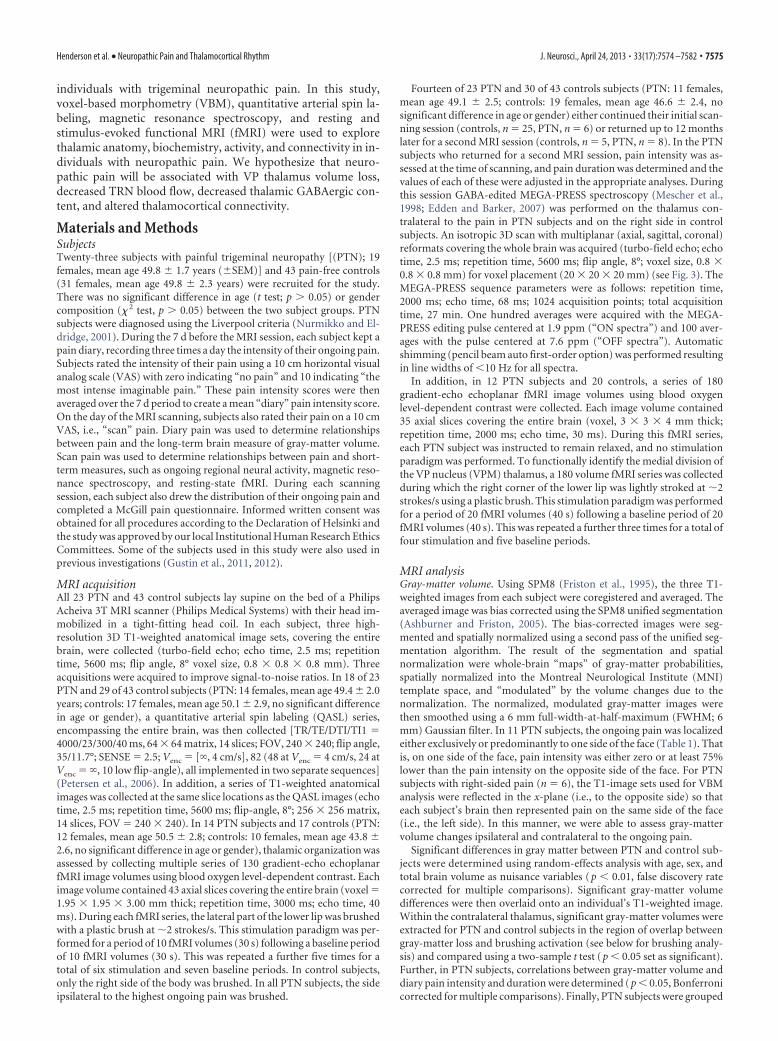
Table 1. Characteristics of patients with PTN (neuropathic pain)
Subject Age SexDuration ofpain (years) Site
Pain intensity(diary VAS) Analgesic Medication Negative sensory symptoms Positive sensory symptoms
MRI analysis performed
VBM QASL MRS rs-fcMRIThalamusfMRI
1 51 M 3.5 Right 2.1 Amitriptyline Molar tooth anesthesia resulting fromendodontic therapy
Unstimulated pain adjacent softtissues
Y* Y Y Y Y
2 48 F 9.0 Bilateral 2.5 Gabapentin Nil 6 months postdental trauma Unstimulated generalized oral pain Y Y — — —3 42 F 2.0 Right 5.5 Gabapentin Molar tooth anesthesia resulting from
endodontic therapyUnstimulated pain in treated tooth
and adjacent soft tissuesY — — — —
4 64 F 11.0 Right 5.0 Gabapentin Nil 6 months postdental treatment Unstimulated pain in treated toothand adjacent tissues
Y — — — Y
5 52 F 3.5 Right 4.5 None Nil 6 months postdental treatment Unstimulated pain in treated toothand adjacent soft tissues
Y — — — —
6 47 F 5.0 Left 1.1 None Molar tooth anesthesia resulting fromendodontic therapy
Unstimulated pain adjacent tissues Y Y Y Y Y
7 53 F 2.5 Right 1.5 None Molar tooth anesthesia resultingfrom endodontic therapy
Unstimulated pain adjacent tissues Y — — — Y
8 52 F 1.5 Bilateral 6.9 Gabapentin, oxycodone Hypoesthesia following surgical damageto inferior alveolar nerve
Unstimulated pain in area of damagednerve distribution
Y Y Y Y Y
9 55 F 2.0 Left 5.2 Amitriptyline, gabapentin Motor deficit resulting from herpeszoster infection
Unstimulated generalized unilateralcraniofacial pain
Y Y Y Y Y
10 46 M 9.0 Bilateral 3.1 None Molar tooth anesthesia resulting fromendodontic therapy
Unstimulated pain adjacent tissues Y Y — — Y
11 46 F 3.0 Bilateral 6.5 Diazepam, ibuprofen (prn) Minor motor impairment resulting fromskull base surgery
Unstimulated generalized unilateralcraniofacial pain
Y — — — Y
12 42 F 11.0 Bilateral 4.8 Carbamazepine Nil 6 months postmandible surgery Unstimulated generalized unilateralpain
Y Y Y Y Y
13 48 F 1.25 Bilateral 2.6 Gabapentin Molar tooth anesthesia resulting fromendodontic therapy
Unstimulated pain adjacent tissues Y Y — — Y
14 34 M 5.0 Bilateral 3.5 Pregabalin, nortriptyline Molar tooth anesthesia resulting fromendodontic therapy
Unstimulated pain adjacent tissues Y Y Y — Y
15 59 F 5.0 Left 3.3 None Molar tooth anesthesia resulting fromendodontic therapy
Unstimulated pain adjacent tissues Y Y — — Y
16 54 F 2.0 Right 1.6 Amitriptyline Molar tooth anesthesia resulting fromendodontic therapy
Unstimulated pain adjacent tissues Y Y Y Y
17 44 F 6.5 Bilateral 6.4 None Molar tooth anesthesia resulting fromendodontic therapy
Unstimulated pain adjacent tissues Y Y Y — Y
18 40 F 3.5 Bilateral 4.0 Carbamazepine Molar tooth anesthesia resulting fromendodontic therapy
Unstimulated pain adjacent tissues Y Y Y Y Y
19 67 F 14.0 Left 8.4 None Nil resulting from trigeminal neuralgiadiagnosis
Allodynia Y Y Y Y —
20 43 M 16.0 Left 5.8 Amitriptyline Nil resulting from dental treatment Unstimulated pain adjacent tissues Y Y Y Y —21 44 F 1.5 Bilateral 1.2 None Nil, following unknown onset Unstimulated pain adjacent tissues Y Y Y Y —22 65 F 7.0 Bilateral 2.5 None Molar tooth anesthesia resulting from
endodontic therapyUnstimulated pain adjacent tissues Y Y Y Y —
23 50 F 7.0 Bilateral 0.5 None Molar tooth anesthesia resulting fromendodontic therapy
Unstimulated pain adjacent tissues Y Y Y Y —
Mean (�SEM) 50 (�2) 5.8 (�0.9) 3.9 (�0.4) n � 23 n � 18 n � 14 n � 12 n � 14
*Y indicates subject was included in a particular MRI analysis.
PRN, Pro re nata (as needed); rs-fcMRI, resting-state functional connectivity magnetic resonance imaging. Note: IASP definition, Neuropathic pain is a clinical description (and not a diagnosis) which requires a demonstrable lesion or a diseasethat satisfies established neurological diagnostic criteria. The term “lesion” is commonly used when diagnostic investigations reveal an abnormality or when there was obvious trauma. The term “disease” is commonly used when theunderlying cause of the lesion is known. The presence of symptoms or signs (e.g., touch-evoked pain) alone does not justify the use of the term “neuropathic.” It is common when investigating neuropathic pain that diagnostic testing mayyield inconclusive or even inconsistent data. In such instances, clinical judgment is required to reduce the totality of findings in a patient into one putative diagnosis or concise group of diagnoses.
7576 • J. Neurosci., April 24, 2013 • 33(17):7574 –7582 Henderson et al. • Neuropathic Pain and Thalamocortical Rhythm
that each subject’s brain then represented pain on the same side of theface. Significant differences in CBF between PTN and control subjectswere determined using random-effects analysis with age and sex as nui-sance variables. Given that we hypothesized that thalamic and primarysomatosensory cortex (SI) regions would display altered blood flow, weused standard regions of interest derived from the MNI PickAtlas tool-box in SPM8, to apply small volume corrections ( p � 0.05, false discov-ery rate corrected for multiple comparisons). Significant CBF differenceswere then overlaid onto an individual’s T1-weighted image.
CBF values from significant clusters were extracted from both the PTNand control subjects and compared using two-sample t tests ( p � 0.05 setas significant). In PTN subjects, correlations between CBF and scan painintensity and duration were determined ( p � 0.05, Bonferroni correctedfor multiple comparisons). In addition, to compare CBF within the VPthalamus of control and PTN subjects, CBF values were extracted from
the region of overlap between gray matter lossand brushing activation (see below for brush-ing analysis) and compared using a two-samplet test ( p � 0.05 set as significant). Finally, PTNsubjects were grouped into those taking anal-gesic medications (n � 10) and those not tak-ing any medications (n � 8), and CBF valuesbetween these two groups were compared us-ing a two-sample t test ( p � 0.05 set assignificant).
GABA-edited MEGA-PRESS spectra. Forquantification of the GABA/creatine ratios, theacquired spectra were analyzed using the Java-based magnetic resonance user’s interface (jM-RUI 4.0, European Union project). First, thedominant water resonance was removed usingthe Hankel Lanczos Singular Valve Decompo-sition algorithm. The ‘‘ON’’ and ‘‘OFF’’ spec-tral subsets were summed producing single ONand OFF 68 ms subspectra for each spectra da-taset. These 68 ms subspectra were then sub-tracted resulting in GABA-edited differencespectra to measure GABA concentration at
3.01 ppm. The GABA-edited difference spectra were then phased withrespect to both the zero-order and first-order phase. Line broadening of5 Hz was used to improve the display. GABA was quantified using AMA-RES, a nonlinear least-square fitting algorithm operating in the timedomain. Peak fitting for GABA was performed after manually definingthe center frequency and line width of the GABA peak and modeling theGABA peak as a singlet. Furthermore, Lorentzian curves were used toobtain the peak amplitude for this resonance.
The OFF spectral subsets were summed producing single OFF 68 mssubspectra for each spectra dataset to measure creatine concentration at3.02 ppm. The single OFF 68 ms subspectra was then phased with respectto both the zero-order and first-order phase. Line broadening of 5 Hz wasused to improve the display. Spectral fitting in AMARES was performedafter manually defining the center frequency and line width of the crea-tine peak, and modeling the creatine peak as a singlet. Furthermore,Lorentzian curves were used to obtain the peak amplitude for this reso-nance. Finally, ratios were calculated for GABA relative to creatine.
Differences in metabolite ratios between PTN and control subjectswere determined using two-sample t tests ( p � 0.05 set as significant). InPTN subjects, correlations between thalamic GABA/Cr (creatine) andscan pain intensity and duration were determined ( p � 0.05, Bonferronicorrected for multiple comparisons). Finally, thalamic GABA/Cr ratiosin PTN subjects taking analgesic medications (n � 8) were comparedwith PTN subjects not taking analgesic medications (n � 6) using atwo-sample t test ( p � 0.05 set as significant).
Functional connectivity. All fMRI images were motion-corrected,global signal drifts removed using the detrending method described byMacey et al. (2004), spatially normalized to the MNI template. To func-tionally identify the location of the VPM thalamus, fMRI images col-lected during lip brushing were smoothed using a 6 mm FWHMGaussian filter, and significant increases in signal intensity were deter-mined using a repeated boxcar model convolved with hemodynamicdelay function (20 volumes baseline, 20 volumes lip brushing; 4 stimu-lation, and 5 baseline periods). Significant activation within the con-tralateral thalamus was determined using a random-effects analysis ( p �0.05, false discovery rate corrected for multiple comparisons, minimumcluster size 10 voxels). This cluster was then used in the VBM analysis todetermine significant changes in gray-matter volume in PTN subjectscompared with controls.
For connectivity analysis, images were smoothed using a 6 mmFWHM Gaussian filter. In all subjects, during the baseline fMRI scan(i.e., no stimulation paradigm), functional connectivity was assessed us-ing signal intensity changes within the VPM thalamus as a “seeding area.”More specifically, signal intensity changes over the 180 volumes wereextracted in each subject from the thalamic region activated by lip brush-ing and also displaying gray-matter loss in PTN subjects (i.e., VPM thal-
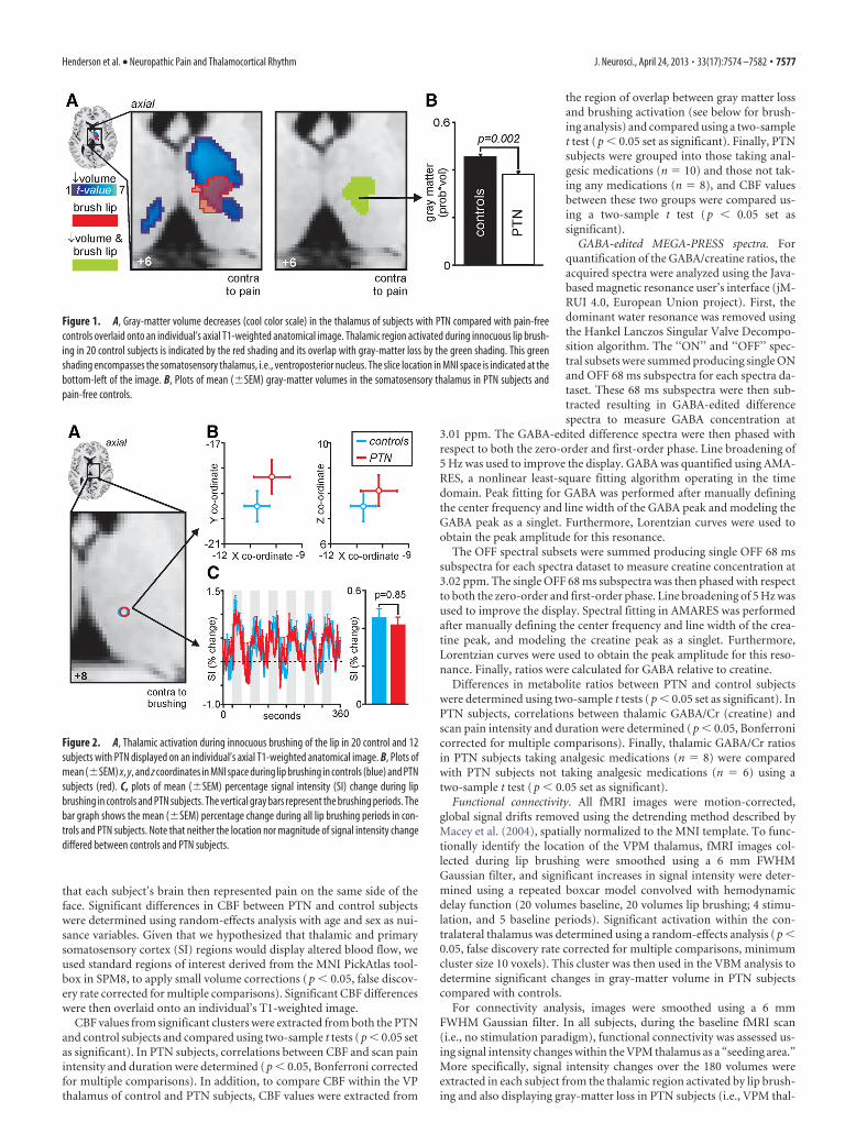
Figure 1. A, Gray-matter volume decreases (cool color scale) in the thalamus of subjects with PTN compared with pain-freecontrols overlaid onto an individual’s axial T1-weighted anatomical image. Thalamic region activated during innocuous lip brush-ing in 20 control subjects is indicated by the red shading and its overlap with gray-matter loss by the green shading. This greenshading encompasses the somatosensory thalamus, i.e., ventroposterior nucleus. The slice location in MNI space is indicated at thebottom-left of the image. B, Plots of mean (�SEM) gray-matter volumes in the somatosensory thalamus in PTN subjects andpain-free controls.
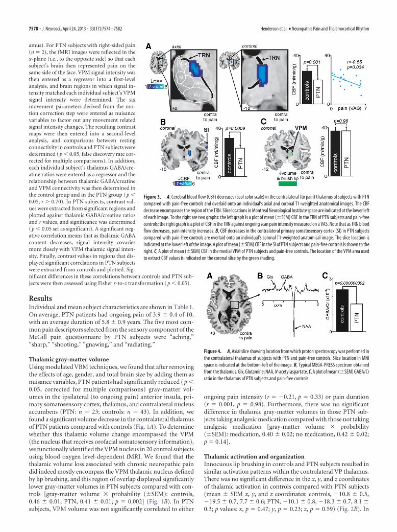
Figure 2. A, Thalamic activation during innocuous brushing of the lip in 20 control and 12subjects with PTN displayed on an individual’s axial T1-weighted anatomical image. B, Plots ofmean (�SEM) x, y, and z coordinates in MNI space during lip brushing in controls (blue) and PTNsubjects (red). C, plots of mean (�SEM) percentage signal intensity (SI) change during lipbrushing in controls and PTN subjects. The vertical gray bars represent the brushing periods. Thebar graph shows the mean (�SEM) percentage change during all lip brushing periods in con-trols and PTN subjects. Note that neither the location nor magnitude of signal intensity changediffered between controls and PTN subjects.
Henderson et al. • Neuropathic Pain and Thalamocortical Rhythm J. Neurosci., April 24, 2013 • 33(17):7574 –7582 • 7577
amus). For PTN subjects with right-sided pain(n � 2), the fMRI images were reflected in thex-plane (i.e., to the opposite side) so that eachsubject’s brain then represented pain on thesame side of the face. VPM signal intensity wasthen entered as a regressor into a first-levelanalysis, and brain regions in which signal in-tensity matched each individual subject’s VPMsignal intensity were determined. The sixmovement parameters derived from the mo-tion correction step were entered as nuisancevariables to factor out any movement relatedsignal intensity changes. The resulting contrastmaps were then entered into a second-levelanalysis, and comparisons between restingconnectivity in controls and PTN subjects weredetermined ( p � 0.05, false discovery rate cor-rected for multiple comparisons). In addition,each individual subject’s thalamus GABA/cre-atine ratios were entered as a regressor and therelationship between thalamic GABA/creatineand VPM connectivity was then determined inthe control group and in the PTN group ( p �0.05, r � 0.70). In PTN subjects, contrast val-ues were extracted from significant regions andplotted against thalamic GABA/creatine ratiosand r values, and significance was determined( p � 0.05 set as significant). A significant neg-ative correlation means that as thalamic GABAcontent decreases, signal intensity covariesmore closely with VPM thalamic signal inten-sity. Finally, contrast values in regions that dis-played significant correlations in PTN subjectswere extracted from controls and plotted. Sig-nificant differences in these correlations between controls and PTN sub-jects were then assessed using Fisher r-to-z transformation ( p � 0.05).
ResultsIndividual and mean subject characteristics are shown in Table 1.On average, PTN patients had ongoing pain of 3.9 � 0.4 of 10,with an average duration of 5.8 � 0.9 years. The five most com-mon pain descriptors selected from the sensory component of theMcGill pain questionnaire by PTN subjects were “aching,”“sharp,” “shooting,” “gnawing,” and “radiating.”
Thalamic gray-matter volumeUsing modulated VBM techniques, we found that after removingthe effects of age, gender, and total brain size by adding them asnuisance variables, PTN patients had significantly reduced ( p �0.05, corrected for multiple comparisons) gray-matter vol-umes in the ipsilateral (to ongoing pain) anterior insula, pri-mary somatosensory cortex, thalamus, and contralateral nucleusaccumbens (PTN: n � 23; controls: n � 43). In addition, wefound a significant volume decrease in the contralateral thalamusof PTN patients compared with controls (Fig. 1A). To determinewhether this thalamic volume change encompassed the VPM(the nucleus that receives orofacial somatosensory information),we functionally identified the VPM nucleus in 20 control subjectsusing blood oxygen level-dependent fMRI. We found that thethalamic volume loss associated with chronic neuropathic paindid indeed mostly encompass the VPM thalamic nucleus definedby lip brushing, and this region of overlap displayed significantlylower gray-matter volumes in PTN subjects compared with con-trols [gray-matter volume � probability (�SEM): controls,0.46 � 0.01; PTN, 0.41 � 0.01; p � 0.002] (Fig. 1B). In PTNsubjects, VPM volume was not significantly correlated to either
ongoing pain intensity (r � �0.21, p � 0.33) or pain duration(r � 0.001, p � 0.98). Furthermore, there was no significantdifference in thalamic gray-matter volumes in those PTN sub-jects taking analgesic medication compared with those not takinganalgesic medication [gray-matter volume � probability(�SEM): medication, 0.40 � 0.02; no medication, 0.42 � 0.02;p � 0.14].
Thalamic activation and organizationInnocuous lip brushing in controls and PTN subjects resulted insimilar activation patterns within the contralateral VP thalamus.There was no significant difference in the x, y, and z coordinatesof thalamic activation in controls compared with PTN subjects(mean � SEM x, y, and z coordinates: controls, �10.8 � 0.5,�19.5 � 0.7, 7.7 � 0.6; PTN, �10.1 � 0.8, �18.3 � 0.7, 8.1 �0.3; p values: x, p � 0.47; y, p � 0.23; z, p � 0.59) (Fig. 2B). In
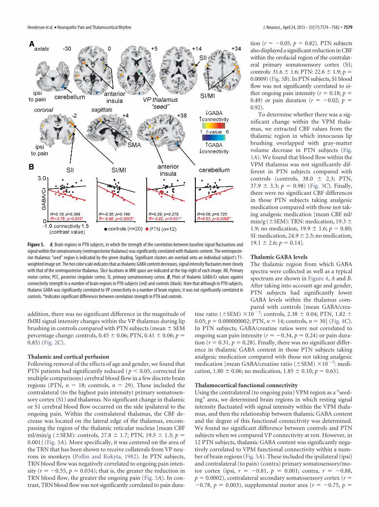
Figure 3. A, Cerebral blood flow (CBF) decreases (cool color scale) in the contralateral (to pain) thalamus of subjects with PTNcompared with pain-free controls and overlaid onto an individual’s axial and coronal T1-weighted anatomical images. The CBFdecrease encompasses the region of the TRN. Slice locations in Montreal Neurological Institute space are indicated at the lower leftof each image. To the right are two graphs: the left graph is a plot of mean (�SEM) CBF in the TRN of PTN subjects and pain-freecontrols; the right graph is a plot of CBF in the TRN against ongoing scan pain intensity measured on a VAS. Note that as TRN bloodflow decreases, pain intensity increases. B, CBF decreases in the contralateral primary somatosensory cortex (SI) in PTN subjectscompared with pain-free controls are overlaid onto an individual’s coronal T1-weighted anatomical image. The slice location isindicated at the lower left of the image. A plot of mean (�SEM) CBF in the SI of PTN subjects and pain-free controls is shown to theright. C, A plot of mean (�SEM) CBF in the medial VPM of PTN subjects and pain-free controls. The location of the VPM area usedto extract CBF values is indicated on the coronal slice by the green shading.
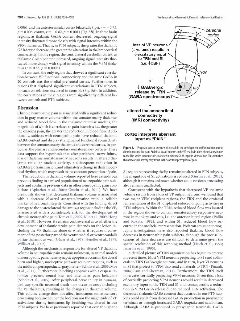
Figure 4. A, Axial slice showing location from which proton spectroscopy was performed inthe contralateral thalamus of subjects with PTN and pain-free controls. Slice location in MNIspace is indicated at the bottom-left of the image. B, Typical MEGA-PRESS spectrum obtainedfrom the thalamus. Glx, Glutamine; NAA, N-acetyl aspartate. C, A plot of mean (�SEM) GABA/Crratio in the thalamus of PTN subjects and pain-free controls.
7578 • J. Neurosci., April 24, 2013 • 33(17):7574 –7582 Henderson et al. • Neuropathic Pain and Thalamocortical Rhythm
addition, there was no significant difference in the magnitude offMRI signal intensity changes within the VP thalamus during lipbrushing in controls compared with PTN subjects (mean � SEMpercentage change: controls, 0.45 � 0.06; PTN, 0.41 � 0.06; p �0.85) (Fig. 2C).
Thalamic and cortical perfusionFollowing removal of the effects of age and gender, we found thatPTN patients had significantly reduced (p � 0.05, corrected formultiple comparisons) cerebral blood flow in a few discrete brainregions (PTN, n � 18; controls, n � 29). These included thecontralateral (to the highest pain intensity) primary somatosen-sory cortex (S1) and thalamus. No significant change in thalamicor S1 cerebral blood flow occurred on the side ipsilateral to theongoing pain. Within the contralateral thalamus, the CBF de-crease was located on the lateral edge of the thalamus, encom-passing the region of the thalamic reticular nucleus [mean CBFml/min/g (�SEM): controls, 27.8 � 1.7; PTN, 19.5 � 1.3; p �0.001] (Fig. 3A). More specifically, it was centered on the area ofthe TRN that has been shown to receive collaterals from VP neu-rons in monkeys (Pollin and Rokyta, 1982). In PTN subjects,TRN blood flow was negatively correlated to ongoing pain inten-sity (r � �0.55, p � 0.034); that is, the greater the reduction inTRN blood flow, the greater the ongoing pain (Fig. 3A). In con-trast, TRN blood flow was not significantly correlated to pain dura-
tion (r � �0.05, p � 0.82). PTN subjectsalso displayed a significant reduction in CBFwithin the orofacial region of the contralat-eral primary somatosensory cortex (S1;controls: 31.6 � 1.6; PTN: 22.6 � 1.9; p �0.0009) (Fig. 3B). In PTN subjects, S1 bloodflow was not significantly correlated to ei-ther ongoing pain intensity (r � 0.18; p �0.49) or pain duration (r � �0.02; p �0.92).
To determine whether there was a sig-nificant change within the VPM thala-mus, we extracted CBF values from thethalamic region in which innocuous lipbrushing overlapped with gray-mattervolume decrease in PTN subjects (Fig.1A). We found that blood flow within theVPM thalamus was not significantly dif-ferent in PTN subjects compared withcontrols (controls, 38.0 � 2.3; PTN,37.9 � 3.3; p � 0.98) (Fig. 3C). Finally,there were no significant CBF differencesin those PTN subjects taking analgesicmedication compared with those not tak-ing analgesic medication [mean CBF ml/min/g (�SEM): TRN: medication, 19.3 �1.9; no medication, 19.9 � 1.6; p � 0.80;SI: medication, 24.9 � 2.5; no medication,19.1 � 2.6; p � 0.14].
Thalamic GABA levelsThe thalamic region from which GABAspectra were collected as well as a typicalspectrum are shown in Figure 4, A and B.After taking into account age and gender,PTN subjects had significantly lowerGABA levels within the thalamus com-pared with controls [mean GABA/crea-
tine ratio (�SEM) �10�3: controls, 2.38 � 0.04; PTN, 1.82 �0.05; p � 0.000000002; PTN, n � 14; controls, n � 30] (Fig. 4C).In PTN subjects, GABA/creatine ratios were not correlated toongoing scan pain intensity (r � �0.34, p � 0.24) or pain dura-tion (r � 0.31, p � 0.28). Finally, there was no significant differ-ence in thalamic GABA content in those PTN subjects takinganalgesic medication compared with those not taking analgesicmedication [mean GABA/creatine ratio (�SEM) �10�3: medi-cation, 1.80 � 0.06; no medication, 1.85 � 0.10; p � 0.65).
Thalamocortical functional connectivityUsing the contralateral (to ongoing pain) VPM region as a “seed-ing” area, we determined brain regions in which resting signalintensity fluctuated with signal intensity within the VPM thala-mus, and then the relationship between thalamic GABA contentand the degree of this functional connectivity was determined.We found no significant difference between controls and PTNsubjects when we compared VP connectivity at rest. However, in12 PTN subjects, thalamic GABA content was significantly nega-tively correlated to VPM functional connectivity within a num-ber of brain regions (Fig. 5A). These included the ipsilateral (ipsi)and contralateral (to pain) (contra) primary somatosensory/mo-tor cortex (ipsi, r � �0.81, p � 0.001; contra, r � �0.88,p � 0.0002), contralateral secondary somatosensory cortex (r ��0.78, p � 0.003), supplemental motor area (r � �0.75, p �
Figure 5. A, Brain regions in PTN subjects, in which the strength of the correlation between baseline signal fluctuations andsignal within the somatosensory (ventroposterior thalamus) was significantly correlated with thalamic content. The ventroposte-rior thalamus “seed” region is indicated by the green shading. Significant clusters are overlaid onto an individual subject’s T1-weighted image set. The hot color scale indicates that as thalamic GABA content decreases, signal intensity fluctuates more closelywith that of the ventroposterior thalamus. Slice locations in MNI space are indicated at the top-right of each image. MI, Primarymotor cortex; PCC, posterior cingulate cortex; SI, primary somatosensory cortex. B, Plots of thalamic GABA/Cr values againstconnectivity strength in a number of brain regions in PTN subjects (red) and controls (black). Note that although in PTN subjects,thalamic GABA was significantly correlated to VP connectivity in a number of brain regions; it was not significantly correlated incontrols. *Indicates significant differences between correlation strength in PTN and controls.
Henderson et al. • Neuropathic Pain and Thalamocortical Rhythm J. Neurosci., April 24, 2013 • 33(17):7574 –7582 • 7579
0.006), and the anterior insular cortex bilaterally (ipsi, r � �0.75,p � 0.006; contra, r � �0.82, p � 0.001) (Fig. 5B). In these brainregions, as thalamic GABA content decreased, ongoing signalintensity fluctuated more closely with signal intensity within theVPM thalamus. That is, in PTN subjects, the greater the thalamicGABAergic decrease, the greater the alteration in thalamocorticalconnectivity. In one region, the contralateral cerebellar cortex, asthalamic GABA content increased, ongoing signal intensity fluc-tuated more closely with signal intensity within the VPM thala-mus (r � 0.83, p � 0.0008).
In contrast, the only region that showed a significant correla-tion between VP functional connectivity and thalamic GABA in20 controls was the medial prefrontal cortex. Furthermore, inregions that displayed significant correlations in PTN subjects,no such correlations occurred in controls (Fig. 5B). In addition,the correlations in these regions were significantly different be-tween controls and PTN subjects.
DiscussionChronic neuropathic pain is associated with a significant reduc-tion in gray-matter volume within the somatosensory thalamusand reduced blood flow in the thalamic reticular nucleus, themagnitude of which is correlated to pain intensity; i.e., the greaterthe ongoing pain, the greater the reduction in blood flow. Addi-tionally, subjects with neuropathic pain have reduced thalamicGABA content and display strengthened functional connectivitybetween the somatosensory thalamus and cerebral cortex, in par-ticular, the primary and secondary somatosensory cortices. Thesedata support the hypothesis that after peripheral nerve injury,loss of thalamic somatosensory neurons results in altered tha-lamic reticular nucleus activity, a subsequent reduction inGABAergic transmission, and ultimately a change in thalamocor-tical rhythm, which may result in the constant perception of pain.
The reduction in thalamic volume reported here extends ourprevious finding in a mixed population of neuropathic pain sub-jects and confirms previous data in other neuropathic pain con-ditions (Apkarian et al., 2004; Gustin et al., 2011). We havepreviously shown that decreased thalamic volume is associatedwith a decrease N-acetyl aspartate/creatine ratio, a reliablemarker of neuronal integrity. Consistent with this finding, directdamage to the posterolateral thalamus, a region including the VP,is associated with a considerable risk for the development ofchronic neuropathic pain (Kim et al., 2007; Klit et al., 2009; Honget al., 2010). However, it remains controversial as to whether thedevelopment of thalamic stroke pain depends on the lesion in-cluding the VP thalamus alone or whether it requires involve-ment of the posterior part of the ventromedial or ventrocaudalisportae thalamic as well (Gucer et al., 1978; Hendler et al., 1979;Willis et al., 2002).
Although the mechanism responsible for altered VP thalamicvolume in neuropathic pain remains unknown, in animal modelsof neuropathic pain, trans-synaptic apoptosis occurs in the dorsalhorn and higher, nociceptive pathway recipient regions, such asthe midbrain periaqueductal gray matter (Scholz et al., 2005; Moret al., 2011). Furthermore, blocking apoptosis with a caspase in-hibitor prevents neural loss and attenuates pain behaviors(Scholz et al., 2005). After peripheral nerve injury in humans,pathway-specific neuronal death may occur in areas includingthe VP thalamus, resulting in the changes in thalamic volume.This volume change does not affect innocuous somatosensoryprocessing because neither the location nor the magnitude of VPactivations during innocuous lip brushing was altered in ourPTN subjects. We have previously reported that even though the
S1 region representing the lip remains unaltered in PTN subjects,the magnitude of S1 activations is reduced (Gustin et al., 2012),although it remains unknown whether acute noxious processingalso remains unaffected.
Consistent with the hypothesis that decreased VP thalamicvolume results from a loss of VP output neurons, we found thattwo major VPM recipient regions, the TRN and the orofacialrepresentation of the S1, displayed reduced ongoing activities inPTN subjects. Within the TRN, reduced blood flow was locatedin the region shown to contain somatosensory responsive neu-rons in monkeys and cats, i.e., the anterior lateral region (Pollinand Rokyta, 1982), and within S1, reduced blood flow oc-curred in the orofacial representation. Positron emission tomog-raphy investigations have also reported thalamic blood flowdecreases in neuropathic pain subjects, although the precise lo-cations of these decreases are difficult to determine given thespatial resolution of this scanning method (Hsieh et al., 1995;Iadarola et al., 1995).
A detailed picture of TRN organization has begun to emergein recent times. Most VPM neurons projecting to S1 send collat-erals to TRN GABAergic neurons, and in turn, layer VI neuronsin S1 that project to VPM also send collaterals to TRN (Pinault,2004; Lam and Sherman, 2011). Furthermore, the TRN itselfinnervates cortically projecting VPM neurons. Given this, a lossof cortically projecting VPM neurons would result in decreasedexcitatory input to the TRN and S1 and, consequently, a reduc-tion in VPM GABA release due to reduced TRN activation. Thedecreased thalamic GABA content that occurred in our PTN sub-jects could result from decreased GABA production in presynapticterminals or through increased GABA reuptake and catabolism.Although GABA is produced in presynaptic terminals, GABA
Figure 6. Proposed central events which result in the development and/or maintenance ofchronic neuropathic pain. An initial loss of neurons in the VP results in a loss of excitatory inputsto the TRN which in turn results in altered inhibitory GABA input in VP thalamus. This disturbedthalamocortical activity may result in the constant perception of pain.
7580 • J. Neurosci., April 24, 2013 • 33(17):7574 –7582 Henderson et al. • Neuropathic Pain and Thalamocortical Rhythm
signal includes contributions from different pools of GABA, in-cluding that stored in presynaptic vesicles, free GABA in the syn-aptic cleft, as well as in glial cells after reuptake. Additionally, inthe macaque, peripheral nerve lesions result in GABA receptordownregulation (Rausell et al., 1992) and somatosensory path-way lesions reduce GABA-immunoreactive synapses by up to50% within the VP thalamus (Ralston, 2005). Although we arethe first to report thalamic GABAergic alteration in human neu-ropathic pain syndromes, indirect evidence for an imbalance inexcitatory/inhibitory neurotransmission also comes from the rel-ative success of the GABA-derivative drugs, gabapentin and pre-gabalin, in the control of neuropathic pain.
More globally, changes in TRN inputs and outputs have a signif-icant effect on overall brain function. Evidence suggests that theTRN regulates all sensory information passing from the thalamus tothe cerebral cortex (Pinault, 2004; Lam and Sherman, 2011). Al-though not conclusive, thalamocortical and TRN neurons also ap-pear to be critical for the production of thalamocortical rhythmswith evidence that the intrinsic and network properties of TRN neu-rons endow them with oscillatory properties (Llinas et al., 1999). Forexample, Steriade et al. (1985) reported spindle-related rhythms indisconnected TRN neurons and abolition of spindle oscillations fol-lowing TRN lesion. It has recently emerged that chronic neuropathicpain is associated with significantly altered thalamocortical rhythm(Sarnthein et al., 2006; Walton and Llinas, 2010). In particular neu-ropathic pain is associated with higher spectral power in the 2–25 Hzrange and a shift of the dominant peak toward lower frequencies(Sarnthein et al., 2006). Furthermore, this altered rhythm is charac-terized by persistent theta and beta overactivations in pain-associated areas, including the primary and secondarysomatosensory cortical regions (Stern et al., 2006).
Consistent with these previous reports, we found altered func-tional connectivity between the VPM thalamus and cortical re-gions including SI, SII (secondary somatosensory cortex), andanterior insula in our neuropathic pain subjects. Because individ-uals with neuropathic pain display increased burst firing of VPthalamic neurons, we speculate that periods of high-intensityneuronal firing and intervening low-intensity activity result inenhanced signal covariation between VP thalamus and con-nected brain regions. We propose that the greater the thalamicGABA decrease, the greater the VP burst firing and the greater theconnectivity. This altered connectivity may not be confined topain processing circuitry, as recently shown in experimental an-imals that neuropathic pain is associated with impaired spatialworking memory and increased spiking phase precision of me-dial prefrontal-hippocampal circuit with respect to hippocampaltheta rhythm (Cardoso-Cruz et al., 2013). Interestingly, this al-tered fronto-hippocampal connectivity and impaired memorywas not associated with a significant change in overall firing rate,just a change in firing rhythm. Similarly, we found no overallincreases in CBF, an index of neural activity, in pain-related brainregions, which is consistent with many previous investigations(Hsieh et al., 1995; Wik et al., 2003; Yunus et al., 2004). These datasuggest that altered thalamocortical rhythm, and not necessarilyincreased overall activity in somatosensory processing regions,can result in the persistent perception of pain. A similar situationmay also occur in other positive sensory conditions. For example,in individuals with tinnitus, increased auditory intensity is ac-companied by decreased medial geniculate nucleus activitywhich results in reduced auditory cortex inhibition (van Gendt etal., 2012). Similar to chronic neuropathic pain, this pattern ofchange is thought to be a manifestation of thalamocortical loopsdysregulation (Llinas et al., 1999).
Together, our MRI results support the thalamocortical con-cept of chronic neuropathic pain (Llinas et al., 1999; Jeanmonodet al., 2001; Sarnthein et al., 2006). This model proposes that afternervous system damage, altered afferent input to specific andnonspecific thalamic nuclei results in disturbed thalamocorticalcircuits involving altered inputs and outputs of cortical somato-sensory areas and the thalamic reticular nucleus. Our data showthat neuropathic pain is associated with significant neural losswithin VP thalamus and that this loss is associated with decreasedactivity in the TRN and S1 (Fig. 6). As a consequence, GABAergiccontent of the thalamus is reduced, likely as a direct result ofdecreased TRN activity. Finally, this reduction in TRN inhibitoryoutput results in altered thalamic firing patterns, alteredthalamocortical rhythm, which in some manner, generates thesubjective feeling of persistent pain.
These data raise the possibility that prevention of thalamicneuronal loss after peripheral nerve injury, for example, usingcaspase inhibitors, may prevent the development of chronic neu-ropathic pain (Scholz et al., 2005). Alternatively, once the painhas been established, altering thalamocortical loops by targetingthalamic lesions, such as to the centrolateral nucleus, may alterthalamocortical processing and relieve chronic pain (Sarnthein etal., 2006). Ultimately, therapeutic interventions aimed at alteringthalamocortical loops may provide better outcomes than are cur-rently available for many subjects suffering with persistent neu-ropathic pain.
ReferencesApkarian AV, Sosa Y, Sonty S, Levy RM, Harden RN, Parrish TB, Gitelman
DR (2004) Chronic back pain is associated with decreased prefrontaland thalamic gray matter density. J Neurosci 24:10410 –10415. CrossRefMedline
Ashburner J, Friston KJ (2005) Unified segmentation. Neuroimage 26:839 –851. CrossRef Medline
Cardoso-Cruz H, Lima D, Galhardo V (2013) Impaired spatial memoryperformance in a rat model of neuropathic pain is associated with reducedhippocampus-prefrontal cortex connectivity. J Neurosci 33:2465–2480.CrossRef Medline
Davis KD, Kiss ZH, Tasker RR, Dostrovsky JO (1996) Thalamicstimulation-evoked sensations in chronic pain patients and in nonpain(movement disorder) patients. J Neurophysiol 75:1026 –1037. Medline
Di Piero V, Jones AK, Iannotti F, Powell M, Perani D, Lenzi GL, FrackowiakRS (1991) Chronic pain: a PET study of the central effects of percutane-ous high cervical cordotomy. Pain 46:9 –12. CrossRef Medline
Edden RA, Barker PB (2007) Spatial effects in the detection of gamma-aminobutyric acid: improved sensitivity at high fields using inner volumesaturation. Magn Reson Med 58:1276 –1282. CrossRef Medline
Friston KJ, Holmes AP, Worsley KP, Proline JP, Frith CD, Frackowiak RSJ(1995) Statistical parametric maps in functional imaging: a general im-aging approach. Hum Brain Mapp 2:189 –210. CrossRef
Gerke MB, Duggan AW, Xu L, Siddall PJ (2003) Thalamic neuronal activityin rats with mechanical allodynia following contusive spinal cord injury.Neuroscience 117:715–722. CrossRef Medline
Gucer G, Niedermeyer E, Long DM (1978) Thalamic EEG recordings inpatients with chronic pain. J Neurol 219:47– 61. CrossRef Medline
Gustin SM, Peck CC, Wilcox SL, Nash PG, Murray GM, Henderson LA(2011) Different pain, different brain: thalamic anatomy in neuropathicand non-neuropathic chronic pain syndromes. J Neurosci 31:5956 –5964.CrossRef Medline
Gustin SM, Peck CC, Cheney LB, Macey PM, Murray GM, Henderson LA(2012) Pain and plasticity: is chronic pain always associated with somato-sensory cortex activity and reorganization? J Neurosci 32:14874 –14884.CrossRef Medline
Head H, Holmes G (1911) Sensory disturbances from cerebral lesions.Brain 34:102–254.
Hendler N, Viernstein M, Gucer P, Long D (1979) A preoperative screeningtest for chronic back pain patients. Psychosomatics 20:801– 808. Medline
Hirayama T, Dostrovsky JO, Gorecki J, Tasker RR, Lenz FA (1989) Record-
Henderson et al. • Neuropathic Pain and Thalamocortical Rhythm J. Neurosci., April 24, 2013 • 33(17):7574 –7582 • 7581
ings of abnormal activity in patients with deafferentation and centralpain. Stereotact Funct Neurosurg 52:120 –126. CrossRef Medline
Hong JH, Bai DS, Jeong JY, Choi BY, Chang CH, Kim SH, Ahn SH, Jang SH(2010) Injury of the spino-thalamo-cortical pathway is necessary for cen-tral post-stroke pain. Eur Neurol 64:163–168. CrossRef Medline
Hsieh JC, Belfrage M, Stone-Elander S, Hansson P, Ingvar M (1995) Centralrepresentation of chronic ongoing neuropathic pain studied by positronemission tomography. Pain 63:225–236. CrossRef Medline
Iadarola MJ, Max MB, Berman KF, Byas-Smith MG, Coghill RC, Gracely RH,Bennett GJ (1995) Unilateral decrease in thalamic activity observed withpositron emission tomography in patients with chronic neuropathic pain.Pain 63:55– 64. CrossRef Medline
Jeanmonod D, Magnin M, Morel A, Siegemund M, Cancro R, Lanz M (2001)Thalamocortical dysrhythmia II: clinical and surgical aspects. ThalamusRelat Syst 1:245–254. CrossRef
Kim JH, Greenspan JD, Coghill RC, Ohara S, Lenz FA (2007) Lesions lim-ited to the human thalamic principal somatosensory nucleus (ventralcaudal) are associated with loss of cold sensations and central pain. J Neu-rosci 27:4995–5004. CrossRef Medline
Klit H, Finnerup NB, Jensen TS (2009) Central post-stroke pain: clinicalcharacteristics, pathophysiology, and management. Lancet Neurol8:857– 868. CrossRef Medline
Lam YW, Sherman SM (2011) Functional organization of the thalamic in-put to the thalamic reticular nucleus. J Neurosci 31:6791– 6799. CrossRefMedline
Latremoliere A, Woolf CJ (2009) Central sensitization: a generator of painhypersensitivity by central neural plasticity. J Pain 10:895–926. CrossRefMedline
Lenz FA, Kwan HC, Dostrovsky JO, Tasker RR (1989) Characteristics of thebursting pattern of action potentials that occurs in the thalamus of pa-tients with central pain. Brain Res 496:357–360. CrossRef Medline
Lenz FA, Seike M, Richardson RT, Lin YC, Baker FH, Khoja I, Jaeger CJ,Gracely RH (1993) Thermal and pain sensations evoked by microstimu-lation in the area of human ventrocaudal nucleus. J Neurophysiol 70:200 –212. Medline
Lenz FA, Garonzik IM, Zirh TA, Dougherty PM (1998) Neuronal activity inthe region of the thalamic principal sensory nucleus (ventralis caudalis) inpatients with pain following amputations. Neuroscience 86:1065–1081.CrossRef Medline
Llinas RR, Ribary U, Jeanmonod D, Kronberg E, Mitra PP (1999) Thalamo-cortical dysrhythmia: a neurological and neuropsychiatric syndromecharacterized by magnetoencephalography. Proc Natl Acad Sci U S A 96:15222–15227. CrossRef Medline
Macey PM, Macey KE, Kumar R, Harper RM (2004) A method for removalof global effects from fMRI time series. Neuroimage 22:360 –366.CrossRef Medline
Melzack R (1999) Pain: an overview. Acta Anaesthesiol Scand 43:880 – 884.CrossRef Medline
Melzack R, Loeser JD (1978) Phantom body pain in paraplegics: evidencefor a central “pattern generating mechanism” for pain. Pain 4:195–210.Medline
Mescher M, Merkle H, Kirsch J, Garwood M, Gruetter R (1998) Simultane-ous in vivo spectral editing and water suppression. NMR Biomed 11:266 –272. CrossRef Medline
Moayedi M, Desouza D, Erpelding N (2011) Making sense of gray matterabnormalities in chronic orofacial pain: synthesizing divergent findings.J Neurosci 31:12396 –12397. CrossRef Medline
Moisset X, Bouhassira D (2007) Brain imaging of neuropathic pain. Neuro-image 37:S80 –S88. CrossRef Medline
Mor D, Bembrick AL, Austin PJ, Keay KA (2011) Evidence for cellular in-jury in the midbrain of rats following chronic constriction injury of thesciatic nerve. J Chem Neuroanat 41:158 –169. CrossRef Medline
Nurmikko TJ, Eldridge PR (2001) Trigeminal neuralgia: pathophysiology,diagnosis and current treatment. Br J Anaesth 87:117–132. CrossRefMedline
Pattany PM, Yezierski RP, Widerstrom-Noga EG, Bowen BC, Martinez-Arizala A, Garcia BR, Quencer RM (2002) Proton magnetic resonancespectroscopy of the thalamus in patients with chronic neuropathic painafter spinal cord injury. Am J Neuroradiol 23:901–905. Medline
Petersen ET, Lim T, Golay X (2006) Model-free arterial spin labeling quan-tification approach for perfusion MRI. Magn Reson Med 55:219 –232.CrossRef Medline
Pinault D (2004) The thalamic reticular nucleus: structure, function andconcept. Brain Res Brain Res Rev 46:1–31. CrossRef Medline
Pollin B, Rokyta R (1982) Somatotopic organization of nucleus reticularisthalami in chronic awake cats and monkeys. Brain Res 250:211–221.CrossRef Medline
Ralston HJ 3rd (2005) Pain and the primate thalamus. Prog Brain Res 149:1–10. CrossRef Medline
Rausell E, Cusick CG, Taub E, Jones EG (1992) Chronic deafferentation inmonkeys differentially affects nociceptive and nonnociceptive pathwaysdistinguished by specific calcium-binding proteins and down-regulatesgamma-aminobutyric acid type A receptors at thalamic levels. Proc NatlAcad Sci U S A 89:2571–2575. CrossRef Medline
Sarnthein J, Stern J, Aufenberg C, Rousson V, Jeanmonod D (2006) In-creased EEG power and slowed dominant frequency in patients with neu-rogenic pain. Brain 129:55– 64. CrossRef Medline
Schmidt-Wilcke T, Luerding R, Weigand T, Jurgens T, Schuierer G,Leinisch E, Bogdahn U (2007) Striatal grey matter increase in pa-tients suffering from fibromyalgia: a voxel-based morphometry study.Pain 132:S109 –S116. CrossRef Medline
Scholz J, Broom DC, Youn DH, Mills CD, Kohno T, Suter MR, Moore KA,Decosterd I, Coggeshall RE, Woolf CJ (2005) Blocking caspase activityprevents transsynaptic neuronal apoptosis and the loss of inhibition inlamina II of the dorsal horn after peripheral nerve injury. J Neurosci25:7317–7323. CrossRef Medline
Schweinhardt P, Kuchinad A, Pukall CF, Bushnell MC (2008) Increasedgray matter density in young women with chronic vulvar pain. Pain 140:411– 419. CrossRef Medline
Steriade M, Deschenes M, Domich L, Mulle C (1985) Abolition of spindleoscillations in thalamic neurons disconnected from nucleus reticularisthalami. J Neurophysiol 54:1473–1497. Medline
Stern J, Jeanmonod D, Sarnthein J (2006) Persistent EEG overactivationin the cortical pain matrix of neurogenic pain patients. Neuroimage31:721–731. CrossRef Medline
van Gendt MJ, Boyen K, de Kleine E, Langers DR, van Dijk P (2012) Therelation between perception and brain activity in gaze-evoked tinnitus.J Neurosci 32:17528 –17539. CrossRef Medline
von Hehn CA, Baron R, Woolf CJ (2012) Deconstructing the neuropathicpain phenotype to reveal neural mechanisms. Neuron 73:638 – 652.CrossRef Medline
Walton KD, Llinas RR (2010) Central pain as a thalamocortical dysrhyth-mia: a thalamic efference disconnection? In: Translational pain research:from mouse to man (Kruger L, Light AR, eds). Boca Raton, FL: CRC.
Wik G, Fischer H, Bragee B, Kristianson M, Fredrikson M (2003) Retro-splenial cortical activation in the fibromyalgia syndrome. Neuroreport14:619 – 621. CrossRef Medline
Willis WD Jr, Zhang X, Honda CN, Giesler GJ Jr (2002) A critical review ofthe role of the proposed VMpo nucleus in pain. J Pain 3:79 –94. CrossRefMedline
Yezierski RP (1996) Pain following spinal cord injury: the clinical problemand experimental studies. Pain 68:185–194. CrossRef Medline
Younger JW, Shen YF, Goddard G, Mackey SC (2010) Chronic myofascialtemporomandibular pain is associated with neural abnormalities in thetrigeminal and limbic systems. Pain 149:222–228. CrossRef Medline
Yunus MB, Young CS, Saeed SA, Mountz JM, Aldag JC (2004) Positronemission tomography in patients with fibromyalgia syndrome andhealthy controls. Arthritis Rheum 51:513–518. CrossRef Medline
7582 • J. Neurosci., April 24, 2013 • 33(17):7574 –7582 Henderson et al. • Neuropathic Pain and Thalamocortical Rhythm