Embed Size (px)
Citation preview
Chronic obstructive pulmonary disease (COPD): epidemiology, risk factors for developing the disease,
mechanisms, and differential diagnosis
Alan Altraja
Department of Pulmonary Medicine,University of Tartu
The learning resources• Harrison’s Pulmonary and Critical Care Medicine.
Joseph Loscalzo (Ed.), McGraw Hill Medical, New York et al. 2010; 580 pp. (ISBN: 978-0-07-166338-0). http://umsha.ac.ir/uploads/Harrisons_Pulmonary_and_Critical.pdf• GOLD 2017 Global Strategy for the Diagnosis, Management and Prevention of COPD. http://goldcopd.org/gold-2017-global-strategy-diagnosis-management-prevention-copd/• GOLD 2017 Pocket Guide. http://goldcopd.org/wp-content/uploads/2016/12/wms-GOLD-2017-Pocket-Guide.pdf
A. Altraja ©2017
Definition of chronic obstructive pulmonary disease (COPD)
• „COPD is a common, preventable and treatable disease that is characterized by persistent respiratory symptoms and airflow limitation that is due to airway and/or alveolar abnormalities usually caused by significant exposure to noxious particles or gases“
• Global Strategy for the Diagnosis, Management, and Prevention of COPD: www.goldcopd.org
A. Altraja ©2017 GOLD, 2017
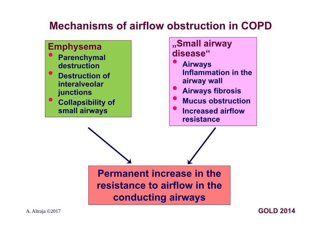
Mechanisms of airflow obstruction in COPD„Small airway disease“• Airways
Inflammation in the airway wall• Airways fibrosis• Mucus obstruction• Increased airflow resistance
Emphysema• Parenchymal
destruction• Destruction of interalveolar junctions• Collapsibility of small airways
Permanent increase in the resistance to airflow in the
conducting airwaysA. Altraja ©2017 GOLD 2014
COPD: conclusions from the definition
• COPD is an increasingly common disease • COPD is treatable, but preferably preventable disease • The respiratory symptoms, rising from airflow limitation, are persistent, as is the airflow limitation itself• The persistent /permanent airflow limitation is due to the airway and/or alveolar abnormalities that are usually caused by significant exposure to noxious particles or gases• Formerly, an augmented inflammation was included into the definition
A. Altraja ©2017 GOLD, 2017
COPD: the main points I• The most common respiratory symptoms include:•Dyspnea•Cough and/or •Sputum production• These symptoms may be under-reported by patients• The main risk factor for COPD is tobacco smoking, but
other environmental exposures do contribute:•Exposures to smoke from biomass fuels and air pollution may contribute•E-cigarette vapors• Besides exposures, host factors predispose individuals
to develop COPD:•Genetic abnormalities (e.g. alpha1-antriprotease deficiency etc.)•Abnormal lung development•Accelerated aging (linked to smoke exposures)•Infections
A. Altraja ©2017 GOLD, 2017
COPD: the main points II• COPD may be punctuated by periods of acute
worsening of respiratory symptoms, called exacerbations• In most patients, COPD is associated with significant concomitant chronic diseases, which have influence on the patients in whole and increase morbidity and mortality• The chronic airflow limitation in COPD is caused by a mixture of two processes: •Small airway disease (e.g. obstructive
bronchiolitis) and•Parenchymal destruction (i.e. emphysema)• One is usually leading, however, the relative contributions of these mechanisms vary from person to person
A. Altraja ©2017 GOLD, 2017
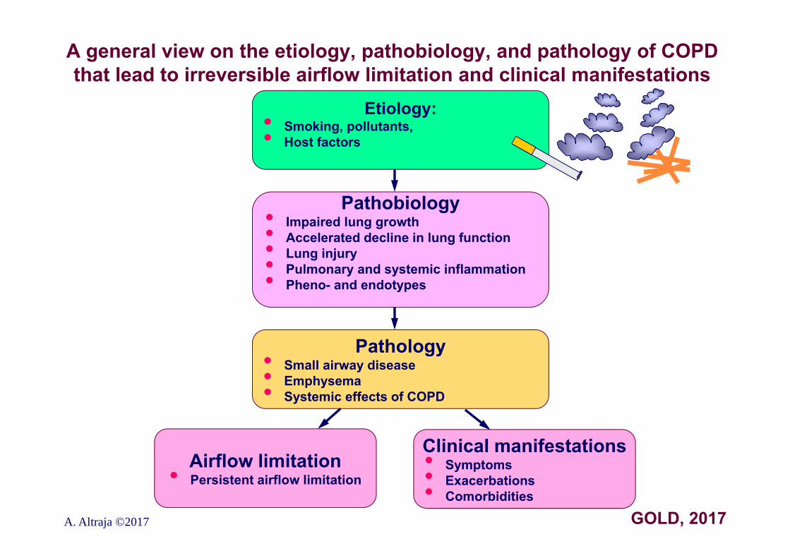
A general view on the etiology, pathobiology, and pathology of COPD that lead to irreversible airflow limitation and clinical manifestations
A. Altraja ©2017 GOLD, 2017
Etiology:• Smoking, pollutants, • Host factors
Pathobiology• Impaired lung growth• Accelerated decline in lung function• Lung injury• Pulmonary and systemic inflammation• Pheno- and endotypes
Pathology• Small airway disease• Emphysema• Systemic effects of COPD
Airflow limitation• Persistent airflow limitation
Clinical manifestations• Symptoms• Exacerbations• Comorbidities
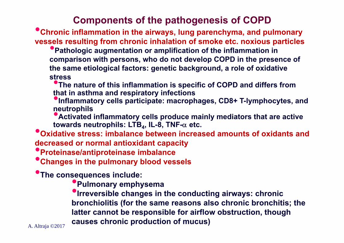
Components of the pathogenesis of COPD•Chronic inflammation in the airways, lung parenchyma, and pulmonary vessels resulting from chronic inhalation of smoke etc. noxious particles •Pathologic augmentation or amplification of the inflammation in
comparison with persons, who do not develop COPD in the presence of the same etiological factors: genetic background, a role of oxidative stress•The nature of this inflammation is specific of COPD and differs from that in asthma and respiratory infections•Inflammatory cells participate: macrophages, CD8+ T-lymphocytes, and neutrophils•Activated inflammatory cells produce mainly mediators that are active towards neutrophils: LTB4, IL-8, TNF- etc.•Oxidative stress: imbalance between increased amounts of oxidants and
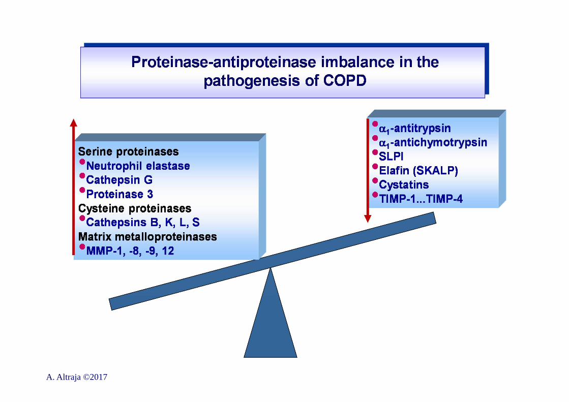
decreased or normal antioxidant capacity•Proteinase/antiproteinase imbalance•Changes in the pulmonary blood vessels•The consequences include: •Pulmonary emphysema•Irreversible changes in the conducting airways: chronic
bronchiolitis (for the same reasons also chronic bronchitis; the latter cannot be responsible for airflow obstruction, though causes chronic production of mucus)
A. Altraja ©2017
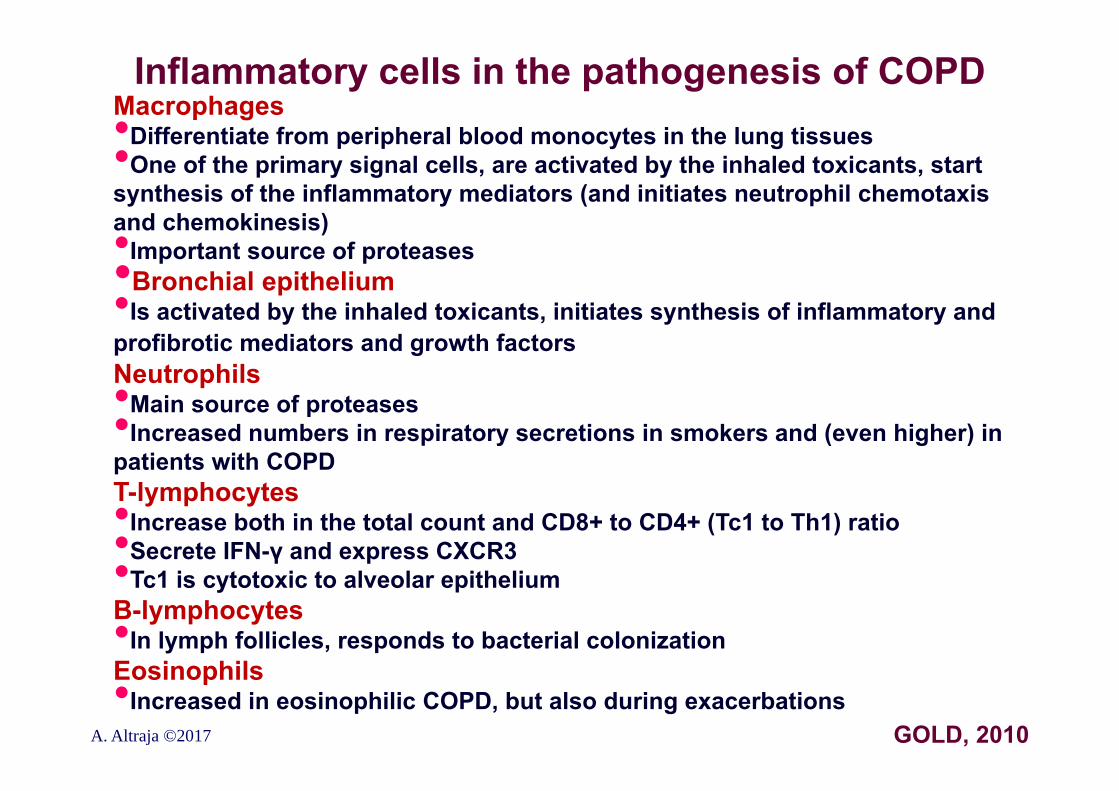
Inflammatory cells in the pathogenesis of COPDMacrophages•Differentiate from peripheral blood monocytes in the lung tissues•One of the primary signal cells, are activated by the inhaled toxicants, start synthesis of the inflammatory mediators (and initiates neutrophil chemotaxis and chemokinesis)•Important source of proteases •Bronchial epithelium•Is activated by the inhaled toxicants, initiates synthesis of inflammatory and profibrotic mediators and growth factorsNeutrophils•Main source of proteases •Increased numbers in respiratory secretions in smokers and (even higher) in patients with COPDT-lymphocytes•Increase both in the total count and CD8+ to CD4+ (Tc1 to Th1) ratio•Secrete IFN-γ and express CXCR3•Tc1 is cytotoxic to alveolar epitheliumB-lymphocytes•In lymph follicles, responds to bacterial colonizationEosinophils•Increased in eosinophilic COPD, but also during exacerbations
A. Altraja ©2017 GOLD, 2010
The main proinflammatory mediators in the pathogenesis of COPD
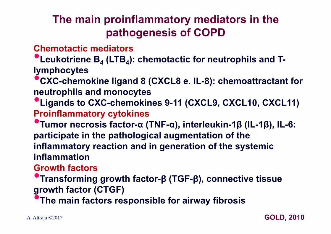
Chemotactic mediators•Leukotriene B4 (LTB4): chemotactic for neutrophils and T-lymphocytes•CXC-chemokine ligand 8 (CXCL8 e. IL-8): chemoattractant for neutrophils and monocytes•Ligands to CXC-chemokines 9-11 (CXCL9, CXCL10, CXCL11)Proinflammatory cytokines•Tumor necrosis factor-α (TNF-α), interleukin-1β (IL-1β), IL-6: participate in the pathological augmentation of the inflammatory reaction and in generation of the systemic inflammationGrowth factors•Transforming growth factor-β (TGF-β), connective tissue growth factor (CTGF)•The main factors responsible for airway fibrosis
A. Altraja ©2017 GOLD, 2010
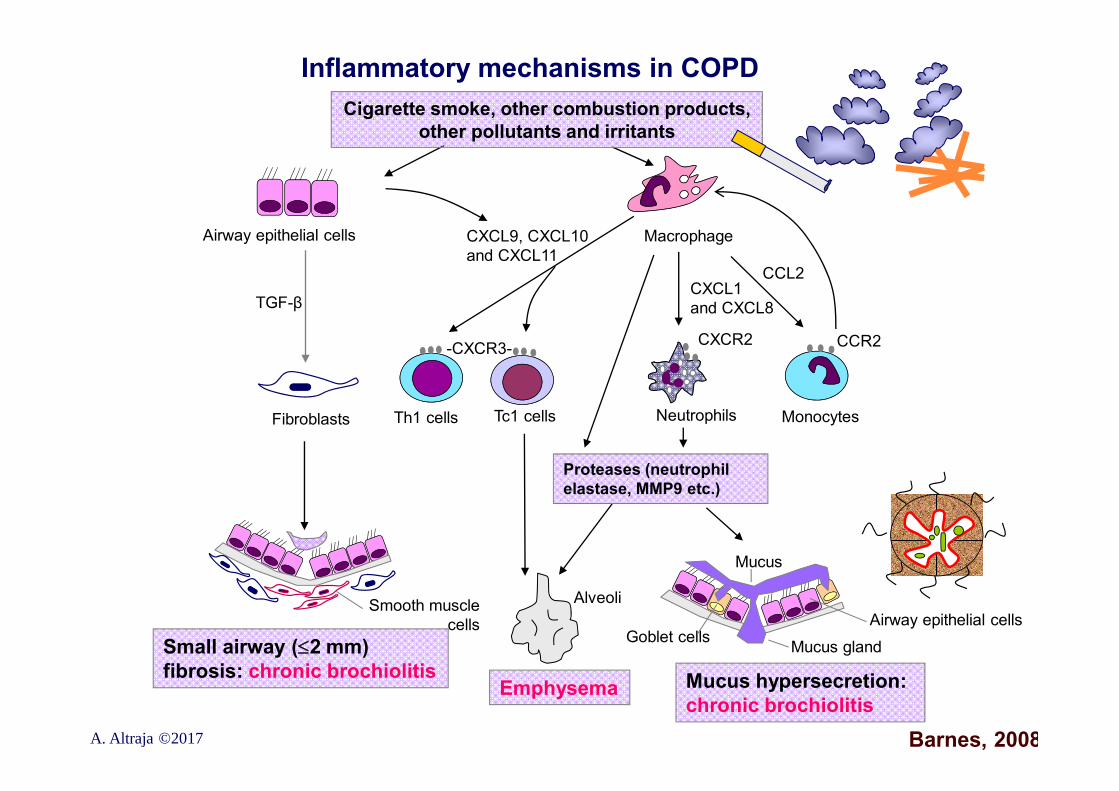
Inflammatory mechanisms in COPD
Barnes, 2008
Small airway (2 mm)fibrosis: chronic brochiolitis
Emphysema Mucus hypersecretion: chronic brochiolitis
Airway epithelial cells
TGF-β
CXCL9, CXCL10and CXCL11
Macrophage
CCL2
CCR2
CXCL1and CXCL8
CXCR2
Neutrophils MonocytesFibroblasts Th1 cells Tc1 cells
Cigarette smoke, other combustion products, other pollutants and irritants
Proteases (neutrophil elastase, MMP9 etc.)
-CXCR3-
Smooth muscle cells
Alveoli
Mucus
Airway epithelial cellsGoblet cells Mucus gland
A. Altraja ©2017
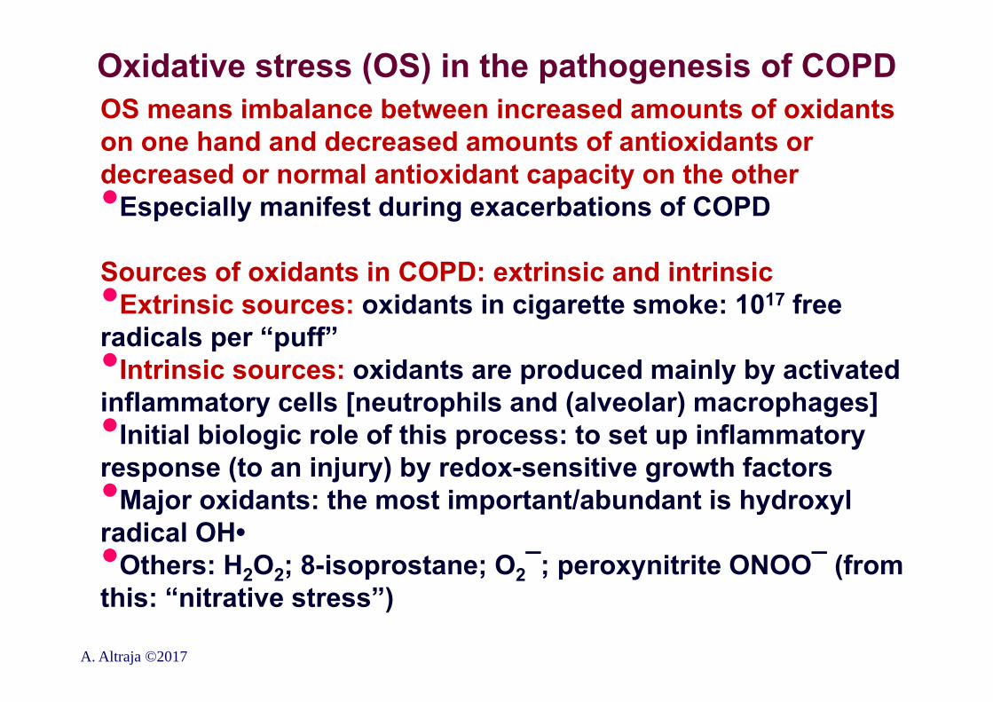
Oxidative stress (OS) in the pathogenesis of COPDOS means imbalance between increased amounts of oxidants on one hand and decreased amounts of antioxidants or decreased or normal antioxidant capacity on the other•Especially manifest during exacerbations of COPD
Sources of oxidants in COPD: extrinsic and intrinsic•Extrinsic sources: oxidants in cigarette smoke: 1017 free radicals per “puff”•Intrinsic sources: oxidants are produced mainly by activated inflammatory cells [neutrophils and (alveolar) macrophages]•Initial biologic role of this process: to set up inflammatory response (to an injury) by redox-sensitive growth factors •Major oxidants: the most important/abundant is hydroxyl radical OH••Others: H2O2; 8-isoprostane; O2¯; peroxynitrite ONOO¯ (from this: “nitrative stress”)
A. Altraja ©2017
Mechanisms of action of the oxidative stress in the context of COPD
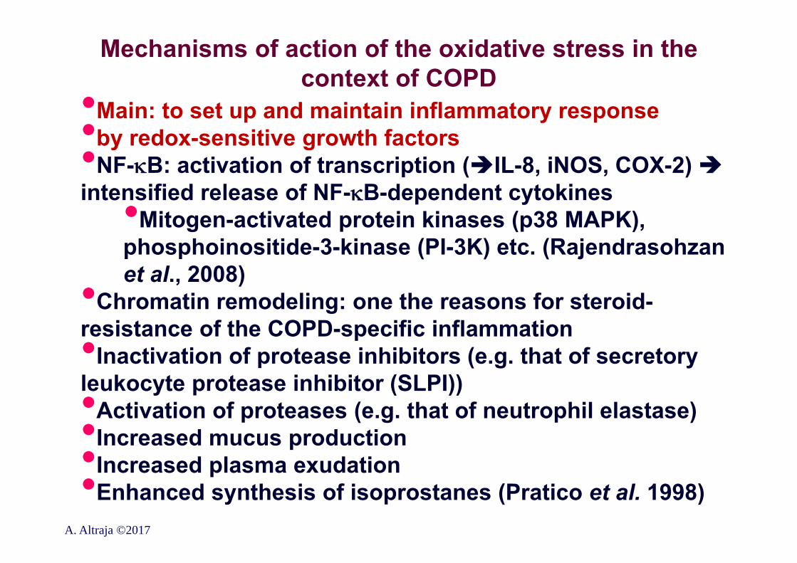
•Main: to set up and maintain inflammatory response •by redox-sensitive growth factors •NF-B: activation of transcription (IL-8, iNOS, COX-2)intensified release of NF-B-dependent cytokines•Mitogen-activated protein kinases (p38 MAPK),
phosphoinositide-3-kinase (PI-3K) etc. (Rajendrasohzan et al., 2008)•Chromatin remodeling: one the reasons for steroid-
resistance of the COPD-specific inflammation•Inactivation of protease inhibitors (e.g. that of secretory leukocyte protease inhibitor (SLPI))•Activation of proteases (e.g. that of neutrophil elastase)•Increased mucus production•Increased plasma exudation•Enhanced synthesis of isoprostanes (Pratico et al. 1998)
A. Altraja ©2017
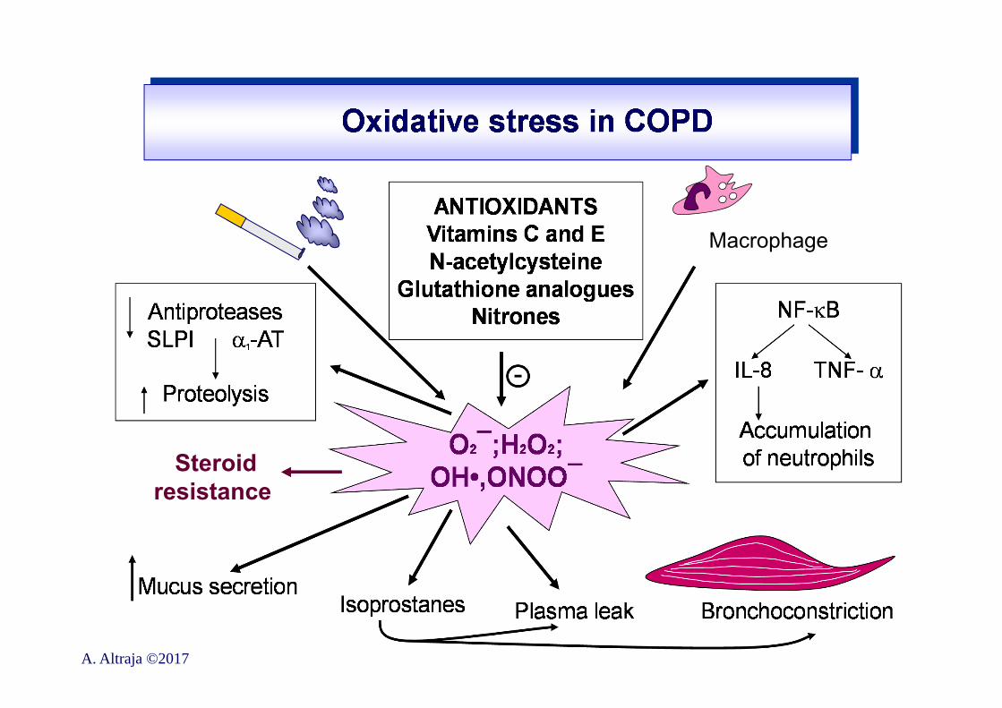
A. Altraja ©2017
Macrophage
Steroid resistance
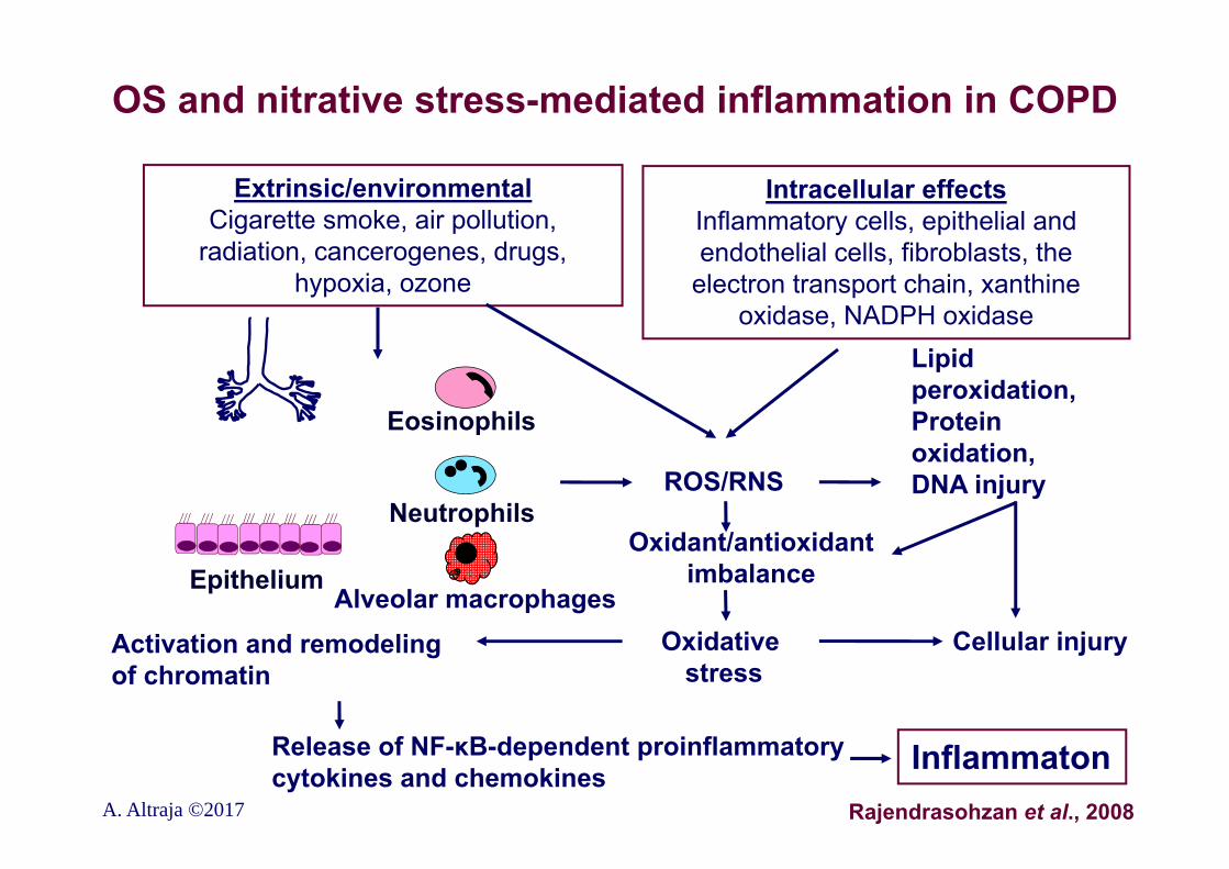
OS and nitrative stress-mediated inflammation in COPD
Extrinsic/environmentalCigarette smoke, air pollution,
radiation, cancerogenes, drugs, hypoxia, ozone
Intracellular effectsInflammatory cells, epithelial and endothelial cells, fibroblasts, the
electron transport chain, xanthine oxidase, NADPH oxidase
ROS/RNS
Oxidant/antioxidant imbalance
Lipid peroxidation,Protein oxidation,DNA injury
Activation and remodeling of chromatin
Oxidative stress
Cellular injury
Release of NF-κB-dependent proinflammatory cytokines and chemokines Inflammaton
Eosinophils
Neutrophils
Alveolar macrophagesEpithelium
A. Altraja ©2017 Rajendrasohzan et al., 2008
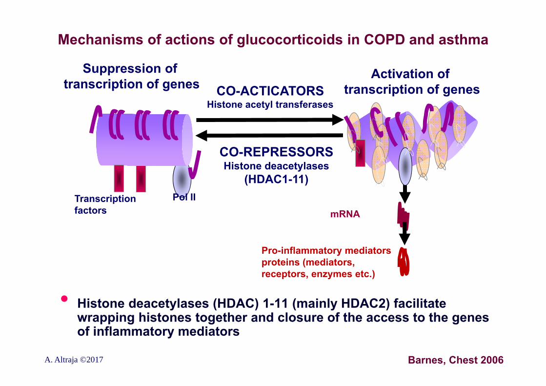
Mechanisms of actions of glucocorticoids in COPD and asthma
• Histone deacetylases (HDAC) 1-11 (mainly HDAC2) facilitate wrapping histones together and closure of the access to the genes of inflammatory mediators
Barnes, Chest 2006
mRNA
Pro-inflammatory mediators proteins (mediators, receptors, enzymes etc.)
CO-REPRESSORSHistone deacetylases
(HDAC1-11)
CO-ACTICATORSHistone acetyl transferases
Suppression of transcription of genes
Activation of transcription of genes
Transcription factors
Pol II
A. Altraja ©2017
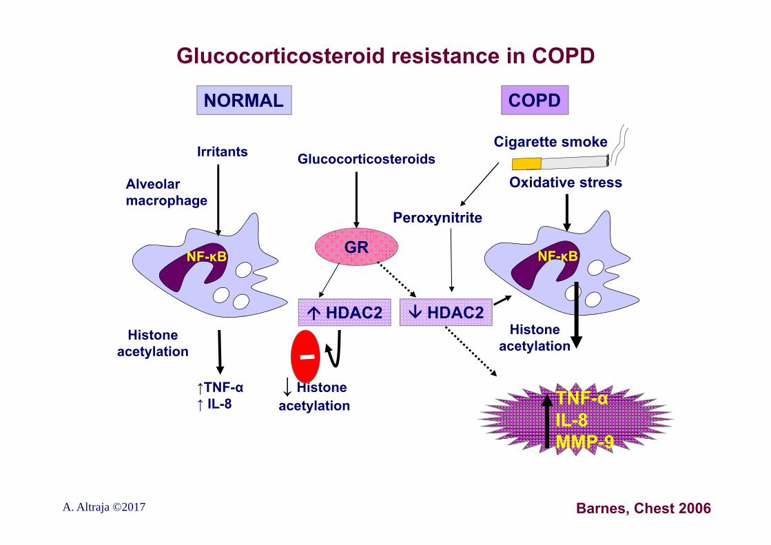
Glucocorticosteroid resistance in COPD
Barnes, Chest 2006
TNF-αIL-8MMP-9
NORMAL COPD
NF-κBNF-κB GR
Cigarette smoke
Oxidative stress
HDAC2 HDAC2
Peroxynitrite
GlucocorticosteroidsIrritants
Alveolarmacrophage
Histoneacetylation
↓ Histoneacetylation
↑TNF-α↑ IL-8
Histoneacetylation
A. Altraja ©2017
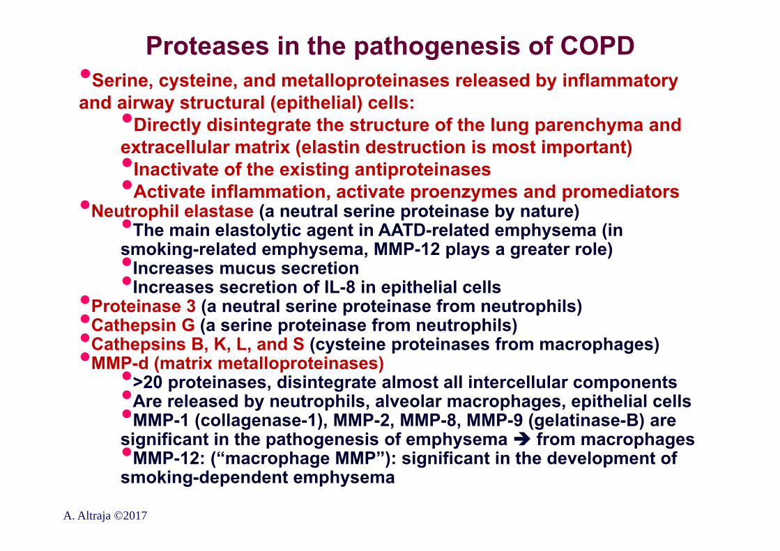
Proteases in the pathogenesis of COPD•Serine, cysteine, and metalloproteinases released by inflammatory and airway structural (epithelial) cells:•Directly disintegrate the structure of the lung parenchyma and
extracellular matrix (elastin destruction is most important)•Inactivate of the existing antiproteinases•Activate inflammation, activate proenzymes and promediators•Neutrophil elastase (a neutral serine proteinase by nature)•The main elastolytic agent in AATD-related emphysema (in smoking-related emphysema, MMP-12 plays a greater role)•Increases mucus secretion•Increases secretion of IL-8 in epithelial cells•Proteinase 3 (a neutral serine proteinase from neutrophils)•Cathepsin G (a serine proteinase from neutrophils)•Cathepsins B, K, L, and S (cysteine proteinases from macrophages)•MMP-d (matrix metalloproteinases)•>20 proteinases, disintegrate almost all intercellular components•Are released by neutrophils, alveolar macrophages, epithelial cells•MMP-1 (collagenase-1), MMP-2, MMP-8, MMP-9 (gelatinase-B) are significant in the pathogenesis of emphysema from macrophages•MMP-12: (“macrophage MMP”): significant in the development of smoking-dependent emphysema
A. Altraja ©2017
A. Altraja ©2017
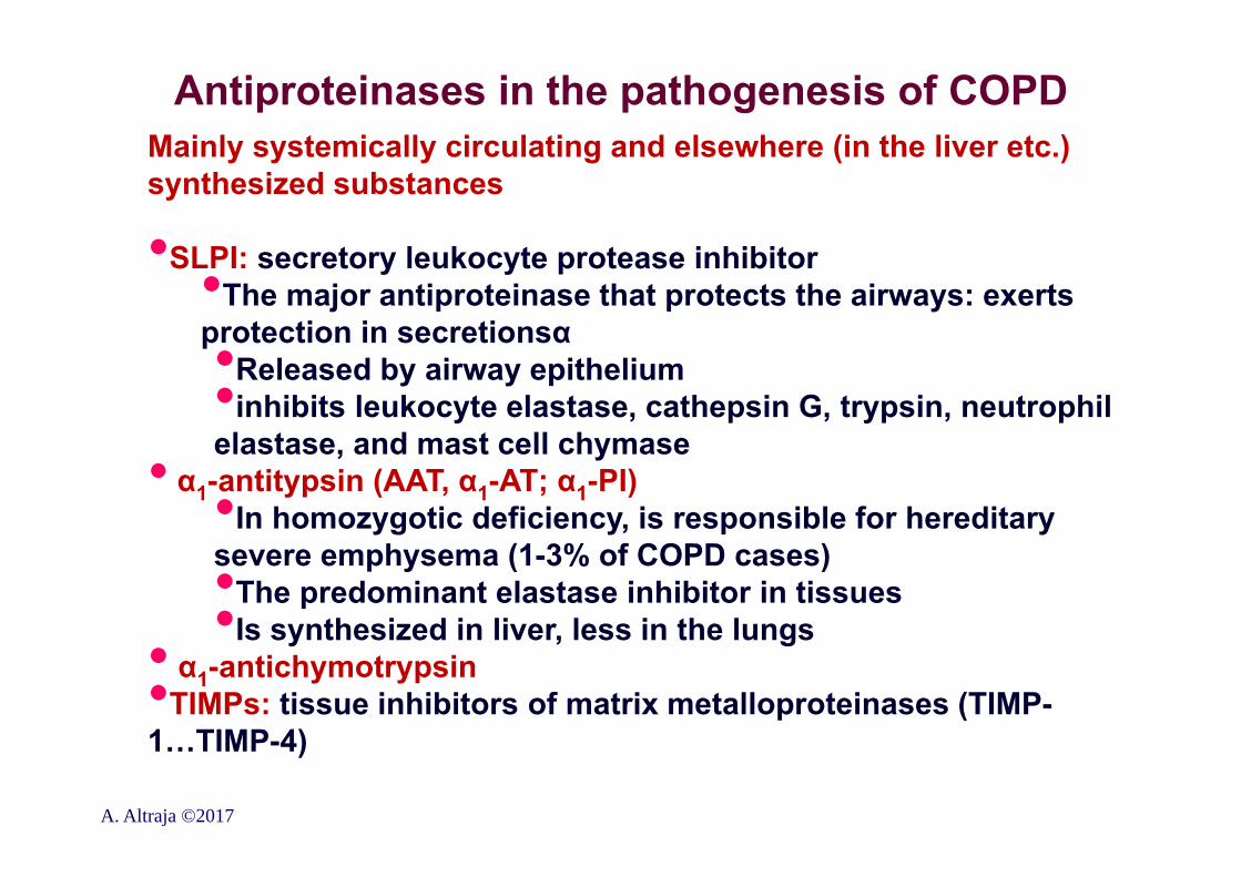
Antiproteinases in the pathogenesis of COPDMainly systemically circulating and elsewhere (in the liver etc.) synthesized substances
•SLPI: secretory leukocyte protease inhibitor•The major antiproteinase that protects the airways: exerts protection in secretionsα•Released by airway epithelium•inhibits leukocyte elastase, cathepsin G, trypsin, neutrophil elastase, and mast cell chymase• α1-antitypsin (AAT, α1-AT; α1-PI)•In homozygotic deficiency, is responsible for hereditary severe emphysema (1-3% of COPD cases)•The predominant elastase inhibitor in tissues•Is synthesized in liver, less in the lungs• α1-antichymotrypsin•TIMPs: tissue inhibitors of matrix metalloproteinases (TIMP-
1…TIMP-4)
A. Altraja ©2017
A. Altraja ©2017
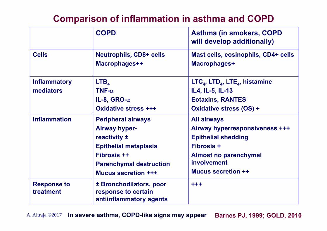
Comparison of inflammation in asthma and COPDCOPD Asthma (in smokers, COPD
will develop additionally)
Cells Neutrophils, CD8+ cellsMacrophages++
Mast cells, eosinophils, CD4+ cellsMacrophages+
Inflammatorymediators
LTB4TNF-IL-8, GRO-Oxidative stress +++
LTC4, LTD4, LTE4, histamineIL4, IL-5, IL-13Eotaxins, RANTESOxidative stress (OS) +
Inflammation Peripheral airwaysAirway hyper-reactivity ±Epithelial metaplasiaFibrosis ++Parenchymal destructionMucus secretion +++
All airwaysAirway hyperresponsiveness +++Epithelial sheddingFibrosis +Almost no parenchymal involvementMucus secretion ++
Response to treatment
± Bronchodilators, poor response to certain antiinflammatory agents
+++
A. Altraja ©2017 In severe asthma, COPD-like signs may appear Barnes PJ, 1999; GOLD, 2010
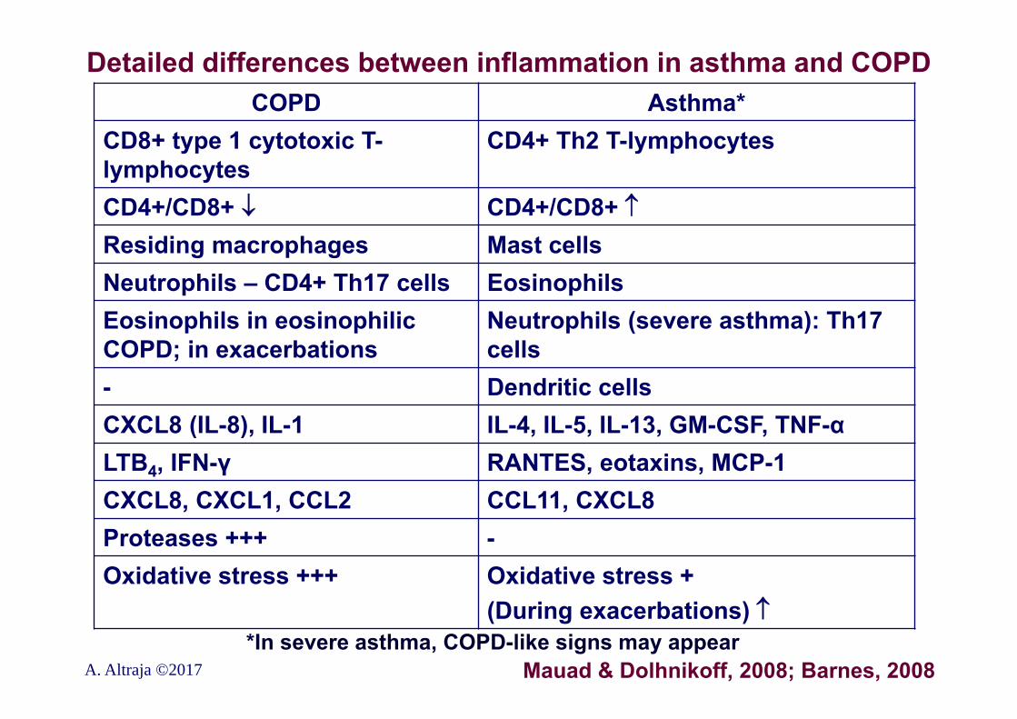
Detailed differences between inflammation in asthma and COPD
A. Altraja ©2017
COPD Asthma*CD8+ type 1 cytotoxic T-lymphocytes
CD4+ Th2 T-lymphocytes
CD4+/CD8+ CD4+/CD8+ Residing macrophages Mast cellsNeutrophils – CD4+ Th17 cells EosinophilsEosinophils in eosinophilic COPD; in exacerbations
Neutrophils (severe asthma): Th17 cells
- Dendritic cellsCXCL8 (IL-8), IL-1 IL-4, IL-5, IL-13, GM-CSF, TNF-αLTB4, IFN-γ RANTES, eotaxins, MCP-1 CXCL8, CXCL1, CCL2 CCL11, CXCL8Proteases +++ -Oxidative stress +++ Oxidative stress +
(During exacerbations)
Mauad & Dolhnikoff, 2008; Barnes, 2008*In severe asthma, COPD-like signs may appear
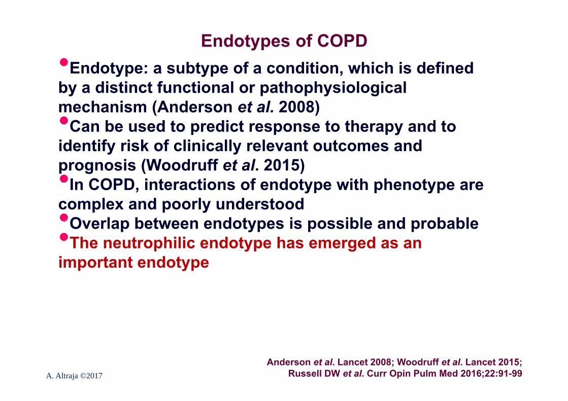
Endotypes of COPD•Endotype: a subtype of a condition, which is defined by a distinct functional or pathophysiological mechanism (Anderson et al. 2008)•Can be used to predict response to therapy and to identify risk of clinically relevant outcomes and prognosis (Woodruff et al. 2015)•In COPD, interactions of endotype with phenotype are complex and poorly understood•Overlap between endotypes is possible and probable•The neutrophilic endotype has emerged as an important endotype
Anderson et al. Lancet 2008; Woodruff et al. Lancet 2015; Russell DW et al. Curr Opin Pulm Med 2016;22:91-99 A. Altraja ©2017
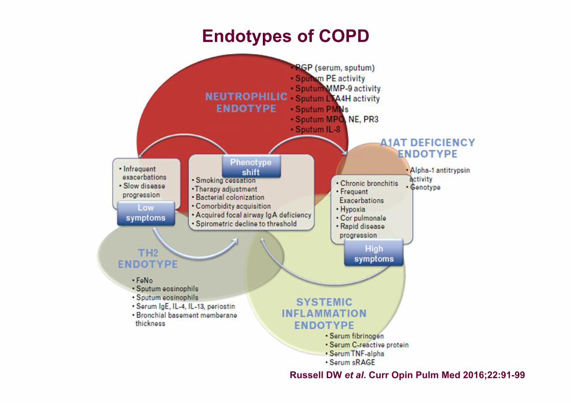
Endotypes of COPD
Russell DW et al. Curr Opin Pulm Med 2016;22:91-99
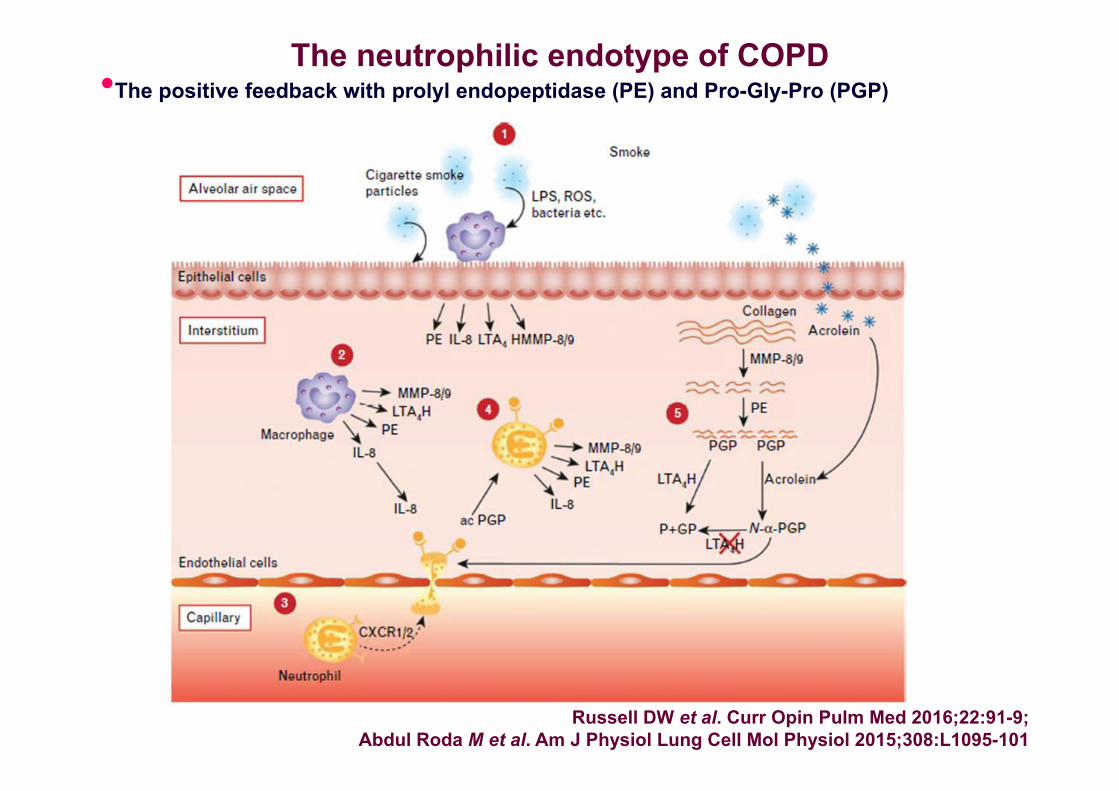
The neutrophilic endotype of COPD
Russell DW et al. Curr Opin Pulm Med 2016;22:91-9; Abdul Roda M et al. Am J Physiol Lung Cell Mol Physiol 2015;308:L1095-101
•The positive feedback with prolyl endopeptidase (PE) and Pro-Gly-Pro (PGP)
Chronic obstructive pulmonary diseaseEncompasses 2 type of conditions that exert different mechanisms of permanent airflow obstruction:•“Small airway disease” or chronic bronchiolitis•Due to the same etiopathogenesis, almost
always, it is accompanied by chronic bronchitis defined as the presence of chronic cough with sputum production for at least 3 months in each of at least 2 consecutive years (ATS, 1962): chronic bronchitis not capable of producing obstruction on its own•Emphysema: abnormal, permanent enlargement of the
airspaces distal to terminal bronchioli (mainly alveoli) with destruction of their walls, but without a clear fibrosis (Snider et al. 1985)
A. Altraja ©2017 GOLD
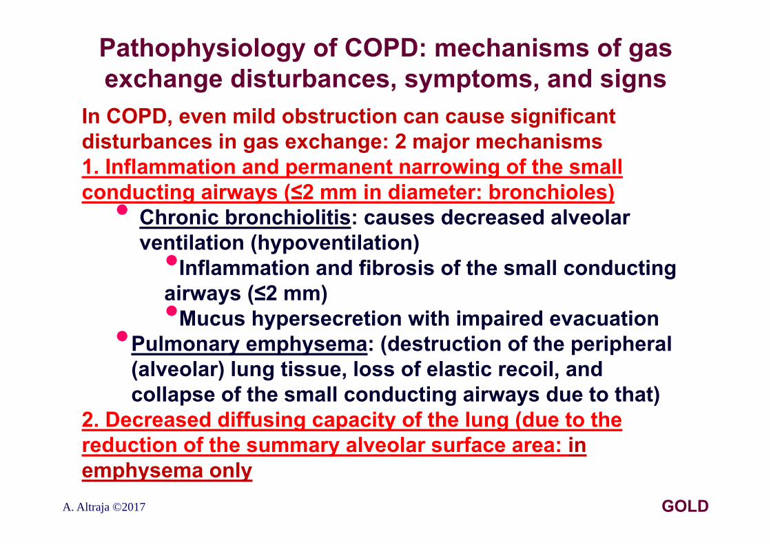
Pathophysiology of COPD: mechanisms of gas exchange disturbances, symptoms, and signs
In COPD, even mild obstruction can cause significant disturbances in gas exchange: 2 major mechanisms1. Inflammation and permanent narrowing of the small conducting airways (≤2 mm in diameter: bronchioles)• Chronic bronchiolitis: causes decreased alveolar
ventilation (hypoventilation)•Inflammation and fibrosis of the small conducting airways (≤2 mm)•Mucus hypersecretion with impaired evacuation•Pulmonary emphysema: (destruction of the peripheral
(alveolar) lung tissue, loss of elastic recoil, and collapse of the small conducting airways due to that)
2. Decreased diffusing capacity of the lung (due to the reduction of the summary alveolar surface area: in emphysema only
A. Altraja ©2017 GOLD
Mechanisms of gas exchange disturbances in COPD•Hypoxemia•In ventilation insufficiency, also hypercapnia (hypoventilation)•Progression of these events with progression of COPD•Small airway-type COPD and emphysema-type COPD are phenotypically different:•Emphysema conveys, in addition to causing collapse-type obstruction, also decreased diffusing capacity of the lung and therefore, produces more hypoxemia (emphysema correlates with decrease in PaO2 and SpO2)•Disease of the peripheral airways with obstruction causes decreased alveolar ventilation, and therefore, also hypercapnia (hypoventilation), in addition to hypoxia •= Decreased alveolar ventilation-to-perfusion ratio (VA/Q↓)•Results in precapillary vasocosntriction/remodeling to avoid perfusion of not-well-ventilated alveoli (→ secondary PH)•Addition of dysfunction of the respiratory muscles
A. Altraja ©2017 GOLD
Fibrosis of the walls of the small airways as a result of the chronic inflammation in COPD
•The basis: chronic inflammation, caused by chronic exposures to irritants
Activation of the epithelium and macrophages
Release of fibrogenic mediators (TGF-β etc.)
Fibrosis, especially in small airways (2 mm in diameter)
A. Altraja ©2017
Mechanisms of mucus hypersecretion in the airways in COPD
1. The result of inhaled irritants•Smoke from combustion of tobacco and other fumes•Other inhaled irritants, incl. e-cigarette vapors
Sensory nerve endings reflex:•Local peptidergic and cholinergic•Spinal cholinergic
Secretion by the glands •Local enzymes (e.g. neutrophil elastase)•The effect of oxidative stress•Stimulating effect of proteases
2. Glandular hyperplasia, proliferation of goblet cells (growth factors): the result of EGFR activation•MUC: expression of different mucin genes (MUC5AC, MUC5B etc.)
A. Altraja ©2017
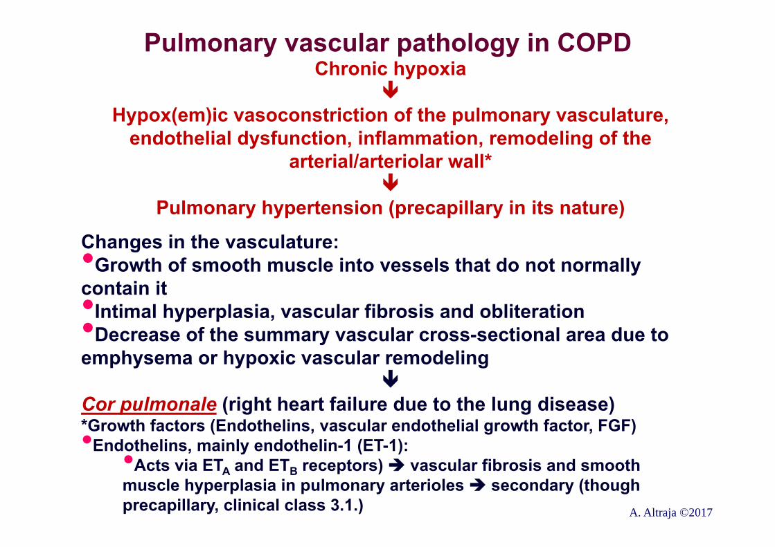
Pulmonary vascular pathology in COPDChronic hypoxia
Hypox(em)ic vasoconstriction of the pulmonary vasculature,
endothelial dysfunction, inflammation, remodeling of the arterial/arteriolar wall*
Pulmonary hypertension (precapillary in its nature)
Changes in the vasculature:•Growth of smooth muscle into vessels that do not normally contain it•Intimal hyperplasia, vascular fibrosis and obliteration•Decrease of the summary vascular cross-sectional area due to emphysema or hypoxic vascular remodeling
Cor pulmonale (right heart failure due to the lung disease) *Growth factors (Endothelins, vascular endothelial growth factor, FGF)•Endothelins, mainly endothelin-1 (ET-1):•Acts via ETA and ETB receptors) vascular fibrosis and smooth
muscle hyperplasia in pulmonary arterioles secondary (though precapillary, clinical class 3.1.) A. Altraja ©2017
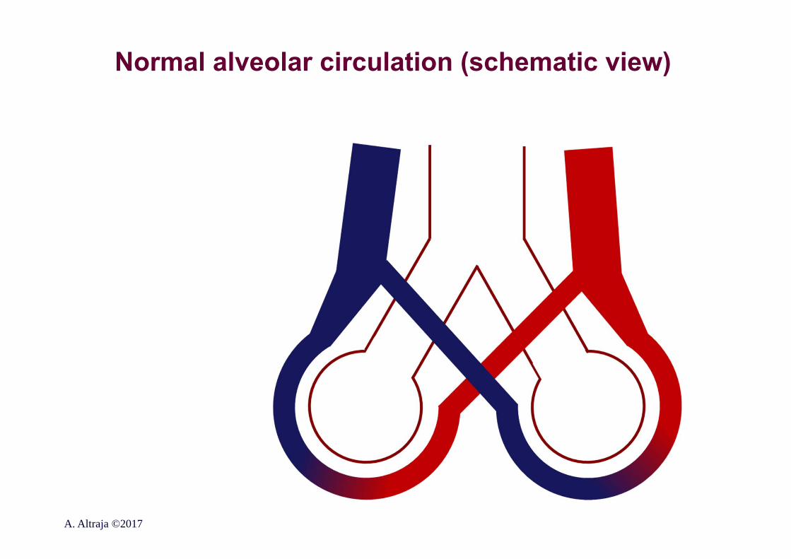
Normal alveolar circulation (schematic view)
A. Altraja ©2017
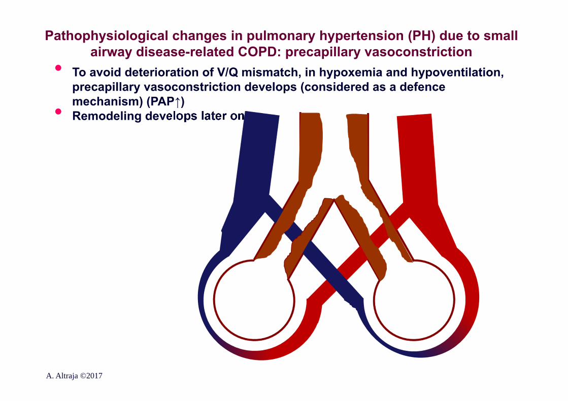
Pathophysiological changes in pulmonary hypertension (PH) due to small airway disease-related COPD: precapillary vasoconstriction
• To avoid deterioration of V/Q mismatch, in hypoxemia and hypoventilation, precapillary vasoconstriction develops (considered as a defence mechanism) (PAP↑)• Remodeling develops later on
A. Altraja ©2017
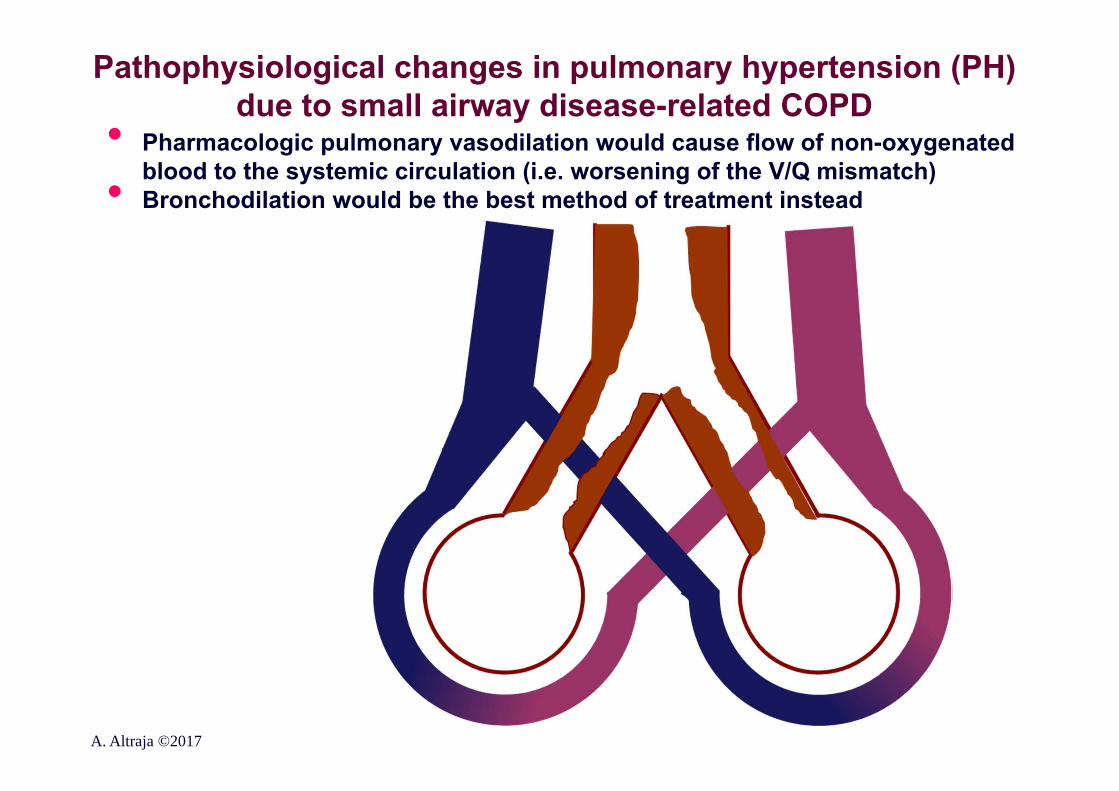
Pathophysiological changes in pulmonary hypertension (PH) due to small airway disease-related COPD
• Pharmacologic pulmonary vasodilation would cause flow of non-oxygenated blood to the systemic circulation (i.e. worsening of the V/Q mismatch)• Bronchodilation would be the best method of treatment instead
A. Altraja ©2017
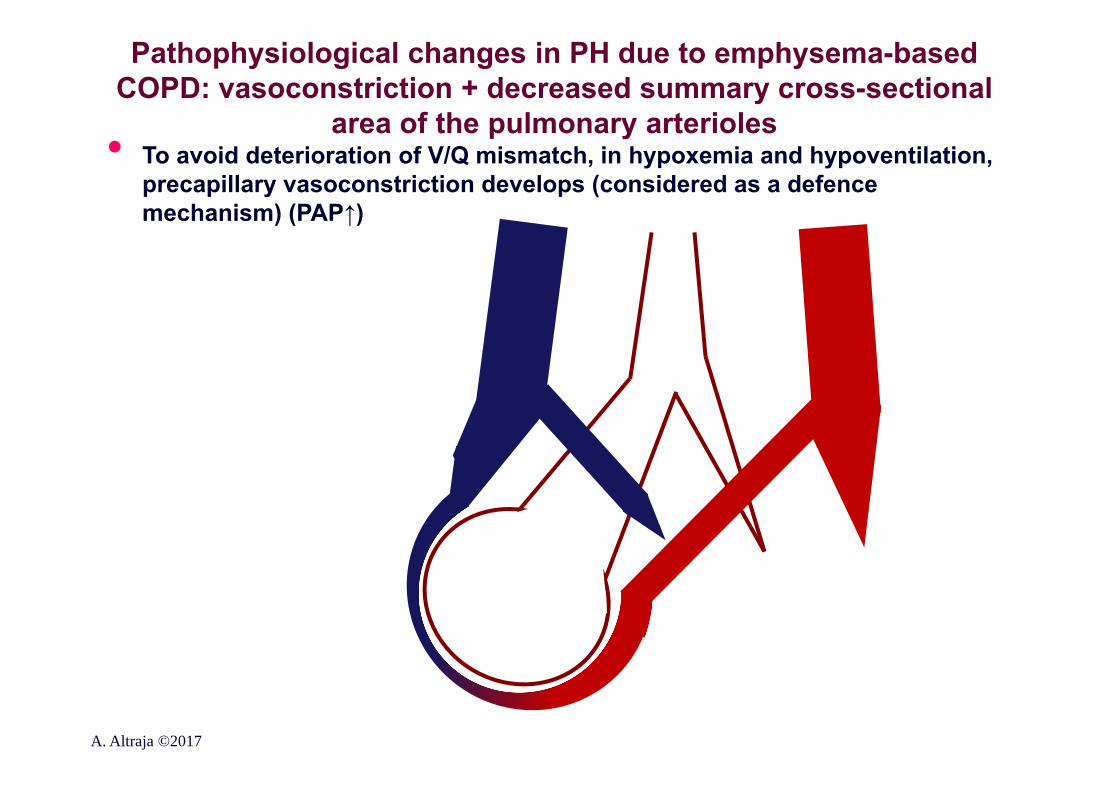
Pathophysiological changes in PH due to emphysema-based COPD: vasoconstriction + decreased summary cross-sectional
area of the pulmonary arterioles• To avoid deterioration of V/Q mismatch, in hypoxemia and hypoventilation, precapillary vasoconstriction develops (considered as a defence mechanism) (PAP↑)
A. Altraja ©2017
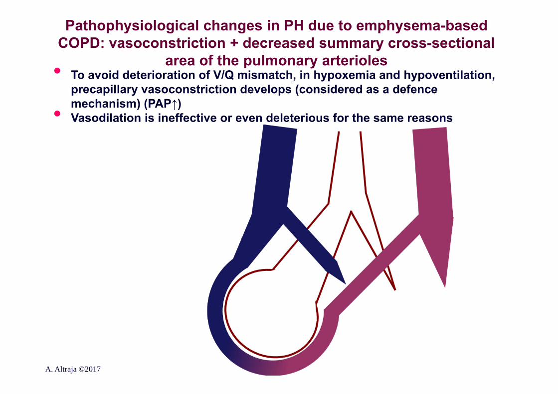
Pathophysiological changes in PH due to emphysema-based COPD: vasoconstriction + decreased summary cross-sectional
area of the pulmonary arterioles• To avoid deterioration of V/Q mismatch, in hypoxemia and hypoventilation, precapillary vasoconstriction develops (considered as a defence mechanism) (PAP↑)• Vasodilation is ineffective or even deleterious for the same reasons
A. Altraja ©2017
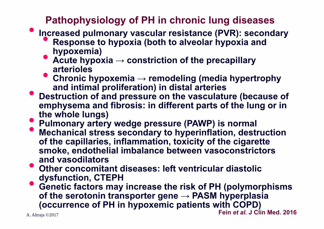
Pathophysiology of PH in chronic lung diseases
A. Altraja ©2017 Fein et al. J Clin Med. 2016
• Increased pulmonary vascular resistance (PVR): secondary• Response to hypoxia (both to alveolar hypoxia and hypoxemia)• Acute hypoxia → constriction of the precapillary arterioles• Chronic hypoxemia → remodeling (media hypertrophy and intimal proliferation) in distal arteries• Destruction of and pressure on the vasculature (because of
emphysema and fibrosis: in different parts of the lung or in the whole lungs)• Pulmonary artery wedge pressure (PAWP) is normal • Mechanical stress secondary to hyperinflation, destruction of the capillaries, inflammation, toxicity of the cigarette smoke, endothelial imbalance between vasoconstrictors and vasodilators• Other concomitant diseases: left ventricular diastolic dysfunction, CTEPH• Genetic factors may increase the risk of PH (polymorphisms of the serotonin transporter gene → PASM hyperplasia (occurrence of PH in hypoxemic patients with COPD)

Pulmonary emphysema: the definitionsAbnormal, permanent enlargement of the airspaces distal to terminal bronchioli (mainly alveoli) with destruction of their walls, but without a clear fibrosis (Snider et al. 1985)•Destruction of the alveolar walls•Macrophages, T-lymphocytes •Neutrophils in the capillaries (low-grade inflammation)The major subtypes of emphysema:•Centriacinar: emphysema (destruction) begins from respiratory
bronchioli and spreads towards the periphery•Centrilobular: a subtype of centriacinar emphysema that results from long-term (heavy) smoking, manifests initially predominantly in the upper parts of the lungs, later in the whole lungs•Distal acinar emphysema: manifests initially in the subpleural areas•Panacinar: emphysema involves almost equally the entire
acinus; predominates initially in the lower parts of the lungs; is characteristic of α1-antitrypsin deficiency; may accompany with centrilobular emphysema, it the patients smoke
A. Altraja ©2017
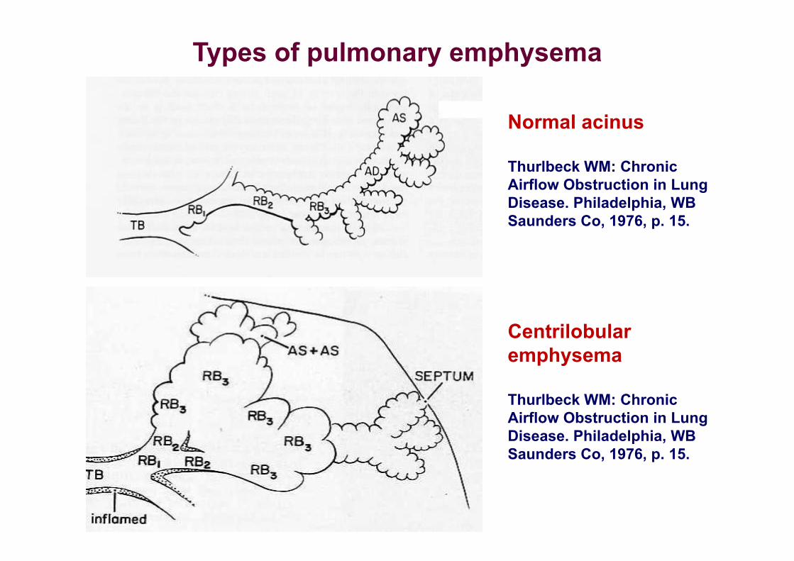
Types of pulmonary emphysema
Normal acinus
Thurlbeck WM: Chronic Airflow Obstruction in Lung Disease. Philadelphia, WB Saunders Co, 1976, p. 15.
Centrilobular emphysema
Thurlbeck WM: Chronic Airflow Obstruction in Lung Disease. Philadelphia, WB Saunders Co, 1976, p. 15.
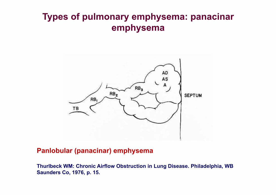
Types of pulmonary emphysema: panacinar emphysema
Panlobular (panacinar) emphysema
Thurlbeck WM: Chronic Airflow Obstruction in Lung Disease. Philadelphia, WB Saunders Co, 1976, p. 15.
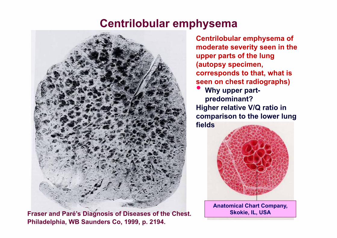
Centrilobular emphysemaCentrilobular emphysema of moderate severity seen in the upper parts of the lung (autopsy specimen, corresponds to that, what is seen on chest radiographs)• Why upper part-
predominant?Higher relative V/Q ratio in comparison to the lower lung fields
Anatomical Chart Company, Skokie, IL, USAFraser and Paré’s Diagnosis of Diseases of the Chest.
Philadelphia, WB Saunders Co, 1999, p. 2194.
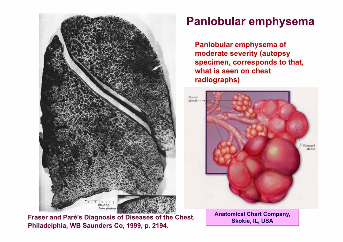
Panlobular emphysema
Panlobular emphysema of moderate severity (autopsy specimen, corresponds to that, what is seen on chest radiographs)
Anatomical Chart Company, Skokie, IL, USAFraser and Paré’s Diagnosis of Diseases of the Chest.
Philadelphia, WB Saunders Co, 1999, p. 2194.
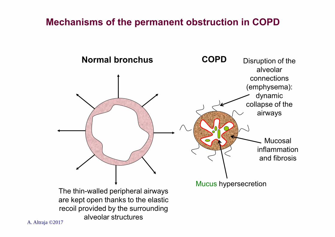
Mechanisms of the permanent obstruction in COPD
Normal bronchus COPD Disruption of the alveolar
connections (emphysema):
dynamic collapse of the
airways
Mucosal inflammation and fibrosis
Mucus hypersecretionThe thin-walled peripheral airways are kept open thanks to the elastic recoil provided by the surrounding
alveolar structuresA. Altraja ©2017
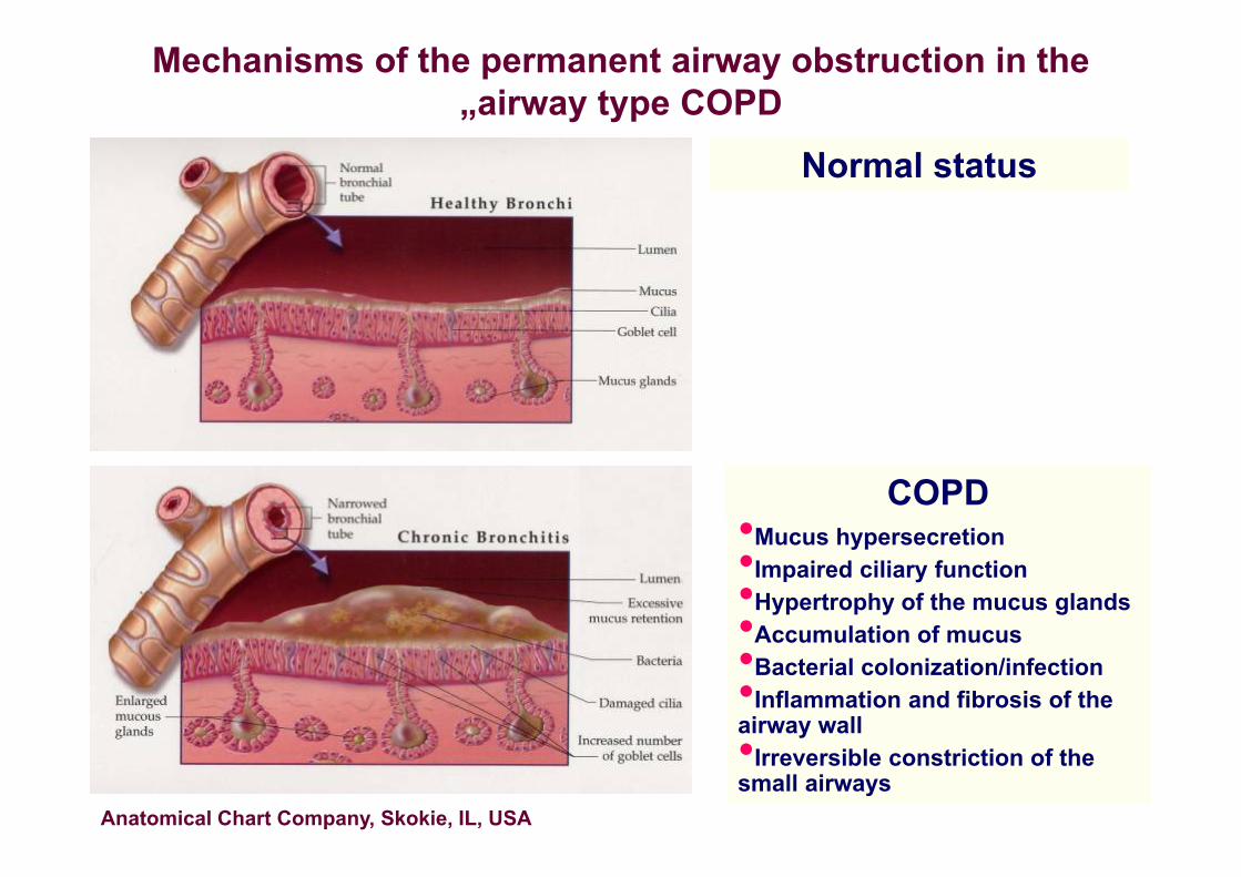
Mechanisms of the permanent airway obstruction in the „airway type COPD
Anatomical Chart Company, Skokie, IL, USA
Normal status
COPD•Mucus hypersecretion•Impaired ciliary function•Hypertrophy of the mucus glands•Accumulation of mucus•Bacterial colonization/infection•Inflammation and fibrosis of the airway wall•Irreversible constriction of the small airways

•With exhaling against positive pressure, the patient can achieve better opening of the small airway lumens
Doing this facilitates both emptying of the peripheral parts of the lungs during expiration and better evacuation of the secretions
The Netter PresenterTM
Respiratory Edition, Version 1.0
Mechanisms of obstruction in patients with emphysema („emphysema-type” COPD)
Mechanisms of permanent airflow obstruction and peripheral air trapping in COPD: a conclusion
•Inflammatory fibrosis of the wall of the small conducting airways that correlates with the decline in FEV1 and FEV1/FVC (Hogg et al., 2004)•This is augmented by the effect of hypersecretion•Pulmonary emphysema (destruction of the peripheral lung tissue) is responsible for impaired diffusion of oxygen (due to the decrease in diffusing capacity of the lung);•Bronchiolar obstruction, especially in emphysema, is complicated by air trapping to the lung periphery and areas of bad ventilation•This air trapping causes abnormal increase in poorly-ventilated lung volumes: hyperinflation of the lungs•Can be heterogeneous or homogeneous (emphysema)•Air trapping interferes with ventilation of the relatively less damaged areas: the basis for lung volume reduction surgery
A. Altraja ©2017 GOLD
Evolution of the pathophysiological changes in COPD
•Prenatal and childhood presumptions and exposures•Exposures during childhood and adolescence•Mucus hypersecretion and impairment of the epithelial ciliary function: is present before the permanent obstruction occurs•Advent of obstruction•Permanent hyperinflation of the lungs•Impairment of the gas exchange•Pulmonary hypertension (COPD-associated PH)•Development of cor pulmonale
A. Altraja ©2017
Pathomorphology of COPDChanges present in all lower airways, from trachea to alveoliIn large bronchi (from trachea to bronchi of >2…4 mm)•Epithelial infiltration with inflammatory cells
(neutrophils)•Hypertrophy of large mucus glands•Increased ration of the goblet-to-ciliary cellsIn small airways ( 2 mm in diameter) •Inflammatory remodeling due to chronic exposure to
irritants (chronic injury and chronic ”repair”)•Increased content of collagens and formation of scar tissue in all layers of the airway wall•Substantial decrease in the airway diameter
Alveolar level: emphysema•Centrilobular (in smokers, inhaled agents)•Panlobular (systemic factors: consequences of the decreased availability of antiproteinases synthesized elsewhere)
A. Altraja ©2017
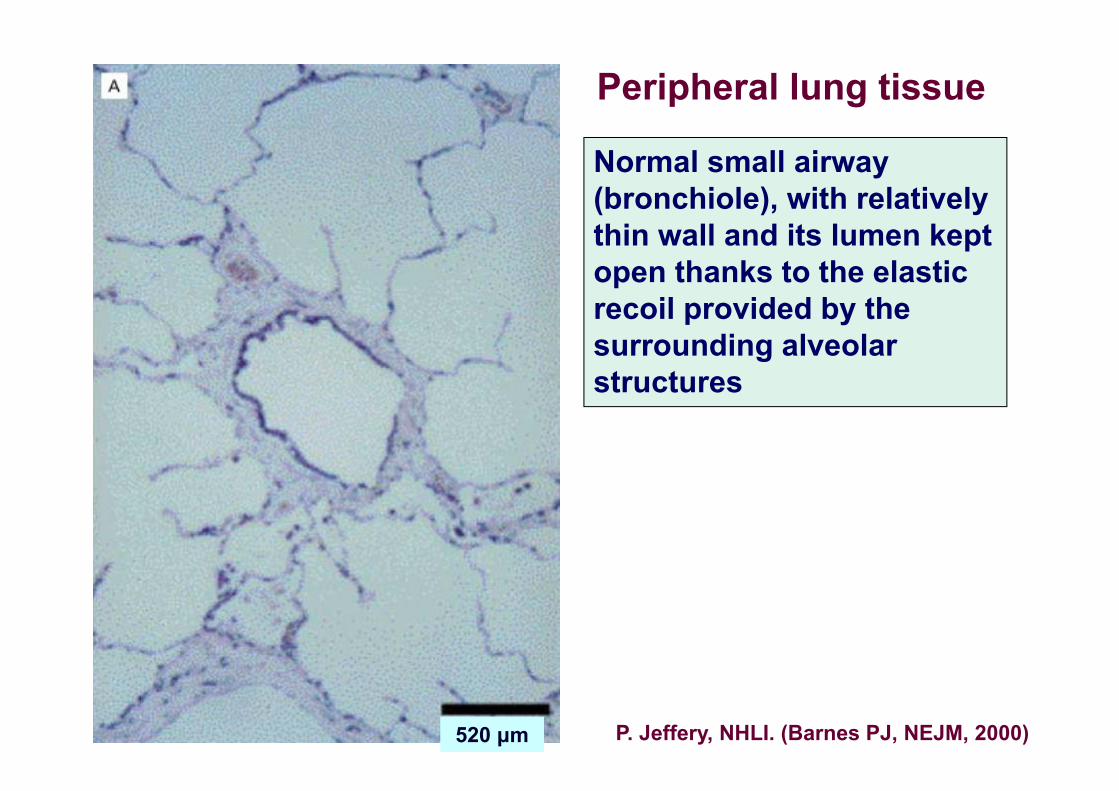
Peripheral lung tissue
Normal small airway (bronchiole), with relatively thin wall and its lumen kept open thanks to the elastic recoil provided by the surrounding alveolar structures
520 μm P. Jeffery, NHLI. (Barnes PJ, NEJM, 2000)
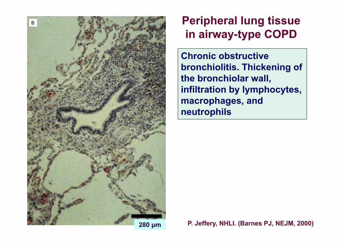
Peripheral lung tissue in airway-type COPD
Chronic obstructive bronchiolitis. Thickening of the bronchiolar wall, infiltration by lymphocytes, macrophages, and neutrophils
280 μm P. Jeffery, NHLI. (Barnes PJ, NEJM, 2000)
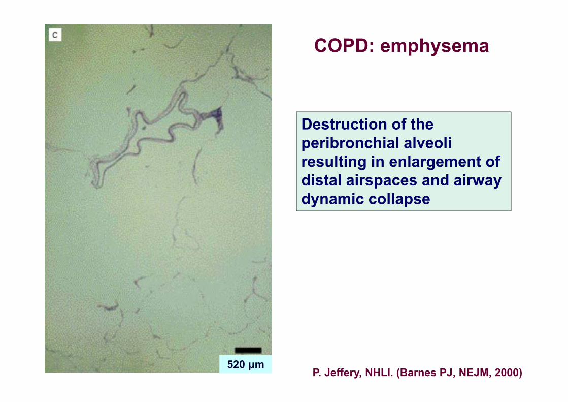
COPD: emphysema
Destruction of the peribronchial alveoli resulting in enlargement of distal airspaces and airway dynamic collapse
520 μmP. Jeffery, NHLI. (Barnes PJ, NEJM, 2000)
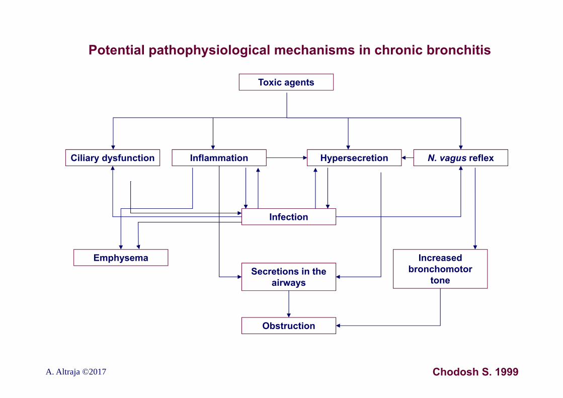
Potential pathophysiological mechanisms in chronic bronchitis
Toxic agents
Obstruction
Inflammation Hypersecretion Ciliary dysfunction
Secretions in the airways
Infection
Emphysema Increased bronchomotor
tone
N. vagus reflex
Chodosh S. 1999A. Altraja ©2017
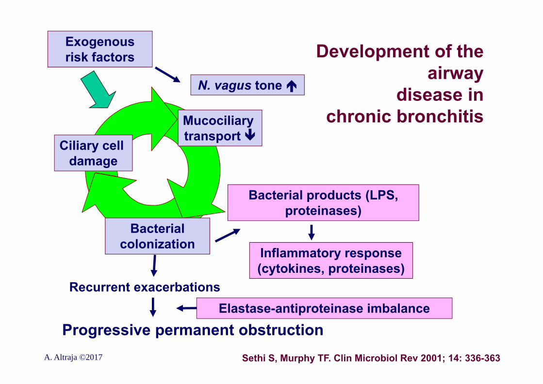
Development of the airway
disease in chronic bronchitis
Sethi S, Murphy TF. Clin Microbiol Rev 2001; 14: 336-363
Recurrent exacerbations
Ciliary cell damage
Mucociliary transport
Bacterial colonization
Progressive permanent obstruction
Exogenous risk factors
Bacterial products (LPS, proteinases)
Inflammatory response (cytokines, proteinases)
Elastase-antiproteinase imbalance
N. vagus tone
A. Altraja ©2017
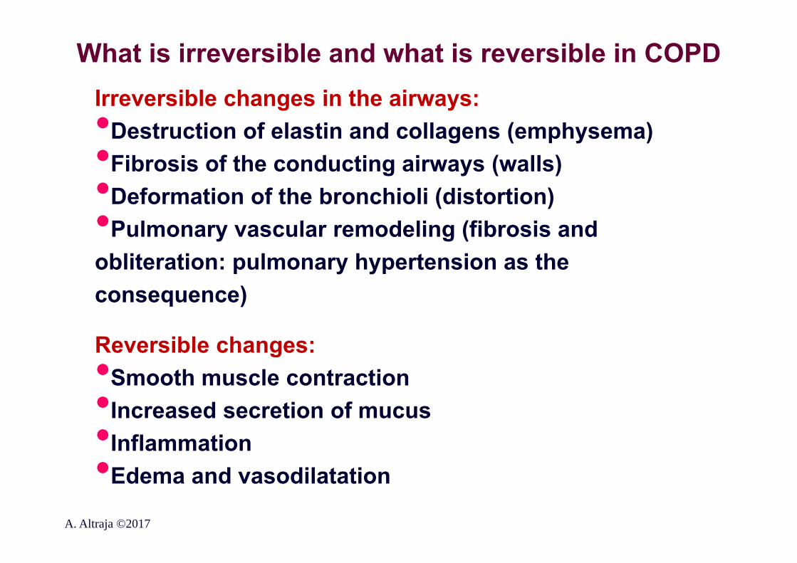
What is irreversible and what is reversible in COPDIrreversible changes in the airways:•Destruction of elastin and collagens (emphysema)•Fibrosis of the conducting airways (walls)•Deformation of the bronchioli (distortion)•Pulmonary vascular remodeling (fibrosis and obliteration: pulmonary hypertension as the consequence)
Reversible changes:•Smooth muscle contraction•Increased secretion of mucus•Inflammation•Edema and vasodilatation
A. Altraja ©2017
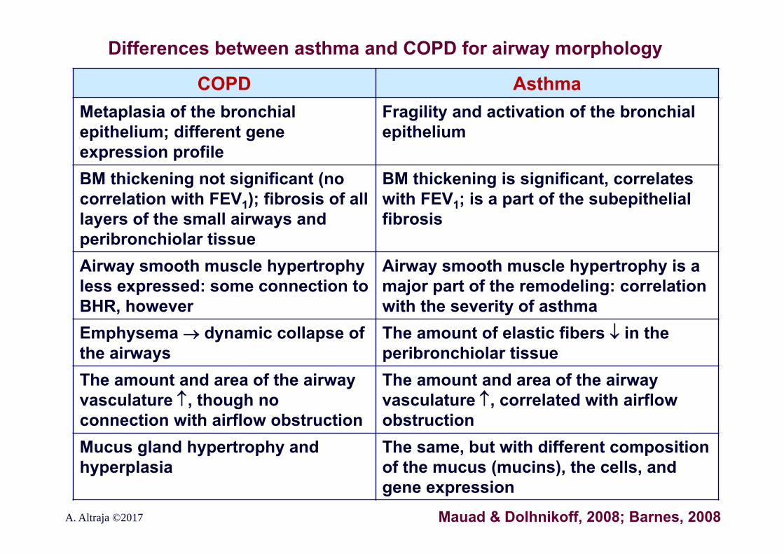
Differences between asthma and COPD for airway morphology
A. Altraja ©2017
COPD AsthmaMetaplasia of the bronchial epithelium; different gene expression profile
Fragility and activation of the bronchial epithelium
BM thickening not significant (no correlation with FEV1); fibrosis of all layers of the small airways and peribronchiolar tissue
BM thickening is significant, correlates with FEV1; is a part of the subepithelial fibrosis
Airway smooth muscle hypertrophy less expressed: some connection to BHR, however
Airway smooth muscle hypertrophy is a major part of the remodeling: correlation with the severity of asthma
Emphysema dynamic collapse of the airways
The amount of elastic fibers in the peribronchiolar tissue
The amount and area of the airway vasculature , though no connection with airflow obstruction
The amount and area of the airway vasculature , correlated with airflow obstruction
Mucus gland hypertrophy and hyperplasia
The same, but with different composition of the mucus (mucins), the cells, and gene expression
Mauad & Dolhnikoff, 2008; Barnes, 2008
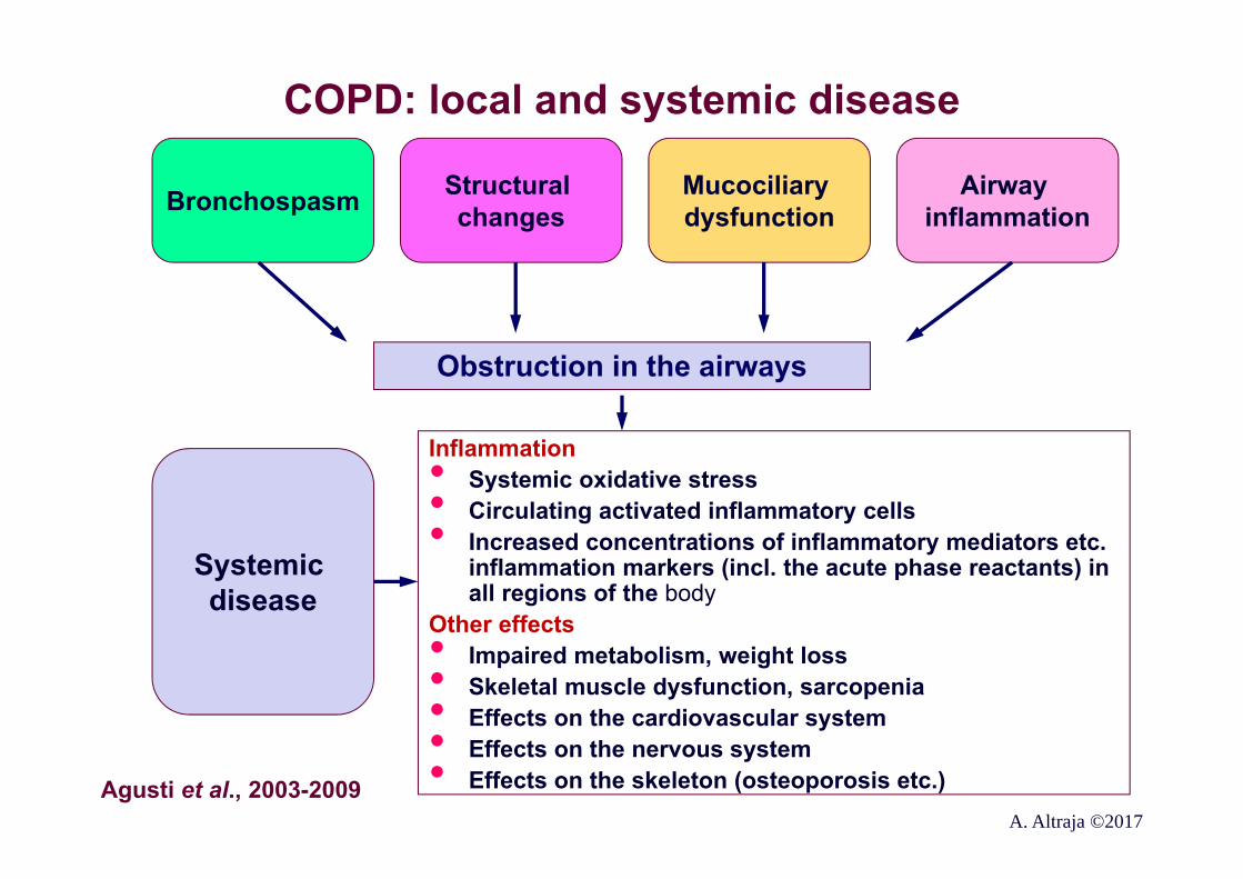
COPD: local and systemic disease
Bronchospasm Structural changes
Mucociliary dysfunction
Airway inflammation
Systemic disease
Obstruction in the airways
Inflammation• Systemic oxidative stress• Circulating activated inflammatory cells• Increased concentrations of inflammatory mediators etc. inflammation markers (incl. the acute phase reactants) in all regions of the body
Other effects• Impaired metabolism, weight loss• Skeletal muscle dysfunction, sarcopenia• Effects on the cardiovascular system• Effects on the nervous system• Effects on the skeleton (osteoporosis etc.)Agusti et al., 2003-2009A. Altraja ©2017

Systemic effects of COPD• Dysfunction of the skeletal muscles with loss of the muscle mass (sarcopenia)• Disturbed metabolism and weight loss (pulmonary cachexia)• Lung cancer (all morphological forms)• Effects on the cardiovascular system•Endothelial dysfunction•Ischemic heart disease•Pulmonary hypertension•Heart failure• Effects on the skeletal system (osteoporosis)• Effects on the endocrine system (diabetes, metabolic syndrome, pituitary, thyroid, gonadal, and pancreatic dysfunction)• Blood changes (normocytic anemia)• Obstructive sleep apnea• Effects on the nervous system (anxiety, depression)• COPD as the senescence of the whole organism
A. Altraja ©2017Decramer et al., 2008; Wouters et al., 2007; 2009; Barnes & Celli, 2009; Laghi
et al., 2009; Man et al., 2009
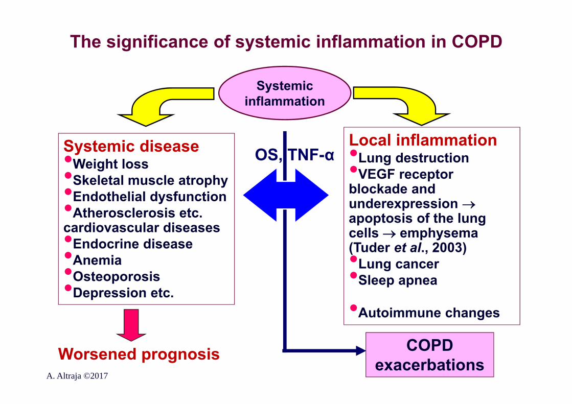
The significance of systemic inflammation in COPD
Systemic disease•Weight loss•Skeletal muscle atrophy•Endothelial dysfunction•Atherosclerosis etc. cardiovascular diseases•Endocrine disease•Anemia•Osteoporosis•Depression etc.
Systemic inflammation
Worsened prognosis
Local inflammation•Lung destruction•VEGF receptor blockade and underexpression apoptosis of the lung cells emphysema (Tuder et al., 2003)•Lung cancer•Sleep apnea
•Autoimmune changes
OS, TNF-α
COPD exacerbations
A. Altraja ©2017
Why does the systemic disease develop in COPD?
The most evidence-based explanation is:
• Inflammation: ”overflow” of mediators and oxidants from airways to the blood circulation and other extra-pulmonary target organs• Inactivity itself: due to the loss of capability to exercise inflammation•COPD and systemic disease are simultaneously
the consequences of the main disease on one hand and mechanisms of further progression of the disease on the other
Decramer et al., 2008A. Altraja ©2017
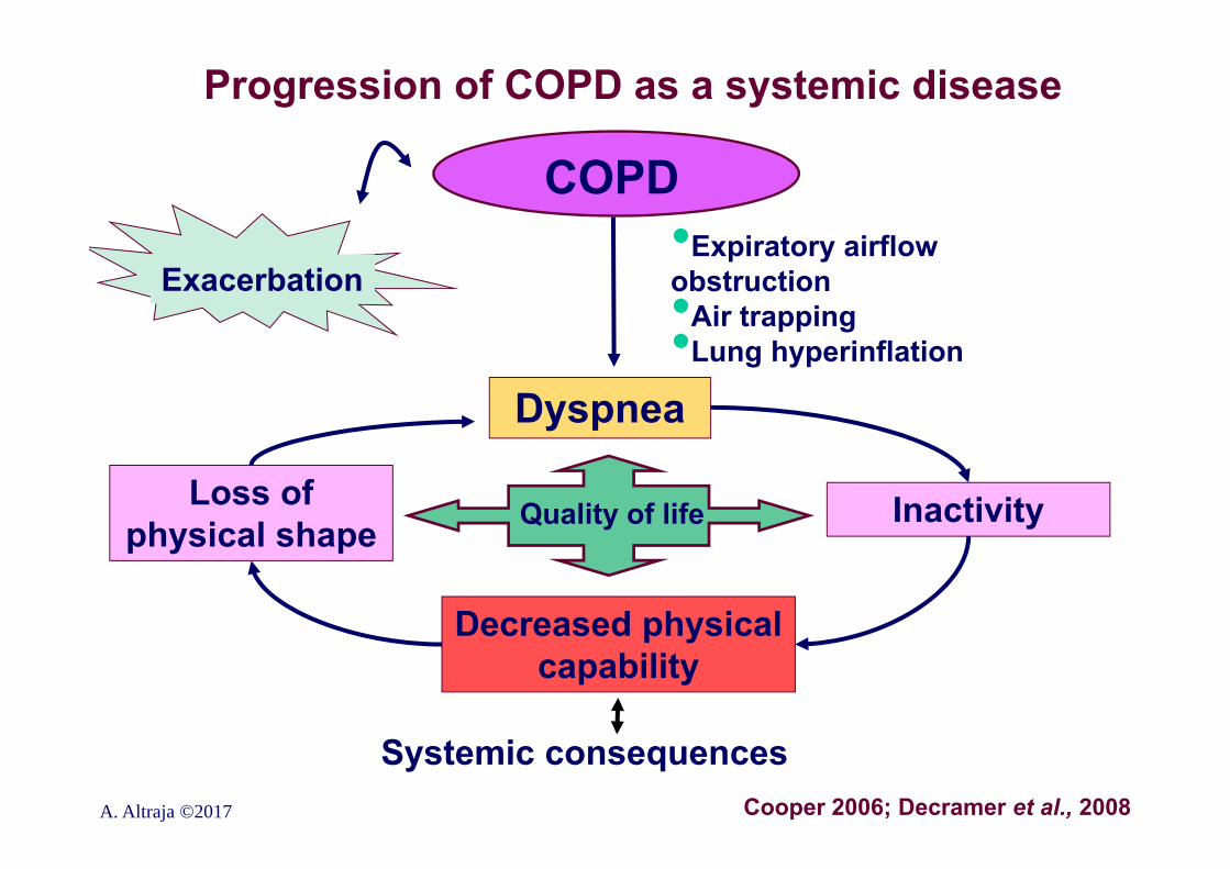
Progression of COPD as a systemic disease
Cooper 2006; Decramer et al., 2008
Dyspnea
Decreased physical capability
•Expiratory airflow obstruction•Air trapping•Lung hyperinflation
COPD
Exacerbation
InactivityLoss of physical shape
Systemic consequences
Quality of life
A. Altraja ©2017
Mechanisms and role of systemic inflammation in COPD• Inflammation in the lungs is the source of systemic
changes: “overflow” to distant targets with circulation• Induction of inflammation and inflammatory burst in distant extra-pulmonary target organs• Inflammation mediated by the adipose tissue• Systemic effects of the risk factors of COPD (smoke)• Certain systemic shifts (genetic background) can facilitate the development of COPD (are still reasons rather than consequences)• Tissue hypoxia
• Systemic inflammation•Varies in intensity from patient to patient•Is significant during exacerbationsWouters et al., 2009; Decramer et al., 2008 Agusti et al., 2006A. Altraja ©2017
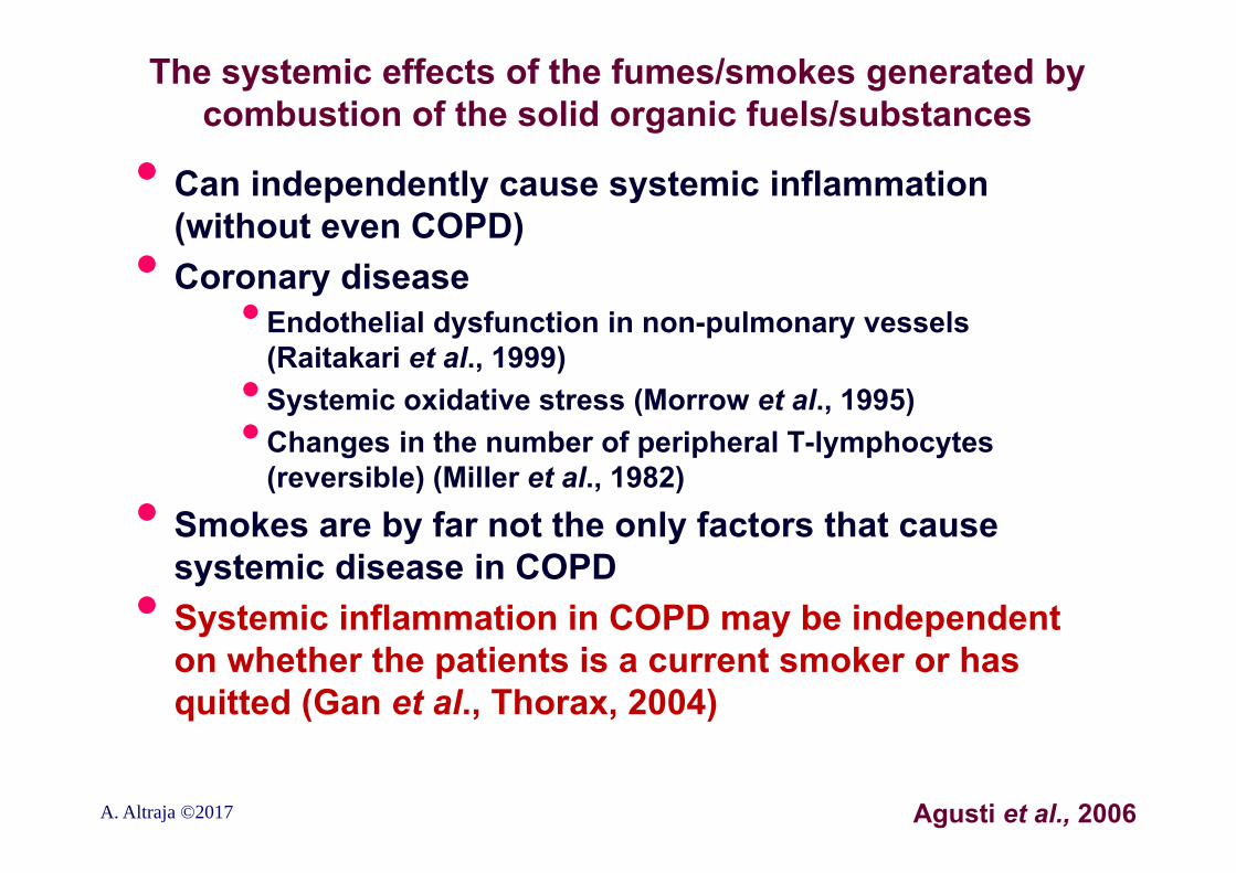
The systemic effects of the fumes/smokes generated by combustion of the solid organic fuels/substances
• Can independently cause systemic inflammation (without even COPD)
• Coronary disease•Endothelial dysfunction in non-pulmonary vessels
(Raitakari et al., 1999)•Systemic oxidative stress (Morrow et al., 1995)•Changes in the number of peripheral T-lymphocytes
(reversible) (Miller et al., 1982)• Smokes are by far not the only factors that cause
systemic disease in COPD• Systemic inflammation in COPD may be independent
on whether the patients is a current smoker or has quitted (Gan et al., Thorax, 2004)
Agusti et al., 2006A. Altraja ©2017
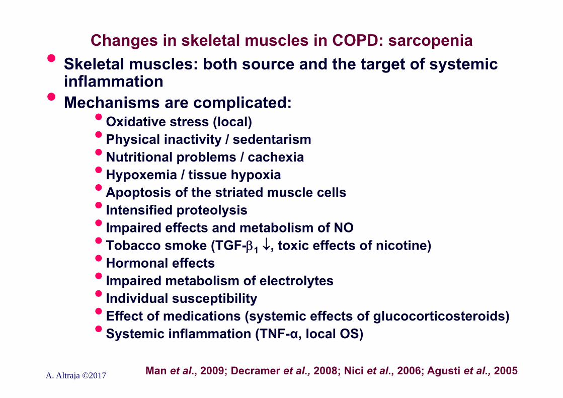
Changes in skeletal muscles in COPD: sarcopenia• Skeletal muscles: both source and the target of systemic
inflammation • Mechanisms are complicated:•Oxidative stress (local)•Physical inactivity / sedentarism•Nutritional problems / cachexia•Hypoxemia / tissue hypoxia•Apoptosis of the striated muscle cells• Intensified proteolysis• Impaired effects and metabolism of NO • Tobacco smoke (TGF-1 , toxic effects of nicotine)•Hormonal effects• Impaired metabolism of electrolytes• Individual susceptibility•Effect of medications (systemic effects of glucocorticosteroids)•Systemic inflammation (TNF-α, local OS)
Man et al., 2009; Decramer et al., 2008; Nici et al., 2006; Agusti et al., 2005A. Altraja ©2017
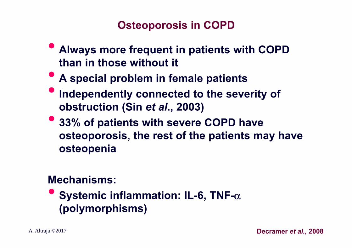
Osteoporosis in COPD
• Always more frequent in patients with COPD than in those without it
• A special problem in female patients• Independently connected to the severity of
obstruction (Sin et al., 2003) • 33% of patients with severe COPD have
osteoporosis, the rest of the patients may have osteopenia
Mechanisms: • Systemic inflammation: IL-6, TNF-
(polymorphisms)
Decramer et al., 2008A. Altraja ©2017
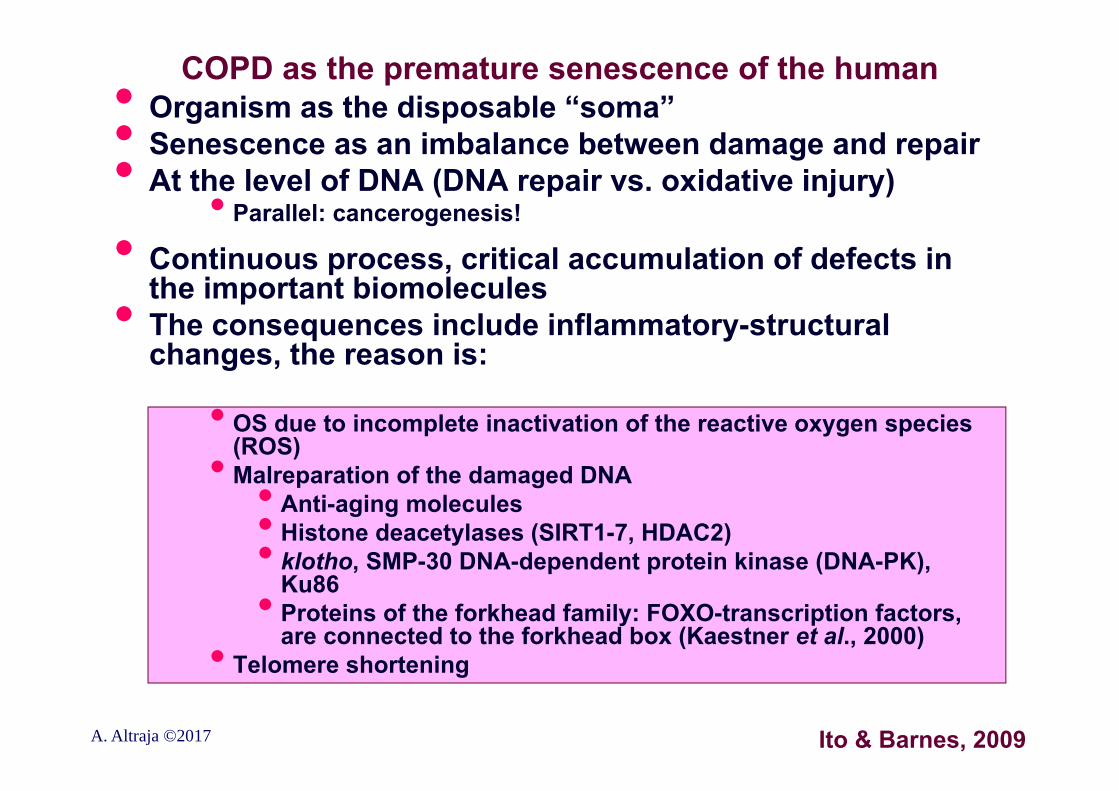
COPD as the premature senescence of the human• Organism as the disposable “soma”• Senescence as an imbalance between damage and repair• At the level of DNA (DNA repair vs. oxidative injury)• Parallel: cancerogenesis!
• Continuous process, critical accumulation of defects in the important biomolecules• The consequences include inflammatory-structural changes, the reason is:
• OS due to incomplete inactivation of the reactive oxygen species (ROS)• Malreparation of the damaged DNA• Anti-aging molecules• Histone deacetylases (SIRT1-7, HDAC2)• klotho, SMP-30 DNA-dependent protein kinase (DNA-PK),
Ku86• Proteins of the forkhead family: FOXO-transcription factors, are connected to the forkhead box (Kaestner et al., 2000)• Telomere shortening
Ito & Barnes, 2009A. Altraja ©2017
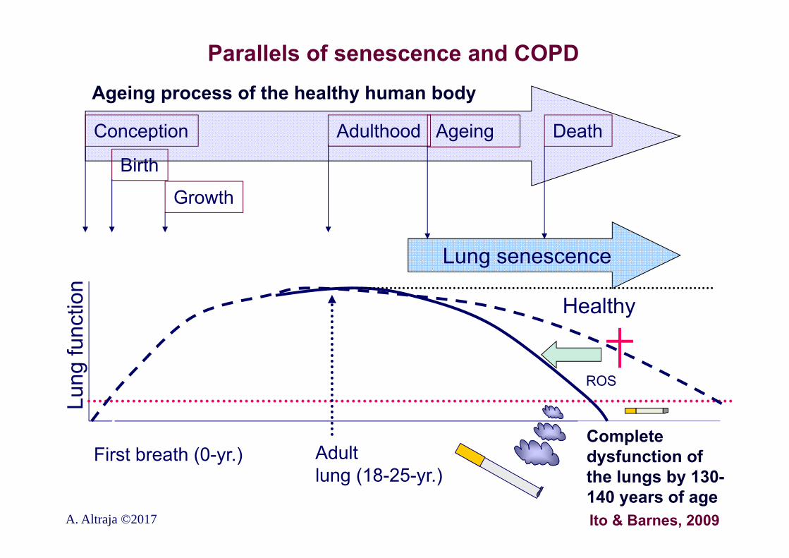
Lung
func
tion
Parallels of senescence and COPD
A. Altraja ©2017
Lung senescence
Conception
Birth
Ageing Death
Growth
Adulthood
First breath (0-yr.) Adult lung (18-25-yr.)
Complete dysfunction of the lungs by 130-140 years of age
Ageing process of the healthy human body
COPD
Healthy
ROS
Ito & Barnes, 2009
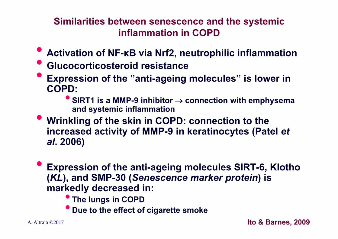
Similarities between senescence and the systemic inflammation in COPD
• Activation of NF-κB via Nrf2, neutrophilic inflammation• Glucocorticosteroid resistance• Expression of the ”anti-ageing molecules” is lower in COPD:
•SIRT1 is a MMP-9 inhibitor connection with emphysema and systemic inflammation• Wrinkling of the skin in COPD: connection to the
increased activity of MMP-9 in keratinocytes (Patel et al. 2006)
• Expression of the anti-ageing molecules SIRT-6, Klotho (KL), and SMP-30 (Senescence marker protein) is markedly decreased in:
• The lungs in COPD•Due to the effect of cigarette smokeA. Altraja ©2017 Ito & Barnes, 2009
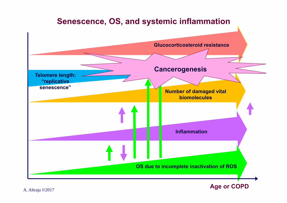
OS due to incomplete inactivation of ROS
Senescence, OS, and systemic inflammation
A. Altraja ©2017 Age or COPD
Inflammation
Number of damaged vital biomolecules
Telomere length: “replicative
senescence”
Glucocorticosteroid resistance
Cancerogenesis
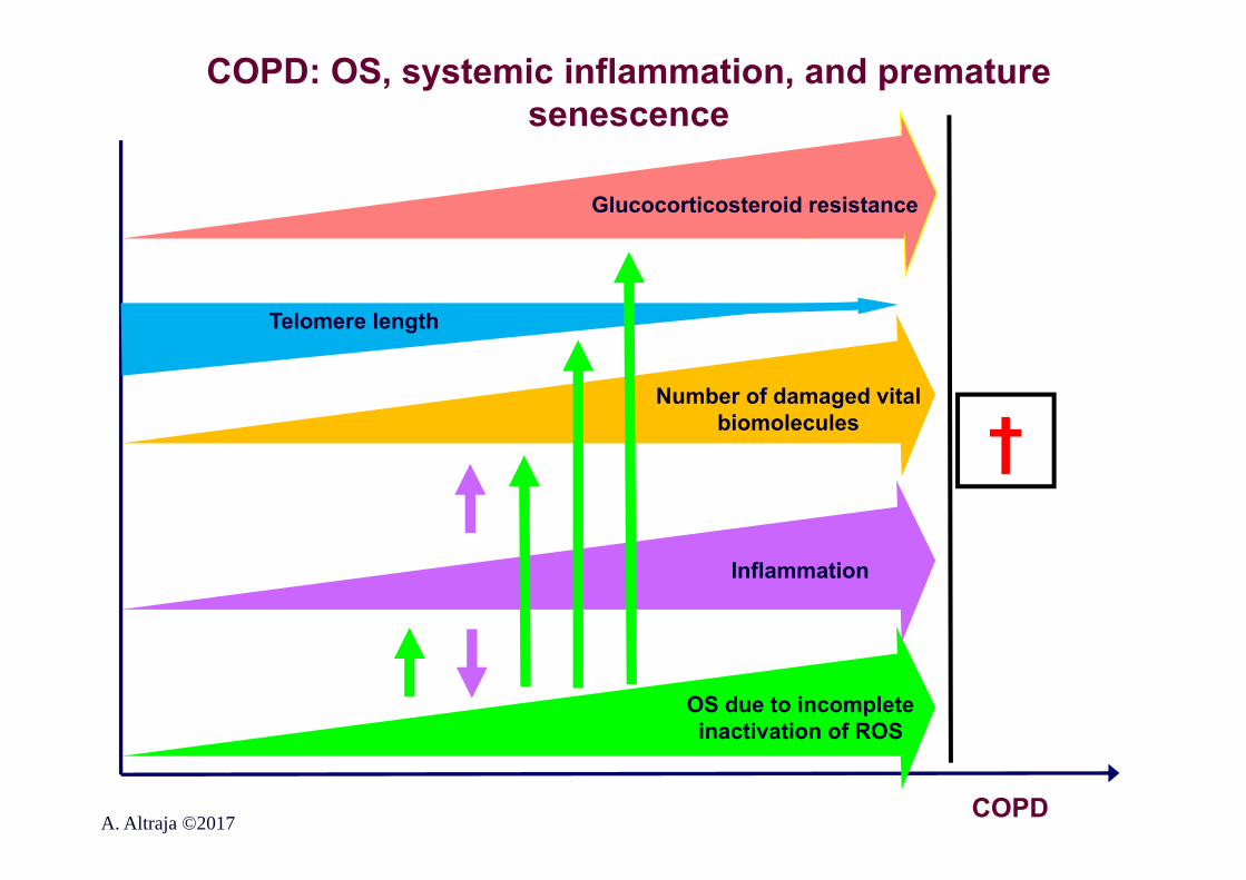
COPD: OS, systemic inflammation, and premature senescence
A. Altraja ©2017 COPD
OS due to incomplete inactivation of ROS
Inflammation
Telomere length
Glucocorticosteroid resistance
Number of damaged vital biomolecules †
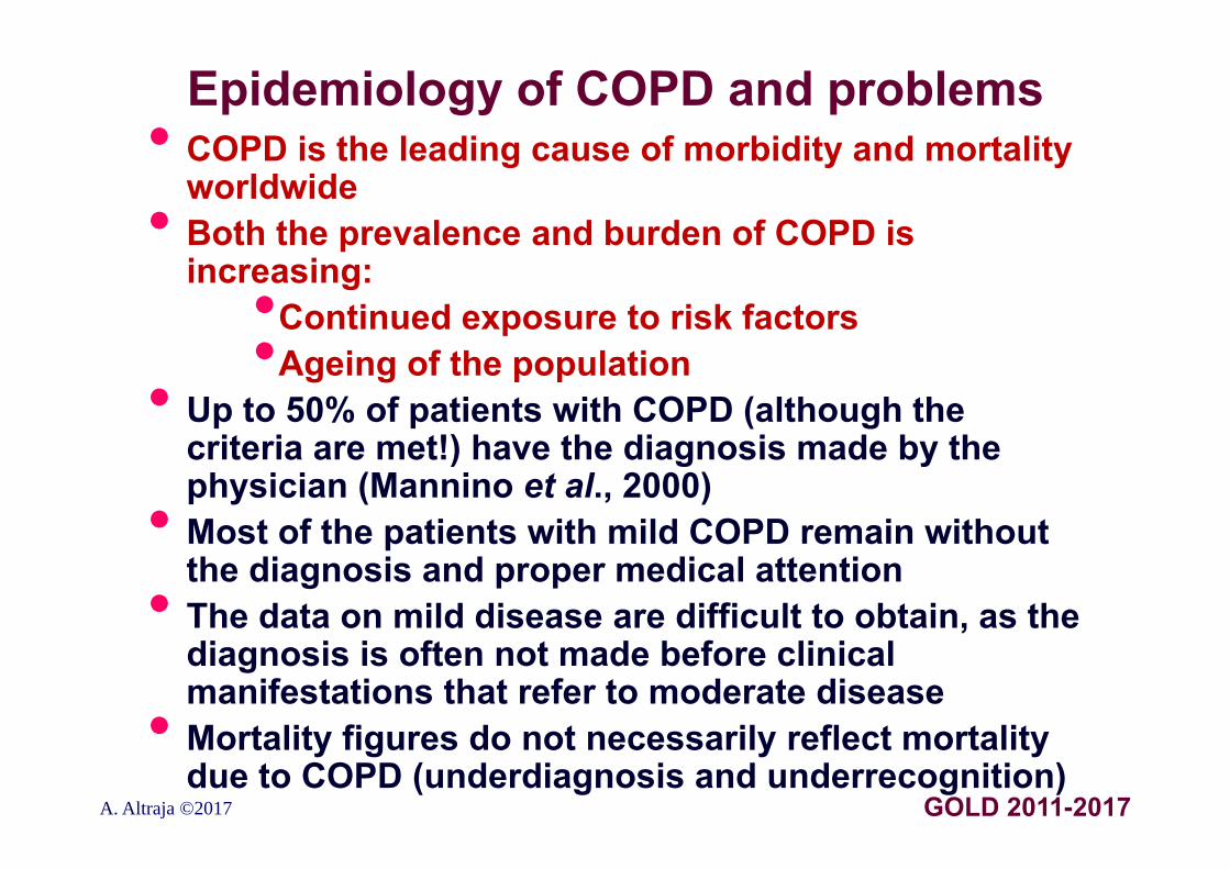
Epidemiology of COPD and problems• COPD is the leading cause of morbidity and mortality
worldwide• Both the prevalence and burden of COPD is increasing: •Continued exposure to risk factors•Ageing of the population• Up to 50% of patients with COPD (although the criteria are met!) have the diagnosis made by the physician (Mannino et al., 2000)• Most of the patients with mild COPD remain without the diagnosis and proper medical attention• The data on mild disease are difficult to obtain, as the diagnosis is often not made before clinical manifestations that refer to moderate disease• Mortality figures do not necessarily reflect mortality due to COPD (underdiagnosis and underrecognition)
GOLD 2011-2017A. Altraja ©2017
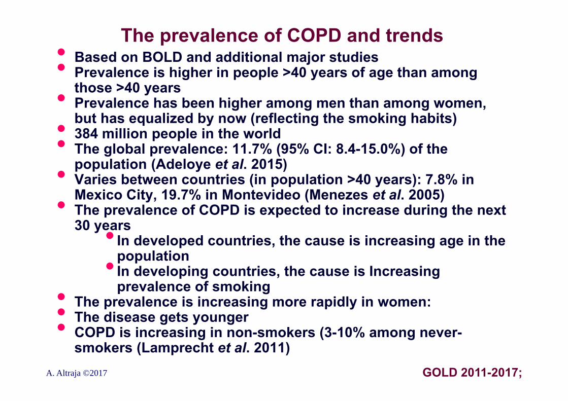
The prevalence of COPD and trends• Based on BOLD and additional major studies• Prevalence is higher in people >40 years of age than among
those >40 years• Prevalence has been higher among men than among women, but has equalized by now (reflecting the smoking habits)• 384 million people in the world• The global prevalence: 11.7% (95% CI: 8.4-15.0%) of the population (Adeloye et al. 2015)• Varies between countries (in population >40 years): 7.8% in Mexico City, 19.7% in Montevideo (Menezes et al. 2005)• The prevalence of COPD is expected to increase during the next 30 years• In developed countries, the cause is increasing age in the
population• In developing countries, the cause is Increasing prevalence of smoking• The prevalence is increasing more rapidly in women:• The disease gets younger• COPD is increasing in non-smokers (3-10% among never-
smokers (Lamprecht et al. 2011) A. Altraja ©2017 GOLD 2011-2017;
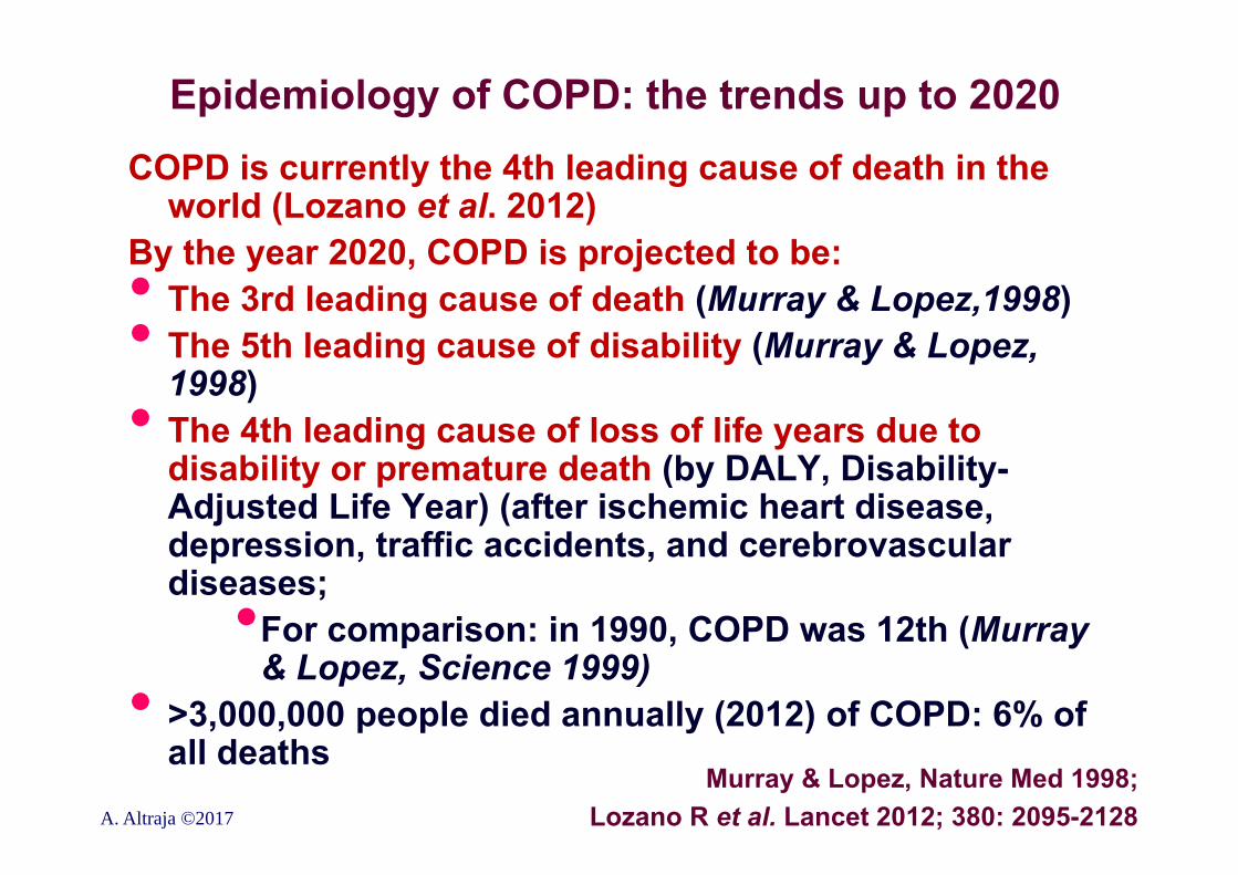
Epidemiology of COPD: the trends up to 2020COPD is currently the 4th leading cause of death in the
world (Lozano et al. 2012)By the year 2020, COPD is projected to be:• The 3rd leading cause of death (Murray & Lopez,1998)• The 5th leading cause of disability (Murray & Lopez,
1998)• The 4th leading cause of loss of life years due to disability or premature death (by DALY, Disability-Adjusted Life Year) (after ischemic heart disease, depression, traffic accidents, and cerebrovascular diseases; •For comparison: in 1990, COPD was 12th (Murray
& Lopez, Science 1999)• >3,000,000 people died annually (2012) of COPD: 6% of all deaths
A. Altraja ©2017
Murray & Lopez, Nature Med 1998;Lozano R et al. Lancet 2012; 380: 2095-2128
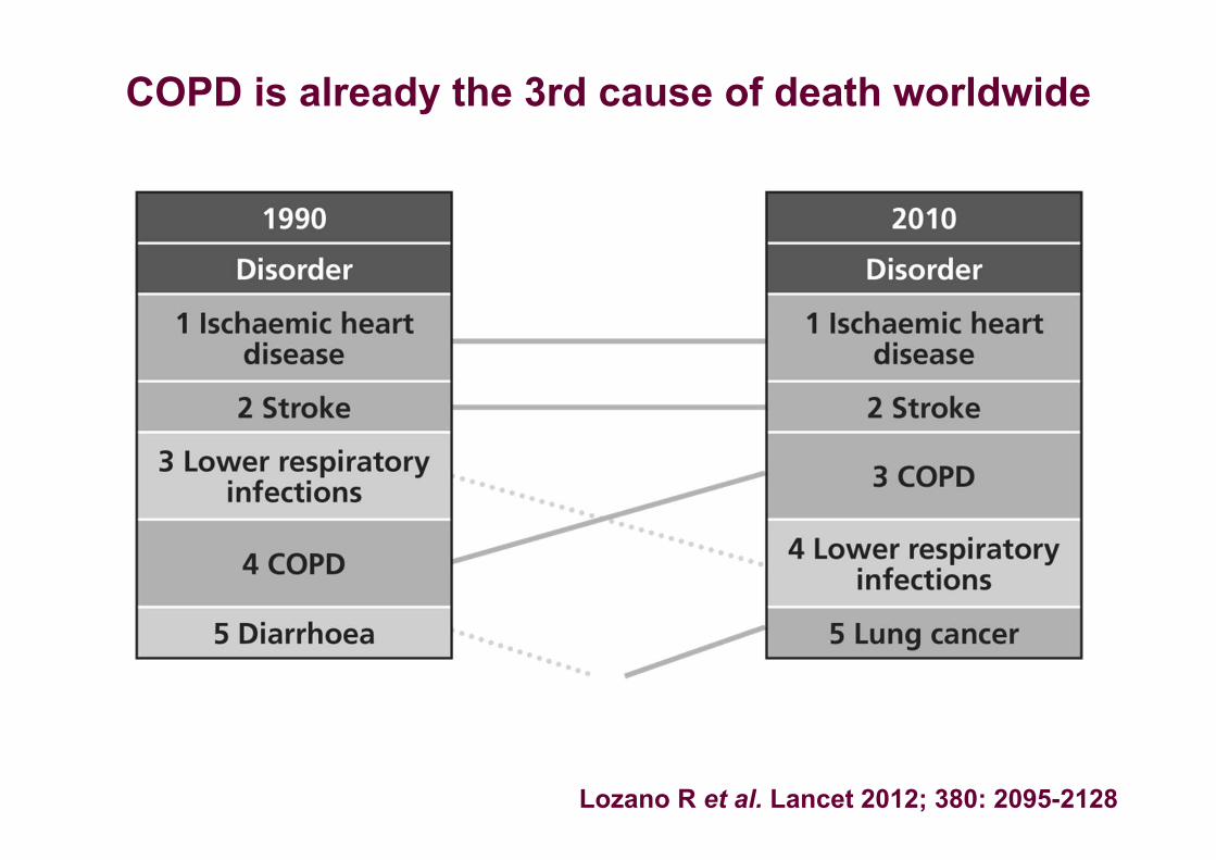
COPD is already the 3rd cause of death worldwide
Lozano R et al. Lancet 2012; 380: 2095-2128
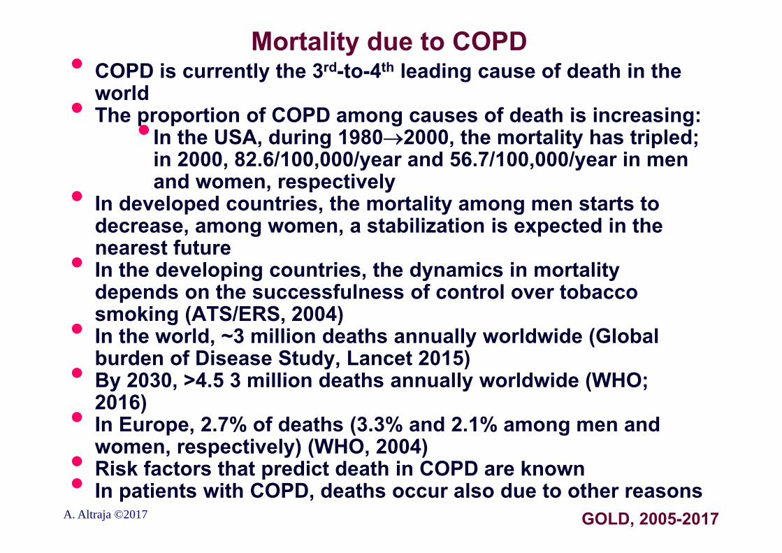
Mortality due to COPD• COPD is currently the 3rd-to-4th leading cause of death in the
world• The proportion of COPD among causes of death is increasing:• In the USA, during 19802000, the mortality has tripled; in 2000, 82.6/100,000/year and 56.7/100,000/year in men and women, respectively• In developed countries, the mortality among men starts to
decrease, among women, a stabilization is expected in the nearest future• In the developing countries, the dynamics in mortality depends on the successfulness of control over tobacco smoking (ATS/ERS, 2004)• In the world, ~3 million deaths annually worldwide (Global burden of Disease Study, Lancet 2015)• By 2030, >4.5 3 million deaths annually worldwide (WHO; 2016)• In Europe, 2.7% of deaths (3.3% and 2.1% among men and women, respectively) (WHO, 2004)• Risk factors that predict death in COPD are known• In patients with COPD, deaths occur also due to other reasons
A. Altraja ©2017 GOLD, 2005-2017
Prevalence of chronic bronchitis
• Higher than that of COPD:• 3-17% in developed countries• 13-27% in less developed countries
A. Altraja ©2017 Gulsvik, 2001; GOLD 2011-2017
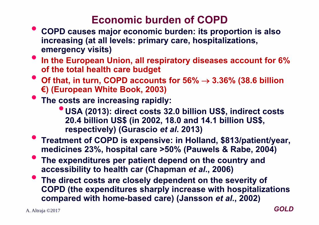
Economic burden of COPD• COPD causes major economic burden: its proportion is also increasing (at all levels: primary care, hospitalizations, emergency visits)• In the European Union, all respiratory diseases account for 6% of the total health care budget• Of that, in turn, COPD accounts for 56% 3.36% (38.6 billion €) (European White Book, 2003)• The costs are increasing rapidly: •USA (2013): direct costs 32.0 billion US$, indirect costs
20.4 billion US$ (in 2002, 18.0 and 14.1 billion US$, respectively) (Gurascio et al. 2013)• Treatment of COPD is expensive: in Holland, $813/patient/year,
medicines 23%, hospital care >50% (Pauwels & Rabe, 2004)• The expenditures per patient depend on the country and accessibility to health car (Chapman et al., 2006)• The direct costs are closely dependent on the severity of COPD (the expenditures sharply increase with hospitalizations compared with home-based care) (Jansson et al., 2002)
A. Altraja ©2017 GOLD
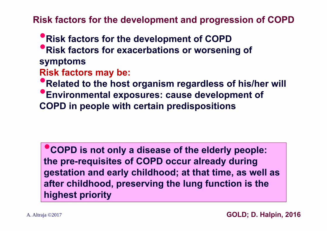
Risk factors for the development and progression of COPD
•Risk factors for the development of COPD•Risk factors for exacerbations or worsening of symptomsRisk factors may be:•Related to the host organism regardless of his/her will•Environmental exposures: cause development of COPD in people with certain predispositions
A. Altraja ©2017
•COPD is not only a disease of the elderly people: the pre-requisites of COPD occur already during gestation and early childhood; at that time, as well as after childhood, preserving the lung function is the highest priority
GOLD; D. Halpin, 2016
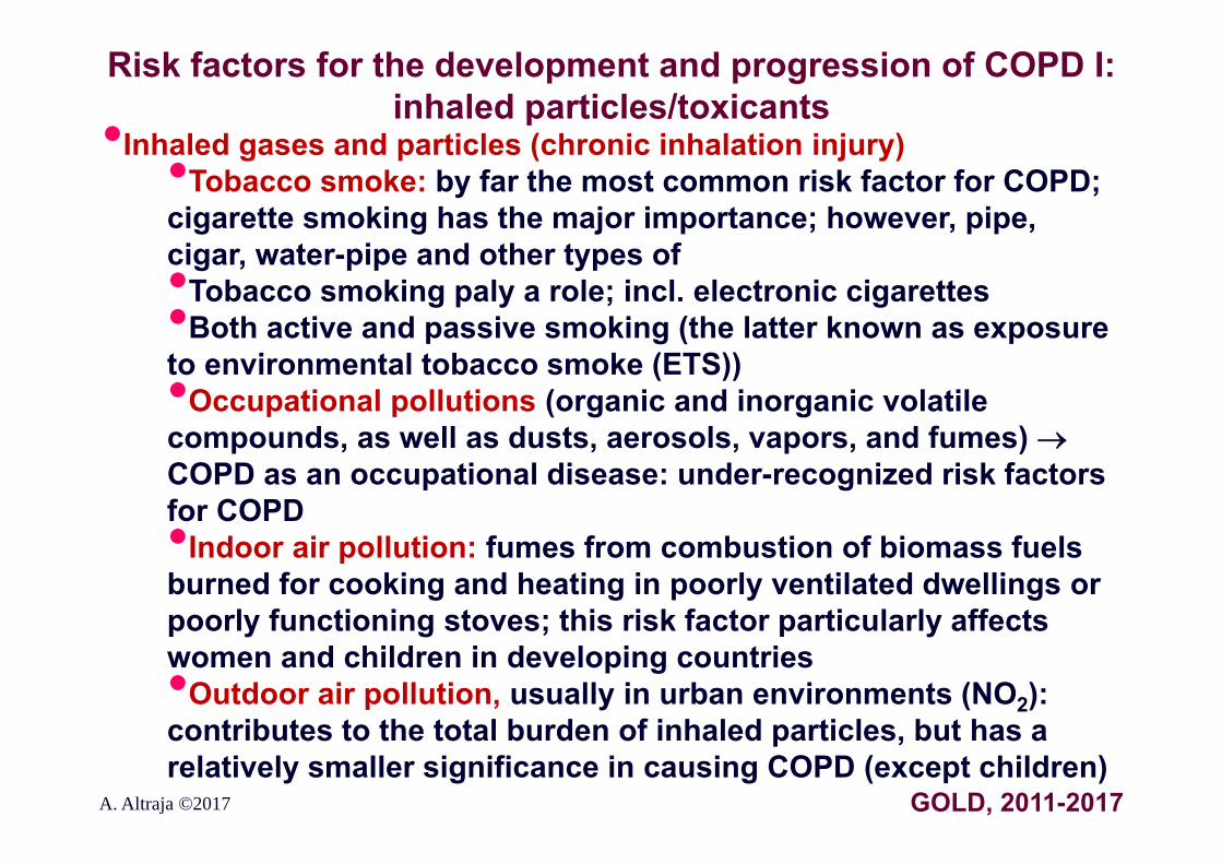
Risk factors for the development and progression of COPD I: inhaled particles/toxicants•Inhaled gases and particles (chronic inhalation injury)•Tobacco smoke: by far the most common risk factor for COPD;
cigarette smoking has the major importance; however, pipe, cigar, water-pipe and other types of•Tobacco smoking paly a role; incl. electronic cigarettes•Both active and passive smoking (the latter known as exposure to environmental tobacco smoke (ETS))•Occupational pollutions (organic and inorganic volatile compounds, as well as dusts, aerosols, vapors, and fumes) COPD as an occupational disease: under-recognized risk factors for COPD•Indoor air pollution: fumes from combustion of biomass fuels burned for cooking and heating in poorly ventilated dwellings or poorly functioning stoves; this risk factor particularly affects women and children in developing countries•Outdoor air pollution, usually in urban environments (NO2): contributes to the total burden of inhaled particles, but has a relatively smaller significance in causing COPD (except children)
A. Altraja ©2017 GOLD, 2011-2017
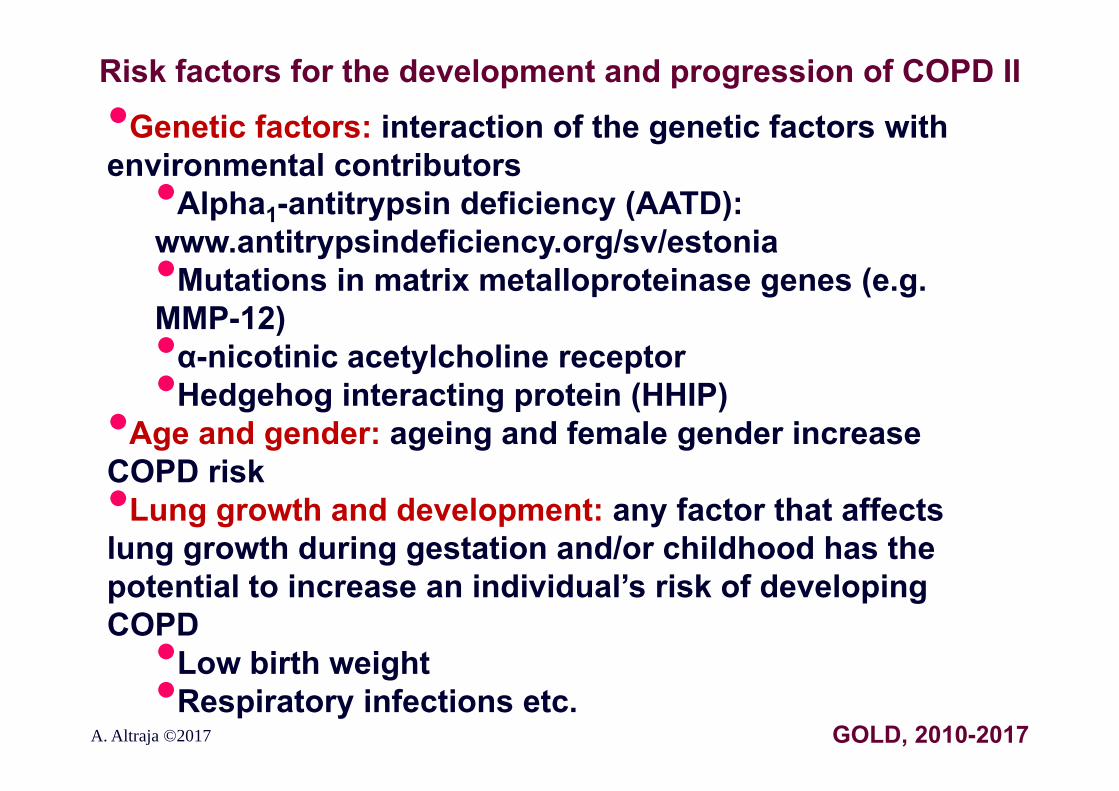
Risk factors for the development and progression of COPD II•Genetic factors: interaction of the genetic factors with environmental contributors•Alpha1-antitrypsin deficiency (AATD):
www.antitrypsindeficiency.org/sv/estonia•Mutations in matrix metalloproteinase genes (e.g. MMP-12)•α-nicotinic acetylcholine receptor•Hedgehog interacting protein (HHIP)•Age and gender: ageing and female gender increase
COPD risk•Lung growth and development: any factor that affects lung growth during gestation and/or childhood has the potential to increase an individual’s risk of developing COPD•Low birth weight•Respiratory infections etc.
A. Altraja ©2017 GOLD, 2010-2017
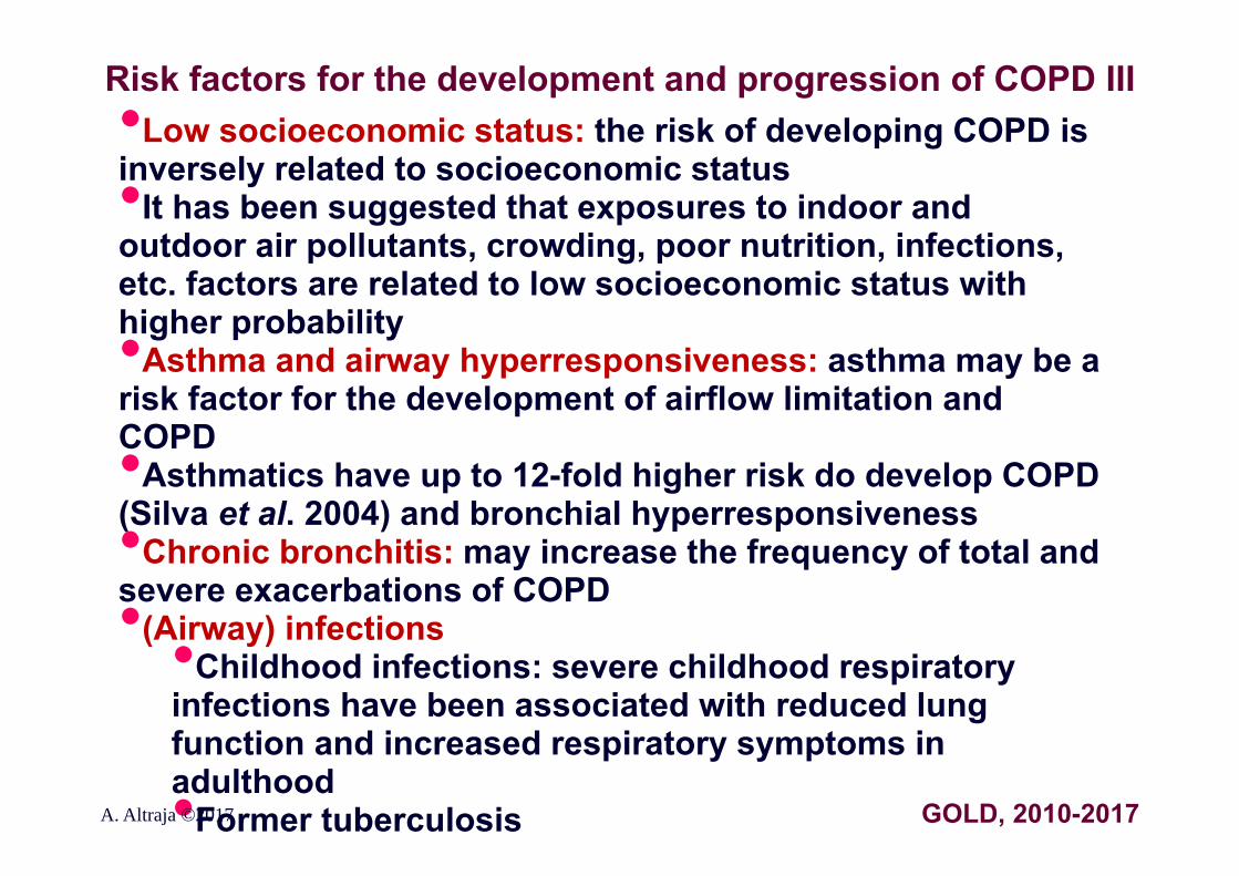
Risk factors for the development and progression of COPD III•Low socioeconomic status: the risk of developing COPD is inversely related to socioeconomic status•It has been suggested that exposures to indoor and outdoor air pollutants, crowding, poor nutrition, infections, etc. factors are related to low socioeconomic status with higher probability•Asthma and airway hyperresponsiveness: asthma may be a risk factor for the development of airflow limitation and COPD•Asthmatics have up to 12-fold higher risk do develop COPD (Silva et al. 2004) and bronchial hyperresponsiveness•Chronic bronchitis: may increase the frequency of total and severe exacerbations of COPD•(Airway) infections•Childhood infections: severe childhood respiratory
infections have been associated with reduced lung function and increased respiratory symptoms in adulthood •Former tuberculosisA. Altraja ©2017 GOLD, 2010-2017
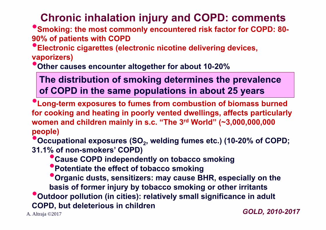
Chronic inhalation injury and COPD: comments•Smoking: the most commonly encountered risk factor for COPD: 80-90% of patients with COPD•Electronic cigarettes (electronic nicotine delivering devices, vaporizers)•Other causes encounter altogether for about 10-20%
•Long-term exposures to fumes from combustion of biomass burned for cooking and heating in poorly vented dwellings, affects particularly women and children mainly in s.c. “The 3rd World” (~3,000,000,000 people)•Occupational exposures (SO2, welding fumes etc.) (10-20% of COPD; 31.1% of non-smokers’ COPD)•Cause COPD independently on tobacco smoking•Potentiate the effect of tobacco smoking•Organic dusts, sensitizers: may cause BHR, especially on the
basis of former injury by tobacco smoking or other irritants•Outdoor pollution (in cities): relatively small significance in adult COPD, but deleterious in children
The distribution of smoking determines the prevalence of COPD in the same populations in about 25 years
GOLD, 2010-2017A. Altraja ©2017
The significance of smoking on the human• COPD develops in up to 50% of smokers (Rennard &
Vestbo, 2005)• Lung cancer develops in 16% of smokers by the age of 75 years or earlier (Peto et al., BMJ 2000)• 50% of all smokers will die prematurely: of smoking-related diseases (Eisen, 2001)
A. Altraja ©2017
Smoking as the main pathogenetic factor of COPD•COPD develops in about 25…50% of smokers, who have smoked at least 10 pack-years (Lokke et al., 2006)•Symptoms of chronic bronchitis in ~50% of smokers•The development of COPD is associated with numerous genetic factors that modify the risk of COPD in smokers•The risk of development of COPD in smokers is dose-dependent (Burrows et al., 1977)•Nicotine toxicity has a role (local and chromosome toxicity) (Kahl et al. 2012)•The mortality in smokers due to COPD depends on:•Cumulative dose (pack-years)•Age at which smoking was started (beginning at
younger age is more dangerous)•Continuation of smoking
A. Altraja ©2017 GOLD, 2010
Smoking as the main pathogenetic factor of COPD•Among smokers, there is: •Higher prevalence of impaired lung function•Quicker annual decline in FEV1•Mortality in the presence of COPD•Smokers of pipe, cigar, water pipe etc. have lower risk of developing COPD and mortality due to than do cigarette smokers•Passive smoking has an important role•Maternal smoking during gestation is a significant risk factor for COPD•Non-smokers with permanent airflow limitation have fewer symptoms, milder disease, and less systemic inflammation and no heightened risk of lung cancer or cardiovascular comorbidities (Thomsen et al. 2013)
A. Altraja ©2017 GOLD, 2010-2017
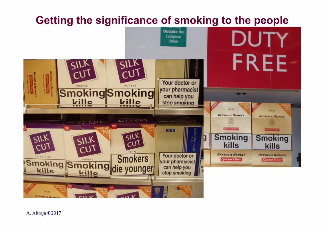
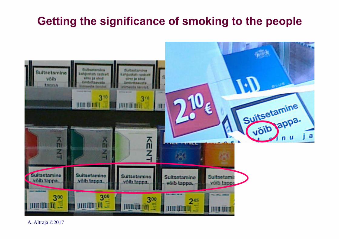
Getting the significance of smoking to the people
A. Altraja ©2017
A. Altraja ©2017
Getting the significance of smoking to the people
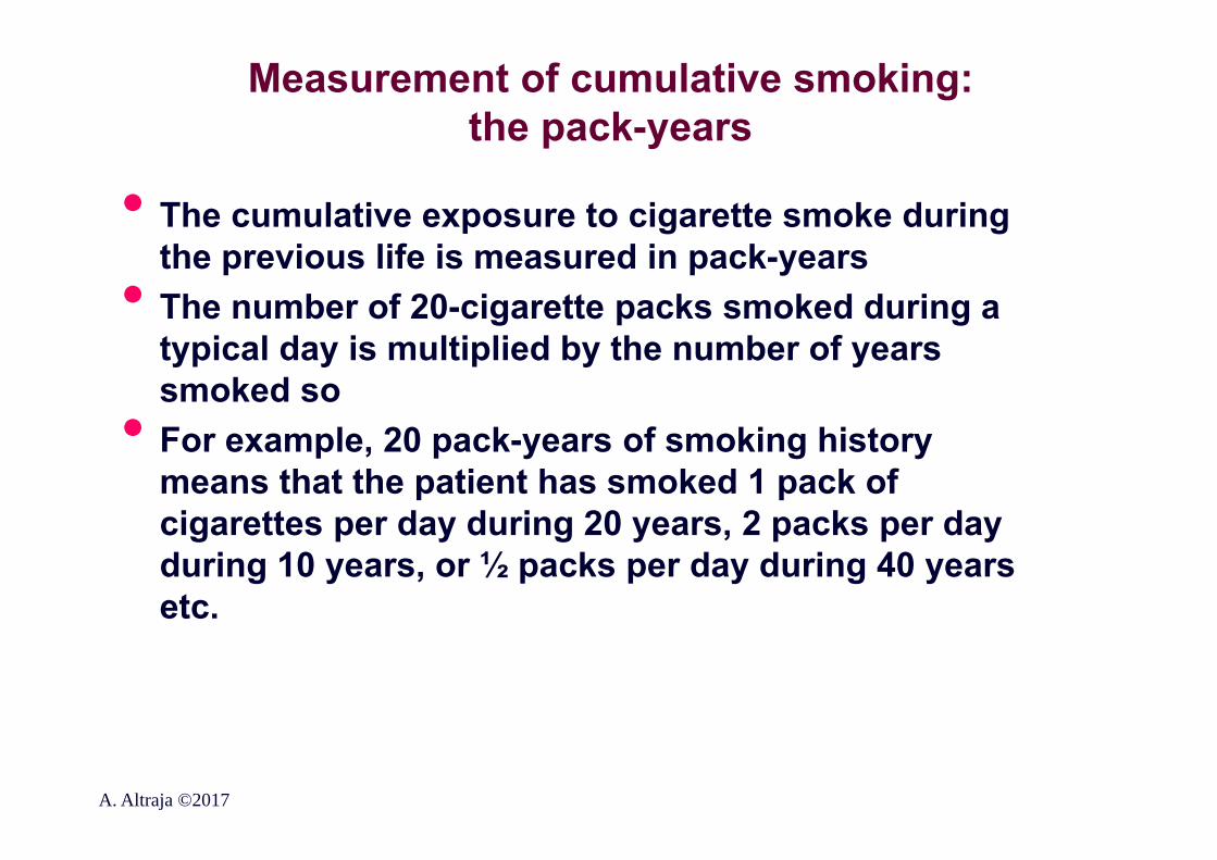
Measurement of cumulative smoking: the pack-years
• The cumulative exposure to cigarette smoke during the previous life is measured in pack-years
• The number of 20-cigarette packs smoked during a typical day is multiplied by the number of years smoked so
• For example, 20 pack-years of smoking history means that the patient has smoked 1 pack of cigarettes per day during 20 years, 2 packs per day during 10 years, or ½ packs per day during 40 years etc.
A. Altraja ©2017
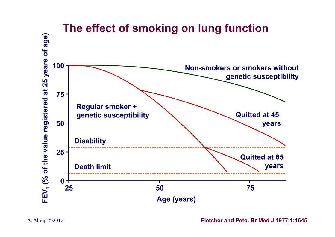
The effect of smoking on lung function
100
75
50
25
025 50 75
Age (years)FEV 1
(% o
f the
val
ue re
gist
ered
at 2
5 ye
ars
of a
ge)
Disability
Death limit
Non-smokers or smokers without genetic susceptibility
Quitted at 45 years
Quitted at 65years
Regular smoker + genetic susceptibility
Fletcher and Peto. Br Med J 1977;1:1645A. Altraja ©2017
100
75
50
25
025 50 75
Age (years)
FEV 1
% p
redi
cted
A. Altraja ©2017
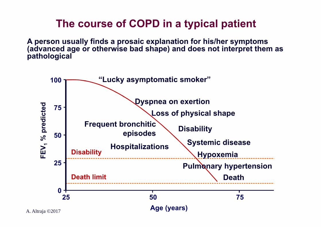
Disability
Death limit
“Lucky asymptomatic smoker”
Dyspnea on exertionLoss of physical shape
Disability
Systemic diseaseHypoxemia
Death
Frequent bronchitic episodes
Hospitalizations
Pulmonary hypertension
A person usually finds a prosaic explanation for his/her symptoms (advanced age or otherwise bad shape) and does not interpret them as pathological
The course of COPD in a typical patient
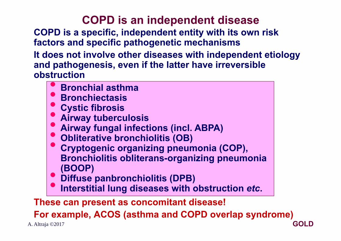
COPD is an independent diseaseCOPD is a specific, independent entity with its own risk factors and specific pathogenetic mechanismsIt does not involve other diseases with independent etiology and pathogenesis, even if the latter have irreversible obstruction
These can present as concomitant disease!For example, ACOS (asthma and COPD overlap syndrome)
• Bronchial asthma• Bronchiectasis• Cystic fibrosis• Airway tuberculosis• Airway fungal infections (incl. ABPA)• Obliterative bronchiolitis (OB)• Cryptogenic organizing pneumonia (COP), Bronchiolitis obliterans-organizing pneumonia (BOOP)• Diffuse panbronchiolitis (DPB)• Interstitial lung diseases with obstruction etc.
A. Altraja ©2017 GOLD
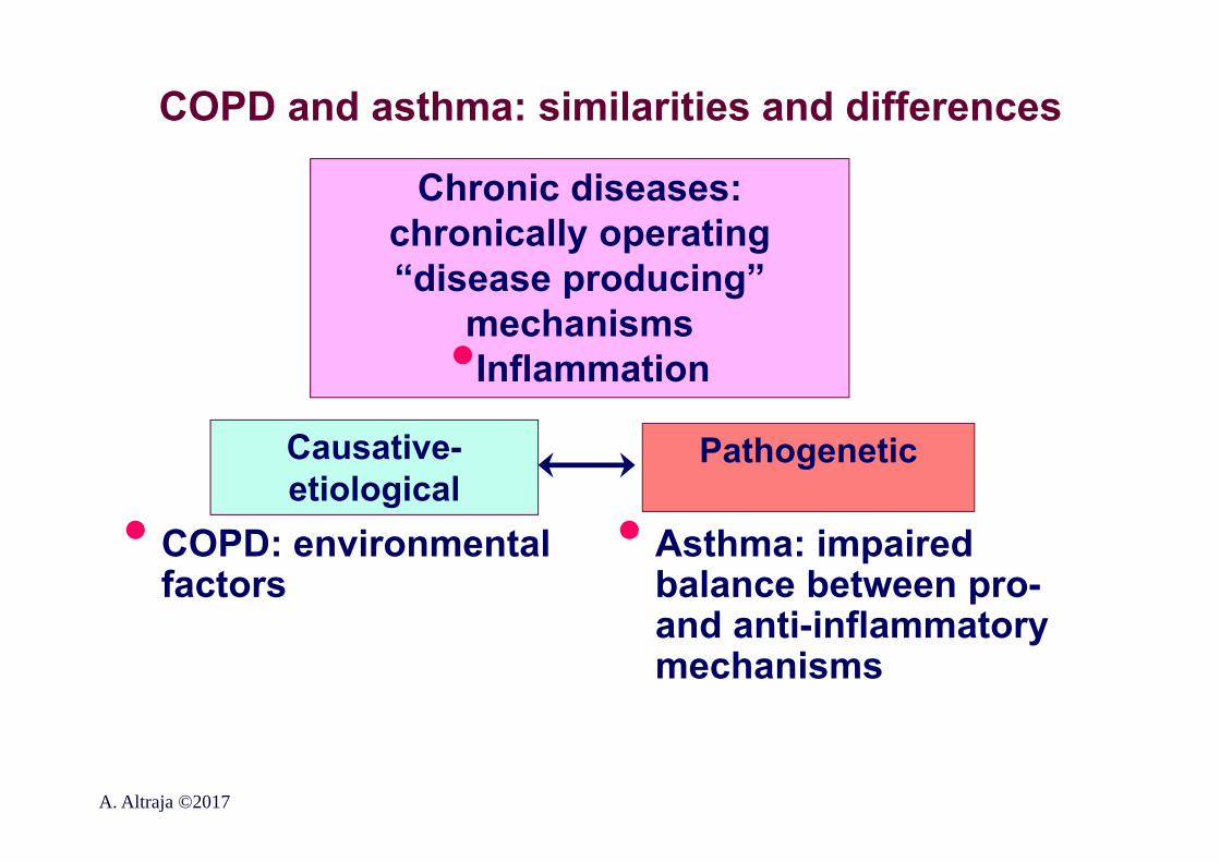
COPD and asthma: similarities and differences
• COPD: environmental factors
PathogeneticCausative-etiological
Chronic diseases: chronically operating “disease producing”
mechanisms•Inflammation
• Asthma: impaired balance between pro-and anti-inflammatory mechanisms
A. Altraja ©2017
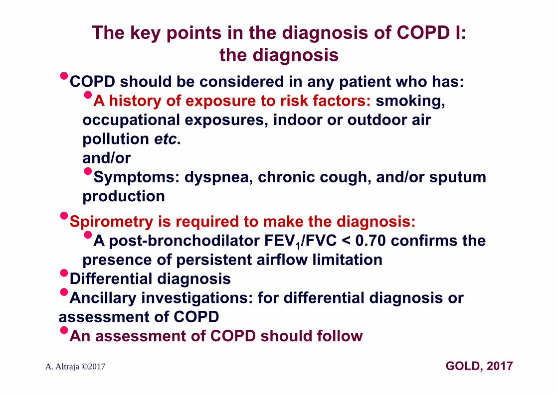
The key points in the diagnosis of COPD I: the diagnosis
•COPD should be considered in any patient who has:•A history of exposure to risk factors: smoking, occupational exposures, indoor or outdoor air pollution etc. and/or•Symptoms: dyspnea, chronic cough, and/or sputum production
•Spirometry is required to make the diagnosis:•A post-bronchodilator FEV1/FVC < 0.70 confirms the presence of persistent airflow limitation•Differential diagnosis•Ancillary investigations: for differential diagnosis or
assessment of COPD•An assessment of COPD should follow
GOLD, 2017A. Altraja ©2017
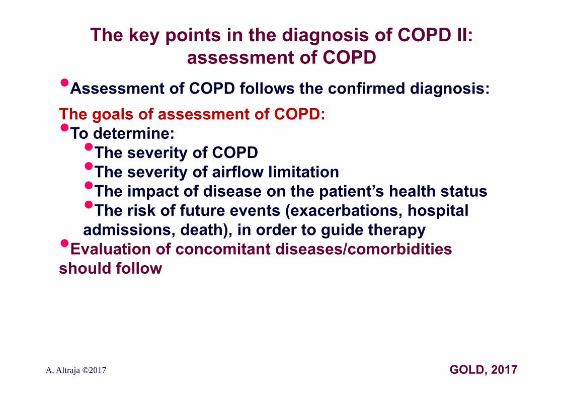
The key points in the diagnosis of COPD II: assessment of COPD
•Assessment of COPD follows the confirmed diagnosis:The goals of assessment of COPD: •To determine:•The severity of COPD•The severity of airflow limitation•The impact of disease on the patient’s health status•The risk of future events (exacerbations, hospital
admissions, death), in order to guide therapy•Evaluation of concomitant diseases/comorbidities should follow
GOLD, 2017A. Altraja ©2017
The key points in the diagnosis of COPD III: concomitant diseases
•Concomitant chronic diseases occur frequently in patients with COPD•Cardiovascular disease•Skeletal muscle dysfunction•Metabolic syndrome or at least diabetes•Osteoporosis•Anxiety and/or depression•Respiratory infections•Lung cancer•These comorbidities, if present, should be actively sought and treated professionally, because they can influence mortality and hospitalizations independently
GOLD, 2017A. Altraja ©2017
Symptoms of COPD by stages•Dyspnea: the main symptom of COPD, though it can be absent in the mildest forms, it progresses in further stages•Chronic cough: often the first symptom, may be productive or dry, may persist diurnally•Sputum production: in all stages, usually progressive, but it may not satisfy the criteria for chronic bronchitis•Wheeze and chest tightness: may be present in all stages, but especially characteristic of advanced COPD and exacerbations of COPDIn more severe forms of COPD in addition: •Weakness, fatigue, loss of appetite, and weight loss (pulmonary cachexia): markers of bad prognosis•Signs of concomitant diseases: malignancies etc.•Syncope during cough paroxysms, rib fractures•Ankle edema: a sign of right heart failure•Anxiety and depression: also markers of bad prognosis
GOLD, 2010-2011A. Altraja ©2017
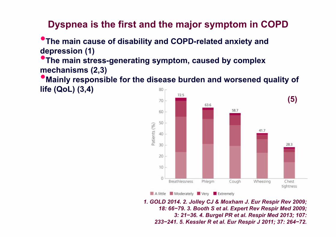
Dyspnea is the first and the major symptom in COPD•The main cause of disability and COPD-related anxiety and depression (1)•The main stress-generating symptom, caused by complex mechanisms (2,3)•Mainly responsible for the disease burden and worsened quality of life (QoL) (3,4)
1. GOLD 2014. 2. Jolley CJ & Moxham J. Eur Respir Rev 2009; 18: 66−79. 3. Booth S et al. Expert Rev Respir Med 2009;
3: 21−36. 4. Burgel PR et al. Respir Med 2013; 107: 233−241. 5. Kessler R et al. Eur Respir J 2011; 37: 264−72.
(5)
The key indicators for considering a diagnosis of COPD•Consider COPD and perform forced flow-volume spirometry to make the diagnosis, if any of these indicators are present in an individual >40 years of age:•History of risk factors:•Host factors (genetic, congenital/developmental risk
factors)•Tobacco smoke (incl. popular local preparations)•Smokes from burning fuels for cooking and heating•Family history and/or childhood factors:•Low birth weight, childhood respiratory Infections etc.•Dyspnea that is: •Progressive over time•Worse with exercise•Persistent in nature•Chronic cough:•May be intermittent and non-productive•Chronic sputum production:•Any pattern may be indicative of COPDGOLD, 2017A. Altraja ©2017

COPD: a simple and convenient way to the diagnosisSuspicion and recognition: within <1 minute!
• Ask, whether the patient smokes / has smoked?• For how long time and how intensively?
• Calculate the pack-years
• If at least 10 pack-years,
Spirometry with reversibility testing to make the diagnosis
A. Altraja ©2017
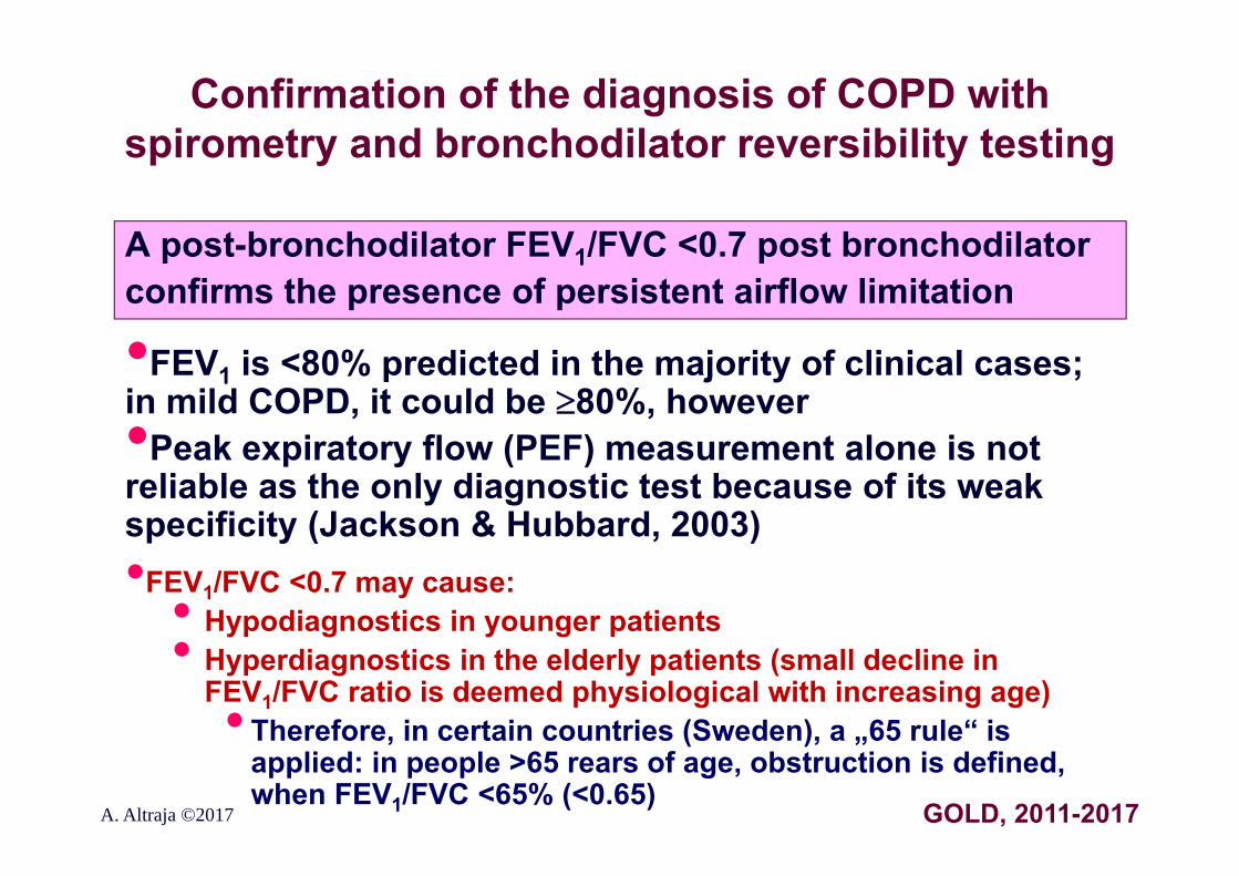
Confirmation of the diagnosis of COPD with spirometry and bronchodilator reversibility testing
A post-bronchodilator FEV1/FVC <0.7 post bronchodilatorconfirms the presence of persistent airflow limitation
•FEV1 is <80% predicted in the majority of clinical cases; in mild COPD, it could be 80%, however•Peak expiratory flow (PEF) measurement alone is not reliable as the only diagnostic test because of its weak specificity (Jackson & Hubbard, 2003)•FEV1/FVC <0.7 may cause:• Hypodiagnostics in younger patients• Hyperdiagnostics in the elderly patients (small decline in
FEV1/FVC ratio is deemed physiological with increasing age)• Therefore, in certain countries (Sweden), a „65 rule“ is applied: in people >65 rears of age, obstruction is defined, when FEV1/FVC <65% (<0.65)
A. Altraja ©2017 GOLD, 2011-2017
Reversibility test in forced spirometry with a bronchodilator
•For the main parameters, FEV1 and FVC, the highest value out of at least 3 technically successful attempts are registered (the variability of FEV1 and FVC cannot exceed 5% or 100 mL between the measurements•The ratio FEV1/FVC is calculated from the attempt, where the sum of FEV1 and FVC is highest•For the bronchodilator test: 400 μg rapidly-acting β2-agonist or up to 160 μg short-acting anticholinergic drug, or their combination is used (Pellegrino et al., 2005)•Repeated measurement occurs 10-15 min. after the administration of the rapidly-acting bronchodilator or 30-45 min. after the administration of the combination•The bronchodilator test is positive, if FEV1 increases by >12% and >200 mL (Pellegrino et al., 2005); otherwise, the airflow limitation is nor fully reversible
A. Altraja ©2017
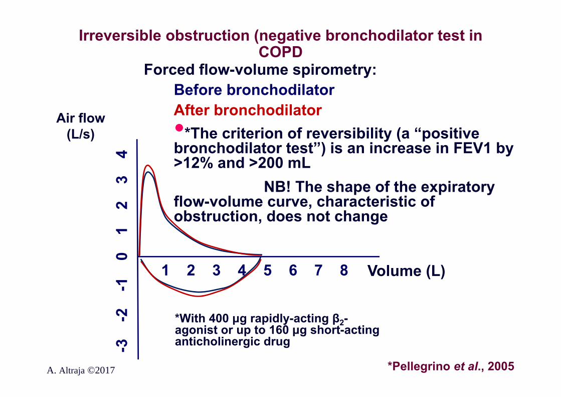
Irreversible obstruction (negative bronchodilator test in COPD
Forced flow-volume spirometry:Before bronchodilatorAfter bronchodilator•*The criterion of reversibility (a “positive bronchodilator test”) is an increase in FEV1 by >12% and >200 mL
NB! The shape of the expiratory flow-volume curve, characteristic of obstruction, does not change
Volume (L)
A. Altraja ©2017
1 2 3 4 5 6 7 8
-3
-2
-1
0
1
2
3
4
*Pellegrino et al., 2005
Air flow (L/s)
*With 400 μg rapidly-acting β2-agonist or up to 160 μg short-acting anticholinergic drug

Forced flow-volume spirometry COPD (volume-to-time curve)
A. Altraja ©2017
NormalM
aht (
L)
1 2 3 4 5 6 7
1
2
3
4
5
6
COPD
Time (s)
FEV1
Volu
me
(L)
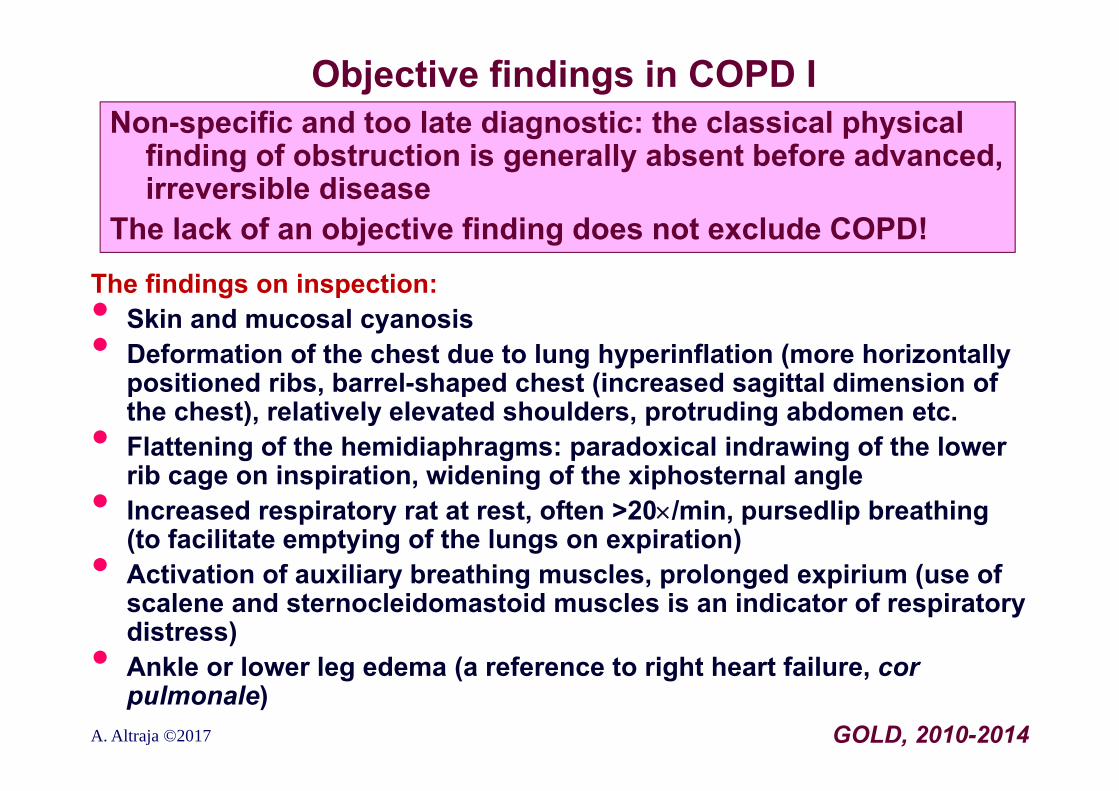
Objective findings in COPD I
The findings on inspection:• Skin and mucosal cyanosis• Deformation of the chest due to lung hyperinflation (more horizontally positioned ribs, barrel-shaped chest (increased sagittal dimension of the chest), relatively elevated shoulders, protruding abdomen etc.• Flattening of the hemidiaphragms: paradoxical indrawing of the lower rib cage on inspiration, widening of the xiphosternal angle• Increased respiratory rat at rest, often >20/min, pursedlip breathing (to facilitate emptying of the lungs on expiration)• Activation of auxiliary breathing muscles, prolonged expirium (use of scalene and sternocleidomastoid muscles is an indicator of respiratory distress)• Ankle or lower leg edema (a reference to right heart failure, cor pulmonale)
Non-specific and too late diagnostic: the classical physical finding of obstruction is generally absent before advanced, irreversible disease
The lack of an objective finding does not exclude COPD!
A. Altraja ©2017 GOLD, 2010-2014
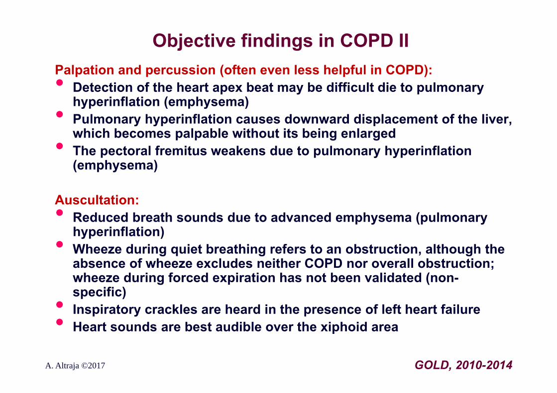
Objective findings in COPD IIPalpation and percussion (often even less helpful in COPD):• Detection of the heart apex beat may be difficult die to pulmonary
hyperinflation (emphysema)• Pulmonary hyperinflation causes downward displacement of the liver, which becomes palpable without its being enlarged• The pectoral fremitus weakens due to pulmonary hyperinflation (emphysema)
Auscultation:• Reduced breath sounds due to advanced emphysema (pulmonary hyperinflation)• Wheeze during quiet breathing refers to an obstruction, although the absence of wheeze excludes neither COPD nor overall obstruction; wheeze during forced expiration has not been validated (non-specific)• Inspiratory crackles are heard in the presence of left heart failure• Heart sounds are best audible over the xiphoid area
A. Altraja ©2017 GOLD, 2010-2014
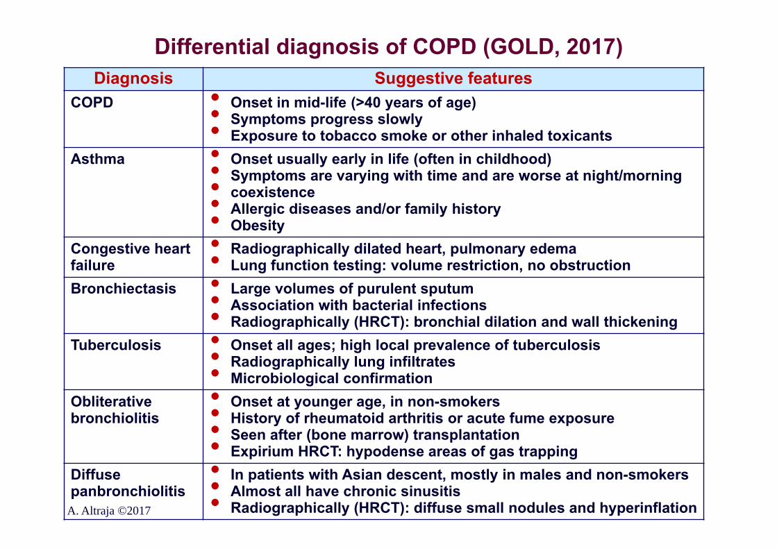
Differential diagnosis of COPD (GOLD, 2017)
A. Altraja ©2017
Diagnosis Suggestive featuresCOPD • Onset in mid-life (>40 years of age)• Symptoms progress slowly• Exposure to tobacco smoke or other inhaled toxicantsAsthma • Onset usually early in life (often in childhood)• Symptoms are varying with time and are worse at night/morning• coexistence• Allergic diseases and/or family history• ObesityCongestive heart failure
• Radiographically dilated heart, pulmonary edema• Lung function testing: volume restriction, no obstructionBronchiectasis • Large volumes of purulent sputum• Association with bacterial infections• Radiographically (HRCT): bronchial dilation and wall thickeningTuberculosis • Onset all ages; high local prevalence of tuberculosis• Radiographically lung infiltrates• Microbiological confirmationObliterative bronchiolitis
• Onset at younger age, in non-smokers• History of rheumatoid arthritis or acute fume exposure• Seen after (bone marrow) transplantation• Expirium HRCT: hypodense areas of gas trappingDiffuse panbronchiolitis
• In patients with Asian descent, mostly in males and non-smokers• Almost all have chronic sinusitis• Radiographically (HRCT): diffuse small nodules and hyperinflation
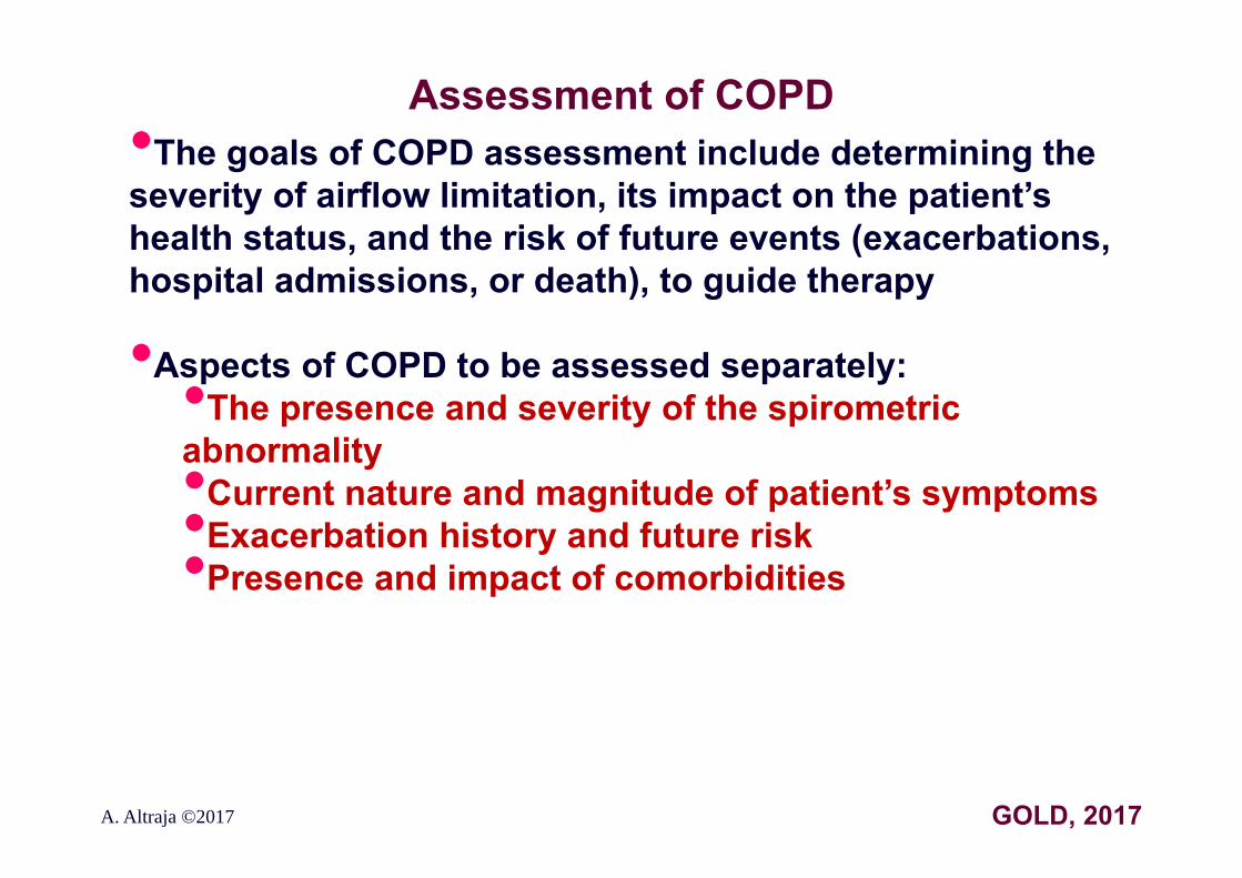
Assessment of COPD•The goals of COPD assessment include determining the severity of airflow limitation, its impact on the patient’s health status, and the risk of future events (exacerbations, hospital admissions, or death), to guide therapy
•Aspects of COPD to be assessed separately:•The presence and severity of the spirometric abnormality•Current nature and magnitude of patient’s symptoms•Exacerbation history and future risk•Presence and impact of comorbidities
GOLD, 2017A. Altraja ©2017
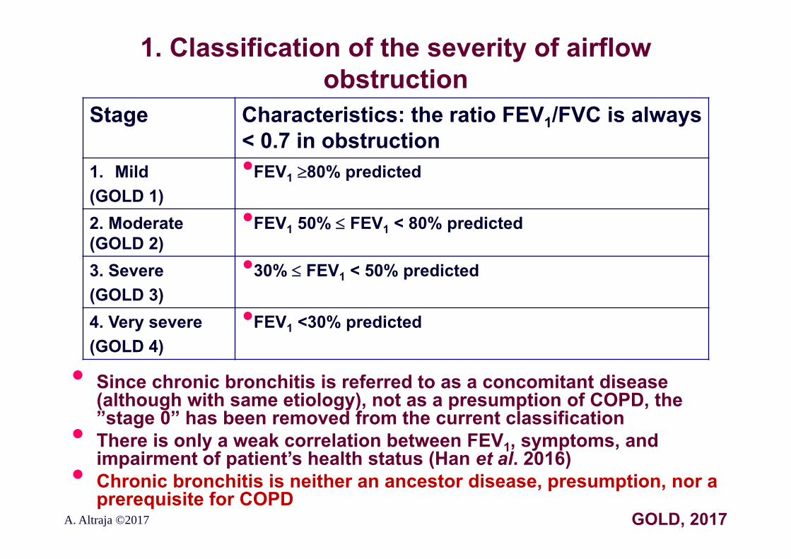
1. Classification of the severity of airflow obstruction
Stage Characteristics: the ratio FEV1/FVC is always < 0.7 in obstruction
1. Mild (GOLD 1)
•FEV1 80% predicted
2. Moderate (GOLD 2)
•FEV1 50% FEV1 < 80% predicted
3. Severe(GOLD 3)
•30% FEV1 < 50% predicted
4. Very severe(GOLD 4)
•FEV1 <30% predicted
A. Altraja ©2017 GOLD, 2017
• Since chronic bronchitis is referred to as a concomitant disease (although with same etiology), not as a presumption of COPD, the ”stage 0” has been removed from the current classification• There is only a weak correlation between FEV1, symptoms, and impairment of patient’s health status (Han et al. 2016)• Chronic bronchitis is neither an ancestor disease, presumption, nor a prerequisite for COPD
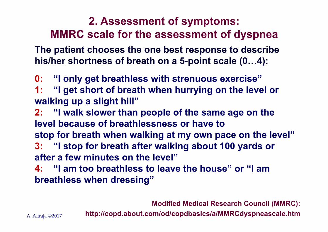
2. Assessment of symptoms: MMRC scale for the assessment of dyspnea
The patient chooses the one best response to describe his/her shortness of breath on a 5-point scale (0…4):
0: “I only get breathless with strenuous exercise”1: “I get short of breath when hurrying on the level or walking up a slight hill”2: “I walk slower than people of the same age on the level because of breathlessness or have to stop for breath when walking at my own pace on the level”3: “I stop for breath after walking about 100 yards or after a few minutes on the level”4: “I am too breathless to leave the house” or “I am breathless when dressing”
Modified Medical Research Council (MMRC):http://copd.about.com/od/copdbasics/a/MMRCdyspneascale.htmA. Altraja ©2017
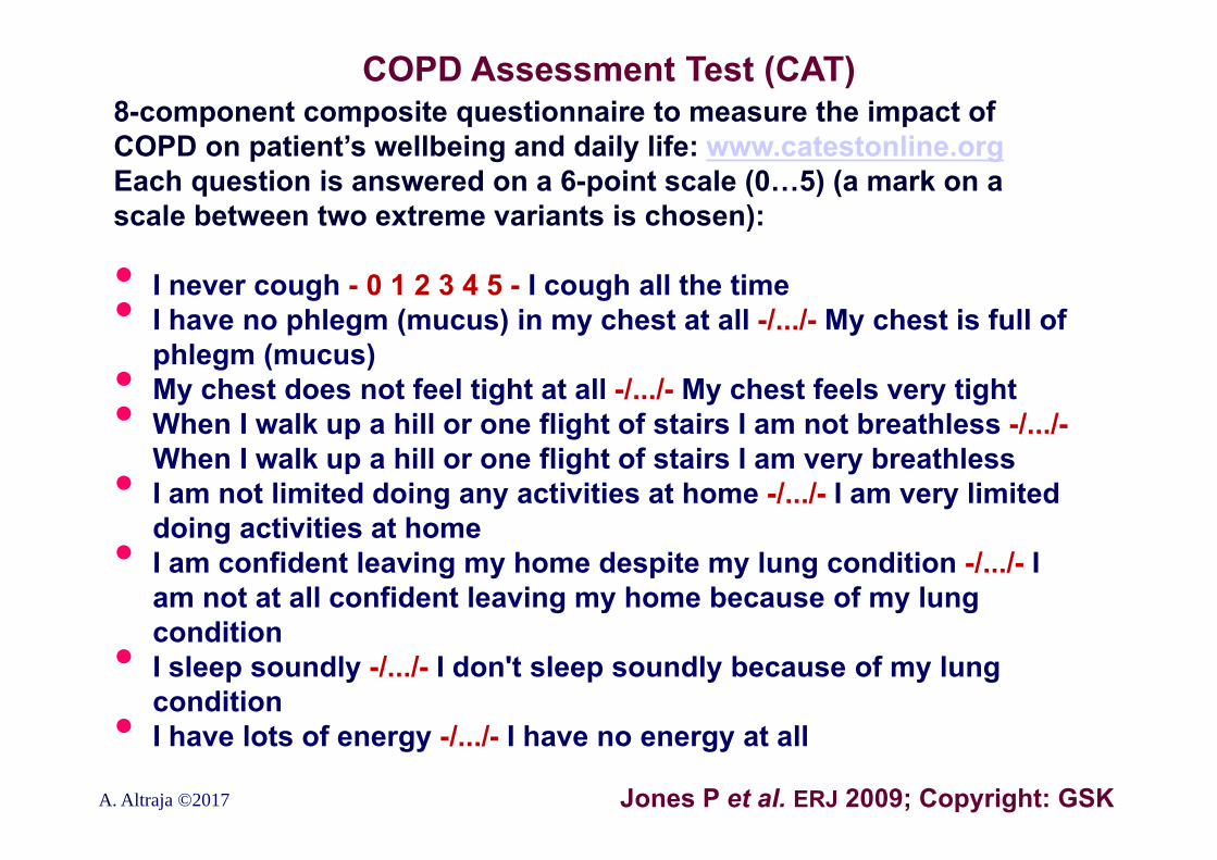
COPD Assessment Test (CAT)8-component composite questionnaire to measure the impact of COPD on patient’s wellbeing and daily life: www.catestonline.orgEach question is answered on a 6-point scale (0…5) (a mark on a scale between two extreme variants is chosen):
• I never cough - 0 1 2 3 4 5 - I cough all the time• I have no phlegm (mucus) in my chest at all -/.../- My chest is full of phlegm (mucus)• My chest does not feel tight at all -/.../- My chest feels very tight• When I walk up a hill or one flight of stairs I am not breathless -/.../-When I walk up a hill or one flight of stairs I am very breathless• I am not limited doing any activities at home -/.../- I am very limited doing activities at home• I am confident leaving my home despite my lung condition -/.../- I am not at all confident leaving my home because of my lung condition• I sleep soundly -/.../- I don't sleep soundly because of my lung condition• I have lots of energy -/.../- I have no energy at all
Jones P et al. ERJ 2009; Copyright: GSKA. Altraja ©2017
The COPD Control Questionnaire (CCQ)10-component, simply self-answered questionnaire developed for assessment of health status by the patient himself/herself (takes ~2 minutes, in >60 languages): www.ccq.nl
A. Altraja ©2017
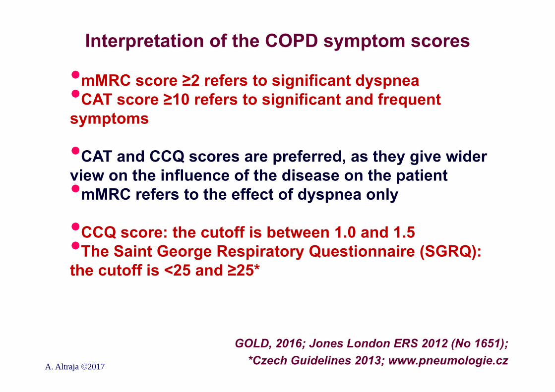
Interpretation of the COPD symptom scores
•mMRC score ≥2 refers to significant dyspnea•CAT score ≥10 refers to significant and frequent symptoms
•CAT and CCQ scores are preferred, as they give wider view on the influence of the disease on the patient•mMRC refers to the effect of dyspnea only
•CCQ score: the cutoff is between 1.0 and 1.5•The Saint George Respiratory Questionnaire (SGRQ): the cutoff is <25 and ≥25*
GOLD, 2016; Jones London ERS 2012 (No 1651);*Czech Guidelines 2013; www.pneumologie.czA. Altraja ©2017
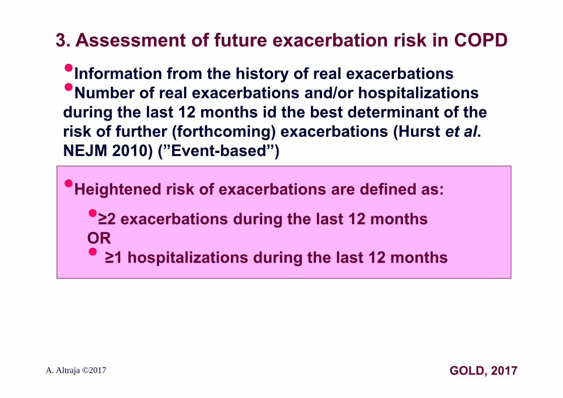
3. Assessment of future exacerbation risk in COPD•Information from the history of real exacerbations•Number of real exacerbations and/or hospitalizations during the last 12 months id the best determinant of the risk of further (forthcoming) exacerbations (Hurst et al. NEJM 2010) (”Event-based”)
•Heightened risk of exacerbations are defined as:
•≥2 exacerbations during the last 12 months OR• ≥1 hospitalizations during the last 12 months
A. Altraja ©2017 GOLD, 2017
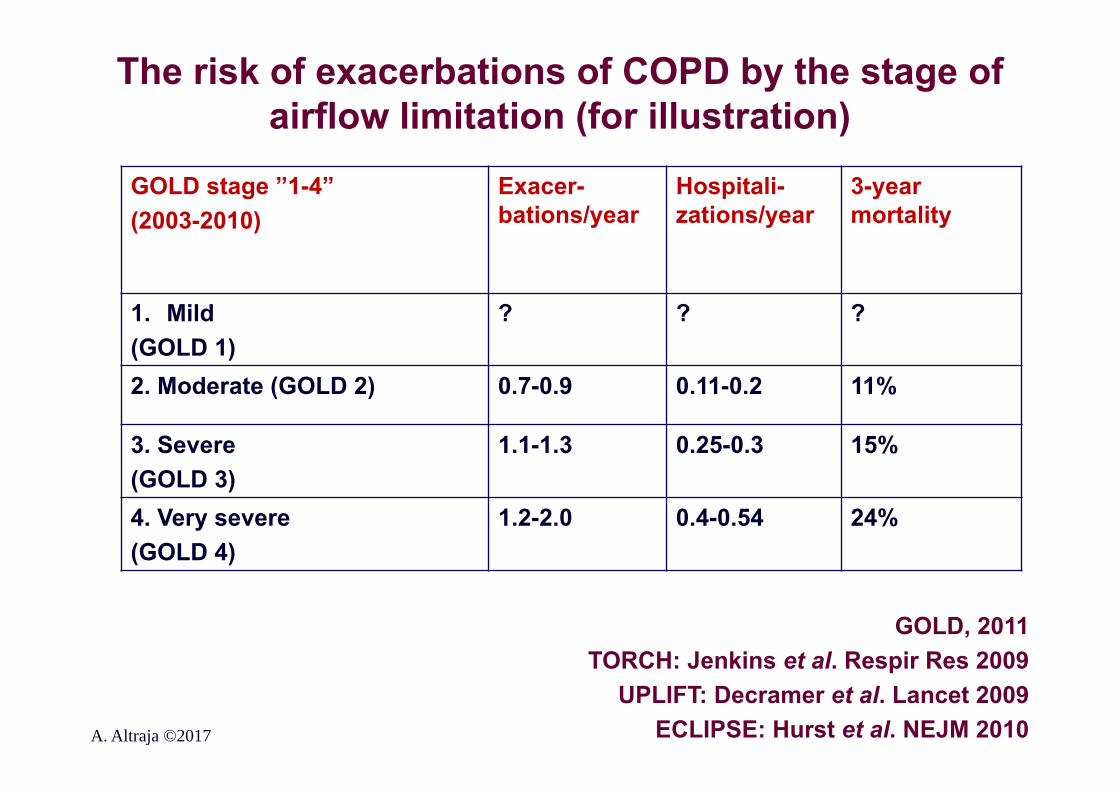
The risk of exacerbations of COPD by the stage of airflow limitation (for illustration)
A. Altraja ©2017
GOLD, 2011TORCH: Jenkins et al. Respir Res 2009
UPLIFT: Decramer et al. Lancet 2009ECLIPSE: Hurst et al. NEJM 2010
GOLD stage ”1-4”(2003-2010)
Exacer-bations/year
Hospitali-zations/year
3-year mortality
1. Mild (GOLD 1)
? ? ?
2. Moderate (GOLD 2) 0.7-0.9 0.11-0.2 11%
3. Severe(GOLD 3)
1.1-1.3 0.25-0.3 15%
4. Very severe(GOLD 4)
1.2-2.0 0.4-0.54 24%
The assessment scheme for COPD
•The patients with COPD should undergo post-bronchodilator spirometry: •1) First, for the diagnosis•2) Thereafter, on a regular basis, to determine the
severity of airflow limitation (i.e. the spirometric grade)•Then, the patients should undergo assessment of either dyspnea using mMRC or symptoms using CAT•Finally, the history of exacerbations (including prior hospitalizations) should be recorded
GOLD, 2017A. Altraja ©2017
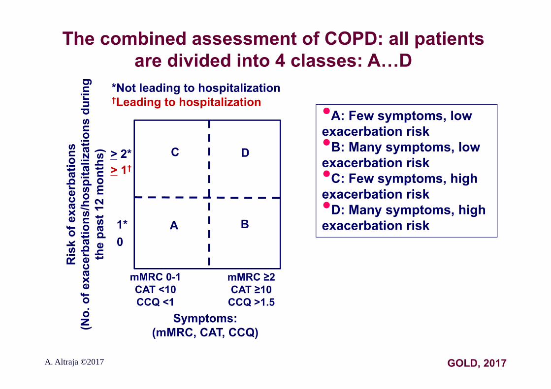
The combined assessment of COPD: all patients are divided into 4 classes: A…D
A. Altraja ©2017
Ris
k of
exa
cerb
atio
ns(N
o. o
f exa
cerb
atio
ns/h
ospi
taliz
atio
ns d
urin
g th
e pa
st 1
2 m
onth
s) > 2*
1*0
C D
A B
•A: Few symptoms, low exacerbation risk•B: Many symptoms, low exacerbation risk•C: Few symptoms, high exacerbation risk•D: Many symptoms, high exacerbation risk
GOLD, 2017
Symptoms:(mMRC, CAT, CCQ)
mMRC 0-1CAT <10CCQ <1
mMRC ≥2CAT ≥10CCQ >1.5
> 1†
*Not leading to hospitalization†Leading to hospitalization
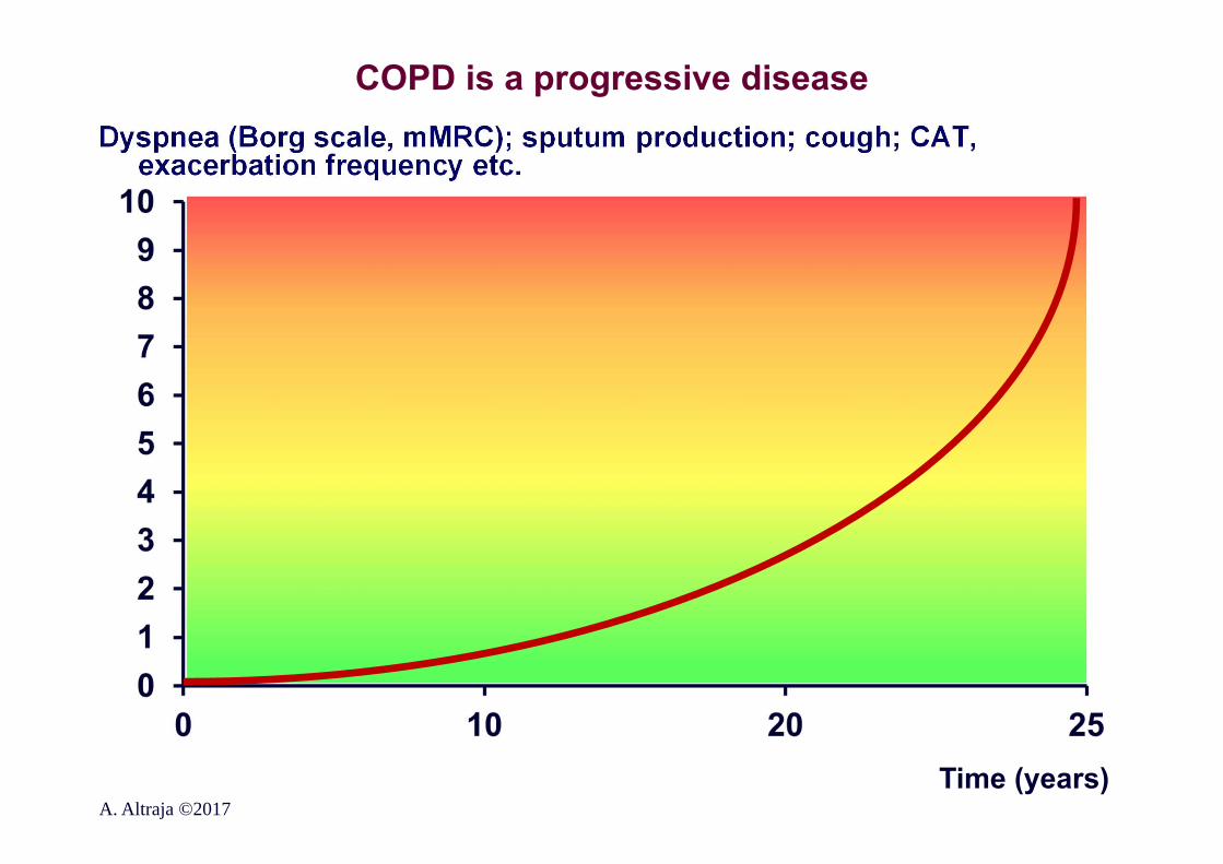
COPD is a progressive diseaseDyspnea (Borg scale, mMRC); sputum production; cough; CAT,
exacerbation frequency etc.
Time (years)A. Altraja ©2017
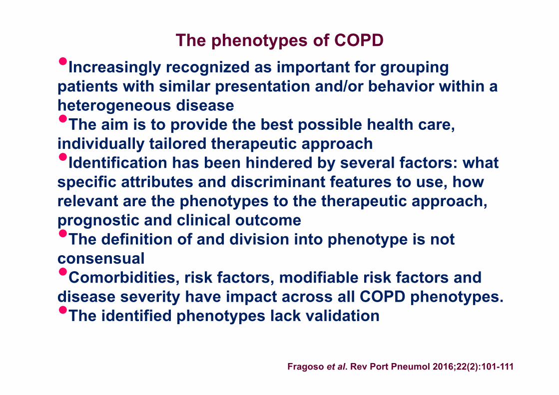
The phenotypes of COPD•Increasingly recognized as important for grouping patients with similar presentation and/or behavior within a heterogeneous disease•The aim is to provide the best possible health care, individually tailored therapeutic approach•Identification has been hindered by several factors: what specific attributes and discriminant features to use, how relevant are the phenotypes to the therapeutic approach, prognostic and clinical outcome•The definition of and division into phenotype is not consensual•Comorbidities, risk factors, modifiable risk factors and disease severity have impact across all COPD phenotypes.•The identified phenotypes lack validation
Fragoso et al. Rev Port Pneumol 2016;22(2):101-111
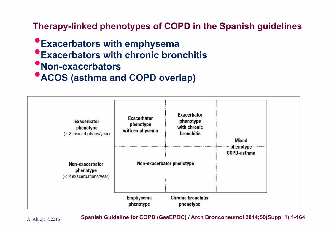
Therapy-linked phenotypes of COPD in the Spanish guidelines
•Exacerbators with emphysema•Exacerbators with chronic bronchitis•Non-exacerbators•ACOS (asthma and COPD overlap)
Spanish Guideline for COPD (GesEPOC) / Arch Bronconeumol 2014;50(Suppl 1):1-164A. Altraja ©2016
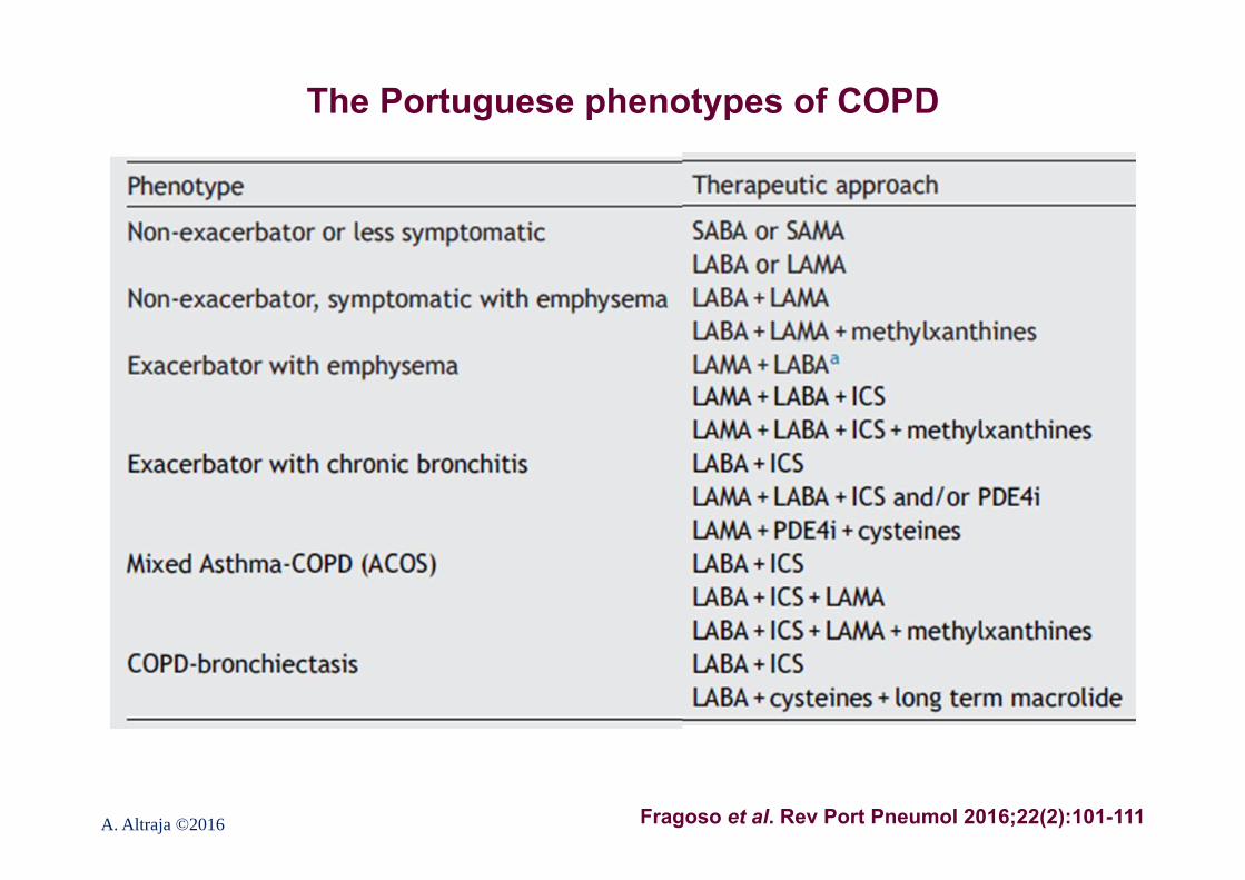
The Portuguese phenotypes of COPD
Fragoso et al. Rev Port Pneumol 2016;22(2):101-111A. Altraja ©2016
Ancillary investigations in COPD•Indicated mainly in moderate-to-severe COPD•To optimize (more personalized) patient management rather than to diagnose COPD itself
•Oral “steroid test” or bronchodilator test usually do not determine the progression of COPD or response to therapy•Radiographic investigations•Chest X-rays•CT/HRCT scans of the chest•Blood gas analysis (a-Astrup)•Measurement of serum 1-antitrypsin•The goal: familial “screening” and counseling• Investigations related to specific measures (surgical or
endoscopic management, incl. lung volume reduction measures, bullectomy or lung transplantation)
A. Altraja ©2017 GOLD
Radiographic investigations in COPDChest X-ray:•Often normal in milder COPD•Rarely diagnostic in the context of the diagnosis•Of COPD itself•To exclude alternative diagnoses and complications (e.g. pneumonia)•To notice e.g. major bullous changes
CT/HRCT of the chest:•Not routinely used•To diagnose/exclude alternative diagnoses (interstitial lung diseases etc.): HRCT (thin-layer HRCT) is needed•For selection of patients for specific measures (surgical or endoscopic management, incl. lung volume reduction measures, bullectomy or lung transplantation)
A. Altraja ©2017
Chest X-ray signs in emphysema-type COPD•Often normal in milder COPDIn more advanced disease: •Pulmonary hyperinflation (increase in volume, s.c. “enlargement of the lungs”)•Hypertransparent lungs, loss of pulmonary vasculature, especially in the periphery; sometimes formation of bullas (hypertransparent areas of air density limited with very thin equal walls)•Barrel-shaped chest, more horizontally positioned ribs, especially with regard to the posterior arch•Bilateral downward displacement and flattening of the diaphragm (in severe cases, even turning to inversely concave)•The heart silhouette gets slim•Protruding pulmonary arteries centrally and enlargement of the heart (with cor pulmonale)On the lateral view additionally:•Enlargement of the retrosternal, retrocardiac, and subcardiac airspaces•Increased sagittal dimension of the chest•Deformation of the sternum accordingly
A. Altraja ©2017
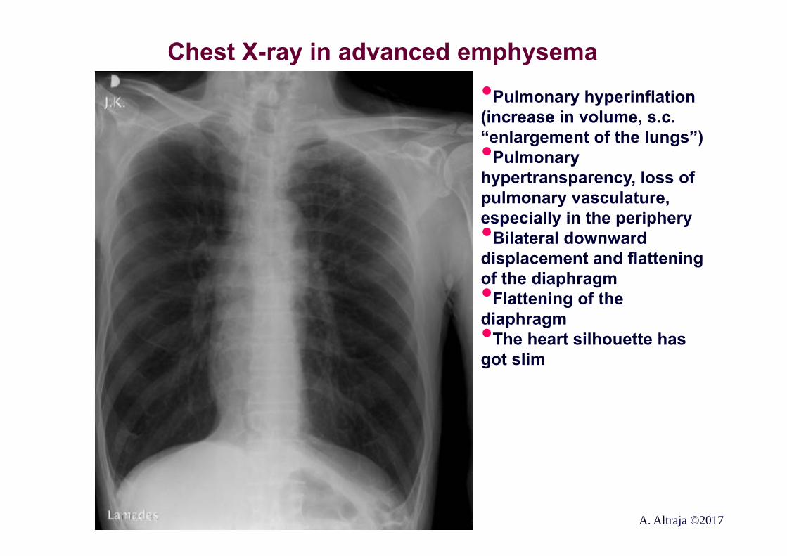
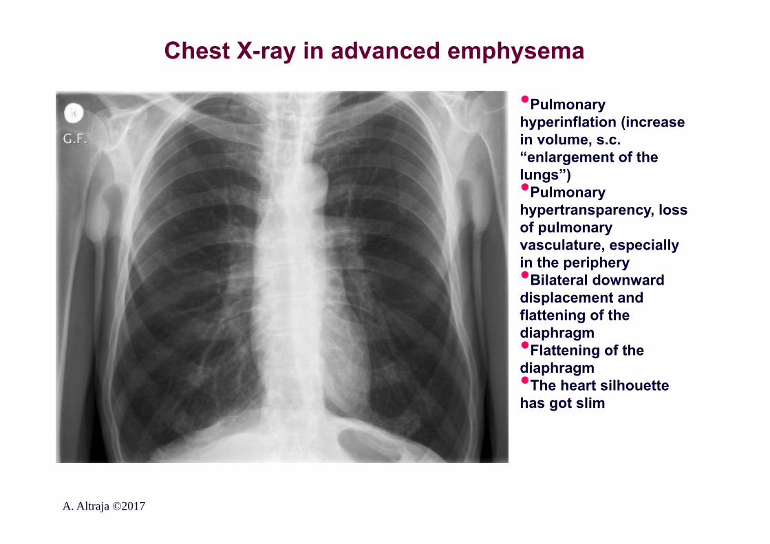
Chest X-ray in advanced emphysema
•Pulmonary hyperinflation (increase in volume, s.c. “enlargement of the lungs”)•Pulmonary hypertransparency, loss of pulmonary vasculature, especially in the periphery•Bilateral downward displacement and flattening of the diaphragm•Flattening of the diaphragm •The heart silhouette has got slim
A. Altraja ©2017
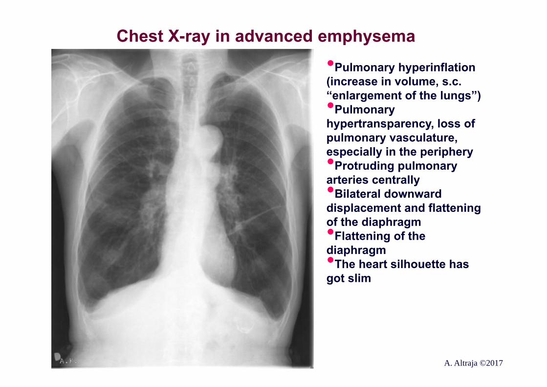
Chest X-ray in advanced emphysema
•Pulmonary hyperinflation (increase in volume, s.c. “enlargement of the lungs”)•Pulmonary hypertransparency, loss of pulmonary vasculature, especially in the periphery•Protruding pulmonary arteries centrally •Bilateral downward displacement and flattening of the diaphragm•Flattening of the diaphragm •The heart silhouette has got slim
A. Altraja ©2017
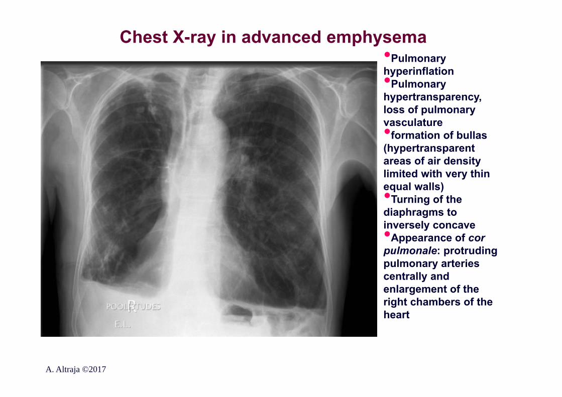
Chest X-ray in advanced emphysema•Pulmonary hyperinflation•Pulmonary hypertransparency, loss of pulmonary vasculature•formation of bullas (hypertransparent areas of air density limited with very thin equal walls)•Turning of the diaphragms to inversely concave•Appearance of cor pulmonale: protruding pulmonary arteries centrally and enlargement of the right chambers of the heart
A. Altraja ©2017
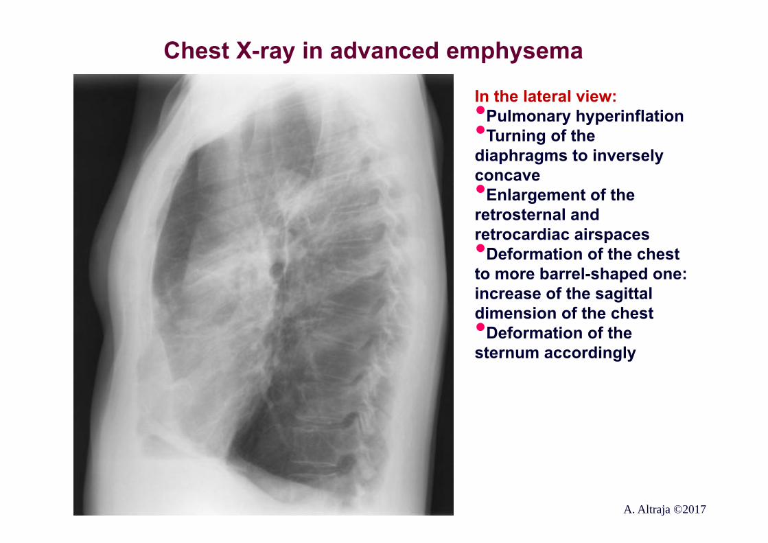
Chest X-ray in advanced emphysema
In the lateral view:•Pulmonary hyperinflation•Turning of the diaphragms to inversely concave•Enlargement of the retrosternal and retrocardiac airspaces•Deformation of the chest to more barrel-shaped one: increase of the sagittal dimension of the chest•Deformation of the sternum accordingly
A. Altraja ©2017
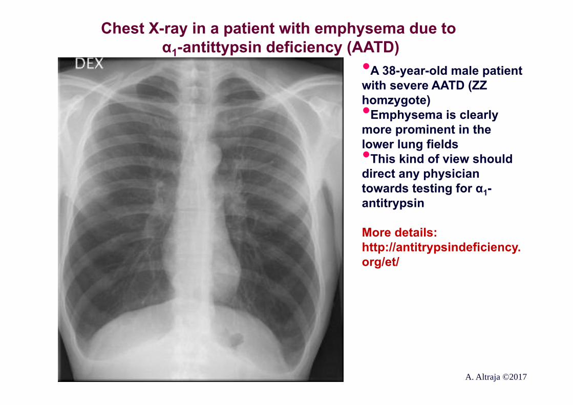
Chest X-ray in a patient with emphysema due to α1-antittypsin deficiency (AATD)
•A 38-year-old male patient with severe AATD (ZZ homzygote)•Emphysema is clearly more prominent in the lower lung fields•This kind of view should direct any physician towards testing for α1-antitrypsin
More details: http://antitrypsindeficiency.org/et/
A. Altraja ©2017
The signs of emphysema on chest CT scan•Most often, emphysema is defined as areas of attenuation of less than minus 950 Hounsfield units•Pulmonary hyperinflation: the lungs enlarge to all directions•In some CT sections, the right and the left lung reach to cover each other like flaps•Flattening of the diaphragms•Enlargement of the retrosternal, retrocardiac, and subcardiac airspaces (visible also on lateral chest X-ray)•The heart silhouette gets slimmer•Areas with low density attenuation and bullas •Abnormal configuration of the vasculature: hypovascularization in the periphery, but prominence of the central (hilar) portions of the pulmonary arteries
A. Altraja ©2017
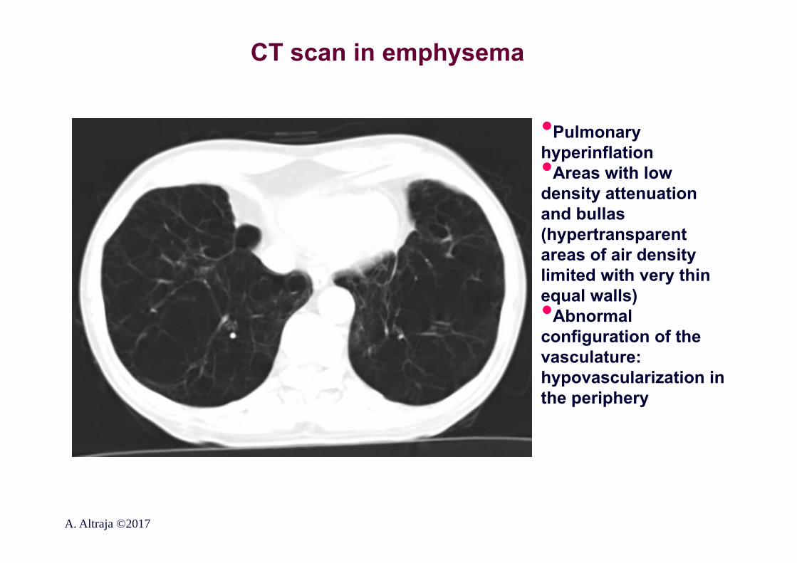
CT scan in emphysema
•Pulmonary hyperinflation•Areas with low density attenuation and bullas (hypertransparent areas of air density limited with very thin equal walls)•Abnormal configuration of the vasculature: hypovascularization in the periphery
A. Altraja ©2017
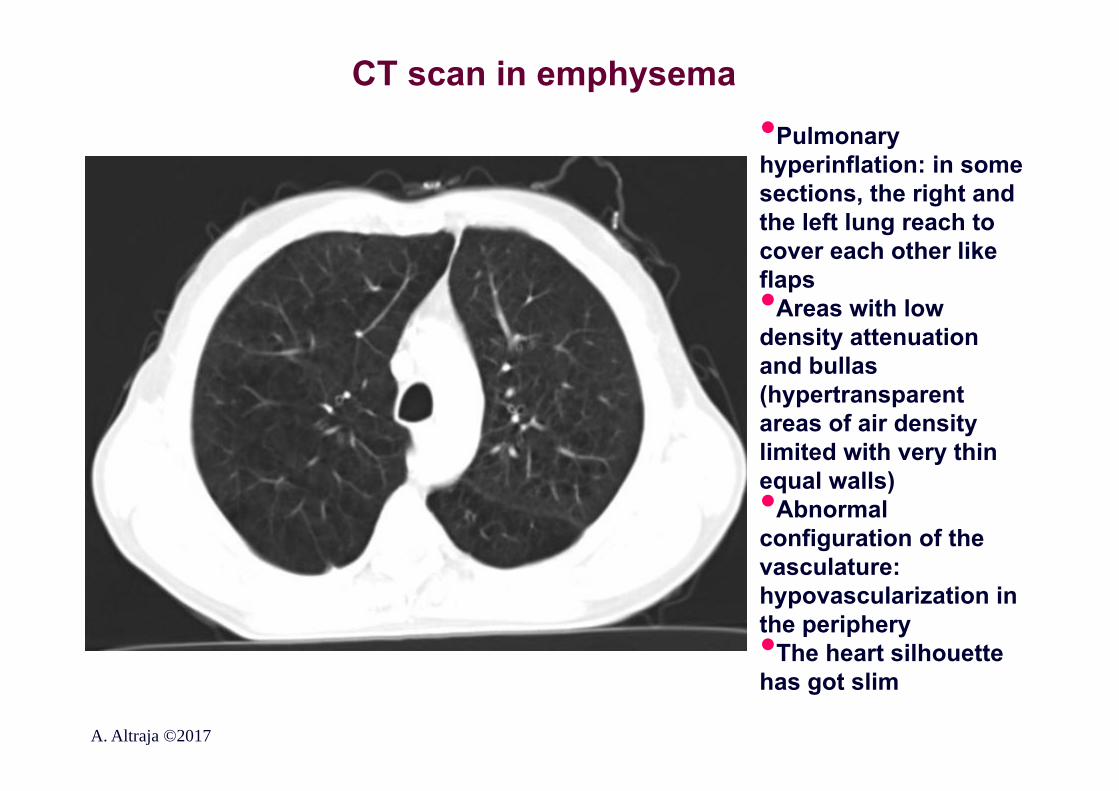
CT scan in emphysema•Pulmonary hyperinflation: in some sections, the right and the left lung reach to cover each other like flaps•Areas with low density attenuation and bullas (hypertransparent areas of air density limited with very thin equal walls)•Abnormal configuration of the vasculature: hypovascularization in the periphery•The heart silhouette has got slim
A. Altraja ©2017
Measurement of the diffusing capacity of the lung with transfer test in COPD
•Enables assessment of the injury to the lung parenchyma (important in emphysema), its magnitude and indirectly, also the homogeneity or heterogeneity of the parenchymal destruction•Is recommended and is often used in expert centers, but is not obligatory for routine use•Special indications: in association with assessment for lung transplantation etc.
GOLD, 2011A. Altraja ©2017
Oximetry and blood gas analyses in COPD•Pulse oximetry is used to evaluate oxygen saturation and the need for supplemental oxygen therapy (measures SaO2, does not show PaCO2) •In all patients suggestive of having respiratory or right heart failure
•Blood gas analyses:•Indicated, it SaO2 is <92%•To diagnose the ventilation insufficiency (hypercapnia)•Criteria of respiratory failure:•PaO2 <60 mmHg (<8,0 kPa)•PaCO2 >45 mmHg (>6,0 kPa)•After changing the FiO2 (on supplemental oxygen therapy) wait for 20-30 min. to let the blood gases to equalize and reflect the status
GOLD, 2017A. Altraja ©2017
Alpha-1 antitrypsin (AAT) deficiency (AATD) screening•The World Health Organization (WHO) recommends that all patients with COPD should be screened once, especially in areas with high AATD prevalence (WHO, 1997)•A low concentration (<20% of normal) is highly suggestive of homozygous deficiency•Family members should also be screened•Estonia and Livonia are endemic areas for the Z-allele•In practice, AATD is markedly underrecognized, underdiagnosed, and underdocumented in Estonia•Both the serum level AAT and phenotype/genotype are determined•Allele variants: M (normal), S, Z, null; others are very rare
GOLD, 2017A. Altraja ©2017
Physical exercise testing in COPD
•A significant objective set of measures to measure the general health status and functional reserves in the patient (for assessment of pre-treatment capability, the effect of treatment, and prognosis)•Non-incremental endurance tests (6 minute walk test):•A sub-maximal exercise test undertaken to assess
aerobic capacity and endurance•The main outcome is the distance covered over 6 minutes, used as the outcome to compare both the performance capacity at the diagnosis and changes over time •Incremental tests (paced 10 m shuttle walk test)
GOLDA. Altraja ©2017
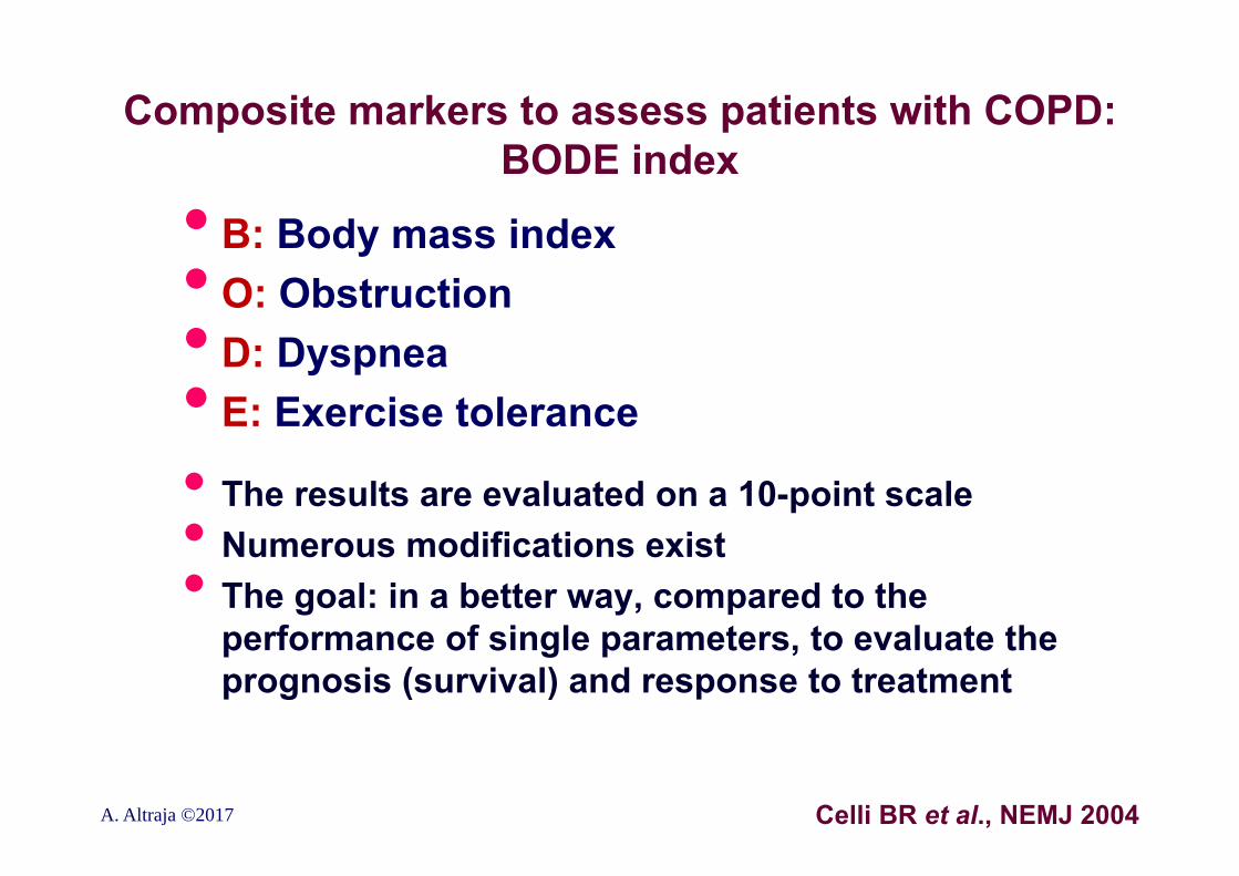
Composite markers to assess patients with COPD: BODE index
• B: Body mass index• O: Obstruction• D: Dyspnea• E: Exercise tolerance
• The results are evaluated on a 10-point scale• Numerous modifications exist• The goal: in a better way, compared to the
performance of single parameters, to evaluate the prognosis (survival) and response to treatment
Celli BR et al., NEMJ 2004A. Altraja ©2017
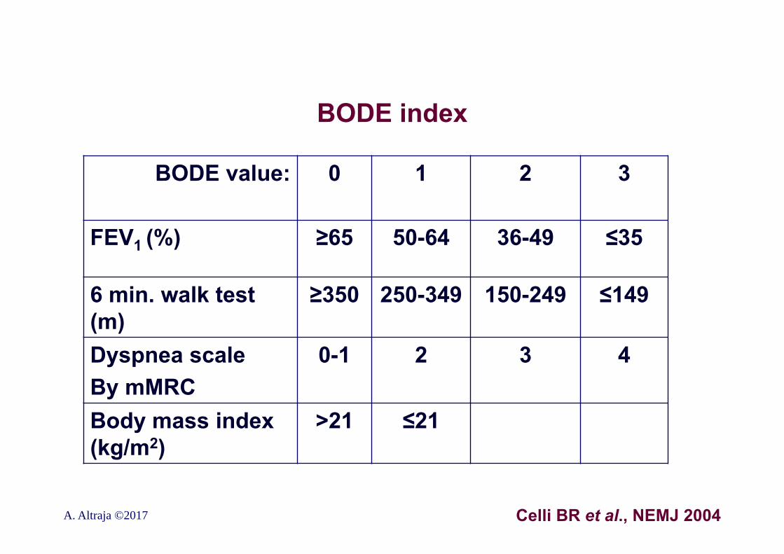
BODE index
BODE value: 0 1 2 3
FEV1 (%) ≥65 50-64 36-49 ≤35
6 min. walk test (m)
≥350 250-349 150-249 ≤149
Dyspnea scaleBy mMRC
0-1 2 3 4
Body mass index (kg/m2)
>21 ≤21
A. Altraja ©2017 Celli BR et al., NEMJ 2004
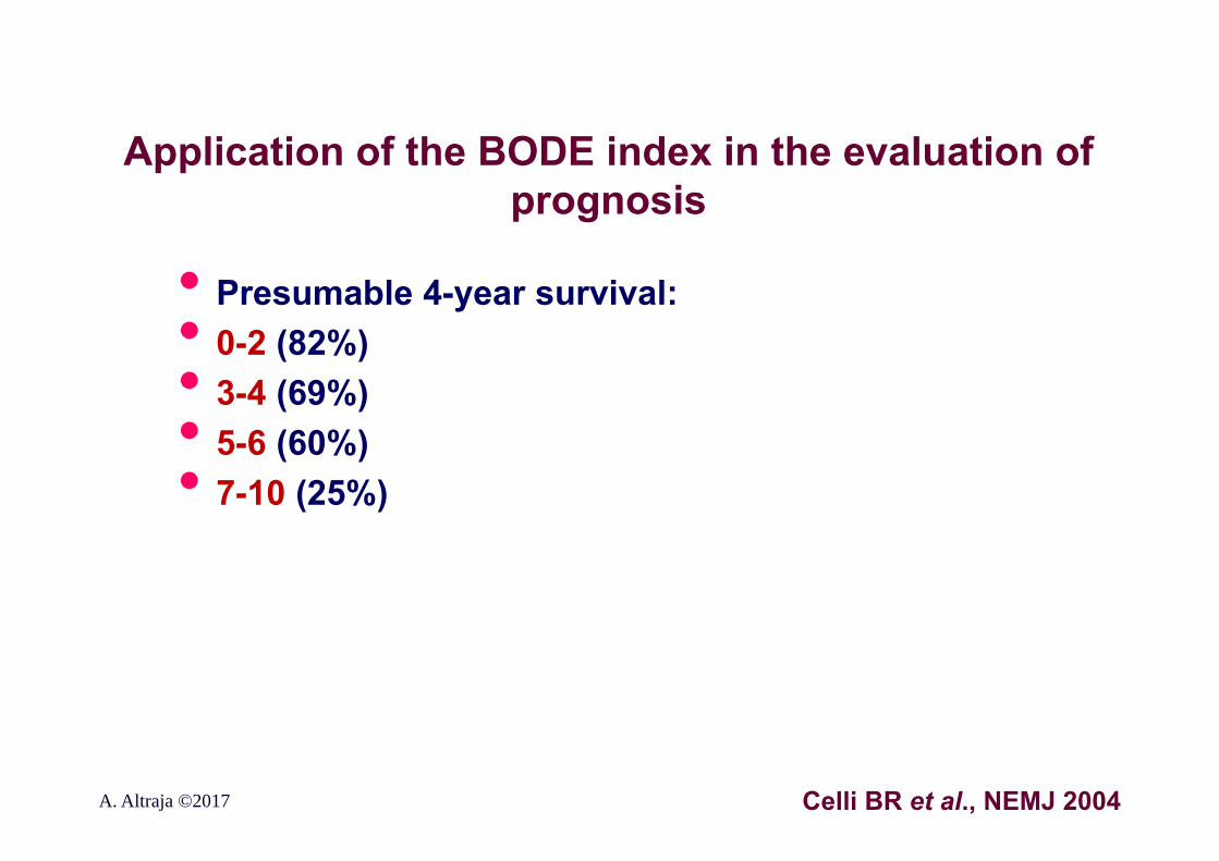
Application of the BODE index in the evaluation of prognosis
• Presumable 4-year survival:• 0-2 (82%)• 3-4 (69%)• 5-6 (60%)• 7-10 (25%)
Celli BR et al., NEMJ 2004A. Altraja ©2017

Definition of arterial hypoxemia•PaO2 <55 mmHg (8.0 kPa) or SaO2 <88%•PaO2 55…<60 mmHg (8.0…<8.5 kPa) with the evidence of pulmonary hypertension, peripheral edema, or polycythemia (hematocrit >55%)•With or without CO2 retention (PaCO2 >50 mmHg (6.7 kPa) on breathing air at sea level
These are the criteria for prescribing supplementary oxygen therapy in COPD (applied also for IPF etc.)• Supplementary oxygen, if the above criteria are met
twice, out of exacerbation over an at last 3-week period• Supplementary oxygen to keep SaO2 <90%• Recheck in 60-90 days: if still indicated, continue long-term
GOLD, 2017A. Altraja ©2017


![Speleotherapy as an Effective Treatment of Chronic ... · Chronic obstructive pulmonary disease (COPD) is a chronic worldwide disease [1-4]. COPD COPD was supposed as the tenth most](https://img.pdfslide.us/doc/110x75/5cd3389988c99399578d07a7/speleotherapy-as-an-effective-treatment-of-chronic-chronic-obstructive-pulmonary.jpg)