Embed Size (px)
Citation preview
International Journal of Green Pharmacy • Jul-Sep 2018 • 12 (3) | 227
Chronic Cannabis-induced oxidative stress and reproductive containment
in female mice
Somenath Ghosh1, S. K. Rai2#
1Department of Zoology, Pineal Research Laboratory, Banaras Hindu University, Varanasi, Uttar Pradesh, India, 2Department of Anatomy, Institute of Medical Science, Banaras Hindu University, Varanasi, Uttar Pradesh, India
#Contributed equally
Abstract
Aim: This study aims to evaluate cannabinoid-induced oxidative stress and reproductive containment in female mice. Settings and Design: Fifteen adult female Parkes strain mice were randomly chosen from the institutional animal house (n = 5/group) with ad libitum access to water and food. Animals were grouped into control (vehicle treated) and treated with 6 mg of Cannabis/100 g of body weight and 12 mg of Cannabis/100 g of body weight. After 15 days, all animals were sacrificed and tissues were collected for histology, immunohistochemistry (IHC), and estimations of different parameters. Methods and Materials: Histology and IHC of cannabinoid receptor 1 (CB1) were performed following standardized protocols. All parameters were estimated either by standard biochemical protocols or by kit following manufacturer’s protocol. Serum level of Estrogen (E2), stress parameters (Super Oxide Dismutase; SOD, Catalase, CAT; Malonaldehyde, MDA and Glutathione Peroxidase; GPx), apoptotic parameters of thecal cells (by Caspase-3 assay), steroidogenic parameters (3β Hydroxy-steroid dehydrogenase; 3βHSD and 17β Hydroxy-steroid dehydrogenase; 17βHSD) and expression of CB1 were noted in ovary. Statistical analyses used: Data were analyzed by one-way analysis of variance followed by Duncan’s Multiple Range post hoc test. Results: We found statistically significant (P < 0.05) decrease in steroidogenic parameters and significant increase (P < 0.05) in free radical and apoptotic parameters and CB1 receptor expressions on dose-dependent Cannabis treatment. Conclusions: We may conclude that chronic treatment of Cannabis causes reproductive containment in females which has never been addressed previously.
Key words: Cannabis, cannabinoid receptor 1, female mice, impairment, reproduction, stress
INTRODUCTION
Cannabis which is a type of marijuana has been used by the people of Indian subcontinent from time unknown.[1] They
not only use this herb as a part of holy practice but also use it for recreational purposes.[2] Irrespective of sex, this hallucinogenic agent is used by most part of the world, particularly by the populations of South America, India, Bangladesh, and Pakistan from a long time ago.[3] Reports suggesting the roles of Cannabis causing systemic neuropathy,[4] neuronal disability,[5] impaired fetal development,[6] and malfunctioning of male reproductive system[7-10] are documented. However, no reports available are depicting the effects of marijuana in the female reproductive system.
The main causative agent of marijuana/cannabinoids is the endocannabinoid (eCBs).
This is a neutral lipid and highly conserved molecule throughout evolutionary history.[11] They are having different derivatives such as anandamide,[12] 2-arachidonoylglycerol,[13] and ∆9-tetrahydrocannabinol (THC).[14] However, among all of the fatty acid derivatives of cannabinods or eCBs, the THC has now been established as the most important hallucinogenic agent of this molecule.[15] There are literatures suggesting the role of this THC in regulation of functions of the central nervous system (CNS) and thus regulating the reproductive functions by affecting/modulating hypothalamo–pituitary–gonadal axis[16] through its receptor CB1 and CB2.[17] Now, it
OR
IGIN
AL
AR
TIC
LE
Address for correspondence: Dr. Somenath Ghosh, Department of Zoology, Pineal Research Laboratory, Banaras Hindu University, Varanasi – 221 005, India. Phone: +91-9936439419. E-mail: [email protected]
Received: 26-05-2018 Revised: 13-07-2018 Accepted: 27-07-2018
Ghosh and Rai: Cannabis affects reproduction and oxidative stress
International Journal of Green Pharmacy • Jul-Sep 2018 • 12 (3) | 228
has been reported that CB1 receptors are localized mostly in whole vertebrate CNS and some peripheral tissues, whereas CB2 receptors are mostly expressed in peripheral tissues and immune cells, however, they have recently been found also in the CNS.[18] However, with all the advancement in psychoneuroendocrine research, till date, it is a matter of debate how THC is going to regulate reproductive system at the peripheral level. Some literature suggests that there is a general agreement on the inhibitory effect exerted by cannabinoids and eCBs on GnRH release.[19] Thus, it is affecting the subsequent follicle-stimulating hormone and luteinizing hormone release in females and impairing female reproduction.[20]
However, all the above-mentioned reported phenomenon is occurring in the CNS and no definitive proof has been reported till date how the endocannabinoids are affecting peripheral reproductive performances in females (in terms of gonadal activity, steroidogenesis, receptor expressions, and free-radical generations). Thus, the aim of the present study was to note the cannabinoid (particularly eCBs)-induced oxidative stress and reproductive impairments in female mice specifically taking peripheral reproductive organs (ovary) in consideration.
SUBJECTS AND METHODS
Animals and Maintenance
In bred adult (12–15 weeks of age) female Parkes strain mice were used for this study. Mice were maintained under hygienic conditions in a well-ventilated room with 12-h photoperiod (8 AM to 8 PM, light) with 50 ± 20% relative humidity, 25 ± 2°C temperature and were fed pelleted food (Mona Laboratory Animal Feeds, Varanasi, India); drinking water was available ad libitum. Five mice in each group were housed in polypropylene cages (430 mm × 270 mm × 300 mm), with dry rice husk as the bedding material. General health condition and body weight of the animals were monitored regularly during the entire tenure of the experiment. All experiments were conducted in accordance with principles and procedures approved by Departmental Research Committee under supervision of Committee for the Purpose of Control and Supervision of Experiments on Animals, Govt. of India (2007).
Preparations of Different Doses of Cannabis Extracts
Leaves and flowers of fresh Cannabis plant (100 g Cannabis plant) were extensively ground in mortar and pestle with 1ml autoclaved double distilled water. From the 1 g/ml paste, 12 mg was weighed and further dissolved in 1 ml autoclaved double-distilled water to make a stock solution of 12 mg/ml. This solution was filtered to get a clear solution. Finally,
the mice were gavaged Cannabis by means of a 100 µl micropipette using the 12 mg/ml stock.
Purity Assessment of Cannabis Preparations
The dry-weight ratio of THC to cannabidiol (CBD) and the percent CBD and THC in the Cannabis variant found in this region of the world has been previously reported.[21] The proportion of high THC/CBD chemotype plants in most accessions assigned to Cannabis sativa was of 25%.[21]
Experimental Design
Mice were randomly allocated into three groups (Groups 1–3). Each group comprised five female mice (n = 5/group). Group I was treated with distilled water (vehicle treated; controls); Group 2 was gavaged with 6 mg/100 g body weight/day aqueous Cannabis preparation; and Group 3 was gavaged with 12 mg/100 g body weight/day aqueous Cannabis preparation. The mode of oral delivery of extracts was following the protocol published previously (21). The tips used for this purpose to deliver the dose from the micropipette had the pointed surface cut to avoid any injury in the mouth of the mouse. The micropipette was used to deliver a small volume of (~20 or 40 µl) dose. The study was continued for 30 days.
Collection of Desired Tissues
Mice were weighed before the start of experiment as well as before killing. The animals were etherized to death and blood was collected from heart. Subsequently, serum was separated and was stored at −20°C until biochemical estimations of total serum cholesterol and estradiol by ELISA. Both the ovaries and uterine horns were excised, blotted free of blood and fat tissues and were weighed. The ovary on one side of the animal was fixed in Bouin’s fluid for histology and immunohistochemical localization of CB1 receptor. The contralateral ovary of each mouse was stored at −20°C until used for enzyme assays (for steroidogenesis, Caspase-3, and free-radical parameters) and Western blot analysis of CB1 receptor.
Antibodies and Reagents
All of the chemicals used for the present study were of analytical grade and were purchased either from Sigma Aldrich (St. Louis, MO, USA) or from Merck (Germany). For Western blot analysis, polyclonal primary antibody against CB1 receptor was purchased from Affinity BioReagents (Rockford, IL, USA, Cat No. RQ4287) and horseradish peroxidase-linked secondary antibody was purchased from Bangalore Genei Pvt., Ltd., (Bengaluru, India). For immunohistochemistry (IHC), ABC Kit was purchased from ABC staining kit (Universal Elite, Vector Laboratories, Burlingame, CA). For 3β Hydroxy-steroid dehydrogenase
Ghosh and Rai: Cannabis affects reproduction and oxidative stress
International Journal of Green Pharmacy • Jul-Sep 2018 • 12 (3) | 229
(3β-HSD) and 17β-HSD assays, pregnenolone was purchased from Sigma Aldrich (St. Louis, MO, USA).
Experimental Approaches
Histological preparations
Ovaries were embedded in paraffin wax and serially sectioned of 6 µm using a microtome (Leica, Germany). One set of the slide was prepared and was further processed for hematoxylin and eosin staining following the protocol published elsewhere.[22] The permanent slides were prepared by mounting with (Distyrene Plasticizer Xylene [DPX], SRL, India), after 24 h were observed under microscope (Leitz MPV3 with photoautomat software) and were documented for general histology.
IHC of CB1 Receptor
IHC for CB1 receptor was performed following the protocol published elsewhere.[21] Ovaries of both treated and untreated adult mice were paraffin embedded and 6 mm sections were analyzed by immunohistochemistry, for CB1 receptor to show where, CB1, receptor is localized in mice ovaries and to have a generalized idea about the receptor expression pattern. For the secondary antibody and enzyme conjugates, ABC staining was used. Briefly after deparaffinization and hydration, and blocking of endogenous peroxidase with 3% H2O2 in methanol, sections were incubated with blocking serum for 1 h, followed by incubation with primary antibody (CB1 at a dilution of 1:50) for 1 h at room temperature. The sections were then washed and incubated with the biotinylated secondary antibody for 30 min at room temperature, followed by another 30 min with horseradish avidin-peroxidase conjugated. After washing, sections were incubated with the chromagen substrate (0.1% 3,3-diaminobenzidine tetrahydrochloride, DAB, Sigma-Aldrich, USA) in 0.05M Tris buffer, pH 7.6, and 0.01% H2O2 for 10 min and then counterstained with Ehrlich’s hematoxylin. The permanent slides were prepared by mounting with (DPX, SRL, India), after 24 h were observed under microscope (Leitz MPV3 with photo-automat software) and were documented.
Estimation of Total Serum Cholesterol
The total serum cholesterol was estimated by commercial cholesterol estimation kit following manufacturer’s protocol (Span Diagnostics, Surat, Gujarat, India).
3β-Hydroxysteroid Dehydrogenase Enzyme Activity
3β-HSD (EC 1.1.1.145) enzyme was assayed according to the protocol of Shivanandappa and Venkatesh[23] using ovarian homogenate. Ten percent tissue homogenate was prepared in 0.1 M Tris–HCl buffer (pH 7.8). The homogenate
was centrifuged at 12,000 × g at 4°C and the supernatant was used as the source of enzyme. The enzyme was assayed in 0.1 M Tris–HCl buffer (pH 7.8) containing 500 mM NAD, 100 mM pregnenolone as substrate, and enzyme (50 ml) in a total volume of 3.0 ml and incubated at 37°C for 1 h. The reaction was stopped by the addition of 2.0 ml of phthalate buffer (pH 3.0) and the absorbance was noted at 490 nm. The enzyme activity was calculated from the standard curve of NADH and expressed as nmol NADH formed/h/mg protein.
17β-Hydroxysteroid Dehydrogenase Enzyme Activity
17β-HSD (EC 1.1.1.62) activity was measured by following the protocol of Blomquist et al., (1985).[24] In brief, 10% homogenate of the ovarian tissues were prepared in normal phosphate-buffered saline (PBS; pH 7.4) and 250 µl of the supernatant was mixed with 250 µl of 440 µM sodium pyrophosphate buffer (pH 10.2), 10 µl ethanol containing 0.3 µM estradiol (Sigma, St. Louis, USA), and 240 µl of 25 mg% BSA. Enzyme activity was measured after addition of 50 µl of 0.5 µM NAD to the mixture in a spectrophotometer at 340 nm against a blank (without NAD). One unit of enzyme activity was the amount causing a change in absorbance of 0.001/min at 340 nm.
Evaluation of Superoxide Dismutase (SOD) Activity in Ovary
SOD; EC 1.15.1.1 activity was assayed following the method of Das et al.[25] Just after sacrifice, 10% homogenates of all ovarian tissues from Group I and set III mice were prepared in 150 mM PBS, pH 7.4 and centrifuged for 30 min at 12,000 g at 4°C. The supernatant was again centrifuged for 60 min at 12,000 g at 4°C and then processed for enzymatic activity based on a modified spectrophotometric method using nitrite formation by superoxide radicals. A 0.5 ml of homogenate was added to 1.4 ml of reaction mixture comprised 50 mM phosphate buffer (pH 7.4), 20 mM L-methionine, 1% (v/v) Triton X- 100, 10 mM hydroxylamine hydrochloride, 50 mM ethylenediaminetetraacetic acid (EDTA) followed by a brief pre-incubation at 37°C for 5 min. Next, 0.8 ml of riboflavin was added to all samples along with a control containing buffer instead of sample and then exposed to two 20 W fluorescent lamps fitted parallel to each other in an aluminum foil-coated wooden box. After 10 min of exposure, 1 ml of Greiss reagent was added and absorbance of the color formed was measured at 543 nm. One unit of enzyme activity is defined as the amount of SOD inhibiting 50% of nitrite formation under assay conditions.
Estimation of Catalase (CAT) Activity in Ovary
CAT; EC 1.11.1.6 activity was measured following the procedure of Sinha.[26] This method is based on the fact that dichromate in acetic acid is reduced to chromic acetate when heated in the presence of H2O2 with the formation of
Ghosh and Rai: Cannabis affects reproduction and oxidative stress
International Journal of Green Pharmacy • Jul-Sep 2018 • 12 (3) | 230
perchromic acid as an unstable intermediate. The chromic acetate thus produced is measured calorimetrically. The CAT preparation is allowed to split H2O2 for different periods of time. The reaction is stopped at a particular time by the addition of dichromate/acetic acid mixture, and the remaining H2O2 is determined by measuring chromic acetate calorimetrically after heating the reaction mixture. There is production of green color at the end of the process. Immediately after sacrifice, 20% homogenate of ovarian tissues from Group I, Group II, and Group III were prepared in PBS (10 mM; pH = 7.0) and then centrifuged at 12,000 g for 20 min at 4°C. Supernatant was taken for enzyme estimation. 5 ml of PBS was added to 4 ml of H2O2 (200 mM) and then 1 ml of enzyme extract was added. After 1 min, 1 ml of this solution was taken in a tube and 2 ml of K2Cr2O7 (5%) solution was added. Then, it was boiled for 10 min and absorbance was measured at 570 nm. The activity of CAT was expressed as amount of H2O2 degraded per minute.
Estimation of Lipid Peroxidation (LPO) Assay by Thiobarbituric Acid Reactive Substances (TBARS) Level Estimation in Ovary
After sacrifice of the mice of all the groups, the ovarian tissues were dissected out on a sterile watch glass placed in ice box, cleaned from adherent tissues, and processed immediately for estimation of LPO. Ovarian tissues of Group I, Group II, and Group III experimental mice were weighed and homogenized in a tenfold excess of 20 mM Tris–HCl buffer (pH 7.4) and the 10% homogenates were centrifuged for 15 min at 3000× g at 4°C. The supernatant was subjected to thiobarbituric acid (TBA) assay by mixing with 8.1% sodium dodecyl sulfate (SDS), 20% acetic acid, 0.8% TBA, and then digested it for 1 h at 95°C. The reaction mixture was immediately cooled in running water, vigorously shaken with 2.5 mL of n-butanol and pyridine reagent (15:1) and centrifuged for 10 min at 1500× g.[27] The absorbance of the upper phase was measured at 534 nm. Total TBARS were expressed as malondialdehyde (MDA; nmol/g tissue weight) taking 1,1,1,1-tetraethoxy propane (TEP) as standard. The standard curve was calibrated using 10 nM TEP.
Glutathione Peroxidase (GPx) Estimation in Ovary
GPx; EC 1.11.1.9 activity was assayed as described by Mantha et al.[28] The reaction mixture (1 ml) contained 50 μl sample, 398 μl of 50 mM phosphate buffer (pH 7.0), 2 μl of 1 mM EDTA, 10 μl of 1 mM sodium azide, 500 μl of 0.5 mM NADPH, 40 μl of 0.2 mM GSH, and 1 U glutathione reductase. The reaction mixture was allowed to equilibrate for 1 min at room temperature. After this, the reaction was initiated by addition of 100 mM H2O2. The absorbance measured kinetically at 340 nm for 3 min. The GPx activity was expressed as nmol of oxidized NADPH oxidized to NADP+ per min per mg of protein using an extinction coefficient (6.22 mM−1 cm−1) for NADPH.
Caspase 3 Activity Assay
Thecal cell suspension was prepared following the protocol of Sharma et al., 2008.[29] In brief, thecal cell suspensions from all the groups were prepared by mincing the entire ovary in ice-cold 1×PBS, at 4°C. After washing, cell pellets were collected by centrifugation at 500 g for 10 min at 4°C and the supernatant was gently removed. Cell pellets were lysed by the addition of 50 ml of cold lysis buffer (5 mM Tris, 20 mM EDTA, 0.5% Triton-X 100, pH 6.0) per 2 × 10⁶ cells and incubated on ice for 10 min. Lysates were centrifuged at 10,000 g for 1 min at 4°C, and the supernatant was transferred to a fresh tube and processed for caspase-3 (EC 3.4.22.xx) activity using a caspase-3 colorimetric assay kit, according to manufacturer’s instructions (R&D Systems, Inc. MN). Each enzymatic reaction, carried out in a 96-well flat bottom microplate, required 50 ml cell lysate, 50 ml reaction buffer and 5 ml caspase-3 colorimetric substrate (DEVD-p-nitroanilide [pNA]). The plate was incubated at 37°C for 2 h with a substrate blank and sample blank. At the end of the incubation period, the absorbance of enzymatically released chromophore pNA was read at 405 nm in a microplate reader (Tecan, Spectra II-micro-ELISA plate reader, Austria). Caspase-3 activity was determined by comparing the absorbance or optical density (OD) of pNA from apoptotic samples with the untreated control and expressed as fold increase in OD405/106 cells per ml.[29]
Serum Level of Estradiol
Estradiol was assayed using ELISA kit (Biotron Diagnostics Inc., USA) according to the manufacturer’s protocol. The coefficient of intra- and inter-assay variation was <4.1% and 6.4%, respectively. The analytical sensitivity was 10 pg/ml.
Western Blot Analysis of Cannabinoid Receptor 1 (CB1) Analysis
The ovarian tissue protein pooled from six mice was extracted as described earlier.[30] For Western blot analysis, 10% ovarian homogenate was prepared. Equal amounts of proteins (50 mg) determined by Bradford’s method were loaded on SDS polyacrylamide gel electrophoresis (10%) for electrophoresis. Thereafter, proteins were transferred electrophoretically to nitrocellulose membrane (NC; Sigma-Aldrich, USA) overnight at 4°C NC was then blocked for 60 min with tris-buffered saline (TBS; Tris 50 mM, pH 7.6) and then incubated with primary antiserum (CB1 at a dilution of 1:250) for 1 h. Then, membranes were washed for 10 min each (three washes) in TBS-Tween 20. Then, NC membrane was incubated with secondary conjugated with serum immunoglobulin (1:500) for 30 min and then washed in TBS for 10 min (3 times). Signals were detected using an ECL kit (Bio-Rad, Hercules, CA). Blot for each protein was repeated for three times. The densitometry analysis of blots was performed by scanning and quantifying the bands for
Ghosh and Rai: Cannabis affects reproduction and oxidative stress
International Journal of Green Pharmacy • Jul-Sep 2018 • 12 (3) | 231
density value using computer-assisted image analysis (Image J 1,38X, NIH). The densitometry data were presented as the mean of the integrated density value ± standard error of the mean (SEM). A pre-stained multicolor broad range marker (Spectra™ multicolour broad range marker; 10 to 260 kDa x SM-1841; Fermentas, MD, USA) was also run along with sample proteins to clarify the position of band obtained as published elsewhere previously to detect the specificity of the bands.[30]
Statistical Analyses
The data were analyzed on Microsoft Office Excel worksheet followed by one-way ANOVA. All data are expressed as mean ± SEM. The data were considered statistically significant if P < 0.05. Further, to note the level of significance between the experimental groups, Duncan’s multiple range post hoc test was applied. All of the estimations were done in single lot using replicates and were repeated thrice. Analyses were done using Statistical Package for the Social Sciences software version 16 for Windows (SPSS, 16.0, IBM, Chicago, IL, USA) and in accordance to Bruning and Knitz.[31]
RESULTS
Histomorphology of Ovary
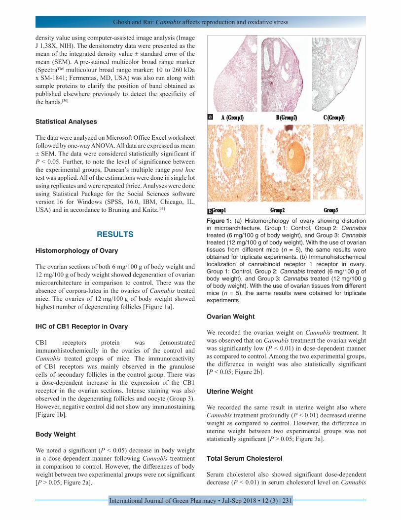
The ovarian sections of both 6 mg/100 g of body weight and 12 mg/100 g of body weight showed degeneration of ovarian microarchitecture in comparison to control. There was the absence of corpora-lutea in the ovaries of Cannabis treated mice. The ovaries of 12 mg/100 g of body weight showed highest number of degenerating follicles [Figure 1a].
IHC of CB1 Receptor in Ovary
CB1 receptors protein was demonstrated immunohistochemically in the ovaries of the control and Cannabis treated groups of mice. The immunoreactivity of CB1 receptors was mainly observed in the granulose cells of secondary follicles in the control group. There was a dose-dependent increase in the expression of the CB1 receptor in the ovarian sections. Intense staining was also observed in the degenerating follicles and oocyte (Group 3). However, negative control did not show any immunostaining [Figure 1b].
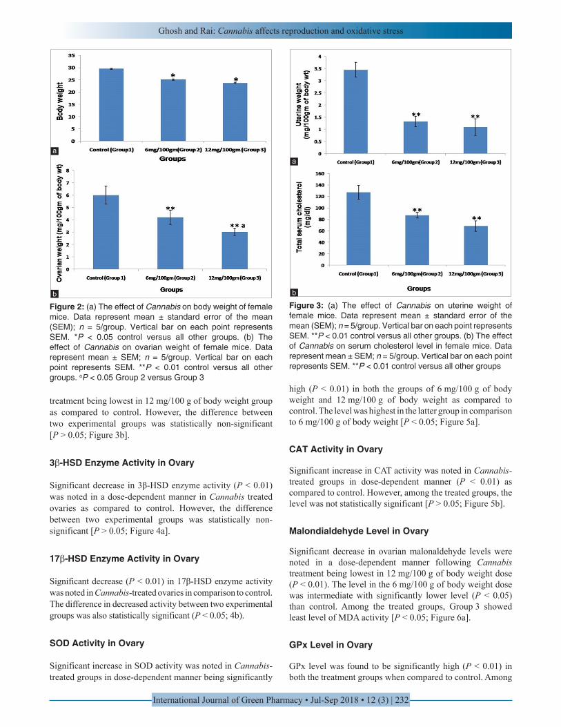
Body Weight
We noted a significant (P < 0.05) decrease in body weight in a dose-dependent manner following Cannabis treatment in comparison to control. However, the differences of body weight between two experimental groups were not significant [P > 0.05; Figure 2a].
Ovarian Weight
We recorded the ovarian weight on Cannabis treatment. It was observed that on Cannabis treatment the ovarian weight was significantly low (P < 0.01) in dose-dependent manner as compared to control. Among the two experimental groups, the difference in weight was also statistically significant [P < 0.05; Figure 2b].
Uterine Weight
We recorded the same result in uterine weight also where Cannabis treatment profoundly (P < 0.01) decreased uterine weight as compared to control. However, the difference in uterine weight between two experimental groups was not statistically significant [P > 0.05; Figure 3a].
Total Serum Cholesterol
Serum cholesterol also showed significant dose-dependent decrease (P < 0.01) in serum cholesterol level on Cannabis
Figure 1: (a) Histomorphology of ovary showing distortion in microarchitecture. Group 1: Control, Group 2: Cannabis treated (6 mg/100 g of body weight), and Group 3: Cannabis treated (12 mg/100 g of body weight). With the use of ovarian tissues from different mice (n = 5), the same results were obtained for triplicate experiments. (b) Immunohistochemical localization of cannabinoid receptor 1 receptor in ovary. Group 1: Control, Group 2: Cannabis treated (6 mg/100 g of body weight), and Group 3: Cannabis treated (12 mg/100 g of body weight). With the use of ovarian tissues from different mice (n = 5), the same results were obtained for triplicate experiments
a
b
Ghosh and Rai: Cannabis affects reproduction and oxidative stress
International Journal of Green Pharmacy • Jul-Sep 2018 • 12 (3) | 232
treatment being lowest in 12 mg/100 g of body weight group as compared to control. However, the difference between two experimental groups was statistically non-significant [P > 0.05; Figure 3b].
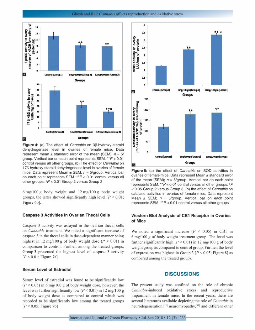
3β-HSD Enzyme Activity in Ovary
Significant decrease in 3β-HSD enzyme activity (P < 0.01) was noted in a dose-dependent manner in Cannabis treated ovaries as compared to control. However, the difference between two experimental groups was statistically non-significant [P > 0.05; Figure 4a].
17β-HSD Enzyme Activity in Ovary
Significant decrease (P < 0.01) in 17β-HSD enzyme activity was noted in Cannabis-treated ovaries in comparison to control. The difference in decreased activity between two experimental groups was also statistically significant (P < 0.05; 4b).
SOD Activity in Ovary
Significant increase in SOD activity was noted in Cannabis-treated groups in dose-dependent manner being significantly
high (P < 0.01) in both the groups of 6 mg/100 g of body weight and 12 mg/100 g of body weight as compared to control. The level was highest in the latter group in comparison to 6 mg/100 g of body weight [P < 0.05; Figure 5a].
CAT Activity in Ovary
Significant increase in CAT activity was noted in Cannabis-treated groups in dose-dependent manner (P < 0.01) as compared to control. However, among the treated groups, the level was not statistically significant [P > 0.05; Figure 5b].
Malondialdehyde Level in Ovary
Significant decrease in ovarian malonaldehyde levels were noted in a dose-dependent manner following Cannabis treatment being lowest in 12 mg/100 g of body weight dose (P < 0.01). The level in the 6 mg/100 g of body weight dose was intermediate with significantly lower level (P < 0.05) than control. Among the treated groups, Group 3 showed least level of MDA activity [P < 0.05; Figure 6a].
GPx Level in Ovary
GPx level was found to be significantly high (P < 0.01) in both the treatment groups when compared to control. Among
Figure 2: (a) The effect of Cannabis on body weight of female mice. Data represent mean ± standard error of the mean (SEM); n = 5/group. Vertical bar on each point represents SEM. *P < 0.05 control versus all other groups. (b) The effect of Cannabis on ovarian weight of female mice. Data represent mean ± SEM; n = 5/group. Vertical bar on each point represents SEM. **P < 0.01 control versus all other groups. aP < 0.05 Group 2 versus Group 3
b
a
Figure 3: (a) The effect of Cannabis on uterine weight of female mice. Data represent mean ± standard error of the mean (SEM); n = 5/group. Vertical bar on each point represents SEM. **P < 0.01 control versus all other groups. (b) The effect of Cannabis on serum cholesterol level in female mice. Data represent mean ± SEM; n = 5/group. Vertical bar on each point represents SEM. **P < 0.01 control versus all other groups
b
a
Ghosh and Rai: Cannabis affects reproduction and oxidative stress
International Journal of Green Pharmacy • Jul-Sep 2018 • 12 (3) | 233
6 mg/100 g body weight and 12 mg/100 g body weight groups, the latter showed significantly high level [P < 0.01; Figure 6b].
Caspase 3 Activities in Ovarian Thecal Cells
Caspase 3 activity was assayed in the ovarian thecal cells on Cannabis treatment. We noted a significant increase of caspase 3 in the thecal cells in dose-dependent manner being highest in 12 mg/100 g of body weight dose (P < 0.01) in comparison to control. Further, among the treated groups, Group 3 presented the highest level of caspase 3 activity [P < 0.01; Figure 7a].
Serum Level of Estradiol
Serum level of estradiol was found to be significantly low (P < 0.05) in 6 mg/100 g of body weight dose, however, the level was further significantly low (P < 0.01) in 12 mg/100 g of body weight dose as compared to control which was recorded to be significantly low among the treated groups [P < 0.05; Figure 7b]
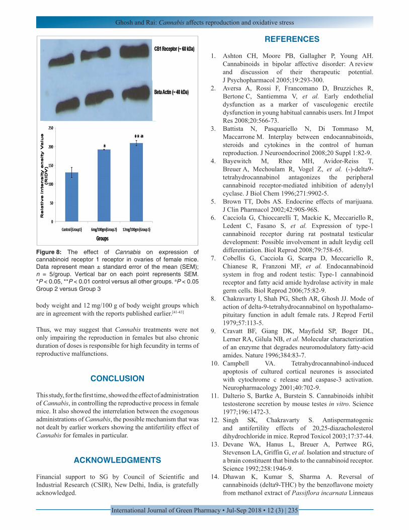
Western Blot Analysis of CB1 Receptor in Ovaries of Mice
We noted a significant increase (P < 0.05) in CB1 in 6 mg/100 g of body weight treatment group. The level was further significantly high (P < 0.01) in 12 mg/100 g of body weight group as compared to control group. Further, the level of expression was highest in Group 3 [P < 0.05; Figure 8] as compared among the treated groups.
DISCUSSIONS
The present study was confined on the role of chronic Cannabis-induced oxidative stress and reproductive impairment in female mice. In the recent years, there are several literatures available depicting the role of Cannabis in neurodegeneration,[32] neuromyopathy,[33] and different other
b
a
Figure 4: (a) The effect of Cannabis on 3β-hydroxy-steroid dehydrogenase level in ovaries of female mice. Data represent mean ± standard error of the mean (SEM); n = 5/group. Vertical bar on each point represents SEM. **P < 0.01 control versus all other groups. (b) The effect of Cannabis on 17β-hydroxy-steroid dehydrogenase level in ovaries of female mice. Data represent Mean ± SEM; n = 5/group. Vertical bar on each point represents SEM. **P < 0.01 control versus all other groups. bP < 0.01 Group 2 versus Group 3
Figure 5: (a) the effect of Cannabis on SOD activities in ovaries of female mice. Data represent Mean ± standard error of the mean (SEM); n = 5/group. Vertical bar on each point represents SEM. **P < 0.01 control versus all other groups. aP < 0.05 Group 2 versus Group 3. (b) the effect of Cannabis on catalase activities in ovaries of female mice. Data represent Mean ± SEM; n = 5/group. Vertical bar on each point represents SEM. **P < 0.01 control versus all other groups
b
a
Ghosh and Rai: Cannabis affects reproduction and oxidative stress
International Journal of Green Pharmacy • Jul-Sep 2018 • 12 (3) | 234
neurological disorders.[34] However, till date, there are no data or reports are available depicting the role of Cannabis treatment in regulating/modulating the female reproduction; however, it had been predicted from prolonged time that Cannabis is potent enough to interfere in reproduction in males[35] and females.
Our study, in relation to the dose-dependent effect of Cannabis treatment in the female reproduction, is the preliminary and elaborated study depicting the deleterious and detrimental effects of Cannabis in female reproduction. Our study is divided into two different parts addressing the role of Cannabis in reproductive impairments in female mice due to oxidative stress and loss in the functions of steroidogenesis.
We noted a significant decrease in body weight, ovarian and uterine weight upon Cannabis treatment, suggesting the first clue in reproductive impairment upon Cannabis treatment. The results were further supported by degeneration in ovarian histomorphology and increase in expressions of CB1 receptors in ovaries of different treatment groups. Cumulatively, the histological and immunohistochemical data suggest a dose-dependent impairment in ovarian as well as reproductive functions which are in agreement with previous reports in
where Cannabis causes reproductive impairment in males.[36] We have also studied the different aspects of free radical as well as reproductive enzyme activities (3β-HSD and 17β-HSD). The SOD, CAT, and GPx levels were significantly high in ovary tissues whereas MDA level was significantly low. The increased results of free-radical scavenging enzyme activities suggest the reproductive impairment in mice is may be due to high generation of free radicals and also due to different physiological malfunctions which are yet to be traced out.[37-40]
Further, significant decrease in total serum cholesterol levels, estradiol levels in circulation, 3β-HSD and 17β-HSD enzyme activities in ovarian tissues upon Cannabis treatment were noted. Thus, we may suggest that upon Cannabis treatment reproduction in females was impaired by Cannabis treatment by generation of free radicals in female reproductive tissues. The results were also discussed in light of apoptosis in thecal cells by Caspase 3 activity assays, and it was found to be significantly high in different doses of Cannabis treatments. To delineate the possible molecular mechanism of Cannabis function in ovary, we checked the CB1 receptor expression in ovarian tissues, and we also found that the CB1 receptor expressions were significantly high in both the 6mg/100g of
Figure 6: (a) the effect of Cannabis on malondialdehyde activities in ovaries of female mice. Data represent mean ± standard error of the mean (SEM); n = 5/group. Vertical bar on each point represents SEM. *P < 0.05, **P < 0.01 control versus all other groups. (b) The effect of Cannabis on GPx activities in ovaries of female mice. Data represent mean ± SEM; n = 5/group. Vertical bar on each point represents SEM. **P< 0.01 control versus all other groups. bP < 0.01 Group 2 versus Group 3
b
a
Figure 7: (a) The effect of Cannabis on Caspase 3 activities in ovarian thecal cells of female mice. Data represent mean ± standard error of the mean (SEM); n = 5/group. Vertical bar on each point represents SEM. **P< 0.01 control versus all other groups. bP< 0.01 Group 2 versus Group 3. (b) The effect of Cannabis on circulatory estradiol level in female mice. Data represents mean ± SEM; n = 5/group. Vertical bar on each point represents SEM. *P < 0.05, **P < 0.01 control versus all other groups
a
b
Ghosh and Rai: Cannabis affects reproduction and oxidative stress
International Journal of Green Pharmacy • Jul-Sep 2018 • 12 (3) | 235
body weight and 12 mg/100 g of body weight groups which are in agreement with the reports published earlier.[41-43]
Thus, we may suggest that Cannabis treatments were not only impairing the reproduction in females but also chronic duration of doses is responsible for high fecundity in terms of reproductive malfunctions.
CONCLUSION
This study, for the first time, showed the effect of administration of Cannabis, in controlling the reproductive process in female mice. It also showed the interrelation between the exogenous administrations of Cannabis, the possible mechanism that was not dealt by earlier workers showing the antifertility effect of Cannabis for females in particular.
ACKNOWLEDGMENTS
Financial support to SG by Council of Scientific and Industrial Research (CSIR), New Delhi, India, is gratefully acknowledged.
Figure 8: The effect of Cannabis on expression of cannabinoid receptor 1 receptor in ovaries of female mice. Data represent mean ± standard error of the mean (SEM); n = 5/group. Vertical bar on each point represents SEM. *P < 0.05, **P < 0.01 control versus all other groups. aP < 0.05 Group 2 versus Group 3
REFERENCES
1. Ashton CH, Moore PB, Gallagher P, Young AH. Cannabinoids in bipolar affective disorder: A review and discussion of their therapeutic potential. J Psychopharmacol 2005;19:293-300.
2. Aversa A, Rossi F, Francomano D, Bruzziches R, Bertone C, Santiemma V, et al. Early endothelial dysfunction as a marker of vasculogenic erectile dysfunction in young habitual cannabis users. Int J Impot Res 2008;20:566-73.
3. Battista N, Pasquariello N, Di Tommaso M, Maccarrone M. Interplay between endocannabinoids, steroids and cytokines in the control of human reproduction. J Neuroendocrinol 2008;20 Suppl 1:82-9.
4. Bayewitch M, Rhee MH, Avidor-Reiss T, Breuer A, Mechoulam R, Vogel Z, et al. (-)-delta9-tetrahydrocannabinol antagonizes the peripheral cannabinoid receptor-mediated inhibition of adenylyl cyclase. J Biol Chem 1996;271:9902-5.
5. Brown TT, Dobs AS. Endocrine effects of marijuana. J Clin Pharmacol 2002;42:90S-96S.
6. Cacciola G, Chioccarelli T, Mackie K, Meccariello R, Ledent C, Fasano S, et al. Expression of type-1 cannabinoid receptor during rat postnatal testicular development: Possible involvement in adult leydig cell differentiation. Biol Reprod 2008;79:758-65.
7. Cobellis G, Cacciola G, Scarpa D, Meccariello R, Chianese R, Franzoni MF, et al. Endocannabinoid system in frog and rodent testis: Type-1 cannabinoid receptor and fatty acid amide hydrolase activity in male germ cells. Biol Reprod 2006;75:82-9.
8. Chakravarty I, Shah PG, Sheth AR, Ghosh JJ. Mode of action of delta-9-tetrahydrocannabinol on hypothalamo-pituitary function in adult female rats. J Reprod Fertil 1979;57:113-5.
9. Cravatt BF, Giang DK, Mayfield SP, Boger DL, Lerner RA, Gilula NB, et al. Molecular characterization of an enzyme that degrades neuromodulatory fatty-acid amides. Nature 1996;384:83-7.
10. Campbell VA. Tetrahydrocannabinol-induced apoptosis of cultured cortical neurones is associated with cytochrome c release and caspase-3 activation. Neuropharmacology 2001;40:702-9.
11. Dalterio S, Bartke A, Burstein S. Cannabinoids inhibit testosterone secretion by mouse testes in vitro. Science 1977;196:1472-3.
12. Singh SK, Chakravarty S. Antispermatogenic and antifertility effects of 20,25-diazacholesterol dihydrochloride in mice. Reprod Toxicol 2003;17:37-44.
13. Devane WA, Hanus L, Breuer A, Pertwee RG, Stevenson LA, Griffin G, et al. Isolation and structure of a brain constituent that binds to the cannabinoid receptor. Science 1992;258:1946-9.
14. Dhawan K, Kumar S, Sharma A. Reversal of cannabinoids (delta9-THC) by the benzoflavone moiety from methanol extract of Passiflora incarnata Linneaus
Ghosh and Rai: Cannabis affects reproduction and oxidative stress
International Journal of Green Pharmacy • Jul-Sep 2018 • 12 (3) | 236
in mice: A possible therapy for cannabinoid addiction. J Pharm Pharmacol 2002;54:875-81.
15. Dixit VP, Lohiya NK. Effects of cannabis extract on the response of accessory sex organs of adult male mice to testosterone. Indian J Physiol Pharmacol 1975;19:98-100.
16. Dixit VP, Gupta CL, Agrawal M. Testicular degeneration and necrosis induced by chronic administration of cannabis extract in dogs. Endokrinologie 1977;69:299-305.
17. El-Gohary M, Eid MA. Effect of cannabinoid ingestion (in the form of bhang) on the immune system of high school and university students. Hum Exp Toxicol 2004;23:149-56.
18. Gammon CM, Freeman GM Jr. Xie W, Petersen SL, Wetsel WC. Regulation of gonadotropin-releasing hormone secretion by cannabinoids. Endocrinology 2005;146:4491-9.
19. Gaoni Y, Mechoulamm R. Isolation, structure and partial synthesis of an active constituent of hashish. J Am Chem Soc 1964;86:1646-7.
20. Gye MC, Kang HH, Kang HJ. Expression of cannabinoid receptor 1 in mouse testes. Arch Androl 2005;51:247-55.
21. Hillig KW, Mahlberg PG. A chemotaxonomic analysis of cannabinoid variation in cannabis (Cannabaceae). Am J Bot 2004;91:966-75.
22. Ghosh S, Haldar C. Histology and immunohistochemical localization of different receptors (MT1, MT2, AR, GR, ERα) in various organs (spleen, thymus, testis, ovary, uterus) of Indian goat C. Hircus. Int J Res Stud Bio Sci 2015;7:1-13.
23. Shivanandappa T, Venkatesh S. A colorimetric assay method for 3beta-hydroxy-delta5-steroid dehydrogenase. Anal Biochem 1997;254:57-61.
24. Blomquist CH, Lindemann NJ, Hakanson EY. 17β-hydroxysteroid and 20α-hydroxysteroid dehydrogenase activities of human placental microsomes: Kinetic evidence for two enzymes differing in substrate specificity. Arch Biochem Biophys 1985;239:206-15.
25. Das K, Samanta L, Chainy GB. A modified spectrophotometric assay of superoxide dismutase using nitrite formation by superoxide radicals. Indian J Biochem Biophys 2000;37:201-4.
26. Sinha AK. Colorimetric assay of catalase. Anal Biochem 1972;47:389-94.
27. Ohkawa H, Ohishi N, Yagi K. Reaction of linoleic acid hydroperoxide with thiobarbituric acid. J Lipid Res 1978;19:1053-7.
28. Mantha SV, Prasad M, Kalra J, Prasad K. Antioxidant enzymes in hypercholesterolemia and effects of vitamin E in rabbits. Atherosclerosis 1993;101:135-44.
29. Sharma S, Haldar C, Chaube SK. Effect of exogenous melatonin on X-ray induced cellular toxicity in lymphatic
tissue of Indian tropical male squirrel, Funambulus pennanti. Int J Radiat Biol 2008;84:363-74.
30. Ghosh S, Singh AK, Haldar C. Seasonal modulation of immunity by melatonin and gonadal steroids in a short day breeder goat Capra hircus. Theriogenology 2014;82:1121-30.
31. Bruning JL, Knitz BL. Computational Handbook of Statistics. 2nd ed. Illinois: Scott Foresman and Company; 1977.
32. Hall W, Solowij N. Adverse effects of cannabis. Lancet 1998;352:1611-6.
33. Harmon J, Aliapoulios MA. Gynecomastia in marihuana users. N Engl J Med 1972;287:936.
34. Jackson AL, Murphy LL. Role of the hypothalamic-pituitary-adrenal axis in the suppression of luteinizing hormone release by delta-9-tetrahydrocannabinol. Neuroendocrinology 1997;65:446-52.
35. Lewis SE, Maccarrone M. Endocannabinoids, sperm biology and human fertility. Pharmacol Res 2009;60:126-31.
36. Maccarrone M, Finazzi-Agró A. The endocannabinoid system, anandamide and the regulation of mammalian cell apoptosis. Cell Death Differ 2003;10:946-55.
37. Matsuda LA, Lolait SJ, Brownstein MJ, Young AC, Bonner TI. Structure of a cannabinoid receptor and functional expression of the cloned cDNA. Nature 1990;346:561-4.
38. Meccariello R, Franzoni MF, Chianese R, Cottone E, Scarpa D, Donna D, et al. Interplay between the endocannabinoid system and gnRH-I in the forebrain of the Anuran amphibian Rana esculenta. Endocrinology 2008;149:2149-58.
39. Moore TH, Zammit S, Lingford-Hughes A, Barnes TR, Jones PB, Burke M, et al. Cannabis use and risk of psychotic or affective mental health outcomes: A systematic review. Lancet 2007;370:319-28.
40. Munro S, Thomas KL, Abu-Shaar M. Molecular characterization of a peripheral receptor for cannabinoids. Nature 1993;365:61-5.
41. Murphy LL, Muñoz RM, Adrian BA, Villanúa MA. Function of cannabinoid receptors in the neuroendocrine regulation of hormone secretion. Neurobiol Dis 1998;5:432-46.
42. Pertwee RG. The therapeutic potential of drugs that target cannabinoid receptors or modulate the tissue levels or actions of endocannabinoids. AAPS J 2005;7:E625-54.
43. Pertwee RG, Ross RA, Craib SJ, Thomas A. (-)-cannabidiol antagonizes cannabinoid receptor agonists and noradrenaline in the mouse vas deferens. Eur J Pharmacol 2002;456:99-106.
Source of Support: Nil. Conflict of Interest: None declared.