Embed Size (px)
Citation preview

Chromosome-level genome assembly andtranscriptome of the green alga Chromochloriszofingiensis illuminates astaxanthin productionMelissa S. Rotha,1, Shawn J. Cokusb,1, Sean D. Gallaherc, Andreas Walterd,e, David Lopezb, Erika Ericksona,f,Benjamin Endelmana,f, Daniel Westcotta,f, Carolyn A. Larabelld,e, Sabeeha S. Merchantc,2, Matteo Pellegrinib,2,and Krishna K. Niyogia,f,2
aHoward Hughes Medical Institute, Department of Plant and Microbial Biology, University of California, Berkeley, CA 94720-3102; bDepartment ofMolecular, Cell and Developmental Biology, University of California, Los Angeles, CA 90095; cDepartment of Chemistry and Biochemistry and Institute forGenomics and Proteomics, University of California, Los Angeles, CA 90095-1569; dDepartment of Anatomy, University of California, San Francisco, CA 94143;eNational Center for X-ray Tomography, Lawrence Berkeley National Laboratory, Berkeley, CA 94720; and fMolecular Biophysics and Integrated BioimagingDivision, Lawrence Berkeley National Laboratory, Berkeley, CA 94720
Contributed by Krishna K. Niyogi, April 12, 2017 (sent for review December 6, 2016; reviewed by C. Robin Buell and Tomas Morosinotto)
Microalgae have potential to help meet energy and food demandswithout exacerbating environmental problems. There is interest inthe unicellular green alga Chromochloris zofingiensis, because itproduces lipids for biofuels and a highly valuable carotenoidnutraceutical, astaxanthin. To advance understanding of its biol-ogy and facilitate commercial development, we present a C. zofin-giensis chromosome-level nuclear genome, organelle genomes,and transcriptome from diverse growth conditions. The assembly,derived from a combination of short- and long-read sequencing inconjunction with optical mapping, revealed a compact genome of∼58 Mbp distributed over 19 chromosomes containing 15,274 pre-dicted protein-coding genes. The genome has uniform gene den-sity over chromosomes, low repetitive sequence content (∼6%),and a high fraction of protein-coding sequence (∼39%) with rela-tively long coding exons and few coding introns. Functional anno-tation of gene models identified orthologous families for themajority (∼73%) of genes. Synteny analysis uncovered localizedbut scrambled blocks of genes in putative orthologous relation-ships with other green algae. Two genes encoding beta-ketolase(BKT), the key enzyme synthesizing astaxanthin, were found inthe genome, and both were up-regulated by high light. Isolationand molecular analysis of astaxanthin-deficient mutants showedthat BKT1 is required for the production of astaxanthin. Moreover,the transcriptome under high light exposure revealed candidategenes that could be involved in critical yetmissing steps of astaxanthinbiosynthesis, including ABC transporters, cytochrome P450 enzymes,and an acyltransferase. The high-quality genome and transcriptomeprovide insight into the green algal lineage and carotenoid production.
Chlorophyceae | carotenoid biosynthesis | de novo genome |genome mapping | RNA-Seq
The growing human population generates an increasing de-mand for food and energy, which intensifies environmental
problems such as global climate change. Microalgae have thepotential to become a major source of sustainable bioproducts,because they use solar energy, grow quickly, consume CO2, andcan be cultivated on nonarable land (1, 2). However, there arepresently considerable practical limitations in the production ofbiofuels from microalgae, resulting in low productivity and highcosts. If microalgae can produce high-value bioproducts inaddition to biofuel components, it could improve the economicviability of commercial algae production. The green microalgaChromochloris zofingiensis is a promising source of lipids forbiofuel and the highly valuable ketocarotenoid astaxanthin,which has nutritive value because of its benefits in humanhealth, making it a leading candidate for commercial scale-up(3–5). However, much remains unknown about the genome andregulation of metabolism in this alga.
C. zofingiensis (division Chlorophyta, class Chlorophyceae,order Sphaeropleales) (6) is a simple ∼4-μm, unicellular, hap-loid, coccoid alga containing multiple mitochondria, which arevisualized typically as a tubular network, and a single inter-connected chloroplast that occupies ∼40% of the cell volumeand contains starch granules (Fig. 1, Movies S1 and S2, and SIAppendix, SI Text). Most of the mitochondria are in close asso-ciation with either the nucleus or the chloroplast. However,neither flagella (cilia) nor pyrenoids were visually observed.Because of the lack of obvious morphological characteristics,C. zofingiensis was originally described as a Chlorella species (6),at times transferred to the genera Muriella and Mychonastes,and finally placed using molecular sequencing into the genusChromochloris (7). Similar to its close relative, the model algaChlamydomonas reinhardtii, C. zofingiensis exhibits multiple fission
Significance
The growing human population generates increasing demandfor food and energy. Microalgae are a promising source ofsustainable bioproducts whose production may not exacerbateworsening environmental problems. The green alga Chromo-chloris zofingiensis has potential as a biofuel feedstock andsource of high-value nutraceutical molecules, including thecarotenoid astaxanthin. We present a high-quality, chromosome-level assembly of the genome by using a hybrid sequencingapproach with independent validation by optical mapping. Ouranalyses of the genome and transcriptome, in addition to ex-periments characterizing astaxanthin production, advance un-derstanding of the green lineage and carotenoid production,and enhance prospects for improving commercial productionof C. zofingiensis.
Author contributions: M.S.R., S.J.C., S.D.G., C.A.L., S.S.M., M.P., and K.K.N. designed re-search; M.S.R., S.J.C., S.D.G., A.W., and B.E. performed research; M.S.R., S.J.C., S.D.G., A.W.,D.L., E.E., and D.W. analyzed data; and M.S.R. and S.J.C. wrote the paper.
Reviewers: C.R.B., Michigan State University; and T.M., Università di Padova.
The authors declare no conflict of interest.
Freely available online through the PNAS open access option.
Data deposition: The Chromochloris zofingiensis version 5 assembly and associated anno-tations and gene families are included as Datasets S1–S19 (see SI Appendix, Datasets Key),and are also available on Phytozome and the project webpage at genomes.mcdb.ucla.edu/Chromochloris/. Raw Illumina and Pacific Biosciences genomic reads are available atNCBI Sequence Read Archive (accession nos. SRR5310949–SRR5310954), and raw RNA-Seqreads are under Gene Expression Omnibus accession no. GSE92515, with FPKM matricesalso available as Datasets S20 and S21.1M.S.R. and S.J.C. contributed equally to this work.2To whom correspondence may be addressed. Email: [email protected], [email protected], or [email protected].
This article contains supporting information online at www.pnas.org/lookup/suppl/doi:10.1073/pnas.1619928114/-/DCSupplemental.
E4296–E4305 | PNAS | Published online May 8, 2017 www.pnas.org/cgi/doi/10.1073/pnas.1619928114
Dow
nloa
ded
by g
uest
on
May
26,
202
0

with temporal separation between cell growth and cell division.C. zofingiensis primarily divides into two or four daughter cells (SIAppendix, Fig. S1 and Movies S3 and S4), but also can divide into16 (SI Appendix, Fig. S1 and Movie S5), 32, or 64 cells (6). Theregulation of cell division timing is unknown, but the daughtercells are the same size. Also like C. reinhardtii (8), the nucleus inC. zofingiensis divides before chloroplast division (SI Appendix, Fig.S1 andMovie S3). Intriguingly, C. zofingiensis has an extremely highphotoprotective capacity compared with other algae and plants (9).Moreover, under specific conditions, C. zofingiensis can dramat-ically increase the production of lipids and secondary carotenoids (3–5, 10). This alga produces triacylglycerols (TAGs), the preferredlipid precursor for biofuel products and accumulates these lipidsto some of the highest levels of 96 microalgae analyzed (3). Thus,C. zofingiensis is considered one of the most promising biofuelfeedstocks for commercial production.Increased production of the highly valuable ketocarotenoid
astaxanthin occurs in concert with accumulation of TAGs (4, 5).Astaxanthin has a broad range of commercial applications, in-cluding pharmaceuticals, nutraceuticals, cosmetics, food, andfeed (11–13). Recent studies have highlighted the antioxidantand antiinflammatory benefits of astaxanthin for applications inhuman health including cancer, cardiovascular disease, neuro-degenerative disease, inflammatory disease, diabetes, and obesitytreatments (11, 12). Although astaxanthin can be producedsynthetically, naturally produced astaxanthin is distinct in itsesterification and stereochemistry (13–15). These differencesresult in natural astaxanthin having >20-fold stronger antioxi-dant activity than synthetic astaxanthin, and only natural astax-anthin has been approved for human consumption (14). BecauseC. zofingiensis is fast growing, can be cultured under many con-ditions (including with wastewater), and reaches high culture densi-ties, C. zofingiensis has a higher potential to meet worldwide demandthan other natural sources, such as the microalga Haematococcuspluvialis, yeast, transgenic plants, and crustaceans (13, 15–17). Thus,C. zofingiensis is a prime candidate to supply the world with naturalastaxanthin and a source of renewable biofuel. However, improve-ments to maximize productivity and yield are needed, and key aspectsof astaxanthin biosynthesis and regulation remain to be elucidated.To better understand astaxanthin production and the biology
of C. zofingiensis, we sequenced and assembled its nuclear, mi-tochondrial, and plastid genomes using a hybrid approach, con-structed a transcriptome from 14 diverse conditions, examinedtranscriptomic changes through a shift from normal growth tothat in high light, generated and analyzed astaxanthin-deficientmutants, and identified candidate genes involved in algal astax-anthin biosynthesis. The high-quality, chromosome-level genomeassembly and accompanying transcriptome, combined with thecapacity for genetic transformation (18), establish a molecularfoundation to facilitate commercial development of C. zofingiensisand broaden understanding of the scope of metabolic and regu-latory mechanisms found in the green lineage.
Results and DiscussionWhole-Genome Sequencing, Assembly, and Global Architecture. Forgenome assembly of C. zofingiensis strain SAG 211–14, we used ahybrid approach blending short reads (Illumina), long reads(Pacific Biosciences of California), and whole-genome opticalmapping (OpGen) (SI Appendix, SI Text and Datasets S1–S19,and refer to SI Appendix, Datasets Key). The combined power ofthese approaches yielded a high-quality haploid nuclear genomeof C. zofingiensis of ∼58 Mbp distributed over 19 chromosomes(Fig. 2) in the tradition of model organism projects, as opposedto the fragmentary “gene-space” assemblies typical of modernprojects using high-throughput methods and associated software.Approximately 99% of reads from the Illumina genomic librarieswere accounted for, and nonplaceholder chromosomal sequencecovers ∼94% of the optical map. Because no automated pipelinewas found able to achieve the desired quality, methods are de-scribed in SI Appendix, SI Text.We compared genome features of C. zofingiensis to four other
green algae: C. reinhardtii, Coccomyxa subellipsoidea C-169,Chlorella sp. NC64A, and Monoraphidium neglectum (the clos-est relative with a sequenced genome), and the model plantArabidopsis thaliana (SI Appendix, Table S1 and SI Text). Similarto most green algae, the genome of C. zofingiensis is about half thesize of A. thaliana and C. reinhardtii; yet C. zofingiensis and all ofthe algae analyzed have more than double the number of chro-mosomes of A. thaliana. There is large variation in overall G+Ccontent, with C. zofingiensis the most balanced (∼51% nuclear,53% coding); although C. subellipsoidea C-169 is similar toC. zofingiensis, the other algal genomes have high G+C contentand A. thaliana has low G+C content, and high G+C is asso-ciated with more fragmentary assemblies. Although Chlorellasp. NC64A has a large number of large regions with distinct G+Ccontent, C. zofingiensis does not. Despite chromosome 5 ap-pearing to end in a large (>1 Mbp) inverted repeat (discussedin SI Appendix, SI Text), the relative repetitive content ofC. zofingiensis, like C. subellipsoidea C-169, appears to be low(∼6%). In contrast, M. neglectum is ∼50% higher and C. sp.NC64A ∼100% higher despite comparable genome sizes, andthe large genomes of C. reinhardtii and A. thaliana are roughlydouble compared with the highest of the other four. AfterC. subellipsoidea C-169, C. zofingiensis contains the most re-petitive fraction from novel repeats not known in RepbaseUpdate (19), which presently focuses only on A. thaliana andC. reinhardtii. Gene density in C. zofingiensis is quite uniformover chromosomes, and there are no grand-scale gradients ingenes or repeats such as found in, e.g., A. thaliana where eachchromosome has several megabase pairs of pericentromericheterochromatin (20), although some smaller scale repeat gradi-ents are found near large assembly gaps and putative (peri)cen-tromeres. RepeatMasker in conjunction with RepeatModeler andRepbase finds ∼5.0% of C. zofingiensis sequence consists ofinterspersed repeats [∼2.0% long interspersed nuclear elements(LINEs), ∼1.5% LTRs, ∼1.2% unclassified, and ∼0.4% DNA ele-ments] with the remainder mostly simple repeats (∼1.0%) and withsome satellites, low complexity sequence, and small RNA (total of∼0.1%). The combination of smaller genome size, balanced G+Ccontent, and low repetitive sequence fraction undoubtedly assistedassembly.Complete (circular with no gaps or ambiguous nucleotides)
mitochondrial and chloroplast genomes for C. zofingiensis strainUTEX 56 (formerly Bracteacoccus cinnabarinus) were alreadyavailable as NCBI accession nos. KJ806268.1 (21) and KT199251.1(22), respectively. We independently assembled equivalent com-plete genomes de novo for strain SAG 211–14 (SI Appendix, TableS1 and SI Text). The two strains were isolated from similar habitatsin localities ∼300 km apart by different people in subsequent years.For the mitochondrial genome, the SAG and UTEX strains wereresolved as 41,733 bp and 44,840 bp, respectively, with the samemajor protein-coding genes, tRNAs, and rRNAs in the same order(SI Appendix, SI Text) (21). However, a pairwise alignment exhibitsonly ∼66% nucleotide identity, with divergence concentrated
A
2 μm
B C D
Fig. 1. C. zofingiensis cell morphology. Cryo-soft X-ray tomography of areconstructed cell with segmented nucleus (purple), chloroplast (green),mitochondria (red), lipids (yellow), and starch granules within the chloro-plast (blue). (A) A representative orthoslice of the reconstructed cell. (SeeMovie S1 for transmission of single cell.) (B) Three-dimensional segmentationover two orthogonal orthoslices. (C) Segmented chloroplast and nucleus.(D) Fully segmented cell (see Movie S2 for rotating view).
Roth et al. PNAS | Published online May 8, 2017 | E4297
PLANTBIOLO
GY
PNASPL
US
Dow
nloa
ded
by g
uest
on
May
26,
202
0

intergenically and in rrnL4, where splicing differs. Restricted tocoding sequence, nucleotide identity rises to ∼98%, and aminoacid identity is ∼99% in translations under the National Centerfor Biotechnology Information (NCBI) Scenedesmus obliquusmitochondrial genetic code. For the chloroplast genome, theSAG and UTEX strains resolved as 181,058 bp and 188,935 bp,respectively, with a ∼6.7 kbp and ∼6.4 kbp, respectively, rRNA-related inverted repeat (SI Appendix, SI Text) (22). Neither theIllumina short reads nor Pacific Biosciences long reads wereable to resolve the relative strand orientation of the two singlecopy regions for the SAG strain; a single contig was constructedwith an arbitrary relative orientation that is opposite that givenfor the UTEX strain. Between the strains, all major protein-coding genes, tRNAs, and rRNAs are again the same in thesame order. (In comparisons, the single copy regions werereoriented to agree.) Nucleotide identity is ∼83%, with diver-gence concentrated intergenically and with the largest singledifference being a loss in the SAG strain of almost all of a ∼9.3-kbpUTEX region annotated as containing a ptz-like ORF. Codingsequence identity is ∼98%, and translation under the NCBI bac-terial, archaeal, and plant plastid code gives ∼97% amino acididentity with lower identity in larger proteins (e.g., FtsH, RpoC2,and Ycf1). The low mitochondrial nucleotide identity was surprisinggiven the presumed closeness of the strains.
The current C. zofingiensis assembly (“ChrZofV5”) successfullyextended into telomere-associated repeats for 25 of 38 chromo-some tips, and unplaced contigs appear to contain another 11 tips,leaving only two tips unaccounted. The canonical unit appearsto be (CCCTAAA)n at 5′ ends [and the reverse complement,(TTTAGGG)n, at 3′ ends], similar to C. subellipsoidea C-169 andC. sp. NC64A and likelyM. neglectum, although C. reinhardtii mayprefer (CCCTAAAA)n. A comparison of counts of apparentlytelomere-associated reads vs. generic nuclear reads (and constraintsimposed by the optical map) suggest an average of ∼3.5-kbptelomeric repeats per tip. Further, based on experience withparticularly difficult sequences during assembly phases and anal-ysis of the chromosomal distributions of specific dispersed andtandem repeat families, for most chromosomes exactly one regionwas identified as a putative (peri)centromeric locus. These loci arecomplex nested insertions of a ∼4.7-kbp circular consensussequence that consists of a ∼4-kbp coding sequence of a type I/Copia LTR retrotransposon together with a ∼0.7-kbp spacer,and some 5S rDNA sequence (but apparently no large tandemarrays of a relatively short unit, such as in A. thaliana). The bestNCBI BLASTX hits are to the filamentous green alga Kleb-sormidium flaccidum and the colonial green alga Volvox carteri.These regions are reminiscent of the Zepp clusters describedin C. subellipsoidea C-169 (23), although the Zepp element is
0 Mbp 1 Mbp 2 Mbp 3 Mbp 4 Mbp
0 Mbp 1 Mbp 2 Mbp 3 Mbp 4 Mbp
1
2
3
4
5
6
7
8
9
10
11
12
13
14
15
16
17
18
19
unplaceds
rDNA (24x tandem)
Fig. 2. C. zofingiensis nuclear genome. The assembled sequence of the 19 chromosomes of the nuclear genome is shown (top bar in each pair) with thematching restriction fragment length fingerprint from the optical map (bottom bar in each pair). Nominal plus strands run 5′ to 3′ left to right. Thin verticaldivisions mark BamHI sites (in silico in top bars, optical consensus in bottom bars). Lines from restriction sites on one bar to another indicate a maximallyscoring alignment computed with a dynamic programming algorithm similar to that used in OpGen’s MapSolver software. Black squares at chromosomeedges indicate sequence assembly has reached telomere-associated repeats. Thick horizontal orange bars indicate explicit assembly gaps (runs of Ns). Thickhorizontal yellow bars indicate additional known assembly issues as cataloged in Dataset S4. Light blue background shading shows where alignments are notone-to-one; shading is light green otherwise. Red dots mark possible (peri)centromeric loci. The end of chromosome 5 is discussed in SI Appendix, SI Text, and∼24× copies of the rDNA unit likely predominate at the beginning of the large sequence gap at the end of chromosome 13. Unplaced contigs/scaffolds and24 copies of the rDNA unit are shown near the bottom right.
E4298 | www.pnas.org/cgi/doi/10.1073/pnas.1619928114 Roth et al.
Dow
nloa
ded
by g
uest
on
May
26,
202
0

LINE-like and not of LTR type. Various analyses (includingconstraints imposed by the optical map) provided a rough es-timate of only ∼25 kbp on average of (peri)centromere perchromosome in C. zofingiensis; perhaps construction of artifi-cial chromosomes may be easier in C. zofingiensis than in someother organisms.The canonical rDNA repeat unit of C. zofingiensis became
apparent early in assembly because of its presence in relativelyhigh copy number. It assembled as a 9,702-bp circular contig an-notated by RNAmmer 1.2 as ∼6.6-kbp 28S followed by ∼1.1 kbpof spacer followed by ∼1.8-kbp 18S followed by ∼0.2 kbp ofspacer. From the presence of homologous sequence on chromo-some 13 leading into the large sequencing gap of that chromo-some, the optical tandem repeat that begins that sequencing gap,and the presence of two BamHI sites in the consensus rDNA unit(creating alternating fragments of ∼6.0 kbp and ∼3.7 kbp that areconsistent with the optical tandem repeat), it is estimated that∼24× tandem copies of the rDNA unit predominate in the first∼40% of the large sequence gap of chromosome 13. (Variousanalyses, e.g., SI Appendix, Table S1, assume 24 exact copies beginthis gap.) The estimated number of copies is similar toM. neglectum,but drastically fewer than in the large genomes of A. thaliana andC. reinhardtii.
Genome Annotation and Transcriptomics. To facilitate annotation,we generated a C. zofingiensis transcriptome by using RNA-Seqdata collected from cells grown under 14 diverse conditionsdesigned to capture a significant fraction of the cell’s transcrip-tional repertoire (SI Appendix, SI Text and Dataset S20). Theseconditions included treatments of different light intensities, nu-trient limitations, and oxidative stress. Paired-end sequencing oftranscriptome libraries was performed to facilitate determinationof splice junctions, resolve close paralogous families, and de novoassembly (used as part of training the AUGUSTUS ab initio genecaller). To capture nonpolyadenylated transcripts, such as thosefrom mitochondria and chloroplasts, libraries were prepared fromtotal RNA depleted of rRNA.RNA-Seq coverage, in conjunction with the de novo tran-
scriptome assembly, was used to select a gene prediction methodfor producing gene models. Multiple pipelines, including Softberry’sFgenesh, MAKER (24), and AUGUSTUS (25), were evaluated byusing metrics such as RNA-Seq coverage capture and intron/exonboundary correlation with coverage. Of all evaluated pipelines, weselected AUGUSTUS as trained on the de novo transcriptome,which identified 15,274 nucleus-encoded protein-coding genes, ofwhich 15,203 begin with a start codon (ATG) and end with a stopcodon (TAA, TGA, TAG). The remaining 71 gene models arelocated on the unplaced contigs/scaffolds and likely extend beyondan edge in the assembly.When the RNA-Seq libraries were aligned to the genome as-
sembly, 95 ± 2% of reads aligned uniquely (mean ± SD, n = 10)and an additional 3 ± 1% aligned to multiple locations, suggestingthat the genome assembly represents nearly all coding genes.Further, 55 ± 3% of RNA-Seq reads overlap by at least 80% withthe coding portion of a gene model on the correct strand (mean ±SD, n = 10); only 1.3 ± 0.3% overlap with a gene model on theopposite strand. Current gene models do not include 5′ and 3′UTRs; extending gene models 1 kbp upstream and downstreamincreases the percentage of reads aligning to the correct strandto 96 ± 2%.To further examine completeness, BUSCO (26) was run to
identify C. zofingiensis orthologs for a set of 303 universal single-copy genes (USCOs) putatively universally found in eukaryotesas single copies (Fig. 3A). Given just the assembly, orthologswere identified by two-pass BUSCO for 92% of USCOs, with97% of these declared complete. Given peptides, orthologs wereidentified for 92% with 91% complete. BUSCO analyses on theother organisms of SI Appendix, Table S1 suggest C. zofingiensisgene model quality is comparable to that of C. subellipsoideaC-169 and C. sp. NC64A, superior to M. neglectum, and inferior toextensively studied model organisms C. reinhardtii and A. thaliana.
They also suggest the C. zofingiensis genome quality is higher thanall of the other algae (including C. reinhardtii), with less fragmentedand fewer missing orthologs.Before this work, NCBI contained 13 gene sequences marked
as from C. zofingiensis. There was 99% or greater identity and1% or fewer gaps as determined by BLAST alignment to our ge-nome for 12 of the 13 (SI Appendix, Table S2). Only AY072815.1,heat shock protein 70, had limited identity, but this sequence wasisolated from a different strain.C. zofingiensis has the highest predicted coding sequence density
(∼39%) of the SI Appendix, Table S1 organisms. The averagelength of its coding sequences (∼482 aa) is the longest apart fromoutlier C. reinhardtii, which helps bring C. reinhardtii to almost ashigh a fraction of coding sequence although its genome is muchlarger. However, the median length of C. zofingiensis coding se-quences (∼347 aa) is more ordinary. The length of individualcoding exons (whether by mean ∼291 bp or median ∼194 bp) ofC. zofingiensis is the longest among the six, whereas the mean(∼5.0) and median (4) number of coding exons per gene is low,being more similar to M. neglectum and A. thaliana rather than thehigher numbers seen in the other algae. The number of identifiedtRNA loci (75, and forming a complete set for the standard aminoacids) is moderate like C. subellipsoidea C-169, rather than low asforC. sp. NC64A andM. neglectum, or high for the two large genomesof C. reinhardtii and A. thaliana.To compare the C. zofingiensis proteome to others in the green
lineage, we functionally annotated gene models by formingfamilies of genes across the six organisms of SI Appendix, TableS1 using a method based on reciprocal near-best global aminoacid alignments (SI Appendix, SI Text and Datasets S9–S18, andrefer to SI Appendix, Datasets Key). This analysis generally per-mits one, many, or no genes per organism per family and sepa-rates genes into closer “primary” (putative orthologs) vs. further“additional” relationships (putative paralogs). The result con-tains 10,490 families involving more than one organism, of which7,904 involve at most two genes per organism. There are somelarge families, with various histones constituting the largest. Ap-proximately 73% of C. zofingiensis genes (and ≥∼60% of everygenome) are placed in a family involving multiple organisms. Allsix genomes (including C. zofingiensis) show evidence of tandemduplication of genes. A phylogram (Fig. 3B) estimated fromputative 1:1:1:1:1:1 orthologs placed C. zofingiensis closest toM. neglectum and then C. reinhardtii, forming a three-memberclade that joins a two-member clade containing C. subellipsoideaC-169 and C. sp. NC64A, consistent with existing literature(27), and this whole-genome data analysis agrees with placingC. zofingiensis into genus Chromochloris (7).Although we do not find large stretches of nucleotide synteny
between C. zofingiensis and the other genomes, we do find amongall members in the green algal lineage (except for M. neglectum,whose current assembly is too fragmented for such an analysis)highly significant genomically localized blocks of genes in putativeorthologous relationships (Fig. 3C and SI Appendix, Figs. S2–S10),extending the result for C. subellipsoidea C-169 vs. C. sp. NC64A(fig. 2 in ref. 23). Whereas block boundaries are rather well defined,gene order and coding strands within blocks are generally com-pletely scrambled. The topology of Fig. 3B can be reconstructedjust from the pattern of syntenies. It is likely that the blocksrepresent random chromosomal rearrangements that accumu-late over time and diverge after speciation.To gain more insight into the metabolic function and cellular
processes associated with specific proteins, we used in silicomethods to predict subcellular localization of proteins encodedby the nuclear genome of C. zofingiensis. Using PredAlgo, analgal-specific subcellular localization prediction program trainedon C. reinhardtii (28), we predicted nucleus-encoded proteins todistribute as ∼15% to the secretory system, ∼12% to the chlo-roplast, and ∼10% to mitochondria. The majority of proteins(∼63%) were predicted to be localized to other areas, which maybe due to unidentified transit peptides, or the transit peptidesof C. zofingiensis being significantly different from PredAlgo’s
Roth et al. PNAS | Published online May 8, 2017 | E4299
PLANTBIOLO
GY
PNASPL
US
Dow
nloa
ded
by g
uest
on
May
26,
202
0

C. reinhardtii training set. Additionally, errors in gene models,especially in terminal regions, may result in inaccurate localiza-tion predictions. The predicted distribution is similar to what hasbeen noted for C. reinhardtii (29).Mitochondrial and chloroplast genes were highly expressed
over a wide range of conditions (SI Appendix, Fig. S11). Despitethe organellar genomes being significantly smaller and express-ing many fewer genes, the transcripts expressed by the chloro-plast and mitochondria represent a substantial portion of thetotal cellular mRNA. In an analysis of the transcriptomic datafrom 14 diverse growth conditions, 31 ± 9% and 7 ± 2% of totalRNA-Seq reads uniquely mapped to the chloroplast and mito-chondrion genomes, respectively (mean ± SD). Thus, RNA ex-pression is dramatically higher for organellar genes: for the73 protein-coding genes encoded in the chloroplast and 22 in themitochondria, median transcript abundance across conditionswas 686 and 419 FPKMs, respectively, in contrast to 5 FPKMsacross all nuclear-encoded genes.To identify genes that were more highly regulated under
specific conditions, we compared expression of every gene overthe 14 conditions and selected those with z-scores beyond ±2,plotting these as heatmaps (SI Appendix, Figs. S12–S21). Themost prominent treatment to affect nuclear and plastid geneexpression was oxidative stress by hydrogen peroxide (SI Ap-pendix, Fig. S12), which significantly affected 3,934 genes. Thesegenes were enriched for ABC-transporter domains (P = 1.0 × 10⁻⁶),suggesting that export of toxics and xenobiotics is a significantmechanism for handling environmental stress in C. zofingiensis.Similarly, singlet oxygen stress induced by the chemical Rose Bengal
affected 1,477 genes (SI Appendix, Fig. S20) and heterotrophicgrowth on glucose identified 853 genes (SI Appendix, Fig. S14).Nutrient deprivation had similar effects on most genes and far fewergenes were identified by analyses; for example, only 21 genes weredetected as highly enriched in the iron-deficient sample.
Cryptic Sex and Motility in C. zofingiensis. Although C. zofingiensishas long been assumed to be asexual and nonmotile, we inves-tigated the presence of putative cilia/flagella and meiosis genesin its genome via the computationally-identified gene families inconjunction with examination of associated gene expressionacross our conditions. The sequencing of the genome of C. sp.NC64A established a precedent for this type of analysis in greenalgae; similar to C. zofingiensis, neither sexual cycle nor flagellahave been observed in C. sp. NC64A, yet its genome revealedmeiosis-specific and primarily motile flagella genes, suggesting acryptic sexual cycle (30). In the C. zofingiensis genome, we foundputative orthologs for 73 of 78 genes (∼94%) in the CiliaCut(31), suggesting that there could be a previously unobserved motilelife cycle stage with flagella in this organism. C. zofingiensis wasmissing only five genes: DLC4, FAP111, FBB5, IFT20, and Tctex1.(All gene symbols in this work are with implicit “[v5.2]” versionsuffixes.) In C. reinhardtii, the ift20 deletion mutant lacks flagellaand is immotile (32), but perhaps C. zofingiensis has an as-yetunidentified gene with similar function. C. zofingiensis seems tohave critical C. reinhardtii genes for flagella motility (FLA14; ref.33) and forming flagella (PF15, PF19), including conservation offunctional residues in these two genes (34, 35). Additionally,FLA14, PF15, and PF19 were expressed in a variety of conditions,
A
B
C. reinhardtii chromosomes then scaffolds (Mbp in concatenated genome)
C
C. zofingiensis chrom
osomes then unplaceds
and rDN
A unit (M
bp in concatenated genome)
A. thaliana
C. reinhardtii
M. neglectum
C. subellipsoidea C-169
C. sp. NC64A0.23
Gen
ome
roun
d 2
Tran
scrip
tsP
eptid
es
Single-copy complete Duplicated complete Fragmented Missing
Fig. 3. Gene families. (A) BUSCO was run on the genomes (two-pass), splice variant-representative transcripts, and representative peptides of the six or-ganisms of SI Appendix, Table S1 to locate copies of its set of 303 putative eukaryotic universal single-copy genes (USCOs). In each run, each USCO is classifiedas exactly one of single-copy complete, duplicated complete, fragmentary, or missing. Although the gene models of established model organism C. reinhardtiiunsurprisingly fare better than the other algae, C. zofingiensis is comparable to the remaining algae, and its genome is the best of the algal genomesanalyzed. A. thaliana’s excellent gene models show high duplication, suggesting BUSCO’s set of eukaryotic USCO’s are not all truly universally single copy. (B)Using a procedure based on reciprocal near-best global amino acid alignments, protein-coding gene families among the six organisms of SI Appendix, TableS1 were formed; a phylogram estimated from restriction to putative 1:1:1:1:1:1 orthologs is consistent with existing literature (27). (C) Scatterplots showscrambled syntenic blocks of conserved genes in the algal lineage (SI Appendix, Figs. S2–S10) (similar to fig. 2 in ref. 23). (Organism pairs involving the highlyfragmented assembly ofM. neglectum are omitted.) Each plot uses those gene families that, for the two organisms selected, have exactly one primary gene ain the first organism and exactly one primary gene b in the second organism. A dot with x, y coordinates at the midpoints of the span of the coding sequencesfor a, b is drawn in red if a and b are on the same nominal genomic strands and in green if they are on opposite strands; dots are plotted in a randomizedorder. Order of assembly sequences (but not nucleotides within sequences) is permuted on both axes so as to compact and emphasize statistically enrichedregions (indicated by orange background shading); small inner numbers give relevant portions of the assembly’s sequence names for sequences at least0.5 Mbp in length (e.g., “1” for “chr1”, i.e., “chromosome 1”). Rightward and downward are 5′ to 3′ on assembly plus strands and light gray lines markassembly sequence boundaries; outer numbers give sequence scales (Mbp positions along concatenated genomes), which are equal horizontally and verti-cally. Small reference sequences (e.g., unplaced contigs labeled as “u. . .” and scaffolds labeled as “s. . .”) group at right and bottom sides (for which light graydividing lines behind red and green orthologous gene dots may merge). Further details are given in SI Appendix, SI Text.
E4300 | www.pnas.org/cgi/doi/10.1073/pnas.1619928114 Roth et al.
Dow
nloa
ded
by g
uest
on
May
26,
202
0
which suggests that these genes are functional despite lackingvisible flagella. Furthermore, we identified putative orthologs of25 of 40 C. reinhardtii meiosis-associated genes (30, 36), whichwas more than we observed for C. sp. NC64A (only 22 of 40). InC. zofingiensis, most of these genes are transcribed under manyconditions, but a few such as GSP1, MER3, and DMC1 had lowtranscript abundance except under a low dose of Rose Bengal(5 μMRose Bengal, 0.5 h dark and 1 h of 100 μmol photons·m−2·s−1).Eleven of the families not found in C. zofingiensis were specific toC. reinhardtii. Although these data cannot rule out the possibilitythat a sexual cycle was recently lost, it is more likely that the highnumber of apparent cilia/flagella and meiosis genes suggests theexistence of sexual reproduction and a motile stage that has not yetbeen observed in C. zofingiensis. Life cycle studies and, in partic-ular, investigations in search of a cryptic sexual cycle, which mayrequire specific conditions, should be a subject of future researchin C. zofingiensis.
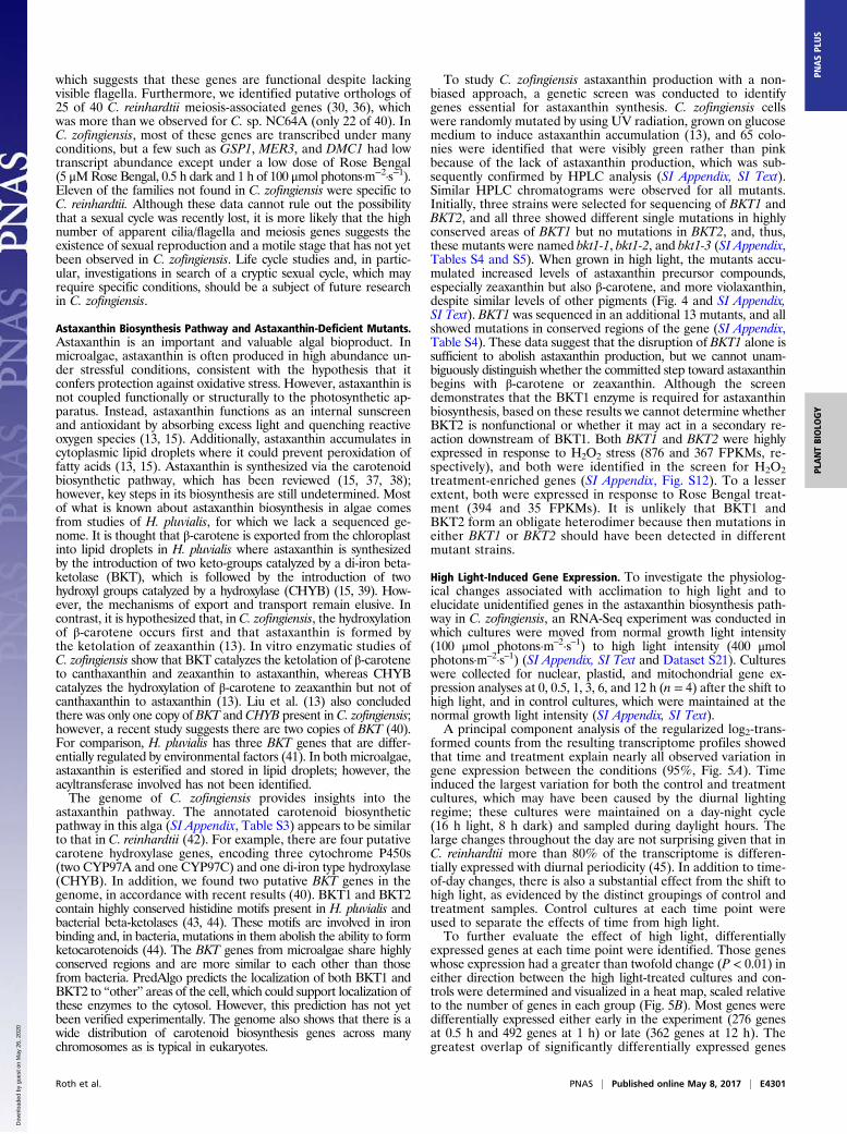
Astaxanthin Biosynthesis Pathway and Astaxanthin-Deficient Mutants.Astaxanthin is an important and valuable algal bioproduct. Inmicroalgae, astaxanthin is often produced in high abundance un-der stressful conditions, consistent with the hypothesis that itconfers protection against oxidative stress. However, astaxanthin isnot coupled functionally or structurally to the photosynthetic ap-paratus. Instead, astaxanthin functions as an internal sunscreenand antioxidant by absorbing excess light and quenching reactiveoxygen species (13, 15). Additionally, astaxanthin accumulates incytoplasmic lipid droplets where it could prevent peroxidation offatty acids (13, 15). Astaxanthin is synthesized via the carotenoidbiosynthetic pathway, which has been reviewed (15, 37, 38);however, key steps in its biosynthesis are still undetermined. Mostof what is known about astaxanthin biosynthesis in algae comesfrom studies of H. pluvialis, for which we lack a sequenced ge-nome. It is thought that β-carotene is exported from the chloroplastinto lipid droplets in H. pluvialis where astaxanthin is synthesizedby the introduction of two keto-groups catalyzed by a di-iron beta-ketolase (BKT), which is followed by the introduction of twohydroxyl groups catalyzed by a hydroxylase (CHYB) (15, 39). How-ever, the mechanisms of export and transport remain elusive. Incontrast, it is hypothesized that, in C. zofingiensis, the hydroxylationof β-carotene occurs first and that astaxanthin is formed bythe ketolation of zeaxanthin (13). In vitro enzymatic studies ofC. zofingiensis show that BKT catalyzes the ketolation of β-caroteneto canthaxanthin and zeaxanthin to astaxanthin, whereas CHYBcatalyzes the hydroxylation of β-carotene to zeaxanthin but not ofcanthaxanthin to astaxanthin (13). Liu et al. (13) also concludedthere was only one copy of BKT and CHYB present in C. zofingiensis;however, a recent study suggests there are two copies of BKT (40).For comparison, H. pluvialis has three BKT genes that are differ-entially regulated by environmental factors (41). In both microalgae,astaxanthin is esterified and stored in lipid droplets; however, theacyltransferase involved has not been identified.The genome of C. zofingiensis provides insights into the
astaxanthin pathway. The annotated carotenoid biosyntheticpathway in this alga (SI Appendix, Table S3) appears to be similarto that in C. reinhardtii (42). For example, there are four putativecarotene hydroxylase genes, encoding three cytochrome P450s(two CYP97A and one CYP97C) and one di-iron type hydroxylase(CHYB). In addition, we found two putative BKT genes in thegenome, in accordance with recent results (40). BKT1 and BKT2contain highly conserved histidine motifs present in H. pluvialis andbacterial beta-ketolases (43, 44). These motifs are involved in ironbinding and, in bacteria, mutations in them abolish the ability to formketocarotenoids (44). The BKT genes from microalgae share highlyconserved regions and are more similar to each other than thosefrom bacteria. PredAlgo predicts the localization of both BKT1 andBKT2 to “other” areas of the cell, which could support localization ofthese enzymes to the cytosol. However, this prediction has not yetbeen verified experimentally. The genome also shows that there is awide distribution of carotenoid biosynthesis genes across manychromosomes as is typical in eukaryotes.
To study C. zofingiensis astaxanthin production with a non-biased approach, a genetic screen was conducted to identifygenes essential for astaxanthin synthesis. C. zofingiensis cellswere randomly mutated by using UV radiation, grown on glucosemedium to induce astaxanthin accumulation (13), and 65 colo-nies were identified that were visibly green rather than pinkbecause of the lack of astaxanthin production, which was sub-sequently confirmed by HPLC analysis (SI Appendix, SI Text).Similar HPLC chromatograms were observed for all mutants.Initially, three strains were selected for sequencing of BKT1 andBKT2, and all three showed different single mutations in highlyconserved areas of BKT1 but no mutations in BKT2, and, thus,these mutants were named bkt1-1, bkt1-2, and bkt1-3 (SI Appendix,Tables S4 and S5). When grown in high light, the mutants accu-mulated increased levels of astaxanthin precursor compounds,especially zeaxanthin but also β-carotene, and more violaxanthin,despite similar levels of other pigments (Fig. 4 and SI Appendix,SI Text). BKT1 was sequenced in an additional 13 mutants, and allshowed mutations in conserved regions of the gene (SI Appendix,Table S4). These data suggest that the disruption of BKT1 alone issufficient to abolish astaxanthin production, but we cannot unam-biguously distinguish whether the committed step toward astaxanthinbegins with β-carotene or zeaxanthin. Although the screendemonstrates that the BKT1 enzyme is required for astaxanthinbiosynthesis, based on these results we cannot determine whetherBKT2 is nonfunctional or whether it may act in a secondary re-action downstream of BKT1. Both BKT1 and BKT2 were highlyexpressed in response to H2O2 stress (876 and 367 FPKMs, re-spectively), and both were identified in the screen for H2O2treatment-enriched genes (SI Appendix, Fig. S12). To a lesserextent, both were expressed in response to Rose Bengal treat-ment (394 and 35 FPKMs). It is unlikely that BKT1 andBKT2 form an obligate heterodimer because then mutations ineither BKT1 or BKT2 should have been detected in differentmutant strains.
High Light-Induced Gene Expression. To investigate the physiolog-ical changes associated with acclimation to high light and toelucidate unidentified genes in the astaxanthin biosynthesis path-way in C. zofingiensis, an RNA-Seq experiment was conducted inwhich cultures were moved from normal growth light intensity(100 μmol photons·m–2·s–1) to high light intensity (400 μmolphotons·m–2·s–1) (SI Appendix, SI Text and Dataset S21). Cultureswere collected for nuclear, plastid, and mitochondrial gene ex-pression analyses at 0, 0.5, 1, 3, 6, and 12 h (n = 4) after the shift tohigh light, and in control cultures, which were maintained at thenormal growth light intensity (SI Appendix, SI Text).A principal component analysis of the regularized log2-trans-
formed counts from the resulting transcriptome profiles showedthat time and treatment explain nearly all observed variation ingene expression between the conditions (95%, Fig. 5A). Timeinduced the largest variation for both the control and treatmentcultures, which may have been caused by the diurnal lightingregime; these cultures were maintained on a day-night cycle(16 h light, 8 h dark) and sampled during daylight hours. Thelarge changes throughout the day are not surprising given that inC. reinhardtii more than 80% of the transcriptome is differen-tially expressed with diurnal periodicity (45). In addition to time-of-day changes, there is also a substantial effect from the shift tohigh light, as evidenced by the distinct groupings of control andtreatment samples. Control cultures at each time point wereused to separate the effects of time from high light.To further evaluate the effect of high light, differentially
expressed genes at each time point were identified. Those geneswhose expression had a greater than twofold change (P < 0.01) ineither direction between the high light-treated cultures and con-trols were determined and visualized in a heat map, scaled relativeto the number of genes in each group (Fig. 5B). Most genes weredifferentially expressed either early in the experiment (276 genesat 0.5 h and 492 genes at 1 h) or late (362 genes at 12 h). Thegreatest overlap of significantly differentially expressed genes
Roth et al. PNAS | Published online May 8, 2017 | E4301
PLANTBIOLO
GY
PNASPL
US
Dow
nloa
ded
by g
uest
on
May
26,
202
0

was during the early time points (0.5 and 1 h), unsurprising giventhat these samples were collected closest together in time. Ad-ditionally, during these early time points, high light had a greatereffect than time (Fig. 5A). Over the course of the experiment,there was greater up-regulation of significantly differentiallyexpressed genes with 67%, 75%, and 94% of genes up-regulatedat 0.5, 6, and 12 h, respectively, but more genes were significantlydown-regulated at 1 h (52%) and 3 h (86%). Most genes hadrelatively modest changes (<fourfold) in the cultures shifted tohigh light, although expression of ELIP8 (early light-inducedprotein) and ELIP10 had >20-fold increases at 0.5 h. Amongchloroplast-encoded genes, both psaA and atpF had significant up-regulation at 1 h. No significantly differentially expressed mito-chondrial genes were found during the shift to high light.Because of the high level of interest in astaxanthin production
in C. zofingiensis, we examined the genes involved in carotenoidbiosynthesis during the shift to high light. High light causes anaccumulation of secondary carotenoids (in particular, astaxanthin)
in C. zofingiensis (46–48). In the present study, both BKT1 andBKT2 have the highest increase in gene expression, which occursimmediately after the light shift at 0.5 h (Fig. 6). Despite the in-crease in BKT2 gene expression in high light, its role in carotenoidbiosynthesis has not been established. Many genes at variouspoints in the carotenoid biosynthesis pathway were up-regulatedearly (0.5 h and 1 h) in response to the high light treatment, in-cluding phytoene synthase (PSY) (Fig. 6), which catalyzes thecommitted step in carotenoid biosynthesis. Previous studies havereported similar up-regulation of PSY, PDS, BKT, and CHYB atlonger time points in response to increases in light (46, 49, 50).However, our study also revealed a significant increase in ex-pression of ZDS at 1 h and a significant decrease in LCYE shortlyafter the shift to high light. Additionally, down-regulation of manygenes in the carotenoid biosynthesis pathway was observed at latertime points (6 and 12 h) in both the treatment and control cul-tures; this regulation is likely an effect of the diurnal cycle. Ahigher expression of carotenoid biosynthesis genes would supportan increase in secondary carotenoids, but does not exclude thepossibility that posttranslational modifications of carotenoid bio-synthetic enzymes may also account for the accumulation ofsecondary carotenoids during high light.The high-quality genome and transcriptome we generated in
combination with the high light RNA-Seq experiment allowed usto identify candidates for additional genes involved in astax-anthin biosynthesis and accumulation. As mentioned above, littleis known about the mechanism of translocation of the astax-anthin precursor(s) out of the chloroplast, the hydroxylation ofthe astaxanthin precursor, transport of astaxanthin into lipiddroplets, or the esterification of astaxanthin. We identified genesputatively involved in astaxanthin biosynthesis by examiningsignificantly differentially expressed genes with high increases ingene expression during the shift to high light, and looking forannotated activity compatible with hypothetical mechanisms ofastaxanthin biosynthesis. Genes that are up-regulated early duringhigh light that may be implicated in the astaxanthin pathway includefour ABC transporters (Cz04g21110, Cz05g17060, Cz09g27180, andCz08g16130), two cytochrome P450 proteins (Cz10g28330 andCz11g14160), and an acyltransferase (Cz02g29020). The ABCtransporters may form a complex that exports the astaxanthinprecursor(s) from the chloroplast. The cytochrome P450 proteinscould be involved in hydroxylation of astaxanthin precursors in thecytosol, and the acyltransferase could be involved in esterificationof astaxanthin. However, these genes need to be experimentallytested in vitro and/or in vivo to determine whether they function inthe pathway. Improving understanding of the astaxanthin pathwaymay provide opportunities to enhance astaxanthin production inthis species and others.In addition to changes in carotenoid biosynthesis, we also in-
vestigated other algal high light responses, including photoprotectivemechanisms and chlorophyll metabolism. In photosynthetic organ-isms, excess light must be safely dissipated to prevent oxidativedamage.C. reinhardtii transiently expresses PSBS at the onset of highlight and LHCSR proteins accumulate under high light, and thisaccumulation is correlated with nonphotochemical quenching ca-pacity (51, 52). Whereas C. reinhardtii has multiple copies of bothLHCSR and PSBS (51, 52), we found only single copies of LHCSRand PSBS in C. zofingiensis, despite having high nonphotochemicalquenching capacity (9). As expected, both LHCSR and PSBS wereup-regulated at the early time points during the shift to high lightand, in particular, at 1 h, which is consistent with observations ofC. reinhardtii during the dark-to-light transition (45). Similar to thecarotenoid biosynthesis genes, under the diurnal cycle LHCSR andPSBS are down-regulated by the end of the day (6 and 12 h) in bothconditions (Fig. 6). Reduction in chlorophyll is another commonphysiological response of algae exposed to high light (53). Accord-ingly, during the shift to high light, many C. zofingiensis genes involvedin chlorophyll synthesis were down-regulated and chlorophylldegradation genes were up-regulated (Fig. 6). The combination ofthese changes would lead to a reduction in chlorophyll contenteither during acclimation or as a stress response to high light.
6 8 10 12 14 16
bkt1-2
WT
bkt1-3
bkt1-1
18Retention Time (min)
0
20
40
60
80
100
120
chlor
ophy
ll
lutein
violax
anthi
n
zeax
anthi
n
neox
anthi
n
-carot
ene
-carot
ene
pmol
/106
cel
ls
****
aa,b
cb
***
a acb*
Abs
orba
nce
(nor
mal
ized
mA
U)
N
V
A
L Z
Chl b
Chl a
AstAstAst
HL MLWT 1 2 3 WT 1 2 3
A
B
Fig. 4. C. zofingiensis astaxanthin-deficient mutants. Astaxanthin-deficientmutants were generated by using forward genetics; mutations were iden-tified in BKT1. (A) HPLC traces of wild type, bkt1-1, bkt1-2, and bkt1-3 grownunder high light (HL, 400–450 μmol photons·m−2·s−1), showing the mutants’lack of astaxanthin production. Pigment abbreviations are as follows: A(antheraxanthin), α (α-carotene), Ast (astaxanthin), β (β-carotene), Chl a(chlorophyll a), Chl b (chlorophyll b), L (lutein), N (neoxanthin), V (viola-xanthin), Z (zeaxanthin). Pigments were detected at 445 nm with referenceat 550 nm (SI Appendix, SI Text). Inset shows high light WT growth withastaxanthin resulting in orange-brown color from astaxanthin (orange) andchlorophylls (green), whereas mutants bkt1-1, bkt1-2, and bkt1-3 do notproduce astaxanthin and remain green. Under medium light (ML, 100 μmolphotons·m−2·s−1), WT does not produce high amounts of astaxanthin andremains green with similar color as bkt1-1, bkt1-2, and bkt1-3. (B) Pigmentlevels (mean ± SD, n = 3 or 4) in HL-grown WT, bkt1-1, bkt1-2, and bkt1-3showing higher levels of carotenoids with similar levels of chlorophyll. *P <0.05, **P < 0.01, ***P < 0.001 (SI Appendix, SI Text).
E4302 | www.pnas.org/cgi/doi/10.1073/pnas.1619928114 Roth et al.
Dow
nloa
ded
by g
uest
on
May
26,
202
0

Annotation of Metabolic Pathways and Photosynthesis-Related Genes.Genes encoding homologs of the primary metabolic pathway en-zymes involved with carbon, carotenoids, chlorophyll, fatty acids,lipids, and proteins involved in the composition, assembly, andregulation of the photosynthetic apparatus, were preliminarilyidentified by using the BLAT sequence search tool (54) against aC. zofingiensis draft genome (SI Appendix, Table S3). Based on thequality of alignments and comparison with well-characterized,closely-related plant and algal query sequences, the targetedgene models in C. zofingiensis were submitted as queries in re-ciprocal BLAST searches against the NCBI RefSeq nonredundantprotein database to confirm coverage, domain architecture, andsimilarity across closely-related homologs. Because of the highquality of the C. zofingiensis assembly, this procedure resulted in anearly complete list of putative genes needed to complete eachpathway. Identified gene models were used to assess the quality ofthe automated gene family analysis across the six species of SI Ap-pendix, Table S1, and the automated analysis was used to confirmadditional candidate models and expand the set of annotations.Based on high sequence similarities and conservation of functionaldomains, we are generally confident in assignments of homology.However, it is possible that additional functional isoforms com-posed of more divergent sequences may also be present, havingbeen missed by the parameters used for BLAT, BLAST, and theautomated gene family analysis.Identification and annotation of genes involved in lipid bio-
synthesis can provide targets for exploitation of C. zofingiensis forbiofuel production. Using other oleaginous organisms as a guide,we would expect a robust oil-producing microalga to have anexpanded family of acyltransferases. The C. zofingiensis diac-ylglycerol acyltransferases (DGAT) are too divergent from theprotein sequences of type 1 DGAT and DGTT (type 2 DGATs;ref. 55) in C. reinhardtii, A. thaliana, and M. neglectum to identifythe corresponding genes via BLAT. Using a more sensitive BLASTsearch with both types of DGAT sequences from C. subellipsoideaC-169 (gij545360296), Chlorella vulgaris (gbjALP13863.1), Nanno-chloropsis gaditana (gbjEWM23187.1), A. thaliana (gij15224779,gij18409359), and C. reinhardtii (Cre01.g045903, Cre03.g205050),additional copies of DGAT type 1- and DGTT-encoding geneswere identified in C. zofingiensis, and yet more were identified byusing the automated gene family analysis. In total, 11 genes wereidentified that have either an LPLAT (lysophospholipid acyl-transferase) domain or a closely-relatedMBOAT (membrane boundO-acyltransferase) domain (SI Appendix, Table S3). We have ten-tatively assigned these genes as encoding proteins with diacylglycerolacyltransferase activity; however, some of these C. zofingiensis genemodels have higher similarity to predicted proteins of unknownfunction than to annotated type 1 or type 2 DGAT proteins fromother closely related organisms. Our finding of multiple copies ofputative DGAT and DGTT genes in C. zofingiensis is consistent withtranscriptome results from the closely-related ATCC 30412 strain(40). It is also possible, although unlikely, that one or multiple ad-ditional copies of DGAT or DGTT may be yet unidentified becauseof a gene modeling or assembly problem.Homologous gene models were also identified for components
of the photosynthetic apparatus and its assembly, including pro-teins that compose PSI, PSII, the major and minor light harvestingantennae, the cytochrome b6f complex, the chloroplast ATP syn-thase complex, soluble electron carriers, and known assemblyfactors for these complexes (SI Appendix, Table S3). Thirteen of theC. zofingiensis light-harvesting complex (LHC) genes are predictedto be more like PSI-associated LHC genes (LHCAs) while nineare predicted to be PSII-associated LHC genes (LHCBs) (SIAppendix, Table S3). This distribution is in contrast to that found
Fig. 5. C. zofingiensis RNA expression during transition to high light. Cul-tures of C. zofingiensis were grown diurnally (16 h light, 8 h dark) in100 μmol photons·m−2·s−1 medium light (ML). At t = 0, cultures were transferredto 400 μmol photons·m−2·s−1 high light (HL). Samples were collected inquadruplicate at 0, 0.5, 1, 3, 6, and 12 h for ML cultures and at 0.5, 1, 3, 6,and 12 h for HL. Transcript abundances for each sample were determined byRNA-Seq. (A) Principal component analysis (PCA) of the regularized log2-transformed counts for all 44 samples. The two most significant components,accounting for 95% of variation, are shown. ML (triangles) and HL (circles)are displayed with time point indicated by color. (B) Differentially expressedgenes during transition to HL. Expression fold change in HL versus ML wasdetermined for each time point for all genes. Genes at least twofold up-regulated or twofold down-regulated are indicated by the height of the barabove or below the line, respectively (P < 0.01); the total is indicated aboveeach bar. The regularized log2-transformed fold change between HL and MLis shown in the black box for each time point as indicated by color. For
comparison, the fold change of each of these genes at the other time pointsis presented flanking the black box. The number of differentially expressedgenes in common between 0.5 and 1 h and between 0.5 and 12 h is indicatedby square brackets.
Roth et al. PNAS | Published online May 8, 2017 | E4303
PLANTBIOLO
GY
PNASPL
US
Dow
nloa
ded
by g
uest
on
May
26,
202
0

in C. reinhardtii, which has nine of each of LHCA and LHCB.Further experimental work is needed to confirm the expressionprofiles and photosystem association of each of these putativeLHC proteins, especially under different light and stress condi-tions. Of note is one LHC model (Lhcb-like3, Cz04g24050) withlittle to no transcriptional expression detected in any of ourRNA-Seq conditions.
ConclusionsOur analyses of the C. zofingiensis genome, transcriptome,astaxanthin-deficient mutants, and RNA expression changesunder high light reveal insights into the basic biology of the greenlineage of photosynthetic organisms and the carotenoid bio-synthesis pathway. We present, in the tradition of model organ-isms, a high-quality chromosome-level assembly with independentgenome validation. The compact ∼58-Mbp genome has balancedG+C content and is rich in protein-coding sequence with few longexons per gene and relatively little repetitive sequence. We iden-tified ortholog families for the majority of C. zofingiensis genes.The gene density is uniform over chromosomes and a synteniccomparison with other algae uncovered highly significantgenomically localized blocks of genes in putative orthologousrelationships; however, gene order and strands within blocksare scrambled. We have shown that BKT1 is critical for theproduction of astaxanthin and have identified candidate genesthat could be missing components in astaxanthin biosynthesisand accumulation pathways. The addition of genomics to the
experimental toolkit for C. zofingiensis makes it an attractivealga not only for fundamental studies of its biology but also theeconomically viable and environmentally sustainable productionof biofuels and important bioproducts.
ACKNOWLEDGMENTS. The cryo-soft X-ray tomography was supportedby the US Department of Energy, Office of Science, Basic Energy Sciences,Chemical Sciences, Geosciences, and Biosciences Division under field workproposal SISGRKN. The whole-genome optical mapping and high lightRNA sequencing was supported by the US Department of Energy, Officeof Science, Basic Energy Sciences, Chemical Sciences, Geosciences, and Bio-sciences Division under field work proposal 449B. The transcriptome se-quencing and long read sequencing was supported by Agriculture andFood Research Initiative Competitive Grant 2013-67012-21272 from the USDepartment of Agriculture National Institute of Food and Agriculture (toM.S.R.). The National Center for X-ray Tomography is supported by theNational Institute of General Medical Sciences of the National Institutesof Health Grant P41GM103445 and the US Department of Energy, Officeof Biological and Environmental Research Grant DE-AC02-05CH11231.S.J.C., S.D.G., S.S.M., and M.P. were supported by a cooperative agreementwith the US Department of Energy Office of Science, Office of Biologicaland Environmental Research program under Award DE-FC02-02ER63421.D.L. was supported by a National Institutes of Health T32 Training Fellow-ship in Genome Analysis 5T32HG002536–13, the Eugene V. Cota-RoblesFellowship, and the Fred Eiserling and Judith Lengyel Doctoral Fellowship.D.W. was supported by a National Science Foundation Graduate ResearchFellowship. K.K.N. is an Investigator of the Howard Hughes Medical In-stitute and the Gordon and Betty Moore Foundation through GrantGBMF3070.
Fig. 6. C. zofingiensis RNA-Seq expression of select genes during the transition to high light. RNA-Seq was performed on cultures following a shift frommedium light (ML, 100 μmol photons·m−2·s−1) to high light (HL, 400–450 μmol photons·m−2·s−1) as described in Fig. 5. C. zofingiensis genes potentially in-volved in carotenoid biosynthesis, nonphotochemical quenching (NPQ), and chlorophyll biosynthesis and degradation were identified by manual curation.(Left) The regularized log2-transformed fold change between HL and ML for each of these genes at each time point is plotted as a heatmap. (Right) Theregularized log2-transformed fold change between each time point relative to t = 0 is plotted. Significantly differential genes that are over twofold up- ordown-regulated are indicated by an asterisk and bold text (P < 0.01).
E4304 | www.pnas.org/cgi/doi/10.1073/pnas.1619928114 Roth et al.
Dow
nloa
ded
by g
uest
on
May
26,
202
0

1. Stephens E, et al. (2010) Future prospects of microalgal biofuel production systems.Trends Plant Sci 15:554–564.
2. Wijffels RH, Barbosa MJ (2010) An outlook on microalgal biofuels. Science 329:796–799.
3. Breuer G, Lamers PP, Martens DE, Draaisma RB, Wijffels RH (2012) The impact of ni-trogen starvation on the dynamics of triacylglycerol accumulation in nine microalgaestrains. Bioresour Technol 124:217–226.
4. Liu J, Mao X, Zhou W, Guarnieri MT (2016) Simultaneous production of triacylglyceroland high-value carotenoids by the astaxanthin-producing oleaginous green micro-alga Chlorella zofingiensis. Bioresour Technol 214:319–327.
5. Mulders KJM, et al. (2014) Effect of biomass concentration on secondary carotenoidsand triacylglycerol (TAG) accumulation in nitrogen-depleted Chlorella zofingiensis.Algal Res 6:8–16.
6. Dönz OC (1934) Chlorella zofingiensis, eine neue Bodenalge. Ber Schweiz Bot Ges 43:127–131.
7. Fucíková K, Lewis LA (2012) Intersection of Chlorella, Muriella and Bracteacoccus:Resurrecting the genus Chromochloris Kol & Chodat (Chlorophyceae, Chlorophyta).Fottea 12:83–93.
8. Goodenough UW (1970) Chloroplast division and pyrenoid formation in Chlamydo-monas reinhardii. J Phycol 6:1–6.
9. Bonente G, et al. (2008) The occurrence of the psbS gene product in Chlamydomonasreinhardtii and in other photosynthetic organisms and its correlation with energyquenching. Photochem Photobiol 84:1359–1370.
10. Ip PF, Wong KH, Chen F (2004) Enhanced production of astaxanthin by the green mi-croalga Chlorella zofingiensis in mixotrophic culture. Process Biochem 39:1761–1766.
11. Hussein G, Sankawa U, Goto H, Matsumoto K, Watanabe H (2006) Astaxanthin, acarotenoid with potential in human health and nutrition. J Nat Prod 69:443–449.
12. Yuan J-P, Peng J, Yin K, Wang J-H (2011) Potential health-promoting effects of as-taxanthin: A high-value carotenoid mostly from microalgae. Mol Nutr Food Res 55:150–165.
13. Liu J, et al. (2014) Chlorella zofingiensis as an alternative microalgal producer of as-taxanthin: Biology and industrial potential. Mar Drugs 12:3487–3515.
14. Capelli B, Bagchi D, Cysewski GR (2013) Synthetic astaxanthin is significantly inferiorto algal-based astaxanthin as an antioxidant and may not be suitable as a humannutraceutical supplement. Nutrafoods 12:145–152.
15. Solovchenko AE (2015) Recent breakthroughs in the biology of astaxanthin accu-mulation by microalgal cell. Photosynth Res 125:437–449.
16. Liu J, et al. (2013) Utilization of cane molasses towards cost-saving astaxanthin pro-duction by a Chlorella zofingiensis mutant. J Appl Phycol 25:1447–1456.
17. Yuan Z, et al. (2013) Scale-up potential of cultivating Chlorella zofingiensis in piggerywastewater for biodiesel production. Bioresour Technol 137:318–325.
18. Liu J, et al. (2014) Genetic engineering of the green alga Chlorella zofingiensis: Amodified norflurazon-resistant phytoene desaturase gene as a dominant selectablemarker. Appl Microbiol Biotechnol 98:5069–5079.
19. Bao W, Kojima KK, Kohany O (2015) Repbase Update, a database of repetitive ele-ments in eukaryotic genomes. Mob DNA 6:11.
20. Arabidopsis Genome Initiative (2000) Analysis of the genome sequence of the flow-ering plant Arabidopsis thaliana. Nature 408:796–815.
21. Fucíková K, Lewis PO, González-Halphen D, Lewis LA (2014) Gene arrangementconvergence, diverse intron content, and genetic code modifications in mitochondrialgenomes of sphaeropleales (chlorophyta). Genome Biol Evol 6:2170–2180.
22. Fucíková K, Lewis PO, Lewis LA (2016) Chloroplast phylogenomic data from the greenalgal order Sphaeropleales (Chlorophyceae, Chlorophyta) reveal complex patterns ofsequence evolution. Mol Phylogenet Evol 98:176–183.
23. Blanc G, et al. (2012) The genome of the polar eukaryotic microalga Coccomyxasubellipsoidea reveals traits of cold adaptation. Genome Biol 13:R39.
24. Cantarel BL, et al. (2008) MAKER: An easy-to-use annotation pipeline designed foremerging model organism genomes. Genome Res 18:188–196.
25. Stanke M, Schöffmann O, Morgenstern B, Waack S (2006) Gene prediction in eu-karyotes with a generalized hidden Markov model that uses hints from externalsources. BMC Bioinformatics 7:62.
26. Simão FA, Waterhouse RM, Ioannidis P, Kriventseva EV, Zdobnov EM (2015) BUSCO:Assessing genome assembly and annotation completeness with single-copy orthologs.Bioinformatics 31:3210–3212.
27. Leliaert F, et al. (2012) Phylogeny and molecular evolution of the green algae. Crit RevPlant Sci 31:1–46.
28. Tardif M, et al. (2012) PredAlgo: A new subcellular localization prediction tool ded-icated to green algae. Mol Biol Evol 29:3625–3639.
29. Lopez D, et al. (2015) Dynamic changes in the transcriptome and methylome ofChlamydomonas reinhardtii throughout its life cycle. Plant Physiol 169:2730–2743.
30. Blanc G, et al. (2010) The Chlorella variabilis NC64A genome reveals adaptation tophotosymbiosis, coevolution with viruses, and cryptic sex. Plant Cell 22:2943–2955.
31. Merchant SS, et al. (2007) The Chlamydomonas genome reveals the evolution of keyanimal and plant functions. Science 318:245–250.
32. Engel BD, et al. (2009) Total internal reflection fluorescence (TIRF) microscopy ofChlamydomonas flagella. Methods Cell Biol 93:157–177.
33. Pazour GJ, Wilkerson CG, Witman GB (1998) A dynein light chain is essential for theretrograde particle movement of intraflagellar transport (IFT). J Cell Biol 141:979–992.
34. Dymek EE, Smith EF (2012) PF19 encodes the p60 catalytic subunit of katanin and isrequired for assembly of the flagellar central apparatus in Chlamydomonas. J Cell Sci125:3357–3366.
35. Dymek EE, Lefebvre PA, Smith EF (2004) PF15p is the chlamydomonas homologue ofthe Katanin p80 subunit and is required for assembly of flagellar central microtu-bules. Eukaryot Cell 3:870–879.
36. Ferris PJ, Armbrust EV, Goodenough UW (2002) Genetic structure of the mating-typelocus of Chlamydomonas reinhardtii. Genetics 160:181–200.
37. Lemoine Y, Schoefs B (2010) Secondary ketocarotenoid astaxanthin biosynthesis inalgae: A multifunctional response to stress. Photosynth Res 106:155–177.
38. Takaichi S (2011) Carotenoids in algae: Distributions, biosyntheses and functions. MarDrugs 9:1101–1118.
39. Grunewald K, Hagen C (2001) β-carotene is the intermediate exported from thechloroplast during accumulation of secondary carotenoids in Haematococcus pluvia-lis. J Appl Phycol 13:89–93.
40. Huang W, et al. (2016) Transcriptome analysis of Chlorella zofingiensis to identifygenes and their expressions involved in astaxanthin and triacylglycerol biosynthesis.Algal Res 17:236–243.
41. Huang JC, Chen F, Sandmann G (2006) Stress-related differential expression of mul-tiple beta-carotene ketolase genes in the unicellular green alga Haematococcuspluvialis. J Biotechnol 122:176–185.
42. Lohr M, Im CS, Grossman AR (2005) Genome-based examination of chlorophyll andcarotenoid biosynthesis in Chlamydomonas reinhardtii. Plant Physiol 138:490–515.
43. Huang J, Zhong Y, Sandmann G, Liu J, Chen F (2012) Cloning and selection of ca-rotenoid ketolase genes for the engineering of high-yield astaxanthin in plants.Planta 236:691–699.
44. Ye RW, Stead KJ, Yao H, He H (2006) Mutational and functional analysis of the beta-carotene ketolase involved in the production of canthaxanthin and astaxanthin. ApplEnviron Microbiol 72:5829–5837.
45. Zones JM, Blaby IK, Merchant SS, Umen JG (2015) High-resolution profiling of asynchronized diurnal transcriptome from Chlamydomonas reinhardtii reveals con-tinuous cell and metabolic differentiation. Plant Cell 27:2743–2769.
46. Li Y, Huang J, Sandmann G, Chen F (2009) High-light and sodium chloride stressdifferentially regulate the biosynthesis of astaxanthin in Chlorella zofingiensis(Chlorophyceae). J Phycol 45:635–641.
47. Del Campo JA, et al. (2004) Accumulation of astaxanthin and lutein in Chlorella zo-fingiensis (Chlorophyta). Appl Microbiol Biotechnol 64:848–854.
48. Rise M, et al. (1994) Accumulation of secondary carotenoids in Chlorella zofingiensis.J Plant Physiol 144:287–292.
49. Cordero BF, Couso I, León R, Rodríguez H, Vargas MA (2011) Enhancement of ca-rotenoids biosynthesis in Chlamydomonas reinhardtii by nuclear transformation usinga phytoene synthase gene isolated from Chlorella zofingiensis. Appl MicrobiolBiotechnol 91:341–351.
50. Huang J, Liu J, Li Y, Chen F (2008) Isolation and characterization of the phytoenedesaturase gene as a potential selective marker for genetic engineering of theastaxanthin-producing green alga Chlorella zofingiensis (Chlorophyta). J Phycol 44:684–690.
51. Correa-Galvis V, et al. (2016) Photosystem II subunit PsbS is involved in the inductionof LHCSR-dependent energy dissipation in Chlamydomonas reinhardtii. J Biol Chem291:17478–17487.
52. Peers G, et al. (2009) An ancient light-harvesting protein is critical for the regulationof algal photosynthesis. Nature 462:518–521.
53. Erickson E, Wakao S, Niyogi KK (2015) Light stress and photoprotection in Chlamy-domonas reinhardtii. Plant J 82:449–465.
54. Kent WJ (2002) BLAT–the BLAST-like alignment tool. Genome Res 12:656–664.55. Boyle NR, et al. (2012) Three acyltransferases and nitrogen-responsive regulator are im-
plicated in nitrogen starvation-induced triacylglycerol accumulation in Chlamydomonas.J Biol Chem 287:15811–15825.
Roth et al. PNAS | Published online May 8, 2017 | E4305
PLANTBIOLO
GY
PNASPL
US
Dow
nloa
ded
by g
uest
on
May
26,
202
0