Embed Size (px)
Citation preview
Chromosomal Replication Initiation Machinery of Low-G�C-ContentFirmicutes
Geoffrey S. Briggs,a Wiep Klaas Smits,b and Panos Soultanasa
School of Chemistry, Centre for Biomolecular Sciences, University Park, University of Nottingham, Nottingham, United Kingdom,a and Department of MedicalMicrobiology, Leiden University Medical Center, Leiden, the Netherlandsb
Much of our knowledge of the initiation of DNA replication comes from studies in the Gram-negative model organism Esche-richia coli. However, the location and structure of the origin of replication within the E. coli genome and the identification andstudy of the proteins which constitute the E. coli initiation complex suggest that it might not be as universal as once thought.The archetypal low-G�C-content Gram-positive Firmicutes initiate DNA replication via a unique primosomal machinery, quitedistinct from that seen in E. coli, and an examination of oriC in the Firmicutes species Bacillus subtilis indicates that it mightprovide a better model for the ancestral bacterial origin of replication. Therefore, the study of replication initiation in organismsother than E. coli, such as B. subtilis, will greatly advance our knowledge and understanding of these processes as a whole. In thisminireview, we highlight the structure-function relationships of the Firmicutes primosomal proteins, discuss the significance oftheir oriC architecture, and present a model for replication initiation at oriC.
The Firmicutes are Gram-positive bacteria encompassing threemajor classes, Bacilli, Clostridia, and Mollicutes. They have a
relatively low G�C content in their genomes and are morpholog-ically and physiologically diverse. Rod-shaped bacilli, sphericalcocci, and aerobic, anaerobic spore-forming, and non-spore-forming bacteria are found in this group. They likely represent themost ancestral phylum of prokaryotes, with high-G�C-contentGram-positive and Gram-negative bacteria having diverged fromthe Firmicutes at a later stage in evolution (13, 70). Bacillus subtilisis the best studied of the Firmicutes and is widely considered theGram-positive model bacterium, but other bacilli, streptococci,staphylococci, and clostridia have been extensively studied be-cause of their medical and industrial importance.
In addition to the universally conserved replication initiationprotein DnaA (32, 65) and the replication restart protein PriA (17,39), the low-G�C-content Firmicutes generally have two uniqueessential genes, dnaD and dnaB, coding for the replication initia-tion proteins DnaD and DnaB, respectively (Table 1) (note thatDnaB in B. subtilis is unrelated to DnaB in Escherichia coli). Insome cases—for example, in several Mollicutes—there are no dis-tinct dnaD and dnaB genes, but there is, instead, a single geneannotated as dnaD-like which may combine both functions. Nohomologous proteins are found outside the Firmicutes, suggestinga replication initiation machinery distinctly different from thoseof other bacteria (31). In addition, there are a number of regula-tory proteins which are not found in the Gram-negative modelorganism Escherichia coli, including YabA, Soj, SirA, and Spo0A,while other regulatory proteins are found in E. coli but not B.subtilis (these regulatory proteins have been subject to a recentreview [29]). DnaA, DnaD, and DnaB, together with the helicaseloader DnaI (called DnaC in E. coli), the replicative helicase DnaC(called DnaB in E. coli), and the primase DnaG, constitute theprimosomal machinery in B. subtilis and related low-G�C-con-tent Gram-positive bacteria (Table 1). Their primosomal activitywas first described by the dependency of a single-stranded initia-tion site, ssiA, on these proteins to prevent accumulation of single-stranded DNA during rolling circle plasmid replication in B. sub-tilis and Staphylococcus aureus (7). During initiation of
chromosomal replication in B. subtilis, they assemble at the origin,oriC, in a strictly hierarchical manner, starting with the associationof the master replication initiation protein DnaA with oriC andthe sequential recruitment of DnaD, DnaB, the DnaI-DnaC com-plex, and, finally, DnaG to complete the active primosome (9, 25,36, 62, 72). The same ordered association of these proteins loadsthe replicative helicase during replication restart at sites other thanoriC in a DnaA-independent but PriA-dependent manner (8, 9,36, 41, 64). Also in Staphylococcus aureus, dnaD, dnaB, and dnaIwere found to be important for replication initiation and restart(33, 34), though detailed information on the hierarchy in theseprocesses is not available.
MODULAR ARCHITECTURE OF THE DnaD AND DnaBPROTEINS
Bioinformatics analyses through hidden-Markov-model-basedtechniques revealed that DnaD and DnaB are structurally related(37). They have a modular architecture comprising two domainstermed DDBH1 and DDBH2 (for DnaD DnaB homology 1 and 2,respectively). B. subtilis DnaD comprises two-domain DDBH1-DDBH2 architecture, whereas DnaB comprises three-domainDDBH1-DDBH2-DDBH2 architecture, with the middle DDBH2domain being degenerate (20, 37, 46) (Fig. 1A). Atomic force andelectron microscopy of B. subtilis DnaB revealed a tetrameric ringstructure with one face of the ring comprising the DDBH1 do-mains forming a closed ring and the opposite face comprising theDDBH2 domains forming an open ring (46, 81).
Two crystal structures of DDBH1 from the B. subtilis (PDBaccession code 2V79) and Geobacillus kaustophilus (PDB acces-sion code 2vn2) DnaD proteins revealed a typical winged-helix(WH) fold with two structural extensions, a helix-strand-helix at
Published ahead of print 13 July 2012
Address correspondence to Panos Soultanas, [email protected].
Copyright © 2012, American Society for Microbiology. All Rights Reserved.
doi:10.1128/JB.00865-12
MINIREVIEW
5162 jb.asm.org Journal of Bacteriology p. 5162–5170 October 2012 Volume 194 Number 19
on May 11, 2018 by guest
http://jb.asm.org/
Dow
nloaded from
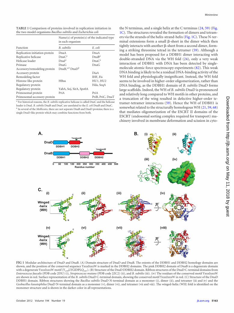
the N terminus, and a single helix at the C terminus (24, 59) (Fig.1C). The structures revealed the formation of dimers and tetram-ers via the strands of the helix-strand-helix (Fig. 1C). These N-ter-minal extensions form a small �-sheet in the dimer which thentightly interacts with another �-sheet from a second dimer, form-ing a striking threonine tetrad in the tetramer (59). Although amodel has been proposed for a DDBH1 dimer interacting withdouble-stranded DNA via the WH fold (24), only a very weakinteraction of DDBH1 with DNA has been detected by single-molecule atomic force spectroscopy experiments (82). This weakDNA binding is likely to be a residual DNA-binding activity of theWH fold and physiologically insignificant. Instead, the WH foldseems to be involved in higher-order oligomerization, rather thanDNA binding, as the DDBH1 domain of B. subtilis DnaD formslarge scaffolds. Indeed, the WH of B. subtilis DnaD is pronouncedand relatively long compared to WH motifs in other proteins, anda truncation of the wing resulted in defective higher-order te-tramer-tetramer interactions (59). Hence the WH of DDBH1 issomewhat related to the structurally homologous WH (21, 59, 68)that mediates oligomerization of the ESCRT II domains of theESCRT (endosomal sorting complex required for transport) ma-chinery involved in membrane deformation and scission in cyto-
TABLE 1 Comparison of proteins involved in replication initiation inthe two model organisms Bacillus subtilis and Escherichia coli
Function
Name(s) of protein(s) of the indicated typein each organism
B. subtilis E. coli
Replication initiation protein DnaA DnaAReplicative helicase DnaCa DnaBa
Helicase loader DnaIa DnaCa
Primase DnaG DnaGAccessory/remodeling protein DnaB,a,b DnaDb
Accessory protein DiaARemodeling factor IHF, FisHistone-like protein HBsu HU1, HU2Regulatory protein Hda, SeqARegulatory protein YabA, Soj, SirA, Spo0APrimosomal protein PriA PriAPrimosomal accessory protein PriB, PriC, DnaTa For historical reasons, the B. subtilis replicative helicase is called DnaC and the helicaseloader is DnaI. B. subtilis DnaB and DnaC are unrelated to the E. coli DnaB and DnaC.b In several of the Mollicutes, there are not separate DnaB and DnaD proteins; there is asingle DnaD-like protein which may combine functions from both.
FIG 1 Modular architecture of DnaD and DnaB. (A) Domain structure of DnaD and DnaB. The extents of the DDBH1 and DDBH2 homology domains areshown, and the position of the conserved sequence YxxxIxxxW is marked in the DDBH2 domains. The pink DDBH2 domain of DnaB is a degenerate domainwith a degenerate YxxxIxxxW motif (Y239LYGIDPLQ247). (B) Structure of the DnaD DDBH2 domain. Ribbon structures of the DnaD C-terminal domains fromEnterococcus faecalis (PDB code 2I5U) (i), Streptococcus mutans (PDB code 2ZC2) (ii), and B. subtilis (iii). (iv) The residues of the conserved motif YxxxIxxxWare shown in red. Surface representation of the B. subtilis DnaD C-terminal domain, showing the conserved motif YxxxIxxxW in red. (C) Structure of the DnaDDDBH1 domain. Ribbon structures showing the Bacillus subtilis DnaD N-terminal domain as a monomer (i), dimer (ii), and tetramer (iii and iv) and theGeobacillus kaustophilus DnaD N-terminal domain as a monomer (v), dimer (vi), and tetramer (vii and viii). The winged-helix (WH) fold is identified on themonomer structure and is shown in the darker color in all representations.
Minireview
October 2012 Volume 194 Number 19 jb.asm.org 5163
on May 11, 2018 by guest
http://jb.asm.org/
Dow
nloaded from
kinesis (1). It remains to be established whether DnaD plays anadditional role in cell division.
Strong single-stranded and double-stranded DNA-binding ac-tivities reside in the DDBH2 domains of B. subtilis DnaD (11) andDnaB (20), suggesting that the DDBH2 domain, rather than theDDBH1 domain, is responsible for DNA binding in replicationinitiation proteins in Firmicutes. DDBH2 is annotated in Pfam asthe DnaB_2 family (accession number PF07261). It is found inmany bacterial and phage proteins of unknown function. In sev-eral cases, two DDBH2 domains are in tandem or linked to otherdomains, such as Rep_3 (PF01051), TrmB (PF01978), IstB_IS21(PF01695), and others. In fact, 16 different architectures that in-clude one or more DDBH2 domains are listed in Pfam. Two crys-tal structures of DDBH2 from DnaD homologous proteins fromStreptococcus mutans UA 159 (PDB accession code 2ZC2) andfrom Enterococcus faecalis (PDB accession code 2I5U) have beendeposited by the Midwest Center for Structural Genomics (Fig.1B). Based upon the former, a structural model for the B. subtilisDDBH2 domain of DnaD was built and verified by nuclear mag-netic resonance (NMR) (37) (Fig. 1B). Bioinformatics analyses,combined with protein NMR, mutagenesis, and genetic comple-mentation studies, revealed that a highly conserved sequence mo-tif within DDBH2, YxxxIxxxW, together with an unstable �-helixand part of a flexible C-terminal region, contributes to DNA bind-ing (37) (Fig. 1B). Binding of DDBH2 to DNA induces the forma-tion of high-order oligomers (11). Interestingly, the cyanobacte-rial Ftn6 protein, which functions in the recruitment of FtsZ andregulates assembly of the Z ring during cell division, has a 77-amino-acid N-terminal domain structurally homologous toDDBH2 (35).
INTERACTIONS WITH DNA DURING REPLICATIONINITIATION/REINITIATION
The B. subtilis DnaD and DnaB proteins possess DNA-remodelingactivities. DnaB laterally compacts DNA (81), while DnaD bendsDNA, eliminates writhe, and converts it to negative twist, i.e.,duplex unwinding (71, 80, 81, 82). Binding of the DnaD DDBH2to DNA untwists the double helix, whereas the scaffolds formedvia the DDBH1 interactions appear to reinforce the helix-untwist-ing activity by forming anchorage points for the DDBH2-DNAcomplexes (80, 82). As DnaD interacts directly with DnaA (26), itis attractive to speculate that the duplex untwisting activity ofDnaD may enhance or stabilize the melting of the chromosomalorigin, oriC, through an oriC-DnaA-DnaD complex during initi-ation of DNA replication.
It is not immediately obvious how the lateral DNA compactionactivity of DnaB contributes to its putative function in a dual-helicase-loader system with DnaI (25, 72). Its association withoriC is dependent on a disordered C-terminal region (20) whichmay be subject to proteolysis or susceptible to degradation in agrowth phase-dependent manner. Truncated DnaB is depletedfrom the B. subtilis oriC, while intact DnaB binds to oriC asym-metrically at the DNA-unwinding element (DUE) half, consistentwith a role in helicase loading (20). Whether this is regulatoryproteolysis and, if so, how it occurs is not clear. However, it mightaffect the double-stranded DNA-binding site located near the Cterminus (between residues 365 and 428) that encompasses astrictly conserved YxxxIxxxW motif (residues 374 to 382) shownto be important in DNA binding in DnaB (our unpublished data)and DnaD (20, 37). DnaB interacts with DnaD (9, 56, 57) and with
the replicative helicase DnaC (72). The latter interaction is alsopreserved when DnaC is in complex with the helicase loader DnaI(72), and DnaB may therefore act to bridge the oriC-DnaA-DnaDcomplex with the DnaC-DnaI complex during replication initia-tion. Loading of DnaC appears to be mediated by a ring assemblymechanism rather than through a preformed ring opening-clos-ing mechanism, and DnaB is essential in this process, acting in adual-loader system together with DnaI (25, 65, 72). Their func-tional cooperation is reflected by the genetic juxtaposition of thetwo genes within the same operon (6, 20).
FUNCTIONS RELATED TO DnaA
At least in B. subtilis, DnaD and DnaB are recruited to DnaA-binding sites outside the chromosomal origin of replication,whereas the DnaC helicase is not (63). The hierarchy at thesesecondary sites is conserved, with DnaA binding first and DnaDand DnaB binding thereafter (63). Though it is possible that therecruitment of the primosomal proteins is a consequence of theirfunctional interaction at oriC, it is equally conceivable that thismay serve a regulatory role. DnaA can act as a transcription factorat the secondary sites (5, 19), and perhaps the interaction with theprimosomal proteins is important for this activity. In E. coli, thedatA locus regulates replication by titrating away replication ini-tiation proteins, and one can envisage that the secondary sites in B.subtilis may serve a similar regulatory role under conditions ofreplication stress. It remains to be established whether DnaD andDnaB also interact with DnaA at secondary sites in other organ-isms.
MEMBRANE ATTACHMENT
It has long been known that DNA replication in bacteria is a mem-brane-associated process (38), and the association of oriC with themembrane appears to be critical for replication initiation (4). In E.coli, DnaA is thought to bind directly to membranes (18, 54),although membrane association of oriC is mediated primarily viaSeqA and SeqB (60). In contrast, membrane association in B. sub-tilis is dependent on DnaB (23, 66, 74), and there is no evidence fora direct association between DnaA and the membrane. Westernblotting studies revealed that DnaB colocalizes with the mem-brane after fractionation (20, 56), and a mutation in dnaB thatsuppresses temperature sensitivity of dnaD and dnaB mutant cellswas found to constitutively recruit DnaD to the membrane (56).No in vivo growth-dependent degradation of the C-terminal partof membrane-bound DnaB was observed, in contrast to cytosolicDnaB, for which degradation caused depletion from oriC (20). ItsC terminus therefore seems protected in membrane-associatedDnaB, and it is likely that this region contains the site of mem-brane attachment. Localization studies of green fluorescent pro-tein fusions of DnaB and DnaD by fluorescence microscopy re-vealed a distinct membrane-proximal location for both proteins(40). Although DnaD is predominantly cytosolic (20), it has beenproposed that membrane-bound DnaB recruits DnaD to themembrane, which, in turn, recruits the DnaA-oriC complex to themembrane, and this provides another level of control for replica-tion initiation (56).
DNA REPAIR
The abundance of DnaD molecules in vivo, 3,000 to 5,000 mole-cules per cell for B. subtilis (9), suggests that DnaD may also playadditional roles beyond its essential role in DNA replication. In
Minireview
5164 jb.asm.org Journal of Bacteriology
on May 11, 2018 by guest
http://jb.asm.org/
Dow
nloaded from
many Firmicutes with dnaD-like genes, including bacilli, lactoba-cilli, staphylococci, acholeplasma, enterococci, streptococci, andclostridia, dnaD is juxtaposed in the same operon to an nth genecoding for an endonuclease III of the Nth/MutY family of DNArepair endonucleases involved in base excision repair (BER) (14).Deletion of nth in B. subtilis results in an H2O2-sensitive pheno-type, whereas the dnaD operon is transiently activated by H2O2 inboth B. subtilis and S. aureus and the dnaD mRNA levels remainrelatively high compared to the marked reduction of the dnaB anddnaI mRNA levels upon H2O2 treatment (12, 14), suggesting apossible regulatory link between DnaD and DNA repair. DnaD isindirectly involved in the Nth-mediated repair of abasic DNAsites, as it alters the DNA topology, which stimulates the activity ofNth (14). Furthermore, mutations in S. aureus dnaD result insensitivity to the DNA-cross-linking agent mitomycin C and toUV (33). The latter was correlated with a defect in the elongationstep of DNA replication, likely linked to defects in replicationrestart. Based on these findings, it seems reasonable to assume thatacross species, DnaD may play a role in DNA repair as well asreplication.
OriC ARCHITECTURE
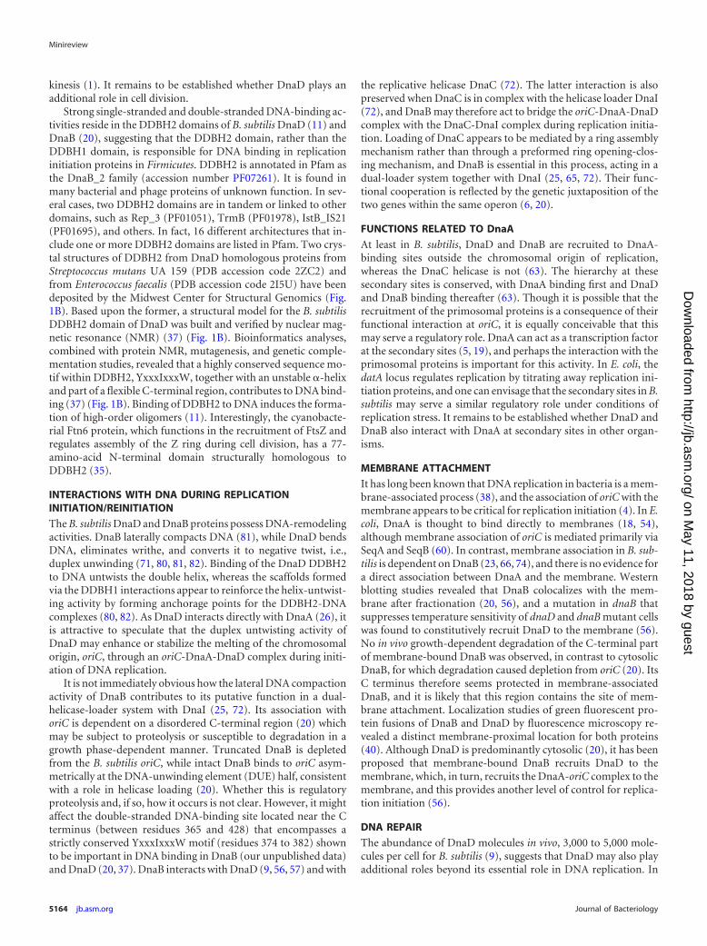
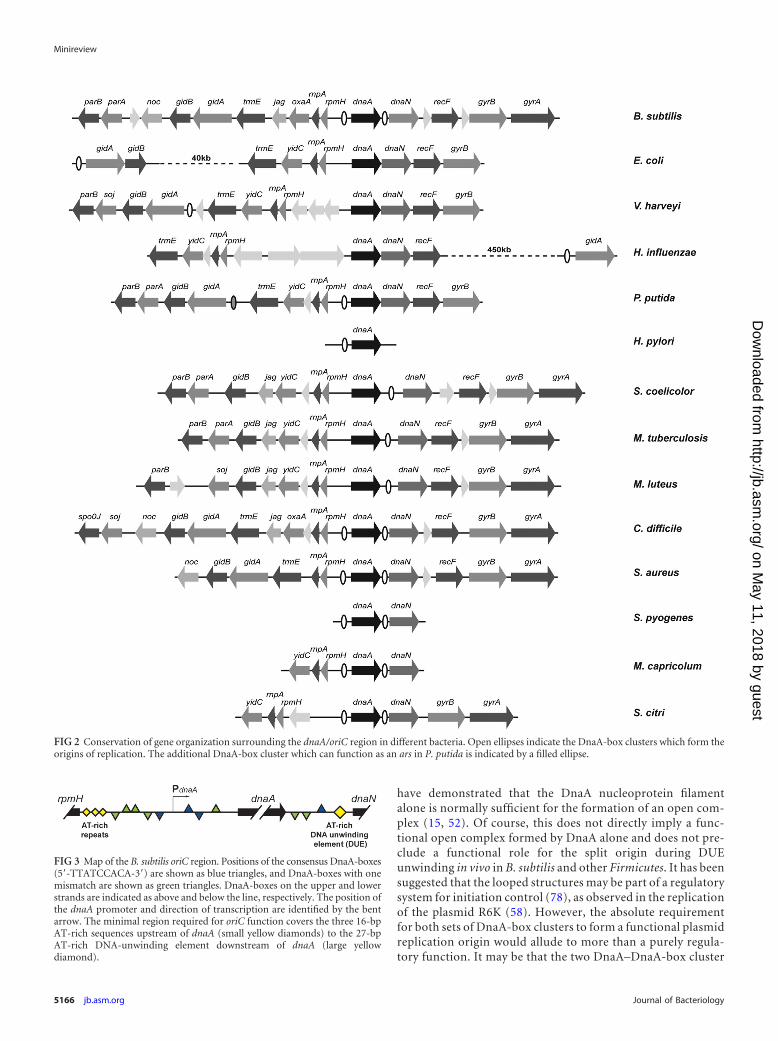
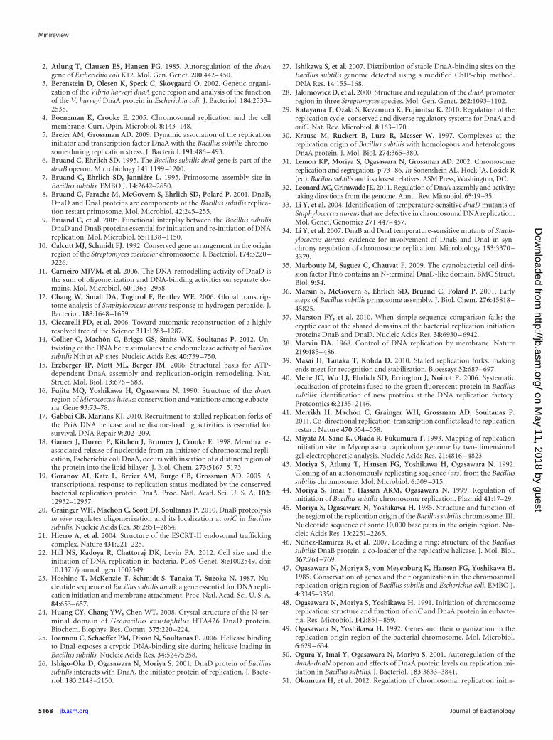
Chromosomal replication in bacteria starts from a single ori-gin, oriC. The position of oriC on the bacterial chromosome isconserved across bacterial species and classes, with most foundin close proximity to the dnaA gene encoding the DNA repli-cation initiation protein DnaA (49, 77). The genes surroundingoriC and dnaA are also commonly conserved, consisting of thegene cluster rnpA-rpmH-dnaA-dnaN-recF-gyrB-gyrA, withoriC located in an intergenic region adjacent to dnaA (Fig. 2).DnaA binds to 9-bp conserved sequences, with multiple suchsites forming DnaA-box clusters which are typically found atthe origin as DnaA binds to these regions to initiate replication(Fig. 3). Origins also contain an AT-rich DUE close to theDnaA-box clusters where the initial unwinding occurs, allow-ing the loading of two bidirectional helicases, one on eachstrand, around which two complete replisomes subsequentlyassemble (65). B. subtilis has the consensus origin gene struc-ture and has DnaA-box clusters in the intergenic regions bothupstream and downstream of the dnaA gene, i.e., in the rpmH-dnaA and dnaA-dnaN regions, with the DUE in the dnaA-dnaNregion (45, 47) (Fig. 2 and 3). Both upstream and downstreamDnaA-box clusters are essential for origin function in B. subtilis(43). The DnaA-box cluster in the promoter region upstreamof the dnaA gene also enables gene expression to be autoregu-lated: binding of DnaA to this DnaA-box cluster has beenshown to decrease transcription and expression of the dnaAgene (50). B. subtilis has six other DnaA-box clusters (51, 63),one of which is close to the dnaA gene, 3 kb upstream in thethdF-jag intergenic region (77), but they are not essential fororigin function. The consensus origin organization with thethree DnaA-box clusters in B. subtilis is often described as theprimordial DNA replication origin. This makes it an excellentmodel for understanding origin structure and regulation.
Replication initiation in E. coli is the most studied system, butit may not provide a good model for bacteria in general. The originin E. coli and in closely related Gram-negative species, includingVibrio harveyi and Haemophilus influenzae, has undergone a ma-jor rearrangement so that the functional origin is no longer prox-imal to the dnaA gene, although the gene organization around
dnaA has been retained (3, 48) (Fig. 2). A small DnaA-box clusterdirectly upstream of the dnaA gene is also retained: this has beenshown to enable autoregulation of dnaA expression (2, 73). Geneanalysis suggests that the E. coli origin is related to the nonessentialDnaA-box cluster in the thdF-jag region of the B. subtilis chromo-some. The Gram-negative bacteria Pseudomonas putida and Pseu-domonas aeruginosa have their origins immediately upstream ofthe dnaA gene, in the rpmH-dnaA intergenic region, and have amuch-shortened dnaA-dnaN intergenic region containing noDnaA-box clusters (61, 76) (Fig. 2). Interestingly, the DnaA-boxcluster linked to thdF in P. putida and P. aeruginosa can function asan autonomously replicating sequence (ars) when subcloned ontoa plasmid. The Helicobacter pylori origin region has undergoneconsiderable rearrangement relative to the consensus, but oriC isstill located directly upstream of the dnaA gene (79) (Fig. 2). TheGram-positive Actinobacteria (high G�C content) conform to theconsensus origin structure found in B. subtilis. Studies have shownthat the replication origins in Streptomyces coelicolor (10), Myco-bacterium tuberculosis (53), and Micrococcus luteus (16) containDnaA-box clusters in both the rpmH-dnaA and dnaA-dnaN inter-genic regions, and the site of initiation is in the dnaA-dnaN region(Fig. 2). However, only the dnaA-dnaN region was required toachieve a functional origin when subcloned onto a plasmid, al-though the rpmH-dnaA DnaA-box clusters did have an effect onorigin replication. As before, dnaA transcription was autoregu-lated by binding of DnaA to DnaA-box clusters in the dnaA pro-moter (28).
In addition to B. subtilis, the origins of chromosomal replica-tion from several other Firmicutes have been characterized. Theconsensus origin organization was seen in the closely related Ba-cilli species Streptococcus pyogenes (67) and the more-distantly re-lated Mollicutes species Mycoplasma capricolum (42) and Spiro-plasma citri (55, 75). The identification of the minimal ars bysubcloning onto a plasmid showed that both the rpmH-dnaA anddnaA-dnaN DnaA-box clusters are required for a functional ori-gin in these bacteria, as is the case with B. subtilis, and it is postu-lated that this may be a common requirement of all the Firmicutesreplication origins. However, the role of this “split origin” is un-clear. Electron microscopy studies show that when B. subtilisDnaA (BsDnaA) was bound to the DnaA-box clusters upstreamand downstream of the dnaA gene in B. subtilis oriC, the tworegions interacted and caused the DNA to “loop” (30). This loop-ing occurred both with a 2.5-kb linear origin fragment containingthe two DnaA-box clusters separated by the dnaA gene and whenthe origin was on a supercoiled circular plasmid. The parallel ex-periment with a fragment of the E. coli origin showed a similarresult, with loops forming between E. coli DnaA (EcDnaA) boundto a DnaA-box cluster at the origin and to a DnaA-box clusterconsisting of two DnaA boxes 500 bp downstream of oriC. Sur-prisingly, EcDnaA was able to bind to the DnaA-box clusters in theB. subtilis origin to promote looping and at a much lower protein/origin ratio (20-fold less) than the cognate BsDnaA. In the recip-rocal experiment, BsDnaA was able to bind to both DnaA-boxclusters in the E. coli origin fragment but could not sustain alooped structure.
It is unlikely that looping at a split origin is absolutely essen-tial for DnaA-mediated unwinding of the AT-rich DUE region.An open complex was observed when BsDnaA bound to a plas-mid containing B. subtilis oriC with deleted upstream DnaA-box clusters (30), and experiments in other bacterial systems
Minireview
October 2012 Volume 194 Number 19 jb.asm.org 5165
on May 11, 2018 by guest
http://jb.asm.org/
Dow
nloaded from
have demonstrated that the DnaA nucleoprotein filamentalone is normally sufficient for the formation of an open com-plex (15, 52). Of course, this does not directly imply a func-tional open complex formed by DnaA alone and does not pre-clude a functional role for the split origin during DUEunwinding in vivo in B. subtilis and other Firmicutes. It has beensuggested that the looped structures may be part of a regulatorysystem for initiation control (78), as observed in the replicationof the plasmid R6K (58). However, the absolute requirementfor both sets of DnaA-box clusters to form a functional plasmidreplication origin would allude to more than a purely regula-tory function. It may be that the two DnaA–DnaA-box cluster
FIG 3 Map of the B. subtilis oriC region. Positions of the consensus DnaA-boxes(5=-TTATCCACA-3=) are shown as blue triangles, and DnaA-boxes with onemismatch are shown as green triangles. DnaA-boxes on the upper and lowerstrands are indicated as above and below the line, respectively. The position ofthe dnaA promoter and direction of transcription are identified by the bentarrow. The minimal region required for oriC function covers the three 16-bpAT-rich sequences upstream of dnaA (small yellow diamonds) to the 27-bpAT-rich DNA-unwinding element downstream of dnaA (large yellowdiamond).
FIG 2 Conservation of gene organization surrounding the dnaA/oriC region in different bacteria. Open ellipses indicate the DnaA-box clusters which form theorigins of replication. The additional DnaA-box cluster which can function as an ars in P. putida is indicated by a filled ellipse.
Minireview
5166 jb.asm.org Journal of Bacteriology
on May 11, 2018 by guest
http://jb.asm.org/
Dow
nloaded from

complexes are involved in loading the two replisomes, with oneloading the first replisome on the leading strand and the otherloading the second replisome on the lagging strand. Whateverthe purpose of the split origin, it is unlikely that random diffu-sion would be sufficient to efficiently bring the distant DnaAfilaments together to form a functional origin in vivo.
The correlation between the conservation of DnaD, a pro-tein which both interacts with DnaA and bends DNA to formloops, and the split-origin structure in the Firmicutes may be
more than coincidence, especially in light of recent studies onthe HobA protein of Helicobacter pylori. HobA is a DnaA-bind-ing protein which is a functional counterpart of diaA in E. coli,and it has been proposed to form a molecular scaffold ontowhich regular oligomers of DnaA can assemble (69). DnaDmay function in a similar manner to bridge the DnaA mole-cules bound to the DnaA-box clusters in the Firmicutes splitorigin, bringing them together to form the functional loopstructure (Fig. 4). In addition, the C-terminal domain of DnaDbinds directly to DNA, enabling it to form a scaffold bridgingdifferent areas of the DNA to create loops and stabilize loopsonce they have formed. This would form a functional opencomplex at oriC, increase the frequency and stability of loopformation, and, thus, promote efficient chromosome replica-tion. This model depends on not only the high intracellularconcentration of DnaD, estimated at 3,000 to 5,000 moleculesper cell (9), but also the efficient targeting of DnaD to theorigin, initially via its interaction with DnaA and then via thegrowing DnaD scaffold.
THREE PROTEINS ARE BETTER THAN ONE?
It is interesting to consider why the Firmicutes have the complexityof the additional proteins DnaB and DnaD and a dual origin whenDnaA alone and a single origin are sufficient for E. coli. The mostlikely explanation is that a three-protein system provides addi-tional levels of regulation and control which may be advanta-geous. The timing of replication initiation in E. coli appears to bedirectly controlled by the levels of intracellular DnaA (29). Recentstudies with a small-cell mutant of E. coli with a delayed replica-tion cycle showed that increasing DnaA levels alleviated the delay(22). In contrast, replication initiation was neither delayed in asmall-cell mutant of B. subtilis nor advanced by additional DnaA.The prevailing opinion is that the DnaA concentration is not thelimiting factor in B. subtilis (44, 48, 51), and increasing intracellu-lar DnaA may actually inhibit replication (48). If the timing ofreplication initiation is no longer directly dependent on DnaA,then additional factors, such as DnaB and DnaD, may have a rolein controlling this critical process. DnaA may also function as amaster regulator: the expression of 34 genes (including dnaA)throughout the B. subtilis genome has been shown to be regulatedby DnaA binding at DnaA-box clusters outside the oriC region(27). These genes include some associated with tolerance to rep-lication stress, while others are associated with the control of thesporulation cycle. The fact that DnaB and DnaD are recruited tomany of these sites suggests that they may also modulate DnaAactivity at the non-oriC DnaA-box clusters (63). The additionallevels of control exerted by DnaB and DnaD on DnaA activity,both at the origin and elsewhere on the genome, may well increasethe ability of Firmicutes such as B. subtilis to thrive in a variety ofenvironments and respond to a wider range of conditions where E.coli cannot.
ACKNOWLEDGMENTS
Research in the P.S. lab is supported by a Wellcome Trust grant (091968/Z/10/Z). W.K.S. is supported by a Veni Fellowship of the NetherlandsOrganization for Scientific Research (NWO-ZonMW) and a Gisela ThierFellowship of the LUMC.
REFERENCES1. Adell MAY, Teis D. 2011. Assembly and disassembly of the ESCRT-III
membrane scission complex. FEBS Lett. 585:3191–3196.
FIG 4 Speculative model of possible roles for DnaD in the initiation of chro-mosome replication of B. subtilis and other Firmicutes. (i) DnaA binds to theDnaA-boxes in the origin region. DnaA-bound DnaD may assist in the recruit-ment of additional DnaA and stabilize the binding to nonperfect DnaA boxes.(ii) DnaD bound to the DnaA nucleoprotein filament may recruit additionalDnaD which invades and binds to the adjacent DNA region. (iii) As the DnaDscaffold extends into the DNA between the DnaA-boxes, it may cause the DNAto bend. (iv) This would have the effect of bringing the two DnaA nucleopro-tein filaments together such that they can interact. DnaD may stabilize thisinteraction, both by forming bridges between the two DnaA complexes and bystabilizing the looped DNA structure through an extended supporting scaf-fold. (v) DnaB is recruited to the DnaA complex, and the AT-rich regionforming the DUE begins to unwind. DnaD may assist in the recruitment ofDnaB and stabilize this interaction. The DnaD-induced conversion of DNAwrithe into negative twist may also assist in the melting of the DUE region. (vi)The DnaC-DnaI complex is recruited to the unwound DNA by DnaA andDnaB, and replication is initiated. A possible explanation for the requirementfor the two DnaA nucleoprotein filaments is that one filament is responsiblefor loading DnaC-DnaI onto one unwound DNA strand in cis while the otheris responsible for loading DnaC-DnaI onto the other unwound DNA strand intrans.
Minireview
October 2012 Volume 194 Number 19 jb.asm.org 5167
on May 11, 2018 by guest
http://jb.asm.org/
Dow
nloaded from
2. Atlung T, Clausen ES, Hansen FG. 1985. Autoregulation of the dnaAgene of Escherichia coli K12. Mol. Gen. Genet. 200:442– 450.
3. Berenstein D, Olesen K, Speck C, Skovgaard O. 2002. Genetic organi-zation of the Vibrio harveyi dnaA gene region and analysis of the functionof the V. harveyi DnaA protein in Escherichia coli. J. Bacteriol. 184:2533–2538.
4. Boeneman K, Crooke E. 2005. Chromosomal replication and the cellmembrane. Curr. Opin. Microbiol. 8:143–148.
5. Breier AM, Grossman AD. 2009. Dynamic association of the replicationinitiator and transcription factor DnaA with the Bacillus subtilis chromo-some during replication stress. J. Bacteriol. 191:486 – 493.
6. Bruand C, Ehrlich SD. 1995. The Bacillus subtilis dnaI gene is part of thednaB operon. Microbiology 141:1199 –1200.
7. Bruand C, Ehrlich SD, Janniére L. 1995. Primosome assembly site inBacillus subtilis. EMBO J. 14:2642–2650.
8. Bruand C, Farache M, McGovern S, Ehrlich SD, Polard P. 2001. DnaB,DnaD and DnaI proteins are components of the Bacillus subtilis replica-tion restart primosome. Mol. Microbiol. 42:245–255.
9. Bruand C, et al. 2005. Functional interplay between the Bacillus subtilisDnaD and DnaB proteins essential for initiation and re-initiation of DNAreplication. Mol. Microbiol. 55:1138 –1150.
10. Calcutt MJ, Schmidt FJ. 1992. Conserved gene arrangement in the originregion of the Streptomyces coelicolor chromosome. J. Bacteriol. 174:3220 –3226.
11. Carneiro MJVM, et al. 2006. The DNA-remodelling activity of DnaD isthe sum of oligomerization and DNA-binding activities on separate do-mains. Mol. Microbiol. 60:1365–2958.
12. Chang W, Small DA, Toghrol F, Bentley WE. 2006. Global transcrip-tome analysis of Staphylococcus aureus response to hydrogen peroxide. J.Bacteriol. 188:1648 –1659.
13. Ciccarelli FD, et al. 2006. Toward automatic reconstruction of a highlyresolved tree of life. Science 311:1283–1287.
14. Collier C, Machón C, Briggs GS, Smits WK, Soultanas P. 2012. Un-twisting of the DNA helix stimulates the endonuclease activity of Bacillussubtilis Nth at AP sites. Nucleic Acids Res. 40:739 –750.
15. Erzberger JP, Mott ML, Berger JM. 2006. Structural basis for ATP-dependent DnaA assembly and replication-origin remodeling. Nat.Struct. Mol. Biol. 13:676 – 683.
16. Fujita MQ, Yoshikawa H, Ogasawara N. 1990. Structure of the dnaAregion of Micrococcus luteus: conservation and variations among eubacte-ria. Gene 93:73–78.
17. Gabbai CB, Marians KJ. 2010. Recruitment to stalled replication forks ofthe PriA DNA helicase and replisome-loading activities is essential forsurvival. DNA Repair 9:202–209.
18. Garner J, Durrer P, Kitchen J, Brunner J, Crooke E. 1998. Membrane-associated release of nucleotide from an initiator of chromosomal repli-cation, Escherichia coli DnaA, occurs with insertion of a distinct region ofthe protein into the lipid bilayer. J. Biol. Chem. 273:5167–5173.
19. Goranov AI, Katz L, Breier AM, Burge CB, Grossman AD. 2005. Atranscriptional response to replication status mediated by the conservedbacterial replication protein DnaA. Proc. Natl. Acad. Sci. U. S. A. 102:12932–12937.
20. Grainger WH, Machón C, Scott DJ, Soultanas P. 2010. DnaB proteolysisin vivo regulates oligomerization and its localization at oriC in Bacillussubtilis. Nucleic Acids Res. 38:2851–2864.
21. Hierro A, et al. 2004. Structure of the ESCRT-II endosomal traffickingcomplex. Nature 431:221–225.
22. Hill NS, Kadoya R, Chattoraj DK, Levin PA. 2012. Cell size and theinitiation of DNA replication in bacteria. PLoS Genet. 8:e1002549. doi:10.1371/journal.pgen.1002549.
23. Hoshino T, McKenzie T, Schmidt S, Tanaka T, Sueoka N. 1987. Nu-cleotide sequence of Bacillus subtilis dnaB: a gene essential for DNA repli-cation initiation and membrane attachment. Proc. Natl. Acad. Sci. U. S. A.84:653– 657.
24. Huang CY, Chang YW, Chen WT. 2008. Crystal structure of the N-ter-minal domain of Geobacillus kaustophilus HTA426 DnaD protein.Biochem. Biophys. Res. Comm. 375:220 –224.
25. Ioannou C, Schaeffer PM, Dixon N, Soultanas P. 2006. Helicase bindingto DnaI exposes a cryptic DNA-binding site during helicase loading inBacillus subtilis. Nucleic Acids Res. 34:52475258.
26. Ishigo-Oka D, Ogasawara N, Moriya S. 2001. DnaD protein of Bacillussubtilis interacts with DnaA, the initiator protein of replication. J. Bacte-riol. 183:2148 –2150.
27. Ishikawa S, et al. 2007. Distribution of stable DnaA-binding sites on theBacillus subtilis genome detected using a modified ChIP-chip method.DNA Res. 14:155–168.
28. Jakimowicz D, et al. 2000. Structure and regulation of the dnaA promoterregion in three Streptomyces species. Mol. Gen. Genet. 262:1093–1102.
29. Katayama T, Ozaki S, Keyamura K, Fujimitsu K. 2010. Regulation of thereplication cycle: conserved and diverse regulatory systems for DnaA andoriC. Nat. Rev. Microbiol. 8:163–170.
30. Krause M, Ruckert B, Lurz R, Messer W. 1997. Complexes at thereplication origin of Bacillus subtilis with homologous and heterologousDnaA protein. J. Mol. Biol. 274:365–380.
31. Lemon KP, Moriya S, Ogasawara N, Grossman AD. 2002. Chromosomereplication and segregation, p 73–86. In Sonenshein AL, Hock JA, Losick R(ed), Bacillus subtilis and its closest relatives. ASM Press, Washington, DC.
32. Leonard AC, Grimwade JE. 2011. Regulation of DnaA assembly and activity:taking directions from the genome. Annu. Rev. Microbiol. 65:19–35.
33. Li Y, et al. 2004. Identification of temperature-sensitive dnaD mutants ofStaphylococcus aureus that are defective in chromosomal DNA replication.Mol. Genet. Genomics 271:447– 457.
34. Li Y, et al. 2007. DnaB and DnaI temperature-sensitive mutants of Staph-ylococcus aureus: evidence for involvement of DnaB and DnaI in syn-chrony regulation of chromosome replication. Microbiology 153:3370 –3379.
35. Marbouty M, Saguez C, Chauvat F. 2009. The cyanobacterial cell divi-sion factor Ftn6 contains an N-terminal DnaD-like domain. BMC Struct.Biol. 9:54.
36. Marsin S, McGovern S, Ehrlich SD, Bruand C, Polard P. 2001. Earlysteps of Bacillus subtilis primosome assembly. J. Biol. Chem. 276:45818 –45825.
37. Marston FY, et al. 2010. When simple sequence comparison fails: thecryptic case of the shared domains of the bacterial replication initiationproteins DnaB and DnaD. Nucleic Acids Res. 38:6930 – 6942.
38. Marvin DA. 1968. Control of DNA replication by membrane. Nature219:485– 486.
39. Masai H, Tanaka T, Kohda D. 2010. Stalled replication forks: makingends meet for recognition and stabilization. Bioessays 32:687– 697.
40. Meile JC, Wu LJ, Ehrlich SD, Errington J, Noirot P. 2006. Systematiclocalisation of proteins fused to the green fluorescent protein in Bacillussubtilis: identification of new proteins at the DNA replication factory.Proteomics 6:2135–2146.
41. Merrikh H, Machón C, Grainger WH, Grossman AD, Soultanas P.2011. Co-directional replication-transcription conflicts lead to replicationrestart. Nature 470:554 –558.
42. Miyata M, Sano K, Okada R, Fukumura T. 1993. Mapping of replicationinitiation site in Mycoplasma capricolum genome by two-dimensionalgel-electrophoretic analysis. Nucleic Acids Res. 21:4816 – 4823.
43. Moriya S, Atlung T, Hansen FG, Yoshikawa H, Ogasawara N. 1992.Cloning of an autonomously replicating sequence (ars) from the Bacillussubtilis chromosome. Mol. Microbiol. 6:309 –315.
44. Moriya S, Imai Y, Hassan AKM, Ogasawara N. 1999. Regulation ofinitiation of Bacillus subtilis chromosome replication. Plasmid 41:17–29.
45. Moriya S, Ogasawara N, Yoshikawa H. 1985. Structure and function ofthe region of the replication origin of the Bacillus subtilis chromosome. III.Nucleotide sequence of some 10,000 base pairs in the origin region. Nu-cleic Acids Res. 13:2251–2265.
46. Núñez-Ramírez R, et al. 2007. Loading a ring: structure of the Bacillussubtilis DnaB protein, a co-loader of the replicative helicase. J. Mol. Biol.367:764 –769.
47. Ogasawara N, Moriya S, von Meyenburg K, Hansen FG, Yoshikawa H.1985. Conservation of genes and their organization in the chromosomalreplication origin region of Bacillus subtilis and Escherichia coli. EMBO J.4:3345–3350.
48. Ogasawara N, Moriya S, Yoshikawa H. 1991. Initiation of chromosomereplication: structure and function of oriC and DnaA protein in eubacte-ria. Res. Microbiol. 142:851– 859.
49. Ogasawara N, Yoshikawa H. 1992. Genes and their organization in thereplication origin region of the bacterial chromosome. Mol. Microbiol.6:629 – 634.
50. Ogura Y, Imai Y, Ogasawara N, Moriya S. 2001. Autoregulation of thednaA-dnaN operon and effects of DnaA protein levels on replication ini-tiation in Bacillus subtilis. J. Bacteriol. 183:3833–3841.
51. Okumura H, et al. 2012. Regulation of chromosomal replication initia-
Minireview
5168 jb.asm.org Journal of Bacteriology
on May 11, 2018 by guest
http://jb.asm.org/
Dow
nloaded from
tion by oriC-proximal DnaA-box clusters in Bacillus subtilis. Nucleic AcidsRes. 40:220 –234.
52. Ozaki S, et al. 2008. A common mechanism for the ATP-DnaA-dependent formation of open complexes at the replication origin. J. Biol.Chem. 283:8351– 8362.
53. Qin MH, Madiraju MV, Rajagopalan M. 1999. Characterization of thefunctional replication origin of Mycobacterium tuberculosis. Gene 233:121–130.
54. Regev T, Myers N, Zarivach R, Fishov I. 2012. Association of thechromosome replication initiator DnaA with the Escherichia coli innermembrane in vivo: quantity and mode of binding. PLoS One 7:e36441.doi:10.1371/journal.pone.0036441.
55. Renaudin J, et al. 1995. Integrative and free Spiroplasma citri oriC plas-mids: expression of the Spiroplasma phoeniceum spiralin in Spiroplasmacitri. J. Bacteriol. 177:2870 –2877.
56. Rokop M, Auchtung JM, Grossman AD. 2004. Control of DNA replica-tion initiation by recruitment of an essential initiation protein to themembrane of Bacillus subtilis. Mol. Microbiol. 52:1757–1767.
57. Rokop M, Grossman AD. 2009. Intragenic and extragenic suppressors oftemperature sensitive mutations in the replication initiation genes dnaDand dnaB of Bacillus subtilis. PLoS One. 4:e6774. doi:10.1371/journal.pone.0006774.
58. Saxena M, Abhyankar M, Bastia D. 2010. Replication initiation at adistance: determination of the cis- and trans-acting elements of replicationorigin alpha of plasmid R6K. J. Biol. Chem. 285:5705–5712.
59. Schneider S, Zhang W, Soultanas P, Paoli M. 2008. Structure of theN-terminal oligomerization domain of DnaD reveals a unique tetramer-ization motif and provides insights into scaffold formation. J. Mol. Biol.376:1237–1250.
60. Shakibai N, et al. 1998. High-affinity binding of hemimethylated oriC byEscherichia coli membranes is mediated by a multiprotein system thatincludes SeqA and a newly identified factor, SeqB. Proc. Natl. Acad. Sci.U. S. A. 95:11117–11121.
61. Smith DW, Yee TW, Baird C, Krishnapillai V. 1991. Pseudomonadreplication origins: a paradigm for bacterial origins? Mol. Microbiol.5:2581–2587.
62. Smits WK, Goranov A, Grossman AD. 2010. Ordered association ofhelicase loader proteins with the Bacillus subtilis origin of replication invivo. Mol. Microbiol. 75:452– 461.
63. Smits WK, Merrikh H, Bonilla CY, Grossman AD. 2011. Primosomalproteins DnaD and DnaB are recruited to chromosomal regions bound byDnaA in Bacillus subtilis. J. Bacteriol. 193:640 – 648.
64. Soultanas P. 2011. The replication-transcription conflict. Transcription2:140 –144.
65. Soultanas P. 2012. Loading mechanisms of ring helicases at replicationorigins. Mol. Microbiol. 84:6 –16.
66. Sueoka N. 1998. Cell membrane and chromosome replication in Bacillussubtilis. Prog. Nucleic Acids Res. Mol. Biol. 59:35–53.
67. Suvorov AN, Ferretti JJ. 2000. Replication origin of Streptococcus pyo-genes, organization and cloning in heterologous systems. FEMS Micro-biol. Lett. 189:293–297.
68. Teo H, Perisic O, Gonzalez B, Williams RL. 2004. ESCRT-II, an endo-some-associated complex required for protein sorting: crystal structureand interactions with ESCRT-III and membranes. Dev. Cell 7:559 –569.
69. Terradot L, Zawilak-Pawlik A. 2010. Structural insight into Helicobacterpylori replication initiation. Gut Microbes 1:330 –334.
70. Tocheva EI, et al. 2011. Peptidoglycan remodelling and conversion of aninner membrane into an outer membrane during sporulation. Cell 146:799 – 812.
71. Turner IJ, Scott DJ, Allen S, Roberts CJ, Soultanas P. 2004. The Bacillussubtilis DnaD protein: a putative link between DNA remodelling and ini-tiation of DNA replication. FEBS Lett. 577:460 – 464.
72. Velten M, et al. 2003. A two-protein strategy for the functional loading ofa cellular replicative DNA helicase. Mol. Cell 11:1009 –1020.
73. Wang QP, Kaguni JM. 1987. Transcriptional repression of the dnaA geneof Escherichia coli by DnaA protein. Mol. Gen. Genet. 209:518 –525.
74. Winston S, Sueoka N. 1980. DNA-membrane association is necessary forinitiation of chromosomal and plasmid replication in Bacillus subtilis.Proc. Natl. Acad. Sci. U. S. A. 77:2834 –2838.
75. Ye F, Renaudin J, Bove JM, Laigret F. 1994. Cloning and sequencing ofthe replication origin (oriC) of the Spiroplasma citri chromosome andconstruction of autonomously replicating artificial plasmids. Curr. Mi-crobiol. 29:23–29.
76. Yee TW, Smith DW. 1990. Pseudomonas chromosomal replication ori-gins: a bacterial class distinct from Escherichia coli-type origins. Proc. Natl.Acad. Sci. U. S. A. 87:1278 –1282.
77. Yoshikawa H, Ogasawara N. 1991. Structure and function of DnaA andthe DnaA-box in eubacteria: evolutionary relationships of bacterial repli-cation origins. Mol. Microbiol. 5:2589 –2597.
78. Yoshikawa H, Wake RG. 1993. Initiation and termination of chromo-some replication, p 507–528. In Sonenshein A, Hoch JA, Losick R (ed),Bacillus subtilis and other Gram-positive bacteria: biochemistry, physiol-ogy and molecular genetics. ASM Press, Washington, DC.
79. Zawilak A, et al. 2001. Identification of a putative chromosomal replica-tion origin from Helicobacter pylori and its interaction with the initiatorprotein DnaA. Nucleic Acids Res. 29:2251–2259.
80. Zhang W, Allen S, Roberts CJ, Soultanas P. 2006. The Bacillus subtilisprimosomal protein DnaD untwists supercoiled DNA. J. Bacteriol. 188:5487–5493.
81. Zhang W, et al. 2005. The Bacillus subtilis DnaD and DnaB proteinsexhibit different DNA remodelling activities. J. Mol. Biol. 351:66 –75.
82. Zhang W, et al. 2008. Single-molecule atomic force spectroscopy revealsthat DnaD forms scaffolds and enhances duplex melting. J. Mol. Biol.377:706 –714.
Geoffrey S. Briggs obtained his B.Sc. (Bio-chemistry) from the University of York in 1990and his Ph.D. from the University of Kent atCanterbury in 1994. He held positions as a post-doctoral research associate at the University ofNottingham and the University of Leicester be-fore joining the group of Professor Bob Lloyd atNottingham in 2001, where he investigated thestructure-function relationships of proteins in-volved in DNA recombination, repair, and rep-lication in Escherichia coli. In 2011, he moved tothe group of Professor Panos Soultanas, where he continued studies in thesame general area, with the emphasis on the proteins and processes involvedin the initiation of DNA replication in the bacterium Bacillus subtilis.
Wiep Klaas Smits obtained his M.Sc. and Ph.D.degrees from the Faculty of Mathematics andNatural Sciences of the University of Gro-ningen, the Netherlands, in 2002 and 2007, re-spectively. After obtaining his Ph.D., he workedas a postdoctoral associate and fellow at the De-partment of Biology of the Massachusetts Insti-tute of Technology in Cambridge (MA), wherehe studied DNA replication initiation in Bacil-lus subtilis. Since 2010, he has worked as a prin-cipal investigator at the Leiden University Med-ical Center in Leiden, the Netherlands, where one of his research linesinvolves the identification and characterization of the DNA replication ma-chinery of Clostridium difficile in relation to that of other Firmicutes.
Continued next page
Minireview
October 2012 Volume 194 Number 19 jb.asm.org 5169
on May 11, 2018 by guest
http://jb.asm.org/
Dow
nloaded from
Panos Soultanas is a Professor of biochemistryand biological chemistry. He obtained his B.Sc.(biochemistry/microbiology) and Ph.D. de-grees from the Department of Molecular Biol-ogy and Biotechnology at the University ofSheffield, United Kingdom, in 1987 and 1991,respectively. He worked as a postdoctoral re-search assistant at the University of Bristol(1991 to 1994), the University of Crete (1995 to1996), and Oxford University (1996 to 2000),where he carried out research on site-specificrecombination, DNA helicases, DNA primases, and DNA replication. In2000, he was appointed to a Lectureship at the School of Chemistry, Univer-sity of Nottingham, United Kingdom. He was promoted to a Readership in2004 and to a Chair in 2008. In 2012, he was elected as a Fellow of the Societyof Biology (FSB). His main area of research is structure-function relation-ships of DNA replication enzymes and DNA replication and repair in theGram-positive model of the Firmicutes species Bacillus subtilis.
Minireview
5170 jb.asm.org Journal of Bacteriology
on May 11, 2018 by guest
http://jb.asm.org/
Dow
nloaded from


![DNA Replication Initiation Is Blocked by a Distant ... · replication and segregation of key chromosomal loci including the origin and terminus [4]. These steps also define two periods](https://img.pdfslide.us/doc/110x75/60020c885f9a0a1b3c145fd4/dna-replication-initiation-is-blocked-by-a-distant-replication-and-segregation.jpg)