Embed Size (px)
Citation preview

Jour
nal o
f Cel
l Sci
ence
COMMENTARY ARTICLE SERIES: IMAGING
Chromophore-assisted laser inactivation – towards aspatiotemporal–functional analysis of proteins, and the ablation ofchromatin, organelle and cell function
Yukimi Sano1, Wataru Watanabe2 and Sachihiro Matsunaga1,*
ABSTRACT
Chromophore-assisted laser or light inactivation (CALI) has been
employed as a promising technique to achieve spatiotemporal
knockdown or loss-of-function of target molecules in situ. CALI is
performed using photosensitizers as generators of reactive oxygen
species (ROS). There are two CALI approaches that use either
transgenic tags with chemical photosensitizers, or genetically
encoded fluorescent protein fusions. Using spatially restricted
microscopy illumination, CALI can address questions regarding, for
example, protein isoforms, subcellular localization or phase-specific
analyses of multifunctional proteins that other knockdown
approaches, such as RNA interference or treatment with
chemicals, cannot. Furthermore, rescue experiments can clarify the
phenotypic capabilities of CALI after the depletion of endogenous
targets. CALI can also provide information about individual events
that are involved in the function of a target protein and highlight them
in multifactorial events. Beyond functional analysis of proteins, CALI
of nuclear proteins can be performed to induce cell cycle arrest,
chromatin- or locus-specific DNA damage. Even at organelle level –
such as in mitochondria, the plasma membrane or lysosomes – CALI
can trigger cell death. Moreover, CALI has emerged as an
optogenetic tool to switch off signaling pathways, including the
optical depletion of individual neurons. In this Commentary, we
review recent applications of CALI and discuss the utility and effective
use of CALI to address open questions in cell biology.
KEY WORDS: Chromophore, Photosensitizer, Spatiotemporal
inactivation, Reactive oxygen species, Imaging
IntroductionChromophore-assisted laser or light inactivation (CALI) is a
powerful technique for microscopic investigation of the functions
of a protein of interest in situ and in a spatiotemporally regulated
manner. CALI enables the selective inactivation of proteins that
are tagged to chromophores by using antibodies (Jay, 1988; Liao,
1994). Since it was first used in the microscopy (Diamond et al.,
1993), CALI has been applied in a variety of experimental
settings and proved to be a promising tool to dissect a number of
complex phenomena in cell biology (reviewed in Hoffman-Kim
et al., 2007; Jacobson et al., 2008).
Through absorption of the laser light, chromophores becomeexcited, react with oxygen and generate reactive oxygen species
(ROS) that, in turn, cause damage to DNA, RNA, lipids andproteins. The principle of CALI is straightforward in thatphotosensitizers are used to harness ROS to eliminate targets ofinterest. ROS are unique molecules that can act as ‘double-edged
swords’, as they are highly oxidative but have a short lifetime;therefore, they only affect the molecules that are in closeproximity to their origins. So, by using a focused laser beam and
controlling the generation of ROS within a specific subcellulararea with micrometer to sub-micrometer accuracy, thespatiotemporal inactivation of targets can be achieved.
The first report of CALI described the inactivation of alkaline
phosphatase and b-galactosidase by using malachite-green-conjugated antibodies (Jay, 1988). More recently, the CALItool box has rapidly improved, mainly through the introduction of
new photosensitizers (see Box 1) or a refinement of themethodology itself. In this Commentary, we summarize anumber of recent investigations that have employed CALI anddiscuss their findings. In particular, we emphasize effective
applications of CALI that make full use of its advantages. Byreflecting on previous research achievements, we also provideperspectives for the potential use of CALI in order to address
outstanding questions in cellular biology that, hopefully, willinspire interesting future applications.
CALI with transgenically encoded tagsThe original CALI technique is based on the use of the dye
Malachite Green that was coupled to proteins of interest throughspecific antibodies (Table 1) (Jay and Keshishian, 1990; Mulleret al., 1996; Schroder et al., 1996; Schroder et al., 1999; Sakurai
et al., 2002). This method is still used to date – especially in thefield of neuroscience – and a number of studies have examinedthe roles of various factors in growth cone dynamics (Abe et al.,
2008; Higurashi et al., 2012; Iketani et al., 2013; Iketani et al.,2009). However, although these examples clearly demonstrate thepracticality of this approach, there are also drawbacks to anantibody-based method, including the necessity to microinject the
antibody into the cell and the risk that antibody binding interfereswith protein function (Keppler and Ellenberg, 2009).
As an alternative to Malachite Green, the fluorescent dyefluorescein isothiocyanate (FITC) and its derivatives have been
introduced for the spatiotemporal inactivation of targets, and thistechnique is often referred to as FALI (fluorophore-assisted lightinactivation) (Beck et al., 2002). Fluorescein is 50-times more
efficient in generating ROS than Malachite Green (Surrey et al.,1998). Recently, FALI was applied to a screening method indeveloping mouse brain (Sato et al., 2011). In order to establish
an even more precise inactivation of target molecules, several
1Department of Applied Biological Science, Faculty of Science and Technology,Tokyo University of Science, 2641 Yamazaki, Noda, Chiba 278-8510, Japan.2Department of Electrical and Electronic Engineering, College of Science andEngineering, Ritsumeikan University, 1-1-1 Noji-higashi, Kusatsu, Shiga525-8577, Japan.
*Author for correspondence ([email protected])
� 2014. Published by The Company of Biologists Ltd | Journal of Cell Science (2014) 127, 1621–1629 doi:10.1242/jcs.144527
1621
Jour
nal o
f Cel
l Sci
ence
approaches have been developed that are based on this potentdye. Instead of using an antibody-based approach for its targeting,fluorescein is mainly used in combination with transgenically
encoded tags (Griffin et al., 1998). These approaches utilize themembrane-permeable property of the fluorescein derivativeFlAsH-EDT2, which is synthesized from 49,59-bis(1,3,2-dithioarsolan-2-yl) fluorescein (Marek and Davis, 2002)
(Table 1). FlAsH-EDT2 in its native form is non-fluorescent,but becomes fluorescent upon binding to an exogenous motif thatconsists of tetra-cysteine residues. However, its use in CALI
might be problematic because nonspecific labeling of endogenouscysteine-rich proteins can take place (Stroffekova et al., 2001)unless the target protein is expressed at a high level. Another
synthetic fluorophore, red arsenical helix (ReAsH), that alsobinds to the tetra-cysteine motif, has even greater efficiency ingenerating ROS (Tour et al., 2003). Although ReAsH can beexcited at a longer wavelength – which can reduce the probability
of damage caused by light irradiation itself – it still has the majordrawback that undesired inactivation effects arise fromnonspecific binding (Hearps et al., 2007). This defect can be
ameliorated by increasing the affinity between the bi-arsenical-tetracysteine motifs and fluorescent dyes as well as the
concentration of dithiols such as 1,2-ethanedithiol (EDT) thatare used in the washing steps (Martin et al., 2005). The
fluorescent dyes mentioned above bind to tetracysteine motifsthrough thiol groups. Therefore, excessive nonspecific binding ofthese dyes to endogenous proteins can be washed off by usingdithiols, which reduces the background.
More refined protein tags for the selective labeling ofintracellular proteins have recently been developed based onadvances in synthetic chemistry (Jing and Cornish, 2011;
Uchinomiya et al., 2014). For example, SNAP-tag is a peptide of182 amino acids that is derived from the human O6-alkylguanine-DNA alkyltransferase. By appending the SNAP-tag to the target in
advance through gene engineering, the administration of O6-benzylguanine bearing a fluorescent dye results in the formation ofa tight connection between the protein of interest and the
fluorescent dye through the SNAP-tag (Keppler et al., 2002)(Fig. 1). Using such a SNAP-tag CALI approach, the inactivationof both a-tubulin and c-tubulin in mammalian cells could beachieved (Keppler and Ellenberg, 2009). Here, inactivation of a-
tubulin lead to mitotic arrest that is accompanied by aberrantmorphology of the mitotic spindle at metaphase. In contrast,inactivation of c-tubulin disrupted nucleation of microtubules
drastically and hampered the growth of microtubules emanatingfrom only those centrosomes that had been exposed to lightirradiation. Overall, these results not only confirmed a highly
specified restriction of localization as well as activation of SNAP-tag-based CALI, but also allowed to identify specific roles of thetubulin isoforms in intracellular molecular dynamics.
Eosin is a photosensitizing chromophore that generates 11-times as much singlet oxygen as fluorescein. Moreover, whencompared with fluorescein-labeled systems, the eosin-labeledsystem exhibits a fivefold greater efficiency in ROS generation,
which was measured using anthracene-9,10-dipropionic acid(Takemoto et al., 2013). Eosin is also used in combination witha protein tag, HaloTag7, a haloalkane dehalogenase mutant
(Ohana et al., 2009), which has been fused to protein kinase Cc(PKCc). Upon the chemical stimulation, PKCc translocates to theplasma membrane. Membrane-permeable eosin, diAc-eosin-AM,
was administrated as a HaloTag ligand to HeLa cells expressingthe HaloTag7–PKCc fusion protein. Upon irradiation withintense green light followed by chemical stimulation, HaloTag–PKCc failed to translocate to the plasma membrane, indicating
that CALI can be used to inactivate PKCc (Takemoto et al.,2011). HaloTag/eosin-based CALI has been also used toinactivate the mitotic kinase Aurora B, resulting in cell-division
arrest and multinuclear formation, consistent with results ofanalyses of Aurora B knockdown (Takemoto et al., 2011). Itshould be noted that the laser power required for the CALI-based
HaloTag/eosin-labelling system is 85-times lower than that for
Table 1. Properties of photosensitizers for CALI
Photosensitizer Molecular classification Excitation (nm) Emission (nm) References
Malachite Green Triarylmethane dye 620 (pulsed laser) – Jay, 1988Fluorescein Xanthene derivative 488 508 Surrey et al., 1998FlAsH-EDT2 Xanthene derivative 508 528 Marek and Davis, 2002ReAsH-EDT2 Xanthene derivative 540 635 Tour et al., 2003EGFP Protein (239 a.a.) 488 507 Rajfur et al., 2002EYFP Protein (239 a.a.) 514 527 McLean et al., 2009KillerRed Protein (273 a.a.) 585 610 Bulina et al., 2006aSuperNova Protein (273 a.a.) 579 610 Takemoto et al., 2013MiniSOG Protein (106 a.a.) 448, 473 500, 528 Qi et al., 2012
Box 1. PhotosensitizersPhotosensitizers are substances that induce particular reactions orlight emission by transmitting energy from the absorbed light toanother molecule. For the most part, photosensitizers are activatedfrom the singlet-ground stage into the singlet-activated stage bylight absorption, and then quickly enter the triplet-activated state bystate transition. A collision between a photosensitizer in the triplet-activated stage and another molecule causes the exchange ofenergy. The photosensitizer returns to the ground stage andthe other molecule enters the triplet-activated stage, which isaccompanied by specific reactions – for instance, fluorescentemissions or the generation of ROS at wavelengths that are uniqueto each molecule.
Types of photosensitizationPhotosensitizers can undergo two types of reaction, whoseprocedures and final products differ from each other. The type-Ireaction involves electron or hydrogen transfer with a reducingsubstrate. Light irradiation induces electron transfer from onemolecule – that becomes oxidized – to a photosensitizer. Thephotosensitizer, in turn, interacts with oxygen, resulting in theformation of a superoxide anion radical, e.g. O2?2, HO?. The type-IIreaction involves the interaction with oxygen. The energy transferoccurs directly to oxygen from a photosensitizer excited trough lightabsorption, and singlet oxygen (also known as dioxigen) is formed.
COMMENTARY Journal of Cell Science (2014) 127, 1621–1629 doi:10.1242/jcs.144527
1622

Jour
nal o
f Cel
l Sci
ence
the SNAP-tag system, implying a considerably lower risk ofnonspecific photodamage. This might be beneficial wheninvestigating photosensitive cellular processes.
CALI with EGFP or EYFPGreen fluorescent protein (GFP) and its variants have achromophore that consists of three amino acid residues within its
b-barrel structure (Cody et al., 1993; Tsien, 1998). ROS formedfollowing the chromophore being hit by light induce theaggregation of proteins that is mediated by crosslinking rather
than breaking a protein backbone (McLean et al., 2009) (Fig. 1).The first application of CALI with enhanced GFP (EGFP) was toinactivate a-actinin in fibroblasts, which resulted in the detachmentof stress fibers from focal adhesions (Rajfur et al., 2002). In
contrast to microinjection of antibodies, the most obvious benefitof using genetically encodable photosensitizers is the precisesubcellular localization of the fusion protein.
CALI with EGFP is particularly useful when wanting toaddress the effect of cell division and cytokinesis in the embryo,because specific cells within the embryo can be accurately
irradiated by the laser. Thus, to determine whether the non-muscle myosin II (MyoII) cable in Drosophila melanogaster
corrects cell mixing through a barrier of cortical tension, CALI
was used to specifically inhibit the MyoII regulatory light chain(MRLC) fused to EGFP in the boundary cables, and combinedwith live imaging of early embryos (Monier et al., 2010). Here,light irradiation of the boundary results in inactivation of MRLC
so that dividing cells are no longer pushed back, resulting incompartmental cell mixing. In contrast, if one side of theingressing furrow in dividing cells is irradiated with light, the cell
fails to divide because MyoII also plays an important role in thisprocess. These results highlight that CALI-mediated proteininactivation can be highly controlled at the subcellular level inlive embryos, and used, for instance, to provide insights into
localization-specific roles of MyoII in the cell.EGFP variants, including enhanced yellow protein (EYFP) and
enhanced cyan protein (ECFP), have also been used in CALI.
Their efficiency with regard to use in CALI follows this order:FLAsH.EGFP.EYFP.ECFP (Table 1) (McLean et al., 2009).The main drawback of CALI together with enhanced fluorescent
proteins is their relatively low efficiency in generating ROS, aswell as the requirement for high-power light irradiation. Toaddress this problem, two-photon- and multiphoton-excitation byusing a femtosecond laser has been applied to CALI (Tanabe
et al., 2005; Shimada et al., 2005). A nonlinear dependence of thesignal intensity on the tightly focused intensity of the excitationlight can be achieved by using femtosecond lasers that emit
ultrashort optical pulses of a duration below 1 picosecond, i.e. inthe order of femtoseconds. This allows for limited interactionregions together with a reduction of detrimental interactions
outside the region of focus, such as photo-induced damage andphotobleaching (Watanabe et al., 2004; Watanabe et al., 2007;Watanabe et al., 2008; Higashi et al., 2013). Two-photon excitation
generates ROS with a high degree of spatial specificity and hasbeen successfully utilized in a number of studies. For instance,CALI-mediated inactivation of connexin 43 – a main component ofgap junctions between neighboring cells – fused to EGFP induces a
decrease in the junctional current (Tanabe et al., 2005).CALI with femtosecond laser makes it possible to perform
time-lapse observations of subcellular organelle dynamics
Target protein
Ligand
Target proteinMembrane-permeable ligand is administered to target-protein–Tagfusion protein
Tag links fluorescentdye with target protein
Light irradiation atappropriate wavelength
Target inactivation
Genetically encoded protein photosensitizer is fused to target protein
Light irradiation at appropriate wavelength
Target inactivation
Tag(SNAP-tag,Halo-tag)
Dye(Fluorescein,
Eosin)
Protein photsensitizer(EGFP, KillerRed,
SuperNova, MiniSOG)
Fig. 1. Schematic overview of CALI using genetically encoded tags or protein sensitizers fused to a target protein. CALI is used for thespatiotemporal inactivation of targeted proteins that are microscopically controlled through light irradiation of specific subcellular regions. CALI is mainlyperformed by using two methods. For one method, the target protein and tag are expressed as a fusion protein. A membrane-permeable photosensitizer isadded at a later point (left). For the other method, the target protein and the photosensitizer are expressed as a fusion protein (right). In both cases, followingchromophore damage ROS are generated that can then inactivate target proteins in close vicinity.
COMMENTARY Journal of Cell Science (2014) 127, 1621–1629 doi:10.1242/jcs.144527
1623
Jour
nal o
f Cel
l Sci
ence
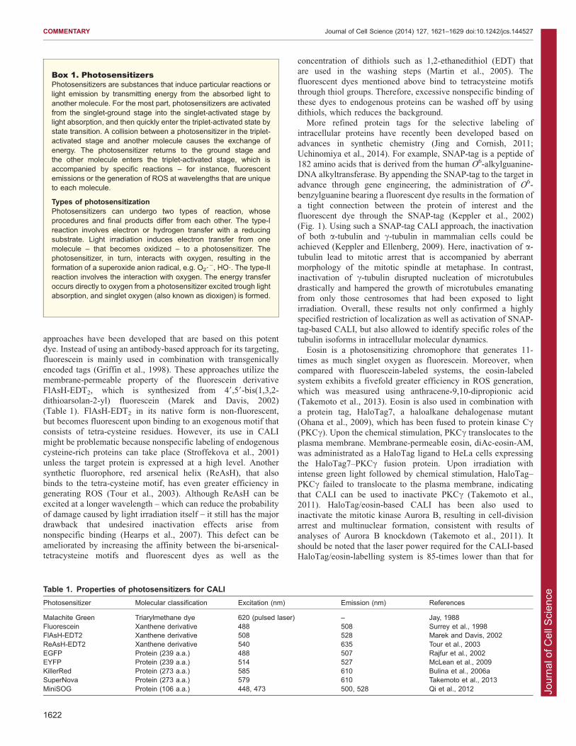
(Shimada et al., 2005). Here, circularization, and arrest ofmitochondrial fusion and fission in a single mitochondrion were
observed in response to mitochondrial membrane depolarizationcaused by inactivation of cytochrome c, when CALI with YFPtagged to sequence that targets cytochrome c oxidase was carriedout (Fig. 2). Although CALI that uses the near-infrared
femtosecond laser has advantages regarding the inactivation ofproteins – such as a high degree of spatial specificity withoutcausing nonspecific photodamage to living cells – it is not that
popular because of the high equipment costs.To further improve the use of EGFP-based CALI, any effects
that arise from endogenous, unlabeled proteins need to be
minimized. Endogenous target proteins that do not contain theEGFP fusion are resistant to CALI, and can compensate for theinactivation of their EGFP-fusion counterparts. To address this
issue, rescue experiments using EGFP-fusion proteins can be usedto evaluate the degree of loss-of-function due to CALI. In fact,they have shown that CALI is comparable with otherconventional methods of abrogating protein function (Vitriol
et al., 2007).
CALI with KillerRedKillerRed is derived from anm2CP (Bulina et al., 2006a), a non-fluorescent chromoprotein isolated from hydrozoa Anthomedusae
sp., and has a barrel-like shape similar to GFP. The chromophore
resides in the internal side of the barrel and connects directly to thesurrounding solvent through a long cavity, which has an importantrole in oxygen and ROS transmission (Carpentier et al., 2009;
Pletnev et al., 2009; Roy et al., 2010; de Rosny and Carpentier,2012). KillerRed is excited by green light; it emits red light(maximum at 585 nm, emission peak at 610 nm) (Table 1) andgenerates superoxide (Vegh et al., 2011; Wang et al., 2012).
KillerRed is the first choice for protein-based CALI because of itshigh efficiency in generating ROS (1000-fold compared withEGFP).
This powerful generation of ROS makes it possible to analyzethe effect of proteins of interest on subcellular dynamics. For
example, CALI with KillerRed bound to b1-integrin – which isinvolved in regulating invadosomes – was used to investigate
whether signaling from b1-integrin is required to either initiate ormaintain the ‘rosette’ structures (formed by several invadosomes)as well as their dynamics (Destaing et al., 2010). The authorsshowed that inactivation of b1-integrin by CALI results in acute
destabilization of both individual invadosomes and rosettes,which indicates the importance of b1-integrin to the entirelifespan of the rosette. The same group subsequently reported
that CALI-mediated dynamin inactivation results in a rapiddisappearance of invadosomes without any changes in focaladhesions, indicating that dynamin specifically regulates the actin
dynamics in invadosome (Destaing et al., 2013).KillerRed-based CALI is particularly useful when specific
antibodies or nontoxic chemical inhibitors are not available
because a protein of interest belongs to the protein family whosemembers are structurally very similar, has isoforms arising fromsplicing variants or has closely related paralogs. The ability toinactivate specific isoforms of a protein of interest would
be particularly valuable to gain insight into their specific anddifferential functions. CALI with KillerRed was used to investigateaquaporins (AQPs), a group of water channels with several
functions in cell physiology (Baumgart et al., 2012). Theirfunctions, however, are not fully understood because of the lackof nontoxic inhibitors. CALI was used to inhibit water permeability
of AQP1 and of two AQP4 isoforms (splicing variants, M1 andM23; AQP4-M1 tetramers freely diffuse in the plasma membrane,whereas AQP4-M23 tetramers are assembled into stationary
aggregates). Water permeability of cells that express AQP fusedto KillerRed was measured by osmotic swelling-induced dilutionof cytoplasmic chloride, in combination with a chloride-sensingfluorescent protein (Galietta et al., 2001). The authors found that
reduction in water permeability upon APQ4 inactivation was muchmore pronounced for the aggregate-forming M23 isoform,implying that intermolecular CALI took place, or that there was
a reciprocal inactivation owing to the recognition of the twoisoforms.
Fig. 2. Visualization of mitochondrial dynamics after CALI-mediated inactivation of cytochrome c by using a femtosecond laser. Mitochondria of HeLacells are visualized in yellow by using EYFP tagged with a targeting sequence from subunit VIII of human cytochrome c oxidase (pEYFP-Mito; Clonetech) ingreen pseudo color, and MitoTracker Red in red (Life Technologies). pEYFP-Mito-expressing mitochondria stained with MitoTracker Red appear yellow. CALI ofcytochrome c oxidase that targets the upper region of a single mitochondrion is performed by irradiation with a femtosecond laser (white arrow in the top leftimage). Time after irradiation is given in the bottom right corner of each image. Immediately after CALI, the red fluorescence of MitoTracker Red disappears fromthe irradiated mitochondrion, suggesting the depolarization of the mitochondrial membrane because MitoTracker Red only stains membrane that has amembrane potential, resulting in fluorescence of YFP only, as green pseudo color, that remains in mitochondria (20 seconds). The depolarized mitochondrionthen becomes circular and does not undergo any fusion and fission (1 min). Depolarization expands (2 min). Depolarized mitochondria become tubular butcannot fuse with adjacent mitochondria (5 min and 7 min). In contrast, the surrounding mitochondria perform fusion and fission repeatedly. The white dashed linedenotes the cell membrane. Scale bar: 5 mm.
COMMENTARY Journal of Cell Science (2014) 127, 1621–1629 doi:10.1242/jcs.144527
1624

Jour
nal o
f Cel
l Sci
ence
Another example for the use of KillerRed when the type ofprotein isoforms is an issue is the mammalian Golgi complex,
which consists of several stacks of membrane-bound structuresdesignated as cisternae. The application of CALI with KillerRedpreviously revealed that formation of cis-cisternae depends onthe recycling of a set of cis-Golgi enzymes through the ER-
Golgi intermediate compartment (ERGIC) (Jarvela and Linstedt,2012). Lateral networks among membranes are highly optimizedand vital for both protein modification and glycan processing,
and membranes are tethered by two reassembly stackingproteins, GRASP55 and GRASP65. Although these proteinsare located in different cisternae, previous studies have reported
that knockdown of either GRASP55 or GRASP65 causesdefective glycosylation. Yet there is little evidence of anydifference in terms of their localization that might be responsible
for compartment integrity. To address this question, CALI-mediated inactivation of each GRASP was performed by usingKillerRed fused to each isoform. This resulted in the loss ofGolgi integrity in an isoform-dependent manner, with
inactivation of GRASP55 and GRASP65 affecting trans-Golgiand cis-Golgi integrity, respectively (Jarvela and Linstedt,2014). These results clearly show that each GRASP isoform
has a cisternae-specific role, ensuring the integrity of Golgicompartmentalization.
CALI is particularly useful to inactivate mutant forms of key
factors in dynamic molecular pathways that are regulated in ahighly spatiotemporal manner. Without changing the localizationof the actin regulator cofilin, CALI was performed separately in
cofilin-depleted cells by using KillerRed coupled to either aconstitutively active cofilin mutant (cofilinS3A) or a dominant-negative cofilin mutant (cofilinS3E) (Vitriol et al., 2013). Cellsthat express either cofilinS3A or cofilinS3E fused to KillerRed
show no obvious change in the quantity or distribution oflamellipodial F-actin. However, only cells that express cofilinS3A-KillerRed show a dramatic increase in F-actin in the lamellipodia
and decrease in the rate of retrograde flow of actin after CALI.These results are consistent with previous studies that have showna role for cofilin in regulating lamellipodia stabilization and in
severing and disassembling of actin filaments. Moreover, thisstudy was not only the first report of a real-time regulation ofactin turnover by cofilin in the living cell, but also clearly showsthat performing CALI in cells in which endogenous protein has
been depleted allows to reveal the characteristics of exogenousproteins, including of phosphomimetic mutants.
KillerRed has also been used as a tool to simply generate ROSin other approaches, including as a chromatin-targeted phototoxicfluorescent protein for the induction of phase-specific arrest ofcell division. In vitro, HeLa cells that express tandem KillerRed
fused to histone H2B normally divide in the dark, but stopproliferation when they are irradiated with green light(Serebrovskaya et al., 2011). Similarly, we performed CALI by
using the cohesion regulator protein RBMX fused to KillerRed toshow that G2-phase-specific inactivation of RBMX by CALIresults in mitotic delay, demonstrating that RBMX specifically
functions in G2-phase nuclei (Matsunaga et al., 2012; Fig. 3).Moreover, in transgenic Xenopus embryos that express H2B-tandem KillerRed under control of tissue-specific promoters,
tissue development is slowed in tadpoles when illuminated withgreen-light, suggesting that CALI can be used to elucidate theeffect of temporal arrests of cell division during organogenesis(Serebrovskaya et al., 2011). KillerRed fused to either lamin B1,
one of the components of inner nuclear membrane, or histoneH2A was shown to induce DNA strand breaks dependent onirradiation time and intranuclear chromatin structure (Waldeck
et al., 2011). Recently, CALI with KillerRed fused to a Tet-repressor or a transcriptional activator that can bind to integratedDNA cassettes (,90 kb), was shown to cause induction of site-
specific DNA damage (Lan et al., 2013). The authors showed thatDNA repair proteins are differentially recruited to euchromatinand heterochromatin.
In addition to its use nuclei, KillerRed-based CALI has mostlybeen performed in organelles by fusing it to signal peptides thatare responsible for their subcellular localization. Accordingly,organelle-specific protein inactivation was performed using
KillerRed targeted to peroxisomes (Ivashchenko et al., 2011),mitochondria (Bulina et al., 2006a; Bulina et al., 2006b; Choubeyet al., 2011; Shibuya and Tsujimoto, 2012) and lysosomes
(Serebrovskaya et al., 2014). CALI with mitochondria-targetingKillerRed in human cells and C. elegans induces mitochondrialmembrane depolarization and morphological changes, respectively,
including fragmentation and swelling, as well as caspase-dependent apoptosis (Shibuya and Tsujimoto, 2012). CALI withlysosome-associated KillerRed also induces cell death, either dueto necrosis following exposure to light of high intensity or due to
A
G2
G2 0
60 90 120 150 180 min
9 18 27 36 45 54 min
0 30B
GFP-H1.2
GFP-H1.2
KR-RBMX
DIC
DIC
Fig. 3. Visualization of cell division arrest upon CALI-mediated inactivation of RBMX with KillerRed. (A) Nuclei andchromosomes of HeLa cells are visualized by using histone H1.2fused to GFP (GFP-H1.2). (B) RBMX, a regulatory protein forcentromeric protection and cohesion, is labeled with KillerRed(KR-RBMX). When a G2-phase nucleus, in which GFP-H1.2 isexpressed, is irradiated with green light, mitosis is typicallyfinished within 60 minutes from nuclear envelope breakdown at0 min (A, control). In contrast, CALI-mediated inactivation ofRMBX in a G2-phase nucleus expressing GFP-H1.2 and KR-RBMX leads to considerable delay of mitosis, which is onlycompleted after 180 minutes (B). Fluorescence of KR-RBMX hasdisappeared following laser irradiation. DIC, differentialinterference contrast microscopy image. Scale bars: 10 mm.
COMMENTARY Journal of Cell Science (2014) 127, 1621–1629 doi:10.1242/jcs.144527
1625

Jour
nal o
f Cel
l Sci
ence
apoptosis following exposure to light of lower intensity(Serebrovskaya et al., 2014). CALI is, therefore, useful in
pathological and pharmacological studies that aim to identifymechanisms underlying aging and certain diseases, by elicitingROS-mediated effects derived from specific organelles orsubcellular regions.
Because it can disrupt the plasma membrane, KillerRed cankill cells. The generated ROS directly affect membrane lipidsand destroy the plasma membrane barrier, together with the
accumulation of toxic ROS compounds (Williams et al., 2013).This cell-death causing feature of KillerRed, has been used todisrupt cells when CALI is applied. These include Muller glia
cells, the primary glial cells in mouse retina (Byrne et al., 2013);habenula neurons in zebrafish (Lee et al., 2010); amphid sensoryneurons in C. elegans (Kobayashi et al., 2013), GABAergic
interneurons in zebrafish (Del Bene et al., 2010); and sensoryneurons, interneurons and motor neurons in C. elegans (Williamset al., 2013). Furthermore, taking advantage of the spatiotemporalROS generation in specific cells, KillerRed-based CALI has
been applied to evaluate ROS cytotoxicity and investigate theoxidative stress response of mammalian cultured cells (Wanget al., 2013; Wang et al., 2012), as well as a screening tool (Liu
et al., 2010; Liu et al., 2011).Despite these remarkable properties, KillerRed also has
some shortcomings. In addition to red fluorescence, KillerRed
also emits weak green fluorescence at 480 nm that is difficultto distinguish from green fluorescent probes, such as GFP.Ideally, KillerRed and green fluorescent probes should, therefore,
not be used together in the same cell (Nordgren et al., 2012). Inaddition, dimerization of KillerRed may hamper the mobility ofthe target protein (Shirmanova et al., 2013). To prevent anyproblems owing to dimerization, a monomeric KillerRed variant
has been developed by introducing several point mutations(Takemoto et al., 2013). This new KillerRed variant, namedSuperNova, possesses the same ability to generate ROS and
shares similar excitation and emission properties as the originalKillerRed.
CALI with miniSOGMiniSOG is a small (106 amino acids) monomeric proteinderived from phototropin 2, which is responsible for detectingthe direction of light irradiation and mediating phototaxis in
Arabidopsis thaliana (Shu et al., 2011). Owing to the presence ofbound flavin mononucleotide (FMN), miniSOG absorbs bluelight at a maximum of 448 nm (with a ‘shoulder’ absorption peak
at 473 nm) and fluoresces green (peaks at 500 and 528 nm)(Ruiz-Gonzalez et al., 2013). FMN is a cofactor for variousenzymes and receptors (Massey, 2000), and generates singlet
oxygen by itself (Baier et al., 2006); it binds to the LOV2 (light,oxygen, voltage) domain of A. thaliana phototropin 2 (Salomonet al., 2000).
Fluorescence of miniSOG depends on the concentration ofFMN; it is reduced when FMN levels decrease (Ryumina et al.,2013). FMN embedded in miniSOG protein enables the photo-induced electron-transfer reaction (type-I reaction), which results
in a relatively poor yield of singlet oxygen compared with its freestate, instead of generating singlet oxygen (type-II reaction) (seeBox 1). Thus, the definition of miniSOG should be changed to
‘mini super oxide generator’ from ‘mini singlet oxygengenerator’ as it was originally named because it was thought togenerate mainly singlet oxygen. (Pimenta et al., 2013). Although
several controversial issues remain with regard to the type or
quantity of generated ROS, miniSOG has been shown to serve asa probe that generates ROS. MiniSOG is not only a CALI
reagent. It has also been used in correlative fluorescence andelectron microscopy, in combination with diaminobenzidine –which forms osmiophilic compounds upon reaction with singletoxygen – and, particularly, in the field of neuroscience, has
enabled the detection of barely visible objects (Boassa et al.,2013; Burgers et al., 2012; Ludwig et al., 2013; Shu, et al., 2011).
Besides its applications mentioned above miniSOG has been
mainly used in the area of neuroscience. Optogenetic techniquesprovide effective ways to manipulate functions of selectedneurons with light. For instance, direct neurotransmitter release
upon focused illumination can be induced by using rhodopsins,the microbial opsin channels, which are valuable tools tomanipulate selected neurons. Using CALI together with
miniSOG to investigate synaptic proteins has helped to achievethis objective. For instance, the fusion of miniSOG to VAMP2 –the SNARE complex protein located at the presynaptic terminalthat mediates vesicular synaptic release – or to synaptophysin
disrupts presynaptic vesicular release upon irradiation with bluelight (i.e. CALI induction) in both cultured neurons andhippocampal organotypic slices (Rambani et al., 2009).
Expression of miniSOG–VAMP2 in whole C. elegans neuronscaused reduced movement and paralysis after CALI was induced.Consequently, this technique has been named inhibition of
synapses with CALI (InSynC) (Lin et al., 2013).In C. elegans, InSynC was used to address the temporal and
spatial requirements of the localization of an UNC-13 isoform
derived from the splicing variant UNC-13L to the active zone,which features a high density of Ca2+ channels and whereneurotransmitter-containing synaptic vesicles are released inresponse to Ca2+ influx (Zhou et al., 2013). In the presynaptic
active zone, UNC-13 isoforms produced by alternative splicinginteract with the synaptic vesicle fusion apparatus and mediatesexocytosis of synaptic vesicles. Upon irradiation with blue light,
worms that express UNC-13L–miniSOG showed impaired rapidmovement accompanied by much reduced slow movement. Thissuggests that the removal of UNC-13L from only the active zone
results in strong inhibition of spontaneous as well as fast releaseof synaptic vesicles, corroborating the idea that UNC-13L at theactive zone is necessary for both types of synaptic vesicle release.
MiniSOG has also been employed as a tool to kill cells. Light-
inducible selective cell ablation was performed using CALI withtransgenically expressed miniSOG targeted at mitochondria(mito-miniSOG) in C. elegans neurons (Qi et al., 2012). Upon
illumination with blue light, mitochondria-tethered miniSOGcaused rapid neuronal death without harming tissues adjacent tothe dead neuron.
Considerations for the use of CALIThere are two distinct technical precautions that need to be
considered when applying CALI in order to determine a truespatiotemporal photo-inactivation of certain targets. The first isthe precise location of chromophores for laser targeting.Targeting can be achieved by using one of three methods: dye-
conjugated antibodies; chemical dyes that recognize specific tagsthat are attached to targets in advance; or genetically encodedphotosensitizers fused to targets (Fig. 1, Table 1). Using
genetically encoded photosensitizers, endogenous targetproteins cannot be labeled and remain functional (Vitriol et al.,2007). Therefore, in combination with molecular genetic
techniques, including RNA interference (RNAi) and genome
COMMENTARY Journal of Cell Science (2014) 127, 1621–1629 doi:10.1242/jcs.144527
1626

Jour
nal o
f Cel
l Sci
ence
editing, the pool of endogenous proteins should be eliminated.Dye-conjugated antibodies are ideal in terms of their effect on
endogenous proteins, but require antibody microinjection intocells. Therefore, dye-conjugated antibodies are less well-suitedwhen using CALI for proteins that have extracellular parts(reviewed in Buchstaller and Jay, 2000). This prevents the use of
CALI in a range of experiments because microinjection istechnically challenging, especially in multicellular organisms.
The second precaution also involves spatial restriction and
should limit nonspecific damage to surrounding molecules or cells,especially when high-intensity light irradiation is used. Indeed,early studies required high-intensity lasers because the efficiency
of ROS production of the probes was low. Irradiation of MalachiteGreen with a 620-nm laser results in reducing the maximal radiusof damage by half (,15 A) (Hoffman-Kim et al., 2007). Using
tetracysteine motifs in combination with chemical dyes, such asReAsH, often shows a high efficiency in the production of ROS;however, the outcomes of CALI experiments may suffer fromexcessive inactivation of target proteins when the dye
concentration is too high (Tour et al., 2003). In contrast, fusingtarget proteins to EGFP requires a high-intensity light irradiation toachieve sufficient inactivation of these proteins because EGFP is
inferior to chemical dyes in its ability to produce ROS (Rajfuret al., 2002). However, intense laser illumination of geneticallyencoded proteins produces deleterious amounts of ROS, which in
close proximity (30–60 A) can cause cleavage or crosslinking ofpeptide backbones (Tanabe et al., 2005; Horskotte et al., 2005;McLean et al., 2009). The negative effects of ROS vary strongly
dependent on molecular crowding, stability of protein complexesand organelle interactions, and CALI should be performed by usingappropriate controls. Thus, although the functional analyses ofproteins by CALI has been impressive, the elucidation of observed
phenotypes needs to be supported by data obtained from otherknockdown or knockout approaches to ensure any secondaryeffects owing to excessive ROS damage are not misinterpreted.
Conclusions and perspectivesThe functional analysis of proteins has mainly been performed
using either RNAi-based knockdown and mutagenesis, or geneticapproaches that involve recombination or tagging-based knockoutexperiments that are based on protein-tagging. However, theseapproaches are limited as they are unable to provide definitive
demonstrations of protein function within living cells. CALI usedtogether with live imaging provides a novel alternative in order toreveal dynamic subcellular function through precise and accurate
spatiotemporal inactivation of proteins in situ. Improvements in thespatial resolution of light irradiation to subcellular target regionsare anticipated with the acceleration of advances in super-
resolution microscopy (Lidke and Lidke, 2012; Sauer, 2013).Additionally, gene editing methods that use transcription-activator-like effector nucleases (TALENs) and clustered regularly
interspaced short palindromic repeats (CRISPRs) in associationwith Cas9 (CRISPR/Cas9) will make it easier to generate cells thatare deficient in the endogenous protein of interest (Gaj et al., 2013;Terns and Terns, 2014), which can then be substituted by target
proteins linked to photosensitizers. CALI experiments in deficientcells that are rescued with functional chromophore-tagged or -fused proteins allow to ascertain the degree of loss-of-function.
Thus, CALI has the potential to develop into a powerful techniquethat provides phenotype ‘snapshots’ through the inactivationdistinct functions of multifunctional proteins by using exogenous
mutants after an endogenous protein has been depleted. CALI
could, thereby, help to dissect the various functions and theirconnectivity within a cell.
Beyond protein analyses, CALI can be extended to investigatedifferent areas of cell biology. CALI can be used to changechromatin organization through the inactivation of chromatin-binding and nuclear factors. Although chromatin organization is
important for gene expression, its exact regulation remainsunknown (Gibcus and Dekker, 2013; Matsunaga et al., 2013).Because chromatin organization dynamically changes depending
on cell phase and developmental stage, CALI may be able toreveal temporally relevant stages of chromatin organization.CALI might also provide insight into DNA-repair mechanisms.
Because X- or gamma-rays induce random DNA damage, manystudies have attempted to induce locus-specific DNA damage thatis physiologically more relevant. Enzymatic systems to induce
DNA breakage include a system that is based on the rare-cutterendonuclease I-SceI – which has a recognition sequence thatoccurs only rarely in a genome; the elicited DNA-repairmechanism may, thus, be different from the naturally occurring
DNA-damage response (Bryant et al., 2004). A system to inducesite-specific oxidative DNA damage based on ROS generators,thus, holds considerable promise to mimick natural DNA-repair
mechanisms. Combination with knock-in systems on the basis ofgenome editing will allow to insert the binding sequence of ROSgenerators into expected genomic loci, inducing locus-specific
sites of DNA damage (Bedell et al., 2012; Auer et al., 2014).Optogenetics is a functional method of protein analysis that
utilizes genetically encoded light-sensitive proteins that are
activated by light. It has recently also been used in behavioralresearch to modify signaling in cranial nerves (Deisseroth, 2011),as well as in studying gene regulation by using a light-inducibletranscriptional effector as an optical switch to control
transcriptional and epigenetic status (Konermann et al., 2013).CALI has also been used as a ‘switch’ in optogenetics because itcan disrupt chromatin and proteins in a locus-specific manner, kill
specific organelles or a single cell in vivo (Yang and Yang, 2011;Lin et al., 2013). Moreover, CALI is beginning to be exploited inphotodynamic therapy of cancer cells (Mironova et al., 2013;
Ryumina et al., 2013; Shirmanova et al., 2013; Liao et al., 2014).Therefore, we anticipate that the portfolio of applications of CALIwill further expand, and contribute to a series of remarkablebiological findings and medical applications in the future.
AcknowledgementsWe are grateful to Hiroshi Ishii, Eri Mizusawa, Kiichi Fukui and Kazuyoshi Itoh forvaluable discussion and comments regarding the use of CALI in live cell imaging.
Competing interestsThe authors declare no competing interests.
FundingThis research was supported by SENTAN and CREST grants from the JapanScience and Technology Agency, a Grant-in-Aid for X-ray Free Electron LaserPriority Strategy Program (MEXT), and grants from MEXT/JSPS KAKENHI(20370027, 20061020, 21027023, 23012027, 23370029, 23120518, 25114514,25120726).
ReferencesAbe, T. K., Honda, T., Takei, K., Mikoshiba, K., Hoffman-Kim, D., Jay, D. G. andKuwano, R. (2008). Dynactin is essential for growth cone advance. Biochem.Biophys. Res. Commun. 372, 418-422.
Auer, T. O., Duroure, K., De Cian, A., Concordet, J. P. and Del Bene, F. (2014).Highly efficient CRISPR/Cas9-mediated knock-in in zebrafish by homology-independent DNA repair. Genome Res. 24, 142-153.
Baier, J., Maisch, T., Maier, M., Engel, E., Landthaler, M. and Baumler, W.(2006). Singlet oxygen generation by UVA light exposure of endogenousphotosensitizers. Biophys. J. 91, 1452-1459.
COMMENTARY Journal of Cell Science (2014) 127, 1621–1629 doi:10.1242/jcs.144527
1627
Jour
nal o
f Cel
l Sci
ence
Baumgart, F., Rossi, A. and Verkman, A. S. (2012). Light inactivation of watertransport and protein-protein interactions of aquaporin-Killer Red chimeras.J. Gen. Physiol. 139, 83-91.
Beck, S., Sakurai, T., Eustace, B. K., Beste, G., Schier, R., Rudert, F. and Jay,D. G. (2002). Fluorophore-assisted light inactivation: a high-throughput tool fordirect target validation of proteins. Proteomics 2, 247-255.
Bedell, V. M., Wang, Y., Campbell, J. M., Poshusta, T. L., Starker, C. G., Krug,R. G., I. I, Tan, W., Penheiter, S. G., Ma, A. C., Leung, A. Y. et al. (2012). Invivo genome editing using a high-efficiency TALEN system. Nature 491, 114-118.
Boassa, D., Berlanga, M. L., Yang, M. A., Terada, M., Hu, J., Bushong, E. A.,Hwang, M., Masliah, E., George, J. M. and Ellisman, M. H. (2013). Mappingthe subcellular distribution of a-synuclein in neurons using genetically encodedprobes for correlated light and electron microscopy: implications for Parkinson’sdisease pathogenesis. J. Neurosci. 33, 2605-2615.
Bryant, P. E., Gray, L. J. and Peresse, N. (2004). Progress towards understandingthe nature of chromatid breakage. Cytogenet. Genome Res. 104, 65-71.
Buchstaller, A. and Jay, D. G. (2000). Micro-scale chromophore-assisted laserinactivation of nerve growth cone proteins. Microsc. Res. Tech. 48, 97-106.
Bulina, M. E., Chudakov, D. M., Britanova, O. V., Yanushevich, Y. G.,Staroverov, D. B., Chepurnykh, T. V., Merzlyak, E. M., Shkrob, M. A.,Lukyanov, S. and Lukyanov, K. A. (2006a). A genetically encodedphotosensitizer. Nat. Biotechnol. 24, 95-99.
Bulina, M. E., Lukyanov, K. A., Britanova, O. V., Onichtchouk, D., Lukyanov, S.and Chudakov, D. M. (2006b). Chromophore-assisted light inactivation (CALI)using the phototoxic fluorescent protein KillerRed. Nat. Protoc. 1, 947-953.
Burgers, P. P., Ma, Y., Margarucci, L., Mackey, M., van der Heyden, M. A.,Ellisman, M., Scholten, A., Taylor, S. S. and Heck, A. J. (2012). A small novelA-kinase anchoring protein (AKAP) that localizes specifically protein kinase A-regulatory subunit I (PKA-RI) to the plasma membrane. J. Biol. Chem. 287,43789-43797.
Byrne, L. C., Khalid, F., Lee, T., Zin, E. A., Greenberg, K. P., Visel, M., Schaffer,D. V. and Flannery, J. G. (2013). AAV-mediated, optogenetic ablation of MullerGlia leads to structural and functional changes in the mouse retina. PLoS ONE8, e76075.
Carpentier, P., Violot, S., Blanchoin, L. and Bourgeois, D. (2009). Structuralbasis for the phototoxicity of the fluorescent protein KillerRed. FEBS Lett. 583,2839-2842.
Choubey, V., Safiulina, D., Vaarmann, A., Cagalinec, M., Wareski, P., Kuum,M., Zharkovsky, A. and Kaasik, A. (2011). Mutant A53T alpha-synucleininduces neuronal death by increasing mitochondrial autophagy. J. Biol. Chem.286, 10814-10824.
Cody, C. W., Prasher, D. C., Westler, W. M., Prendergast, F. G. and Ward, W. W.(1993). Chemical structure of the hexapeptide chromophore of the Aequoreagreen-fluorescent protein. Biochemistry 32, 1212-1218.
deRosny, E. andCarpentier, P. (2012). GFP-like phototransformationmechanismsin the cytotoxic fluorescent protein KillerRed unraveled by structural andspectroscopic investigations. J. Am. Chem. Soc. 134, 18015-18021.
Deisseroth, K. (2011). Optogenetics. Nat. Methods 8, 26-29.Del Bene, F., Wyart, C., Robles, E., Tran, A., Looger, L., Scott, E. K., Isacoff,E. Y. and Baier, H. (2010). Filtering of visual information in the tectum by anidentified neural circuit. Science 330, 669-673.
Destaing, O., Planus, E., Bouvard, D., Oddou, C., Badowski, C., Bossy, V.,Raducanu, A., Fourcade, B., Albiges-Rizo, C. and Block, M. R. (2010). b1Aintegrin is a master regulator of invadosome organization and function.Mol. Biol.Cell 21, 4108-4119.
Destaing, O., Ferguson, S. M., Grichine, A., Oddou, C., De Camilli, P., Albiges-Rizo, C. and Baron, R. (2013). Essential function of dynamin in the invasiveproperties and actin architecture of v-Src induced podosomes/invadosomes.PLoS ONE 8, e77956.
Diamond, P., Mallavarapu, A., Schnipper, J., Booth, J., Park, L., O’Connor,T. P. and Jay, D. G. (1993). Fasciclin I and II have distinct roles in thedevelopment of grasshopper pioneer neurons. Neuron 11, 409-421.
Gaj, T., Gersbach, C. A. and Barbas, C. F., III (2013). ZFN, TALEN, and CRISPR/Cas-based methods for genome engineering. Trends Biotechnol. 31, 397-405.
Galietta, L. J., Haggie, P. M. and Verkman, A. S. (2001). Green fluorescentprotein-based halide indicators with improved chloride and iodide affinities.FEBS Lett. 499, 220-224.
Gambe, A. E., Ono, R. M., Matsunaga, S., Kutsuna, N., Higaki, T., Higashi, T.,Hasezawa, S., Uchiyama, S. and Fukui, K. (2007). Development of amultistage classifier for a monitoring system of cell activity based on imagingof chromosomal dynamics. Cytometry 71A, 286-296.
Gibcus, J. H. and Dekker, J. (2013). The hierarchy of the 3D genome. Mol. Cell49, 773-782.
Griffin, B. A., Adams, S. R. and Tsien, R. Y. (1998). Specific covalent labeling ofrecombinant protein molecules inside live cells. Science 281, 269-272.
Hearps, A. C., Pryor, M. J., Kuusisto, H. V., Rawlinson, S. M., Piller, S. C. andJans, D. A. (2007). The biarsenical dye Lumio exhibits a reduced ability tospecifically detect tetracysteine-containing proteins within live cells. J. Fluoresc.17, 593-597.
Higashi, T., Watanabe, W. and Matsunaga, S. (2013). Application of visualizationtechniques for cell and tissue engineering. J. Biosci. Bioeng. 115, 122-126.
Higurashi, M., Iketani, M., Takei, K., Yamashita, N., Aoki, R., Kawahara, N. andGoshima, Y. (2012). Localized role of CRMP1 and CRMP2 in neurite outgrowthand growth cone steering. Dev. Neurobiol. 72, 1528-1540.
Hoffman-Kim, D., Diefenbach, T. J., Eustace, B. K. and Jay, D. G. (2007).Chromophore-assisted laser inactivation. Methods Cell Biol. 82, 335-354.
Horstkotte, E., Schroder, T., Niewohner, J., Thiel, E. and Henning, S. W.(2005). Toward understanding the mechanism of chromophore-assisted laserinactivation--evidence for the primary photochemical steps. PhotochemPhotobiol 81, 358-366.
Iketani, M., Imaizumi, C., Nakamura, F., Jeromin, A., Mikoshiba, K., Goshima,Y. and Takei, K. (2009). Regulation of neurite outgrowth mediated by neuronalcalcium sensor-1 and inositol 1,4,5-trisphosphate receptor in nerve growthcones. Neuroscience 161, 743-752.
Iketani, M., Iizuka, A., Sengoku, K., Kurihara, Y., Nakamura, F., Sasaki, Y.,Sato, Y., Yamane, M., Matsushita, M., Nairn, A. C. et al. (2013). Regulation ofneurite outgrowth mediated by localized phosphorylation of protein translationalfactor eEF2 in growth cones. Dev. Neurobiol. 73, 230-246.
Ivashchenko, O., Van Veldhoven, P. P., Brees, C., Ho, Y. S., Terlecky, S. R.and Fransen, M. (2011). Intraperoxisomal redox balance in mammaliancells: oxidative stress and interorganellar cross-talk. Mol. Biol. Cell 22, 1440-1451.
Jacobson, K., Rajfur, Z., Vitriol, E. and Hahn, K. (2008). Chromophore-assistedlaser inactivation in cell biology. Trends Cell Biol. 18, 443-450.
Jarvela, T. and Linstedt, A. D. (2012). Irradiation-induced protein inactivationreveals Golgi enzyme cycling to cell periphery. J. Cell Sci. 125, 973-980.
Jarvela, T. and Linstedt, A. D. (2014). Isoform-specific tethering links the Golgiribbon to maintain compartmentalization. Mol. Biol. Cell 25, 133-144.
Jay, D. G. (1988). Selective destruction of protein function by chromophore-assisted laser inactivation. Proc. Natl. Acad. Sci. USA 85, 5454-5458.
Jay, D. G. and Keshishian, H. (1990). Laser inactivation of fasciclin I disruptsaxon adhesion of grasshopper pioneer neurons. Nature 348, 548-550.
Jing, C. and Cornish, V. W. (2011). Chemical tags for labeling proteins insideliving cells. Acc. Chem. Res. 44, 784-792.
Keppler, A. and Ellenberg, J. (2009). Chromophore-assisted laser inactivation ofalpha- and gamma-tubulin SNAP-tag fusion proteins inside living cells. ACSChem. Biol. 4, 127-138.
Keppler, A., Gendreizig, S., Gronemeyer, T., Pick, H., Vogel, H. and Johnsson,K. (2002). A general method for the covalent labeling of fusion proteins withsmall molecules in vivo. Nat. Biotechnol. 21, 86-89.
Kobayashi, J., Shidara, H., Morisawa, Y., Kawakami, M., Tanahashi, Y., Hotta,K. and Oka, K. (2013). A method for selective ablation of neurons in C. elegansusing the phototoxic fluorescent protein, KillerRed. Neurosci. Lett. 548, 261-264.
Konermann, S., Brigham, M. D., Trevino, A. E., Hsu, P. D., Heidenreich, M.,Cong, L., Platt, R. J., Scott, D. A., Church, G. M. and Zhang, F. (2013). Opticalcontrol of mammalian endogenous transcription and epigenetic states. Nature500, 472-476.
Lan, L., Nakajima, S., Wei, L., Sun, L., Hsieh, C. L., Sobol, R. W., Bruchez, M.,Van Houten, B., Yasui, A. and Levine, A. S. (2013). Novel method for site-specific induction of oxidative DNA damage reveals differences in recruitment ofrepair proteins to heterochromatin and euchromatin. Nucl. Acids Res. [Epubahead of print] doi:10.1093/nar/gkt1233.
Lee, A., Mathuru, A. S., Teh, C., Kibat, C., Korzh, V., Penney, T. B. andJesuthasan, S. (2010). The habenula prevents helpless behavior in larvalzebrafish. Curr. Biol. 20, 2211-2216.
Liao, J. C., Roider, J. and Jay, D. G. (1994). Chromophore-assisted laserinactivation of proteins is mediated by the photogeneration of free radicals. Proc.Natl. Acad. Sci. USA. 91, 2659-2663.
Liao, Z. X., Li, Y. C., Lu, H. M. and Sung, H. W. (2014). A genetically-encodedKillerRed protein as an intrinsically generated photosensitizer for photodynamictherapy. Biomaterials 35, 500-508.
Lidke, D. S. and Lidke, K. A. (2012). Advances in high-resolution imaging– techniques for three-dimensional imaging of cellular structures. J. Cell Sci.125, 2571-2580.
Lin, J. Y., Sann, S. B., Zhou, K., Nabavi, S., Proulx, C. D., Malinow, R., Jin, Y.and Tsien, R. Y. (2013). Optogenetic inhibition of synaptic release withchromophore-assisted light inactivation (CALI). Neuron 79, 241-253.
Liu, X., Liu, X., Zhou, Y., Zou, D., Shi, R., Li, Z. and Zheng, D. (2010). T vectorbearing KillerRed protein marker for red/white cloning screening. Anal.Biochem. 405, 272-274.
Liu, X., Shi, R., Zou, D., Li, Z., Liu, X., Chen, Y., Yang, X., Zhou, Y. and Zheng,D. (2011). Positive selection vector using the KillerRed gene. Anal. Biochem.412, 120-122.
Ludwig, A., Howard, G., Mendoza-Topaz, C., Deerinck, T., Mackey, M., Sandin,S., Ellisman, M. H. and Nichols, B. J. (2013). Molecular composition andultrastructure of the caveolar coat complex. PLoS Biol. 11, e1001640.
Marek, K. W. and Davis, G. W. (2002). Transgenically encoded proteinphotoinactivation (FlAsH-FALI): acute inactivation of synaptotagmin I. Neuron36, 805-813.
Martin, B. R., Giepmans, B. N., Adams, S. R. and Tsien, R. Y. (2005).Mammalian cell-based optimization of the biarsenical-binding tetracysteine motiffor improved fluorescence and affinity. Nat. Biotechnol. 23, 1308-1314.
Massey, V. (2000). The chemical and biological versatility of riboflavin. Biochem.Soc. Trans. 28, 283-296.
Matsunaga, S., Takata, H., Morimoto, A., Hayashihara, K., Higashi, T.,Akatsuchi, K., Mizusawa, E., Yamakawa, M., Ashida, M., Matsunaga, T. M.et al. (2012). RBMX: a regulator for maintenance and centromeric protection ofsister chromatid cohesion. Cell Rep. 1, 299-308.
COMMENTARY Journal of Cell Science (2014) 127, 1621–1629 doi:10.1242/jcs.144527
1628

Jour
nal o
f Cel
l Sci
ence
Matsunaga, S., Katagiri, Y., Nagashima, Y., Sugiyama, T., Hasegawa, J.,Hayashi, K. and Sakamoto, T. (2013). New insights into the dynamics of plantcell nuclei and chromosomes. Int. Rev. Cell Mol. Biol. 305, 253-301.
McLean, M. A., Rajfur, Z., Chen, Z., Humphrey, D., Yang, B., Sligar, S. G. andJacobson, K. (2009). Mechanism of chromophore assisted laser inactivationemploying fluorescent proteins. Anal. Chem. 81, 1755-1761.
Mironova, K. E., Proshkina, G. M., Ryabova, A. V., Stremovskiy, O. A.,Lukyanov, S. A., Petrov, R. V. and Deyev, S. M. (2013). Genetically encodedimmunophotosensitizer 4D5scFv-miniSOG is a highly selective agent fortargeted photokilling of tumor cells in vitro. Theranostics 3, 831-840.
Monier, B., Pelissier-Monier, A., Brand, A. H. and Sanson, B. (2010). Anactomyosin-based barrier inhibits cell mixing at compartmental boundaries inDrosophila embryos. Nat. Cell Biol. 12, 60-65, 1-9.
Muller, B. K., Jay, D. G. and Bonhoeffer, F. (1996). Chromophore-assisted laserinactivation of a repulsive axonal guidance molecule. Curr. Biol. 6, 1497-1502.
Nordgren, M., Wang, B., Apanasets, O., Brees, C., Veldhoven, P. P. andFransen, M. (2012). Potential limitations in the use of KillerRed for fluorescencemicroscopy. J. Microsc. 245, 229-235.
Ohana, R. F., Encell, L. P., Zhao, K., Simpson, D., Slater, M. R., Urh, M. andWood, K. V. (2009). HaloTag7: a genetically engineered tag that enhancesbacterial expression of soluble proteins and improves protein purification.Protein Expr. Purif. 68, 110-120.
Pimenta, F. M., Jensen, R. L., Breitenbach, T., Etzerodt, M. and Ogilby, P. R.(2013). Oxygen-dependent photochemistry and photophysics of ‘‘miniSOG,’’ aprotein-encased flavin. Photochem. Photobiol. 89, 1116-1126.
Pletnev, S., Gurskaya, N. G., Pletneva, N. V., Lukyanov, K. A., Chudakov,D. M., Martynov, V. I., Popov, V. O., Kovalchuk, M. V., Wlodawer, A., Dauter,Z. et al. (2009). Structural basis for phototoxicity of the genetically encodedphotosensitizer KillerRed. J. Biol. Chem. 284, 32028-32039.
Qi, Y. B., Garren, E. J., Shu, X., Tsien, R. Y. and Jin, Y. (2012). Photo-induciblecell ablation in Caenorhabditis elegans using the genetically encoded singletoxygen generating protein miniSOG. Proc. Natl. Acad. Sci. USA 109, 7499-7504.
Rajfur, Z., Roy, P., Otey, C., Romer, L. and Jacobson, K. (2002). Dissecting thelink between stress fibres and focal adhesions by CALI with EGFP fusionproteins. Nat. Cell Biol. 4, 286-293.
Rambani, K., Vukasinovic, J., Glezer, A. and Potter, S. M. (2009). Culturingthick brain slices: an interstitial 3D microperfusion system for enhanced viability.J. Neurosci. Methods 180, 243-254.
Roy, A., Carpentier, P., Bourgeois, D. and Field, M. (2010). Diffusion pathwaysof oxygen species in the phototoxic fluorescent protein KillerRed. Photochem.Photobiol. Sci. 9, 1342-1350.
Ruiz-Gonzalez, R., Cortajarena, A. L., Mejias, S. H., Agut, M., Nonell, S. andFlors, C. (2013). Singlet oxygen generation by the genetically encoded tagminiSOG. J. Am. Chem. Soc. 135, 9564-9567.
Ryumina, A. P., Serebrovskaya, E. O., Shirmanova, M. V., Snopova, L. B.,Kuznetsova, M. M., Turchin, I. V., Ignatova, N. I., Klementieva, N. V.,Fradkov, A. F., Shakhov, B. E. et al. (2013). Flavoprotein miniSOG as agenetically encoded photosensitizer for cancer cells. Biochim. Biophys. Acta1830, 5059-5067.
Sakurai, T., Wong, E., Drescher, U., Tanaka, H. and Jay, D. G. (2002). Ephrin-A5restricts topographically specific arborization in the chick retinotectal projectionin vivo. Proc. Natl. Acad. Sci. USA 99, 10795-10800.
Salomon, M., Christie, J. M., Knieb, E., Lempert, U. and Briggs, W. R. (2000).Photochemical and mutational analysis of the FMN-binding domains of the plantblue light receptor, phototropin. Biochemistry 39, 9401-9410.
Sato, Y., Iketani, M., Kurihara, Y., Yamaguchi, M., Yamashita, N., Nakamura, F.,Arie, Y., Kawasaki, T., Hirata, T., Abe, T. et al. (2011). Cartilage acidic protein-1B (LOTUS), an endogenous Nogo receptor antagonist for axon tract formation.Science 333, 769-773.
Sauer, M. (2013). Localization microscopy coming of age: from concepts tobiological impact. J. Cell Sci. 126, 3505-3513.
Schroder, R., Tautz, D. and Jay, D. G. (1996). Chromophore-assisted laserinactivation of even skipped in Drosophila precisely phenocopies genetic loss offunction. Dev. Genes Evol. 206, 86-88.
Schroder, R., Jay, D. G. and Tautz, D. (1999). Elimination of EVE protein by CALIin the short germ band insect Tribolium suggests a conserved pair-rule functionfor even skipped. Mech. Dev. 80, 191-195.
Serebrovskaya, E. O., Gorodnicheva, T. V., Ermakova, G. V., Solovieva, E. A.,Sharonov, G. V., Zagaynova, E. V., Chudakov, D. M., Lukyanov, S., Zaraisky,A. G. and Lukyanov, K. A. (2011). Light-induced blockage of cell division with achromatin-targeted phototoxic fluorescent protein. Biochem. J. 435, 65-71.
Serebrovskaya, E. O., Ryumina, A. P., Boulina, M. E., Shirmanova, M. V.,Zagaynova, E. V., Bogdanova, E. A., Lukyanov, S. A. and Lukyanov, K. A.(2014). Phototoxic effects of lysosome-associated genetically encodedphotosensitizer KillerRed. J. Biomed. Opt. 19, 071403.
Shibuya, T. and Tsujimoto, Y. (2012). Deleterious effects of mitochondrial ROSgenerated by KillerRed photodynamic action in human cell lines and C. elegans.J. Photochem. Photobiol. B 117, 1-12.
Shimada, T., Watanabe, W., Matsunaga, S., Higashi, T., Ishii, H., Fukui, K.,Isobe, K. and Itoh, K. (2005). Intracellular disruption of mitochondria in a livingHeLa cell with a 76-MHz femtosecond laser oscillator. Opt Express 13, 9869-9880.
Shirmanova, M. V., Serebrovskaya, E. O., Lukyanov, K. A., Snopova, L. B.,Sirotkina, M. A., Prodanetz, N. N., Bugrova, M. L., Minakova, E. A., Turchin,I. V., Kamensky, V. A. et al. (2013). Phototoxic effects of fluorescent proteinKillerRed on tumor cells in mice. J. Biophotonics 6, 283-290.
Shu, X., Lev-Ram, V., Deerinck, T. J., Qi, Y., Ramko, E. B., Davidson, M. W.,Jin, Y., Ellisman, M. H. and Tsien, R. Y. (2011). A genetically encoded tag forcorrelated light and electron microscopy of intact cells, tissues, and organisms.PLoS Biol. 9, e1001041.
Stroffekova, K., Proenza, C. and Beam, K. G. (2001). The protein-labelingreagent FLASH-EDT2 binds not only to CCXXCC motifs but also non-specifically to endogenous cysteine-rich proteins. Pflugers Arch. 442, 859-866.
Surrey, T., Elowitz, M. B., Wolf, P. E., Yang, F., Nedelec, F., Shokat, K. andLeibler, S. (1998). Chromophore-assisted light inactivation and self-organizationof microtubules and motors. Proc. Natl. Acad. Sci. USA 95, 4293-4298.
Takemoto, K., Matsuda, T., McDougall, M., Klaubert, D. H., Hasegawa, A., Los,G. V., Wood, K. V., Miyawaki, A. and Nagai, T. (2011). Chromophore-assistedlight inactivation of HaloTag fusion proteins labeled with eosin in living cells.ACS Chem. Biol. 6, 401-406.
Takemoto, K., Matsuda, T., Sakai, N., Fu, D., Noda, M., Uchiyama, S., Kotera,I., Arai, Y., Horiuchi, M., Fukui, K. et al. (2013). SuperNova, a monomericphotosensitizing fluorescent protein for chromophore-assisted light inactivation.Sci. Rep. 3, 2629.
Tanabe, T., Oyamada, M., Fujita, K., Dai, P., Tanaka, H. and Takamatsu, T.(2005). Multiphoton excitation-evoked chromophore-assisted laser inactivationusing green fluorescent protein. Nat. Methods 2, 503-505.
Terns, R. M. and Terns, M. P. (2014). CRISPR-based technologies: prokaryoticdefense weapons repurposed. Trends Genet. 30, 111-118.
Tour, O., Meijer, R. M., Zacharias, D. A., Adams, S. R. and Tsien, R. Y. (2003).Genetically targeted chromophore-assisted light inactivation. Nat. Biotechnol.21, 1505-1508.
Tsien, R. Y. (1998). The green fluorescent protein. Annu. Rev. Biochem. 67, 509-544.
Uchinomiya, S., Ojida, A. and Hamachi, I. (2014). Peptide tag/probe pairs basedon the coordination chemistry for protein labeling. Inorg. Chem. 53, 1816-1823.
Vegh, R. B., Solntsev, K. M., Kuimova, M. K., Cho, S., Liang, Y., Loo, B. L.,Tolbert, L. M. and Bommarius, A. S. (2011). Reactive oxygen species inphotochemistry of the red fluorescent protein ‘‘Killer Red’’. Chem. Commun.(Camb.) 47, 4887-4889.
Vitriol, E. A., Uetrecht, A. C., Shen, F., Jacobson, K. and Bear, J. E. (2007).Enhanced EGFP-chromophore-assisted laser inactivation using deficient cellsrescued with functional EGFP-fusion proteins. Proc. Natl. Acad. Sci. USA 104,6702-6707.
Vitriol, E. A., Wise, A. L., Berginski, M. E., Bamburg, J. R. and Zheng, J. Q.(2013). Instantaneous inactivation of cofilin reveals its function of F-actindisassembly in lamellipodia. Mol. Biol. Cell 24, 2238-2247.
Waldeck, W., Mueller, G., Wiessler, M., Toth, K. and Braun, K. (2011).Positioning effects of KillerRed inside of cells correlate with DNA strand breaksafter activation with visible light. Int. J. Med. Sci. 8, 97-105.
Wang, Y., Nartiss, Y., Steipe, B., McQuibban, G. A. and Kim, P. K. (2012). ROS-induced mitochondrial depolarization initiates PARK2/PARKIN-dependentmitochondrial degradation by autophagy. Autophagy 8, 1462-1476.
Wang, B., Van Veldhoven, P. P., Brees, C., Rubio, N., Nordgren, M.,Apanasets, O., Kunze, M., Baes, M., Agostinis, P. and Fransen, M. (2013).Mitochondria are targets for peroxisome-derived oxidative stress in culturedmammalian cells. Free Radic. Biol. Med. 65, 882-894.
Watanabe, W., Arakawa, N., Matsunaga, S., Higashi, T., Fukui, K., Isobe, K.and Itoh, K. (2004). Femtosecond laser disruption of subcellular organelles in aliving cell. Opt. Express 12, 4203-4213.
Watanabe, W., Shimada, T., Matsunaga, S., Kurihara, D., Fukui, K., Shin-IchiArimura, S., Tsutsumi, N., Isobe, K. and Itoh, K. (2007). Single-organelletracking by two-photon conversion. Opt. Express 15, 2490-2498.
Watanabe, W., Matsunaga, S., Higashi, T., Fukui, K. and Itoh, K. (2008). In vivomanipulation of fluorescently labeled organelles in living cells by multiphotonexcitation. J. Biomed. Opt. 13, 031213.
Williams, D. C., Bejjani, R. E., Ramirez, P. M., Coakley, S., Kim, S. A., Lee, H.,Wen, Q., Samuel, A., Lu, H., Hilliard, M. A. et al. (2013). Rapid and permanentneuronal inactivation in vivo via subcellular generation of reactive oxygen withthe use of KillerRed. Cell Rep. 5, 553-563.
Yang, J. Y. and Yang, W. Y. (2011). Spatiotemporally controlled initiation of Parkin-mediated mitophagy within single cells. Autophagy 7, 1230-1238.
Zhou, K., Stawicki, T. M., Goncharov, A. and Jin, Y. (2013). Position of UNC-13in the active zone regulates synaptic vesicle release probability and releasekinetics. eLife. 2, e01180.
COMMENTARY Journal of Cell Science (2014) 127, 1621–1629 doi:10.1242/jcs.144527
1629