Embed Size (px)
Citation preview

7/30/2019 Chp14 retina
http://slidepdf.com/reader/full/chp14-retina 1/76
Diseases of Retina
The 4th Affilitated Hospital of China Medical University
Eye Hospital of China Medical University
7/30/2019 Chp14 retina
http://slidepdf.com/reader/full/chp14-retina 2/76
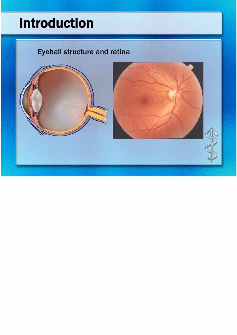
Introduction
Eyeball structure and retina
7/30/2019 Chp14 retina
http://slidepdf.com/reader/full/chp14-retina 3/76
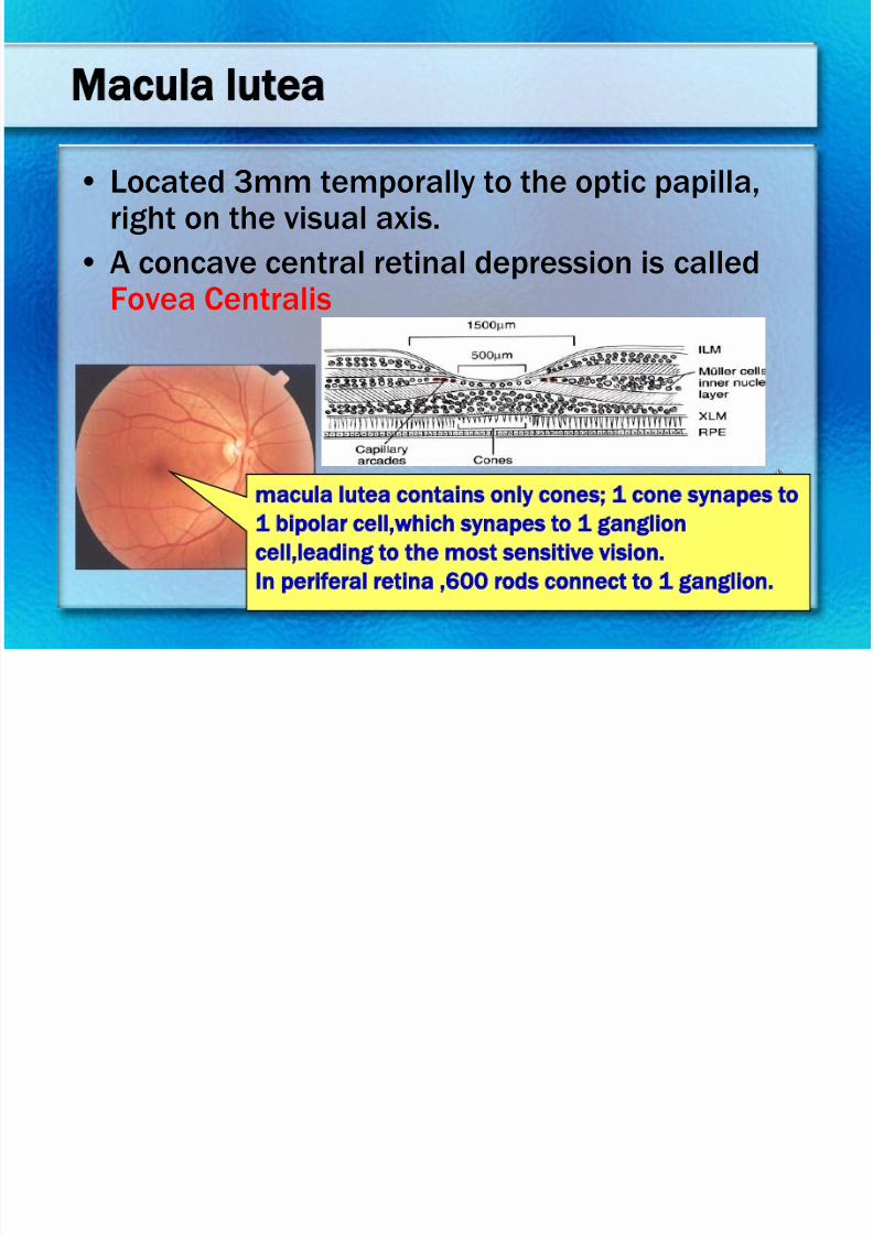
Macula lutea
• Located 3mm temporally to the optic papilla,right on the visual axis.
• A concave central retinal depression is called
Fovea Centralis
macula lutea contains only cones; 1 cone synapes to
1 bipolar cell,which synapes to 1 ganglion
cell,leading to the most sensitive vision.
In periferal retina ,600 rods connect to 1 ganglion.
7/30/2019 Chp14 retina
http://slidepdf.com/reader/full/chp14-retina 4/76
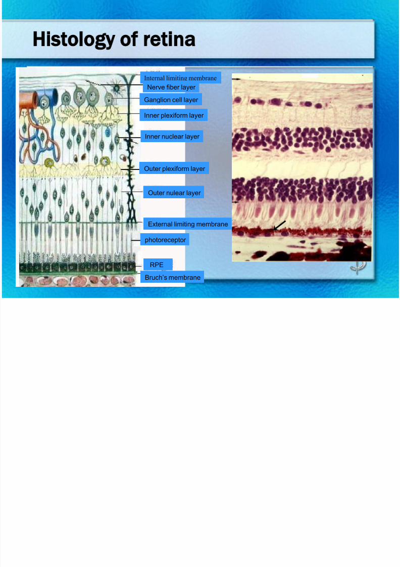
Histology of retina
Internal limiting membrane
Nerve fiber layer
Ganglion cell layer
Inner plexiform layer
Inner nuclear layer
Outer plexiform layer
Outer nulear layer
External limiting membrane
photoreceptor
RPE
Bruch’s membrane
7/30/2019 Chp14 retina
http://slidepdf.com/reader/full/chp14-retina 5/76
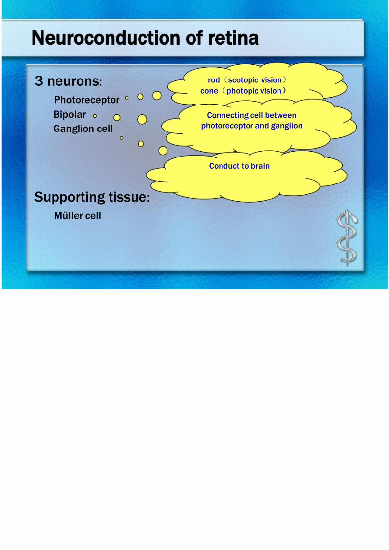
Neuroconduction of retina 3 neurons:
Photoreceptor
Bipolar
Ganglion cell
Supporting tissue:Müller cell
rod(scotopic vision)
cone(photopic vision)
Connecting cell between
photoreceptor and ganglion
Conduct to brain
7/30/2019 Chp14 retina
http://slidepdf.com/reader/full/chp14-retina 6/76
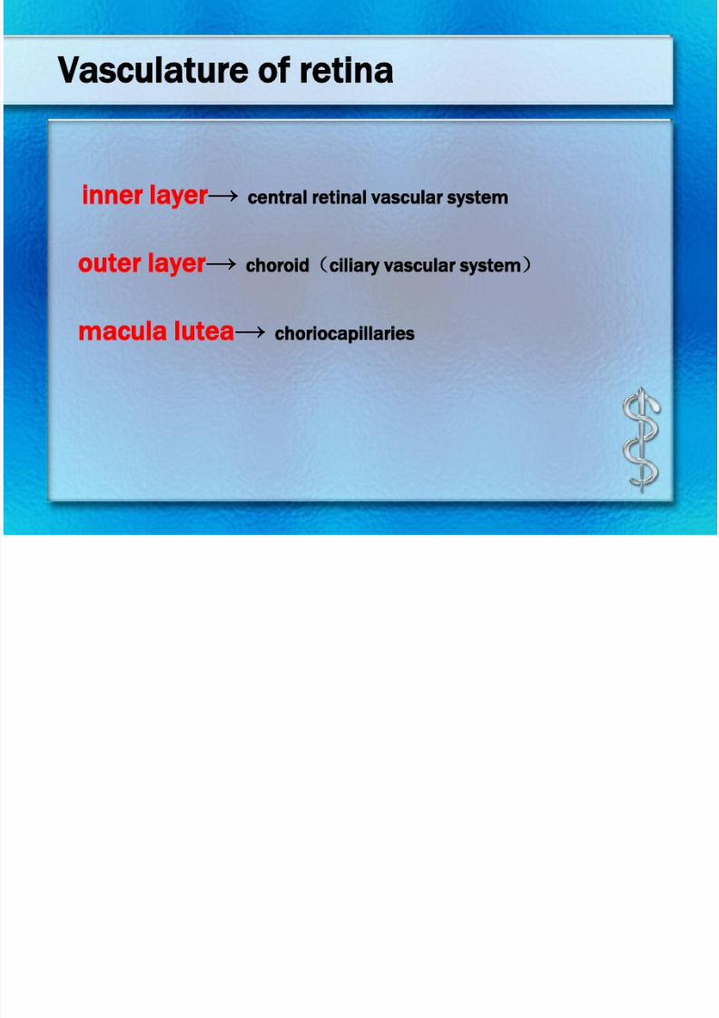
Vasculature of retina
inner layer→ central retinal vascular system
outer layer→ choroid(ciliary vascular system)
macula lutea→ choriocapillaries
7/30/2019 Chp14 retina
http://slidepdf.com/reader/full/chp14-retina 7/76
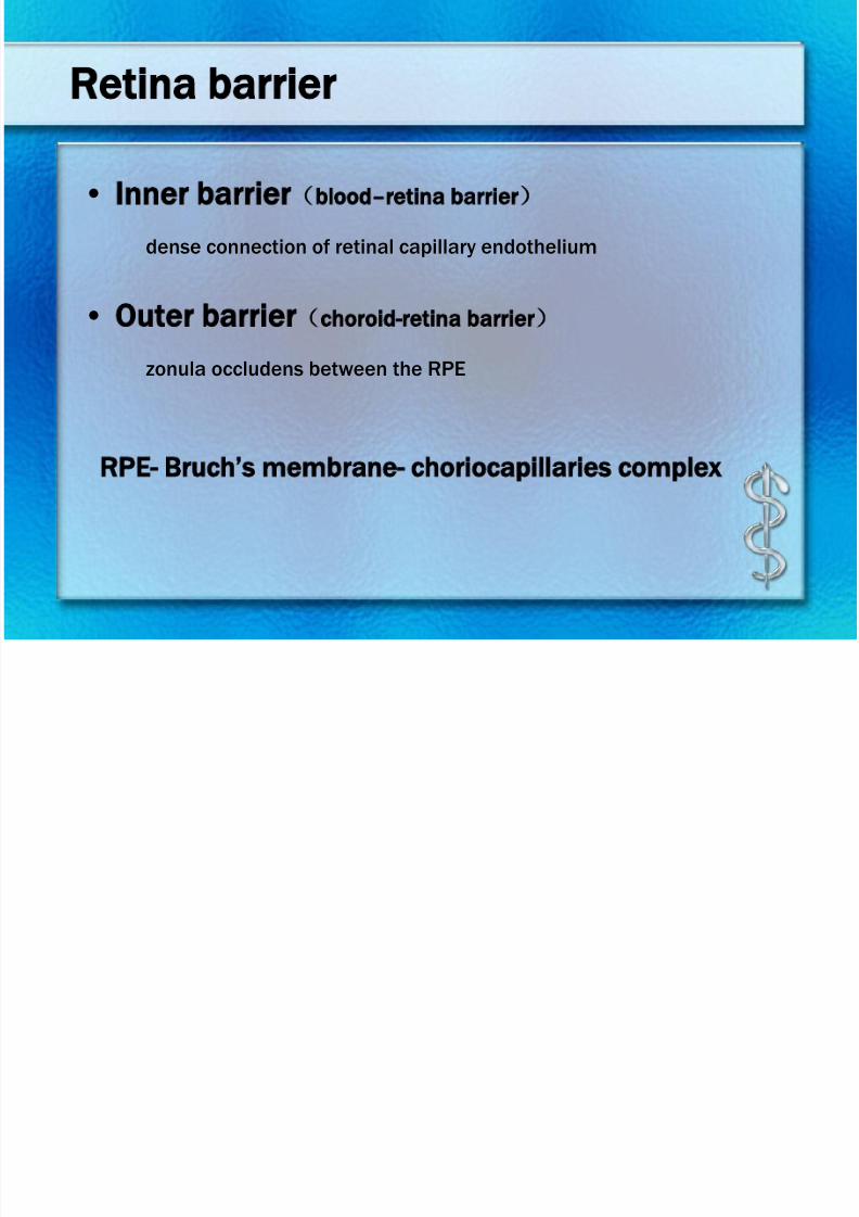
Retina barrier
• Inner barrier(blood–retina barrier)
dense connection of retinal capillary endothelium
• Outer barrier(choroid-retina barrier)
zonula occludens between the RPE
RPE- Bruch’s membrane- choriocapillaries complex
7/30/2019 Chp14 retina
http://slidepdf.com/reader/full/chp14-retina 8/76
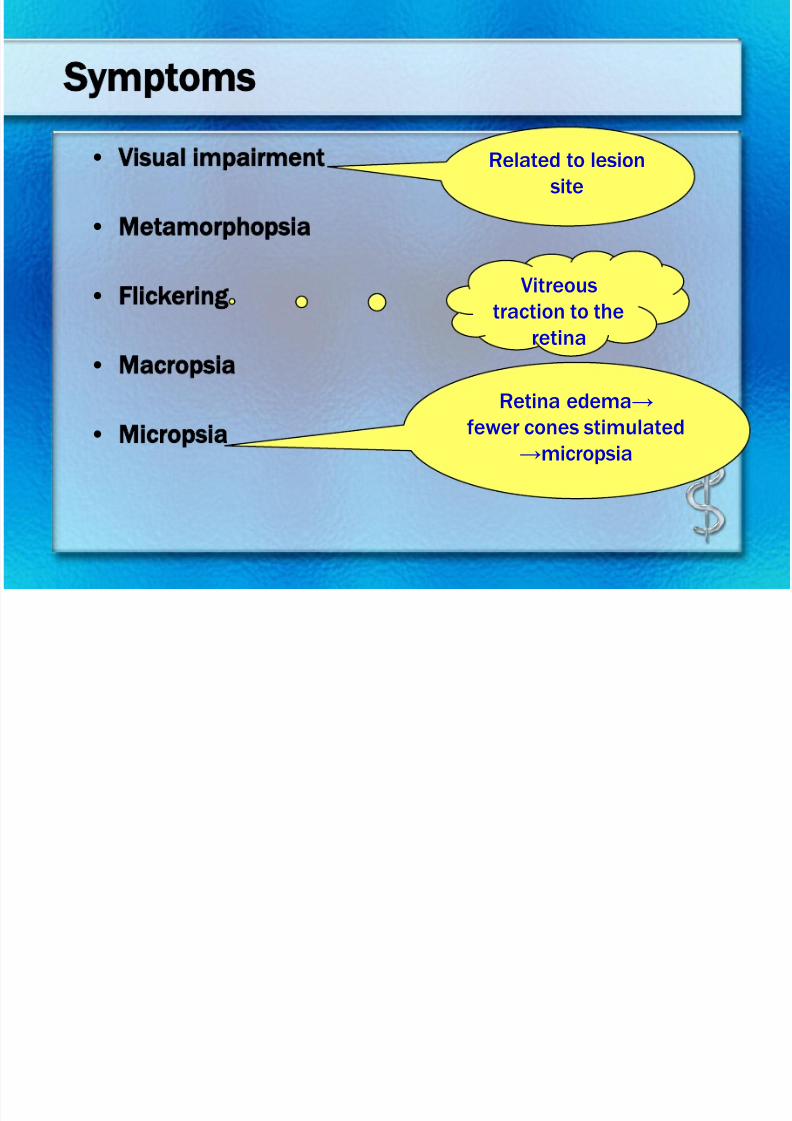
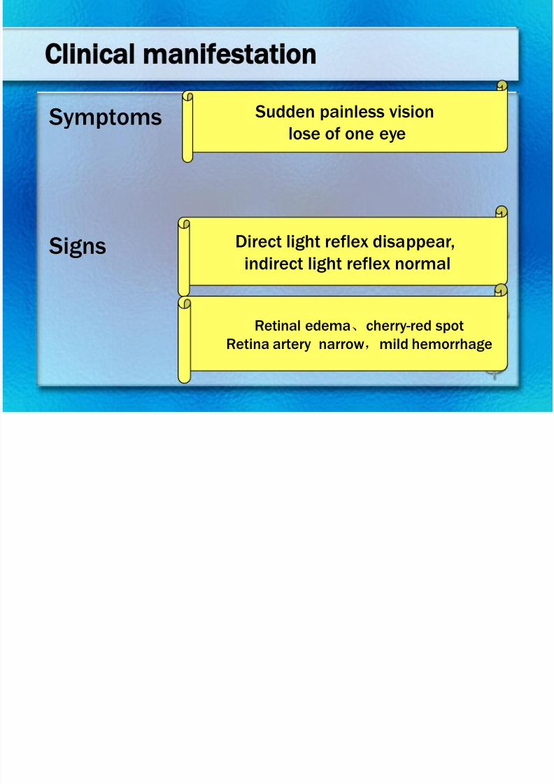
Symptoms
• Visual impairment
• Metamorphopsia
• Flickering
• Macropsia
• Micropsia
Related to lesion
site
Vitreous
traction to the
retina
Retina edema→
fewer cones stimulated
→micropsia
7/30/2019 Chp14 retina
http://slidepdf.com/reader/full/chp14-retina 9/76
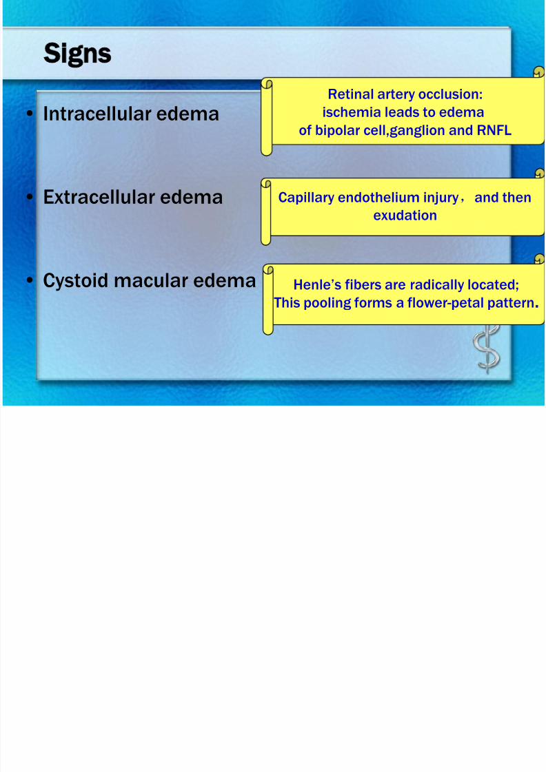
Signs
• Intracellular edema
• Extracellular edema
• Cystoid macular edema
Retinal artery occlusion:ischemia leads to edema
of bipolar cell,ganglion and RNFL
Capillary endothelium injury,and then
exudation
Henle’s fibers are radically located;This pooling forms a flower-petal pattern.
7/30/2019 Chp14 retina
http://slidepdf.com/reader/full/chp14-retina 10/76
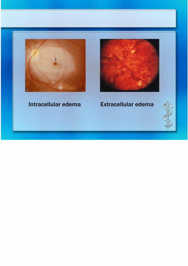
Intracellular edema Extracellular edema
7/30/2019 Chp14 retina
http://slidepdf.com/reader/full/chp14-retina 11/76
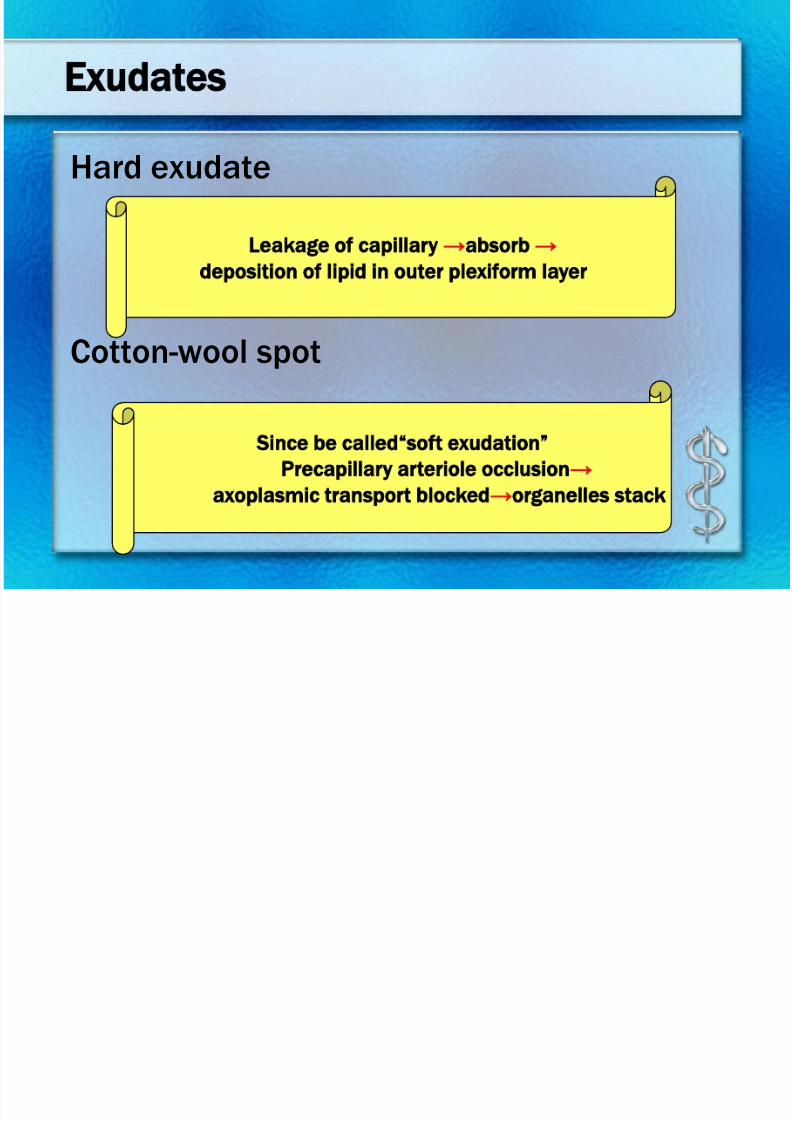
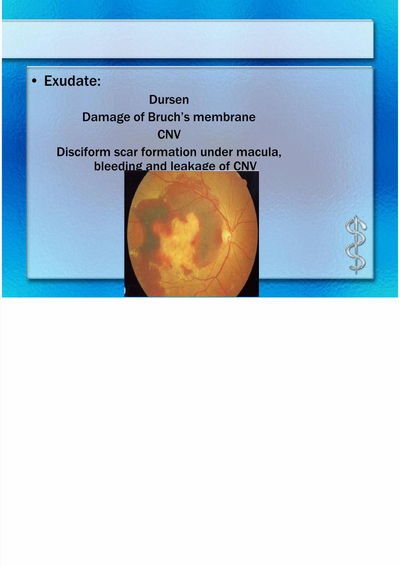
Exudates
Hard exudate
Cotton-wool spot
Leakage of capillary →absorb →
deposition of lipid in outer plexiform layer
Since be called“soft exudation”
Precapillary arteriole occlusion→
axoplasmic transport blocked→organelles stack
7/30/2019 Chp14 retina
http://slidepdf.com/reader/full/chp14-retina 12/76
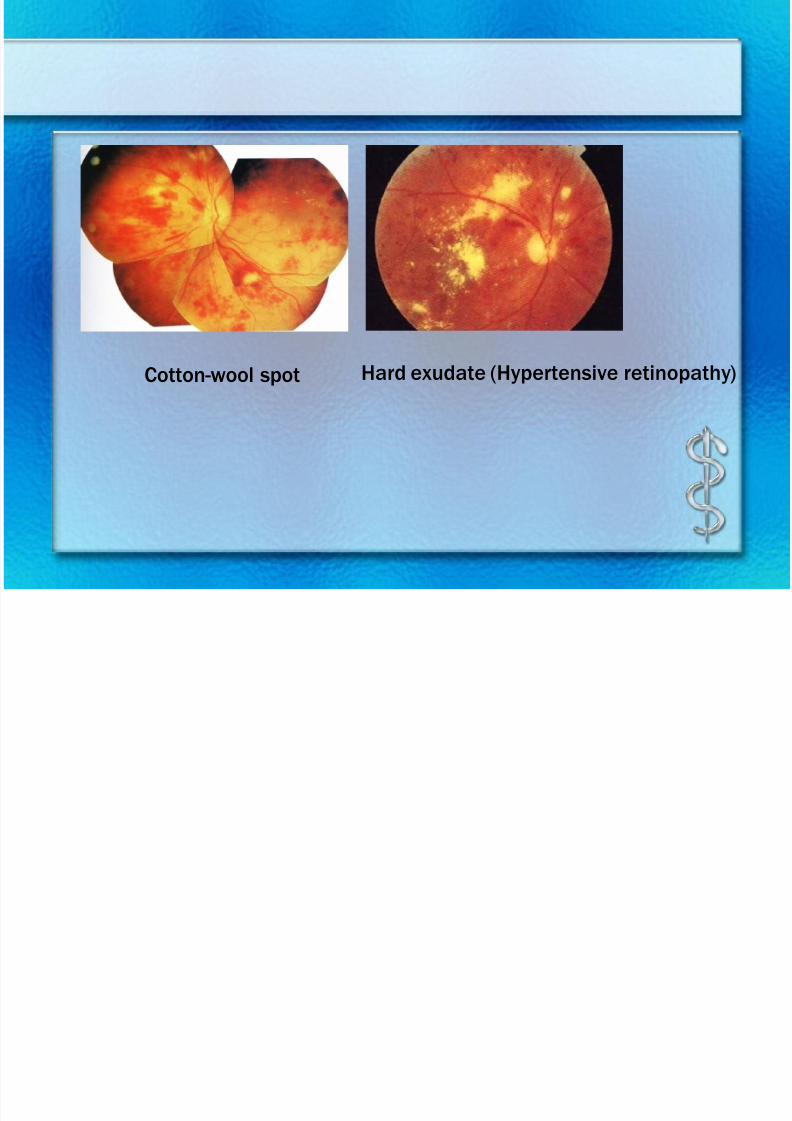
Cotton-wool spot Hard exudate (Hypertensive retinopathy)
7/30/2019 Chp14 retina
http://slidepdf.com/reader/full/chp14-retina 13/76
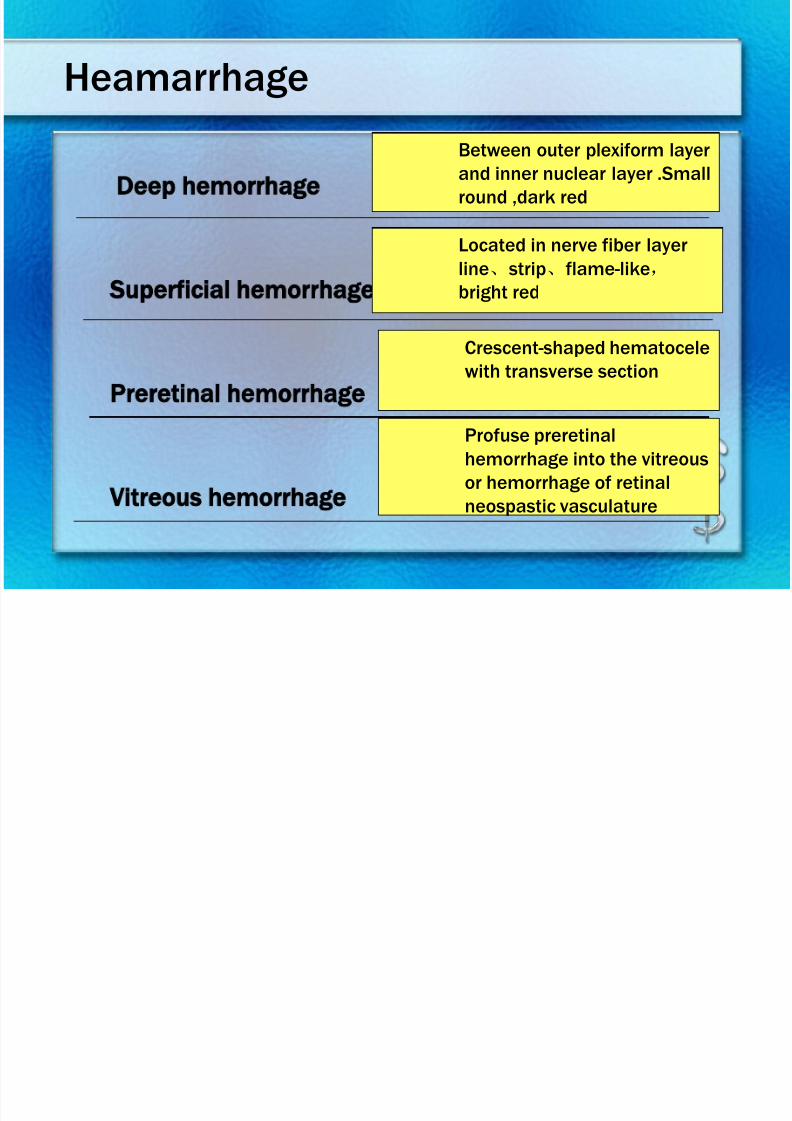
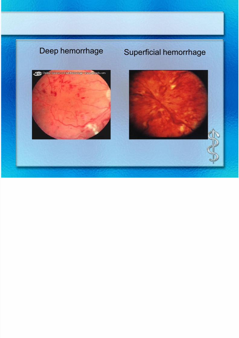
Heamarrhage
Deep hemorrhage
Superficial hemorrhage
Preretinal hemorrhage
Vitreous hemorrhage
Between outer plexiform layer
and inner nuclear layer .Small
round ,dark red
Located in nerve fiber layer
line、
strip、
flame-like,
bright red
Crescent-shaped hematocele
with transverse section
Profuse preretinal
hemorrhage into the vitreous
or hemorrhage of retinal
neospastic vasculature
7/30/2019 Chp14 retina
http://slidepdf.com/reader/full/chp14-retina 14/76
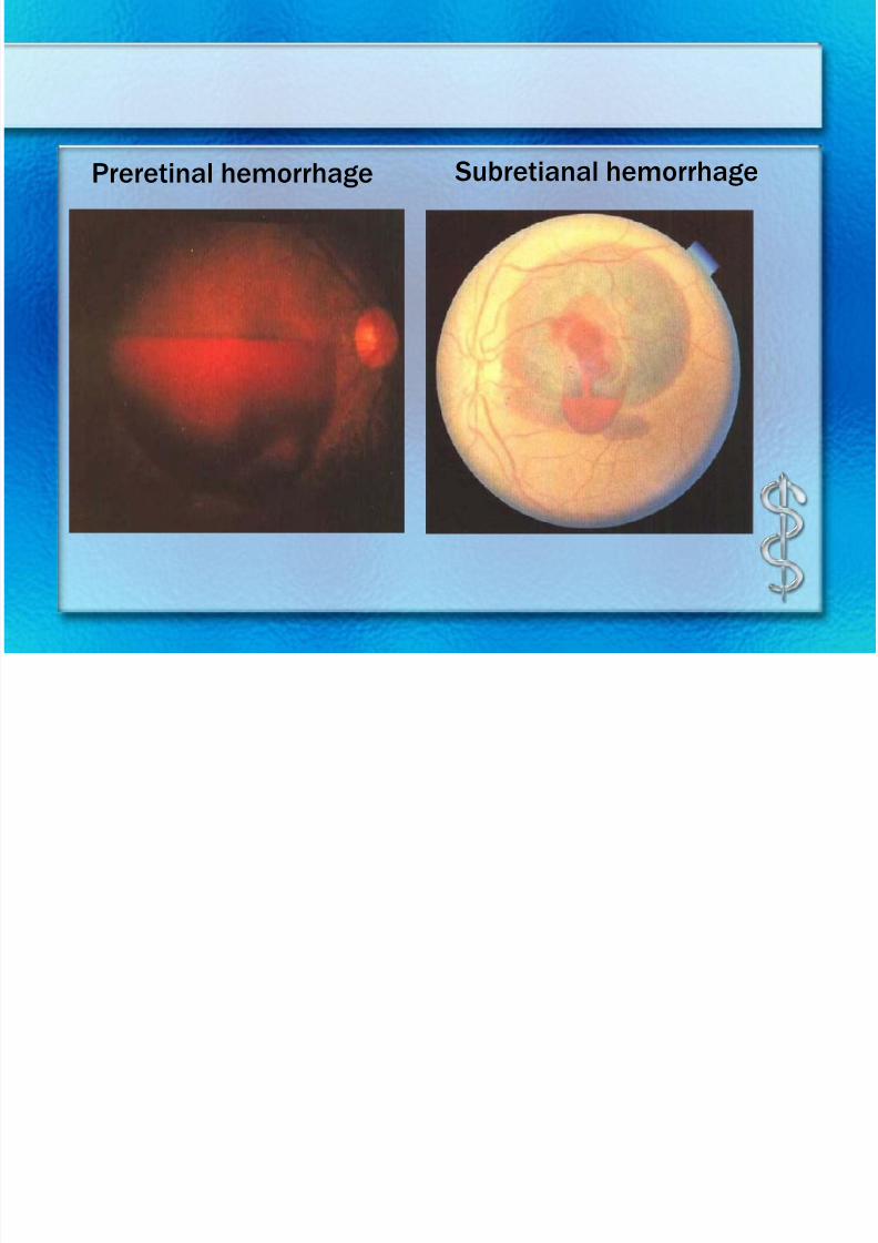
Preretinal hemorrhage Subretianal hemorrhage
7/30/2019 Chp14 retina
http://slidepdf.com/reader/full/chp14-retina 15/76
Deep hemorrhage Superficial hemorrhage
7/30/2019 Chp14 retina
http://slidepdf.com/reader/full/chp14-retina 16/76
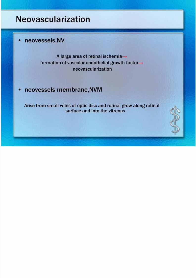
Neovascularization
• neovessels,NV
A large area of retinal ischemia→
formation of vascular endothelial growth factor→
neovascularization
• neovessels membrane,NVM
Arise from small veins of optic disc and retina; grow along retinalsurface and into the vitreous
7/30/2019 Chp14 retina
http://slidepdf.com/reader/full/chp14-retina 17/76
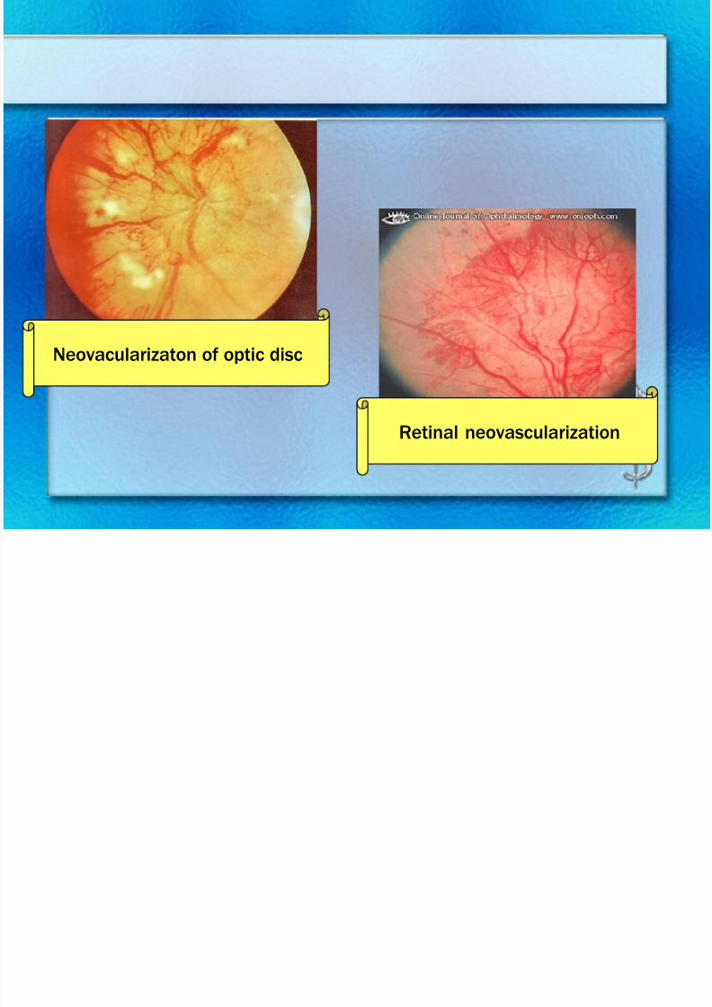
Retinal neovascularization
Neovacularizaton of optic disc
7/30/2019 Chp14 retina
http://slidepdf.com/reader/full/chp14-retina 18/76
7/30/2019 Chp14 retina
http://slidepdf.com/reader/full/chp14-retina 19/76
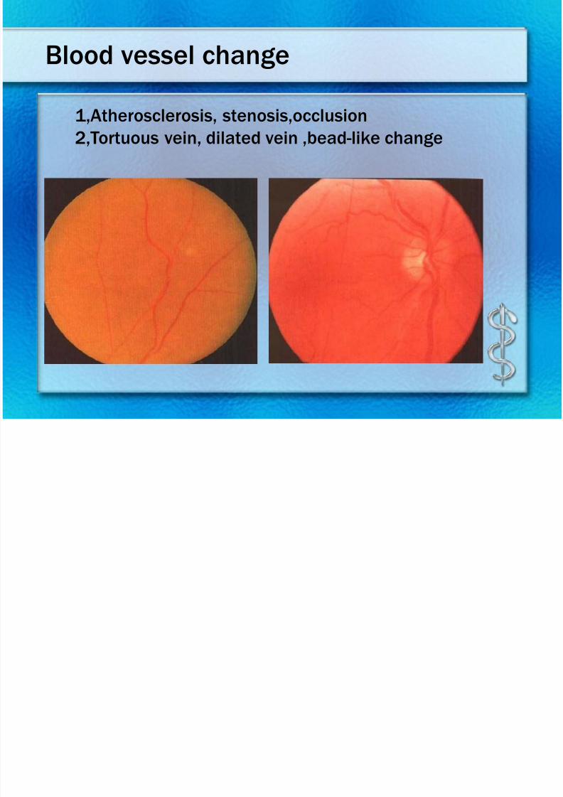
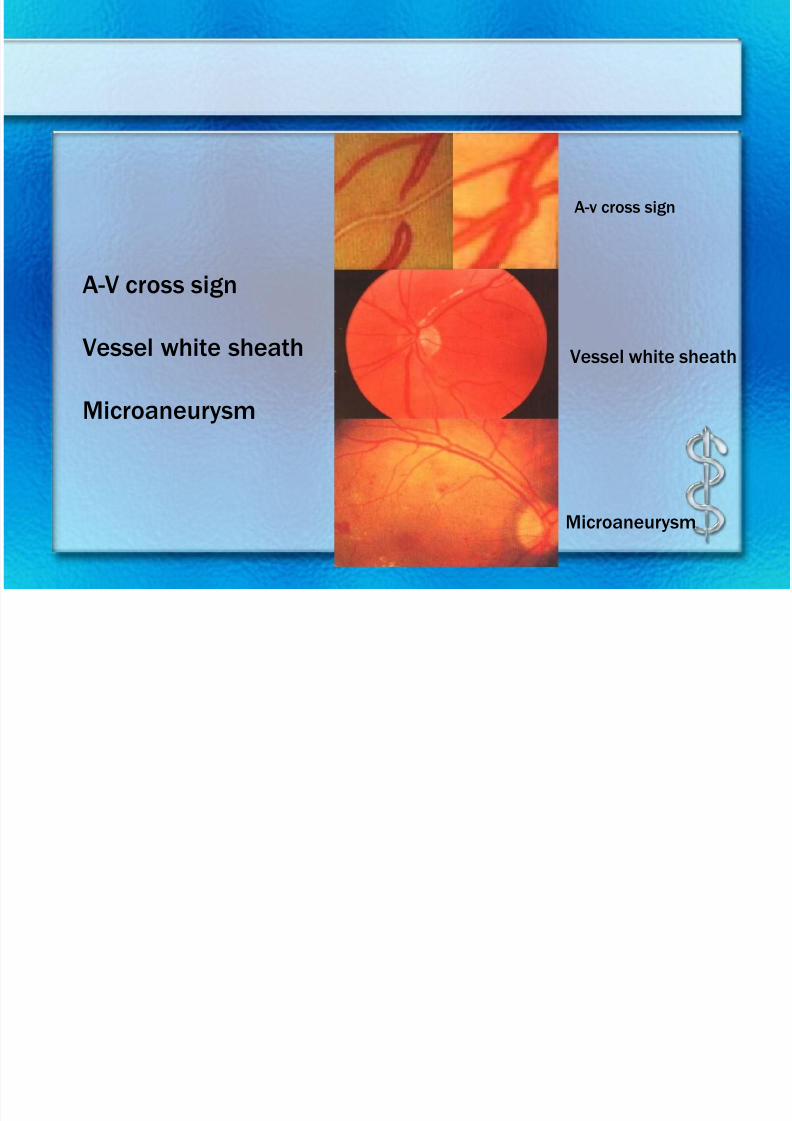
A-V cross sign
Vessel white sheath
Microaneurysm
Microaneurysm
A-v cross sign
Vessel white sheath
7/30/2019 Chp14 retina
http://slidepdf.com/reader/full/chp14-retina 20/76
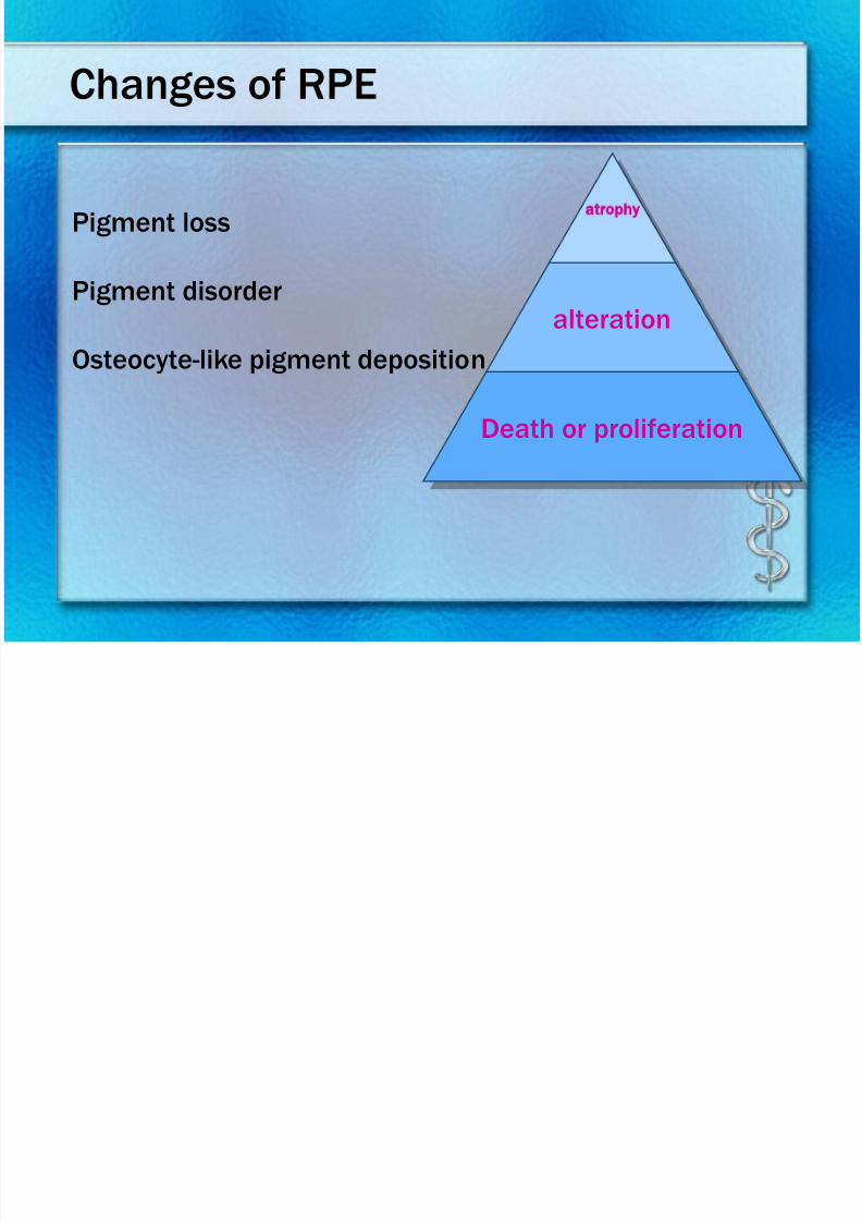
Changes of RPE
atrophy
alteration
Death or proliferation
Pigment loss
Pigment disorder
Osteocyte-like pigment deposition
7/30/2019 Chp14 retina
http://slidepdf.com/reader/full/chp14-retina 21/76
7/30/2019 Chp14 retina
http://slidepdf.com/reader/full/chp14-retina 22/76
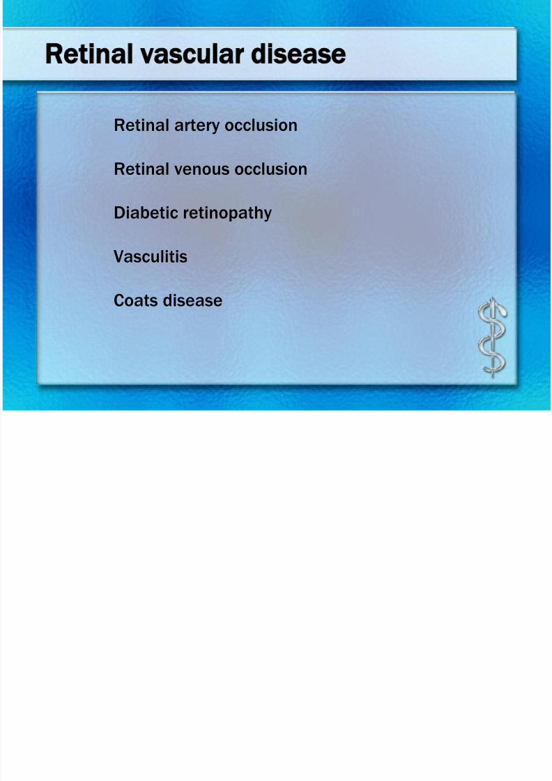
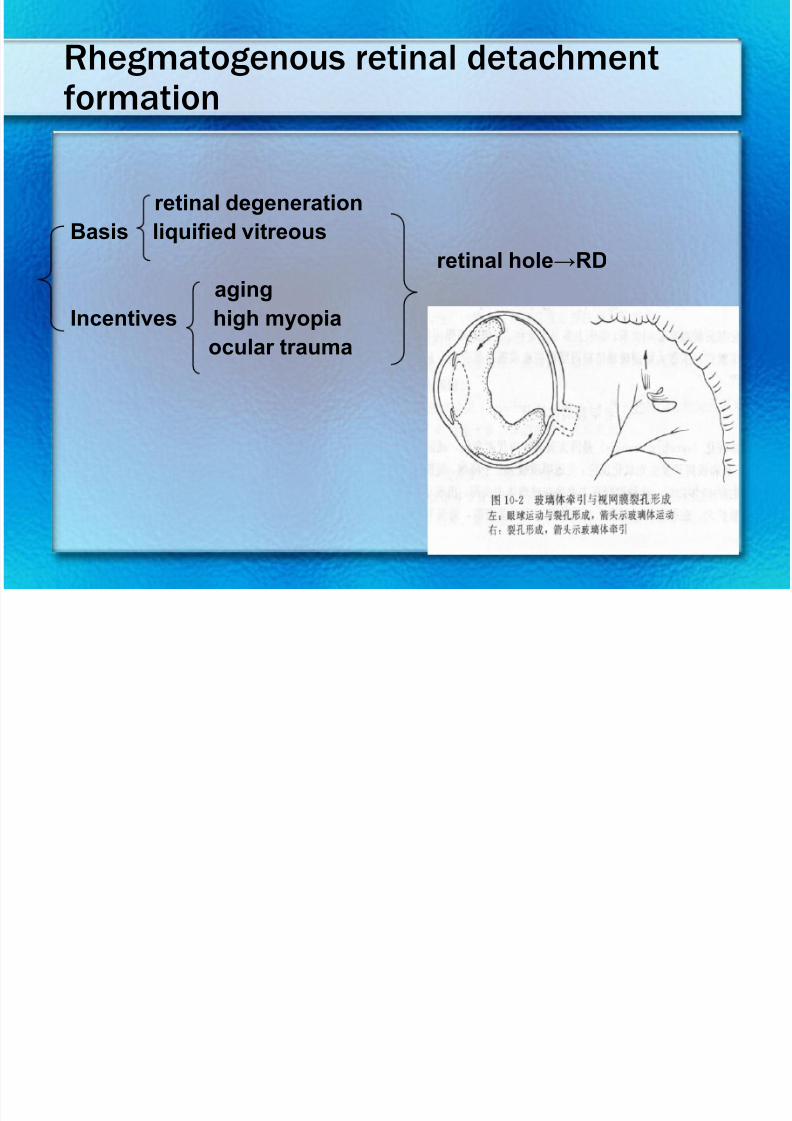
Classification of retinal diseases
• Vascular diseases
• Macular diseases
• Retinal detachment
• Retinal degeneration
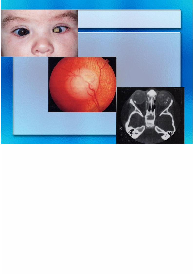
• Retinal tumor
• Ocular manifestation of general diseases
7/30/2019 Chp14 retina
http://slidepdf.com/reader/full/chp14-retina 23/76
7/30/2019 Chp14 retina
http://slidepdf.com/reader/full/chp14-retina 24/76
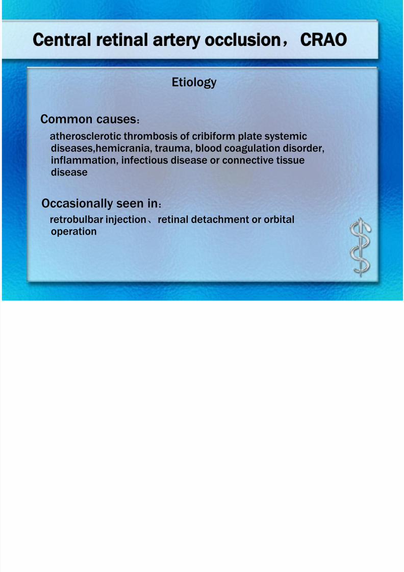
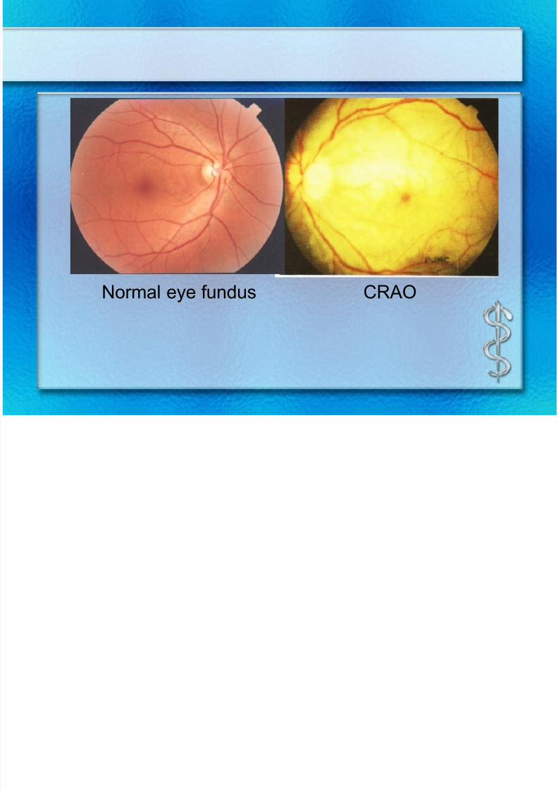
Central retinal artery occlusion,CRAO
Common causes:
atherosclerotic thrombosis of cribiform plate systemicdiseases,hemicrania, trauma, blood coagulation disorder,inflammation, infectious disease or connective tissuedisease
Occasionally seen in:
retrobulbar injection、retinal detachment or orbitaloperation
Etiology
7/30/2019 Chp14 retina
http://slidepdf.com/reader/full/chp14-retina 25/76
7/30/2019 Chp14 retina
http://slidepdf.com/reader/full/chp14-retina 26/76
7/30/2019 Chp14 retina
http://slidepdf.com/reader/full/chp14-retina 27/76
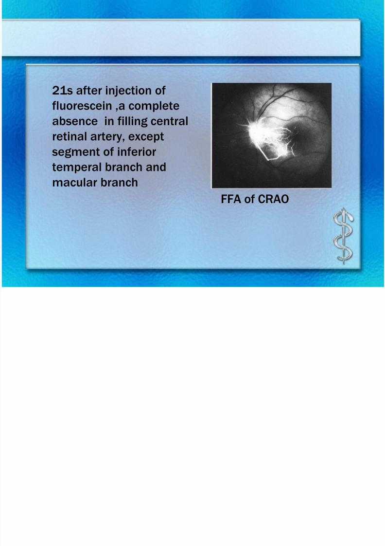
FFA of CRAO
21s after injection of
fluorescein ,a complete
absence in filling central
retinal artery, exceptsegment of inferior
temperal branch and
macular branch
7/30/2019 Chp14 retina
http://slidepdf.com/reader/full/chp14-retina 28/76
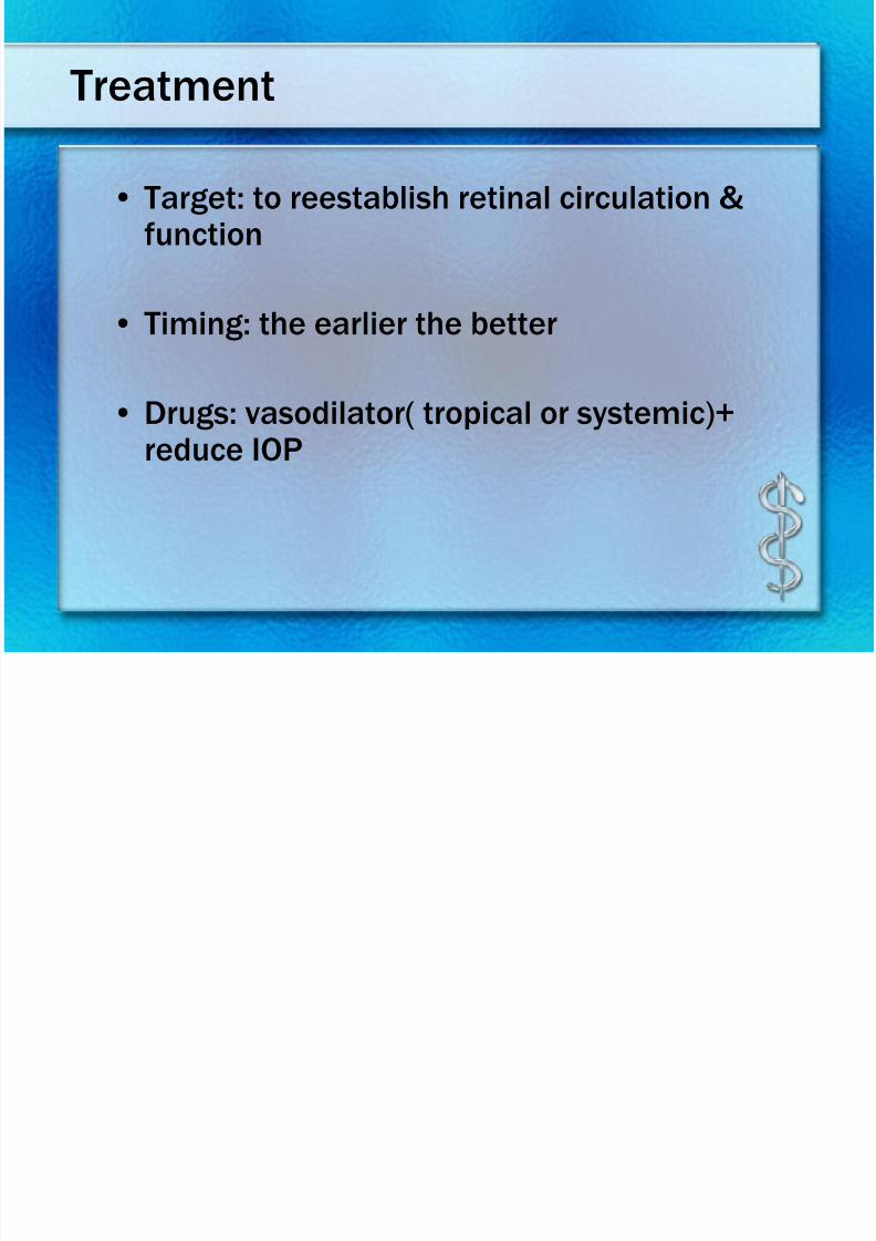
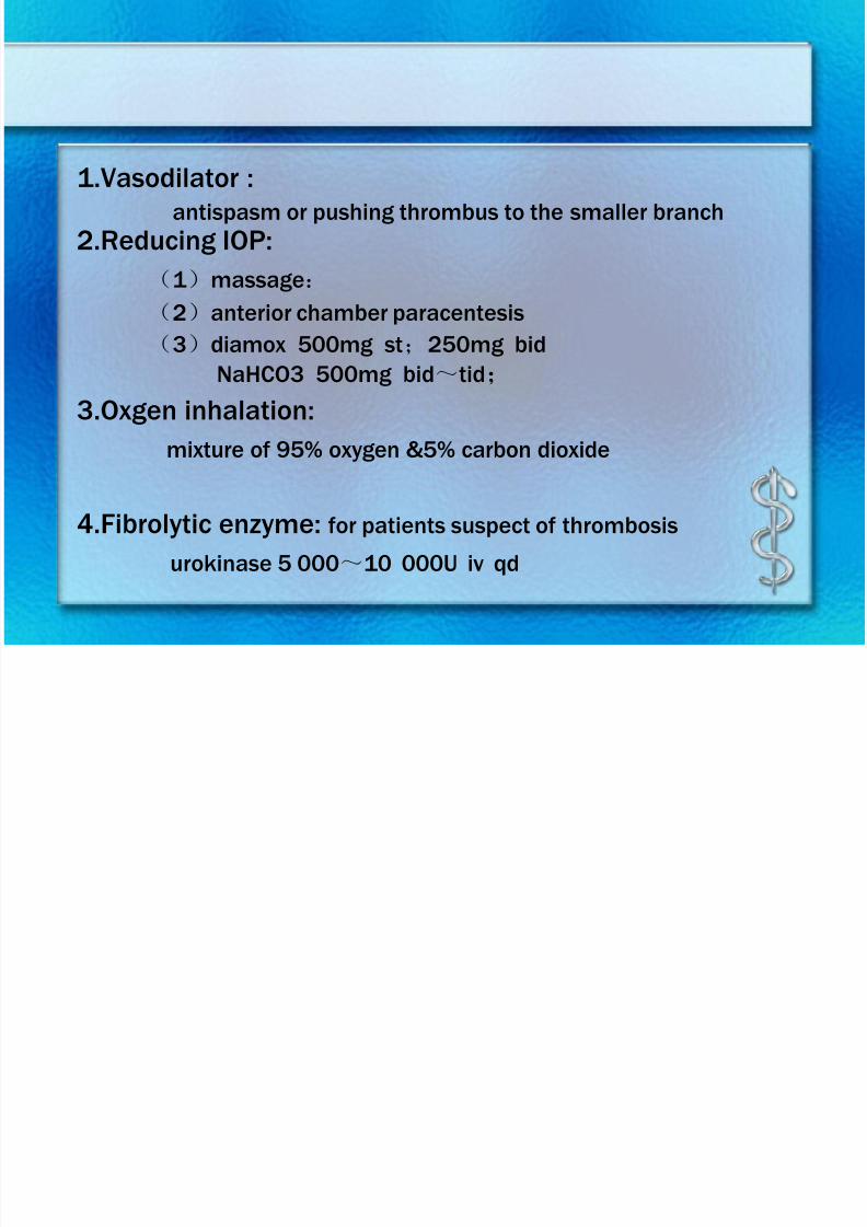
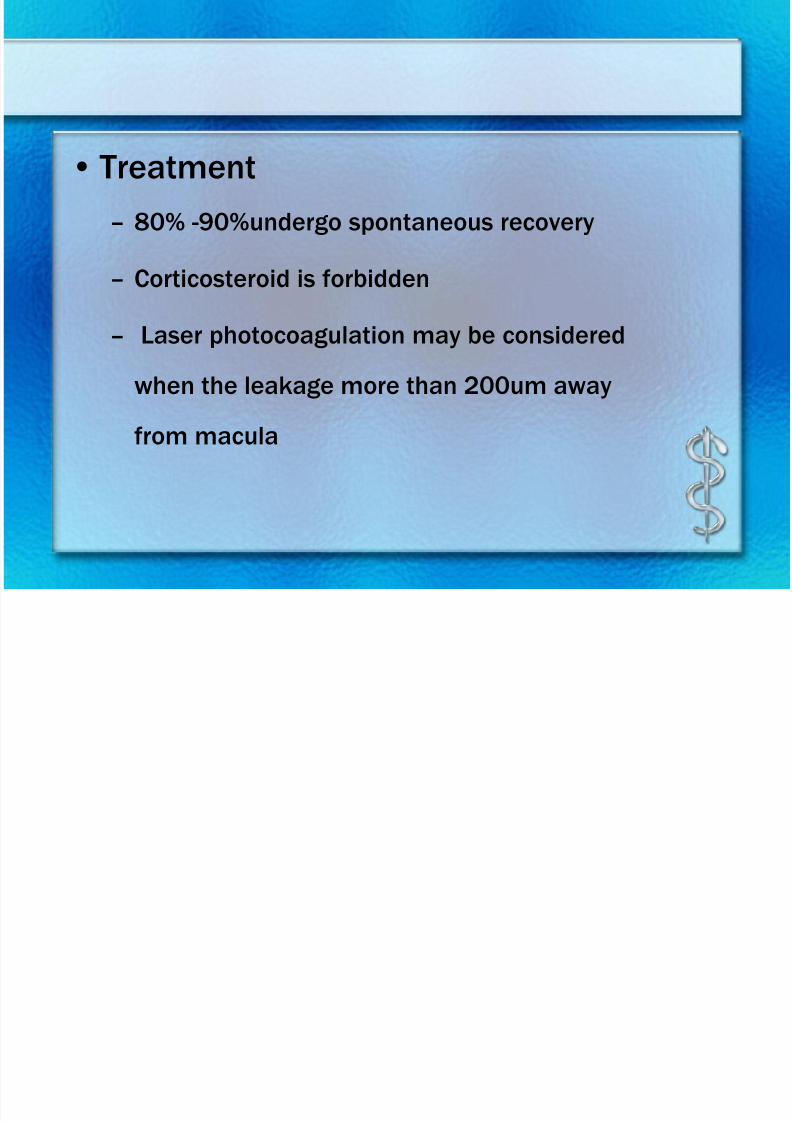
Treatment
• Target: to reestablish retinal circulation &function
• Timing: the earlier the better
• Drugs: vasodilator( tropical or systemic)+
reduce IOP
7/30/2019 Chp14 retina
http://slidepdf.com/reader/full/chp14-retina 29/76
7/30/2019 Chp14 retina
http://slidepdf.com/reader/full/chp14-retina 30/76
7/30/2019 Chp14 retina
http://slidepdf.com/reader/full/chp14-retina 31/76
7/30/2019 Chp14 retina
http://slidepdf.com/reader/full/chp14-retina 32/76
7/30/2019 Chp14 retina
http://slidepdf.com/reader/full/chp14-retina 33/76
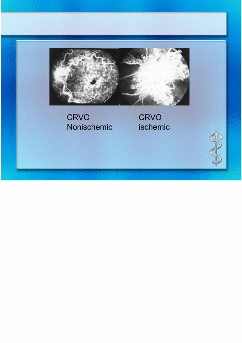
CRVO
Nonischemic
CRVO
ischemic
7/30/2019 Chp14 retina
http://slidepdf.com/reader/full/chp14-retina 34/76
7/30/2019 Chp14 retina
http://slidepdf.com/reader/full/chp14-retina 35/76
Treatment
– Chinese medicine
– Anitplatelet or antithrombotic drugs: unknown
therapeutic effects
– Systemic examination to find out causes
– Corticosteroid if vasculitis exist
– Grid pattern photocoagulation of macula、PRP
– Laser induced retina-choroid vascular anastomosis
7/30/2019 Chp14 retina
http://slidepdf.com/reader/full/chp14-retina 36/76
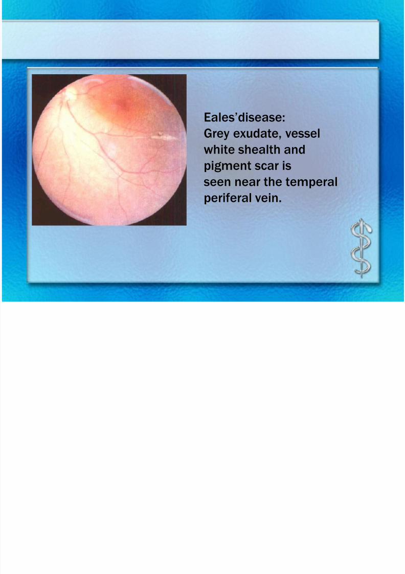
Vasculitis
• Idiopathic retinal vasculitis
Eales disease(Retinalperiphlebitis)
Both A. and V. are involved
Causes is unclear,patient tuberculin reaction (+)
Seen in 20-40 years old men
Bilateral peripheral small vessels occlusion,
recurrent vitreous hemorrhage,retinalneovascularization
7/30/2019 Chp14 retina
http://slidepdf.com/reader/full/chp14-retina 37/76
7/30/2019 Chp14 retina
http://slidepdf.com/reader/full/chp14-retina 38/76
7/30/2019 Chp14 retina
http://slidepdf.com/reader/full/chp14-retina 39/76
7/30/2019 Chp14 retina
http://slidepdf.com/reader/full/chp14-retina 40/76
7/30/2019 Chp14 retina
http://slidepdf.com/reader/full/chp14-retina 41/76
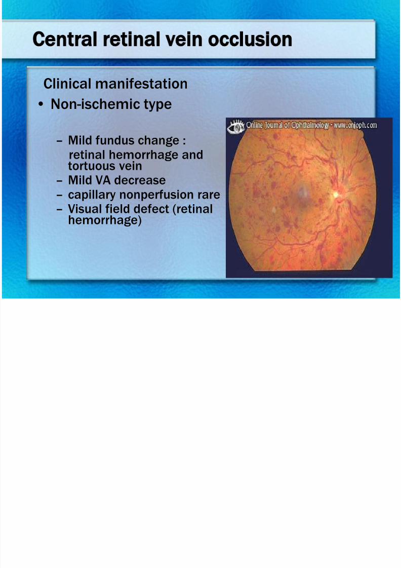
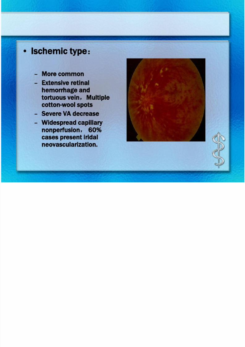
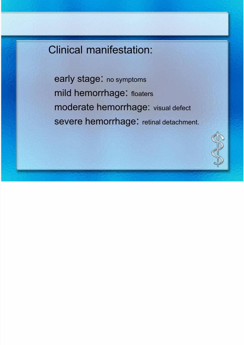
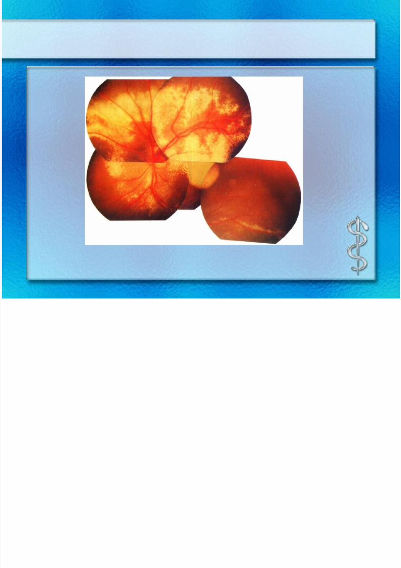
Clinical manifestation
– Visual disturbance、strabismus、“leucoria”
– Fundus:
extensive yellow-white lipid exudation withfaring cholesterol crystal;capillary and veindilate,microaneurysm;capillary nonfusion; Secondary glaucoma, exudative RD, uvitis,complicated cataract
– Rare neovascularization
7/30/2019 Chp14 retina
http://slidepdf.com/reader/full/chp14-retina 42/76
7/30/2019 Chp14 retina
http://slidepdf.com/reader/full/chp14-retina 43/76
7/30/2019 Chp14 retina
http://slidepdf.com/reader/full/chp14-retina 44/76
Treatment
– Photocoagulation or cryocoagulation ofcapillary dilation
7/30/2019 Chp14 retina
http://slidepdf.com/reader/full/chp14-retina 45/76
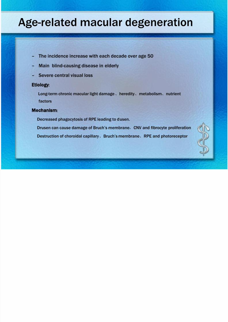
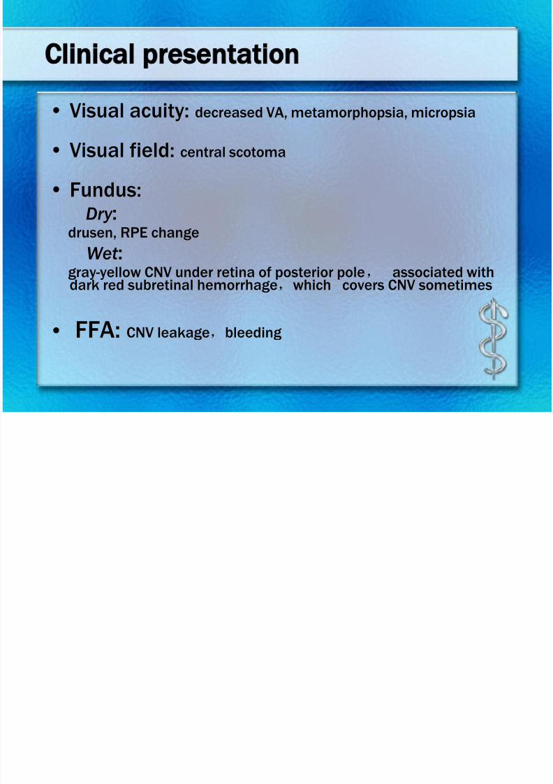
Macular diseases
• Central serous chorioretinopathy
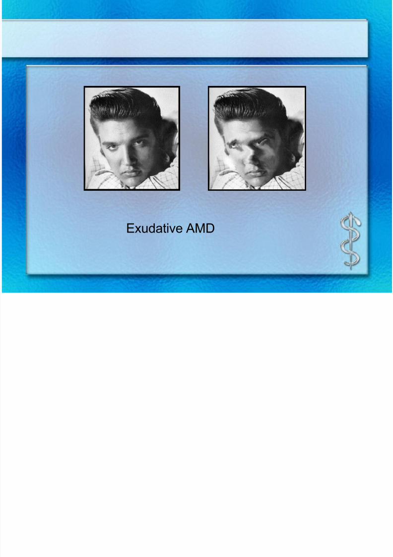
• Age-related macular degeneration
• Central exudative chorioretinopathy
7/30/2019 Chp14 retina
http://slidepdf.com/reader/full/chp14-retina 46/76
– Often seen in 20-45 years old men
– Self-limitted
– Related to stress reaction
Central serous chorioretinopathy
7/30/2019 Chp14 retina
http://slidepdf.com/reader/full/chp14-retina 47/76
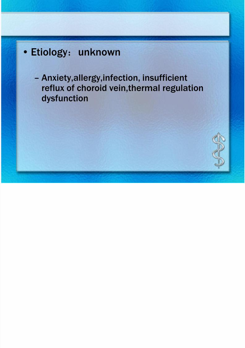
• Etiology:unknown
– Anxiety,allergy,infection, insufficient
reflux of choroid vein,thermal regulationdysfunction
7/30/2019 Chp14 retina
http://slidepdf.com/reader/full/chp14-retina 48/76
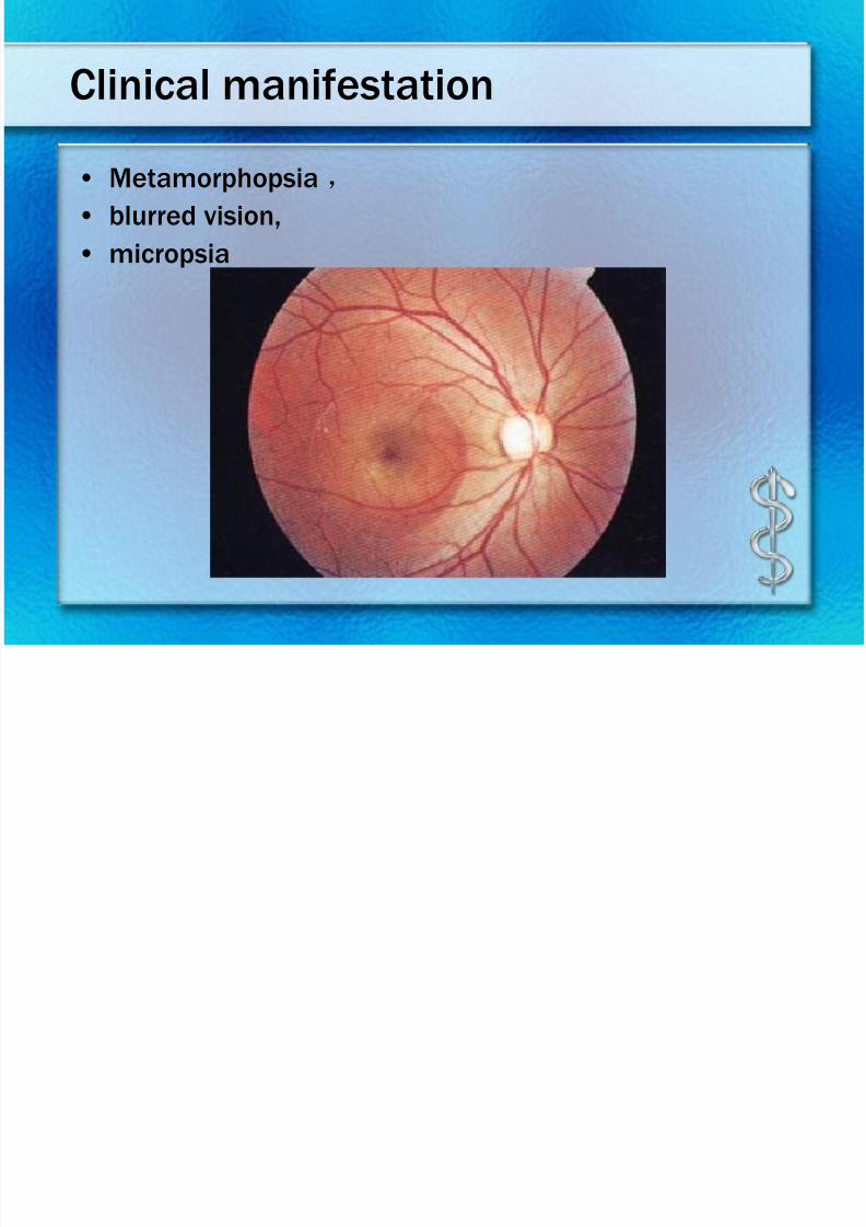
Clinical manifestation
• Metamorphopsia,
• blurred vision,
• micropsia
7/30/2019 Chp14 retina
http://slidepdf.com/reader/full/chp14-retina 49/76
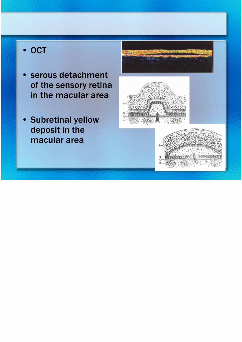
• OCT
• serous detachment
of the sensory retinain the macular area
• Subretinal yellow
deposit in themacular area
7/30/2019 Chp14 retina
http://slidepdf.com/reader/full/chp14-retina 50/76
7/30/2019 Chp14 retina
http://slidepdf.com/reader/full/chp14-retina 51/76
7/30/2019 Chp14 retina
http://slidepdf.com/reader/full/chp14-retina 52/76
7/30/2019 Chp14 retina
http://slidepdf.com/reader/full/chp14-retina 53/76
7/30/2019 Chp14 retina
http://slidepdf.com/reader/full/chp14-retina 54/76
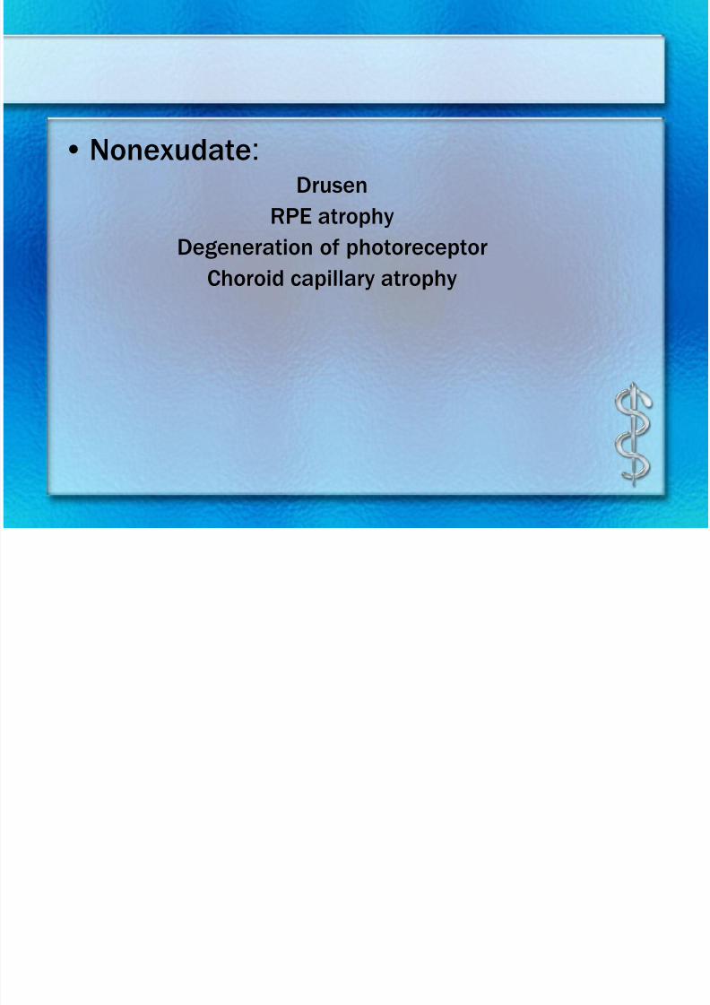
• Nonexudate:Drusen
RPE atrophy
Degeneration of photoreceptorChoroid capillary atrophy
7/30/2019 Chp14 retina
http://slidepdf.com/reader/full/chp14-retina 55/76
7/30/2019 Chp14 retina
http://slidepdf.com/reader/full/chp14-retina 56/76
7/30/2019 Chp14 retina
http://slidepdf.com/reader/full/chp14-retina 57/76
7/30/2019 Chp14 retina
http://slidepdf.com/reader/full/chp14-retina 58/76
7/30/2019 Chp14 retina
http://slidepdf.com/reader/full/chp14-retina 59/76
7/30/2019 Chp14 retina
http://slidepdf.com/reader/full/chp14-retina 60/76
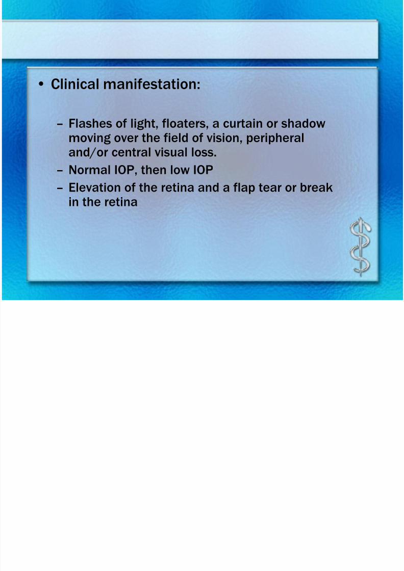
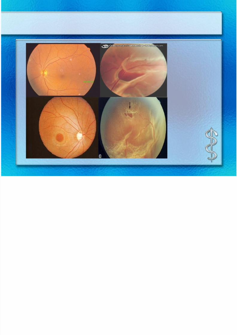
• Clinical manifestation:
– Flashes of light, floaters, a curtain or shadowmoving over the field of vision, peripheraland/or central visual loss.
– Normal IOP, then low IOP
– Elevation of the retina and a flap tear or breakin the retina
7/30/2019 Chp14 retina
http://slidepdf.com/reader/full/chp14-retina 61/76
7/30/2019 Chp14 retina
http://slidepdf.com/reader/full/chp14-retina 62/76
7/30/2019 Chp14 retina
http://slidepdf.com/reader/full/chp14-retina 63/76
Retinitis Pigmentosa
• Chronic ,progressive,inherited disease
• Cone cell、rod cell and RPE distrophy
• Inheritance pattern: AD, AR,X-link
• Onset age: childhood,
• Bilateral eyes involved
7/30/2019 Chp14 retina
http://slidepdf.com/reader/full/chp14-retina 64/76
Clinical manifestation
• Constriction of visual field
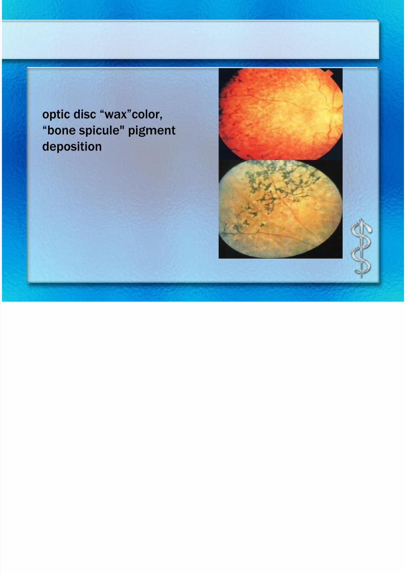
• Fundus :
optic disc “wax”color, "bone spicule" pigment deposition
• Nyctalopia is the first symptom
• FFA:
window defect, blocked fluorescein in pigment deposition at
early stage, hypofluorescence and the fluorescein of choroidis seen at late stage
7/30/2019 Chp14 retina
http://slidepdf.com/reader/full/chp14-retina 65/76
7/30/2019 Chp14 retina
http://slidepdf.com/reader/full/chp14-retina 66/76
Treatment
• Genetic counselling
• Avoiding sunlight and UV
• Vasodilator, Vitamins
• Suppliment of taurine
• Low vision aids
• Grid laser coagulation is used with caution for CME
7/30/2019 Chp14 retina
http://slidepdf.com/reader/full/chp14-retina 67/76
7/30/2019 Chp14 retina
http://slidepdf.com/reader/full/chp14-retina 68/76
7/30/2019 Chp14 retina
http://slidepdf.com/reader/full/chp14-retina 69/76
7/30/2019 Chp14 retina
http://slidepdf.com/reader/full/chp14-retina 70/76
Staging
• intraocular stage
• glaucomatous stage
• extraocular stage
• metastasis stage
7/30/2019 Chp14 retina
http://slidepdf.com/reader/full/chp14-retina 71/76
7/30/2019 Chp14 retina
http://slidepdf.com/reader/full/chp14-retina 72/76
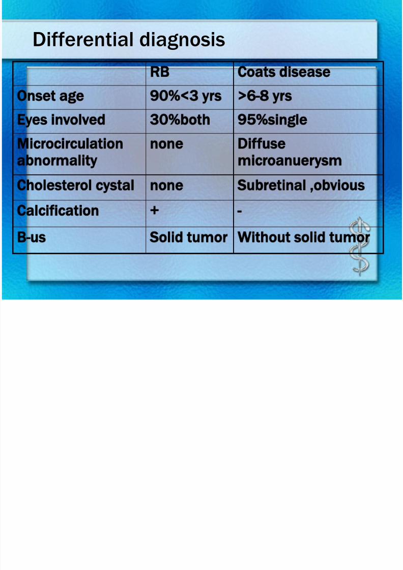
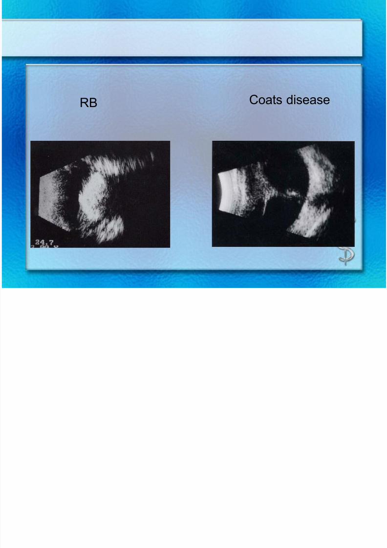
RB Coats disease
7/30/2019 Chp14 retina
http://slidepdf.com/reader/full/chp14-retina 73/76
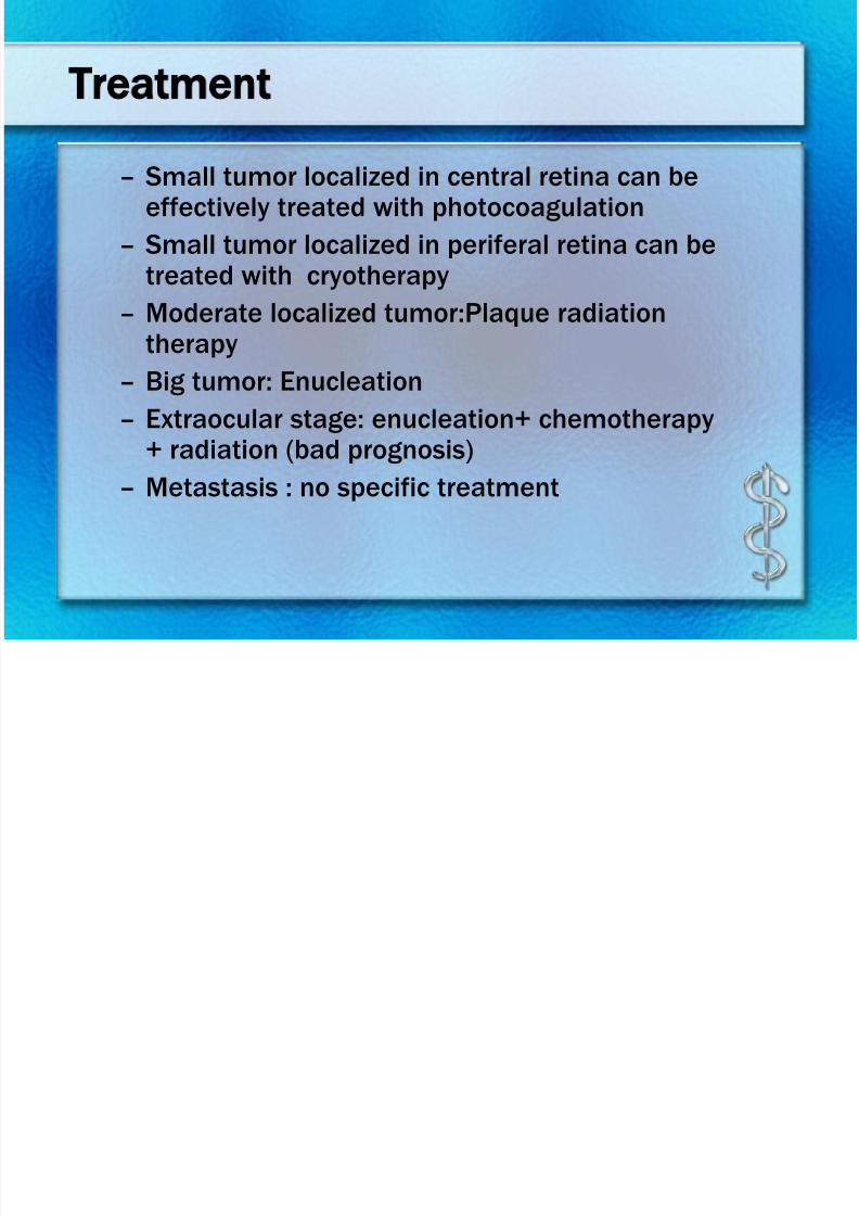
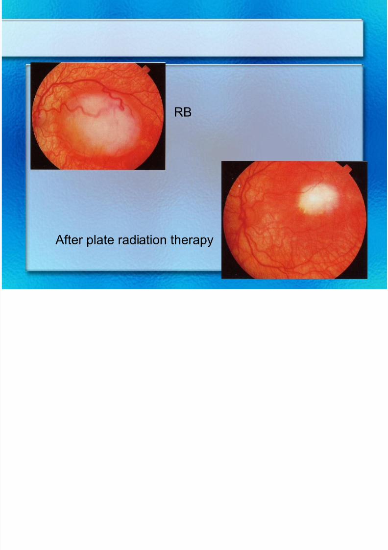
Treatment
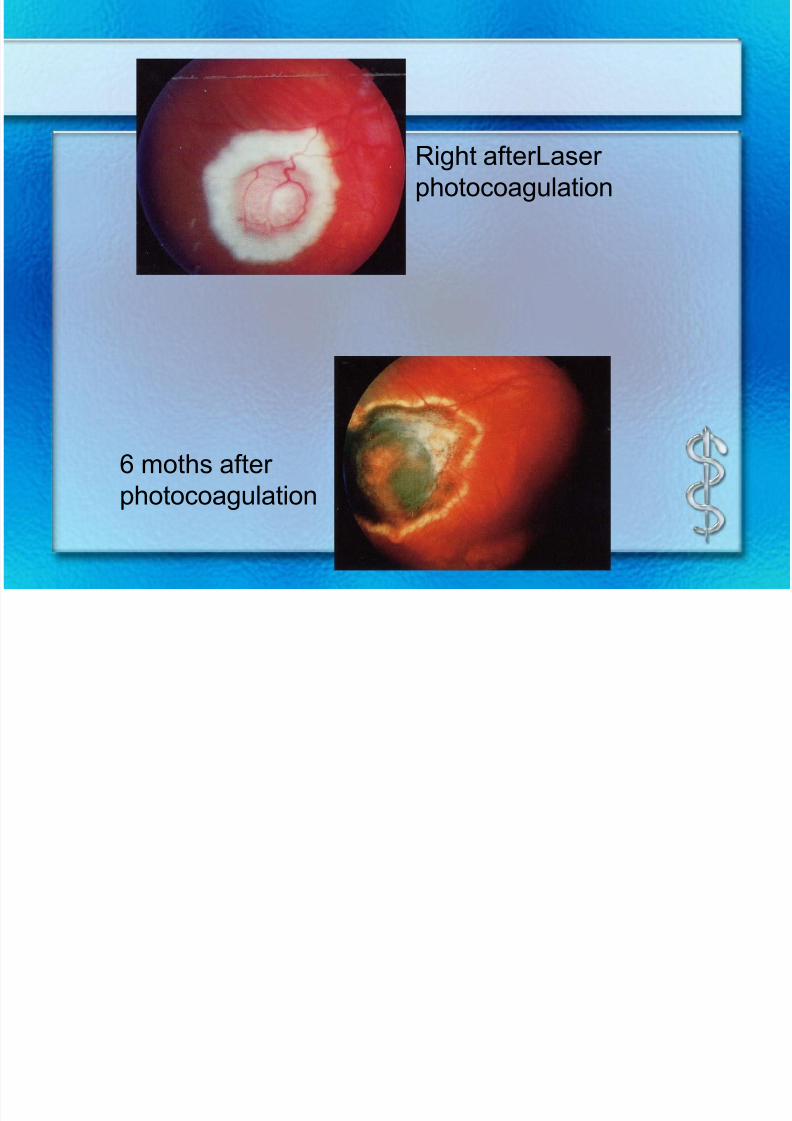
– Small tumor localized in central retina can beeffectively treated with photocoagulation
– Small tumor localized in periferal retina can be
treated with cryotherapy
– Moderate localized tumor:Plaque radiationtherapy
– Big tumor: Enucleation
– Extraocular stage: enucleation+ chemotherapy
+ radiation (bad prognosis)– Metastasis : no specific treatment
7/30/2019 Chp14 retina
http://slidepdf.com/reader/full/chp14-retina 74/76
7/30/2019 Chp14 retina
http://slidepdf.com/reader/full/chp14-retina 75/76
RB
After plate radiation therapy

7/30/2019 Chp14 retina
http://slidepdf.com/reader/full/chp14-retina 76/76