Embed Size (px)
Citation preview
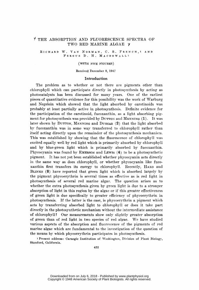
THE ABSORPTION AND FLUORESCENCE SPECTRA OFTWO RED MARINE ALGAE y
RICHARD W. VAN NORMAN, C. S. FRENCH, 1 ANDFERGUS D. H. MACDOWALLI
(WITH FIVE FIGURES)
Received December 8, 1947
Introduction
The problem as to whether or not there are pigments other thanchlorophyll which can participate directly in photosynthesis by acting asphotocatalysts has been discussed for many years. One of the earliestpieces of quantitative evidence for this possibility was the work of Warburgand Negelein which showed that the light absorbed by carotinoids wasprobably at least partially active in photosynthesis. Definite evidence forthe participation of the carotinoid, fucoxanthin, as a light absorbing pig-ment for photosynthesis was provided by DUTTON and MANNING (1). It waslater shown by DUTTON, MANNING and DUGGAR (2) that the light absorbedby fucoxanthin was in some way transferred to chlorophyll rather thanitself acting directly upon the remainder of the photosynthesis mechanismii.This was established by showing that the fluorescence of chlorophyll wasexcited equally well by red light which is primarily absorbed by chlorophylland by blue-green light which is primarily absorbed by fucoxanthin.Phycocyanin was found by EMERSON and LEWIS (4) to be a photosyntheticpigment. It has not yet been established whether phycocyanin acts directlyin the same way as does chlorophyll, or whether phycocyanin like fuco-xanthin first transfers its energy to chlorophyll. Recently, HAXO andBLINKS (8) have reported that green light which is absorbed largely bythe pigment phycoerythrin is several times as effective as is red light inphotosynthesis of several red marine algae. The question arises as towhether the extra photosynthesis given by green light is due to a strongerabsorption of light in this region by the algae or if this greater effectivenessof green light is due specifically to greater efficiency of phycoerythrin inphotosynthesis. If the latter is the case, is phycoerythrin a pigment whichacts by transferring absorbed light to chlorophyll or does it take partdirectly in the photosynthetic mechanism without the intermediate assistanceof chlorophyll? Our measurements show only slightly greater absorptionof green than of red light in two species of red algae. NVe have studiedvarious aspects of the absorption and fluorescence of the pigments of redmarine algae which are fundamental to the investigation of the question ofthe means by which phycoerythrin participates in photosynthesis.
1 Present address: Carnegie Institution of WVashiiigton, Division of Plant Biology,Stanford, California.
455
www.plantphysiol.orgon July 6, 2018 - Published by Downloaded from Copyright © 1948 American Society of Plant Biologists. All rights reserved.
PLANT PHYSIOLOGY
Results
MATERIALS
Two batches of algae were available to us for these experiments. Thefirst batch was collected by one of us (F.D.H.M.) near Victoria, BritishColumbia, and brought to the laboratory by airplane. The second batchwas collected in the same place and shipped by air express in two one-gallonthermos jugs. The temperature of this shipment on arrival was 17° C.Several species of algae from each batch were tested for their capacity toperform photosynthesis. In the first batch Gigartina radula, Iridaea sp.,and Ulva were found to give no photosynthesis. There was no gas evolutionwhen this Gigartina was tested for 02 evolution by the Hill reaction whilebeing illuminated in a solution of quinone or in a mixture of ferric oxalateand ferricyanide. From these experiments it appears unlikely that any ofthe algae in the first batch were photosynthetically active at the time thefluorescence experiments were performed with them. In the second batch ofalgae Gigartin exasperata and Iridaea showed photosynthesis rates meas-ured manometrically in sea water of about half of those reported by EMERSONand GREEN (3) for a species of Gigartina. Our lower rate may have beendue to the use of a somewhat lower light intensity, which in our case was250 f.c. The Gigartitn radula appeared to be the least stable of all thealgae since some of its red pigment diffused out into the sea water duringtransit. It appeared to be much more fragile than the other species withwhich we were dealing and did not show any photosynthesis.
THE ABSORPTION SPECTRA OF SEVERAL INTACT ALGAE
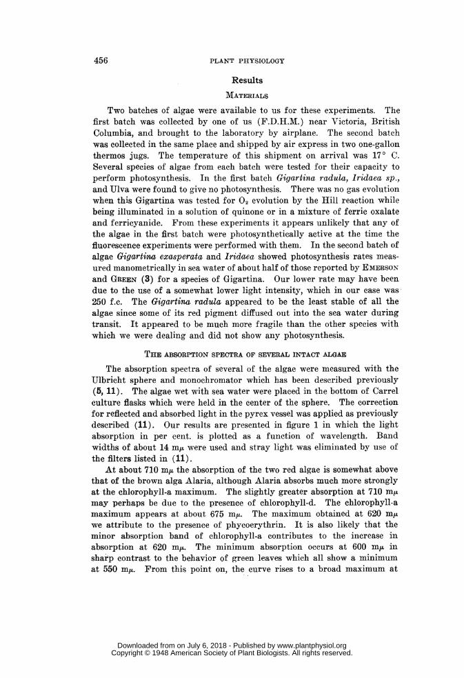
The absorption spectra of several of the algae were measured with theUlbricht sphere and monochromator which has been described previously(5, 11). The algae wet with sea water were placed in the bottom of Carrelculture flasks which were held in the center of the sphere. The correctionfor reflected and absorbed light in the pyrex vessel was applied as previouslydescribed (11). Our results are presented in figure 1 in which the lightabsorption in per cent. is plotted as a function of wavelength. Bandwidths of about 14 m,u were used and stray light was eliminated by use ofthe filters listed in (11).
At about 710 mn, the absorption of the two red algae is somewhat abovethat of the brown alga Alaria, although Alaria absorbs much more stronglyat the chlorophyll-a maximum. The slightly greater absorption at 710 mp,may perhaps be due to the presence of chlorophyll-d. The chlorophyll-amaximum appears at about 675 m/A. The maximum obtained at 620 mjLwe attribute to the presence of phycoerythrin. It is also likely that theminor absorption band of chlorophyll-a contributes to the increase inabsorption at 620 mu. The minimum absorption occurs at 600 m,u insharp contrast to the behavior of green leaves which all show a minimumat 550 m1L. From this point on, the curve rises to a broad maximum at
456
www.plantphysiol.orgon July 6, 2018 - Published by Downloaded from Copyright © 1948 American Society of Plant Biologists. All rights reserved.
VAN NORMNIAN ET AL.: RED MARINE ALGAE
about 550 m,u. In Gigartina, there appears to be a slight depression andanother rise at 530 mu. This rise has not been identified with any particularpigment. In Iridaea there is but one very broad band with the maximumat about 550 m,u. The maximum found at 495 mu is a band also presentin green leaves and may be presumed to be due either to a chlorophyll or toa carotinoid. However, we see in figure 2 below that the water extract ofIridaea containing primarily phycoerythrin also shows a sharp maximumat this point. It may be that the absorption spectrum of the intact algaerises in region of 495 mu because of the combined effects of several pigmentsincluding phycoerythrin. From 470 m/z on toward the shorter wavelengths
w60zw0 4(1)W GIGARTINA RADULAo 2 4-IRIDAEA SP
LA0I0(i ~~~ALARIA SR./ 4'-~~~~IRIDAEA SR
FS GIGARTINA RD
co 60 -
49~ ~ ~ AELNTINM
40-w
-0
5w0 600 700 800WAVELENGTH IN MMi
FIG. 1. The absorption spectra of two red and one brown marine algae, with thefluorescence spectra of the two red algae plotted above.
the curves rise rather sharply. This also is noted in both the methanol-soluble and the water-soluble pigments which will be discussed later. Inthe curve for Iridaea the bands are much less pronounced, but the maximaand minima are found in the same positions as in the curve for Giga'rtinaradula. A point worthy of note is that the absorption of Gigartina at550 m/ is 78% while the absorption at the red maximum, in this case 675 m,uis 71%. If the algae used by Haxo and Blinks are even approximatelysimilar, their several times higher yield of photosynthesis in green lightcan not have been due to increased absorption and therefore must be dueto participation of phycoerythrin in the process of photosynthesis. Incomparing the height of the absorption band at 550 mu in Gigartinawith that of Lactuca (11), we conclude that the absorption due to phyco-erythrin in Gigartina radula is about three times that of the absorptiondue to chlorophyll at this wavelength.
457
www.plantphysiol.orgon July 6, 2018 - Published by Downloaded from Copyright © 1948 American Society of Plant Biologists. All rights reserved.
PLANT PHYSIOLOGY
The curve for Alaria, a brown alga, is much more like that of a typicalgreen leaf. The red maximum appears to be at 675 mju with a minimum at570 mu. From this point on toward the blue the rise is presumed to beprimarily due to absorption by fucoxanthin.
A PARTIAL SEPARATION OF THE PIGMENTS OF IRIDAEA IN EXTRACTS
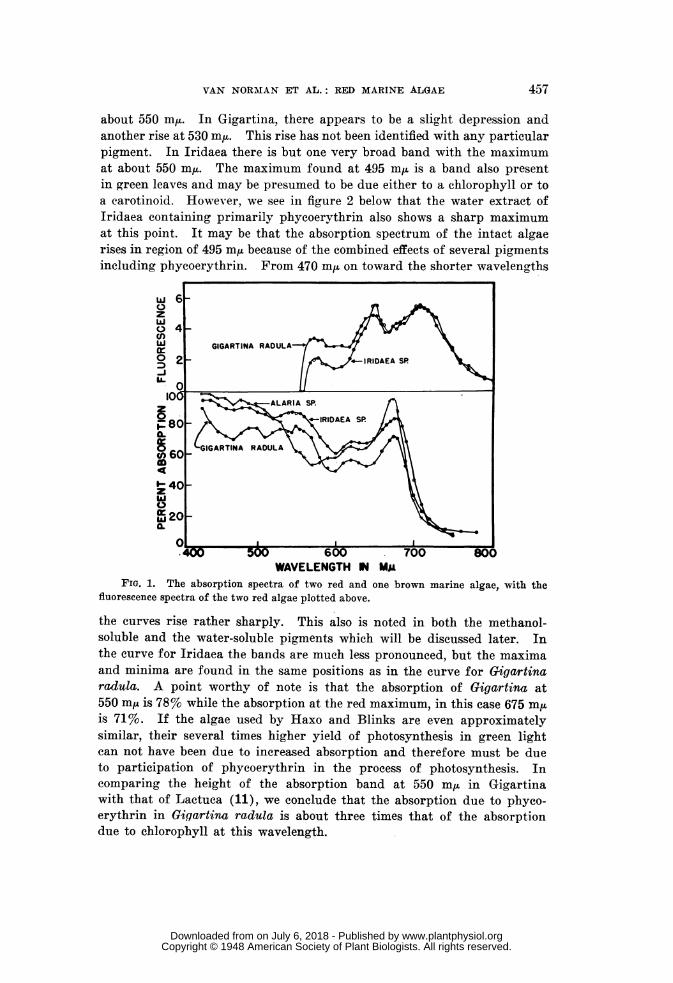
In order to study both the absorption and the fluorescence of phyco-erythrin and of chlorophyll for the most part separated from each other,extracts were made from a fresh piece of Iridaea. This was done by grind-ing with sand and fine quartz powder with added water. This maceratedmaterial was centrifuged at low speed to remove the larger particles andthen placed in a high speed centrifuge at 12,000 x g for 20 minutes. Two
'514 1.4-
1231.2005060 -
10 '.0 lOWuUz
80- 00.8~~~~~~~~~~~~~~~~~~~~~~~~~~~~~~rWz
w ~~~~~~~~~~~~06w ~~~0.6-
4 0.4-
gni=)1-ABS0R9PTION2 2 FLUORESCENCE-
300 400 500 600 700 800WAVELENGTH IN My&
FIG. 2. The absorption and fluorescence spectra of a water extract of Iridaea sp.,containing primarily phycoerythrin.
successive water extracts were combined for the high speed centrifugation.The supernatant material will be referred to hereafter as the water extractof Iridaea. To the residue of the water extraction a volume of methylalcohol was added which was equal to the total volume of cell material andquartz. The first methyl alcohol extract was slightly pinkish but containedno appreciable amount of pigment and was rejected. The second extractionwith an equal volume of methyl alcohol was green in color. This, combinedwith a third methyl alcohol extract which was deep green was saved andwill be referred to later as the methanol extract of Iridaea. A fourth ex-traction with methanol, made by heating the mixture to boiling, wasbrownish presumably due to the formation of some phaeophytin. Thisfraction was rejected.
The water extract was pink and highly fluorescent. It was nearly clearbut did show some cloudiness which was hard to distinguish due to the
458
www.plantphysiol.orgon July 6, 2018 - Published by Downloaded from Copyright © 1948 American Society of Plant Biologists. All rights reserved.
VAN NORMAN ET AL.: RED MARINE ALGAE
intense orange fluorescence. Its absorption curve is shown in figure 2.The absorption maximum at 675 mu indicated the presence of some chloro-plastin. The absorption due to chlorophyll present compared to that ofphycoerythrin appeared to be quite small. Maximum of phycoerythrinabsorption appeared at a wavelength of 550 mM,. It is a rather broadband extending from 540 to 570 m,u. We did not find a minimum at 550m,u between two peaks as described by KYLIN (10). This band is followedby a minimum at 515 miu and another maximum at 495 mi/. A broadminimum at 460 m,uL leads to a steep rise at the blue end of the spectrum.In the near ultraviolet a very dense band was found with a maximum at329 m,u. This absorption was ten times that of any of the visible part ofthe spectrum. Since this was a crude extract of the algae and not a
1.2
1.0
0.8
U)0.6 6
z.w~~~~0.4~~~~ ~ ~ ~
ABSORPTION--$~ ~ ~ ~ ~ aFLUORESCENCE WU
0.2 ABOPTOr.J-J
0.0 LA._400 500 600 700 800
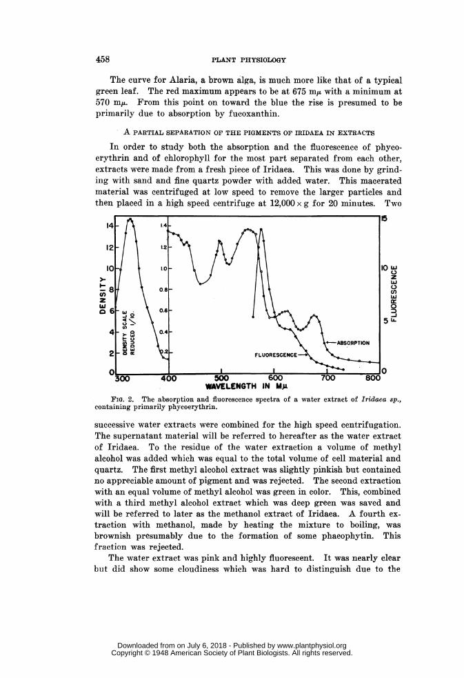
WAVELENGTH IN MgFIG. 3. The absorption and fluorescence spectra of a methanol extract of Iridaea
sp., containing primarily chlorophylls and carotinoids.
purified preparation it remains to be determined if this band is due tophycoerythrin.
The absorption curve of the methanol extract of Iridaea is shown infigure 3. It was hoped that this extract might reveal some trace of thechlorophyll-d band in the neighborhood of 690 mn,u. It is, however, difficultto tell whether or not chlorophyll-d is present in traces in this extract.The chlorophyll-a maximum is found to be at 666 m/A. HARRIS andZSCHEILE (9) report a major maximum for chlorophyll in methanol at 664min.
We have attempted to reproduce the absorption curve of the algae bvthe addition of curves of the water extract and of the methyl alcohol extract.To make such an addition, an appreciable broadening of the chlorophyllband is required in addition to the shifting of its maximum. This appearsto be reasonable in that the pigment combined with the protein may well have
459
www.plantphysiol.orgon July 6, 2018 - Published by Downloaded from Copyright © 1948 American Society of Plant Biologists. All rights reserved.
PLANT PHYSIOLOGY
a broader band than a pigment in true solution. However, any procedurewhich we were able to devise for broadening this red band resulted incurves which did not fit at the blue end of the spectrum so the attempt toreproduce the intact algae curves by some manipulation of the extract curveswas abandoned. We did not, however, find any definite evidence for thepresence of other pigments than those accounted for in one or another ofthese extracts. A possible exception to this statement may be the smallmaximum found at 530 mu in the absorption of Gigartina radula.
THE INTENSITY OF RED FLUORESCENCE EMITTED BY INTACT ALGAE WHENEXCITED BY EQUAL NUMBERS OF QUANTA OF VARIOUS WAVELENGTHS
We have tried to find out if the energy which is absorbed by phyco-erythrin is transferred to chlorophyll. If this were the case one should be
c--PHOTRONIC CELLPHOTOCELL - SAMPLEGLASS FILTER-7"
PARABOLICMIRROR
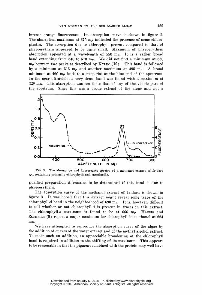
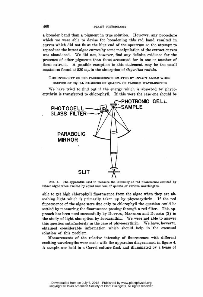
SLITFIG. 4. The apparatus used to measure the intensity of red fluorescence emitted by
intact algae when excited by equal numbers of quanta of various wavelengths.
able to get high chlorophyll fluorescence from the algae when they are ab-sorbing light which is primarily taken up by phycoerythrin. If the redfluorescence of the algae were due only to chlorophyll the question could besettled by measuring the fluorescence passing through a red filter. This ap-proach has been used successfully by DUTTON, MANNING and DUGGAR (2) inthe study of light absorption by fucoxanthin. We were not able to answerthis question satisfactorily in the case of phycoerythrin. We have, however,obtained considerable information which should help in the eventualsolution of this problem.
Measurements of the relative intensity of fluorescence with differentexciting wavelengths were made with the apparatus diagrammed in figure 4.A sample was held in a Carrel culture flask and illuminated by a beam of
460
www.plantphysiol.orgon July 6, 2018 - Published by Downloaded from Copyright © 1948 American Society of Plant Biologists. All rights reserved.
VAN NORMAN ET AL.: RED MARINE ALGAE
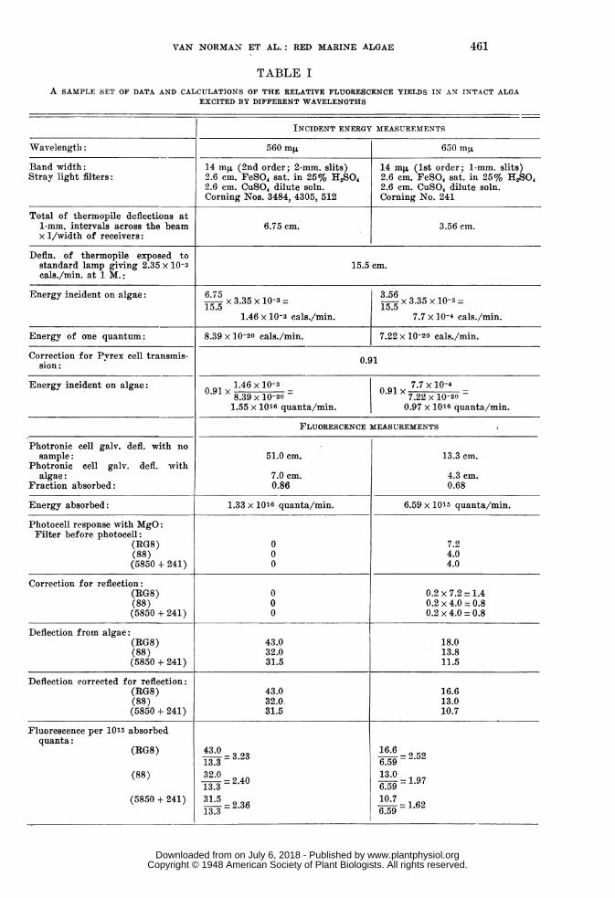
TABLE IA SAMPLE SET OF DATA AND CALCULATIONS OF THE RELATIVE FLUORESCENCE YIELDS IN AN INTACT ALGA
EXCITED BY DIFFERENT WAVELENGTHS
INCIDENT ENERGY MEASUREMENTS
Wavelength: 560 mR 650 myt
Band width: 14 mj. (2nd order; 2-mm. slits) 14 mg, (lst order; 1-mm. slits)Stray light filters: 2.6 cm. FeSO, sat. in 25% H,SO 2.6 cm. FeSO4 sat. in 25% HA2SO
2.6 cm. CuSO4 dilute soln. 2.6 cm. CuSO, dilute soln.Corning Nos. 3484, 4305, 512 Corning No. 241
Total of thermopile deflections at1-mm. intervals across the beam 6.75 cm. 3.56 cm.x 1/width of receivers:
Defln. of thermopile exposed tostandard lamp giving 2.35 x 10⁻³ 15.5 cm.cals./min. at 1 M.:
Energy incident on algae: 6.75 3.5615. 3.35 X10⁻³ 15.5 x3.35 X10-3
1.46 x 10⁻³ cals./min. 7.7 x 10⁻⁴ cals./min.
Energy of one quantum: 8.39 x 10⁻²⁰ cals./min. 7.22 x 10⁻²⁰ cals./min.
Correction for Pvrex cell transmis- 0.91sion:
Energy incident on algae: 0.91 .146 x 10-3 7091X.7 X 10-409x8.39 x10-20 01x7.22X10-20=1.55 x 10¹⁶ quanta/min. 0.97 x 10¹⁶ quanta/min.
FLUORESCENCE MEASUREMENTS
Photronic cell galv. defl. with nosample: 51.0 cm. 13.3 cm.
Photronic cell galv. defl. withalgae: 7.0 cm. 4.3 cm.
Fraction absorbed: 0.86 0.68
Energy absorbed: 1.33 x 10¹⁶ quanta/min. 6.59 x 10¹⁵ quanta/min.
Photocell response with MgO:Filter before photocell:
(RG8) 0 7.2(88) 0 4.0(5850 + 241) 0 4.0
Correction for reflection:(RG8) 0 0.2 x 7.2 = 1.4(88) 0 0.2 x 4.0 = 0.8(5850 + 241) 0 0.2 x 4.0 = 0.8
Deflection from algae:(RG8) 43.0 18.0(88) 32.0 13.8(5850 + 241) 31.5 11.5
Deflection corrected for reflection:(RG8) 43.0 16.6(88) 32.0. 13.0(5850 + 241) 31.5 10.7
Fluorescence per 1015 absorbedquanta:
(RG8) 43.0- 3.23
16.6= 2.5213.3-32 6.59
(88) 32.03- 2 40 1359 = 1.9713.3-=2 6.59
(5850 + 241) 31.5- 6107 =16
13.3-23 6.59
461
www.plantphysiol.orgon July 6, 2018 - Published by Downloaded from Copyright © 1948 American Society of Plant Biologists. All rights reserved.
PLANT PHYSIOLOGY
light of about two sq. cm. area which came from the monochromator. Filters(11) were used in front of the monochromator to reduce stray light of otherwavelengths. The intensity of the incident ligrht was measured by athermopile calibrated in absolute units. The fraction of light which wasabsorbed by the algae was measured by a photronic cell placed directlybehind the vessel. Its diffuse reflection was estimated as a fraction ofincident light, by coating half the vessel with magnesium oxide and weaken-ing that portion of the beam of light which fell on the magnesium oxidewith a set of filters of known transmission until the brightness of themagnesium oxide and of the algae appeared equal. This value was usedin making, a correction for scattered light as noted below. The fluorescentlight was collected by means of a curved mirror which concentrated a con-stant fraction thereof on a photocell located behind red glass filters. Thephotocell was connected to a "Photovolt" amplifier which permitted reading
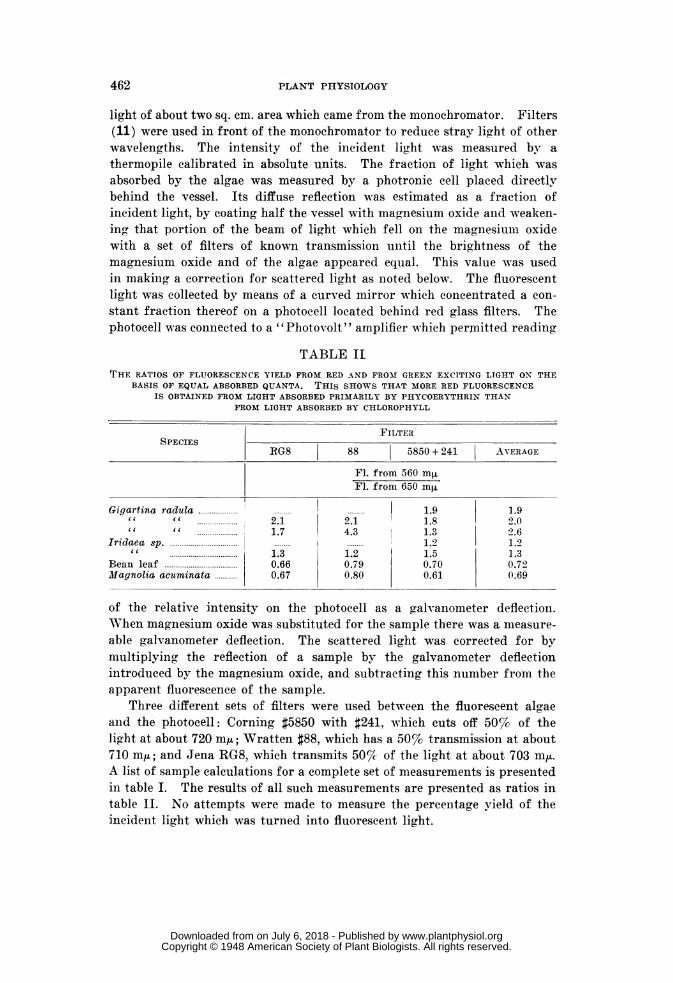
TABLE IITHE RATIOS OF FLUORESCENCE YIELD FROM RED AND FRO-M GREEN EXCITING LTGHT ON THE
BASIS OF EQUAL ABSORBED QUANTA. THIS SHOWS THAT MORE RED FLUORESCENCEIS OBTAINED FROM LIGHT ABSORBED PRIMARILY BY PHYCOERYTHRIN THAN
FROM LIGHT ABSORBED BY CHLOROPHYLL
FILTERSPECIES
RG8 88 5850 + 241 AVERAGE
Fl. from 560 mjFl. from 650 mAt
Gigartina radula 1..........................119 1.9it " 2.1 2.1 1.8 2.0cc it ............1.7 4.3 1 1.3 2.6
Iridaea sp 2................. 1.2cc 1.3 1.2 1.5 1.3
Bean leaf 0.66 0.79 0.70 0.72Magnolia acuminata 0.67 0.80 0.61 0.69
of the relative intensity on the photocell as a galvanometer deflection.When magnesium oxide was substituted for the sample there was a measure-able galvanometer deflection. The scattered light was corrected for bymultiplying the reflection of a sample by the galvanometer deflectionintroduced by the magnesium oxide, and subtracting this number from theapparent fluorescence of the sample.
Three different sets of filters were used between the fluorescent algaeand the photocell: Corning #5850 with #241, which cuts off 50% of thelight at about 720 m,; Wratten #88, which has a 50%/ transmission at about710 mpt; and Jena RG8, which transmits 50%c of the light at about 703 my.A list of sample calculations for a complete set of measurements is presentedin table I. The results of all such measurements are presented as ratios intable II. No attempts were made to measure the percentage yield of theincident light which was turned into fluorescent light.
462
www.plantphysiol.orgon July 6, 2018 - Published by Downloaded from Copyright © 1948 American Society of Plant Biologists. All rights reserved.
VAN NORMAN ET AL.: RED MARINE ALGAE
It was observed (table II) that in the red algae the fluorescence excitedby green light having a wavelength of 560 mu is definitely larger than thatexcited by red light having a wavelength of 650 mfA. The green light islargely absorbed by phycoerythrin and the red by chlorophyll. Incomparison with this behavior of red algae it is seen that in green leaves therelative fluorescence yields are reversed; that is, more red fluorescence isexcited by red light than by green light. These results could be due eitherto a red fluorescence from other pigments than chlorophyll or to a moreefficient excitation of chlorophyll fluorescence by phycoerythrin than bychlorophyll itself. A study of the fluorescence spectra of the live algaeand of the extracts was therefore undertaken.
THE FLUORESCENCE SPECTRA OF INTACT ALGAE AND OF THEIR EXTRACTS
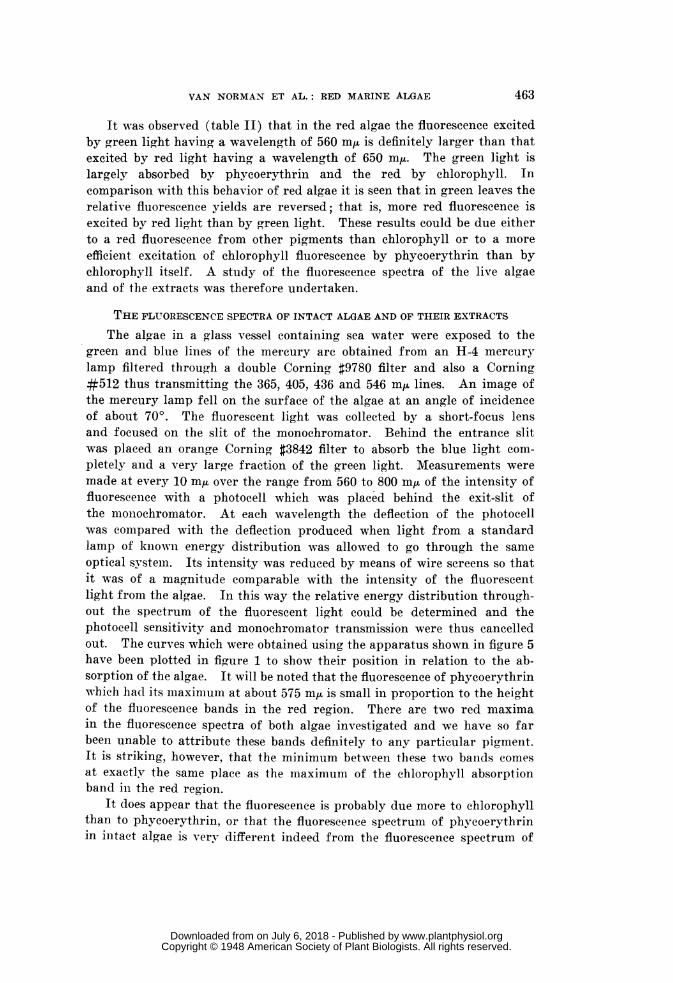
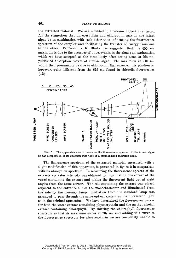
The algae in a glass vessel containing sea water were exposed to thegreen and blue lines of the me-rcury arc obtained from an H-4 mercurylamp filtered through a double Corning #9780 filter and also a Corning#512 thus transmitting the 365, 405, 436 and 546 m[u lines. An image ofthe mercury lamp fell on the surface of the algae at an angle of incidenceof about 700. The fluorescent light was collected by a short-focus lensand focused on the slit of the monochromator. Behind the entrance slitwas placed an orange Corning #3842 filter to absorb the blue light com-pletely and a very large fraction of the green light. Measurements weremade at every 10 m,u over the range from 560 to 800 mu of the intensity offluorescenee with a photocell which was placed behind the exit-slit ofthe monochromator. At each wavelength the deflection of the photocellwas comiipared with the deflection produced when light from a standardlamp of knowni energy distribution was allowed to go through the sameoptical system. Its intensity was reduced by means of wire screens so thatit was of a magnitude comparable with the intensity of the fluorescentlight from the algae. In this way the relative energy distribution through-out the spectrum of the fluorescent light could be determined and thephotocell sensitivity and monochromator transmission were thus cancelledout. The curves which were obtained using the apparatus shown in figure 5have been plotted in figure 1 to show their position in relation to the ab-sorption of the algae. It will be noted that the fluorescence of phycoerythrinwhich had its maximum at about 575 mnj is small in proportion to the heightof the fluorescence bands in the red region. There are two red maximain the fluorescence spectra of both algae investigated and we have so farbeen unable to attribute these bands definitely to any particular pigment.It is striking, however, that the minimum between these two bands comesat exactly the same place as the maximum of the chlorophyll absorptionband in the red region.
It does appear that the fluorescence is probably due more to chlorophyllthan to phycoerythrin, or that the fluorescence spectrum of phycoerythrinin intact algae is very different indeed from the fluorescence spectrum of
463
www.plantphysiol.orgon July 6, 2018 - Published by Downloaded from Copyright © 1948 American Society of Plant Biologists. All rights reserved.
the extracted material. We are indebted to Professor Robert Livingstonfor the suggestion that phycoerythrin and chlorophyll may in the intactalgae be in combination with each other thus influencing the fluorescencespectrum of the complex and facilitating the transfer of energy from oneto the other. Professor L. R. Blinks has suggested that the 655 mumaximum is due to the presence of phycocyanin in the algae; an explanationwhich we have accepted as the most likely after seeing some of his un-published absorption curves of similar algae. The maximum at 710 m,uwould then presumably be due to chlorophyll fluorescence. Its position is,however, quite different from the 675 mix found in chlorella fluorescence(12).
PHOTOCELLLL
p ,10 20 30 ,40CENTIMETERS
0~~~~0 Z~~ WL.(l()Zc z W ) Iz wJCfW W -J wn ZcrD_L i.-DJ 0
31) C/ wJ 0c w2 CD wC8Zz_J r D<C/) Cf)_J _~~~JCD 0D
FIG. 5. The apparatus used to measure the fluorescence spectra of the intact algaeby the comparison of its emission with that of a standardized tungsten lamp.
The fluorescence spectrum of the extracted material, measured with aslight modification of this apparatus, is presented in figure 2 in comparisonwith its absorption spectrum. In measuring the fluorescence spectra of theextracts a greater intensity was obtained by illuminating one corner of thevessel containing the extract and taking the fluorescent light out at rightangles from the same corner. The cell containing the extract was placedadjacent to the entrance slit of the monochromator and illuminated fromthe side by the mercury lamp. Radiation from the standard lamp was
arranged to pass through the same optical system as the fluorescent light,as in the original apparatus. We have determined the fluorescence curves
for both the water extract containing phycoerythrin and the methyl alcoholextract containing chlorophyll. By shifting the chlorophyll fluorescencespectrum so that its maximum comes at 707 m/A and adding this curve tothe fluorescence spectrum for phycoerythrin we are completely unable to
AL
zwU,CD
g1..
464 PLANT PHYSIOLOGY
www.plantphysiol.orgon July 6, 2018 - Published by Downloaded from Copyright © 1948 American Society of Plant Biologists. All rights reserved.
VAN NORMAN ET AL.: RED MARINE ALGAE
reproduce the shape of the fluorescence curves of the intact cells. It musttherefore be concluded that the fluorescence spectrum of the phycoerythrinand of the chlorophyll is widely different in the intact algae from its shapein extracts, or that other pigments not present in either the water or themethyl alcohol extracts are causing some of the fluorescence of the intactmaterial. The determination of the fluorescence curves of algae when ex-cited by various wavelengths has not yet been attempted since it would re-quire two monochromators and a more sensitive photocell. Until this canbe done it will probably be difficult to come to a definite conclusion as towhether or not the energy absorbed by phycoerythrin is used directly forphotosynthesis or is first transferred to chlorophyll.
TEE BEHAVIOR OF WATER EXTRACTS OF ALGAE WHEN ILLUMINATED IN THE
PRESENCE OF DYES
The dye phenol-indophenol has been shown by HOLT and FRENCH (6, 7)to be reduced to the leuco form when illuminated in the presence of activechloroplasts of spinach with a concomitant evolution of oxygen. This isbelieved to take place by means of a part of the photosynthesis system of thecell. Chloroplasts from our first shipment of Gigartina radula were notactive in the reduction of the dye. A very slight activity was obtained fromthe Ulva chloroplasts. The extract of phycoerythrin from the first batch ofGigartina appeared to lose color when illuminated with added dye at pH 6.5.At this pH phenol-indophenol and phycoerythrin are both pink. Furtherinvestigation using slightly alkaline solutions of the dye which are blue oracid solutions of 2,6-dichlorophenol-indophenol showed that it was not thedye but the phycoerythrin which was bleached upon illumination in suchsolutions. Dye reduction experiments were later made with fresh phyco-erythrin extracts of algae which had been shown to be photosyntheticallyactive, again with negative results.
SummaryThe absorption and fluorescence spectra of several marine algae are
presented. The fluorescence yield in Gigartina radula and in Iridaea sp.is greater from green light than from red light. The fluorescence spectra oftwo red algae show a band at 575 mju corresponding to phycoerythrinfluorescence, and also peaks at 675 mju and 710 m/.
It is a pleasure to thank the Graduate School of the University of Minne-sota for grants which made this work possible. DRS. C. 0. ROSENDAHL andJOHN H. MOORE gave invaluable assistance in the identification of the algaeused. The photosynthesis experiments and the dye reduction experimentswere done by DR. A. S. HOLT to whom we are also greatly indebted.
DEPARTMENT OF BOTANYUNIVERSITY OF MINNESOTA
MINNEAPOLIS, MINNESOTA.
465
www.plantphysiol.orgon July 6, 2018 - Published by Downloaded from Copyright © 1948 American Society of Plant Biologists. All rights reserved.
PLANT PHYSIOLOGY
LITERATURE CITED
1. DUTTON, H. J., and MANNING, W. M. Evidence for carotinoid-sensi-tized photosynthesis in the diatom Nitzschia closterium. Amer.Jour. Bot. 28: 516-526. 1941.
2. DUTTON, H. J., MANNING, W. M., and DUGGAR, B. M. Chlorophyllfluorescence and energy transfer in the diatom Nitzscahia closterium.Jour. Physical Chem. 47: 308-313. 1943.
3. EMERSON, ROBERT, and GREEN, LOWELL. Manometric measurements ofphotosynthesis in the marine alga Gigartina. Jour. GeneralPhysiol. 17: 817-843. 1934.
4. EMERSON, ROBERT, and LEWIS, CHARLTON M. The photosynthetic effi-ciency of phycocyanin in Chroococcus and the problem of caroti-noid participation in photosynthesis. Jour. General Physiol. 25:579-595. 1942.
5. FRENCH, C. S., RABIDEAU, G. S., and HOLT, A. S. The construction andperformance of a large grating monochromator with a high energyoutput for photochemical and biological investigations. Rev. Sci-entific Instruments 18: 11-17. 1947.
6. FRENCH, C. S., and HOLT, A. S. The evolution of oxygen with thesimultaneous reduction of a dye by illuminated suspensions ofchloroplasts. Amer. Jour. Bot. 33: 19a (abstract). 1946.
7. HOLT, A. S., and FRENCH, C. S. The evolution of oxygen by isolatedchloroplasts immersed in solutions of various oxidizing agents.Amer. Jour. Bot. 33: 21a (abstract). 1946.
8. HAxo, FRANCIS, and BLINKS, L. R. Photosynthetic action spectra inred algae. Amer. Jour. Bot. 33: 20a (abstract). 1946.
9. HARRIS, D. G., and ZSCHErLE, F. P. The effects of solvents upon ab-sorption spectra of chlorophylls a and b; their ultra-violet absorp-tion spectra in ether solution. Bot. Gaz. 104: 515-527. 1943.
10. KYLIN, H. A marine Porphyridium. Kgl. Fysiograf. Siillskap. LundF6rh. 7: 119-123. 1938.
11. RABIDEAU, G. S., FRENCH, C. S., and HOLT, A. S. The absorption andreflection spectra of leaves, chloroplast suspensions and chloroplastfragments as measured in an Ulbricht sphere. Amer. Jour. Bot.33: 769-777. 1946.
12. VERMEULEN, D., WASSINK, E. C., and REMAN, G. H. On the fluor-escence of photosynthesizing cells. Enzymologia 4: 254-268.1937.
466
www.plantphysiol.orgon July 6, 2018 - Published by Downloaded from Copyright © 1948 American Society of Plant Biologists. All rights reserved.