Embed Size (px)
Citation preview

446 SHORT COMMUNICATIONS
of absorbance around 75o--87 o n m . Upon raising to pH 9.5, 138oo was partially re- generated at the expense of the 75o-78o mn absorption. This suggests that the B77o-79 o intermediates are capable of being converted back to BSoo. Fig. 2 also shows that B88o disappears within 5 min at pH 1.55 and is not regenerated by changing the pH. Another pronounced absorption band at 85 ° n m (B85o) was hardly affected. At this pH, the photoactivity underwent irreversible inactivation despite the partial regeneration of BSoo. Chromatium chromatophores behaved similarly, though the regeneration of BSoo was less pronounced than with R. spheroidcs.
When R. spheroides and Chromatium chromatophores were kept at the pH range 2.o-2. 4, B8oo was gradually reduced but B88o was not strongly affected. By bringing back the pH to neutral or alkaline (pH 6 - I I ) , the photoactivity was regenerated in the same way as shou~n for R. rubrum (Fig. IB). The acid-induced spectral changes reported here are generally in agreement with the results by THOMAS et al), except that they observed little change of B8oo in the case of Chromatium. The reason for this discrepancy is not clear.
The pH-induced modification and regeneration of the photoreactive bacterio- chlorophyll complex (P8oo-P87o) and B8oo, reported in this communication, indicate that these species differ considerably from other forms of bacteriochlorophyll (B85o and B88o). B88o and B85o are more resistant to low pH, but once affected, the changes are almost irreversible. B8oo and P8oo-P87o, on the other hand, are ap- parently in specific and intimate association with proteins. Their reversible response to acid-base transition suggests the occurence of pH-dependent reversible confor- mational changes in their protein environments.
Photochemistry Section, Energy Conversion Branch, Space Physics Laboratory, Air Force Cambridge Research Laboratories, Bedford, Mass. oz73 o (U.S.A.)
E I J I FUJIMORI
1 J. B. THOMAS, J. C. GOEDHEER AND J. G. KOMEN, Biochim. Biophys. Acta, 22 (1956) I. 2 E. FUJIMORI, Biochim. Biophys. Acta, 18o (i969) 360. 3 A. K. GHOSH AND J. M. OLSON, Biochim. Biophys. Acta, 162 (1968) z35. 4 A. K. GHOSH, T. t~. BROKER AND J. M. OLSON, Biochim. Biophys. Mcta, 162 (1968) 402. 5 t3. KE, L. P. VERNON, A. GARCIA AND E. NGO, Biochemistry, 7 (1968) 311.
Received August 7th, 197 °
Biochim. Biophys. Acta, 223 (z97 o) 444-446
BBA 43 285
Chlorophyll-pheophytin interactions"
Long wavelength forms of chlorophyll occur in green plants 1 and are considered important in photosynthesis ~. The origin of these red-shifted spectral transitions and the chlorophyll species responsible for them are still obscure. Long wavelength forms
* Based on work pe r fo rmed u n d e r t he auspices of t he U.S. A tomic E n e r g y Commiss ion .
Biochim. Biophys. Mcta, 223 (197 o) 446-449

SHORT COMMUNICATIONS 447
of chlorophyll can be prepared in the laboratory (see, for example ref. 3), the best characterized of these being the oligomer 4, (Chl2)n, which absorbs in the red at 662 and 678 nm, and a chlorophyll-water adductS, 6, (Chl. H20)n, which absorbs near 74 ° nm. Recently, we have observed and characterized a chlorophyll a -pheophyt in a - water interaction product that is markedly red-shifted and has spectral properties remarkably similar to those encountered in some in vivo systemsL
Water is essential for the red shift to occur in the chlorophyll a-pheophyt in a interaction. Dry films or solutions (in aliphatic hydrocarbon solvents) of chlorophyll a - pheophytin a mixtures show spectra in tile visible and the infrared that are not greatly perturbed. Pure pheophytin a films or concentrated solutions of pheophytin a in ali- phatic hydrocarbon solvents absorb at 695 nm. In more dilute solutions in aliphatic hydrocarbon solvents (approx. I mM), or in polar solvents, pheophytin a absorbs at 668 nm. Water does not influence the optical spectra of the pure pheophytin a. How- ever, when an anhydrous film of chlorophyll a containing lO-6O % pheophytin a is exposed to water vapor at 55 ° for I h, a new absorption maximum appears at 712 :k 2 nm. Varying the pheophytin a content between these limits does not affect the position of this absorption maximum, but the intensity of absorption at 712 nm increases with increasing pheophytin content. Hydration of ctdorophyll a films containing 5 to lO % ptleophytin a for I h produces a small 712 nm shoulder, and hydration pro- longed for several hours forms predominately the 74 ° n m (Chl. H 20)n species. Mixtures with more than IO O/io pheophytin a form the 712 nm species, and only a trace amount of 740 nm species is evident even after 12 h of hydration. The 712 nm maximum is always accompanied by a blue shoulder near 695 nm, as revealed by graphical de- convolution of the red absorption envelope. The 712 nm fraction of the total red absorption is proportional to the pheophytin a content until approx. 30 % pheophytin is present in the film. In the range 30-50 % pheophytin, the contribution of the 712 nm fraction is roughly constant. Assuming no large changes in extinction coefficients in the mixture as compared to the pure components, the red shift is consistent with a chlorophyll a -pheophyt in a complex in the ratio of 2:1.
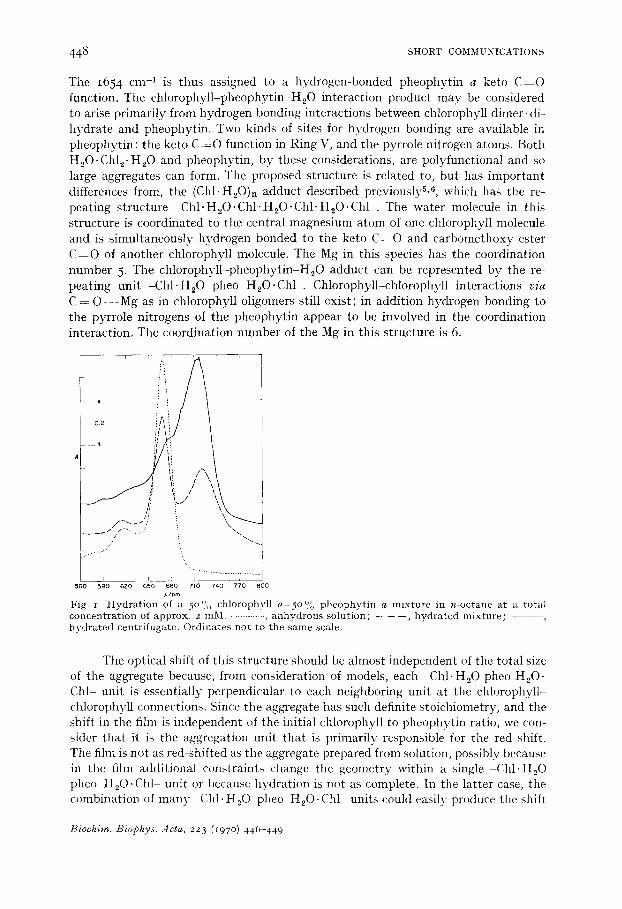
Hydration of a solution of pheophytin a and chlorophyll a (with a pheophytin a content between 25 and 50 % and a total concentration of 2 mM) in dodecane or n-octane produces a sharp absorption peak with an absorption maximum at 720 ~ 2 nm without formation of a 74 ° n m species (Fig. i). Centrifugation at low field removes the 720 nm species. The centrifugate has a pronounced peak at 718 nm with a shoulder near 695 nm and a small 668 nm peak. By spectral analysisS, 9 on a diethyl ether solution of the centrifugate the stoichiometric ratio of chlorophyll a:pheophytin a is 2.00 ± 0.09. Since the aggregate is easily sedimented at low centrifugal fields, the gross composition of the species is (2Chl a: IPheo a:xH20)n, where the aggregation number, n, appears very large.
Infrared spectra and the stoichiometry established by analysis provide a basis for advancing a structure for the 720 nm species. Infrared maxima in the CO region with proposed assignments follow:
1735 cm -1 free e s t e r C = O
1675 cm -1 Chl ke to C ~ O - - - M g - - - O H 2
1654 cm -1 (pheo keto) C = O - - - H - O - - - M g
H
Biochim. Biophys. Acta, 223 (197o) 446-449
448 SHORT COMMUNICATIONS
The I654 cm 1 is thus assigned to a hydrogen-bonded pheophytin a keto C--~O function. The chlorophyll-pheophytin-H20 interaction product may be considered to arise primarily front hydrogen bonding interactions between chlorophyll dimer, di- hydrate and pheophytin. Two kinds of sites for hydrogen bonding are available in pheophytin : the keto C = O function in Ring V, and the pyrrole nitrogen atoms. Both H20.Chle. H20 and pheophytin, by these considerations, are polyfunctional and so large aggregates can form. The proposed structure is related to, but has important differences from, the (Chl'H20)n adduct described previouslyS, 6, which has the re- peating structure -Chl- H20"Chl 'H,O'Chl" HzO.Chl-. The water molecule in this structure is coordinated to the central magnesium atom of one chlorophyll molecule and is simultaneously hydrogen bonded to the keto C = O and carbomethoxy ester C = O of another chlorophyll molecule. The Mg in this species has the coordination number 5. The chlorophyll-pheophytin-H20 adduct can be represented by the re- peating unit -Chl .H20 pheo H20.Chl -. Chlorophyll-chlorophyll interactions via C = O - - - M g as in chlorophyll oligomers still exist; in addition hydrogen bonding to the pyrrole nitrogens of the pheophytin appear to be involved in the coordination interaction. The coordination number of the Mg in this structure is 6.
i i ; . r i
i ̧ i ? it!
0.2 !1~ I ~ i
i l i
\ / ~ 'i / '
/ ~f i . / ', /i i
i / k
, , ' , ............. i t , .............. ] sso 59o 6zo o~o 68o 7,o ~4o 77o soo
2,/r*m Fig. r Hydration ot a 5 ° % chlorophyll a-5o % pheophytin a mixture in n-octane at a total concentration of approx, z raM. • .......... , anhydrous solution; - - - - - , hydrated mixture; - - - - - hydrated centrifugate. Ordinates not to the same scale.
The optical shift of this structure should be almost independent of the total size of the aggregate because, from consideration of models, each -Chl. H20 pheo H20. Chl- unit is essentially perpendicular to each neighboring unit at the chlorophyll- chlorophyll connections. Since the aggregate has such definite stoichiometry, and the shift in the film is independent of the initial chlorophyll to pheophytin ratio, we con- sider that it is the aggregation unit that is primarily responsible for the red shift. The film is not as red-shifted as tile aggregate prepared from solution, possibly because in the film additional constraints change tile geometry within a single Chl. H20 pheo H20.Chl unit or because hydration is not as complete. In the latter case, tile combination of many ChI.H20 pheo H20.Chl units could easily produce the shift
t?iochim. Biophys. Acta, 223 (r97 o) 446-449

SHORT COMMUNICATIONS 449
from 712 to 72o nm. The 695 nm component in the spectra of the aggregates must have at least two different origins: (I) A hydrated chlorophyll film containing 50 % pheophytin produces a much larger 695 nm component than does a 25 % pheophytin film. This increase must represent a pheophytin-pheophytin interaction not involving H20 , originating in the pheophytin excluded from the aggregate for stoichiometric reasons. (2) The centrifugate from solution hydration requires a deconvolution peak near 695 nm constituting as much as one-third of the total area, thus suggesting a second means for producing 695 nm absorption. This 695 nm absorption most likely reflects a transition associated with the pheophytin component of the aggregate, but the origin of the transition is still uncertain.
The Soret region of the optical spectra also shows large changes in the chloro- phyl l -pheophyt in-H20 aggregates; these are being studied. The pheophytin a peaks at 505 and 534 nm are essentially absent, and tile 408 nm peak is diminished in the aggregate.
Absorption maxima at or near 695 nm, 712 nm and 720 nm have been reported in green plants 7, generally under conditions where there is reason to suppose that large amounts of pheophytin are present in the plant TM. The origins of these maxima now seem clearer, and there is reason to question whether they have any real sig- nificance for photosynthesis. Preliminary experiments with chlorophyll b-pheo- phytin b, bacteriochlorophyll-bacteriopheophytin, and chlorophyll a baeteriopheo- phytin have produced no new optical maxima under hydration conditions used for the chlorophyll a pheophytin a system. However, the hydration behavior of chloro- phyll b and bacteriochlorophyll is much more complicated n and the possibility that the bacteriochlorophyll-bacteriopheophytin system may show interactions similar to those described here for the chlorophyll a system cannot be excluded at this time.
Chemistry Division, Argonne National Laboratory, Argonne, Ill. 6o 439 (U.S.A.)
J. R. NORRIS R. A. UPHAUS T. M. COTTON
J. J. KATZ
I W. L. BUTLER, in L. P. VERNON AND G. R. SEELY, The Chlorophylls, Academic Press , New York, 1966, p. 343.
2 R. K. CLAYTON, Molecular Physics in Photosynthesis, Blaisdell Pub l i sh ing Co., New York, 1965, p. 149.
3 K. BALLSCHMITER AND J. J. I~ATZ, Nature, 22o (1968) 1231. 4 K. BALLSCHMITER, I~. TRUESDELL AND J. J. KATZ, Biochim. Biophys. -dcta, 184 (1969) 604. 5 K. BALLSCHMITER AND J. J. KATZ, J. Am. Chem. Sot., 91 (1969) 2661. 6 J. J. KATZ, K. BALLSCHMITER, M. GARCIA-MORIN, H. H. STRAIN AND R. A. [JPHAUS, Proc.
Natl. Acad. Sci. U.S., 60 (1968) ioo. 7 J. S. BROWN, Photochem. Photobiol., 2 (1963) 159. 8 H. A. W. SCHNEIDER, Photosynthetica, i (1967) 259. 9 C. S. FRENCH, in Handbuch cler Pfianzenphysiologie, V/I , Springer Verlag, Berl in, 196o, p. 259.
io C L GREENBLATT AND J. A. SCHIFF, J. Protozool., 6 (1959) 23. i1 K. BALLSCHMITER, T. M. COTTON, H. H. STRAIN AND J. J. KATZ, Biochim. Biophys. Acta, 18o
(1969) 347.
Received September 4th, 197o
Biochim. Biophys. Acta, 223 (197 o) 446 449