Embed Size (px)
Citation preview

ORIGINAL ARTICLE
CHK1-targeted therapy to deplete DNA replication-stressed, p53-deficient, hyperdiploid colorectalcancer stem cellsGwenola Manic,1 Michele Signore,2 Antonella Sistigu,3 Giorgio Russo,3,4
Francesca Corradi,1 Silvia Siteni,3,5 Martina Musella,3,6 Sara Vitale,4
Maria Laura De Angelis,2 Matteo Pallocca,7 Carla Azzurra Amoreo,8
Francesca Sperati,9 Simone Di Franco,10 Sabina Barresi,11 Eleonora Policicchio,2,12
Gabriele De Luca,2 Francesca De Nicola,7 Marcella Mottolese,8 Ann Zeuner,2
Maurizio Fanciulli,7 Giorgio Stassi,10 Marcello Maugeri-Saccà,13 Marta Baiocchi,2
Marco Tartaglia,11 Ilio Vitale,1,3 Ruggero De Maria4
▸ Additional material ispublished online only. To viewplease visit the journal online(http://dx.doi.org/10.1136/gutjnl-2016-312623).
For numbered affiliations seeend of article.
Correspondence toDr. Ilio Vitale, Department ofBiology, University of Rome“Tor Vergata”, via dellaRicerca Scientifica 1, Rome00133, Italy; [email protected] Ruggero De Maria,Catholic University and GemelliPolyclinic, Largo Francesco Vito1, Rome 00168, Italy;[email protected]
GM, MS and AS contributedequally.IV and RDM are seniorcoauthors.
Received 15 July 2016Revised 3 February 2017Accepted 28 February 2017
To cite: Manic G,Signore M, Sistigu A, et al.Gut Published Online First:[please include Day MonthYear] doi:10.1136/gutjnl-2016-312623
ABSTRACTObjective Cancer stem cells (CSCs) are responsible fortumour formation and spreading, and their targeting isrequired for tumour eradication. There are limitedtherapeutic options for advanced colorectal cancer(CRC), particularly for tumours carrying RAS-activatingmutations. The aim of this study was to identify novelCSC-targeting strategies.Design To discover potential therapeutics to beclinically investigated as single agent, we performed ascreening with a panel of FDA-approved orinvestigational drugs on primary CRC cells enriched forCSCs (CRC-SCs) isolated from 27 patients. Candidatepredictive biomarkers of efficacy were identified byintegrating genomic, reverse-phase protein microarray(RPPA) and cytogenetic analyses, and validated byimmunostainings. DNA replication stress (RS) wasincreased by employing DNA replication-perturbing orpolyploidising agents.Results The drug-library screening led to theidentification of LY2606368 as a potent anti-CSCagent acting in vitro and in vivo in tumour cells from aconsiderable number of patients (∼36%). By inhibitingcheckpoint kinase (CHK)1, LY2606368 affected DNAreplication in most CRC-SCs, including RAS-mutatedones, forcing them into premature, lethal mitoses.Parallel genomic, RPPA and cytogenetic analysesindicated that CRC-SCs sensitive to LY2606368displayed signs of ongoing RS response, including thephosphorylation of RPA32 and ataxia telangiectasiamutated serine/threonine kinase (ATM). This wasassociated with mutation(s) in TP53 and hyperdiploidy,and made these CRC-SCs exquisitely dependent onCHK1 function. Accordingly, experimental increase ofRS sensitised resistant CRC-SCs to LY2606368.Conclusions LY2606368 selectively eliminatesreplication-stressed, p53-deficient and hyperdiploidCRC-SCs independently of RAS mutational status.These results provide a strong rationale forbiomarker-driven clinical trials with LY2606368in patients with CRC.
Significance of this study
What is already known on this subject?▸ Cancer stem cells (CSCs), the subpopulation of
cells driving tumour initiation and spreading,are associated with cancer relapse, therapeuticresistance and poor patient prognosis therebylimiting the efficacy of antineoplastic regimens.
▸ Patient-derived cancer models enriched forCSCs are successfully employed for discoveringnovel cancer drugs, predicting the clinicalefficacy of novel antineoplastic regimens andrapidly translating these novelties into theclinical setting.
▸ We and others previously showed that thetherapeutic resistance of CSCs is due to theirhigh efficiency in activating the DNA damageresponse (DDR) in the presence of DNA lesions,but further investigations are required toelucidate the status and molecular mechanismsof DDR in CSCs and how these insights can beexploited for cancer therapy.
▸ Previous evidence indicated that the inhibitionof the ataxia telangiectasia mutated serine/threonine kinase (ATM)-checkpoint kinase(CHK)2 or ataxia telangiectasia mutated andRad3 related serine/threonine kinase(ATR)-CHK1 axis of DDR, alone or incombination with DNA-damagingchemotherapeutics, could kill cancer cellsdisplaying genomic instability, but the effect onCSC survival needs further investigations.
What are the new findings?▸ We identified LY2606368 as a potent in vitro
and in vivo anti-CSC agent able to kill asignificant fraction (approximately a third) ofour large panel of primary colorectal cancer(CRC) cells enriched for CSCs (CRC-SCs).
Manic G, et al. Gut 2017;0:1–15. doi:10.1136/gutjnl-2016-312623 1
GI cancer Gut Online First, published on April 7, 2017 as 10.1136/gutjnl-2016-312623
Copyright Article author (or their employer) 2017. Produced by BMJ Publishing Group Ltd (& BSG) under licence.
on 9 April 2019 by guest. P
rotected by copyright.http://gut.bm
j.com/
Gut: first published as 10.1136/gutjnl-2016-312623 on 7 A
pril 2017. Dow
nloaded from

INTRODUCTIONThe management of patients with colorectal cancer (CRC)remains a major clinical challenge, owing to the high incidenceof tumour relapse and development of treatment-refractory dis-eases.1 Despite the discovery of novel regimens combining bio-logical therapies to the chemotherapy backbone over the pastdecade, intrinsic and acquired resistance remains a criticalhurdle. The mechanisms of therapeutic resistance, often emer-ging after initial tumour responses, include (but are not limitedto) mutation(s) in the gene encoding the target protein,2 induc-tion of bypass or parallel cascade(s),3 restoration of the defectivetargeted pathway4 and outgrowth of pre-existing minor ordormant clones.5
The development of effective therapeutics against solid malig-nancies is further hampered by their considerable genetic, epi-genetic, functional and phenotypical heterogeneity over spaceand time.6–8 This intratumour heterogeneity is reported to ariseearly in the development of CRC and feeds therapeutic resist-ance.9 Mounting evidence indicates that intratumour heterogen-eity and therapeutic resistance is driven by transient/stable
subsets of immature, undifferentiated cells known as cancer stemcells (CSCs).6 10 11 CSCs have been isolated from multiple malig-nancies, including CRC, by specific cell-surface markers orserum-free in vitro culture enrichment.12 13 This stem-like popu-lation is at the apex of the hierarchical organisation of the cancersystem, where it constitutes a perpetual pool generating non-tumorigenic progeny with variable degree of differentiation andplasticity, and in dynamic crosstalk with the tumour microenvir-onment.10 CSCs are responsible for tumour initiation, and areinvolved in cancer progression, recurrence, metastasis and thera-peutic failure.10 14 15 Underscoring their clinical importance, spe-cific CSC-related gene-expression signatures have been associatedto inferior clinical outcomes,6 16 17 and CRC patients with stemcell-like tumours have been documented to display lower 5-yeardisease-free survival rates than those having differentiatedtumours.18 The therapeutic targeting of CSCs is thus advocatedas essential for the development of effective drugs.
Multicellular spheroids (best known as tumourspheres) are acancer model employed to efficiently purify, enrich and propa-gate patient-derived CSCs,13 hence representing a powerful toolfor discovering anticancer drugs and companion predictive bio-markers. Considering the enormous potential of CSC-basedassays as hypothesis-generating models for early clinical trials,we decided to screen freshly generated patient-derived CRCtumourspheres enriched for CSCs (hereafter referred to asCRC-SCs) with a panel of >300 drugs already approved or inclinical development.
MATERIALS AND METHODSCell lines, culture conditions and chemicalsUnless otherwise indicated, media, supplements and plasticwarewere purchased from Gibco-Thermo Fisher Scientific (Waltham,Massachusetts, USA) or Corning-Life Sciences (Corning, New York,USA). CSC isolation and culture from samples of human patientswith CRC (obtained in accordance with the standards of the institu-tional Ethics Committee on human experimentation, authorisationCE5ISS-09/282) were performed as previously reported19 anddescribed in online supplementary information. All CRC-SCs werevalidated for their capability to generate neoplasms faithfully phe-nocopying the original patient tumour when xenotransplanted intoimmunocompromised mice.20 Selected CRC-SCs belonging to thethree categories (#1 and #4 for high; #3 and #6 for medium; and#8, #10 and #12 for low sensitive) were employed in most invitro experiments. Of these CRC-SCs, #1, #3 and #8 were used invivo. Additional CRC-SCs were employed depending on cell avail-ability and status always being well balanced towards the point ofLY2606368 sensitivity. Compounds were provided from SelleckChemicals (Houston, Texas, USA) with the exception of aphidico-lin, colchicine, Mps1-IN-3, nocodazole, reversine, thymidine,UCN-01 (all purchased from Sigma-Aldrich), LY2606368 (alsoknown as prexasertib, provided from Eli Lilly, Indianapolis,Indiana, USA) and PV1019 (obtained from Calbiochem-MerckMillipore, Billerica, Massachusetts, USA).
Compound screening and measurement of cell proliferation/viabilityTo determine the IC50 or drug-combination efficiency, disso-ciated cells were seeded in 96-well plates (6×10³ cells/100 mLmedium/well), cultured for 24 hours and then treated as indi-cated in the figure legends. Cell viability/proliferation was deter-mined by evaluating the ATP levels via CellTiter-GloLuminescent Cell Viability Assay (Promega, Madison,Wisconsin, USA) with a multimode reader (DTX-880; BeckmanCoulter, Brea, California, USA). For drug screening, CRC-SCs
Significance of this study
▸ The mechanism of CRC-SC killing by LY2606368 involves thespecific inhibition of CHK1, which alters the DNA replicationand intra-S checkpoint of CRC-SCs, in turn resulting in thegeneration of an intolerable DNA damage burden and theconsequent cell demise via replication catastrophe.
▸ The fraction of CRC-SCs displaying signs of DNA replicationstress, such as phosphorylation of RPA32 or ATM, deficiencyin p53 and increased number of chromosomes(hyperdiploidy) was shown to be highly dependent on CHK1activity and thus targetable with CHK1 inhibitors.
▸ Boosting replication stress or increasing the number ofchromosomes by employing DNA replication-perturbing ormitosis-perturbing agents was proven to be a strategy tosensitise formerly resistant CRC-SCs to LY2606368.
How might it impact on clinical practice in theforeseeable future?▸ Our results, together with the recent evidence on tolerable
safety profiles and preliminary antineoplastic activity ofLY2606368 as monotherapy in a phase I, non-randomised,open-label, dose-escalation trial in patients affected byadvanced or metastatic solid tumours who underwent atleast three previous lines of treatment, support the furtherclinical development of LY2606368.
▸ The selective targeting of CHK1 by LY2606368 can berapidly translated into the clinical settings for the eradicationof tumours with replication-stressed, p53-deficient andhyperdiploid CRC-SCs, which can be easily identified by p53sequencing combined with immunohistochemical analysis forreplication stress markers (phosphorylation of ATM andRPA32) and ploidy status (measurement of nuclear area).
▸ Our findings also guide for the further development oftherapeutic regimens based on the induction of replicationstress in patients with low DNA replication-stressed CRC(identified as reported above) so as to broaden thetherapeutic use of LY2606368 to a large number of subjects.
2 Manic G, et al. Gut 2017;0:1–15. doi:10.1136/gutjnl-2016-312623
GI cancer on 9 A
pril 2019 by guest. Protected by copyright.
http://gut.bmj.com
/G
ut: first published as 10.1136/gutjnl-2016-312623 on 7 April 2017. D
ownloaded from
were seeded onto 96-well plates. Triplicate plates for eachCRC-SC/drug-library plate combination were generated. Upontreatment for 72 hours, CRC-SC proliferation/viability wasdetermined by CellTiter-Glo assay. Normalised viability for eachdrug (VD) was obtained, for each plate, by referencing lumines-cence values (LD) to the averaged luminescence values ofDimethyl sulfoxide (DMSO)/controls (mLC) using the formula:VD=(LD/mLC)×100. The z scores (ZD) were calculated by ref-erencing each normalised viability replicate (VD) to the grandmean (Gm) and grand SD (Gsd) of all compounds using theformula: ZD=(VD-Gm)/Gsd. Drug screening data were ana-lysed using the ‘R’ software (R-Foundation for StatisticalComputing, Vienna, Austria).
Genetic and reverse-phase protein microarray analysesFor targeted deep DNA resequencing, the sequencing librarywas prepared with the Truseq Custom Amplicon Kit (Illumina,San Diego, California, USA), while sequencing was performedon a MiSeq instrument. FASTQ files were analysed with MiseqReporter software using the somatic variant caller algorithm,and variant annotation was performed with the VariantStudiosuite (all from Illumina). The OncoPrint was generated usingcustom ‘R’ scripting and complex heatmaps library (https://github.com/jokergoo/ComplexHeatmap). Microsatellite instabil-ity (MSI) analysis was performed with a reference panel of fivefluorescent dye-labelled microsatellite primers (NR-21, BAT-25,MONO-27, NR-24, BAT-26) using the MSI Analysis System kit,Version_1.2 (Promega). Amplified fragments were detected byloading the PCR products for capillary electrophoresis using theABI Prism 3500 Genetic Analyser and the POP-4 polymer (bothfrom Applied Biosystems, Foster City, California, USA) accord-ing to manufacturer’s instructions (Promega). MSI status wasdetermined upon analysis with GeneMapper software,Version_4.1 (Applied-Biosystems). Reverse-phase protein micro-array (RPPA) was performed as previously described.21
In vivo studyFor xenograft studies, CRC-SCs were resuspended in 50%Matrigel Basement Membrane Matrix (BD Biosciences, FranklinLakes, New Jersey, USA)/50% growth medium, and 5×10⁵ cellswere injected subcutaneously in the flank of mice as reported byDe Angelis et al.20 When tumours were palpable (after 3/5 weeks), mice were randomised to control and treatmentgroups (10 mice/group) and treated subcutaneously with vehicleonly (Captisol; CyDex Pharmaceuticals, La Jolla, California,USA), or 5 or 10 mg/kg of the mesylate salt formulation ofLY2606368 (which has comparable in vitro activity andincreased in vivo bioavailability). For in vivo rescue studies,CRC-SC-derived xenografts were harvested from vehicle-treatedor LY2606368-treated mice (6 tumours/group), dissociated assingle cells and injected into secondary mice as described above.All animals were left untreated and in vivo tumour growth andvolumes were measured as reported by De Angelis et al.20
Immunohistochemistry (IHC)Sections from formalin-fixed/paraffin-embeddedCRC-SC-derived xenografts were stained as described in onlinesupplementary information. Images were obtained using anEclipse 55i microscope (Nikon, Melville, New York, USA)equipped with Eureka Interface System (Menarini, Florence,Italy). The levels of ataxia telangiectasia mutated serine/threo-nine kinase (ATM) and replication protein A (RPA)32 phosphor-ylation were evaluated in terms of proportion of expressingtumour cells (0–100%) and staining intensity (0:negative, 1+:
weak, 2+:moderate, 3+:strong) and analysed as categoricalvariables. The two scores were multiplied (maximum=300) andthe median score of all tumours was used to classify low-expressing and high-expressing CRC-SC-derived xenografts.
Statistical proceduresUnless otherwise specified, all experiments were performed intriplicate parallel instances and independently repeated at leastthree times, data were analysed with Microsoft Excel(Microsoft, Redmond, Washington, USA), GraphPad Prism(GraphPad Software, San Diego, California, USA) or ‘R’ soft-ware. Statistical significance of data from most in vitro and invivo studies was evaluated by one-way or two-way ANOVA andBonferroni multiple comparisons test (figures 1, 5, 7 and onlinesupplementrary figures S2, S5–S9). Statistical analyses of RPPAand COMET data were performed by: Kruskal-Wallis non-parametrical test followed by Wilcoxon signed-rank test andp value adjustment using Benjamini-Hochberg’s correction(false discovery rate, FDR); and/or factorial ANOVA (type-IIIsums of squares) followed by Tuckey’s honestly significant differ-ence (HSD) multiple comparison’s test (figures 3, 4, 5 andonline supplementary figures S4 and S10). The Pearson’s χ2 testand Fisher’s exact test, when appropriate, were applied to evalu-ate the relationship between LY2606368 sensitivity and thephosphorylation of RPA32, ATM, the ploidy status, MSI statusand tumour protein 53 (TP53/p53) mutational status (figures 4,6 and online supplementary text) with SPSS softwareVersion_21 (SPSS, Chicago, Illinois, USA).
The detailed description of all materials and methods arereported in online supplementary information.
RESULTSIdentification of the checkpoint kinase (CHK)1/CHK2inhibitor LY2606368 as a potent in vitro and in vivoanti-CSC agentTo discover novel compounds targeting CSCs, we took advan-tage of a large collection of primary CRC-SCs12 characterisedfor the mutational status of recurrently CRC-altered genes,whose mutational frequency is consistent with that reported inThe Cancer Genome Atlas (TCGA) (see online supplementarytable S1) (http://cancer.sanger.ac.uk/cosmic/).22 Throughout thisstudy only early passage CRC-SCs were used.
For the initial screening, we selected three CRC-SCs differingfor Kirsten rat sarcoma viral oncogene homolog (KRAS) status,one wild type (#1) and two mutated (#2 and #3). Moreover, wemanually curated a library of 305 clinically relevant (as mostlyUS Food and Drug Administration (FDA)-approved or investiga-tional) compounds targeting most cancer-related pathways (seeonline supplementary table S2). CRC-SCs were treated with thefull drug library at 20 nM and assessed 72 hours later for theirproliferation/viability by measuring ATP levels. This high-throughput drug-sensitivity assay allowed us to identify threecompounds exerting the most potent anti-CSC activity(figure 1A). The novel CHK1-CHK2 inhibitor LY260636823 wasselected for further investigation, considering that: (1) theATM-CHK2 and ATM and Rad3 related serine/threonine kinase(ATR)-CHK1 axes of the DNA damage response (DDR) wereshown to be involved in CSC therapeutic resistance;24 25 and (2)LY2606368 demonstrated acceptable safety profile and prelimin-ary evidence of antineoplastic activity in a recent phase I,dose-expansion study,26 and is being employed in ongoing phaseI and phase II clinical trials (https://clinicaltrials.gov/).
Further confirming its potent anti-CRC-SC activity, in adose-response study LY2606368 resulted the most effective
Manic G, et al. Gut 2017;0:1–15. doi:10.1136/gutjnl-2016-312623 3
GI cancer on 9 A
pril 2019 by guest. Protected by copyright.
http://gut.bmj.com
/G
ut: first published as 10.1136/gutjnl-2016-312623 on 7 April 2017. D
ownloaded from

DDR inhibitor in killing CSCs (IC50<13 nM in four out of fiveCRC-SCs) (figure 1B and online supplementary figure S1).When we extended our analyses to multiple CRC-SCs, we con-firmed the dose-dependent and time-dependent anti-CRC-SCactivity of LY2606368, and classified CRC-SCs in three categor-ies according to their LY2606368-sensitivity: high sensitive,medium sensitive, low sensitive/resistant, with approximately athird of samples for each group (figure 1C and onlinesupplementary table S3). Administration of LY2606368
significantly decreased the clonogenic potential of medium sen-sitive and high sensitive but not that of resistant CRC-SCs(figure 1D), and dwindled the fraction of cells expressing specificcolorectal CSC markers—CD44v6 and ephrin B2—or displayingelevated WNT activity exclusively in LY2606368-sensitiveCRC-SCs (figure 1E and online supplementary figure S2).Consistent with our in vitro approach, the in vivo growth of#1-derived xenografts and, to a lesser extent, that of xenografted#3 were arrested by the administration of LY2606368, whereas
4 Manic G, et al. Gut 2017;0:1–15. doi:10.1136/gutjnl-2016-312623
GI cancer on 9 A
pril 2019 by guest. Protected by copyright.
http://gut.bmj.com
/G
ut: first published as 10.1136/gutjnl-2016-312623 on 7 April 2017. D
ownloaded from
that of #8-derived xenografts was unaffected by LY2606368(figure 1F). To confirm the decrease in the CSC fraction byLY2606368 administration, we recovered high sensitive CRC-SCsfrom LY2606368-treated mice and reimplanted cells to form sec-ondary heterotopic xenografts. Such tumours displayed less clo-nogenicity and growth delay in comparison to those generated byCRC-SCs recovered from vehicle-treated mice (figure 1G).
Taken together, these results demonstrate that LY2606368efficiently targets a large subset of CRC-SCs in vitro and in vivoby preferentially depleting CSCs.
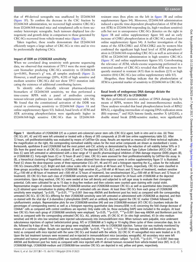
Impact of DDR on LY2606368 sensitivityWhen we correlated drug sensitivity with genome sequencingdata, we observed that mutations of TP53 were the most signifi-cant biomarker predicting CRC-SCs sensitivity to LY2606368(p=0.001, Pearson’s χ2 test, all samples analysed) (figure 2).However, a small percentage (20%, 4/20) of high sensitive andmedium sensitive CRC-SCs did not show TP53 mutation indi-cating the existence of additional contributing factors.
To identify other clinically relevant pharmacodynamicbiomarkers of LY2606368 sensitivity, we thus performed atime-course RPPA with a panel of antibodies coveringtumour-relevant pathways (see online supplementary table S4).We found that the constitutional activation of the DDR wascrucial in conferring sensitivity to LY2606368 (figure 3A andonline supplementary figure S3). Indeed, basal levels of ATM andATR activating phosphorylation were significantly higher inLY2606368-high sensitive CRC-SCs than in LY2606368-
resistant ones (box plots on the left in figure 3B and onlinesupplementary figure S4). Moreover, LY2606368 administrationinduced a specific time-dependent phosphorylation of ATM (butnot ATR) in LY2606368-responding (ie, high+medium sensitive)cells but not in unresponsive CRC-SCs (kinetics on the right infigure 3B and online supplementary figure S4) and an earlyincrease of CHK1 phosphorylation in all CRC-SCs analysed (seeonline supplementary figure S4). A further investigation of thestatus of the ATR-CHK1 and ATM-CHK2 axis by western blotconfirmed the significant high basal level of ATM phosphoryl-ation in LY2606368-responding CRC-SCs as well as activation ofATM on LY2606368 administration in high sensitive CRC-SCs(figure 3C and online supplementary figures S5). Corroboratingthe relevance of ATM, whole-exome sequencing performed in arestricted number of CRC-SCs revealed that ATM was more fre-quently mutated in resistant (4/5) than in medium (2/5) or highsensitive (0/6) CRC-SCs (see online supplementary table S5).
Altogether, these findings indicate that the phoshorylation ofATM is a potential marker of sensitivity to LY2606368 in CRC-SCs.
Basal levels of endogenous DNA damage dictate theresponse of CRC-SCs to LY2606368We then performed extensive analyses of DNA damage levels bymeans of RPPA, western blot and immunofluorescence studies.These analyses revealed that basal phosphorylation levels of RPA2/RPA32, a signalling intermediate of ongoing DNA replication stress(RS) response,27 and H2A histone family, member X (γH2AX), adouble strand break (DSB)-sensitive marker, were significantly
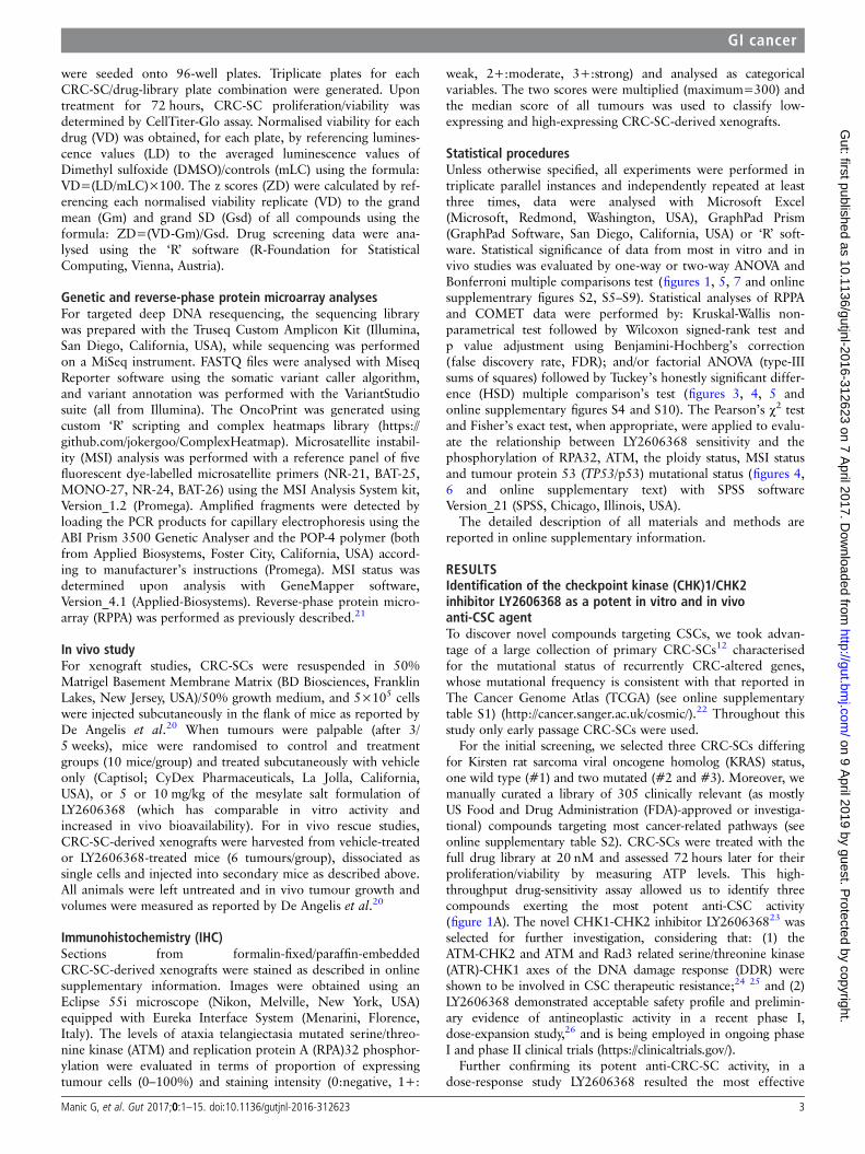
Figure 1 Identification of LY2606368 (LY) as a potent anti-colorectal cancer stem cells (CRC-SCs) agent, both in vitro and in vivo. (A) ThreeCRC-SCs (#1, #2 and #3) were left untreated or treated with a library of 305 compounds at 20 nM (see online supplementary table S2). After72 hours, cell proliferation/viability was evaluated by means of CellTiter-Glo assay. Results are reported as means±SD of three technical replicates. Inthe magnification on the right, the corresponding normalised viability values for the most active compounds are shown as standardised z scores.Bortezomib, epothilone B and LY2606368 had the most potent anti-CSC activity as demonstrated by the reduction of cell viability below 50% or 3SDs from the grand mean in all CRC-SCs analysed. (B and C) The depicted CRC-SCs (B,C) and American Type Culture Collection (ATCC) CRC celllines (C) were left untreated or administered with a dose range of 13 specific pharmacological inhibitors of DNA damage response (DDR) players (B),or LY2606368 (C). Forty-eight hours (C) or 72 hours (B and C) later, CRC-SCs were assessed for their proliferation/viability by CellTiter-Glo assay. In(B), a hierarchical clustering of logarithmic scaled IC50 values obtained from dose-response curves in online supplementary figure S1 is illustrated.Panel (C) shows the dose-response curves of three representative CSCs (#1, #6 and #7) and a histogram reporting the IC50 values for the indicatedCRC-SCs (means±SEM; n≥2). Bright and dark colour scales refer to end points at 48 hours and 72 hours, respectively. CRC-SCs were classified inthree groups according to their sensitivity to LY2606368: high sensitive (IC50<100 nM at 48 hours and 72 hours of treatment), medium sensitive(IC50>100 nM at 48 hours of treatment and <500 nM at 72 hours of treatment), low sensitive/resistant (IC50>500 nM at 48 hours and 72 hours oftreatment). (D) CRC-SCs from each class of LY2606368 sensitivity were left untreated or treated for 24 hours with LY2606368 at the depictedconcentrations. Upon drug washout, CRC-SCs were seeded at low cell density and subjected to soft agar assay to evaluate their clonogenicpotential. Cells were cultivated for up to 15 days in drug-free medium and then colonies were counted upon staining with crystal violet.Representative images of colonies formed from LY2606368-sensitive and LY2606368-resistant CRC-SCs as well as quantitative data (means±SEM;n≥3) obtained upon normalisation to plating efficiency of untreated cells are shown. At least three CRC-SCs from each group of LY2606368sensitivity were employed. *p<0.05, **p<0.01, ***p<0.001 (two-way ANOVA and Bonferroni post hoc tests) as compared with the correspondinguntreated CRC-SCs. (E) CRC-SCs differing for their LY2606368 sensitivity were left untreated or administered with LY2606368 as depicted, and thenco-stained with the vital dye 4',6-diamidino-2-phenylindole (DAPI) and an antibody directed against the CRC-SC marker CD44v6 followed bycytofluorimetric analysis. Representative plots for one LY2606368-sensitive (#4) and one LY2606368-resistant (#7) CRC-SCs (numbers indicate thepercentages of corresponding events) as well as a histogram reporting quantitative data (means±SEM; n=4) concerning the fold change decrease ofCD44v6+ cells as compared with control conditions are reported. Only viable cells (ie, those excluding DAPI) were included in the analysis. At leasttwo CRC-SCs from each group of LY2606368 sensitivity were employed. *p<0.05, **p<0.01, ***p<0.001 (two-way ANOVA and Bonferroni post hoctests) as compared with the corresponding untreated CRC-SCs. AU, arbitrary units. (F) CRC-SC #1 (in vitro high sensitive), #3 (in vitro mediumsensitive) and #8 (in vitro low sensitive) were injected subcutaneously into immunodeficient mice. When tumours were palpable, mice underwentsubcutaneous injections of captisol (vehicle/control group, NT) or LY2606368 at 5 mg/kg or 10 mg/kg (three cycles of three consecutive days, bis indie, followed by 4 days of rest). Ten mice per group were employed. Arrows refer to LY2606368 injection. Tumour size was routinely monitored bymeans of a common calliper. Results are reported as means±SEM. *p<0.05, **p<0.01, ***p<0.001 (two-way ANOVA and Bonferroni post hoctests) as compared with mice injected with the same CRC-SCs and treated with the vehicle. (G) CRC-SC #1-xenografted mice were treated as in (F).Upon the last treatment tumours were recovered and re-injected in immunodeficient mice (secondary xenografts). All secondary mice were leftuntreated and tumour growth (means±SEM) was routinely evaluated by means of a common calliper. *p<0.05, **p<0.01, ***p<0.001 (two-wayANOVA and Bonferroni post hoc tests) as compared with mice injected with #1-derived tumours recovered from vehicle-treated mice (NT). In (C–E)LY2606368-high, LY2606368-medium and LY2606368-low sensitive CRC-SCs are depicted in red, yellow and green, respectively.
Manic G, et al. Gut 2017;0:1–15. doi:10.1136/gutjnl-2016-312623 5
GI cancer on 9 A
pril 2019 by guest. Protected by copyright.
http://gut.bmj.com
/G
ut: first published as 10.1136/gutjnl-2016-312623 on 7 April 2017. D
ownloaded from
higher in LY2606368-sensitive than in LY2606368-resistantCRC-SCs (box plots on the left in figure 4A–C and see onlinesupplementary figures S6–S8). These results indicate an elevatedamount of basal endogenous DNA damage and RS in these cells.In addition, LY2606368 significantly induced DNA damage (figure4D) and increased the levels of RPA32 phosphorylation andγH2AX in CRC-SCs (box plots on the right in figure 4A, B and seeonline supplementary figure S6). IHC analyses performed onparaffin-embedded sections of >15 CRC-SCs-derived xenograftsconfirmed a significant association between LY2606368 sensitivityand phosphorylation of ATM or RPA32 (p=0.043 or 0.013,respectively; high+medium vs low) (figure 4E, F).
Altogether, these results indicate that high basal levels of RScoupled to ATM phosphorylation represent reliable in vitro andin vivo markers for predicting the response of CRC-SCs toLY2606368.
Mechanisms of CSCs killing by, and of resistance to, LY2606368We then explored the impact of LY2606368 on CRC-SC cellcycle progression. We observed that LY2606368 affected cellcycle distribution selectively in LY2606368-sensitive (high+medium) CRC-SCs by enriching the percentage of cells with aDNA content between 2n and 4n (figure 5A). S-phase accumula-tion was accompanied by a significant augmentation of themitotic cell fraction (pH3+) in high but not medium sensitiveor low sensitive CRC-SCs (figure 5A and online supplementaryfigure S4E). Moreover, upon LY2606368 exposure a consider-able percentage of pH3+ cells in high+medium sensitive
CRC-SCs displayed <4n DNA content, while the fraction ofpremature mitoses did not significantly vary amongLY2606368-unresponsive CRC-SCs (figure 5A). LY2606368induced a significant increase in the percentage ofDNA-replicating (5-ethynyl-20-deoxyuridine (EdU)+) cells onlyin responsive CRC-SCs (figure 5B), suggesting the deregulationof the replication process. These results, which are reminiscentof those found in the absence of CHK1,23 28 indicate thatCHK1 is the main target of LY2606368. Accordingly, the deple-tion of CHK1 induced an accumulation of cells with a DNAcontent between 2n and 4n (figure 5C), triggered cell death(figure 5D and see online supplementary figure S9), andimpaired the clonogenic potential (figure 5E) exclusively inLY2606368-sensitive CRC-SCs. The absence of thep53-dependant G1 checkpoint forces S-phase entry in the pres-ence of DNA damage. In line with this evidence, the expressionlevels of the p53 target cyclin-dependent kinase inhibitor 1A(CDKN1A/p21) were higher in resistant than sensitive cells (seeonline supplementary figure S10). This confirms that p53 defi-ciency is a marker of LY2606368 sensitivity and that the p53pathway protects from the lethal effect of LY2606368.CRC-SCs responding to LY2606368 could not endure cell cyclederegulation and eventually die for the activation of the caspase-dependent pathway of apoptosis (figure 5F).
Altogether these results indicate that LY2606368 killsCRC-SCs by inhibiting CHK1 resulting in the deregulation ofDNA replication, impairment of cell cycle checkpoints andlethal replication catastrophe.
Figure 2 Oncoprint of mutations for the 16 most commonly mutated genes in colorectal cancer (CRC) found in CRC stem cells (CRC-SCs) by deepsequencing1.1http://cancer.sanger.ac.uk/cosmic, online supplementary table S1. 2No Catalogue Of Somatic Mutations In Cancer (COSMIC) mutations were foundfor: ACVR1B, KIAA1804, MAP2K4, NRAS, SMAD2.
6 Manic G, et al. Gut 2017;0:1–15. doi:10.1136/gutjnl-2016-312623
GI cancer on 9 A
pril 2019 by guest. Protected by copyright.
http://gut.bmj.com
/G
ut: first published as 10.1136/gutjnl-2016-312623 on 7 April 2017. D
ownloaded from

Impact of the ploidy status on the sensitivity of CRC-SCs toLY2606368We then evaluated the impact of chromosomal content onLY2606368 activity. Through metaphase spreading, we found aheterogeneous modal chromosome number with ∼57% (13/23)CRC-SCs exceeding the near-to-diploid set (>50, ie,
hyperdiploid) (figure 6A, B). Cell cycle profiling by flow cyto-metry confirmed the significant association between LY2606368sensitivity and increased chromosome number (p=0.005;Pearson’s χ2 test, high+medium vs low) (figure 6C–E). Thus,almost all hyperdiploid CRC-SCs (∼95.2%, 20/21) were high+medium sensitive to LY2606368, whereas 88.9% (8/9) of
Figure 3 Phosphorylation of ataxia telangiectasia mutated serine/threonine kinase (ATM) as a marker of colorectal cancer stem cells (CRC-SCs)sensitivity to LY26063668. (A and B) Nine representative CRC-SCs, three from each group of LY2606368 sensitivity, and the ATCC cell lines HCT 116and HT-29 were left untreated or treated with LY2606368 at low doses (10 nM, 50 nM and 100 nM) for 1 hour, 4 hours, 9 hours or 24 hours andthen subjected to reverse-phase protein microarray (RPPA) analysis. Panel (A) represents the hierarchical clustering of RPPA results obtained onuntreated CRC-SCs (white) and CRC-SCs exposed for 24 hours to 100 nM LY2606368 (black). In the left part of (B), basal levels of phosphorylated(p)ATM (pATM_S1981) in untreated CRC-SCs at all time points were pooled for each sensitivity group and box-plotted. Statistical analysis: Wilcoxonsigned-rank test followed by p value adjustment by false discovery rate (FDR) for the comparison of high versus low sensitive CRC-SCs (*p<0.05,**p<0.01, ***p<0.001). In (B), on the right, the full time-dependent and dose-dependent RPPA kinetics of pATM are shown as means±SD ofCRC-SCs grouped by LY2606368 sensitivity. Statistical analysis: factorial ANOVA design followed by p value adjustment (Tuckey’s honestly significantdifference (HSD)) for the comparison of (1) 100 nM LY2606368-treated versus untreated CRC-SCs of the same sensitivity group (*p<0.05, **p<0.01,***p<0.001) or (2) high versus low sensitive CRC-SC treated with 100 nM LY2606368 (§p<0.05, §§p<0.01, §§§p<0.001). A.U., arbitrary units. (C)The illustrated CRC-SCs were left untreated (–) or administrated with 100 nM LY2606368 (LY) for 6 hours (+) and then subjected to western blotanalyses with antibodies recognising the phosphorylated or total forms of ATM, checkpoint kinase (CHK)2, ataxia telangiectasia mutated and Rad3related serine/threonine kinase (ATR) or CHK1. Nucleolin or β-actin levels were monitored to ensure equal loading of lanes. Note that six CRC-SCs,two from each sensitivity class, were the same used in RPPA studies. One representative western blot is shown. For the densitometric and statisticalanalysis of the western blots, refer to online supplementary figure S5 and online supplementary information. LY2606368-high (H),LY2606368-medium (M) and LY2606368-low (L) sensitive CRC-SCs are depicted in red, yellow and green, respectively.
Manic G, et al. Gut 2017;0:1–15. doi:10.1136/gutjnl-2016-312623 7
GI cancer on 9 A
pril 2019 by guest. Protected by copyright.
http://gut.bmj.com
/G
ut: first published as 10.1136/gutjnl-2016-312623 on 7 April 2017. D
ownloaded from

Figure 4 Endogenous DNA damage and replication stress predict the response of colorectal cancer stem cells (CRC-SCs) to LY2606368, both in vitro andin vivo. (A–C) reverse-phase protein microarray (RPPA) box plots or time-response and dose-response plots (A and B) and western blot analyses (untreatedconditions) performed with antibodies recognising the phosphorylated (p) form of RPA32 (pRPA32_S4/S8) and total RPA32 (C) on a representative panel ofCRC-SCs. In (A) and (B), the box plots on the left represent basal levels of pRPA32_S4/S8 or γH2AX of untreated CRC-SCs pooled for each sensitivity group,while line plots on the right report the RPPA kinetics of pRPA32_S4/S8, and γH2AX shown as means±SD of CRC-SCs grouped by LY2606368 sensitivity. Formore insights on RPPA data statistical analysis refer to legend of figure 3B. A.U., arbitrary units. In (C) β-actin levels were monitored to ensure equalloading of lanes. One representative western blot is shown. For the densitometric and statistical analyses of the western blots, refer to online supplementaryfigure S5 and online supplementary information. (D) CRC-SCs from each class of LY2606368 (LY) sensitivity were left untreated or treated for 24 hours with100 nM LY2606368, and then subjected to single cell gel electrophoresis (also known as COMET assay) to measure DNA lesions. The panel showsrepresentative microphotographs of one sensitive CRC-SC (#4) and one resistant CRC-SC (#7) as well as box plots of the tail moments of at least 100 nucleiper experimental point measured by image analysis. Scale bar=100 mm. Statistical analysis: Wilcoxon signed-rank test followed by p value adjustment byfalse discovery rate (FDR) for the comparison of high versus low sensitive CRC-SCs (*p<0.05, **p<0.01, ***p<0.001). (E and F) Representative IHC imagesobtained from CRC-SC-derived xenograft sections stained with antibodies recognising pRPA32_S4/S8 (E) and phosphorylated ATM (pATM_S1981) (F) (scalebar=30 μm). Upon IHC analyses specimens were divided into two groups, one with low staining (pRPA32_S4/S8low or pATM_S1981low) and one with highstaining (pRPA32_S4/S8high or pATM_S1981high) according to the median intensity values and by scoring on a continuous scale of 0–300. Numbers in thetables refer to the number (N) and the percentage (%) of samples in each group. The association between phosphorylation levels and LY2606368 sensitivityin all samples was evaluated by Pearson’s χ2 test, whereas the comparison of high+medium (med) versus low sensitive CRC-SCs by Fisher’s exact test.p values <0.05 were considered statistically significant. LY2606368-high (H), LY2606368-medium (M) and LY2606368-low (L) sensitive CRC-SCs aredepicted in red, yellow and green, respectively.
8 Manic G, et al. Gut 2017;0:1–15. doi:10.1136/gutjnl-2016-312623
GI cancer on 9 A
pril 2019 by guest. Protected by copyright.
http://gut.bmj.com
/G
ut: first published as 10.1136/gutjnl-2016-312623 on 7 April 2017. D
ownloaded from

Manic G, et al. Gut 2017;0:1–15. doi:10.1136/gutjnl-2016-312623 9
GI cancer on 9 A
pril 2019 by guest. Protected by copyright.
http://gut.bmj.com
/G
ut: first published as 10.1136/gutjnl-2016-312623 on 7 April 2017. D
ownloaded from

resistant CRC-SCs were near-to-diploid (figure 6D, E). Asopposed to ploidy, MSI status was not significantly associated tothe response of CRC-SCs to LY2606368 (p=0.108; Pearson’sχ2 test, high+medium vs low) (figure 6D). Consistent with therole of p53 in controlling ploidy,29 ∼81% (13/16) ofTP53-mutated CRC-SCs were hyperdiploid, while ∼73% (8/11)TP53 wild type CRC-SCs displayed near-to-diploid karyotypes(association TP53 mutation and ploidy status: p=0.043;Pearson’s χ2 test) (figure 2). Taken together these results indicatethat hyperdiploidy is a cytogenetic marker of LY2606368sensitivity.
Strategies to modulate CRC-SC sensitivity to LY2606368We finally assessed the relevance of the predictive markers iden-tified above. We first evaluated the impact of ATM activity dem-onstrating that the pharmacological inhibition of ATM partiallyprotected medium/high sensitive CRC-SCs from LY2606368(figure 7A). Thereafter, we assessed the contribution of p53showing that the constitutive knockdown of p53 via transduc-tion of shRNA-expressing lentiviral vectors sensitised previouslyresistant CRC-SCs to LY2606368 (figure 7B). To analysewhether an artificial increase in cellular ploidy might confer sen-sitivity to LY2606368, we generated clones bearing a doubleDNA content (hereafter called tetraploid) from onep53-mutated (#14) and three p53-proficient (#7, #8, #10)near-to-diploid CRC-SCs with an optimised protocol.30 Weobtained two tetraploid clones only from CRC-SC #14, bothdisplaying an augmented sensitivity to LY2606368 (figure 7C).Corroborating the positive impact of RS on LY2606368 sensitiv-ity, by perturbing DNA replication with low/sublethal doses ofaphidicolin, hydroxyurea or thymidine, we were able to signifi-cantly sensitise previously non-responding CRC-SCs toLY2606368 (figure 7D). Along similar lines, LY2606368
cytotoxicity was highly increased in LY2606368-resistantCRC-SCs by coadministering gemcitabine (figure 7E).
These results confirm the reliability of the phosphorylation ofATM, p53 deficiency, hyperdiploidy and high RS as markers ofLY2606368 sensitivity, thus validating our experimental strategy.
DISCUSSIONIn this study, after a pharmacological screening with therapeuticcompounds on CRC-SCs, LY2606368 was identified as a potentanti-CRC-SC single agent acting independently of RAS muta-tional status. We also demonstrated that CHK1 targeting byLY2606368 potently killed CRC-SCs from approximately athird of tumours by preferentially depleting the CSC fraction.Cell death occurred via a mechanism involving the alterationand deregulation of DNA replication, which resulted in the gen-eration of an intolerable DNA damage burden and induction ofreplication catastrophe.31 By correlating drug sensitivity withdata coming from genome sequencing, RPPA and cytogeneticanalyses, we identified four predictive and interlinked markersof CRC-SC sensitivity to LY2606368: (1) high basal levels of RSmarkers; (2) overactivation of the DDR player ATM; (3) muta-tions of TP53; and (4) hyperdiploidy (figure 7F).
Compelling evidence indicates that the DDR acts as a barrierduring oncogenesis by limiting genomic instability induced byRS.32 Deregulated DDR pathways are frequently found in humanneoplasms, where they are linked to increased genomic instabilityand therapeutic failure and/or sensitivity.22 33 34 Moreover, it isbecoming evident that, once established, tumours overactivatethe DDR to endure high levels of RS.35 CSCs efficiently activatethe DDR24 25 and are responsible for tumour resilience. From atherapeutic perspective, here, we demonstrated that CRC-SCsdisplaying RS are highly dependent on the activity of CHK1, akinase frequently overexpressed in neoplasms and regulating
Figure 5 LY2606368 kills colorectal cancer stem cells (CRC-SCs) by inducing replication catastrophe and premature mitosis entry followingcheckpoint kinase (CHK)1 inhibition. (A) CRC-SCs from each class of LY2606368 sensitivity were left untreated or treated for 6 hours or 24 hourswith LY2606368 at 10 nM or 100 nM, then fixed with ethanol and stained with the DNA dye 4’,6-diamidino-2-phenylindole (DAPI) and an antibodydirected against phospho-histone H3 (pH3_S10), for cytofluorimetric analysis of DNA content and mitotic cells. In the upper part, cell cycle profilesand dot plots of pH3+ cells with the indicated DNA content are shown for one representative high sensitive and low sensitive CRC-SC. Orange andred quadrants highlight premature mitosis (pH3+ cells with a <4n DNA content) and normal mitosis (pH3+ cells with a 4n DNA content) events,respectively. Numbers indicate the percentages of corresponding events. The histograms report quantitative data (means±SEM; n=4; at least threeCRC-SCs from each group of LY2606368 sensitivity were employed) concerning the percentage or the fold change (as compared with controlconditions) of S-phase, pH3+ cells and premature mitosis. Note that the sub-G1 fraction was excluded from the analysis. *p<0.05, **p<0.01,***p<0.001 (two-way ANOVA and Bonferroni post hoc tests) as compared with the corresponding untreated CRC-SCs. (B) CRC-SCs differing fortheir LY2606368 sensitivity were left untreated or treated for 15 hours with LY2606368 as indicated, then fixed and labelled with the thymidineanalogue 5-ethynyl-20-deoxyuridine (EdU). Representative dot plots of EdU+ cells for LY2606368-high sensitive CRC-SCs and quantitative data(means±SEM; n=3; at least three CRC-SCs from each group of LY2606368 sensitivity were employed) for the three groups of sensitivity toLY2606368 are reported. Note that the sub-G1 fraction was excluded from the analysis. *p<0.05, **p<0.01, ***p<0.001 (two-way ANOVA testfollowed by Bonferroni post hoc test) as compared with the corresponding CRC-SCs left untreated. (C–E) CRC-SCs from each class of LY2606368sensitivity were transfected with an unrelated small interfering (si) RNA (siCTR) or two specific siRNAs directed against CHK1 (siCHK1A andsiCHK1B) (C and E) or, alternatively were transduced with lentiviral vectors expressing non-silencing short hairpin (sh) RNA (shCTR) orCHK1-targeting shRNAs (shCHK1A and shCHK1B) (D). Samples were collected, dissociated as single cells and then either analysed for their DNAcontent by flow cytometry upon staining with the DNA dye Hoechst 33342 (C), subjected to the cytofluorimetric assessment of mitochondrialmembrane potential loss (a cell death-associated parameter) upon staining with Tetramethylrhodamine, methyl ester (TMRM) (D), or evaluated fortheir clonogenic potential by soft agar assay (E) as described in figure 1D. At least two CRC-SCs from a distinct group of LY2606368 sensitivity wereemployed in all the assays. Histograms reporting quantitative data (means±SEM; n≥3) concerning the percentage of S-phase cells or viable cells atthe indicated time upon transfection/transduction are shown in (C) or (D), respectively. ‘d2’ and ‘d7’ refer to the day after the puromycin selectionround. NT, non-transfected cells. In (C) the sub-G1 fraction was excluded from the analysis, while in (D) only the transduced cell population (greenfluorescent protein+, GFP+ cells) was assessed. Quantitative data of clonogenicity (means±SEM; n≥2) obtained upon normalisation to platingefficiency of siCTR-transfected CRC-SCs are shown in (E). *p<0.05, **p<0.01, ***p<0.001 (two-way ANOVA and Bonferroni post hoc tests) ascompared with the corresponding siCTR-transfected or shCTR-transduced CRC-SCs. (F) Reverse-phase protein microarray (RPPA) time-response anddose-response plots of cleaved Caspase-3 (cCASP3), cleaved Caspase-7 (cCASP7), cleaved poly(ADP-ribose) polymerase 1 (cPARP-1) and cleavedFodrinα (cFODRINα) of CRC-SCs grouped by LY2606368 sensitivity. Results are shown as means±SD. For more insights on RPPA data statisticalanalysis refer to legend of figure 3B. LY2606368-high, LY2606368-medium and LY2606368-low sensitive CRC-SCs are depicted in red, yellow andgreen, respectively. A.U., arbitrary units.
10 Manic G, et al. Gut 2017;0:1–15. doi:10.1136/gutjnl-2016-312623
GI cancer on 9 A
pril 2019 by guest. Protected by copyright.
http://gut.bmj.com
/G
ut: first published as 10.1136/gutjnl-2016-312623 on 7 April 2017. D
ownloaded from
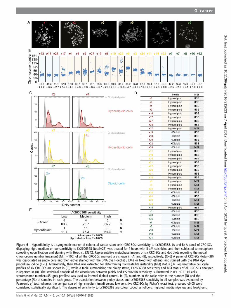
Figure 6 Hyperdiploidy is a cytogenetic marker of colorectal cancer stem cells (CRC-SCs) sensitivity to LY2606368. (A and B) A panel of CRC-SCsdisplaying high, medium or low sensitivity to LY2606368 (total=23) was treated for 4 hours with 5 μM colchicine and then subjected to metaphasespreading upon fixation and staining with Hoechst 33342. Representative metaphase images of six CRC-SCs and dot plots reporting the modalchromosome number (means±SEM; n=100) of all the CRC-SCs analysed are shown in (A) and (B), respectively. (C–E) A panel of CRC-SCs (total=38)was dissociated as single cells and then either stained with the DNA dye Hoechst 33342 or fixed with ethanol and stained with the DNA dyepropidium iodide (C–E). Alternatively, their DNA was extracted for determining microsatellite instability (MSI) status (D). Representative cell cycleprofiles of six CRC-SCs are shown in (C), while a table summarising the ploidy status, LY2606368 sensitivity and MSI status of all CRC-SCs analysedis reported in (D). The statistical analysis of the association between ploidy and LY2606368 sensitivity is illustrated in (E). HCT 116 cells(chromosome number=45; grey profiles) was used as internal diploid control. In (E), numbers in the table refer to the number (N) and thepercentage (%) of samples in each group. The association between ploidy status and LY2606368 sensitivity in all samples was evaluated byPearson’s χ2 test, whereas the comparison of high+medium (med) versus low sensitive CRC-SCs by Fisher’s exact test. p values <0.05 wereconsidered statistically significant. The classes of sensitivity to LY2606368 are colour coded as follows: high/red, medium/yellow and low/green.
Manic G, et al. Gut 2017;0:1–15. doi:10.1136/gutjnl-2016-312623 11
GI cancer on 9 A
pril 2019 by guest. Protected by copyright.
http://gut.bmj.com
/G
ut: first published as 10.1136/gutjnl-2016-312623 on 7 April 2017. D
ownloaded from

DNA replication, RS response and cell cycle progression.36 37 Wesurmise that the essential role of CHK1 in CRC-SCs is to main-tain a high but tolerable level of RS (figure 7F). This ‘thresholdlevel’ hypothesis is supported by two observations. First,LY2606368-mediated inhibition of CHK1 increased the level ofRS and induced a lethal replication catastrophe31 exclusively inCRC-SCs responding to LY2606368. This occurred through a
mechanism involving: (1) unscheduled DNA replication accom-panied by the slowing down of the replication process and RS,ultimately leading to an excess of stretches of single-strandedDNA recognised by phosphorylated RPA32;31 (2) the deregula-tion of S-phase progression due to the impairment of theCHK1-dependent intra-S checkpoint; and (3) replication forkcollapse eventually resulting in the accumulation of DSBs during
12 Manic G, et al. Gut 2017;0:1–15. doi:10.1136/gutjnl-2016-312623
GI cancer on 9 A
pril 2019 by guest. Protected by copyright.
http://gut.bmj.com
/G
ut: first published as 10.1136/gutjnl-2016-312623 on 7 April 2017. D
ownloaded from

replication. This evidence confirms recent observations demon-strating that CHK1, which is a key player for the correct execu-tion and regulation of DNA replication, rather than CHK2,which operates mainly during the G2-M transition, is the majortarget of LY2606368.23 It also proves that LY2606368 inducesDNA damage and, at the same time, inhibits the DDR. Second,strategies aimed at perturbing replication (eg, gemcitabine coad-ministration) sensitised non-responding CRC-SCs to LY2606368.These results, which are in line with previous observations,38 39
suggest that the activity of CHK1 becomes essential in CRC-SCsas a means to cope with enhanced RS.
In this preclinical study, CRC-SC sensitivity to LY2606368 isassociated with the phosphorylation of ATM, suggesting acrucial role of ATM in coordinating the RS response, which isin line with previous findings.40 Accordingly, we found a signifi-cant association between the phosphorylation of ATM andRPA32, and responsiveness of CRC-SCs to LY2606368. Theseresults confirm the crosstalk between the ATM-CHK2 andATR-CHK1 cascade.27 Reportedly, ATR-CHK1 inhibitors dis-played synthetic lethality with deficiency in other DDRplayers.41 42 Here, we provide evidence that the inhibition ofATM protects CRC-SCs to LY2606368, suggesting a functionalrole of ATM in the mechanism of CRC-SC killing byLY2606368. Further experiments are needed to elucidate theprecise link between ATM and CHK1.
We also provided evidence of a cytogenetic cause of RS intumours. CRC-SCs having a hyperdiploid chromosome setshowed higher levels of RS than near-to-diploid CRC-SCs, whichexplains their exquisite sensitivity to LY2606368. We surmisethat the imbalance/augmentation in copy number of multiplechromosomes is behind the increased RS displayed by hyperdi-ploid cells.43 44 Moreover, a hyperdiploid karyotype may lead tothe deregulation of cancer-related proteins involved in RSresponse. In this context, hyperdiploid CRC-SCs always harbourmutation(s) in TP53, which may promote RS by favouring thegeneration/tolerance of hyperdiploidy or diminishing DNArepair efficiency. Accordingly, we demonstrated that the absenceof p53 significantly increased the cytotoxicity caused by the
inhibition of CHK1. Moreover, the few diploid CRC-SCs sensi-tive to LY2606368 mostly presented TP53 mutations and highRS levels, while p53-proficient near-to-diploid CRC-SCs consti-tutionally activated the p53 pathway to limit RS and LY2606368toxicity. These findings are coherent with previous observationsreporting enhanced sensitivity to CHK1 inhibition of tetraploidand/or p53-deficient cancer cells.45 46
Patient-derived cancer models are promising tools for drugdiscovery and clinical efficacy prediction.47–50 The majorstrength of this preclinical study is the vast collection of molecu-larly and functionally characterised CRC-SCs. This enabled usto (1) identify a clinically relevant agent with broad anti-CSCactivity at low nanomolar doses, and effective againstKRAS-mutated, non-hypermutated and/or p53-deficientCRC-SCs, (2) uncover and validate in vitro and in vivo candi-date predictive markers of CRC-SC responsiveness, and (3)design strategies to overcome the intrinsic resistance to thistherapeutic regimen. Importantly, LY2606368 demonstrated tol-erable safety profiles and preliminary evidence of antineoplasticactivity in a recent phase I, non-randomised, open-label,dose-escalation trial in patients affected by advanced or meta-static solid tumours who underwent at least three previous linesof treatment.26 In this study, partial response and disease stabil-isation was obtained in 4.4% and 33.3% of patients, respect-ively. LY2606368 is currently employed alone or combined withother therapeutic agents in ongoing phase I or phase II studiesalso in patients with CRC (eg, NCT02203513, NCT02124148;https://clinicaltrials.gov/). Our preclinical study supports thefurther clinical development of LY2606368 as we demonstratedits potent anti-CSC activity in a subset of CRC-SCs and uncov-ered for the first time biomarkers associated with its efficacy inCRC. The prospective validation of the predictive value of thesebiomarkers may thus provide valuable background to the defin-ition of prediction nomograms. In this context, the identifica-tion of reliable markers of CSCs and enhanced/ongoing RSresponse will allow to prospectively distinguish CSCs andreplication-stressed CSCs within the tumour mass and thusconfirm the true potential of this anti-CSC strategy.
Figure 7 Strategies to modulate the sensitivity of colorectal cancer stem cells (CRC-SCs) to LY2606368. (A) Three representative LY2606368-highor LY2606368-medium sensitive CRC-SCs were left untreated or were treated with LY2606368 and/or the ataxia telangiectasia mutated serine/threonine kinase (ATM) inhibitor KU-60019 at the indicated dose. Upon 48 hours, CRC-SC proliferation/viability were assessed by CellTiter-Glo assay(means±SEM; n=5). The percentage of viable cells shown for co-treatments with KU-60019 and LY2606368 is normalised over the correspondenttreatment with KU-60019 as single agent. Note that KU-60019 alone decreases CRC-SC viability of 0%, ∼9% and ∼27% at doses 1 mM, 5 mM and10 mM, respectively. *p<0.05, **p<0.01, ***p<0.001 (two-way ANOVA and Bonferroni post hoc test) as compared with the correspondingCRC-SCs left untreated or treated only with LY2606368. (B) Representative p53-proficient CRC-SCs displaying resistance to LY2606368 (LY) weretransduced with lentiviral vectors expressing non-silencing short hairpin (sh) RNA (shCTR) or p53-targeting shRNAs (shp53A and shp53B). Upon5 days of selection with 1.5 mg/mL puromycin, cells were amplified, seeded and then left untreated or treated with the indicated concentration ofLY2606368. Finally, cells were subjected to the assessment of mitochondrial membrane potential loss (a cell death-associated parameter) by flowcytometry upon staining with Tetramethylrhodamine, methyl ester (TMRM). Only the transduced cell population (GFP+ cells) was evaluated.Representative plots (numbers refer to the percentage of TMRMlow cells), quantitative data (means±SEM; n≥3), and western blot analysesperformed with antibodies recognising the p53 or β-actin (whose levels were monitored to ensure equal loading of lanes) are reported. *p<0.05,**p<0.01, ***p<0.001 (two-way ANOVA and Bonferroni post hoc tests) as compared with CRC-SCs transduced with the same shRNA but leftuntreated. (C) CRC-SCs #14 were left untreated or treated with nocodazole for 48 hours, washed and then cultured in standard medium. Upon2 weeks, cells were subcloned by limiting dilution to isolate diploid (2n) and tetraploid (4n) clones. The ploidy of proliferating clones was assessedas in figure 6C. The IC50 values of the parental cells (#14), one diploid (#A1) and two tetraploid clones (#A2 and #A3) were calculated byCellTiter-Glo assay after LY2606368 administration as indicated. Results from a single polyploidising series are reported. Note that following∼30 days of culture #A2 and #A3 clones spontaneously reverted to near-to-diploidy decreasing their sensitivity to LY2606368. (D and E) Threerepresentative LY2606368-low sensitive CRC-SCs were left untreated or treated with the indicated concentration of LY2606368 alone or togetherwith sublethal doses of aphidicolin, hydroxyurea or thymidine (C), or three doses of gemcitabine (D). Cell viability, as assessed by CellTiter-Glo assayafter 72 hours of treatment, is reported as mean±SEM (n≥3). *p<0.05, **p<0.01, ***p<0.001 (two-way ANOVA and Bonferroni post hoc tests) ascompared with the corresponding CRC-SCs left untreated or treated only with LY2606368. (F) Proposed model accounting for LY2606368 sensitivityof CRC-SCs. RS, replication stress; pATM, phosphorylated ATM; pRPA32, phosphorylated RPA32.
Manic G, et al. Gut 2017;0:1–15. doi:10.1136/gutjnl-2016-312623 13
GI cancer on 9 A
pril 2019 by guest. Protected by copyright.
http://gut.bmj.com
/G
ut: first published as 10.1136/gutjnl-2016-312623 on 7 April 2017. D
ownloaded from

Author affiliations1Department of Biology, University of Rome “Tor Vergata”, Rome, Italy2Department of Oncology and Molecular Medicine, Istituto Superiore di Sanità,Rome, Italy3Department of Research, Advanced Diagnostics and Technological Innovation,Regina Elena National Cancer Institute, Rome, Italy4Institute of General Pathology, Catholic University and A. Gemelli Polyclinic, Rome,Italy5Department of Science, University “Roma Tre”, Rome, Italy6Department of Molecular Medicine, University “La Sapienza”, Rome, Italy7SAFU, Department of Research, Advanced Diagnostics, and TechnologicalInnovation, Translational Research Area, Regina Elena National Cancer Institute,Rome Italy8Department of Pathology, Regina Elena National Cancer Institute, Rome, Italy9Biostatistical Unit, Regina Elena National Cancer Institute, Rome, Italy10Department of Surgical Oncological and Stomatological Sciences, University ofPalermo, Palermo, Italy11Genetics and Rare Diseases Research Division, Ospedale Pediatrico “BambinoGesù”, Rome, Italy12Department of Experimental Medicine, University “La Sapienza”, Rome, Italy13Division of Medical Oncology 2, Regina Elena National Cancer Institute, Rome,Italy
Acknowledgements The authors thank Emanuela Pilozzi for providing theparaffin-embedded xenograft sections, and Paola Di Matteo, Roberto Ricci andStefano Guida for technical assistance. In vivo experiments were performed atPlaisant Castel Romano and Istituto Superiore di Sanità (Rome, Italy).
Contributors GM, MS, AS, GR designed and performed experiments, analysed andinterpreted data; FC, SS, MM, SV, MLDA, CAA, SDF, SB, GDL performedexperiments; MP, EP, FDN, MF analysed data and performed bioinformatic studies;FS, MM-S analysed data and performed statistical analysis; MM, AZ, GS, MB, MTdesigned experiments; IV obtained funding, supervised the project, designed andperformed experiments, analysed and interpreted data and wrote the manuscript;RDM obtained funding, supervised the project, designed experiments and wrote themanuscript.
Funding This work was supported by the Associazione Italiana per la Ricerca sulCancro (AIRC, MFAG 2013 grant number 14641 to IV, 5 per Mille grant number9979 to GS, MT and RDM), Ministero Italiano della Salute (grant numberRF_GR-2011-02351355 to IV), Ministero Italiano dell’Istruzione, dell’Universitae della Ricerca (MIUR, Programma per i Giovani Ricercatori ‘Rita Levi Montalcini’2010 to IV and Fondo per gli Investimenti della Ricerca di Base, FIRB, grant numberRBAP11WCRZ-005 U54 2010 to RDM). AS is supported by AIRC (Start-Up 2016#18418). GM is supported by AIRC (Triennial Fellowship ‘Antonietta Latronico’2014). AZ is supported by AIRC (grant number 15749). SDF is an AIRC fellowshiprecipient for 2016.
Competing interests None declared.
Provenance and peer review Not commissioned; externally peer reviewed.
Open Access This is an Open Access article distributed in accordance with theCreative Commons Attribution Non Commercial (CC BY-NC 4.0) license, whichpermits others to distribute, remix, adapt, build upon this work non-commercially,and license their derivative works on different terms, provided the original work isproperly cited and the use is non-commercial. See: http://creativecommons.org/licenses/by-nc/4.0/
REFERENCES1 Siegel R, Desantis C, Jemal A. Colorectal cancer statistics, 2014. CA Cancer J Clin
2014;64:104–17.2 Montagut C, Dalmases A, Bellosillo B, et al. Identification of a mutation in the
extracellular domain of the Epidermal Growth Factor Receptor conferring cetuximabresistance in colorectal cancer. Nat Med 2012;18:221–3.
3 Bardelli A, Corso S, Bertotti A, et al. Amplification of the MET receptor drivesresistance to anti-EGFR therapies in colorectal cancer. Cancer Discov2013;3:658–73.
4 Bouwman P, Aly A, Escandell JM, et al. 53BP1 loss rescues BRCA1 deficiency andis associated with triple-negative and BRCA-mutated breast cancers. Nat Struct MolBiol 2010;17:688–95.
5 Kreso A, O’Brien CA, van Galen P, et al. Variable clonal repopulation dynamicsinfluence chemotherapy response in colorectal cancer. Science 2013;339:543–8.
6 Dalerba P, Kalisky T, Sahoo D, et al. Single-cell dissection of transcriptionalheterogeneity in human colon tumors. Nat Biotechnol 2011;29:1120–7.
7 Gerlinger M, Horswell S, Larkin J, et al. Genomic architecture and evolution of clearcell renal cell carcinomas defined by multiregion sequencing. Nat Genet2014;46:225–33.
8 Sharma SV, Lee DY, Li B, et al. A chromatin-mediated reversible drug-tolerant statein cancer cell subpopulations. Cell 2010;141:69–80.
9 Sottoriva A, Kang H, Ma Z, et al. A Big Bang model of human colorectal tumorgrowth. Nat Genet 2015;47:209–16.
10 Kreso A, Dick JE. Evolution of the cancer stem cell model. Cell Stem Cell2014;14:275–91.
11 Meacham CE, Morrison SJ. Tumour heterogeneity and cancer cell plasticity. Nature2013;501:328–37.
12 Ricci-Vitiani L, Lombardi DG, Pilozzi E, et al. Identification and expansion of humancolon-cancer-initiating cells. Nature 2007;445:111–15.
13 Zeuner A, Todaro M, Stassi G, et al. Colorectal cancer stem cells: from the crypt tothe clinic. Cell Stem Cell 2014;15:692–705.
14 Todaro M, Gaggianesi M, Catalano V, et al. CD44v6 is a marker of constitutive andreprogrammed cancer stem cells driving colon cancer metastasis. Cell Stem Cell2014;14:342–56.
15 Beck B, Blanpain C. Unravelling cancer stem cell potential. Nat Rev Cancer2013;13:727–38.
16 de Sousa E Melo F, Colak S, Buikhuisen J, et al. Methylation ofcancer-stem-cell-associated Wnt target genes predicts poor prognosis in colorectalcancer patients. Cell Stem Cell 2011;9:476–85.
17 Merlos-Suárez A, Barriga FM, Jung P, et al. The intestinal stem cell signatureidentifies colorectal cancer stem cells and predicts disease relapse. Cell Stem Cell2011;8:511–24.
18 Dalerba P, Sahoo D, Paik S, et al. CDX2 as a Prognostic Biomarker in Stage II andStage III Colon Cancer. N Engl J Med 2016;374:211–22.
19 Ricci-Vitiani L, Pallini R, Biffoni M, et al. Tumour vascularization viaendothelial differentiation of glioblastoma stem-like cells. Nature 2010;468:824–8.
20 De Angelis ML, Zeuner A, Policicchio E, et al. Cancer stem cell-based models ofcolorectal cancer reveal molecular determinants of therapy resistance. Stem CellsTransl Med 2016;5:511–23.
21 Marziali G, Signore M, Buccarelli M, et al. Metabolic/proteomic signaturedefines two glioblastoma subtypes with different clinical outcome. Sci Rep2016;6:21557.
22 Cancer Genome Atlas Network. Comprehensive molecular characterization of humancolon and rectal cancer. Nature 2012;487:330–7.
23 King C, Diaz HB, McNeely S, et al. LY2606368 causes replication catastrophe andantitumor effects through CHK1-dependent mechanisms. Mol Cancer Ther2015;14:2004–13.
24 Bao S, Wu Q, McLendon RE, et al. Glioma stem cells promote radioresistanceby preferential activation of the DNA damage response. Nature 2006;444:756–60.
25 Bartucci M, Svensson S, Romania P, et al. Therapeutic targeting of Chk1 in NSCLCstem cells during chemotherapy. Cell Death Differ 2012;19:768–78.
26 Hong D, Infante J, Janku F, et al. Phase I study of LY2606368, a checkpoint kinase1 inhibitor, in patients with advanced cancer. J Clin Oncol 2016;34:1764–71.
27 Maréchal A, Zou L. RPA-coated single-stranded DNA as a platform forpost-translational modifications in the DNA damage response. Cell Res2015;25:9–23.
28 Lam MH, Liu Q, Elledge SJ, et al. Chk1 is haploinsufficient for multiple functionscritical to tumor suppression. Cancer Cell 2004;6:45–59.
29 Vitale I, Galluzzi L, Senovilla L, et al. Illicit survival of cancer cells duringpolyploidization and depolyploidization. Cell Death Differ 2011;18:1403–13.
30 Castedo M, Coquelle A, Vivet S, et al. Apoptosis regulation in tetraploid cancercells. EMBO J 2006;25:2584–95.
31 Toledo LI, Altmeyer M, Rask MB, et al. ATR prohibits replication catastrophe bypreventing global exhaustion of RPA. Cell 2013;155:1088–103.
32 Gorgoulis VG, Vassiliou LV, Karakaidos P, et al. Activation of the DNA damagecheckpoint and genomic instability in human precancerous lesions. Nature2005;434:907–13.
33 Aguilera A, García-Muse T. Causes of genome instability. Annu Rev Genet2013;47:1–32.
34 Pearl LH, Schierz AC, Ward SE, et al. Therapeutic opportunities within the DNAdamage response. Nat Rev Cancer 2015;15:166–80.
35 Norquist B, Wurz KA, Pennil CC, et al. Secondary somatic mutations restoringBRCA1/2 predict chemotherapy resistance in hereditary ovarian carcinomas. J ClinOncol 2011;29:3008–15.
36 Zeman MK, Cimprich KA. Causes and consequences of replication stress. Nat CellBiol 2014;16:2–9.
37 Zhang Y, Hunter T. Roles of Chk1 in cell biology and cancer therapy. Int J Cancer2014;134:1013–23.
38 Brooks K, Oakes V, Edwards B, et al. A potent Chk1 inhibitor is selectivelycytotoxic in melanomas with high levels of replicative stress. Oncogene2013;32:788–96.
39 Murga M, Campaner S, Lopez-Contreras AJ, et al. Exploiting oncogene-inducedreplicative stress for the selective killing of Myc-driven tumors. Nat Struct Mol Biol2011;18:1331–5.
40 McNeely S, Conti C, Sheikh T, et al. Chk1 inhibition after replicative stress activatesa double strand break response mediated by ATM and DNA-dependent proteinkinase. Cell Cycle 2010;9:995–1004.
14 Manic G, et al. Gut 2017;0:1–15. doi:10.1136/gutjnl-2016-312623
GI cancer on 9 A
pril 2019 by guest. Protected by copyright.
http://gut.bmj.com
/G
ut: first published as 10.1136/gutjnl-2016-312623 on 7 April 2017. D
ownloaded from

41 Al-Ahmadie H, Iyer G, Hohl M, et al. Synthetic lethality in ATM-deficientRAD50-mutant tumors underlies outlier response to cancer therapy. Cancer Discov2014;4:1014–21.
42 Kwok M, Davies N, Agathanggelou A, et al. Synthetic lethality in chroniclymphocytic leukaemia with DNA damage response defects by targeting the ATRpathway. Lancet 2015;385(Suppl 1):S58.
43 Burrell RA, McClelland SE, Endesfelder D, et al. Replication stress links structuraland numerical cancer chromosomal instability. Nature 2013;494:492–6.
44 Vitale I, Manic G, Senovilla L, et al. Karyotypic aberrations in oncogenesis andcancer therapy. Trends in Cancer 2015;1:124–35.
45 Ma CX, Cai S, Li S, et al. Targeting Chk1 in p53-deficient triple-negative breastcancer is therapeutically beneficial in human-in-mouse tumor models. J Clin Invest2012;122:1541–52.
46 Toledo LI, Murga M, Zur R, et al. A cell-based screen identifies ATR inhibitors withsynthetic lethal properties for cancer-associated mutations. Nat Struct Mol Biol2011;18:721–7.
47 Bertotti A, Migliardi G, Galimi F, et al. A molecularly annotated platform ofpatient-derived xenografts (“xenopatients”) identifies HER2 as an effectivetherapeutic target in cetuximab-resistant colorectal cancer. Cancer Discov2011;1:508–23.
48 van de Wetering M, Francies HE, Francis JM, et al. Prospective derivation of a livingorganoid biobank of colorectal cancer patients. Cell 2015;161:933–45.
49 Francescangeli F, Patrizii M, Signore M, et al. Proliferation state and polo-like kinase1dependence of tumorigenic colon cancer cells. Stem Cells 2012;30:1819–30.
50 Gupta PB, Onder TT, Jiang G, et al. Identification of selective inhibitors of cancerstem cells by high-throughput screening. Cell 2009;138:645–59.
Manic G, et al. Gut 2017;0:1–15. doi:10.1136/gutjnl-2016-312623 15
GI cancer on 9 A
pril 2019 by guest. Protected by copyright.
http://gut.bmj.com
/G
ut: first published as 10.1136/gutjnl-2016-312623 on 7 April 2017. D
ownloaded from