Embed Size (px)
Citation preview
www.activemotif.com
ChIP-IT® Express HTHigh Throughput Magnetic
Chromatin Immunoprecipitation Kit
(version A3)
Catalog No. 53018
Active Motif North America 1914 Palomar Oaks Way, Suite 150 Carlsbad, California 92008, USA Toll free: 877 222 9543 Telephone: 760 431 1263 Fax: 760 431 1351
Active Motif Europe Avenue Reine Astrid, 92 B-1310 La Hulpe, Belgium UK Free Phone: 0800 169 31 47 France Free Phone: 0800 90 99 79 Germany Free Phone: 0800 181 99 10 Telephone: +32 (0)2 653 0001 Fax: +32 (0)2 653 0050
Active Motif Japan Azuma Bldg, 7th Floor 2-21 Ageba-Cho, Shinjuku-Ku Tokyo, 162-0824, Japan Telephone: +81 3 5225 3638 Fax: +81 3 5261 8733
Copyright 2011 Active Motif, Inc.

www.activemotif.com
Information in this manual is subject to change without notice and does not constitute a commit-ment on the part of Active Motif, Inc. It is supplied on an “as is” basis without any warranty of any kind, either explicit or implied. Information may be changed or updated in this manual at any time.
This documentation may not be copied, transferred, reproduced, disclosed, or duplicated, in whole or in part, without the prior written consent of Active Motif, Inc. This documentation is proprietary information and protected by the copyright laws of the United States and interna-tional treaties.
The manufacturer of this documentation is Active Motif, Inc.
© 2011 Active Motif, Inc., 1914 Palomar Oaks Way, Suite 150; Carlsbad, CA 92008. All rights reserved.
All trademarks, trade names, service marks or logos referenced herein belong to their respective companies.
www.activemotif.com
TABLE OF CONTENTS Page
Overview . . . . . . . . . . . . . . . . . . . . . . . . . . . . . . . . . . . . . . . . . . . . . . . . . . . . . . . . . . . . . . . . . . . . . . . . . . . . 1
Flow Chart of Process & Example . . . . . . . . . . . . . . . . . . . . . . . . . . . . . . . . . . . . . . . . . . . . . . . . . . . . . .2
ChIP-IT Express HT Advantages. . . . . . . . . . . . . . . . . . . . . . . . . . . . . . . . . . . . . . . . . . . . . . . . . . . . . . . .3
Kit Performance . . . . . . . . . . . . . . . . . . . . . . . . . . . . . . . . . . . . . . . . . . . . . . . . . . . . . . . . . . . . . . . . . . . . .4
Kit Components and Storage . . . . . . . . . . . . . . . . . . . . . . . . . . . . . . . . . . . . . . . . . . . . . . . . . . . . . . . . .5
Additional Materials Required . . . . . . . . . . . . . . . . . . . . . . . . . . . . . . . . . . . . . . . . . . . . . . . . . . . . . . . .6
ChIP-IT Express HT Experimental Design . . . . . . . . . . . . . . . . . . . . . . . . . . . . . . . . . . . . . . . . . . . . . . .7
Protocols – ChIP-IT Express HT Assay A. Immunoprecipitation . . . . . . . . . . . . . . . . . . . . . . . . . . . . . . . . . . . . . . . . . . . . . . . . . . . . . . . 10 B. Wash Magnetic Beads . . . . . . . . . . . . . . . . . . . . . . . . . . . . . . . . . . . . . . . . . . . . . . . . . . . . . . . 11 C. Elute Chromatin, Reverse Cross-links and Treat with Proteinase K. . . . . . . . . . . . . . . . 11
References. . . . . . . . . . . . . . . . . . . . . . . . . . . . . . . . . . . . . . . . . . . . . . . . . . . . . . . . . . . . . . . . . . . . . . . . . . 12
Appendix Section A. Troubleshooting Guide . . . . . . . . . . . . . . . . . . . . . . . . . . . . . . . . . . . . . . . . . . . . . . 12 Section B. PCR Analysis . . . . . . . . . . . . . . . . . . . . . . . . . . . . . . . . . . . . . . . . . . . . . . . . . . . . . . . . 14 Section C. Analysis of PCR Products . . . . . . . . . . . . . . . . . . . . . . . . . . . . . . . . . . . . . . . . . . . . . 16 Section D. Related Products . . . . . . . . . . . . . . . . . . . . . . . . . . . . . . . . . . . . . . . . . . . . . . . . . . . . 17
Technical Services . . . . . . . . . . . . . . . . . . . . . . . . . . . . . . . . . . . . . . . . . . . . . . . . . . . . . . . . . . . . . . . . . . 20

1www.activemotif.com
Overview
Chromatin Immunoprecipitation (ChIP) is a powerful tool for studying protein/DNA interactions1, 2. In this method, intact cells are fixed using formaldehyde, which cross-links and preserves protein/DNA interactions. The DNA is then sheared into small, uniform fragments using either sonication or enzymatic digestion and specific protein/DNA complexes are immunoprecipitated using an antibody directed against the DNA-binding protein of interest. Following immunoprecipitation, cross-linking is reversed, the proteins are removed by treatment with Proteinase K and the DNA is recovered. The DNA is then analyzed to determine which DNA fragments were bound by the protein of interest (see Figure 1 on page 2).
ChIP is extremely useful for the study of chromatin biology and transcriptional regulation because it enables localization of chromatin proteins, modified histones and transcription factors that are bound to specific DNA regions in specific cells. Furthermore, because protein/DNA interactions are fixed while in an endogenous, chromosomal context, ChIP results reflect the influence of chromosomal topology and the effects of cellular regulatory proteins3, 4, 5.
ChIP, in particular ChIP performed with antibodies directed against transcription factors rather than abundant histone proteins, can be technically demanding. The method requires high-quality antibodies to recognize the fixed, target-bound proteins of interest, and an efficient reagent (usually protein A or G agarose beads) to precipitate the antibody/chromatin complex. In addition, specialized buffers, inhibitor cocktails and blocking reagents are required to minimize non-specific enrichment and reduce protein degradation.
Active Motif’s ChIP-IT® Express HT Kit contains proven ChIP reagents that enable convenient, accurate monitoring of protein/DNA interactions in an high-throughput microplate-based format. The ChIP-IT Express HT Kit utilizes protein G-coated magnetic beads, which have made it possible to greatly simplify and streamline the ChIP protocol. A number of steps have been reduced in length or completely eliminated (see page 3 for details), making ChIP-IT Express HT an extremely rapid and efficient way to perform ChIP.
product format catalog no.
ChIP-IT® Express HT 25 rxns 53018
2www.activemotif.com
Flow Chart of Process & Example
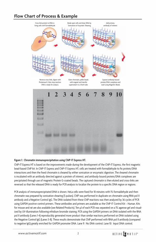
Figure 1: Chromatin immunoprecipitation using ChIP-IT Express HT.
ChIP-IT Express HT is based on the improvements made during the development of the ChIP-IT Express, the first magnetic bead-based ChIP kit. In ChIP-IT Express and ChIP-IT Express HT, cells are treated with formaldehyde to fix protein/DNA interactions and then the fixed chromatin is sheared by either sonication or enzymatic digestion. The sheared chromatin is incubated with an antibody directed against a protein of interest, and antibody-bound protein/DNA complexes are precipitated through use of magnetic Protein G-coated beads. The captured chromatin is then eluted and cross-links are reversed so that the released DNA is ready for PCR analysis to localize the protein to a specific DNA region or regions.
PCR analysis of immunoprecipitated DNA is shown. HeLa cells were fixed for 10 minutes with 1% formaldehyde and then chromatin was prepared by sonication shearing (5 pulses). ChIP was performed in duplicate on chromatin using RNA pol II antibody and a Negative Control IgG. The DNA isolated from these ChIP reactions was then analyzed by 36 cycles of PCR using GAPDH positive control primers. These antibodies and primers are available as the ChIP-IT Control Kit – Human. Kits for mouse and rat are also available (see Related Products). Ten µl of each PCR was separated on a 1% agarose gel and visual-ized by UV-illumination following ethidium bromide staining. PCR using the GAPDH primers on DNA isolated with the RNA pol II antibody (Lanes 1-4) reproducibly generated more product than similar reactions performed on DNA isolated using the Negative Control IgG (Lanes 5-8). These results demonstrate that ChIP performed with RNA pol II antibody (compared to negative IgG) greatly enriched for GAPDH promoter DNA. Lane 9: No DNA control. Lane 10: Input DNA control.
3www.activemotif.com
ChIP-IT Express HT Advantages
• Process up to 96 ChIP reactions simultaneously
• Faster plate-based protocol
• Fewer cells required per ChIP
• Compatible with ChIP-chip and ChIP-seq methodologies
The ChIP-IT Express HT Kit contains sufficient components to perform 96 ChIP reactions. The included LSV (Low Sample Volume) Protein G-coated magnetic beads are provided ready-to-use. These beads have a high binding capacity for IgG, enabling them to be used in smaller volumes that reduce non-specific binding. Because of this lower non-specific binding, magnetic beads require fewer washing steps than agarose beads, and it is not necessary to pre-clear the chromatin or to block the beads. An added advantage is that the magnetic beads pellet much more quickly than agarose beads, which are pelleted by centrifugation. In addition, magnetic stands are designed to pellet the beads onto the side of the tube. This makes it easy to remove buffers without disturbing the beads, so washing can be performed using multi-channel pipettors. This dramatically reduces hands-on time and improves sample-to-sample consistency. The LSV Protein G-coated magnetic beads are also optimized for use in the small sample size of a microplate well.
ChIP-IT Express HT is based on the improvements made during the development of the ChIP-IT Express, the first magnetic bead-based ChIP kit. This means that many of the steps in the ChIP protocol have been optimized for efficiency. The specialized ChIP Elution Buffer, coupled with a reagent to inactivate Proteinase K, eliminates the need for DNA purification after the ChIP is complete. This saves time, minimizes manipulations and eliminates the DNA loss that can occur during purification.
These improvements enable ChIP-IT Express HT assays to be performed rapidly. As the hands-on time is greatly reduced, it is possible to perform 24, 48 or 96 ChIP experiments at the same time. This is not possible with traditional ChIP methods, which are time- and manipulation-intensive.
Because appropriate controls make performing, troubleshooting and interpreting ChIP much easier, Active Motif sells ChIP-IT Control Kits for human, mouse and rat samples. These useful kits contain positive and negative control antibodies, appropriate positive control PCR primers, PCR buffer and loading dye (see Related Products in Appendix).
Another product designed to make your time more productive is Ready-to-ChIP HeLa Chromatin. High-quality chromatin from HeLa, Hep G2, K-562 and NIH/3T3 cells has been sheared by sonica-tion, and is ready for use in ChIP.
4www.activemotif.com
Kit Performance
The ChIP-IT Express HT Kit provides reagents and protocols to perform chromatin IP experiments in a 96-well microplate-based format. The ChIP-IT Express HT Kit can be used in conjunction with Active Motif’s Chromatin Shearing Kits, which are used to prepare chromatin after first determining the optimal conditions for shearing chromatin from a particular cell line. Although not included in this kit, a brief discussion of the reagents provided in the shearing kits is included below for your reference.
Preparation of chromatin – Sonication ShearingThe ChIP-IT Express Shearing Kit (Catalog No. 53032) includes all buffers (excluding formaldehyde) required for cell fixation, nuclei purification and chromatin shearing by sonication. A Glycine “Stop-Fix” Buffer is included to prevent excessive cross-linking and a Protease Inhibitor Cocktail (PIC) and PMSF are included to ensure that the protein/DNA interactions are preserved during the chromatin purification and immunoprecipitation steps.
Preparation of chromatin – Enzymatic DigestionThe ChIP-IT Express Enzymatic Shearing Kit (Catalog No. 53035) is similar to the ChIP-IT Express Shearing Kit, except that it uses a proprietary Enzymatic Shearing Cocktail and Digestion Buffer to enable enzymatic shearing, rather than sonication shearing. Enzymatic shearing is easily controlled by time and temperature to yield fragments that are ideal for performing ChIP. The ChIP-IT Express Enzymatic Shearing Kit replaces sonication shearing, thus eliminating problems due to variability in sonication power as well as complications arising from the emulsification of chromatin during sonication.
Performing ChIP in a microplate-based high-throughput formatThe protocol for ChIP-IT Express HT is based on that of ChIP-IT Express, modified for efficiency using a 96-well microplate instead of Eppendorf tubes. This does not mean that you need to perform all 96 reactions at once. You may find it convenient to perform 24 or 48 reactions at once. In this case, simply cap the unused wells with the supplied capstrips before working in the other wells to avoid contamination during the ChIP procedure and save the plate for later use. Also, it is likely that you will wish to incorporate the use of a multi-channel pipettor and reagent reservoir. The Thermo Fisher MatrixTech 25 ml Reagent Reservoir (part no. 8094) is a good choice, as it allows for minimal solution waste. Be careful not to use an excess of the supplied reagents if you are not performing all 96 ChIP reactions at one time, or you may not have sufficient reagents for subsequent experiments.
You will also need to have a 96-well magnetic sorter, such as Active Motif’s MAG-96 (Catalog No. 90096), Ambion’s Magnetic Stand-96 or the Novagen Magnetight HT96.
5www.activemotif.com
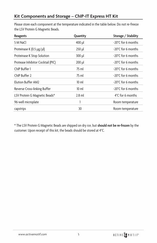
Kit Components and Storage – ChIP-IT Express HT Kit
Please store each component at the temperature indicated in the table below. Do not re-freeze the LSV Protein G Magnetic Beads.
Reagents Quantity Storage / Stability
5 M NaCl 400 µl -20°C for 6 months
Proteinase K (0.5 µg/µl) 250 µl -20°C for 6 months
Proteinase K Stop Solution 300 µl -20°C for 6 months
Protease Inhibitor Cocktail (PIC) 200 µl -20°C for 6 months
ChIP Buffer 1 75 ml -20°C for 6 months
ChIP Buffer 2 75 ml -20°C for 6 months
Elution Buffer AM2 10 ml -20°C for 6 months
Reverse Cross-linking Buffer 10 ml -20°C for 6 months
LSV Protein G Magnetic Beads* 2.8 ml 4°C for 6 months
96-well microplate 1 Room temperature
capstrips 30 Room temperature
* The LSV Protein G Magnetic Beads are shipped on dry ice, but should not be re-frozen by the customer. Upon receipt of this kit, the beads should be stored at 4ºC.
6www.activemotif.com
Additional materials required• A ChIP-validated antibody directed against the protein of interest
• 96-well magnetic sorter (e.g. Active Motif’s MAG-96 (Catalog No. 90096) Ambion’s Magnetic Stand-96 or the Novagen Magnetight HT96).
• 37% Formaldehyde (Fixation)
• Glycogen, 20 mg/ml (Purification of Input DNA)
• Phenol/chloroform (Purification of Input DNA and purification of sheared DNA prior to checking concentration by spectrophotometry or gel electrophoresis)
• 3 M Sodium Acetate pH 5.2 (Purification of Input DNA and purification of sheared DNA prior to checking concentration by spectrophotometry or gel electrophoresis)
• 100% ethanol (Purification of Input DNA and purification of sheared DNA prior to checking concentration by spectrophotometry or gel electrophoresis)
• 70% ethanol (Purification of Input DNA and purification of sheared DNA prior to checking concentration by spectrophotometry or gel electrophoresis)
• DNase-free H2O (Purification of Input DNA)
• PCR strip tubes and caps (e.g. Thermo Fisher part no. AB-0451 for Purification of DNA from chromatin after elution)
• Rocking platform for culture plates
• Apparatus to rotate tubes end-to-end at 4°C (e.g. a Labquake from Barnstead/Thermolyne)
• Microcentrifuge and microcentrifuge tubes
• Microplate centrifuge
• Spectrophotometer
• Pipettors and tips (a multi-channel pipettor and filter tips are recommended)
• Reagent reservoir (e.g. Thermo Fisher MatrixTech 25 ml Reagent Reservoir part no. 8094)
• Agarose gel electrophoresis apparatus
• Minimal cell culture media
• Cell scraper (rubber policeman)
• 10 ml pipette, aspirator and 100 ml graduated cylinder
Optional materials• Chromatin IP DNA Purification Kit (e.g. Active Motif Catalog No. 58002) .
7www.activemotif.com
ChIP-IT Express HT Experimental Design
PLEASE READ THE ENTIRE PROTOCOL BEFORE STARTING!
Points to consider:• Cell growth and chromatin preparation. This kit contains Protein G-coated magnetic beads
and other reagents sufficient to perform a total of 96 ChIP reactions. Chromatin can be prepared for use in ChIP through sonication by using Active Motif’s ChIP-IT Express Shear-ing Kit (Catalog No. 53032) or by enzymatic digestion with the ChIP-IT Express Enzymatic Shearing Kit (Catalog No. 53035). ChIP-IT Express HT is also compatible for use with Active Motif’s Ready-to-ChIP Chromatin. Before starting an experiment, calculate the number of chromatin preparations you require and determine the number of ChIP reactions you plan to perform on each chromatin preparation. Be sure to include the appropriate control ChIP reactions in your calculations. Also, note that if you wish to analyze the effect of particular compounds or culturing conditions on transcription factor/DNA interactions, you should prepare chromatin from control (untreated) cells as a reference sample. See Troubleshooting in the Appendix for comments regarding chromatin yield and the amount of chromatin used for each ChIP reaction.
• Shearing by Sonication. Chromatin sheared to a size of 200-1500 bp is usually used for ChIP experiments. You may wish to optimize shearing conditions using chromatin that is not intended for use in ChIP. In general, shearing efficiency is improved through the use of a small shearing volume and a V-bottom tube rather than a round-bottom tube. Also, note that shearing is inefficient if the chromatin sample becomes emulsified. This can be avoided by using lower shearing power and by turning the power on gradually. If a shearing reaction is allowed to emulsify, discontinue shearing and centrifuge the sample at 4ºC for 4 minutes at 8000 rpm in a microcentrifuge to remove trapped air. Finally, to prevent overheating and denaturation of chromatin, samples should be kept on ice as much as possible during shearing, and shearing should be performed discontinuously (i.e. sonicate for 15 seconds, then place on ice for 30 seconds, sonicate again for 15 seconds, etc.).
• Shearing by Enzymatic Digestion. While sonication is the most common method used to shear chromatin for ChIP, in some cases (e.g. when the sample is limited, when nucleosome ChIP is to be performed, when a sonicator is not available, or for high-throughput applica-tions) enzymatic shearing is an excellent alternative. To provide a robust and user-friendly enzymatic shearing method, Active Motif offers the ChIP-IT Express Enzymatic Shearing Kit, which uses a proprietary Enzymatic Shearing Cocktail to quickly digest the chromatin DNA to 200-1500 bp fragments. Enzymatic activity can be easily controlled by time and temperature.
• LSV Protein G-coated magnetic beads. The supplied LSV beads are optimized for use in the small volumes of a 96-well plate and are ready to use following complete resuspension to a homogeneous slurry. There is no need to pre-block the beads or pre-clear the sample. For best results, gently shake and roll the tube. The beads settle quickly, thus resuspend just before pipetting. The LSV Protein G Magnetic Beads are shipped on dry ice but should not be re-frozen by the customer. Upon receipt, the beads should be stored at 4ºC.
8www.activemotif.com
• Antibodies must be suitable for ChIP. ChIP antibodies must recognize fixed, native protein that is bound to DNA and/or complexed with other proteins. Many antibodies that perform well in other applications do not perform in ChIP. Thus, ChIP performed with an unproven antibody must include appropriate controls (such as Active Motif’s RNA pol II antibody, Catalog No. 39097) to demonstrate that the antibody and the prepared chromatin are appropriate for ChIP. For your convenience, Active Motif sells ChIP-IT Control Kits for human, mouse and rat samples; these kits contain positive and negative control antibodies, appropri-ate positive PCR primers, PCR buffer and loading dye (see Related Products in Appendix).
• Perform ChIP-IT Express HT in the provided 96-well plate. You will need a 96-well mag-netic sorter, such as Active Motif’s MAG-96 (Catalog No. 90096), Ambion’s Magnetic Stand-96 or Novagen’s Magnetight HT96, to perform many of the steps in the ChIP-IT Express HT protocol. First-time users of magnetic beads in the PCR tube format should familiarize themselves with the manipulations before performing a ChIP.
• PCR analysis of immunoprecipitated DNA. A successful ChIP results in an enrichment of chromatin fragments that are bound by the protein of interest, not complete purification. Thus, DNA isolated by ChIP is unavoidably contaminated with non-specifically captured DNA. For this reason, Real-Time Quantitative PCR analysis is preferred. If this method is not available, PCR of ChIP DNA should be performed such that cycling is stopped while the reaction is still in the linear stage of amplification. Hot-start PCR methods are recommended to ensure consistent PCR amplification results.
If you intend to analyze the binding of a known protein to a known binding site, design the PCR primers so that they flank the binding site and generate a 100-250 bp amplicon. Alternatively, if you hope to identify a protein binding site within a region of DNA, it may be best to design several primer pairs so that the DNA region in question can be systematically analyzed. In this case, design a series of primer pairs that can be used to generate amplicons that overlap one another and span the region of interest. To facilitate this, the amplicons can be 250-400 bp in length. After these primer pairs have been used to roughly localize the binding site, design a more focused set of primers. Use of PCR design programs can be help-ful in selecting good primer pairs. Also, as PCR analysis is extremely sensitive, precautions against contamination should be taken throughout the entire ChIP protocol.
• PCR primers. PCR primers should efficiently and specifically amplify the desired target. This should be proven on a relevant template, such as genomic or Input DNA. In addition, negative control primers can be useful to control for DNA shearing efficiency and to map putative protein binding sites. See Troubleshooting for discussion.
• Maximum volume of chromatin. Chromatin shearing (sonication) buffers usually contain detergents (e.g. 0.1% SDS and 0.5% sodium deoxycholate is typical). If you plan to use more than 60 µl sonicated chromatin in a ChIP reaction, use the 200 µl reaction volume from Table 1 (page 10). This will ensure that the detergent in the shearing buffer does not interfere with antibody activity. This is not a concern with chromatin prepared by the Active Motif ChIP-IT Express Enzymatic Shearing Kit because the relevant buffers contain little or no detergent.
9www.activemotif.com
• Resuspend solutions completely. Thaw the PIC and the Proteinase K Stop Solution at room temperature until fully dissolved. Vortex gently and spin down briefly before use.
• Quantity of antibody. Best results are typically obtained by use of 1-3 µg antibody. However, this will vary according to the activity of the antibody and the quality of the chromatin, and you may need to use more of a particular antibody.
• Stopping points in the protocol. Convenient stopping points are mentioned in the Troubleshooting section in the Appendix.
• Safety precautions. Formaldehyde is a highly toxic chemical. Appropriate safety precautions (i.e. safety glasses, gloves and lab coat) should be used. Also, formaldehyde is highly toxic by inhalation and should be used only in a ventilated hood.
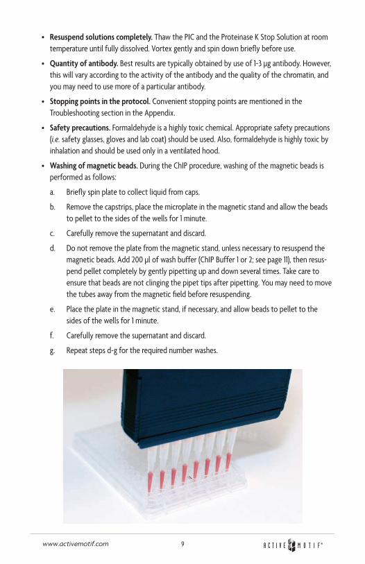
• Washing of magnetic beads. During the ChIP procedure, washing of the magnetic beads is performed as follows:
a. Briefly spin plate to collect liquid from caps.
b. Remove the capstrips, place the microplate in the magnetic stand and allow the beads to pellet to the sides of the wells for 1 minute.
c. Carefully remove the supernatant and discard.
d. Do not remove the plate from the magnetic stand, unless necessary to resuspend the magnetic beads. Add 200 µl of wash buffer (ChIP Buffer 1 or 2; see page 11), then resus-pend pellet completely by gently pipetting up and down several times. Take care to ensure that beads are not clinging the pipet tips after pipetting. You may need to move the tubes away from the magnetic field before resuspending.
e. Place the plate in the magnetic stand, if necessary, and allow beads to pellet to the sides of the wells for 1 minute.
f. Carefully remove the supernatant and discard.
g. Repeat steps d-g for the required number washes.
10www.activemotif.com
Protocols – ChIP-IT Express HT Assay
A. Immunoprecipitation
1. Thaw chromatin samples (if necessary). Transfer 10 µl of each to a microcentrifuge tube; this tube is the “Input DNA” that will be processed in Step C6. It will then be used as a control in PCR analysis. Store this reserved chromatin at 4ºC if it will be used within 6 hours; otherwise, store at -20ºC.
2. Set up the ChIP reactions by adding the components shown in Table 1 below to the provided 96-well plate. If you are not setting up 96 ChIPs, cap the unused wells with the supplied capstrips before beginning to avoid contamination, then save the plate for later use. Before pipetting the magnetic beads, they should be resuspended by inverting and/or vortexing the bottle. The LSV beads should be the first component added to the reaction; add the anti-body last. If you are using Active Motif’s Ready-to-ChIP Chromatin, use 25 µl per reaction.
Note: You can make a master mix of all the components without the magnetic beads and antibody, which need to be added to the wells first and last. You may find it convenient to use a multi-channel pipette reservoir such as the Thermo Fisher MatrixTech 25 ml Reagent Reservoir (part no. 8094). If you use this procedure for less than all 96 ChIP reactions, be careful not to use an excess of the supplied reagents, or you may run out of some components in subsequent ChIPs.
Table 1 One reaction One reaction (if using less than (if using more than Reagent 60 µl of chromatin) 60 µl of chromatin)
LSV Protein G Magnetic Beads 25 µl 25 µl
ChIP Buffer 1 10 µl 20 µl
Sheared Chromatin (~7 µg)* 20-60 µl 61-100 µl
Protease Inhibitor Cocktail (PIC) 1 µl 1 µl
dH20 Add enough so that the final Add enough so that the final
reaction volume will be 100 µl reaction volume will be 200 µl
Antibody (added last) 1-3 µg 1-3 µg
Total Volume 100 µl 200 µl
*Note: If you used one of our shearing kits using a 15 cm plate and did not quantify the chromatin, 25 µl will contain ~7 µg of chromatin. Depending on the application, ChIP can be performed using anywhere from 1-50 µg of chromatin. An important factor is the volume of the chromatin being added, especially if the chromatin was prepared using sonication. The detergents used during sonication will impact ChIP. Use the 200 µl ChIP in the right column above if the volume of chromatin will be greater than 60 µl. See Troubleshooting in the Appendix for discussions on the amount of chromatin to use and for methods to quantify chromatin.
11www.activemotif.com
3. Mix thoroughly by gentle pipetting and cap wells with the supplied capstrips.
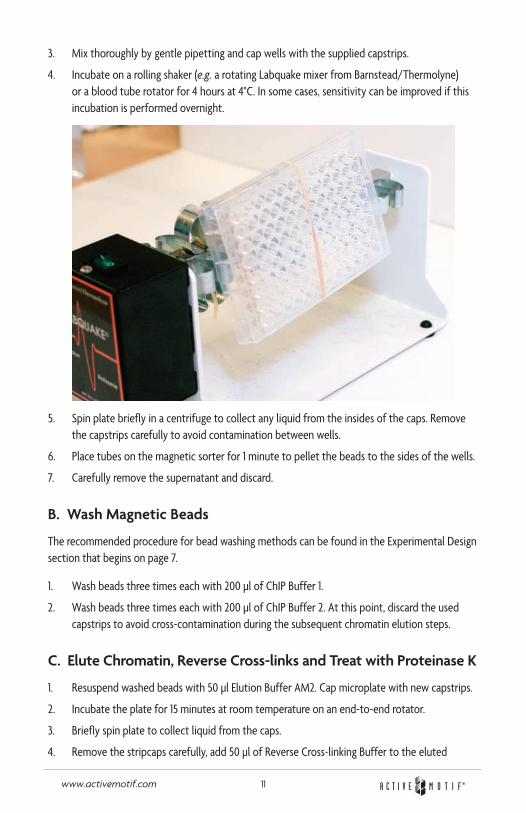
4. Incubate on a rolling shaker (e.g. a rotating Labquake mixer from Barnstead/Thermolyne) or a blood tube rotator for 4 hours at 4°C. In some cases, sensitivity can be improved if this incubation is performed overnight.
5. Spin plate briefly in a centrifuge to collect any liquid from the insides of the caps. Remove the capstrips carefully to avoid contamination between wells.
6. Place tubes on the magnetic sorter for 1 minute to pellet the beads to the sides of the wells.
7. Carefully remove the supernatant and discard.
B. Wash Magnetic Beads
The recommended procedure for bead washing methods can be found in the Experimental Design section that begins on page 7.
1. Wash beads three times each with 200 µl of ChIP Buffer 1.
2. Wash beads three times each with 200 µl of ChIP Buffer 2. At this point, discard the used capstrips to avoid cross-contamination during the subsequent chromatin elution steps.
C. Elute Chromatin, Reverse Cross-links and Treat with Proteinase K
1. Resuspend washed beads with 50 µl Elution Buffer AM2. Cap microplate with new capstrips.
2. Incubate the plate for 15 minutes at room temperature on an end-to-end rotator.
3. Briefly spin plate to collect liquid from the caps.
4. Remove the stripcaps carefully, add 50 µl of Reverse Cross-linking Buffer to the eluted
12www.activemotif.com
chromatin, then immediately place plate on magnetic sorter and allow beads to pellet to sides of wells for 1 minute.
5. Quickly transfer the supernatant, which contains the chromatin, to PCR strip tubes.
6. “Input DNA” sample: take the 10 µl Input DNA aliquot (that was reserved in Step C1 above) from the ice. Add 88 µl ChIP Buffer 2 and 2 µl 5M NaCl to the Input DNA sample only, so that its final volume is 100 µl.
7. Incubate the ChIP and Input DNA samples at 95°C for 15 minutes in a thermocycler.
Note: If you are using larger microcentrifuge tubes, it may be easier to perform a 2.5 hour incubation at 65°C.
8. Return tubes to room temperature, spin tubes briefly if liquid has collected on the inside of the caps, then add 2 µl Proteinase K.
9. Cap tubes, mix well and incubate at 37°C for 1 hour.
10. Return the tubes to room temperature and add 2 µl Proteinase K Stop Solution. Briefly centrifuge the tubes to collect liquid from the caps. DNA can be used immediately in PCR or stored at -20°C.
11. For PCR analysis, we recommend to use 5 µl of immunoprecipitated DNA in a 25 µl PCR reaction. Input DNA should be diluted 10 fold before use in PCR. (See Appendix – Section B for recommendations for PCR.) For ChIP-chip or ChIP-seq, additional DNA cleanup may be required.
References
1. Solomon, M.J. et al. (1988) Cell 53(6): 937-47.2. Solomon, M.J. and Varshavsky A. (1985) PNAS USA 82(19): 6470-4.3. Kuo, M.H. and Allis, C.D. (1999) Methods 19(3): 425-33.4. Weinman, A.S. and Farnham, P.J. (2002) Methods 26: 37-47.5. Caretti, G. et al. (2003) J Biological Chem. 278: 30435-30440.
Appendix
Section A. Troubleshooting Guide
Problem/question Recommendation
At what points in the protocol can I stop?
The protocol can be paused and samples stored at -20°C at any of the following steps:1. After bead washing2. After the cross-link reversal3. After DNA clean up
How much sheared chromatin should I use for a ChIP reaction?
Best results are obtained when the amount of chromatin used in ChIP is from 1-3 x 10⁶ cells. If it is assumed that human diploid cells contain 6.6 picograms of DNA and recovery after cell fixation and shearing is 70%, then this is between 4.6 and 13.9 µg DNA. The mini-mum amount of sheared chromatin that can be used is 1 µg while the maximum is 50 µg.
Preparing a ChIP reaction with a large volume
It is better to set up several small ChIP reactions (200 µl each) and pool the samples at the end, rather than trying to ChIP a single large sample.
13www.activemotif.com
Problem/question Recommendation
Poor enrichment with ChIP antibody
In some cases, use of an antibody in ChIP results in lower-than-expected enrichment of a target of interest. This is often because the antibody does not efficiently recognize fixed proteins, either because the epitope is destroyed by fixation or because the epitope is masked by other proteins in a larger complex. In this case, use more antibody when performing the ChIP. Alternatively, try to find an antibody that has been proven to work in ChIP, or that is known to recognize an epitope distinct from the one recognized by the unsatisfactory antibody.
There is no difference in band intensity between negative control and positive control.
See the recommendation below regarding increasing washing stringency.
Decrease the number of PCR cycles (30, 32, 34 PCR cycles). The exponential phase of amplification occurs in PCR cycles where reaction components are still in excess and PCR products are accumulating at a constant rate. During this phase, each copy of DNA is being actively amplified, making it a better measure than endpoint PCR. In endpoint PCR, reagents such as nucleotides or primers may become exhausted. This can result in inefficient amplification, which can cause inaccurate quantification of the gene of inter-est. Thus, high background due to endpoint PCR can be decreased if the number of PCR cycles are reduced, so the results reflect exponential PCR. Real-time PCR can also be used in such cases.
Shearing should produce DNA fragments that are small enough to exclude background from neighboring chromosomal sequences, but still large enough that there is a good pos-sibility your amplicon remains intact. We recommend 200-1500 bp fragments. If the DNA fragments are too large, you the background is increased. So, consider increasing the time of the enzymatic digestion, or the time and/or number of pulses for sonication.
Confirm species specificity of your primers. You may need to redesign your primers.
See the recommendation below regarding blocking the magnetic beads.
Strong PCR signal when using target PCR primers to amplify ChIP DNA that was isolated with a negative control (non-target) antibody.
In most cases, the washing procedure in the enclosed protocol is appropriate. However, when the background is high you can increase washing stringency in several ways:
1) After adding ChIP Buffer 1 and/or ChIP Buffer 2 during the wash steps, gently agitate the samples for several minutes before removing the buffer.
2) Perform additional washes. Sufficient ChIP Buffer 1 is provided for two “extra” washes per sample. Sufficient ChIP Buffer 2 is provided for one additional wash.
3) Add two washes using a high-salt buffer (20 mM Tris-Cl, 1 mM EDTA, 0.1% SDS, 1% Triton X-100, 500 mM NaCl, pH 7.4), which is not provided. These additional washes should be performed after the washes with ChIP Buffer 1. Then, proceed with the ChIP Buffer 2 washes, as outlined in the protocol.
Confirm the species specificity of your primers. You may need to redesign your primers.
See the next recommendation regarding blocking the magnetic beads.
Is blocking of the magnetic beads ever required?
The beads provided are ready to use for most ChIPs. However, for applications highly sensitive to non-specific binding (such as when cloning ChIP DNA or using antibodies that require extra blocking), you may add blocking reagents to the ChIP reaction. In these cases, a combination of BSA (e.g. Sigma Cat. No. 4503) and either tRNA (e.g. Sigma Cat. No. R3629) or salmon sperm DNA (e.g. Sigma Cat. No. A-7888) can be added directly to the ChIP reaction. 2.5 µg/µl BSA and 1.25 µg/µl tRNA or 2.5 µg/µl Salmon sperm DNA (final concentrations) can be used as a starting point and more or less can be added as desired.
The PCR products are the correct size, but are very light.
Load more PCR product, and/or use smaller wells for the agarose gel. It should be noted that because the PCRs should be stopped while the reactions are in the linear phase of amplification, the yield of PCR product will be lower than in typical PCR amplifications, which are performed for maximum product yield. You can also perform more PCR cycles.
No PCR products with Real-time PCR.
The DNA should be purified before performing real-time PCR. We recommend Active Motif’s Chromatin IP DNA Purification Kit (Catalog No. 58002) prior to amplification. Its columns yield 50 µl; 2 µl is used for each PCR, providing enough DNA for 25 PCR reactions.
14www.activemotif.com
Problem/question Recommendation
No PCR bands for Input DNA or ChIP’d samples.
In the presence of 0.8 mM total dNTP concentration, perform a MgCl2 titration series in
0.5 mM increments over a range of 1-4 mM. This will identify the magnesium ion concen-tration that produces the highest yield of a specific PCR product. When using Taq DNA Polymerase, too little free magnesium ion results in little or no PCR product, while excess free magnesium ion can cause unwanted products and promote misincorporation.
Confirm species specificity of your primers. You may need to redesign your primers.
How do I design PCR primers to analyze shearing efficiency and to map putative DNA-binding sites?
Negative control PCR primers can be used to demonstrate that chromatin was sufficiently sheared. For example, negative control primers can be designed to amplify a DNA fragment that is 2 kb away from the “Target DNA” (the region bound by the protein of interest). Following ChIP reactions (performed with antibody against the protein of interest and with a negative IgG), PCR is performed with the negative control primers and with primers that amplify the Target DNA. The PCR should show that the anti-protein-of-interest ChIP enriches for Target DNA, but not for the negative control DNA. This result would support the conclusion that the enrichment was due to protein binding to (or near) the putative Target, and not due to binding elsewhere on a very large (poorly sheared) chromatin fragment.
Similarly, appropriately designed PCR primers can be used to roughly map the DNA Target of the protein of interest. For example, primers can be designed to amplify short (approximately 100 bp) DNA fragments that are progressively closer to the putative Target DNA (e.g. within 1.5 kb, 1 kb, 500 bp, 250 bp). This type of analysis can help confirm the exact binding site of the protein of interest. For such higher-resolution mapping, the chromatin must be extensively sheared (DNA fragment size should be less than 500 bp).
Section B. PCR Analysis
The protocol below is a guideline for optimizing PCR analysis of DNA collected through ChIP. Accurate PCR analysis of ChIP DNA requires that the PCR be stopped during linear amplification. The appropriate number of PCR cycles must be determined empirically. In the example below, PCR is performed on four DNA templates: DNA from ChIP with the positive control RNA pol II antibody and the Negative Control IgG (that are included in the ChIP-IT Control Kit – Human), the Input DNA and DNA from ChIP with the test antibody. A water-only control PCR is also performed to ensure that the PCR reagents are not contaminated.
In the example below, PCR reactions are set up using 2 different PCR cocktails, which contain positive & negative PCR primer sets. If you are using Active Motif’s ChIP-IT Control Kit – Human, only GAPDH positive control primers would be used for the first DNA template because the posi-tive control RNA pol II antibody can bind many regions along a chromosome, making it difficult to design “negative control” primers that function with all cell types and shearing conditions. For PCR analysis of ChIP performed with other antibodies, we recommend that you use both positive and negative PCR primer sets that are appropriate for your antibody. See the Experimental Design section that begins on page 7 and Troubleshooting in the Appendix for details.
Note: PCR is extremely sensitive and all precautions should be taken to guard against contamination. Gloves should be worn and filter-tip pipettes should be used.
1. Program the thermocycler. The program should start with a initial melt step at 94°C for 3 minutes, then 36 cycles of [94°C for 20 seconds, 59°C for 30 seconds and 72°C for
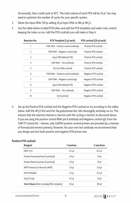
15www.activemotif.com
30 seconds], then a hold cycle at 10°C. The total volume of each PCR will be 25 µl. You may need to optimize the number of cycles for your specific system.
2. Dilute the Input DNA 1:10 by adding 20 µl Input DNA to 180 µl dH2O.
3. Use the table below to label PCR tubes and add the PCR templates and water-only control, keeping the tubes on ice. Add the PCR cocktails you will make in Step 4:
Reaction No. PCR Template (5 µl each) PCR cocktail (20 µl each)
1 ChIP DNA – Positive control antibody Positive PCR cocktail
2 ChIP DNA – Negative control IgG Positive PCR cocktail
3 Input DNA (diluted 1:10) Positive PCR cocktail
4 ChIP DNA – Test antibody Positive PCR cocktail
5 H2O (no DNA control) Positive PCR cocktail
6 ChIP DNA – Positive control antibody Negative PCR cocktail
7 ChIP DNA – Negative control IgG Negative PCR cocktail
8 Input DNA (diluted 1:10) Negative PCR cocktail
9 ChIP DNA – Test antibody Negative PCR cocktail
10 H2O (control) Negative PCR cocktail
4. Set up the Positive PCR cocktail and the Negative PCR cocktail on ice according to the tables below. Add the dH
2O first and the Taq polymerase last. Mix thoroughly and keep on ice. This
ensures that the reaction mixture is inactive until the cycling is started. As discussed above, if you are using the positive control RNA pol II antibody and Negative control IgG from the ChIP-IT Control Kit – Human, only GAPDH positive control primers are provided (as a mixture of forward and reverse primers). However, for your own test antibody we recommend that you design and test both positive and negative PCR primer sets.
Positive PCR cocktail:Reagent 1 reaction 5 reactions
DEPC H2O 12.3 µl 61.5 µl
Positive Forward primer (5 pmol/µl) 2.0 µl 10 µl
Positive Reverse primer (5 pmol/µl) 2.0 µl 10 µl
dNTP mixture (5 mM each dNTP) 1.0 µl 5.0 µl
10X PCR Buffer 2.5 µl 12.5 µl
Taq (5 U/µl) 0.2 µl 1.0 µl
Total Volume (Not including DNA template) 20 µl 100 µl
16www.activemotif.com
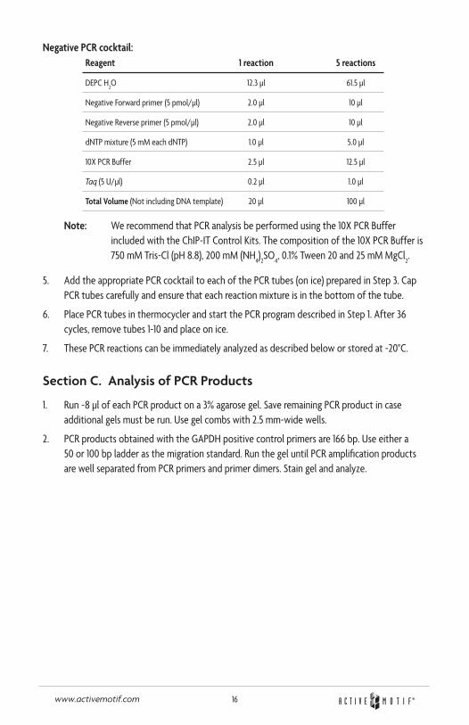
Negative PCR cocktail:Reagent 1 reaction 5 reactions
DEPC H2O 12.3 µl 61.5 µl
Negative Forward primer (5 pmol/µl) 2.0 µl 10 µl
Negative Reverse primer (5 pmol/µl) 2.0 µl 10 µl
dNTP mixture (5 mM each dNTP) 1.0 µl 5.0 µl
10X PCR Buffer 2.5 µl 12.5 µl
Taq (5 U/µl) 0.2 µl 1.0 µl
Total Volume (Not including DNA template) 20 µl 100 µl
Note: We recommend that PCR analysis be performed using the 10X PCR Buffer included with the ChIP-IT Control Kits. The composition of the 10X PCR Buffer is 750 mM Tris-Cl (pH 8.8), 200 mM (NH
4)2SO
4, 0.1% Tween 20 and 25 mM MgCl
2.
5. Add the appropriate PCR cocktail to each of the PCR tubes (on ice) prepared in Step 3. Cap PCR tubes carefully and ensure that each reaction mixture is in the bottom of the tube.
6. Place PCR tubes in thermocycler and start the PCR program described in Step 1. After 36 cycles, remove tubes 1-10 and place on ice.
7. These PCR reactions can be immediately analyzed as described below or stored at -20°C.
Section C. Analysis of PCR Products
1. Run ~8 µl of each PCR product on a 3% agarose gel. Save remaining PCR product in case additional gels must be run. Use gel combs with 2.5 mm-wide wells.
2. PCR products obtained with the GAPDH positive control primers are 166 bp. Use either a 50 or 100 bp ladder as the migration standard. Run the gel until PCR amplification products are well separated from PCR primers and primer dimers. Stain gel and analyze.
17www.activemotif.com
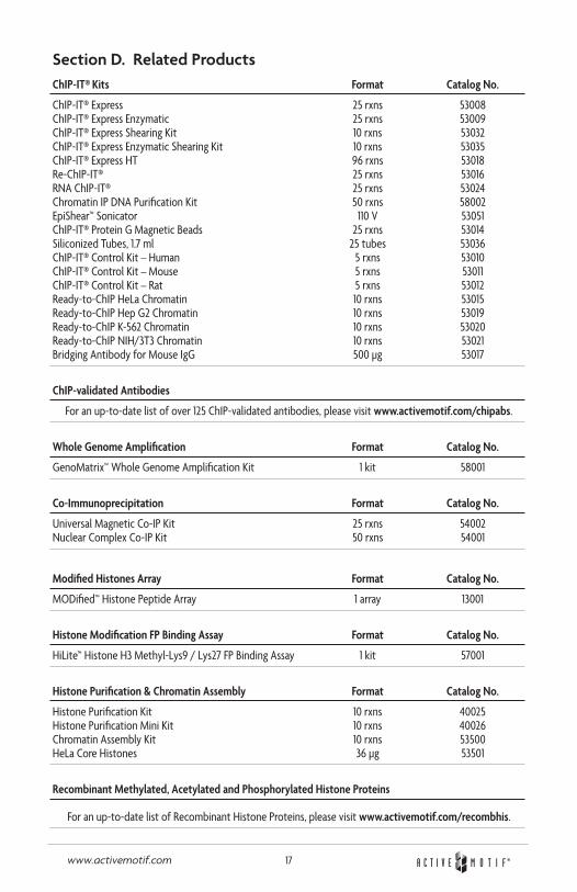
Section D. Related ProductsChIP-IT® Kits Format Catalog No.
ChIP-IT® Express 25 rxns 53008 ChIP-IT® Express Enzymatic 25 rxns 53009 ChIP-IT® Express Shearing Kit 10 rxns 53032 ChIP-IT® Express Enzymatic Shearing Kit 10 rxns 53035 ChIP-IT® Express HT 96 rxns 53018 Re-ChIP-IT® 25 rxns 53016 RNA ChIP-IT® 25 rxns 53024 Chromatin IP DNA Purification Kit 50 rxns 58002 EpiShear™ Sonicator 110 V 53051 ChIP-IT® Protein G Magnetic Beads 25 rxns 53014 Siliconized Tubes, 1.7 ml 25 tubes 53036 ChIP-IT® Control Kit – Human 5 rxns 53010 ChIP-IT® Control Kit – Mouse 5 rxns 53011 ChIP-IT® Control Kit – Rat 5 rxns 53012 Ready-to-ChIP HeLa Chromatin 10 rxns 53015 Ready-to-ChIP Hep G2 Chromatin 10 rxns 53019 Ready-to-ChIP K-562 Chromatin 10 rxns 53020 Ready-to-ChIP NIH/3T3 Chromatin 10 rxns 53021 Bridging Antibody for Mouse IgG 500 µg 53017
ChIP-validated Antibodies
For an up-to-date list of over 125 ChIP-validated antibodies, please visit www.activemotif.com/chipabs.
Whole Genome Amplification Format Catalog No.
GenoMatrix™ Whole Genome Amplification Kit 1 kit 58001
Co-Immunoprecipitation Format Catalog No.
Universal Magnetic Co-IP Kit 25 rxns 54002 Nuclear Complex Co-IP Kit 50 rxns 54001
Modified Histones Array Format Catalog No.
MODified™ Histone Peptide Array 1 array 13001
Histone Modification FP Binding Assay Format Catalog No.
HiLite™ Histone H3 Methyl-Lys9 / Lys27 FP Binding Assay 1 kit 57001
Histone Purification & Chromatin Assembly Format Catalog No.
Histone Purification Kit 10 rxns 40025 Histone Purification Mini Kit 10 rxns 40026 Chromatin Assembly Kit 10 rxns 53500 HeLa Core Histones 36 µg 53501
Recombinant Methylated, Acetylated and Phosphorylated Histone Proteins
For an up-to-date list of Recombinant Histone Proteins, please visit www.activemotif.com/recombhis.
18www.activemotif.com
Histone Acetyltransferase and Deacetylase Activity Format Catalog No.
HAT Assay Kit (Fluorescent) 1 x 96 rxns 56100 Recombinant p300 protein, catalytic domain 5 µg 31205 Recombinant GCN5 protein, active 5 µg 31204 HDAC Assay Kit (Fluorescent) 1 x 96 rxns 56200 HDAC Assay Kit (Colorimetric) 1 x 96 rxns 56210
Histone ELISAs Format Catalog No.
Histone H3 monomethyl Lys4 ELISA 1 x 96 rxns 53101 Histone H3 dimethyl Lys4 ELISA 1 x 96 rxns 53112 Histone H3 trimethyl Lys4 ELISA 1 x 96 rxns 53113 Histone H3 acetyl Lys9 ELISA 1 x 96 rxns 53114 Histone H3 dimethyl Lys9 ELISA 1 x 96 rxns 53108 Histone H3 trimethyl Lys9 ELISA 1 x 96 rxns 53109 Histone H3 phospho Ser10 ELISA 1 x 96 rxns 53111 Histone H3 acetyl Lys14 ELISA 1 x 96 rxns 53115 Histone H3 monomethyl Lys27 ELISA 1 x 96 rxns 53104 Histone H3 trimethyl Lys27 ELISA 1 x 96 rxns 53106 Histone H3 phospho Ser28 ELISA 1 x 96 rxns 53100 Total Histone H3 ELISA 1 x 96 rxns 53110
DNA Methylation Format Catalog No.
hMeDIP 10 rxns 55010 MeDIP 10 rxns 55009 MethylDetector™ 50 rxns 55001 MethylCollector™ 25 rxns 55002 MethylCollector™ Ultra 30 rxns 55005 UnMethylCollector™ 30 rxns 55004 DNMT Activity / Inhibition Assay 96 rxns 55006 Methylated DNA Standard Kit 3 x 2.5 µg 55008 Fully Methylated Jurkat DNA 10 µg 55003 Jurkat genomic DNA 10 µg 55007
Histone Demethylase Activity Format Catalog No.
Histone Demethylase Assay (Fluorescent) 48 rxns 53200
SUMOylation Format Catalog No.
SUMOlink™ SUMO-1 Kit 20 rxns 40120 SUMOlink™ SUMO-2/3 Kit 20 rxns 40220
Transcription Factor ELISAs Format Catalog No.
TransAM® AML-1/Runx1 1 x 96-well plate 47396 TransAM® AML-3/Runx2 1 x 96-well plate 44496 TransAM® AP-1 Family 2 x 96-well plates 44296 TransAM® AP-1 c-Fos 1 x 96-well plate 44096 TransAM® AP-1 c-Jun 1 x 96-well plate 46096 TransAM® AP-1 FosB 1 x 96-well plate 45096 TransAM® AP-1 JunD 1 x 96-well plate 43496 TransAM® ATF-2 1 x 96-well plate 42396 TransAM® c-Myc 1 x 96-well plate 43396 TransAM® C/EBP a/b 1 x 96-well plate 44196
19www.activemotif.com
Transcription Factor ELISAs (continued) Format Catalog No.
TransAM® CREB 1 x 96-well plate 42096 TransAM® pCREB 1 x 96-well plate 43096 TransAM® Elk-1 1 x 96-well plate 44396 TransAM® ER 1 x 96-well plate 41396 TransAM® FKHR (FOXO1/4) 1 x 96-well plate 46396 TransAM® GATA Family 2 x 96-well plates 48296 TransAM® GATA-4 1 x 96-well plate 46496 TransAM® GR 1 x 96-well plate 45496 TransAM® HIF-1 1 x 96-well plate 47096 TransAM® HNF Family 2 x 96-well plates 46296 TransAM® HNF-1 1 x 96-well plate 46196 TransAM® IRF-3 (Human) 1 x 96-well plate 48396 TransAM® IRF-3 (Mouse) 1 x 96-well plate 48496 TransAM® IRF-7 1 x 96-well plate 50196 TransAM® MAPK Family 2 x 96-well plates 47296 TransAM® MEF2 1 x 96-well plate 43196 TransAM® MyoD 1 x 96-well plate 47196 TransAM® NF-YA 1 x 96-well plate 40396 TransAM® NFATc1 1 x 96-well plate 40296 TransAM® NFkB Family 2 x 96-well plates 43296 TransAM® Flexi NFkB Family 2 x 96-well plates 43298 TransAM® NFkB p50 1 x 96-well plate 41096 TransAM® NFkB p50 Chemi 1 x 96-well plate 41097 TransAM® Flexi NFkB p50 1 x 96-well plate 41098 TransAM® NFkB p52 1 x 96-well plate 48196 TransAM® NFkB p52 Chemi 1 x 96-well plate 48197 TransAM® NFkB p65 1 x 96-well plate 40096 TransAM® NFkB p65 Chemi 1 x 96-well plate 40097 TransAM® Flexi NFkB p65 1 x 96-well plate 40098 TransAM® Nrf2 1 x 96-well plate 50296 TransAM® Oct-4 1 x 96-well plate 42496 TransAM® p53 1 x 96-well plate 41196 TransAM® PPARg 1 x 96-well plate 40196 TransAM® Sp1 1 x 96-well plate 41296 TransAM® Sp1/Sp3 1 x 96-well plate 40496 TransAM® STAT Family 2 x 96-well plates 42296 TransAM® STAT3 1 x 96-well plate 45196 TransAM® T-bet 1 x 96-well plate 51396
For a complete, up-to-date list of available TransAM® Kits, please visit www.activemotif.com/transam
20www.activemotif.com
Technical Services
If you need assistance at any time, please call Active Motif Technical Service at one of the numbers listed below.
Active Motif North America 1914 Palomar Oaks Way, Suite 150 Carlsbad, CA 92008 USA Toll Free: 877 222 9543 Telephone: 760 431 1263 Fax: 760 431 1351 E-mail: [email protected]
Active Motif Europe Avenue Reine Astrid, 92 B-1310 La Hulpe, Belgium UK Free Phone: 0800 169 31 47 France Free Phone: 0800 90 99 79 Germany Free Phone: 0800 181 99 10 Telephone: +32 (0)2 653 0001 Fax: +32 (0)2 653 0050 E-mail: [email protected]
Active Motif Japan Azuma Bldg, 7th Floor 2-21 Ageba-Cho, Shinjuku-Ku Tokyo, 162-0824, Japan Telephone: +81 3 5225 3638 Fax: +81 3 5261 8733 E-mail: [email protected]
Visit Active Motif on the worldwide web at http://www.activemotif.com
At this site:
• Read about who we are, where we are, and what we do
• Review data supporting our products and the latest updates
• Enter your name into our mailing list to receive our catalog, MotifVations newsletter and notification of our upcoming products
• Share your ideas and results with us
• View our job opportunities
Don’t forget to bookmark our site for easy reference!