Embed Size (px)
Citation preview
Handbook of
PEDIATRIC ANESTHESIA
2
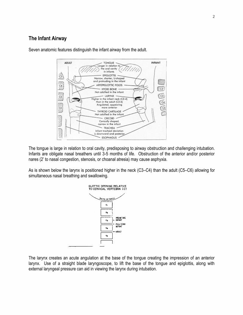
The Infant Airway Seven anatomic features distinguish the infant airway from the adult. The tongue is large in relation to oral cavity, predisposing to airway obstruction and challenging intubation. Infants are obligate nasal breathers until 3-5 months of life. Obstruction of the anterior and/or posterior nares (2’ to nasal congestion, stenosis, or choanal atresia) may cause asphyxia. As is shown below the larynx is positioned higher in the neck (C3–C4) than the adult (C5–C6) allowing for simultaneous nasal breathing and swallowing. The larynx creates an acute angulation at the base of the tongue creating the impression of an anterior larynx. Use of a straight blade laryngoscope, to lift the base of the tongue and epiglottis, along with external laryngeal pressure can aid in viewing the larynx during intubation.
3
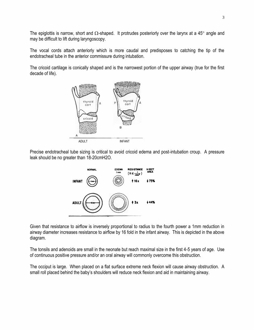
The epiglottis is narrow, short and -shaped. It protrudes posteriorly over the larynx at a 45 angle and may be difficult to lift during laryngoscopy. The vocal cords attach anteriorly which is more caudal and predisposes to catching the tip of the endotracheal tube in the anterior commissure during intubation. The cricoid cartilage is conically shaped and is the narrowest portion of the upper airway (true for the first decade of life).
ADULT INFANT
Precise endotracheal tube sizing is critical to avoid cricoid edema and post-intubation croup. A pressure leak should be no greater than 18-20cmH2O. Given that resistance to airflow is inversely proportional to radius to the fourth power a 1mm reduction in airway diameter increases resistance to airflow by 16 fold in the infant airway. This is depicted in the above diagram. The tonsils and adenoids are small in the neonate but reach maximal size in the first 4-5 years of age. Use of continuous positive pressure and/or an oral airway will commonly overcome this obstruction. The occiput is large. When placed on a flat surface extreme neck flexion will cause airway obstruction. A small roll placed behind the baby’s shoulders will reduce neck flexion and aid in maintaining airway.
4
Pediatric Respiratory Physiology Lower Airway
The alveolar bed is incompletely developed at birth; mature alveoli are seen at 5 weeks of age and alveolar multiplication with adult morphology being reached by eight years of life. Infant lung compliance is extremely high due to absence of elastic fibers (resembles the emphysematous lung). It is prone to airway collapse and premature airway closure secondary to low elastic recoil. The cartilaginous rib cage and poorly developed intercostal muscles results in a highly compliant chest wall – leading to inefficient ventilation. Circular configuration of the rib cage (ellipsoid in adult) and horizontally attached diaphragm (oblique in adult) leads to poor respiratory mechanics. Chest wall begins to stiffen at 6 months of age improving outward recoil of chest wall. The diaphragm has fewer Type I muscle fibers (sustained twitch, highly oxidative, fatigue resistant) and is susceptible to fatigue. Adult diaphragm contains 55%, neonate 25% and preterm only 10% Type I fibers. Lung Volumes
Functional residual capacity (FRC) in the spontaneously breathing infant is dynamically maintained at 40% of total lung capacity (similar to adults). The following mechanisms play a role in dynamically maintaining FRC in the awake infant:
1. Termination of the expiratory phase before the lung volume reaches FRC, “auto-PEEP” 2. Glottic closure during the expiratory phase (grunting) maintaining lung volumes 3. Diaphragmatic braking; diminished diaphragmatic activity extending to the expiratory phase 4. Tonic activity of the diaphragmatic and intercostal muscles stiffening the chest wall and maintaining
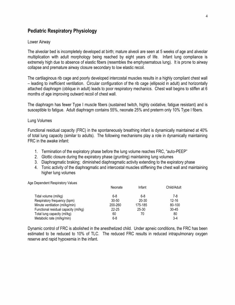
higher lung volumes Age Dependent Respiratory Values
Neonate Infant Child/Adult
Tidal volume (ml/kg) 6-8 6-8 7-8 Respiratory frequency (bpm) 30-50 20-30 12-16 Minute ventilation (ml/kg/min) 200-260 175-185 80-100 Functional residual capacity (ml/kg) 22-25 25-30 30-45 Total lung capacity (ml/kg) 60 70 80 Metabolic rate (ml/kg/min) 6-8 3-4
Dynamic control of FRC is abolished in the anesthetized child. Under apneic conditions, the FRC has been estimated to be reduced to 10% of TLC. The reduced FRC results in reduced intrapulmonary oxygen reserve and rapid hypoxemia in the infant.
5
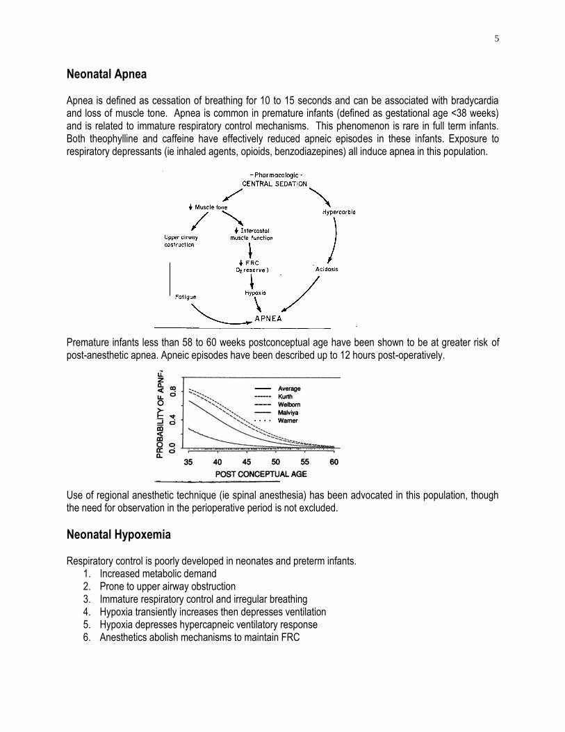
Neonatal Apnea
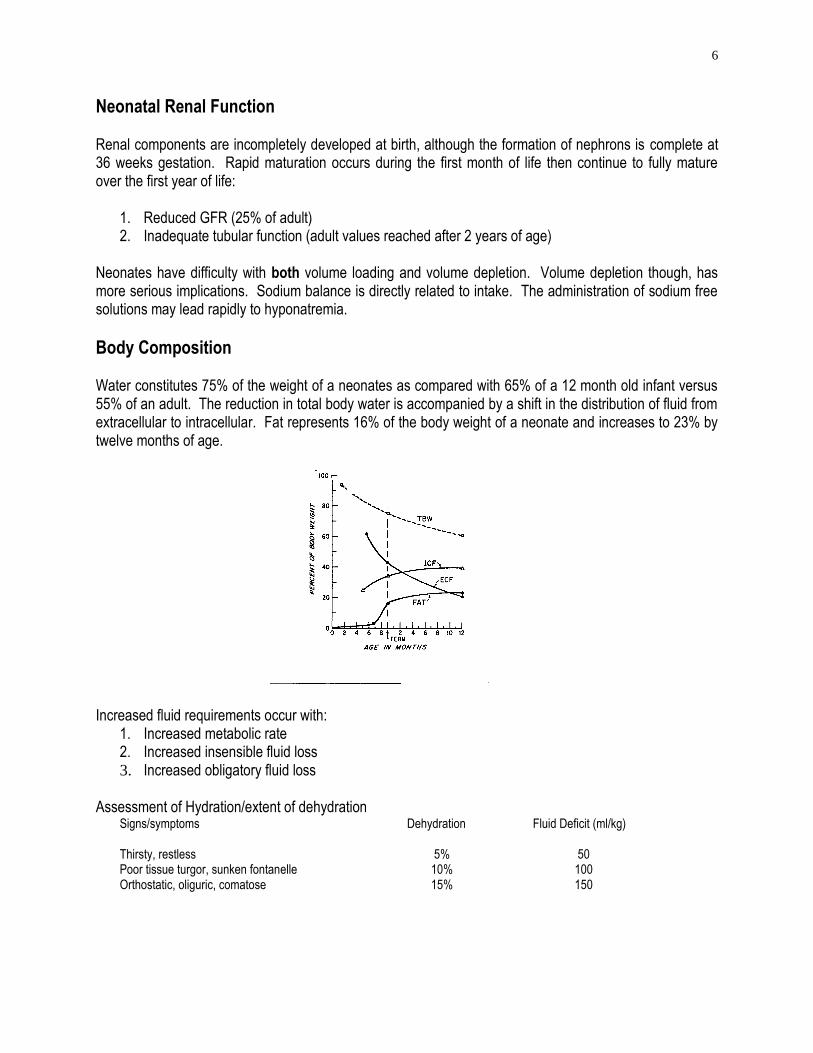
Apnea is defined as cessation of breathing for 10 to 15 seconds and can be associated with bradycardia and loss of muscle tone. Apnea is common in premature infants (defined as gestational age <38 weeks) and is related to immature respiratory control mechanisms. This phenomenon is rare in full term infants. Both theophylline and caffeine have effectively reduced apneic episodes in these infants. Exposure to respiratory depressants (ie inhaled agents, opioids, benzodiazepines) all induce apnea in this population. Premature infants less than 58 to 60 weeks postconceptual age have been shown to be at greater risk of post-anesthetic apnea. Apneic episodes have been described up to 12 hours post-operatively. Use of regional anesthetic technique (ie spinal anesthesia) has been advocated in this population, though the need for observation in the perioperative period is not excluded.
Neonatal Hypoxemia Respiratory control is poorly developed in neonates and preterm infants.
1. Increased metabolic demand 2. Prone to upper airway obstruction 3. Immature respiratory control and irregular breathing 4. Hypoxia transiently increases then depresses ventilation 5. Hypoxia depresses hypercapneic ventilatory response 6. Anesthetics abolish mechanisms to maintain FRC
6
Neonatal Renal Function Renal components are incompletely developed at birth, although the formation of nephrons is complete at 36 weeks gestation. Rapid maturation occurs during the first month of life then continue to fully mature over the first year of life:
1. Reduced GFR (25% of adult) 2. Inadequate tubular function (adult values reached after 2 years of age)
Neonates have difficulty with both volume loading and volume depletion. Volume depletion though, has more serious implications. Sodium balance is directly related to intake. The administration of sodium free solutions may lead rapidly to hyponatremia.
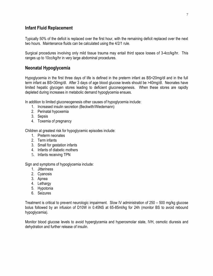
Body Composition Water constitutes 75% of the weight of a neonates as compared with 65% of a 12 month old infant versus 55% of an adult. The reduction in total body water is accompanied by a shift in the distribution of fluid from extracellular to intracellular. Fat represents 16% of the body weight of a neonate and increases to 23% by twelve months of age. Increased fluid requirements occur with:
1. Increased metabolic rate 2. Increased insensible fluid loss 3. Increased obligatory fluid loss
Assessment of Hydration/extent of dehydration
Signs/symptoms Dehydration Fluid Deficit (ml/kg) Thirsty, restless 5% 50 Poor tissue turgor, sunken fontanelle 10% 100 Orthostatic, oliguric, comatose 15% 150
7
Infant Fluid Replacement Typically 50% of the deficit is replaced over the first hour, with the remaining deficit replaced over the next two hours. Maintenance fluids can be calculated using the 4/2/1 rule. Surgical procedures involving only mild tissue trauma may entail third space losses of 3-4cc/kg/hr. This ranges up to 10cc/kg/hr in very large abdominal procedures.
Neonatal Hypoglycemia Hypoglycemia in the first three days of life is defined in the preterm infant as BS<20mg/dl and in the full term infant as BS<30mg/dl. After 3 days of age blood glucose levels should be >40mg/dl. Neonates have limited hepatic glycogen stores leading to deficient gluconeogenesis. When these stores are rapidly depleted during increases in metabolic demand hypoglycemia ensues. In addition to limited gluconeogenesis other causes of hypoglycemia include:
1. Increased insulin secretion (Beckwith/Wiedemann) 2. Perinatal hypoxemia 3. Sepsis 4. Toxemia of pregnancy
Children at greatest risk for hypoglycemic episodes include:
1. Preterm neonates 2. Term infants 3. Small for gestation infants 4. Infants of diabetic mothers 5. Infants receiving TPN
Sign and symptoms of hypoglycemia include:
1. Jitteriness 2. Cyanosis 3. Apnea 4. Lethargy 5. Hypotonia 6. Seizures
Treatment is critical to prevent neurologic impairment. Slow IV administration of 250 – 500 mg/kg glucose bolus followed by an infusion of D10W in 0.45NS at 65-85ml/kg for 24h (monitor BS to avoid rebound hypoglycemia). Monitor blood glucose levels to avoid hyperglycemia and hyperosmolar state, IVH, osmotic diuresis and dehydration and further release of insulin.
8
Infant Temperature Regulation The newborn is a homeotherm - compensatory mechanisms exist, but regulate only within a limited temperature range (see chart below). The newborn is easily overwhelmed by decreases in environmental temperature. This is compounded by small size, large surface area to volume ratio (especially the head 20% of the surface area vs 9% in the adult), thin skin and limited fat stores. Thermal conductance (heat lose through skin) is inevitable.
Neutral Temperature Critical Temperature
Preterm infant 34C 28C
Term infant 32C 23C
Adult 28C 1C
Neutral temperature - ambient temperature which results in minimal oxygen consumption Critical temperature - temperature below which the unanesthetized patient cannot maintain normal core temperature
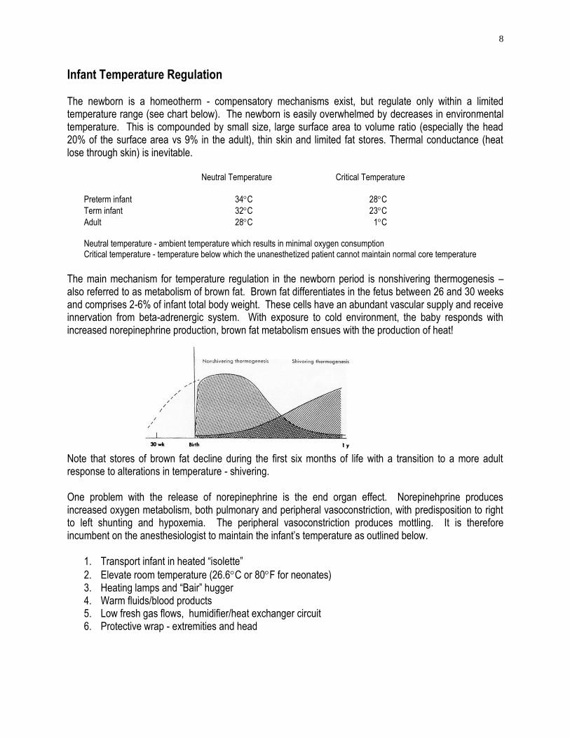
The main mechanism for temperature regulation in the newborn period is nonshivering thermogenesis – also referred to as metabolism of brown fat. Brown fat differentiates in the fetus between 26 and 30 weeks and comprises 2-6% of infant total body weight. These cells have an abundant vascular supply and receive innervation from beta-adrenergic system. With exposure to cold environment, the baby responds with increased norepinephrine production, brown fat metabolism ensues with the production of heat! Note that stores of brown fat decline during the first six months of life with a transition to a more adult response to alterations in temperature - shivering. One problem with the release of norepinephrine is the end organ effect. Norepinehprine produces increased oxygen metabolism, both pulmonary and peripheral vasoconstriction, with predisposition to right to left shunting and hypoxemia. The peripheral vasoconstriction produces mottling. It is therefore incumbent on the anesthesiologist to maintain the infant’s temperature as outlined below.
1. Transport infant in heated “isolette”
2. Elevate room temperature (26.6C or 80F for neonates) 3. Heating lamps and “Bair” hugger 4. Warm fluids/blood products 5. Low fresh gas flows, humidifier/heat exchanger circuit 6. Protective wrap - extremities and head
9
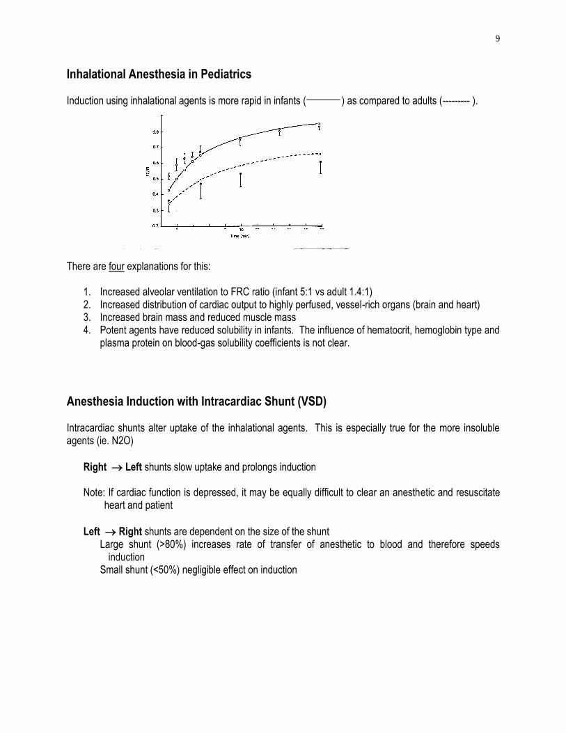
Inhalational Anesthesia in Pediatrics Induction using inhalational agents is more rapid in infants ( ) as compared to adults (--------- ). There are four explanations for this:
1. Increased alveolar ventilation to FRC ratio (infant 5:1 vs adult 1.4:1) 2. Increased distribution of cardiac output to highly perfused, vessel-rich organs (brain and heart) 3. Increased brain mass and reduced muscle mass 4. Potent agents have reduced solubility in infants. The influence of hematocrit, hemoglobin type and
plasma protein on blood-gas solubility coefficients is not clear.
Anesthesia Induction with Intracardiac Shunt (VSD) Intracardiac shunts alter uptake of the inhalational agents. This is especially true for the more insoluble agents (ie. N2O)
Right Left shunts slow uptake and prolongs induction Note: If cardiac function is depressed, it may be equally difficult to clear an anesthetic and resuscitate
heart and patient
Left Right shunts are dependent on the size of the shunt Large shunt (>80%) increases rate of transfer of anesthetic to blood and therefore speeds
induction Small shunt (<50%) negligible effect on induction
10
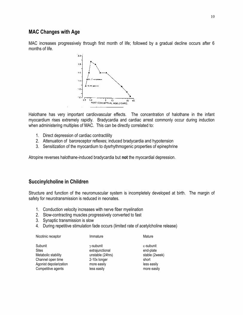
MAC Changes with Age MAC increases progressively through first month of life; followed by a gradual decline occurs after 6 months of life. Halothane has very important cardiovascular effects. The concentration of halothane in the infant myocardium rises extremely rapidly. Bradycardia and cardiac arrest commonly occur during induction when administering multiples of MAC. This can be directly correlated to:
1. Direct depression of cardiac contractility 2. Attenuation of baroreceptor reflexes; induced bradycardia and hypotension 3. Sensitization of the myocardium to dysrhythmogenic properties of epinephrine
Atropine reverses halothane-induced bradycardia but not the myocardial depression.
Succinylcholine in Children Structure and function of the neuromuscular system is incompletely developed at birth. The margin of safety for neurotransmission is reduced in neonates.
1. Conduction velocity increases with nerve fiber myelination 2. Slow-contracting muscles progressively converted to fast 3. Synaptic transmission is slow 4. During repetitive stimulation fade occurs (limited rate of acetylcholine release)
Nicotinic receptor Immature Mature
Subunit -subunit -subunit Sites extrajunctional end-plate Metabolic stability unstable (24hrs) stable (2week) Channel open time 2-10x longer short Agonist depolarization more easily less easily Competitive agents less easily more easily
11
Succinylcholine (a depolarizing agonist) is useful for rapid tracheal intubation and for treatment of laryngospasm (iv or im). Features in infants and children include:
1. Increased dose requirement based on weight 2. Duration of action is unaffected despite reduced pseudocholinesterase activity 3. Both increased dose and limited duration appear to be due to rapid redistribution into a larger ECF
volume 4. No phase II block on first dose
Pediatric Contraindications: Succinylcholine These are similar to adults with one notable exception - the myopathic child. FDA attempted to limit its use due to a number of hyperkalemic cardiac arrests in children with unrecognized myopathies. Side effects:
1. Cardiac arrhythmias (bradycardia, asystole, ventricular fibrillation) 2. Hyperkalemia 3. Post-anesthetic myalgias 4. Pulmonary edema 5. Increased gastric, intraocular and intracranial pressure 6. Associated masseter stiffness, spasm and malignant hyperthermia
Rocuronium: Differences Young vs Old Rocuronium (non-depolarizing antagonist) is considered a long-acting relaxant in infants, especially neonates. A larger volume of distribution and slower clearance results in a prolonged neuromuscular block in infants (56 min vs 26 min children). Onset time is slightly faster in infants. The duration of action is markedly prolonged when repeated doses are administered. Neuromuscular function must be evaluated carefully to avoid hypoventilation-related acidosis and potentiation of relaxant. Observe the infant prior to induction (muscle tone, depth of respiration, vigor of cry) and aim for return of this function post-operatively. Useful clinical signs include: ability to flex arms and lift legs, negative inspiratory force less than –25cm H2O, crying vital capacity greater than 15ml/kg. The neostigmine requirement is less in children. Onset of edrophonium is 2-3 min faster than neostigmine.
Oral Premedication in Pediatrics Benzodiazepine derivatives are widely used for premedicating children. They are given to calm patients, allay anxiety, and diminish recall of perianesthetic events. At low doses minimal drowsiness and cardiovascular or respiratory depression are produced. Nausea/vomiting is rare. Midazolam is a short acting, water-soluble molecule with a half-life of 2 hours. It is currently the most widely used premed due to its rapid uptake and elimination. After oral administration there is incomplete absorption and extensive first pass hepatic extraction (explaining need for administration of high oral dose).
12
Other features include: 1. Peak plasma concentrations in 53 minutes 2. No increase in gastric pH or residual volume 3. Calmer child less - likely to need 5% halothane induction 4. Acceptable taste for most children 5. Fewer behavioral changes than unpremedicated child 6. Does not affect the time to recovery
Fentanyl Oralet is most effective when absorbed via oral mucosa - not swallowed (1st pass metabolism through liver is high). The effect is dose dependent signs of sedation in 10 minutes after receiving 10-15 ug/kg. Desaturation and preoperative nausea is minimized if the child is brought to OR within 10min of completion of the oralet. In doses greater than 15ug/kg there is an increased incidence of nausea, vomiting, pruritis and desaturation.
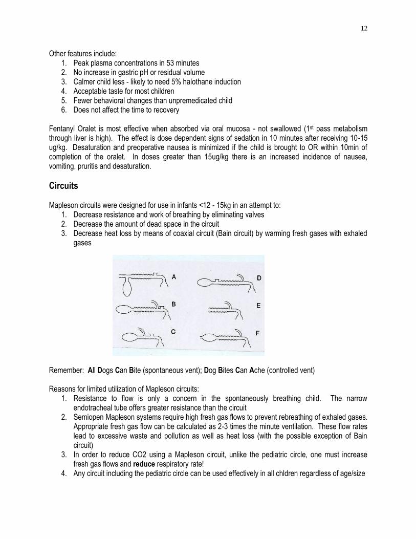
Circuits Mapleson circuits were designed for use in infants <12 - 15kg in an attempt to:
1. Decrease resistance and work of breathing by eliminating valves 2. Decrease the amount of dead space in the circuit 3. Decrease heat loss by means of coaxial circuit (Bain circuit) by warming fresh gases with exhaled
gases
Remember: All Dogs Can Bite (spontaneous vent); Dog Bites Can Ache (controlled vent) Reasons for limited utilization of Mapleson circuits:
1. Resistance to flow is only a concern in the spontaneously breathing child. The narrow endotracheal tube offers greater resistance than the circuit
2. Semiopen Mapleson systems require high fresh gas flows to prevent rebreathing of exhaled gases. Appropriate fresh gas flow can be calculated as 2-3 times the minute ventilation. These flow rates lead to excessive waste and pollution as well as heat loss (with the possible exception of Bain circuit)
3. In order to reduce CO2 using a Mapleson circuit, unlike the pediatric circle, one must increase fresh gas flows and reduce respiratory rate!
4. Any circuit including the pediatric circle can be used effectively in all chldren regardless of age/size
13
Regional Anesthesia Advantages
1. Faster awakening; reduced anesthetic requirement 2. Autonomic nervous system suppression 3. Limb immobilization perioperatively 4. Reduced stress response
Indications
1. Premature infants (<58-60 weeks prematurity) 2. “Floppy” infants with neuromuscular disease 3. Infants with chronic pulmonary disease 4. Children at risk for malignant hyperthermia 5. Older children who wish to remain awake
Contraindications
1. Infection at site 2. Coagulopathy 3. Anatomical anomaly (ie spina bifida)
Local Anesthetics Children are not more resistant to local anesthetic toxicity than adults:
1. Decreased albumin and -1-acid glycoprotein, both responsible for binding local anesthetics 2. Reduced hepatic degradation and therefore slower rate of elimination 3. Younger infants exhibit greater free fraction of local anesthetic
Dosing for infants <6months and <10kg need to be adjusted Anatomic differences of Lower CNS Neonate Infant Adult
Low end spinal cord L3 L1 L1 Low end dural sac S4 S2 S2 CSF volume (ml/kg) 4 3 2 Epidural Fat Loose Firm
14
Spinal in the neonate 1. Position: sitting or lateral decubitus, support neck and administer oxygen 2. Drug: Tetracaine (0.6-1 mg/kg with epinephrine wash); 22G spinal needle distance to from skin
to subarachnoid space <1 cm in preterm infant 3. Duration: 45-60 minutes
Complications
1. Total spinal 2. Apnea 3. Bradycardia
24 hour observation mandatory in high risk infants even after spinal anesthetic
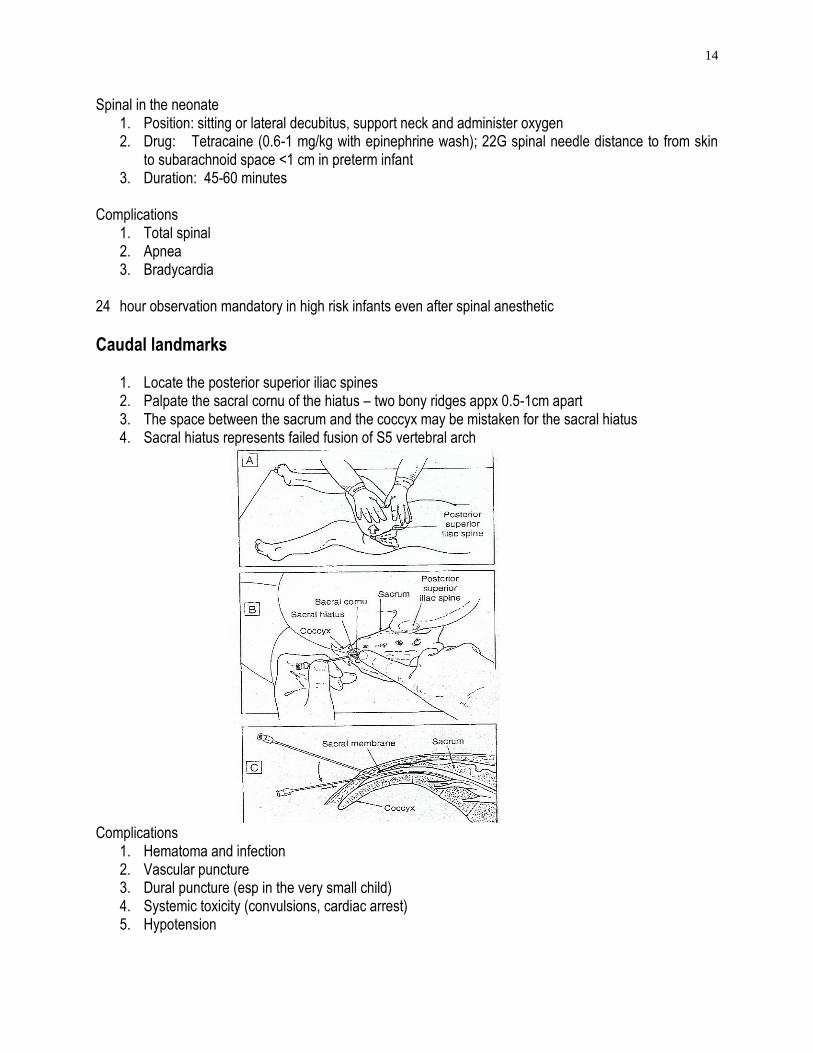
Caudal landmarks
1. Locate the posterior superior iliac spines 2. Palpate the sacral cornu of the hiatus – two bony ridges appx 0.5-1cm apart 3. The space between the sacrum and the coccyx may be mistaken for the sacral hiatus 4. Sacral hiatus represents failed fusion of S5 vertebral arch
Complications 1. Hematoma and infection 2. Vascular puncture 3. Dural puncture (esp in the very small child) 4. Systemic toxicity (convulsions, cardiac arrest) 5. Hypotension
15
Omphalocele and Gastroschisis Omphalocele
1. Herniation of the intestine into the base of the umbilical cord 2. Peritoneal sac is always present but may be ruptured 3. Sizes dependent on organs contained in sac (intestine, liver, spleen, stomach, bladder) 4. >50% association with congenital anomalies
GI tract: malrotation, biliary atresia, imperforate anus Cardiac: tetralogy of Fallot Genitourinary: bladder extrophy Craniofacial: cleft palate lip/palate Beckwith-Wiedemann: visceral macrosomia, macroglossia, microcephaly, hypoglycemia
Gastroschisis
1. Defect of the abdominal wall lateral (right) to the umbilicus through which intestinal evisceration occurs
2. No peritoneal sac allowing for fluid/heat loss, edema, infection and chemical peritonitis 3. Higher incidence of prematurity as compared to omphalocele 4. Associated congenital anomalies uncommon
Perioperative Care
1. Heat loss - cover exposed viscera with bowel bag 2. Decompress stomach 3. Sepsis – broad spectrum antibiotics 4. Trauma to herniated organs 5. Hypovolemia, acidosis, shock 6. Fluids
Anesthetic Management
1. Maintenance of temperature 2. Rapid sequence or awake tracheal intubation with oxyscope 3. Avoid N2O 4. Fluids D10W at maintenance and lactated Ringer’s at 10 – 15 cc/kg/hr 5. Muscle relaxants to facilitate abdominal wall closure; plan on prolonged period of mechanical
ventilation (24-48 hours) depending on size of defect
6. Underdeveloped abdominal cavity may not accommodate herniated bowel lung compression. Use airway pressures and oximetry as indicator of successful closure. Heart rate, blood pressure are unreliable indicators
7. Inferior vena cava compression and hypotension has been described 8. May require staged repair (use of silo) allowing viscus to settle, and final closure in 7-10 days.
Prolonged ileus not uncommon
16
Necrotizing Enterocolitis NEC is a disease of prematurity (<38weeks) and low birth weight (<2500gm). Less common in full term infants. Etiology
Reduced mesenteric blood flow secondary to ischemic event, resulting in intestinal mucosal injury. Mesenteric blood flow may be compromised: reduced cardiac output, fetal asphyxia, postnatal apnea, cardiac failure, arrhythmia, bradycardia, RDS, umbilical artery catheter.
Presentation
Irritability / lethargy, abdominal distention, bloody mucus in stool, vomiting. Radiographic findings
Pneumoperitoneum (perforation)
Medical Management
1. Reserved for those infants without perforation 2. Discontinuation of enteral feeds and gastric decompression 3. Intravenous fluids 4. Antibiotics
Indications for Surgery
1. Intestinal perforation 2. Failed medical therapy 3. Surgical goals: excision of all necrotic bowel, preserve length of bowel, remove necrotic debris
Anesthetic Management
1. Rapid sequence or awake oral intubation (carries risk of intracranial hemorrhage secondary to increases in ICP)
2. Air/oxygen, avoid N2O 3. Inotropic support 4. Volume resuscitation 5. Correction of metabolic disturbance 6. D10W infusion 7. Temperature management 8. Post-operative mechanical ventilation and parenteral alimentation (expect prolonged ileus)
17
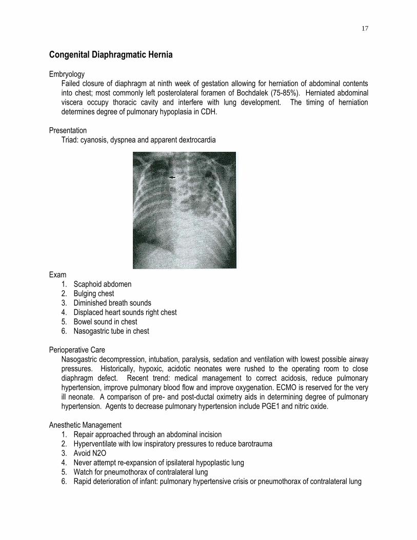
Congenital Diaphragmatic Hernia Embryology
Failed closure of diaphragm at ninth week of gestation allowing for herniation of abdominal contents into chest; most commonly left posterolateral foramen of Bochdalek (75-85%). Herniated abdominal viscera occupy thoracic cavity and interfere with lung development. The timing of herniation determines degree of pulmonary hypoplasia in CDH.
Presentation
Triad: cyanosis, dyspnea and apparent dextrocardia
Exam 1. Scaphoid abdomen 2. Bulging chest 3. Diminished breath sounds 4. Displaced heart sounds right chest 5. Bowel sound in chest 6. Nasogastric tube in chest
Perioperative Care
Nasogastric decompression, intubation, paralysis, sedation and ventilation with lowest possible airway pressures. Historically, hypoxic, acidotic neonates were rushed to the operating room to close diaphragm defect. Recent trend: medical management to correct acidosis, reduce pulmonary hypertension, improve pulmonary blood flow and improve oxygenation. ECMO is reserved for the very ill neonate. A comparison of pre- and post-ductal oximetry aids in determining degree of pulmonary hypertension. Agents to decrease pulmonary hypertension include PGE1 and nitric oxide.
Anesthetic Management
1. Repair approached through an abdominal incision 2. Hyperventilate with low inspiratory pressures to reduce barotrauma 3. Avoid N2O 4. Never attempt re-expansion of ipsilateral hypoplastic lung 5. Watch for pneumothorax of contralateral lung 6. Rapid deterioration of infant: pulmonary hypertensive crisis or pneumothorax of contralateral lung
18
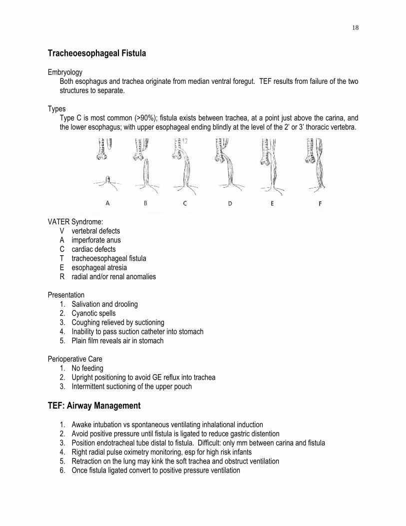
Tracheoesophageal Fistula Embryology
Both esophagus and trachea originate from median ventral foregut. TEF results from failure of the two structures to separate.
Types
Type C is most common (>90%); fistula exists between trachea, at a point just above the carina, and the lower esophagus; with upper esophageal ending blindly at the level of the 2’ or 3’ thoracic vertebra.
VATER Syndrome: V vertebral defects A imperforate anus C cardiac defects T tracheoesophageal fistula E esophageal atresia R radial and/or renal anomalies
Presentation
1. Salivation and drooling 2. Cyanotic spells 3. Coughing relieved by suctioning 4. Inability to pass suction catheter into stomach 5. Plain film reveals air in stomach
Perioperative Care
1. No feeding 2. Upright positioning to avoid GE reflux into trachea 3. Intermittent suctioning of the upper pouch
TEF: Airway Management
1. Awake intubation vs spontaneous ventilating inhalational induction 2. Avoid positive pressure until fistula is ligated to reduce gastric distention 3. Position endotracheal tube distal to fistula. Difficult: only mm between carina and fistula 4. Right radial pulse oximetry monitoring, esp for high risk infants 5. Retraction on the lung may kink the soft trachea and obstruct ventilation 6. Once fistula ligated convert to positive pressure ventilation
19
Management of Pediatric Stridor Stridor may have a congenital or acquired origin. Most cases are of extrathoracic origin but some originate in the trachea and major bronchi. Stridor may be caused by fixed obstruction (airway diameter does not change with changes in transmural pressure) or variable obstruction (airway caliber responds to changes in transmural pressure). Congenital
Pierre Robin syndrome Treacher-Collins syndrome Macroglossia (Beckwith syndrome, Trisomy 21) Laryngomalacia Subglottic stenosis Vascular ring
Acquired
Infectious (epiglottitis, croup) Foreign body Laryngeal trauma Neoplasia (papillomatosis)
Management of stridor in the otherwise healthy child will depend on the etiology.
1. Large occiput - head position to prevent airway collapse esp in the newborn 2. Large tongue - simple jaw thrust opening the mouth and removing the tongue from the palate, use
oral airway if necessary 3. Tonsillar hypertrophy - continuous positive airway pressure and/or oral airway 4. Intramuscular succinylcholine for laryngospasm 5. Post intubation edema:
mild – humidified air severe – racemic epinephrine, ICU admission
URI in Children: Anesthetic Complications There is no consensus regarding the safety of anesthetizing children with active or recent URI. Reported complications include:
1. Bronchospasm 2. Laryngospasm 3. Stridor secondary to subglottic edema 1. Hypoxia 2. Atelectasis
These complications occur more often in children < 2 years of age. All of these complications are usually easily treated and not associated with prolonged morbidity.
20
Incidence of Laryngospasm In a large population study from Stockholm the incidence of laryngospasm in children less than 9 years of age was shown to be 1.7% Factors affecting the incidence of laryngospasm include:
1. Children with a history of respiratory infection (9.6%) 2. The presence of obstructive lung disease (6.4%) 3. Previous anesthetic complications (5.5%)
Tetralogy of Fallot: Rx of Hypoxemic Spell This disease comprises 10% of all congenital heart disease. Four anomalies
1. Right ventricular outflow tract obstruction 2. Subaortic VSD 3. Overriding aorta 4. Right ventricular hypertrophy
Surgical Repair
Traditionally, palliative shunts to improve pulmonary blood flow were placed. Currently, primary pulmonary artery/RVOT reconstruction at an earlier age is advocated.
“Tet spell”
Hypercyanotic episode resulting when infundibular spasm reduces pulmonary blood flow and increases
RL shunting across the VSD. Management of “Tet Spell”:
1. Optimize pulmonary blood flow: reduce pulmonary vascular resistance 100% oxygen Morphine Hydration
2. Reduce RL shunt flow across VSD: increase systemic vascular resistance Squatting and abdominal compression Phenylephrine 3. Reduce infundibular spasm: reduce inotropy Propranolol Halothane

21
Pyloric Stenosis: Electrolytes Etiology
Hypertrophy of the circular muscles of the pyloris resulting in gradual gastric outlet obstruction. Presentation
1. Non-bilious projectile vomiting between 2 and 8 weeks of age 2. Palpation of an olive-sized mass in the upper abdomen 3. Confirmed by abdominal ultrasound
Perioperative Care:
1. Not a surgical emergency; metabolic issues must be addressed prior to surgical repair to avoid introperative hypotension and arrhythmias
2. Vomiting causes severe dehydration and loss of gastric secretions (H+ and Cl- ions) resulting in hypochloremic, hypokalemic and metabolic alkalosis
3. Treatment: fluid and electrolyte replacement (D5 ½NS +KCL) over 24 to 48 hour period Anesthetic Management:
1. Empty stomach completely with nasogastric tube 2. Insert endotracheal tube using rapid sequence/cricoid pressure or awake oral (oxyscope) 3. Surgical release of circular layer of the pyloris
Anesthetic Implications of Trisomy 21 Physical defects:
1. Developmentally delayed 2. Small mouth, hypoplastic mandible, protruding tongue 3. Congenital subglottic stenosis 4. Obstructive sleep apnea (large tongue, mid face hypoplasia, large tonsils) 5. Ligamentous laxity at the atlantoaxial joint 6. 40% manifest congenial heart disease (VSD, AV canal) 7. Pulmonary hypertension
Anesthetic issues:
1. Premedication to reduce anxiety 2. Oral airway 3. Prepare to intubate with smaller than expected ETT 4. Cervical spine films recommended but not universally practiced 5. Minimal neck flexion to prevent C1-C2 subluxation (predisposition to spinal cord injury)