Embed Size (px)
Citation preview

Clinical Guidelines
February 2011 to January 2012
1

TABLE OF CONTENTS
TABLE OF CONTENTS ............................................................................................2
1 GENERAL ADMINISTRATION .......................................................................6 1.1 UHW Telephone Numbers ............................................................................6
1.1.1 Consultant Medical Staff and Speciality Doctor ...................................6 1.1.2 Secretarial Staff and Specialist Cardiac Nurses.....................................6 1.1.3 Junior Medical Staff/Trainees................................................................7 1.1.4 Technical and Support Staff...................................................................7 1.1.5 Useful E-Mail Addresses .......................................................................7 1.1.6 Useful Contact Numbers........................................................................8
1.2 Welsh Hospitals ...........................................................................................10 1.3 Specialist Cardiac Nurses ............................................................................11
1.3.1 Contact Numbers .................................................................................11 1.3.2 Roles and Responsibilities ...................................................................11 1.3.3 Nurse-Led Clinic..................................................................................11
1.4 Bristol Children’s Hospital ..........................................................................12 1.4.1 Medical Staff – Paeds ..........................................................................12 1.4.2 Medical Staff – GUCH (Bristol Heart Institute)..................................12 1.4.3 Other Useful Numbers .........................................................................12 1.4.4 Bristol E-Mail Addresses.....................................................................12
1.5 UK and Ireland Paediatric Cardiac Units.....................................................13 1.6 Annual and Study Leave..............................................................................14 1.7 On-Call Arrangements .................................................................................14
1.7.1 Consultant Staff ...................................................................................14 1.7.2 Junior Medical Staff.............................................................................14
1.8 Audit and Research ......................................................................................14 1.9 Computers and Cardiobase® ........................................................................15 1.10 Medical Notes and Correspondence Headings ............................................15
1.10.1 Categories/Definitions of Admissions and Reviews............................16 1.10.2 Correspondence Headings ...................................................................16
2 DAY TO DAY BUSINESS ................................................................................17 2.1 Daily Timetable ...........................................................................................17 2.2 UHW Clinics................................................................................................18
2.2.1 General Clinics............................................................................................18 2.2.2 Specialist Clinics.........................................................................................18 2.2.3 Echo Clinic..................................................................................................18
2.3 Outreach Clinics...........................................................................................18 2.4 Teaching Topics...........................................................................................19 2.5 Teaching Ward Round .................................................................................19
3 ADMISSIONS AND REFERRALS .................................................................20 3.1 Routine Admissions .....................................................................................20 3.2 Day Case Admissions ..................................................................................21
3.2.1 Echocardiogram under sedation...........................................................21 3.2.2 ACE inhibitor protocol ........................................................................22 3.2.3 Carvedilol protocol ..............................................................................23 3.2.4 Adrenaline/Epinephrine challenge for LQTS ......................................24
2

3.2.5 Brugada syndrome - Flecainide or Ajmaline Challenge......................27 3.3 Admissions for Transoesophageal Echocardiography.................................28 3.4 Admissions for MRI or CT scan..................................................................28
3. me
3. me7.17.2
3.3.
4 4.4. ran4.
5 ST5.
2.1
5.5.4.1 Indications for exercise test .................................................................43
4.34.4
5.
5.2
5.8 MRI or CT Scan...........................................................................................48 5.
9.19.2
6 6.
3.4.1 Non-General Anaesthetic...........................................................................28 3.4.2 General Anaesthetic3.4.3 Adenosine Stress Test by MRI
...................................................................................29 ..................................................................29
3.5 ................................30 Non-Cardiac Admissions and Casual Ward Attenders6 E rgency admissions and procedures .......................................................31 3.6.1 .......................................................31 Patients known to the Department3.6.2 ......................................................................................31 New Referrals3.6.3 ...........................................................................31 Neonatal Admissions3.6.4 Protocol for PGE infusion (NB risk of apnoea) ........................................32 7 E rgency Interventional Procedures .........................................................32 3. Balloon Atrial Septostomy ...................................................................33 3. ..................................................................33 DC Cardioversion (DCCV)3.7.3 ............................................34 Pericardiocentesis for cardiac tamponade8 Post Surgical Transfers (in) .........................................................................36 9 Inpatient Referrals ........................................................................................36
DISCHARGE AND TRANSFER PROCEDURES.........................................37 1 General Principles ........................................................................................37 2 T sfers to Other Hospitals ........................................................................38 3 Discharges Following Cardiac Surgery .......................................................39
4.4 .....................................................................................39 Discharge Checklist4.5 .........................................................................................39 Death of a Patient
INVE IGATIONS...........................................................................................40 1 Drug Monitoring and/levels .........................................................................40
5.2 ........................................................................................40 Electrocardiogram5. .....................................................................41 Basic ECG Interpretation
5.3 ...........................................................................................42 Echocardiogram5.3.1 .....................42 Echocardiogram to rule out cardiac source of embolism
4 Exercise Test ................................................................................................43
5.4.2 ....................................................................................43 Bruce protocols5. ..................................................................44 Indications for termination:5. .................................................................44 Difficulties in interpretation5.4.5 Helpful tips ...........................................................................................44
5 Ambulatory ECG Monitoring ......................................................................45 5.5.1 Holter monitoring .................................................................................45 5. ....................................................................................45 Event recorders
5.6 .........................................................................45 Ambulatory BP Monitoring5.7 ........................................................................................................47 Tilt Test
9 Isotope Scans ...............................................................................................48 5. Cardiac Nuclear Scanning ....................................................................48 5. .............................................................................48 Lung perfusion scan
CLINICAL PROBLEMS ..................................................................................49 1 Anti-platelet Therapy and Anticoagulation .................................................49
3

6.1.1 ...................................................49 Aspirin and Anti-platelet Therapy116.1.2
1.3
1.71.8
6.2.1
6.6.
6.7 Endocarditis and Endocarditis Prophylaxis .................................................70
7.2
6.8.4 Recommendations in Post-Operative Patients ...........................................79
6.
6.6.
6.
6.
...............................................................................49 Devices and Stents6. ...............................................................................50 Valve replacement6.1.4 Cavopulmonary shunt / Fontan ............................................................50 6.1.5 Other indications ..................................................................................50 6.1.6 Commencing anticoagulation (heparin and warfarin) .........................51 6. ......................................................................................53 INR Sampling6. ........................................................................................53 INR Protocol6.1.9 ........................................55 Warfarin dosage table (doses shown in mg)6.1.10 Cessation of warfarin for surgical or invasive procedure ....................56 6.1.11 Factors that influence the efficacy of warfarin ....................................56 2 Asplenia and Immunodeficiency .................................................................57 6. ...............................................................................................57 Asplenia6.2.2 ....................58 DiGeorge syndrome and chromosome 22 microdeletion3 Cardiac Failure .............................................................................................59 4 Cardiac Tamponade - See section 3.7.2 .......................................................63
6.5 ...........................................................................................63 Cardiomyopathy6.6 .................................................................................68 Chest Pain in Children
6.7.1 Infective Endocarditis ..........................................................................70 6. .....................................................................71 Endocarditis prophylaxis
6.8 ........................................................72 Exercise in Paediatric Cardiac Patients6.8.1 Myocardial Abnormalities .........................................................................75 6.8.2 Coronary Abnormalities.............................................................................75 6.8.3 Congenital Lesions and Conditions ...........................................................76
6.8.5 Recommendations for Athletes with Arrhythmias ....................................81 6.9 Fits, Faints and Funny Turns .......................................................................82
6.9.1 Reflex Syncope .........................................................................................83 10 Hypercyanotic Spells ...................................................................................85
6.11 ........................................................................................86 Kawasaki Disease6.12 MRSA Infection...........................................................................................88 6.13 Nutrition in Cardiac Patients........................................................................89
6.13.1 Faltering Growth..................................................................................89 6.13.2 Gastro-Oesophageal Reflux (GOR).........................................................89 14 Oxygen Therapy in Cardiac Patients ...........................................................90 15 Premature Beats in Newborn Babies ...........................................................91
6.16 Prescribing Drugs Safely .............................................................................92 6.16.1 Medication Errors ....................................................................................92 6.16.2 Quick Calculations of Drug Concentrations for Infusions ......................93 17 Propranolol for the Treatment of Capillary Haemangiomas ........................94
6.18 Protein-losing enteropathy (PLE) / Plastic bronchitis (PB) after Fontan ....96 6.19 Pulmonary Hypertension in Childhood .......................................................97
6.19.1 Definition, Classification and WHO Functional Status.......................97 6.19.2 Pathophysiology ...................................................................................98 6.19.3 Treatment of Pulmonary Hypertension................................................99 6.19.4 Persistent Pulmonary Hypertension of the Newborn (PPHN)...........101 20 Rheumatic Fever ........................................................................................105 6.20.1 ..............................................................................105
.......106 Diagnostic criteria
6.20.2 Secondary Prevention of Rheumatic Fever (Recurrent Attacks)
4

6.20.3 6.6.
6.
6.
IN
Duration of Secondary Rheumatic Fever Prophylaxis.......................106 21 RSV Infection in Cardiac Patients .............................................................106 22 Screening for Cardiac Disease (genetic, familial, etc) ...............................107 6.22.1 Heart muscle disease..........................................................................107 6.22.2 Heart Rhythm.....................................................................................111 6.22.3 Heart Structure ...................................................................................112 23 Supraventricular Tachycardia ....................................................................114 6.23.1 SVT in the Fetus ................................................................................116 24 Transplantation ..........................................................................................116
DEX ......................................................................................................................119
5

1 GENERAL ADMINISTRATION
1.1 1. Clinician Secretary
UHW Telephone Numbers
1.1 Consultant Medical Staff and Speciality Doctor
Office Radiopage Mobile Home
Dr Victor Ofoe 4746 3869 07623 905 928 07815 510 833 Via UHW Switchboard
r Obed Onuzo 4606 2908 07623 906 018 07815 902 866 Via UHW Switchboard
D
r Orhan Uzun 4743 4745 07623 906 121 07967 337 319 Via UHW Switchboard
D
Dr Dirk G Wilson 4749 5156 07623 905 734 07968 822 824 Via UHW Switchboard
Dr Peter Groves 3533 2354 07623 905 821 07899 727 937 Via UHW Switchboard
Dr Navroz Masani 4086 4569 07623 905 821 07710 272 928 Via UHW Switchboard
Dr Helen Wallis Contact via Neath Port Talbot Hospital Switchboard or Secretary (01639 722049)
Dr Amos Wong 4759 Hospital page 6343
Speciality Doctor in Paediatric Cardiology
Preferred contact is via radiopager or mobile phones. Most hospital mobiles do not work in Welsh hospitals, so radiopage is often
more reliable during working hours. If you cannot make contact successfully try the home phone number via
Switchboard. If all efforts to contact the on-call consultant are unsuccessful, contact one of
the other consultants. In a dire emergency, speak to the on-call Bristol consultant for advice.
1.1.2 Secretarial Staff and Specialist Cardiac Nurses Name Extension Other information Sarah Wooller (Dr Onuzo) 4606 08:30 – 17:00 Mon-Fri
Angela Butters (Dr Uzun) 4743 09:00 – 17:30 Mon-Fri
Suzanne Cornish (Dr Wilson) 4749 08:30 – 17:00 Mon-Wed 08:00 – 14:30 Thu
Amanda Doyle (Dr Ofoe) 4746 09:00 – 17:00 Mon-Wed, Fri
Sarah Grinter (Dr Masani) 4086 08:30 – 17:00 Mon-Fri
Karen McCarthy (Dr Groves) 2354 08:30 – 17:00 Mon-Fri
Specialist Cardiac Nurse (Area) Office Mobile Claire Logan (Cardiff and the Vale + Rhondda Cynon Taff) 5184 07811 197 136
Wendy Williams (Bridgend and West) 4753 07813 922 441
6

Alison Pearce (Gwent, western Valleys and erthyr) 5524 07966 461 421 M
Ann Jermyn (Transition Ca 7 re 12-19 years) 8046 07980 635 17
es 1.1.3 Junior Medical Staff/Traine
Title Bleep Extension
Cardiac Registrar 5391 or
64
07623 90 359 759
Paediatric Registrar 5394 4759
SHO/ST2 5334 4759
1.1.4 Techn
ical a pport S
ad ten
nd Su taff
Staff Bleep/R iopage Ex sion
Viv Booker (Echo) 5503 3920
Bethany Glasser (D n) 5 6 90 ieticia 07623 90 07 31
Tony Bradley (Soci rk) ia 76 al Wo Mobile v Switch 27
David “Wally”Support)
James (Audit, Data and IT 3889
1.1.5 Useful E-Mail Addresses
Name E-mail address
Booker, Viv [email protected]
Butters, Angela [email protected]
Cornish, Suzanne [email protected]
Doyle, Amanda [email protected]
James, Wally [email protected]
Jermyn, Ann [email protected]
Logan, Claire [email protected]
Ofoe, Victor Vict alesor.Ofoe@w .nhs.uk
Onuzo, Obed Obed [email protected] s.nhs.uk
Pearce, Alison Aliso [email protected] es.nhs.uk
Uzun, Orhan Orha [email protected] s.nhs.uk
Wallis, Helen [email protected]
Wilson, Dirk Dirk [email protected] s.nhs.uk
Wooller, Sarah Sara [email protected] les.nhs.uk
Wong, Amos [email protected]
7

1.1.6 Useful Contact Numbers
Department/Individual Extensio lee mation n B p Other Infor
Adult Cardiac ICU 3265
Anaes hetic Department 3107 t
B1 (Adult Cardiology) 3382/4603
Bacteriology 2044 Bleep for out-of-hours samples 5388
Biochemistry (Main Dep 3 t) 2805/26 7
Biochemistry (Emergenc 5278 Call Lab for urgent processing y)
Blood Bank 2157/215 8 5268
CARDIAC ARREST 2222
Cardiac Day Case Unit 4414
Catheter Laboratory 4642/3329/4607
Clinical Investigation Unit 3765
Children’s Assessment Unit 5441
Coronary Care Unit 2110
Coagulation 2214 5270
Coroner’s Office 20 222 111 Outside line required
Delivery Suite 2679
Dental Surgeon Paeds 2260
Drug Information 2251
ECG (Main Dept, Pacing) 3325
ECG (Inpatient requests) 1 OPD (Holters also) 6396 Suite 1
EEG/Neurophysiology 3194
Exercise/Tilt Test 3465
Fetal Medicine 2279/3341
General ICU 5319
Genetics 4028
Haematology 2805 5269 Bleep for out-of-hours samples
Hand over room General 8930
Hand over room Specialty 8820
Heulwen Ward in Desk 4755/5375 Ma
Histology 2714
Holter (24 hour tape) 6396
Immunology 5814
IT help desk 5073
Medical Illustration 3305/3307
MRI 3063
MRI/CT CD Copying 6631
8

Department/Individual Extension Bleep Other Information
Neonatal Unit Reception 2680/2684
NICU (ICU Room 1) 6873 NNU HDU 7847
OPD (Adult Cardiac) dult Congenital Clinic 3266 A
OPD (Paediatric) 3364
OPD (Room 4) 2242
Oncology Sky Ward 802 8801/8
Pacing Clinic 4600
Pacing Lab 3081
Paediatric HDU (Heulwen) 4751
Paediatric ICU 3282/4622
Paeds Land Ward 3274/3276
Paeds Ocean Ward 3359/3370
Paeds South 3277/2650
Pathology 2710
Personnel (Human resources) 3887
Pharmacy 2988
Porters 2667
Postnatal Ward 3343
Public Health 2236
SALT team 3736
Sophie Pearson Room 6355
Switchboard 100 Emergency 2222
Teenage Cancer Trust 6784/6915 Dr’s Office 2973
Theatres (Reception) 2993
Toxicology 72 6894 Drug levels
Ultrasound 4834
X-ray 3027 5299 ut of hours – call extn 8084 O
9

1.2 Welsh Hospitals
District General Hospital WHTN l Number Externa
Aberdare General Hospital 01753 01685 872 411
Brecon War Memorial Hospita 01762 01874 622 443 l
Bronglais General Hospital(Ab 01822 01970 623 131 eryswyth)
Caerphilly Miner’s Hospital 01755 029 2085 1811
Llandough Hospital
01776
(From UHW
dial 72 then
extension)
029 2071 1711
Morriston Hospital (Swansea) 01789 01792 702 222
Neath Port Talbot Hospital 01881 01639 862 000
Nevill Hall Hospital (Abergave 01736 01873 732 732 nny)
Prince Charles Hospital (Merth 01854 01685 721 721 yr)
Princess of Wales Hospital (Bridgend) 01855 01656 752 752
Prince Phillip Hospital (Llanell 01824 01554 756 567 i)
Royal Glamorgan Hospital 01751 3 443 01443 44
Royal Gwent Hospital (Newpo 01738 4 234 rt) 01633 23
Saint David’s Hospital 01771 2053 6666 029
Singleton Hospital (Swansea) 01883 01792 205 666
West Wales General Hospital (Carmarthen) 01827 01267 235 151
Withybush Hospital (Haverfordwest) 01720 01437 764 545
Ysbyty Glan Clwyd (Rhyl) 01815 01745 583 910
Ysbyty Gwynedd (Bangor) 01746 01248 384 384
Add “100” to the WHTN number for Switchboard Operator or direct-dial if you know the extension
10

1.3 Specialist Cardiac Nurses
alent paediatric cardiac liaison nurses: Claire Logan, n and Alison Pearc n is part t ded
or “adopted” by the British Heart Foundation.
ffice iopage
1.3.1 Contact Numbers There are 3.4 whole-time-equivWendy Williams, Ann Jermy e (Aliso ime). All are fun
Nurse (Area) O Rad Mobile
Claire Logan (Cardiff and the Vale + Rhondda Cynon Taff)
5184 23 905 75 136 076 8 07811 197
Wendy Williams (Bridgend and West)
4753 23 906 121 07813 922 441 076
Alison Pearce - Part time Th/Fr (Gwent, western Valleys, Merthyr)
5524 6 29 421 07623 90 7 07966 461
Ann Jermyn (Transition care, age 12-19)
8046 07980 635 177
1.3.2 Roles and Responsi
Nurse-led clinic (seebilities 1.3.2 below)
e (section Link between clinicians and parents
ristol – participation in weekly Planning Meeting
be contacted a Newly diagnosed children
o the ward n preparatioatient (so the eep track
ious children or parent Ward discharges
nd 1:30 – 4 RUF
The clinic is supported by the cardiac dietician and echo technician; junior to review patients attending the clin
e review o Nutritional review
ents is kept in the Liaison Nurses’ Office. The person booking the appointment is responsible for informing the
patient/parents of the date, time and location of the clinic.
Primary contact in INR servic 6.1)
Link between Cardiff and B Parent education and advice
The cardiac liaison nurses should bout:
Cardiac admissions t Cardiac catheter or operatio Any decision to transfer a p
n y can k of transfers out)
Distressed or anx s
1.3.3 Nurse-Led Clinic
Nurse-Led Clinic runs on Mondays a Fridays :00 pm in the KUnit (extension 6782).
doctors may be asked ic.
Aspects of care provided include o Post-operativ
Patients are booked by an appointment system allowing 15 minutes per patient.
A diary for these appointm
11

1.4 Bristol Children’s Hospital 1.4.1 Medical Staff – Paeds Consultant Secretary Office
Dr Alison Hayes 0117 342 8856 8848
Dr Rob Martin 0117 342 8855 8849
Dr Gareth Morgan 0117 342 8852
Dr Graham Stuart 0117 342 8852 8859
Dr Andrew Tometzki 0117 342 8853 8858
Dr Bev Tsai-Goodman 0117 342 8856 8923
Dr Rob Tulloh 0117 342 8856 8176
Mr Massimo Caputo
Mr Andrew Parry
Mr Serban Stoica
0117 3(Christine
42 8854 McF
a BRI rd 01179 215 411
01179 276 998
adden)
Radiopage/Mobile/Home
Vi Switchboa
or
1.4.2 Medical Staff – GUCH (Bristol Heart Institute)
r Stephanie Curtis 0117 342 5967 D
Dr Rob Martin Dr Gareth Morgan 0117 342 6576 Dr Graham Stuart
Dr Mark Turner 0117 342 6575 0478
Via BRI Switchboard 01179 215 411 or 01179 276 998
1.4.3 Other Useful Numbers
witchboard S 01179 215 411 or 01179 276 998
BHI Coronary Care Unit 0117 342 2278
Paediatric Cardiac Ward (32) 0117 342 8332 / 8679
Catheter Laboratory 0117 342 8282 / 8456
Echocardiography Laboratory 0117 342 8722
GUCH Liaison Nurse (Sheena) 0117 342 0463
PCLN (Cathy + Debbie) 0117 342 8286
Paediatric ICU 0117 342 8377 or 8437
Paeds OPD 0117 342 8401 or 8402
SCBU (St Michael’s) 0117 342 5275 or 5275
1.4.4 Bristol E-Mail Addresses Dr Stephanie Curtis [email protected]
Dr Alison Hayes [email protected]
Dr Rob Martin [email protected]
Dr Graham [email protected] Stuart
Dr Gareth Morgan [email protected]
Dr Andrew Tometzki [email protected]
Dr Beverly Tsai-Goodman [email protected]
Dr Rob Tulloh [email protected]
Dr Mark Turner [email protected]
Cont’d overleaf
12

r Massimo Caputo [email protected]
Mr And ew Parry r [email protected]
Mr Serban Stoica serban uhbris.stoica@ tol.nhs.uk
1.5 UK and Ireland Pa iac Uni
Swi
ediatric Card ts
Centre tchboard
Alder Hey Children’s Hosp l) 0151ital (Liverpoo 228 4811
Birmingham Children’s Ho 0121 333 9999 spital
Bristol Royal Hospital for Children
01179 230 000
01179 276 998
01173 428 460
Freeman Hospital (Newcastle-upon-Tyne) 0191 233 6161
Glenfield Hospital (Leicester) 0116 287 1471
Great Ormond Street, The Hospital for Sick Children 0207 405 9200
Guys Hospital (Evelina Children’s Hospital, London) 0207 188 7188
Harefield Hospital (London) 01895 823 737
John Radcliffe Hospital (Oxford) 741 166 01865
Leeds General Infirmary 0113 243 2799
Our Lady’s Hospital for Sick Children (Dub 00 353 1409 6100 lin)
Royal Belfast Hospital for Sick Children 40 503 02890 2
Royal Brompton & Harefield NHS Trust (L 52 8121 ondon) 0207 3
Royal ospital for Sick Children (E H dinburgh) 0131 536 0000
Royal Hospital for Sick Children (Glasgow) 0141 201 0000
Royal Manchester Children's Hospital 696 0161 794 4
Wessex Cardiothoracic Unit (Southampton) 02380 777 222
13

1.6 Annual and Study Leave ntitlement should be take
ve 6 weeks in advance usin nery forms g study leave passport)
Inform junior doctor rota coordinator, enter leave in Junior Doctors’ Leave Diary, which is held by Suzanne (DGW’s secretary)
uch as examinations or job interviews) two junior doctors must be present to cover the unit during normal
orking hours.
The consultant staff work a 1:4 rota, one week a hand-over taking place on Monday mornings in the Planning
ell in advance. All junior staff should ensure they know which consultant is on call. If you are unable to contact the on-call consultant
a dire emergency, the consultant covering the Bristol unit).
ic Cross-cover tea
c unit iatric specialities or NNU m .
on-call 1 night in 5 (non-aeds rota; he or she may be asked to provide cross-
ng an emergency. ll specialities SHO/ST2 and specialities registrar provide
ents, inc U. Clear hand-overs between clinical staff are essential. Hand-over rounds take place
30. The team-m the cardiac
nts. W rs should be they are the cardiac
1.8 Audit and Research tient care. The broad principles behind medical
parison of current practice to that the audit by ard of
are.
This Department is actively involved in medical audit, both with the Cardiology service and General Paediatrics. Rotating paediatric junior staff should attend the paediatric audit meetings. Each junior doctor will be expected to undertake an audit project during his or her post.
Full leave e n Book lea g approved Hospital and Dea
(includin
Unless there are exceptional circumstances (s
w
1.7 On-Call Arrangements
1.7.1 Consultant Staff at a time, with Meeting.
An on-call rota is circulated w
in an emergency, contact one of the other consultants (or, in
1.7.2 Junior Medical Staff
The SHO/ST2 participates in the paediatr on-call rota. arrangements are in place with other speciality
The rotating general paediatrics registrar attached to the cardiams.
participates in the paed iddle grade rota The cardiology registrar is
participate in the general president) and does not
cover support duri The resident on-ca
out-of-hours cross cover for cardiac pati luding those on HD
daily (Mon-Fri) at 08:30 and 16: ember coveringpatients should attend the relevant hand-over round to pick up and convey any relevant information about the cardiac patie ritten hand-oveprovided to the paediatric team whenpatients.
cross-covering
Effective medical audit can improve paaudit are the setting of an accepted standard, comstandard, making alterations and completing re-assessing the standc
14
Part ation in research projects iicip s expected from all junior staff and opportunities r to bodies such as the Welsh Paediatric
e
ided with a password for accessing the hospital network. Dr Uzun bears responsibility with the IT Department for use of the network by
Any abuse will be dealt with harshly. password to non-unit staff.
ient identifiable information
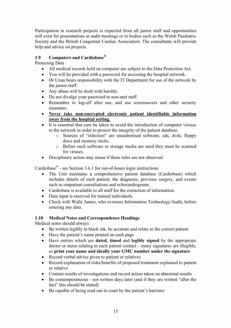
will exist for presentations at audit meetings oSociety and the British Congenital Cardiac Association. The consultants will providhelp and advice on projects. 1.9 Computers and Cardiobase® Protecting Data
All medical records held on computer are subject to the Data Protection Act. You will be prov
the junior staff.
Do not divulge your Remember to log-off after use, and use screensavers and other security
measures. Never take non-encrypted electronic pat
away from the hospital setting. It is essential that care be taken to avoid the introduction of computer viruses
to the network in order to protect the integrity of the patient database. o Sources of “infection" are unauthorised software, cds, dvds, floppy
discs and memory sticks. tware or storage media are used they must be scanned
erved.
chocardiograms.
legibly in black ink, be accurate and relate to the correct patient Have the patient’s name printed on each page
dated, timed legibly signed
Record explanation of risks/benefits of proposed treatment explained to patient
d out in court by the patient’s barrister
o Before such soffor viruses.
Disciplinary action may ensue if these rules are not obs Cardiobase® - see Section 3.6.1 for out-of-hours login instructions
The Unit maintains a comprehensive patient database (Cardiobase) which includes details of each patient, the diagnosis, previous surgery, and events such as outpatient consultations and e
Cardiobase is available to all staff for the extraction of information. Data input is reserved for trained individuals. Check with Wally James, who oversees Information Technology/Audit, before
entering any data. 1.10 Medical Notes and Correspondence Headings Medical notes should always
Be written
Have entries which a re and by the appropriate doctor or nurse relating to each patient contact – many signatures are illegible, so print your name and ideally your GMC number under the signature
Record verbal advice given to patient or relatives
or relative Contain results of investigations and record action taken on abnormal results Be contemporaneous - not written days later (and if they are written “after the
fact” this should be stated) Be capable of being rea
15

At discharge contain a list of all diagnoses including co-morbidities and procedures (get a senior member of staff to confirm the entries), avoiding the use of ambiguous abbreviations
1.10.1 Categories/Definitions of Admissions and Reviews
Comment Category Explanation
Admission Patient is admitted to a bed. Nursing resource is used.
Dictate a “Discharge Summary” on Cardiobase
The patient stays overnight at least one night.
Day Case Admission Patient is admitted to a bed Dictate a brief “Discharge Summary” on Cardiobase – mark
as a Day Case for an investigation or procedure (e.g. Captopril itchallenge, sedated echo). Nursing resource is used. The patient is discharged the same day.
Casual Ward Attender The patient attends the ward without being admitted to a
Dictate a “General Letter” on Cardiobase if treatment is
bed (e.g. blood test). changed or if the outcome of the visit needs to be communicated to the GP
Outpatient clinic (including or “Nurse-Led Clinic” letter on
tpatient Visit A patient seen in a booked ou
Dictate and “Outpatient Letter”
Nurse-Led Clinic). Cardiobase
Inpatient Referral A patient is referred by another team for review
Dictate and “Inpatient Report” on Cardiobase
whilst they are an inpatient. This may be at UHW or another hospital.
1.10.2 Correspondence Headings Medical Discharge Summaries Inpatient Reports (see Section 3.9) Date of Admission Date of consultation
ate of Discharge Referring Doctor iologist
Ward
rtinent history/examination
w-up
dict o
DCardiologist CardDiagnosis Procedure Reason for referral History/Examination PeInvestigations Investigations Management Final diagnosis Status at time of discharge Advice given Weight at discharge FolloDischarge medication Follow-up Copies to __________ Risk of endocarditis Y/N Top copy of form to patient notes Copies to __________ Bottom copy for
ati n/secretaries
16

The rwhich Outpatient Letters
se vice doctor of the day bears the responsibility of dictating the correspondence, should be dispatched to the clinician/GP by 5 working days.
ing noses
oses Medication (+ any changFindings, including heig
h parFollow-up Risk of endocarditis Y/NCopies to __________ Parents are sent a copy inic letters. Ensure that the summary at the end of the
tan on uld understand. If the paren take sitive, confidential details of th A pro forma Conference Reports is incorporated in propriate section on
Date of clinic / dictation Date of typCardiac diagOther diagn
es) ht and weight
Investigations Communication wit ents
of clletter will be unders dable to the parents and c
ts are separated/divorced e estranged family.
tains terms a layperson wo care not to divulge sen
to the apCardiobase.
2 DAY TO DAY BUSINESS
Planning Meeting
OU and OCO C
2.1 Daily Timetable
Monday 08:15
09:00 linic
e Meeting
Sophie Pearson Room
Paeds OPD
WHRI
m
12:30 CVS Scienc
12:30 Microbiology and Paeds Seminar Room or
X-Ray Meeting (Paeds) KRUF Seminar R
13:30 Nurse-Led Clinic KRUF Unit
oo
Tuesday 09:00 DGW and VDO Clinic Paeds OPD
Adult ASD closures/ MRI list Cath Lab / Ra
(non-GA)
13:30 Speciality Doctor Echo Clinic POPD
13:30 BRHC Weekly M&M Meeting
14:00 BRHC Surgical Conference
diology
Meeting Room 6
Meeting Room 6
Wednesday 08:00 Echo Meeting C3 Seminar Room
09:00 Academic Session Sophie Pearson Room
17

10:00
13:30
Teaching Ward Round
Adult Congenital Clinic
Sophie Pearson Room
Cardiology OPD
14:00 Paediatric Grand Rounds Academic Seminar Rm
Thursday 09:00
0
Fetal Clinic (OU)
SHO In-House Teaching
S Imaging MDT Meeting
Antenatal
KRUF or Paeds SemRm
4th Thursdays, SP Room
13:0
14:00 CV
Friday 09:30 Speciality Do
13:00 Paed
13:00 Cardiology Academ
ctor Echo Clinic
iatric Case Presentations
ic Meeting
Nurse-Led Clinic
POPD
Paeds Seminar Room
WHRI
KRUF Unit 13:30
2.2 UHW Clinics 2.2.1 General Clinics The outpatient setting will provide good experience in listening to murmurs and
these clinics by attending regularly.
2.2.2 Specialist Clinics Experience is also offered in the specialist linics, including Marfan, Pacemaker (held
Adult Congenital and Fetal clinics.
ClinThe paediatric sp akes an “Echo Clinic” on Tuesday PM and Friday AM ral via the con edical staff. The on call paedia rd ultant suppfollowing types rra ic:
Oncolog patient tment scans where the question is ?LV function
Nephrology patients where the question is ?LV fun pertrophy or
ients or ultant gen ician with a murmur here th is iby echo is desired (patients where pathology is suspected should be referred to a formal paediatric cardiology clinic)
The Echo Clini in or G rals.
2.3 Outreac icsUHW consultan ert ession u make the effort to attend some of these clinics you will be rewarded with a wealth of
o tients should be slo local clinics, where appropriate – check with the consultant.
dealing with some of the long-term management issues in paediatric cardiac patients. Make use of
cmonthly on 3rd Mondays), 2.2.3 Echo ic
cardiac eciality doctor undert. Refertric ca
s to the clinic are madeiologist provides cons
sultant mort for this clinic. The
of refe l are seen in this cliny s having routine inter-trea
ction, ?LV hythose pre- or post- renal transpla
Patnt needing “routine” echocardiography
discussed with a cons seen eral paediatr w e clinical assessment is that it nnocent and confirmation
c is not tended as a rapid access clinic f P refer
h Clin ts und ake >200 peripheral clinic s s each year. If yo
training opportunities. P st-discharge pa tted into one of these
18

Neath Port Tal pithly Dr Ofoe (+ Quarterly
GW
bot Hos tal Bi- mon
UCH clinic Dr ilson)
Nevill Hall Hos be onthly Dpital (A rgavenny) M r Onuzo
Prince Charles Hospital ( Dr Ofoe (+ Dr WG
Merthyr) Monthlyilson
UCH 2x/yr)
Princess of Wal ic DDr Ofoe
es Clin (Bridgend) Monthly Bi-monthly
r Uzun
Royal Glamorg it D on/Onuzo an Hosp al 1.5 / month rs Wils
Royal Gwent Hospital DU
3.5 / month rs Wilson / Onuzo / zun
Singleton Hospital (Swansea) Weekly
Drs Uzun / Onuzo / Ofoe (+ Dr Wilson Marfan Clinic 2x/yr, monthly GUCH clinic)
West Wales General Hospital Bi-monthly Dr Ofoe (Dr Wilson 2 extra clinics per year) (Carmarthen)
Withybush Hospital (Haverfordwest) Bi-monthly Dr Ofoe (Dr Wilson 2 extra clinics per year)
2.4 Teaching Topics
arditis
hing session. Each patient is discussed prior to being seen. When presenting the patient’s details the
ered:
Reason for admission
Each rotation will have core teaching topics, including: Acyanotic heart disease Cyanotic heart disease Cardiac emergencies Basic ECG interpretation Arrhythmias Fundamentals of echocardiography Genetic influence on cardiac disease Case presentations Infective endoc Pacemakers Cardiac transplantation Surgical treatment of congenital heart disease
Other more specialised topics can be taught on request. 2.5 Teaching Ward Round The main round is on Wednesday commencing at 10 am after the teac
following points should be cov Name Age Diagnosis Date of admission
19

Pertinent social history Developmental and immunisation
Results of investigations
Current management plan.
and gr luding head
artment of P rdio ke the
cardiography (e.g. recognise an effusion). CPCH examinatio focu attention on exam
preparation and additional coaching will be provided. A variety of books and journals on congenital heart disease may be borrowed on request m the
st not be from the hospital and should be returned promptly.
history Clinical findings
Latest CXR
All patients must have an up-to-date problcircumference in infants.
em list owth chart, inc
Whilst attached to the Dep aediatric Ca logy, you should taopportunity to learn the basics of echoSHOs undertaking MR ns need to s their
, but you must inforrelevant consultant. They mu removed
3 ADMISSIONS AND REFERRALS
3.1 Routine Admissions ll admissions receive a pack on arrival in Heulwen Ward that includes general
t, hospital procedures, layout and social service quired for all admissions.
nt, as neurological problems noted post-operatively w patients with dysmorphic features or
e analysis, renal and cranial ultrasound and a
heters and operations vaccination, coagulation (excessive bleeding/bruising)
se ability ese on a centile chart)
not been performed in the past three months
of any investigations undertaken
Ainformation about the Unientitlements. A routine paediatric clerking is reDevelopmental status is importamay have been present pre-operatively! Necomplex disease should have chromosomgenetics referral. You must document:
Diagnosis Previous cardiac cat Social, developmental,
history Clinical status and exerci Height, weight and head circumference (plot th 4-limb blood pressure (all new patients) Oxygen saturation ECG - arrange if one has Chest X-ray - consider the need Echocardiogram findings Action plan and problem list Results
20

3.2 Day Case Admissions 3.2.1 Echocardiogram under sedation
admitted for echo under sedation. Admissions are through the medical secretaries. The degree of sedation required
moderate” (refer to NICE guideline).
It is common for infants to be arranged in advancefor successful echocardiography is “
efore sedation:B Ascertain the following:
o respiratory disease or and airway
for and timing of ECG and CXR Availability of echo machine Availability of consultant to perform the echo Obtain inform orm Document all clinical findings
Patient weight and baseline observations Fitness of the patient for sedation
Patients who do not have significant problem should be NBM for 2 hours pre-sedation
o In patients with significant respiratory disease or symptoms of airway obstruction the “2, 4, 6” rule should be applied (NBM 2o for clear fluid, 4o for breast milk and 6o for formula milk or solids)
Possible role of play therapist for toddlers (using distraction as an alternative to sedation)
Possible need
ed consent using a standard hospital consent f
Exposure: Ensure the top half of the patient is adequately exposed before the patient is asleep. Monitoring: Saturation and ECG monitoring is required during and after sedation, until the patient is fully awake. Sedation: When you have ensured the above, ascertain the consultant’s preference, then EITHER give :
Chloral hydrate 75 mg/kg po or pr (maximum dose 1000 mg). NB in some circumstances it is reasonable to give chloral with a milk feed – this can be done on consultant instruction.
OR 0.1 mg/kg into each nostril (total dose 0.2 mg/kg). Older patients
al preparation is available (oral dose
ay not fall asleep, but should become co-is not er top-
olam).
ay
Midazolamdo not tolerate the intranasal route – an or0.5 mg/kg).
With intranasal midazolam, the patient moperative within 5-10 minutes. When chloral is used, if the patient
consultant and check whethadequately sedated by 20 minutes, contact theen (e.g. intranasal or oral midazup sedation should be giv
sInform the echocardiographer as soon a the patient is sedated, as the effects m
only last a few minutes. Discharge: The nurses will work to a pro form
ischarge. The discharging doctor should be satisa that indicates the patient is fit for fied that:
Vital signs have returned to normal and that the airway, breathing and haemodynamic state have returned to baseline
The patient is easily roused The patient has taken a feed
d
21

Doc ent any problems in theum patient record, e.g. difficulties with sedation, and any omplications. When the patient is discharged, ensure the handwritten GP note is c
given to the patient and a very brief summary is dictated. Reference: www.nice.org.uk/guidance/CG112
3.2.2 ACE inhibitor protocol ACE inhibitors are indicated in:
Large LR shunt Severe aortic regurgitation Severe mitral regurgitation Ventricular dysfunction Hypertension (e.g. following coarctation repair)
tant recognised side effects of ACE inhibitors include: Impor
ment } on diuretics
emature babies and infants < 1 ram/kg (0.01
/kg (0.05
5 sultant preference – captopril may be
6 for repeat U+E/creatinine 1 week after discharge
is not uncommon
gular basis as the dose is increasing.
ing substituted, a straight swap ibitor can be made without needing a test dose.
First dose hypotension } particularly in young infants Renal impair Blood dyscrasias
ACE inhibitors must be used with great caution in prmonth of age. When therapy is first instituted a test dose of 10 microg
g. The target dose is 50 microgrammg/kg) is used with careful monitorinmg/kg). Protocol for institution of ACE inhibitors in older children:
1 Ascertain that the patient is physically fit, including baseline BP 2 Check renal function BEFORE the dose is given 3 Give test dose of CAPTOPRIL (0.1 mg/kg) 4 Arrange for nursing staff to check BP every 15 minutes for 90-120
minutes If there is no marked hypotensive response, prescribe a maintenance dose of ACE inh r (check conibitocontinued [up to 0.5 mg/kg/dose tds; doses of up to 1 mg/kg tds may be used in older children], or it may be preferable to give a longer-acting ACE inhibitor such as enalapril or lisinopril) Arrange
7 Remember the GP note and brief typed discharge summary 8 Ensure appropriate OPD follow-up
Young infants with a lean systemic output due to a large LR shunt may tolerate aptopril poorly. It is therefore usually instituted in inpatients and it c
to increase the dose very slowly over 3-5 days. If doses over 0.3 to 0.5 mg/kg/dose tds are used, potassium-sparing diuretics may need to be discontinued. Renal function hould be checked on a res
If a patient is intolerant to captopril or other ACE inhibitor because of a “captopril cou ”gh an angiotensin II antagonist should be used instead. There is most experience with losartan. The initial dose is 0.5 mg/kg once daily, increasing to 1-2 mg once
CE inhibitor is bedaily according to response. If an Aof losartan in place of the ACE inh
22

Alternatives to losartan in older patients are valsartan or candesartan (indications: hypertension and/or heart failure). 3.2.3 Carvedilol protocol Somebe commenced on a -blocker – u
patients with dilated cardiomyopathy, or with other causes of heart failure may sually carvedilol. In older children, use of bisoprolol red. The recommended carvedilol protocol for
ws:
e of carvedilol Comment
or metoprolol can be consideintroduction of therapy is as follo Event Dosag
Day case admission (see note below). If tolerated
0.05 mg/kg bd. Test dose 0.05 mg/kg
discharge on
First increment (1 week after 0.1 mg/kg bd
test dose)
Outpatient or wacheck clinical st
rd review: atus,
including weight and BP. Before increase: repeat ECG looking at HR and intervals. Consider need for echo.
Second increment (1 week later)
Ward review: check clinical status including weight and
0.25 mg/kg bd BP. Before increase: repeat ECG and echo – discuss
consultant findings with
Third increment (1 week er, if clinically indic
0.5 mg/kg bd Before increase: repeat ECG and echo – discuss with
lat ated)
review: heck clinical status
g weight and BP.
Outpatient or wardcincludin
consultant and increase dose if clinically indicated. Consider need for repeat U&E/creat/LFT.
a, discuss with
, including blood pressure
Table above).
Day Case admission for Test Dose Ascertain patient’s clinical status; if there is a history of asthm
the consultant Check baseline weight and observations Obtain baseline ECG (?bradycardia/heart block) and echo including LV
dimensions and fractional shortening Check blood for baseline U&E/creat/LFTs Give commencing dose of carvedilol 90-120 minutes either side of other
medications that could cause hypotenstion (e.g. captopril and furosemide) Prescribe/give a dose of 0.05 mg/kg orally Check BP every 30 min for 2 hours post administration If tolerated well with no significant drop in BP, discharge on a dose of 0.05
mg/kg/dose twice daily – unless hypotension was a problem, the timing of doses can coincide with other vasodilators such as captopril
Arrange for review in 1 week for reassessment and ?increase in dose (see
23

NB – patients with DCM who are under observation on the ward may have an accelerated up-titration of the carvedilol dose over the period of a few days, epending on clinical response.
hortens QTS 2 – T-wave morphology may change (becoming notched)
– pronounced shortening of the QT interval.
s
d 3.2.4 Adrenaline/Epinephrine challenge for LQTS This may be of value in assessing catecholamine-sensitive forms of LQTS, specifically LQTS 1, LQTS 2 and LQTS 3 (to a lesser extent). LQTS 1 – raw QT prolongs rather than sLLQTS 3 Indication :
Suspected LQTS (e.g. suspicious ECG, history of re Screening for LQTS (family history of LQTS, but no genetic mutation
information available)
e
current collapse)
In Advanc :
An HDU bed should be booked 2 weeks beforehand Book the procedure with ECG (extn 3325) Pre-test U&E/creat (?K+ normal) and bone profile (
Patients already on -blockers should discontinue tr
Mixing up the infusion
?Ca2+ normal) – preferably
eatment 3 days beforehandprior to admission
: 1. Prepare a solution of adrenaline/epinephrine 1 mc f
adrenaline 1:1000 to 1000mL saline) Dilution: Adrenaline 1 in 1000 = 1 mg/mL = 1000 mc
dd 1mL Ad in 1000) into 1000 me ion) required for
Total volume required for test: (Body weight in kg x3. Prescribed infusion rate in mL/hour as per the table
Infusion rate (mL/min) = Wt x dose in mcg/Infusion rate (mL/hr) = Wt x dose in mcg/kg
Infu
g/mL (by adding 1 mL o
g/mL mL normal saline = 1 mcg/mL test 0.025) mL x 80 below kg/min /min x 60
A2. Calculate total volu
renaline (1 (of this dilut
sion Protocol:
Duration Dose Volume Per Volume Per Volume to be Minute (ml) Hour (ml) given (ml)
10 min 0.025 mcg/kg/min
(Wt x 0.025) ml
(Wt x 0.025 x 60) ml
(Wt x 0.025 x 10) ml
5 min 0.05 mcg/kg/min
(Wt x 0.05 x 60) ml
(Wt x 0.05 x 5) ml
(Wt x 0.05) ml
5 min
0.1 mcg/kg/min (Wt x 0.1) ml
(Wt x 0.1 x 60) ml
(Wt x 0.1 x 5) ml
5 min 0.2 mcg/kg/min
(Wt x 0.2) ml
(Wt x 0.2 x 60) ml
(Wt x 0.2 x 5) ml
Total Volume to be given (Wt x 0.025 x 80)
24

Patient should be in a monitored HDU bed, but if this is not available, the supervising consultant mbed on Heulwen.
ay allow the test to be done on a monitored non-HDU
K+ anl
f VF) ions (HR, BP, RR,
sats) See above for . Start adrenaline
g/kg/min, continue for 10 minutes, then increase infusion 0.05, 0.1 and 0 eve (b
us ) ont oring G and ) at ase , 20 in, 25 min, 30 min and 35 min
Cont for 30 rwards (risk of torsades de pointes in susceptible individuals – if this occurs give IV Esmolol 0.5 mg/kg over 1 min followed by 0.05 m in ).
For this to take place there must be the requisite nursing and junior doctor support and the resuscitation trolley must be immediately to hand. C d Ca results were normal onfirm 2+
Check availability of resuscitation equipment + IV EsmoloConsent for procedure (small risk of torsades or VT, very small risk o
Pre-test ECG (25 mm/sec and 50 mm/sec) and observat
instructions on how to mix up the infusioninfusion at 0.025
pw steinf
ise toion rate
.2 g/kg/min
+ 12-lead EC
ry 5 minutes
(25 mm/sec
y doubling the
50 mm/sec Cb
inuous ECG monitline, 10 min, 15 min m
inue monitoring min afte
g/kg/m for 4 min
25

Interpretation:
Comparison of the change in absolute QT intervals (ΔQT) among the genotypes. LQTS 1: >30 msec prolongation of absolute (i.e. non-corrected) QT interval recorded from an average of 4 measurements taken from lead II and lead V5 ± induction of torsades. (Sensitivity 92%, Specificity 86%, Positive Predictive Value 75%, Negative
redictive Value 96%).
LQTS 3: No paradoxical QT prolongation, but accentuated shortening of QT intervals (this finding is suggestive, but not diagnostic) Reference and Figures: H.Vyas et al; Epinephrine QT stress testing in congenital long QT intervals; Journal of Electrocardiology, Vol 39 (2006), S107-S113
P LQTS 2: No paradoxical QT prolongation, but low-dose adrenaline elicits G1 or G2 notching in greater than 50% on patients with LQT2 who have non-diagnostic resting ECG (see figure below).
26

3.2.5 Brugada syndrome - Flecainide or Ajmaline Challenge
http://pmj.bmj.com/content/80/950/723/F2.large.jpg
dden
cardiac death and is generally due to mutations of the SCN5A gene on omo
ways obvious in the resting ECG, but may be invoked or
s, e.g. flecainide or ajmaline.
Brugada syndrome is an autosomal-dominantly inherited cause of su
chr some 3. The hallmark ECG features of RBBB pattern with ST elevation in leads V1-
V3 are not alaccentuated by blocking myocyte sodium channels using Class I anti-arrhythmic agent
Indications:
Screening of first degree relative with a confirmed diagnosis of Brugada syndrome (usually with symptoms or suspicious ECG).
An HDU bed must be booked in advance, ideally 2 weeks before, but if this is not available, the supervising consultant may allow the test to be done on a monitored non-HDU bed on Heulwen. For this to take place there must be the requisite nursing and junior doctor support and the resuscitation trolley must be immediately to hand. Inform ECG department well in advance of the procedure. Drug administration:
1. Ensure drug available from pharmacy BEFORE the planned admission 2. Inform ECG Dept in advance (location and timing) – extn 3325 3. Ensure monitored bed 4. Obtain consent (1:200 risk of VF requiring DC cardioversion) 5. Patient to wear gown (open to the front to enable access to the precordium) 6. Baseline physical examination and observations 7. IV access
ing with 12-lead ECG every
Ajmaline 1 mg/kg given iv over 5 mins (usual solution is 5 mg/mL, so volume of ajmaline to be infused is body weight divided by 5 – NB check
Suspected Brugada syndrome (abnormal ECG with symptoms of palpitation or syncope)
8. Continuous cardiac monitoring 9. Baseline 12 lead ECG, continuous rhythm record
minute pre- and post- drug infusion 10. Drug infusion:
27

calculation!). Record ECG every minute for 5 minutes during infusion, then every minute for 5 mins after infusion, and at any time ST elevation is noted.
OR Flecainide 2 mg/kg of neat solution iv over 10 minutes. Record ECG every minute for 10 minutes during infusion, then every minute for 10 minutes after infusion, and at any time ST elevation is noted. 11. If the test is positive – continue with 12-lead ECG every 2 minutes until ECG
normalises. Continue to monitor patient for at least 30 minutes if the test is negative and for 60 minutes if it is positive or equivocal.
12. Inform consultant of results and enter results of study in “Exercise Test” section on Cardiobase.
Positive Test: 2 mm ST elevation in one or more RV leads (V1-3). The ST
In d
ography dmission
segment is measured 0.08 sec (2 small squares) after the J point (junction of QRS complex with ST segment).
all positive individuals a VStim study should be considered to assess ucibility of ventrin icular arrhythmias and to measure the conduction intervals
(the H-V interval is frequently prolonged in Brugada syndrome. Reference: Brugada.org
3.3 Admissions for Transoesophageal EchocardiA : These patients are normally admitted the day before the procedure. This
cardiac evaluation and anaesthetic review. Straightforward, older TOE permits full patients may be admitted as day cases with prior anaesthetic approval. Consent: This is obtained by the person undertaking the procedure. Discharge: Patients who have undergone TOE only may be discharged when they are drinking normally and have managed something to eat. 3.4 Admissions for MRI or CT scan All patients for MRI must have renal function checked prior to admission – the result should be made available to the MRI coordinator in Radiology. 3.4.1 Non-General Anaesthetic Admission: Children having cross-sectional imaging without GA should attend the
aediatric day case unit 2 hours prior to the procedure. pIV Access: Venous access should be established as early as possible to enable a perPla
iod of heart rate recovery (use Emla or Amitop). y Therapy: If the play therapist is to be involved, this should also be scheduled
within this 2-hour window. -Blocker: Patients having CT should be prescribed propranolol 0.5 mg/kg 1 hour
Conpre-procedure to attain a HR <80 bpm. (Ensure no contra-indication)
sent: Obtained by clerking doctor State that iv contrast medium is likely to be administered [small risk of contrast rea arries a very small risk of tumour devDischa
ction]; if for CT – dose of radiation celopment (~1:2500 or less).
rge: Patients who have undergone CT without GA may be discharged imm rocedure if there is no evidence of an anaphylactic reaction to the iv c
ediately after the pontrast medium.
28
3.4.2 GAdmiss
eneral Anaesthetic ion: Children admitted for cross-sectional imaging under GA are routinely
adm ow anaesthetic assessment. IV ce
itted the afternoon before the procedure to allac ss: Not necessary for MRI scan (line placed by anaesthetist) C angiogram, there is a requirement for a heart rate < 80 bpm – this may be ie d by anaesthesia alone, but check with the consu
For Tach ve ltant paediatric cardiologist / radiologist / anaesthetist whether propranolol should be prescribed (ensure no conprocedhours p cker is not to be givConsen
traindication). If so, a dose of 0.5 mg/kg should be given 1-2 hours before the ure. In infants, place an IV line and prescribe maintenance dextrose/saline for 4 re-procedure to prevent hypoglycaemic reaction. If a -blo
en pre-procedure, there is no need for line placement. t: Obtained by clerking doctor (state that iv contrast medium is likely to be
adm nistered – small risk of contrast reaction); if for CT – dose of radiation carries a verDis
iy small risk of tumour development (~1:2500 or less). charge: Patients who have undergone CT under GA may be discharged when they drinking normally and have mare anaged something to eat. NB – infants under 60
wkobs
denosine dose: l) into 80ml normal saline (total 100 mL)
Adenosine bag of 1mg/mL)
d
esthetist / radiologist
s post conceptual age (i.e 20 wks/5 months corrected age) must be kept in for ervation overnight (small risk of apnoea).
3.4.3 Adenosine Stress Test by MRI Aim To evaluate myocardial perfusion reserve Protocol Baseline MRI scan (with or without GA) A
Mix 4 amps of Adenosine (25mg/5mThis gives a concentration of 1mg/mL (Alternatively pre-order from pharmacy a pre-mix 120ml
Give Adenosine at 140 g/kg/min for 6 minutes using an infusion pump. (i.e 140 g X weight = ………..g of adenosine per minute for a total of 6
minutes) Inject contrast 3 minutes into Adenosine infusion Monitor heart rate, BP, ECG at rest, 1 minute intervals during adenosine infusion ancontinue for 5 minutes post termination of infusion Practical considerations Need 2 IV access lines – 1 for the adenosine, 1 for anaesthetic drugs Infusion pump for Adenosine - ensure availability of Adenosine and saline Resuscitation equipment Aminophylline as antidote for Adenosine-induced bronchospasm Ensure availability of ana
29

Contraindications Patients with active bronchospasm / reactive airway disease Patients with more than first-degree heart block Patients with low BP Patients on dipyridamole or methylxanthines Early Termination Severe hypotension Symptomatic Mobitz-I second-degree heart block Mobitz-II or third-degree heart block Bronchospasm Severe chest pain associated with ECG changes Adverse effects Systemic effects include dizziness, headache, symptomatic hypotension, dyspnoea, and flushing. The main GI side effect is nausea. Cardiac effects include chest pain and ST-segment changes. Monitoring Chart for Adenosine Stress Test (with or without GA)
Heart Rate Blood Pressure ECG changes Remarks Baseline 1 minute (infusion)
st
nd minute (infusion) 2 3rd minute (infusion) 4th minute (infusion
)
5th minu
te (infusion)
6th minute (infusion)
1st minute (recovery)
2nd minu
te (recovery)
3rd minute (recovery) 4th minute (recovery)
5th minute (recovery)
NB-Print out the above chart for documentation when overseeing this test (Highlight desired xt, then “print selection”)
ward for urgery or ENT procedures. The main reason is to provide
main theatres with for preventing infective endocarditis
te 3.5 Non-Cardiac Admissions and Casual Ward Attenders Occasionally children with congenital heart disease are admitted to thedental treatment, plastic scardiac supervision during the admission and allow anaesthesia infull cardiac back up. The new NICE guideline
30

should apply – unless specified by the consultant (e.g. very high risk cases) no
ward for a number of reasons, including post-transplant review, parental concern, and so on.
ome of these patients can be reviewed in the Nurse-Led Clinic to avoid burdening he patient's diagnosis, active problems, weight, and current oted. If there are any concerns then the registrar or consultant
rapy should be communicated with
.6 Emergency admissions and procedures
judge whether the patient should be seen urgently on the ward ardiac problem) or by their GP and referred to the Children’s Assessment Unit (e.g.
ollowing a full clerk sulta ld of ssion hatever the time day or night.
e given, please se the “Telephone Advice” pro forma kept n .
ccess to Cardiobase out of hours
antibiotic cover is given. Patients attend the supervision, INR estimation, post-operative Sthe ward nurses. Tprogress should be nshould be informed. Any significant changes in thethe patient's GP. 3 3.6.1 Patients known to the Department The Unit operates an "open door" policy for all known patients. If parents contact you about their child, (cfor general paediatric problems). F ing, the con nt on-call shou be informed the admiw If telephone advic
Heulwen wardis u
o A : Patient information is held on Cardiobase. The
ble out of hours from Heulwen W rd PCs. Log-in to any of the li on the Cardiobase icon, log in as Doctor (username) Junior
-only access – do not attempt to alter the patient record.
.6.2 New Referrals e ferred to the U it from a variety of sources (e.g. clinic, GPs,
ultant ma er to see neonatal referrals at the cotside it l, so check befo arranging transfer to UHW. Many neonates, W cardiologist, are referred directly from the referring hospital to
e surgical centre.
e basic history and clinical status of the patient from the referring a e of child, ox gen saturation, presence of cardiac failure,
a s, and overall condition). The name and telephone number of the referring doctor should be noted and
ntact the referring hospital when the cardiac condition is
congenital heart disease as many will have duct dependent lesions and some single
database is accessi award PCs, double-c
assword). This is readck
(p 3New patients will b re npaediatricians etc.). The consn the referring hosp
y prefre i a
once seen by the UHth
Ascertain thdoctor (e.g. g yventilation st tu
then the cardiologist contacted. Remember to co
established and update them on the diagnosis and treatment plan. 3.6.3 Neonatal Admissions Ensure the birth weight and gestational age are documented in the medical records. Special considerations apply to the management of neonates with suspected
31

ventricle physiology. The history and examination are important in distinguishing cardiac from respiratory causes. Investigations should include the O2 saturation, -limb BP, chest X-ray, ECG, FBC, U&E and capillary blood gas (if unwell).
es with dysmorphic features (e.g. DiGeorge Syndrome) irradiated, MV negative blood products.
Diagnosis Presentation
4 Many will have duct dependent lesions and there should be a low threshold for starting prostaglandin. Even despite this mixing may be inadequate and urgent balloon atrial septostomy may be necessary. Those with single ventricle physiology (eg HLHS) will require a different ventilation strategy and babiC Indications for prostaglandin E therapy:
Physiology
Low pulmonary blood flow Critical PS Pulmonary atresia Tricuspid atresia
Profound cyanosis Murmur (Cardiogenic shock)
Low systemic blood flow Severe CoA HLH
Cardiac failure Cardiogenic shock
Critical AS
S Interrupted aortic arch
Poor or unequal pulses
Inadequate mixing Transposition of the great
Cyanosis
arteries Tachypnoea Collapse
3.6.4 Protocol for PGE infusion (NB risk of apnoea)
(a) Wt x 30 = number of microgram of PGE added to 50 mL of 5% or 10% dextrose
1 mL/hr 10 nanogram/kg/min OR (b) 50 microgram in 50 mL of PGE added to 50 mL of 5% or 10% dextrose 0.3 mL/kg/hour is equivalent to 5 nanogram/kg/min
Protoco d by BRHC. Sta gIn unand rcanetween
l (a) is recommended, as this is the one usertin dose is 5-10 nanogram/kg/min
well and/or ventilated patients doses of up to 50 nanogram/kg/min can be used, tit ated downwards depending on response. Caution: high initial doses of PGE precipitate apnoea in unventilated patients. There is also an association
the use of high-dose PGE and necrotising enterocolitis. b .7 Emergency Interventional Procedures 3
32

3.7.1 Balloon Atrial Septostomy BAS may be required to improve mixing between the pulmonary and systemic circulations in conditions such as:
Transposition of the great arteries
d semi-electively in the surgical centre, but in any cases it is a life-saving procedure in a collapsed neonate. In most circumstances
ations including:
ulsion Mitral valve damage
Arrhythmia. The risk is higher in certain e.g. in the context of juxtaposed atrial a aneu rruption of the the
ed the risk of delaying Except in life-threatening situa uld bea surgical centre.
onal consultant may be asked to undertake emergency y in a Welsh hospital. They will bring a septos em.
onitoring and resuscitation equipment (e.g.
Equipment necessary for the procedure (brought by Bristol consultant) 2 units of cross-matched blood
atients with a shockable rhythm in cardiac arrest should receive DCCV as part of the being managed and
esthesia).
l
ock, or where semi-rgent DC cardioversion is needed as “first-line” therapy, e.g. atrial fibrillation or utter.
rior to carrying out DCCV, ensure the following:
Tricuspid atresia Pulmonary atresia with intact septum.
In most cases BAS may be performemBAS is performed with sedation and local anaesthesia muscle relaxant in a ventilated neonate. In experienced hands BAS is usually a very safe procedure, but there is a risk of serious complic
Bleeding Pericardial tamponade IVC av
Stroke
situations (ppendages, atrial septal
procedure must be weighrysm, or inte IVC). The risks of
life-saving interventioagainst n. delayed until transfer to tions, high risk BAS sho
The on-call Bristol interventiseptostom
tomy kit with th
Prerequisites for BAS Skilled operator Support staff trained in safe airway management (this may be an
intensivist, neonatal consultant or an experienced NICU registrar) Safe environment with full m
PICU or NICU) Informed consent from the parent/guardian
3.7.2 DC Cardioversion (DCCV) Presuscitation algorithm (this is undertaken wherever the arrest isdoes not require separate sedation or ana DCCV outside the context of cardiac arrest may be needed in cases of shockable atriaor ventricular arrhythmia with cardiovascular compromise, e.g. rapid SVT not responsive to medication, VT with a pulse but with features of shufl P
33

The patient should be nil by mouth (the “2,4,6” rule applies except in dire
thetist tres (3099)
ired afterwards)
nt is anaesthetized and stable, set up the defibrillator as per
you are recording the process on paper. n the machine if this facility is available.
ar paddles).
oor. onsider what prophylactic anti-arrhythmic medication is required.
3.7.3
Ca company a variety of illnesses, such as viral infections une
post-operative complication. It usually resolves
e ay only become
Chest pain
Tachycardia
emergency: NBM 2 hours for clear fluid, 4 hours for breast milk and 6 hours for formula milk or solids)
Inform the on call paediatric anaes Inform Main Thea Inform PICU (in case admission is requ Obtain formal consent. The potential risks include:
o Intractable cardiac arrest o Neurological deficit o Surface irritation or burns to skin o Risks of anaesthesia
Procedure:
Once the patieAPLS/PLS guidance.
Ensure Mark events o Apply appropriate energy – usually 0.5 J/kg as the first shock. The energy is
increased to 1, then 2 J/kg as necessary for future shocks. If an energy level of 2 J/kg is unsuccessful, consider changing pad position to anterior-posterior (apex and L subscapular), or reverse polarity (reverse position of apex and subclavicul
After DCCV, admission to PICU should be considered if the patient remains haemodynamically unstable – have a low threshold if ventricular function is pC
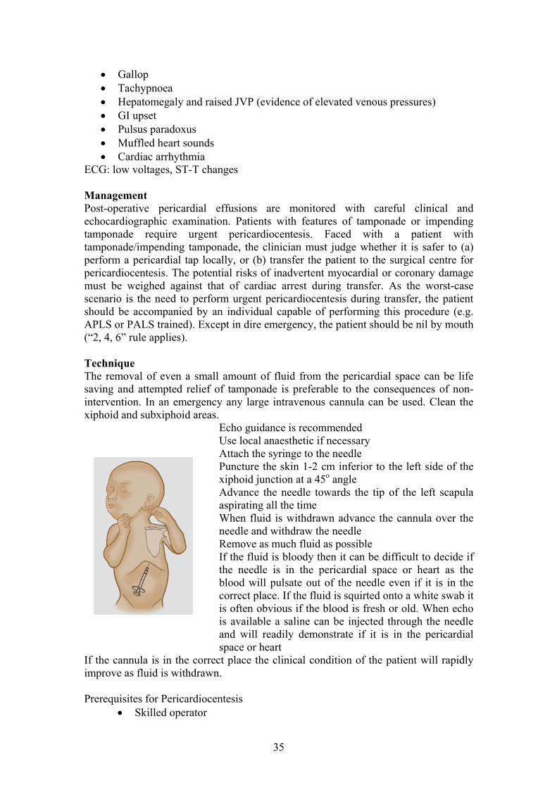
Pericardiocentesis for cardiac tamponade
Aetiology rdiac tamponade may ac
(e.g. coxsackie, mumps, adenovirus, and HIV), bacterial infection (e.g. TB), immwasaki disease), connective tissue diseases mediated diseases (e.g. rheumatic fever, Ka
(e.g. JRA, SLE) and uraemia. Pericardial effusion is also a commonspontaneously or following treatment with diuretics anti-inflammatory drugs such as aspirin or ibuprofen. Often the effusion is a feature in the early post-operative period and patients are discharged from the surgical centre only when it is clear thollection is not increasing in size. In other cases the effusion mc
apparent many days or even weeks post-operatively and forms part of the post-cardiotomy (Dressler’s) syndrome. Clinical features
Prodromal illness Disappearance of a previous pericardial rub
34
Gallop Tachypnoea Hepatomegaly and raised JVP (evidence of elevated venous pressures)
ECG: l ManagPost-operative pericardial effusions are monitored with careful clinical and chocardiographic examination. Patients with features of tamponade or impending
require urgent pericardiocentesis. Faced with a patient with tam (a) perform lly, or (b) transfer the patient to the surgical centre for per r yocardial or coronary damage mu b er. As the worst-case sce i t should APLS uth (“2, 4, Technique
n be life on-
e used. Clean the iphoid and subxiphoid areas.
Echo guidance is recommended necessary
needle m inferior to the left side of the
n advance the cannula over the
ilable a saline can be injected through the needle and will readily demonstrate if it is in the pericardial space or heart
If the cannula is in the correct place the clinical condition of the patient will rapidly improve as fluid is withdrawn. Prerequisites for Pericardiocentesis
erator
GI upset Pulsus paradoxus Muffled heart sounds Cardiac arrhythmia
ow voltages, ST-T changes
ement
etamponade
ponade/impending tamponade, the clinician must judge whether it is safer to a pericardial tap loca
ica diocentesis. The potential risks of inadvertent mst e weighed against that of cardiac arrest during transfnar o is the need to perform urgent pericardiocentesis during transfer, the patien
be accompanied by an individual capable of performing this procedure (e.g. or PALS trained). Except in dire emergency, the patient should be nil by mo6” rule applies).
The removal of even a small amount of fluid from the pericardial space casaving and attempted relief of tamponade is preferable to the consequences of nintervention. In an emergency any large intravenous cannula can bx
Use local anaesthetic ifAttach the syringe to thePuncture the skin 1-2 c
needle and withdraw the needle
xiphoid junction at a 45o angle Advance the needle towards the tip of the left scapula aspirating all the time When fluid is withdraw
Remove as much fluid as possible If the fluid is bloody then it can be difficult to decide if the needle is in the pericardial space or heart as the blood will pulsate out of the needle even if it is in the correct place. If the fluid is squirted onto a white swab it is often obvious if the blood is fresh or old. When echo is ava
Skilled op
35

Anaesthetist or intensivist and appropriate support staff onment with full monitoring and resuscitation equipment (e.g.
ment necessary for the procedure plications or the patient’s
ictate atched blood
e parent/guardian
ios
n (monitored bed). Transfer to PICU for post-arrest management.
Feeding problem Persisting haem Pericardial effusion Persisting res Persisting neu Ongoing infection
See the relevant sectiontransferred to Cardifand echocardiogram. 3.9 Inpatient Referrals
Inpatient referrals arsurgeons and general
Before reviewing the
by/discussed with the It is essential to clar
ve an outpatient-style consultation, a full examination , and chest x-ray (where appropriate).
Safe enviroperating theatre, PICU)
Equip Ability to admit/transfer to PICU should com
clinical condition d 2 units of cross-m Informed consent from th
Possible scenar 1. Well child with increasing significant pericardial collection with no signs of
response to treatment. Appropriate management – arrange for transfer to surgical centre for semi-urgent pericardiocentesis.
2. Child with significant pericardial collection and impending collapse due to
tamponade. Appropriate management – transfer to Main Theatres or PICU for urgent pericardiocentesis (paediatric anaesthetist / intensivist support).
3. Collapsed child. Appropriate management – perform pericardiocentesis at bedside
as part of resuscitatio
3.8 Post Surgical Transfers (in) Following cardiac surgery some cases are transferred back to Cardiff prior to final discharge home. This is usually because their postoperative course is slow or complicated. They may have:
s odynamic problems
piratory problems rological problems
in Clinical Problems or Surgical Complications. All patients f for post-operative management need an up-to-date ECG, CXR
e frequently received from neonatologists, paediatric paediatricians.
patient, ensure the consultation has been requested referring consultant. ify the clinical question being posed by the referring
team. If the question is not clear, discuss this with the cardiac consultant on-call. These patients should ha
and an ECG, echocardiogram
36

The findings should be discussed with the on-call consultant, preferably well d
by mented in the case record notes,
ort dic
nt to the parents, the GP and the patient’s sure the summary at the end is
in plain English). eferrals should be reviewed as necessary and follow-up
arrangements should be clearly stated.
, or chemo patients having LV function
ssessment between courses (if there have been no previous problems). All other echo
before the end of the normal working day – remember all echos are reviewea consultant.
Details of the consultation should be docuincluding information about the echocardiogram, and an inpatient rep
tated. Ensure copies of this report are seDGH (since the parents are receiving a copy, en
Inpatient r
NB – There is almost no such thing as a referral for “echo only”. Exceptions arefollow-up neonatal echos (check known PDA)arequests are regarded as a “referral for cardiology opinion”. Inpatient referral checklist:
Was the referral generated by a consultant? Is the clinical question clear?
Take a history and perform a clinical examination Arrange essential investigations (consider need for CXR)
Are the findings adequately documented in the medical record? Has the case been discussed with the consultant?
Has the clinical problem been addressed? Has the outcome been clearly communicated to the referring team/GP/DGH
paediatrician? Has an inpatient report been dictated, checked, signed and dispatched?
Are any outpatient arrangements in place?
ings. See Section 1.10 for Inpatient Report head
4 DISCHARGE AND TRANSFER PROCEDURES 4.1 General Principles
scharging a patient, imagine yourself as the doctor
ic in a few weeks’ time. Also picture yourself as the parent taking their child home after a stressful time in a
ey know whom to contact if they
ritten (and often verbal) communication is paramount. to
eded that long).
Before transferring or direceiving the transfer, or the paediatrician in the DGH, the GP, or the consultant seeing the patient in clin
specialist centre. Have they received clear communication about the status of their child and the management plan? Do thare worried? Clear w
On discharge, patients will be given the standard hand-written note to taketheir GP. Sufficient drugs should be prescribed for up to one month (if ne
37

Parents should be told to contact their GP and local pharmacy soon after discharge for a repeat prescription as many of the drugs commonly prescribed from hospital (e.g. furosemide suspension) need to be specially ordered and
It is the responsibility of the SHO or registrar on-call the day of the patient’s disc rdischa Summaor chec um standard, all discharge summaries hould be dictated, typed and posted within five working days of discharge from the
e undertaken. e transferred safely in the family car or in a hospital taxi
with a nurse escort. Patients on m n by bolus, patient
receiving low-flow o transferred by the nursing staff.
Patient fusion) must be ff with a paramedic ferred under the superv
tail all of the relevant m an up-to-date medication list.
ld also be sent.
If time constraints do not permit a typed summary, a clear hand-written note is r typing later. This permits accurate
udit of transfers out of the Unit.
Transfer Checklist Is there a clear indication for transfer?
the chemist will require a few days to obtain them.
ha ge to ensure a summary is dictated that day. See Section 1.10 for a guide to rge summary headings.
ries on patients with protracted or complicated admissions should be dictated ked by one of the registrars. As a minim
shospital. 4.2 Transfers to Other Hospitals Before transferring a child to the surgical centre consider what level of transfer should b
Well patients may b
inimal support (e.g. intravenous drugs givexygen) may be appropriately
s receiving a higher level of support (e.g. prostaglandin intransferred with an experienced member of the medical sta
crew. Intubated patients will normally be transision of NICU or PICU consultant staff.
Wherever possible, a typed summary should be provided and this should deedical details, including
In some circumstances a photocopy of the patient’s medical notes shou
require, but a summary should be dictated foa
Ensure patient is fit for transfer Communicate with medical staff in the receiving unit
Ensure appropriate ambulance has been arranged Ascertain which members of the medical/nursing team should accompany the patient
Written transfer letter (preferably typed) Document current medications and recent lab results
Should any of the notes be copied? Ensure the proper equipment is available (transfer pack)
Ensure safe level of monitoring equipment Telephone receiving hospital just prior to departure
Ensure a discharge summary is dictated
38

4.3 Discharges Following Cardiac Surgery The standard hand-written GP note should be provided and the post-surgical
handout for the parents should also be given. Where the post-operative course has been prolonged or complicated, the DGH
consultant and GP should be informed of the discharge by telephone.
y: Normal bathing is permitted but the wound should be protected until the scabs
st children should not attend school until after their first review in Nurse-al rule the child should avoid PE (or equivalent
4.4
A typewritten summary should be dispatched within five working days. Wound Care and Physical Activit
have come off. Parents should know that if the child is unwell prior to the OPD appointment,
the ward should be informed and a review will be arranged. Mo
Led Clinic or OPD. As a generactivity) for 6/52 and contact sports (or equivalent) for 3/12. Other disease-related activity restrictions may apply.
Follow-up Arrangements: 2 week appointment Nurse-Led Clinic, 4 week appointment Cardiology OPD (at local hospital where possible) – ensure these arrangements are in place.
Discharge Checklist Is the patient fit for discharge?
Are the medical notes complete with full documentation of the patient’s status? Has there been adequate communication (with parents, nursing staff, GP, referring
paediatrician? Is the GP letter/TTH complete?
Are suitable follow-up arrangements in place? Has the discharge summary been dictated?
Death of4.5 a Patient
Inform the on-call consultant immediately and the child’s consultant, even if not on-call, at the earliest opportunity. Consider the need for the coroner's office to be informed of the death. It is Unit policy to ask the family to consider a hospital PM
a coroner’s case. Use the new hospital PM
Contact:
GP and H
sent to Ca Tony Bra ent counselling)
y have given ng the initial
bereavem C Consul Consultant Cardiac Surgeon (in the case of post-operative death)
(either full or limited) if the death is notconsent form.
ealth Visitor DGH Cons Birth Clerk/Community Paediatrics (to prevent routine appointments being
ultant
rdiff dley (So ereavem
patients) cial Worker, who is also trained in b
– NB Tony can only be directly involved with the family if thetheir consent t duri– if appropriate, try to seek this consen
ent discussions ardiac Liaison Nurse
tant Obstetrician (in cases of neonatal death)
39

If the death was unexpected, consider the need to inform the Clinical Director
As should hould be sen o evious
ospitals. If not all the information is available for the summary at this time explain
A leaflet is available on the ward that gives advice to relatives of the deceased about obt iconsulteve t
and the Child Death Review team
the notes are usually “lost” in the pathology department after death a summary be dictated at the time the death certificate is signed. Copies of this s
t t everyone who has been involved in the patient's care, including prhan additional report will follow.
ain ng the death certificate and contacting funeral directors. The patient’s ant will normally write to the family of the deceased child a short time after the
nt o see if they want to discuss the death of their child further.
5 INVESTIGATIONS 5.1
Drug Investigation imen Comments
Drug Monitoring and/levels
Spec
Amiodarone Opt
Check TFT and LFT at baseline, at 1 month and then every 6 months.
rm CXR every 12 months.
TFT, LFT, CXR and consider
halmology Lithium heparin Perfo
(changes are bottle Drug levels (+ metabolites) can be
reversible) checked through Toxicology Lab, but rarely indicated.
Bosentan
U&Esp
aminases may become elevated sentan.
onthly. GOS 0207 905 2321
, LFT (must ecify ALT and
Lithium heparin bottle (plus
Transon bo
AST); add FBC if EDTA if having Bloods done mEisenmenger FBC) Fax results to
DT ottle Desired range varies – check with transplant team. A bCiclosporine Drug level EFax results to GOS 0207 813 8440
Digoxin Drug level Plain/clotted sample
Trough level is needed (6 hours post dose) Desired level = 1.0-2.0 g/mL Check U&E at same time. Consider need for ECG if level abnormal.
Flecainide ECG Drug level and Plain/clotted
sample
Trough level 6 hours after last dose. QRS duration on ECG should lengthen by <25% of baseline. Desired level = 0.15-0.9
Tacroli for Tacrolimus -Switchboard for dosing advice). mus LFT, U&E, FBC
and drug levels
Specimen bottle Desired range varies – check with transplant team (bleep 0600 via GOS
EDTA Fax results to GOS 0207 813 8440
5.2 During working hours these are requested via the ECG Department (6396). Fill in a yel EC ulwen Ward. Familiarise yourself with the use of this machine.
Electrocardiogram
low request form.
Gs may be performed out-of-hours using the machine kept on He
40

` Patient thmia should have a 12-lead ECG in the rrhythmia, during pharmacological or other cardioversion therapy, and once sinus
ents” section of the shared directory. hen assessing and ECG you should determine the following:
PQRST morphology Ventricular forces Intervals
PR
o QTThe ECG computer pack ulate the intervalsmeasurement are common and you should ideally calcu al
lf. A 440- sig T ere t ain dr ontraindica
www.QTdrugs.co
s admitted with a suspected arrhyarhythm has been restored. 5.2.1 Basic ECG Interpretation For more detailed information about quantitative ECG interpretation (e.g. age-related intervals), look on the “MeasuremW
Correct patient demographics Heart rate Rhythm Electrical axis
oo QRS duration
age will calc
QT interval >he use of certm
for you, but errors in the QT late the corrected QT intervnify the presence of long Qted (for a list see
(QTc) yoursesyndrome wh
460 msec couldugs is c
). The most common fo on ly-used method r QTc calculati is the Bazett formula:
R-R interval
QT interval
R-R
rval
interval
QT inte
QTc = QT_______ RR interval
41

The Bazett formula over-corrects for slow and under-corrects for fast heart rates. An
ec) + (1000 – RR interval in msec / 6.5)
During working hours, echos on cardiac patients are arranged via the Echo Technician (Ms Viv Booker – Extension 3920, Bleep 5503). Requests for echo should be communicated to her early in the day. She has clinic commitments Monday, Tuesday and Thursday AM, and Wednesday and PM. Ech e ms:
such thing as a referral for “echo only”. hos (check known PDA), or chemo
patients having LV function assessment between courses (if there have been no previous problem
re echos for ught to the attention of the relevant consultant well before the
end of the normal working day. urs, this
must be discussed with the consultant on call.
5.3.1 Echocardiogram to rule out cardiac source of embolism Young patients with stroke should have a detailed echo to exclude a cardiac source of embolism. This includes assessment of right-to-left shunt across PFO with bubble-contrast echocardiography which requires an experienced echocardiographer and assistant. The recommended protocol is as follows:
Intravenous cannulation is usually performed in the upper limb. The procedure for Valsalva with release is explained to both the subject and
the parents. The subject is instructed to inhale deeply and exhale through the nose. Concurrently either the subject or the parent blocks the nasal passages externally with release on indication from the echocardiographer.
Baseline transthoracic echocardiogram is performed. 5 ml of normal saline is drawn up into a 10 ml Luer-lock syringe and attached to a three-way tap with a further 10 ml Luer-lock syringe attached to another port. The final port is connected to the subject.
The saline is agitated with air and the subject’s blood (5 ml 0.9% saline to 0.5 ml of air and 0.5 ml of patient’s own blood) by plunging it between the two syringes, ensuring age of blood. When the saline m th the patient breathing normal assage of contrast into the left atrium. Sniffing can be requested at this time as this can sometimes open up a PFO and trigger a bubble shunt. A short sharp sniff is needed.
alternative method, particularly for faster heart rates, is the Framingham formula: QTc (in msec) = QT (in ms 5.3 Echocardiogram For more detailed information about normal values (including Z-score graphs), lookon “Measurements” section of the shared directory.
o r ferrals from other tlmost no
ea There is a Exceptions are follow-up neonatal ec
s). All other echo requests are regarded as a “referral for cardiology opinion”. Ensure the referral has been discussed by the child’s lead consultant. Ensure
the formal referral request document is filled in with adequate information. Remember – all echo reports are confirmed by a consultant - ensu
review are bro
If there are referrals for echocardiography out of normal working ho
that they are fully screwed on to prevent leakix is sufficiently agitated, initial injection wi
ly is performed to assess spontaneous p
42

If no shunt is seen, the subject is instructed to exhale forcefully against the blocked nasal passages and mouth while a slow injection of agitated saline is performed.
ent in the right atrium throughout the Valsalva. After an adequate Valsalva, where the left
isibly smaller, and with contrast present in the right
sed. This is repeated on up to five subsequent occasions, ensuring an adequate study. Adequacy in this case
Large shunt >20 bubbles ere would be opacification of the left ventricle.
visualised coming down the pulmonary veins usually
ss
ophic cardiomyopathy*
5.4.2 The me n on theincreas the standa ). In
A slow injection is needed to ensure adequate contrast is pres
heart should become vatrium, the echocardiographer indicates to release the Valsalva and the number of bubbles crossing to the left atrium is asses
means that a Valsalva caused a reduced size in the left heart and was released when bubbles were filling the right atrium.
o Small shunt - <6 bubbles o Medium shunt 6–20 bubbles oo Massive shunt - th
False-positives:
Pulmonary arteriovenous malformations (these always give a shunt without Valsalva, bubbles can bewithin 3 heart beats
Use of colloid rather than saline to mix the contrast (in some patients, colloid contrast may pass through normal pulmonary capillaries allowing microbubbles to reach the left atrium when no PFO is present).
Reference: Arch Dis Child 2008;93:255-259
5.4 Exercise Test Contact the ECG Department and fill in a request form. Most children over 4 years of
hould be able to manage the exercise treadmill. age s 5.4.1 Indications for exercise test These include:
Objective assessment of exercise tolerance Assessment of sinus node disease Provocation of exercise-induced arrhythmias Heart rate response in complete heart block Risk assessment in WPW Assessment of long QT syndrome (use modified Bruce protocol, regardle
of age) Assessment of peak oxygen consumption (VO2 peak) and aerobic threshold
(cardio-pulmonary exercise test, CPET) Assessment of severity of asymptomatic severe aortic stenosis* Risk assessment in hypertr
*meticulous attention to BP required (see below)
Bruce protocols thod of assessment at UHW is the exercise treadmill. It provides informatio
haemodynamic and electrocardiographic response to a staged, progressive e in load over a short period of time. In relatively fit, older patients rd Bruce protocol should be used (arrange with the ECG technician
43

you e ing assess Oth m
Exercis
Pat Che inutes (prompted by machine) Wa Con the
s reached the end of Stage 5 (standard Bruce) and the target HR is
, consultant
mination:
icular ectopy, VT, CHB) Hypotension or failure to increase BP with symptoms of dizziness or chest
c BP >220-240 in with significant flat or downsloping ST depression or ST elevation
undle branch block) 5.4
a) ly LBBB
and may mask ischaemia– consider
5.4.5 Helpful tips
Continual gentle encouragement of the patient will maximise the information y
ng r patients, those with moderate or severe exercise limitation, and those befor long QT syndrome, the modified Bruce protocol should be employed.
er easurements employed on occasion: Saturation data (does the patient desaturate on exertion?) Peak flow rate pre- and post-exertion (is there exercise-induced asthma?) Echocardiography (how high does the aortic gradient go on exertion?)
e test protocol: Check resting ECG (technician) and BP (doctor) Checking standing ECG (technician)
ient commences exercise ck BP and 12-lead ECG every 3 mtch for arrhythmias tinue until patient exhausted, there is a medical indication to stop, or
patient haachieved
Document reason for cessation Take report to consultant’s secretary (a report will be entered on Cardiobase
by the consultant or one of the registrars) If the test was terminated early because of an unexpected abnormality
please discuss this with the patient’s consultant, or the servicellowed home before the patient is a
5.4.3 Indications for ter
Genuine patient exhaustion Significant arrhythmia (SVT, increasing ventr
pain; hypertension with systoli Chest pa
(>2-2.5 mm in the absence of pre-existing b
.4 Difficulties in interpretation Breathlessness due to other cause (e.g. asthm Pre-existing BBB, especial LVH WPW Digoxin (affects ST segments – consider stopping drug prior to exercise test) -blocker therapy (blunts HR response
stopping drug prior to test)
obtained in the stud BP should be measured with the patient’s arm lifted off the equipment and
supported by the operator – this minimises machine noise If there are difficulties auscultating the BP, use the palpation technique (radial
or brachial artery)
44
a surrogate for peak BP
ntial in patients being exercised with ssure to rise by more
itate 87-2991, J
Am Coll Cardiol 2000;36:2212–8)
reful BP measurement also applies in AS – a flat BP response
ng e filed in
the patient record until the result 5.5.Uses in
atypical seizures and syncope of unknown
n rrhythmic medications
Ho to o
are issued for et up as a loop recorder (attached via leads to
ton the device saves the latest symptoms (patient activated).
subcutaneously (Reveal device), but this severe, infrequent episodes. If an episode is experienced, the patient
needs to attend the ECG department for the device to be interrogated. 5.6 Blood pressure varies significantly over a 24 hour period and single, elevated
easurements do not necessarily reflect the true situation. Ambulatory monitoring tivities provides several readings throughout the day and night,
and fimportant in differentiating spuriously high readings (white coat effect) from true
mmenced (e.g. after
ol in patients taking anti-hypertensives
Meticulous BP measurement is esseHOCM – a flat BP response (failure of systolic blood prethan 20–30 mm Hg from baseline), drop in BP during exertion, or precipdrop in BP post-exertion are all poor prognostic signs (Circulation. 1997;96:29
The need for casignifies important AS
5.5 Ambulatory ECG MonitoriThis is requested through the ECG Department (6396). Reports should not b
has been entered on to Cardiobase.
1 Holter monitoring clude:
Investigation of palpitations, aetiology
Assessment of severity of known arrhythmias Assessing cardiac rhythm in those at risk of arrhythmia (e.g. Tetralogy of
Fallot, post Senning or Mustard procedure, Fontan circulation, pre-excitation on ECG, 1st or 2nd degree heart block)
Risk assessment of cardiomyopathy, CHB and LQTS Assessment of pacemaker functio
ti-a Assessment of response to an
lter monitoring can be undertaken in any age group and can run from 24 hours up ne week (you need to specify the required duration on the request).
5.5.2 Event recorders
wo types of device are provided: CardioCall and King of Hearts. TheyTup to two weeks. Both devices can be sthe ech st continuously; when the patient presses a butloop) or placed on the chest for recording during Rarely, a recording device may be implantedis reserved for
Ambulatory BP Monitoring
mduring routine daily ac
of ers a profile of blood pressure during rest as well as activities. It is especially
sustained hypertension. It is helpful in: Assessing whether anti-hypertensive therapy should be co
CoA repair) Assessing BP contr
45

Deciding on optimal time to take anti-hypertensive medications Evaluation of hypotensive symptoms in patients taking anti-hypertensives,
diuretics and ACE inhibitors The unon-docheck There onitors available for use in paediatric patients. Most
on the graph. These monitors print the actual adings and mean BP values during time periods throughout the 24 hours.
les show a fall in e blood pressure level during sleep - evidence suggests that patients with no fall
d risk of end organ damage.
Osc o
c ff is permanently on the arm and the recorder placed on a belt or rucksack. The minant arm is normally used, but in coarctation patients it is mandatory to the right arm BP (there may be a BP gradient between the upper limbs).
are several ambulatory muse the oscillometric technique and correlate the reading with simultaneous recording of the heart rate which is also plotted re Treatment decisions should be based on the mean values over the monitored period and take into account the shape of the profile. Normal 24 hour profith(non-dippers) have an increase
ill metric mean ambulatory BP values in healthy children:
Percentile for 24 hr period Daytime percentile * Nighttime percentile †
Height in cm (n) 50th 95th 50th 95th 50th 95th
Boys
120 (33) 105/65 113/72 112/73 123/85 95/55 104/63
130 (62) 105/65 117/75 113/73 125/85 96/55 107/65
140 (102) 107/65 121/77 114/73 127/85 97/55 110/67
150 (108) 109/66 124/78 115/73 129/85 99/56 113/67
160 (115) 112/66 126/78 118/73 132/85 102/56 116/67
170 (83) 115/67 128/77 121/73 135/85 104/56 119/67
180 (69) 120/67 130/77 124/73 137/85 107/56 122/67
Girls
120 (40) 103/65 113/73 111/72 120/84 96/55 107/66
130 ( 105/66 117/7558) 112/72 124/84 97/55 109/66
140 (70) 108/66 120/76 114/72 127/84 98/55 111/66
150 (111) 110/66 122/76 115/73 129/84 99/55 112/66
160 (156) 111/66 124/76 116/73 131/84 100/55 113/66
170 (109) 112/66 124/76 118/74 131/84 101/55 113/66
180 ) (25 113/66 124/76 120/74 131/84 103/55 114/66
*Daytime: 8 am to 8 pm / †Nighttime: midnight to 6 am Reference: The Journal of Pediatrics Volume 130, Issue 2, February 1997, Pages 178-184
46

5.7Ind t
ollapse
rocedure
ose)
specified on the request e for collapse, or (b) ntil baseline heart rate
rises 25%-30%]); use Ametop or Emla as appropriate; allow 15-20 min recovery s
i ire up the ble
Continuous heart rate and BP m ring is required atient or 10-15 mins
tage I: Dr e Stao Tilt the table (head up!) to 70 degrees for 20 minutes, recording heart
rate and BP continuously o Make a note of any significant ms sea, ing,
presyncope) and check BP imo If the patient bla t lay le flat immediately and ensure the
ECG is being recorded continuously o If th nt doe xper ync thin nute eed
dire Stage hout in tie pin Stage II: Pharmacological challenge with sublingual GTN 400-500
icrogram : o Administer 400 microgram of sublingual GTN and continue the tilt for
a fu -20 m o Make a note of any significant ms sea, ing,
presyncope) and check BP im telo If the patient bla t lay le flat immediately and ensure the
ECG and blood pressure are being recorded continuously o If no syncope occurs with GTN term he ure turn
patient to supine position. Allow patient to rest f 0 m ntil they feel ready to stand up.
rovide a w t-ou ults e a report is entered into Cardiobase
Tilt Test ica ions:
Investigation of unexplained c Suspicion of vasovagal syncope (a.k.a. neurocardiogenic syncope or neurally
mediated syncope) Assessment of therapy
P The test is booked through the ECG department The test is overseen by a doctor with APLS or PALS certification Check resuscitation equipment and drugs prior to beginning Ensure you have a drug chart and a supply of GTN (400 microgram d Explain to the patient and parents what the procedure involves; mention that if
GTN is used it may cause a headache No IV line is required for sublingual GTN Tilt Test but make sure that an
immediate access to IV line insertion is available in any emergency situation. NB - Insert IV cannula before the test only if this isform (possible reasons: (a) the history is very positivisoprenaline is to be given [dose 0.02-0.05mcg/kg/min u
before commencing te t The ECG technician w
straps onll “w ” the patient and secure the supporting
tilt taonito
Allow the p
to lie supine f
S ug Fre te:
sympto (nau sweatmediately.
cks ou the tab
e patie s not e ience s ope wi 20 mi s, procctly to II wit return g the pa nt to su e
m spray
rther 15 inutes.sympto
y. (nau sweat
mediacks ou the tab
inate t proced and reor 10-2 inutes u
P ritten assessment of the test on the prin t. Discuss the reswith the requesting consultant and ensur
47

5.8 MRI or CT Scan l in assessing cardiac anatomy (particularly the great vessels), cardiac
n and other haemodynamic issues. r
to be booked well in advance (see section 3.4 for ission arrangements for inpatient scans).
there is a small risk of a rare rosis.
rement to provide nths
and ventricular function. r (adult cardiologist).
Organise thpulmonarybranch PS)
Usefufunction, severity of valve regurgitatio
Scans are organised through Dr Andrew Wood, Consultant CardiovasculaRadiologist.
Scans under GA have adm
If the patient has pre-existing renal dysfunction but irreversible skin complication called nephrogenic fibrosing scle
o To guard against this adverse effect it is now a requithe MRI coordinator with “up-to-date” U&E/creat (i.e. within 4 moof the scan).
o Depending on the age and location of the patient this test can be done by the GP, the local DGH or at UHW prior to the investigation.
o In practice it is best to have the results at least a week before the scan is due – try not to leave until day of admission for MRI.
5.9 Isotope Scans These use a small dose of radio-isotope, which is excreted in the urine. Ensure the child’s mother is not pregnant! Sedation may be required; intravenous access will be required – co-ordinate with the children’s Clinical Investigation Unit. 5.9.1 Cardiac Nuclear Scanning
aemia Main indications include assessing myocardial ischequests need to be discussed with Dr Richard WheeleR
5.9.2 Lun pg erfusion scan
rough Dr John Rees, Consultant Radiologist. Indications are exclusion of embolus (V/Q scan) and assessment of differential lung perfusion (e.g. in .
48

6 CLINICAL PROBLEMS
6.1 6.1Indicat les below):
o
An l
er syndrome – relative indication)
alve with history of embolism despite anticoagulation
agents such as ypridamole or clopidogrel (limited data in children).
of ontinuing aspirin vs the small risk of Reye’s syndrome. Parents should be instructed
n this situation.
rile illness; however patients with a weaker indication for aspirin (e.g.
hronic cyanosis with a cavopulmonary shunt, Kawasaki disease) should discontinue aspirin temporarily during the feverish phase of an illness. The consultant may consider use of dypridamole or clopidogrel during this period (NB the data sheet for clopidogrel also advises discontinuation during chicken pox, etc). 6.1.2 Devices and Stents Indication Treatment Duration
Anti-platelet Therapy and Anticoagulation
.1 Aspirin and Anti-platelet Therapy ions for use of aspirin in children (plus see tab
Anti-inflammatory action – treatment of o Acute pericarditis o Kawasaki disease (acute phase)
Acute rheumatic fever
ti-p atelet therapy – treatment of o Kawasaki disease (convalescent phase) o Systemic-pulmonary shunt o Chronic cyanosis (e.g. cavopulmonary shunt, Eisenmeng
o Prosthetic v(added therapy)
If aspirin cannot be used (e.g. allergy), consider the use of other d In the event of development of chicken pox, herpes, influenza, rubella, or other severe flu-like febrile illness, the clinician will determine whether the risks / benefitscto telephone to ask for advice i Patients on aspirin for a B-T shunt should not have therapy discontinued, evenduring a febc
ASD device Aspirin 3-5 mg/kg once daily, usual maximum 75 mg once daily*
6 months
VSD device Aspirin 3-5 mg/kg once daily, usual maximum 75 mg once daily*
6 months
Aortic stent Aspirin 3-5 mg/kg once daily, usual maximum 75 mg once daily*
6 months
Pulmonary artery stent Aspirin 3-5 mg/kg once daily, usual maximum 75 mg once daily*
6 months
PDA device Not indicated
49

*NB – there may be individual clinical reasons to extend treatment, to supplement with other antipla icoagulation.
spirin dose of 150 mg or 300 mg may be used in older adolescents. ing intercurrent illness.
on
telet agents (e.g. clopidogrel), or to use formal antASee section 6.1.1 for advice regarding use of aspirin dur 6.1.3 Valve replacement Indication Treatment DuratiTis (triscusp
nce um 75
6 months sue valve id, m
Aspirin 3-5 mg/kg o, usual maximitral, aortic) daily
mg once daily* Tissue RV
ulmonary valve
tran thoperation)
-5 mg/kg once aximum 75
6 months -PA conduit or Aspirin 3daily, usual mp
(any type, including sca eter l
mg once daily*imp ant, Ross
Prosthetic (any type inc naortic)
rget INR 3.0
ange 2.5-3.5)**
Indefinite valve Warfarin – taludi g mitral or ± 0.5
(i.e. r*NB – there m
ith other anti g. clopidogrel), or to use formal anticoagulation.
a higher range, e.g. small alve, or increased risk of thrombosis – this needs to be clearly specified in the
dication Treatme
ay be individual clinical reasons to extend treatment, to supplement platelet agents (e.w
Aspirin dose of 150 mg or 300 mg may be used in older adolescents. See section 6.1.1 for advice regarding use of aspirin during intercurrent illness. **NB – there may be individual clinical reasons to usevmedical notes and in the INR booklet. Our unit experience tells us that an upper range of 4 has been very safe in the past. 6.1.4 Cavopulmonary shunt / Fontan In nt Duration Glenn / superior cavopulmonary shunt
Aspirin 3-5 mg/kg once daily, usual maximum 75 mg once daily*
To continue until Fontan / TCPC**
Fontan / TCPC Warfarin – target INR = 2.5 ± 0.5 (i.e. range 2-3)
Indefinite
*NB – See section 6.1.1 for advice regarding use of aspirin during intercurrent illness. – easons to extend treatment, to supplement
(e.g. clopidogrel), or to use formal anticoagulation.
indications Indication Treatment Duration
**NB There may be individual clinical rwith other antiplatelet agents 6.1.5 Other
Modified Blalock-Taussig Shunt
Aspirin 3-5 mg/kg once daily, usual maximum 75 mg once daily
To continue until ndefinitive s
ext urgery;
treatment to continue with ess febrile illn
Kawasaki Disease HA or UK guidelines
Refer to AHA or UK guidelines
Refer to A
Pulmonary Arterial Hypertension
EITHER Aspirin 3-5 mg/kg once daily, usual maximum 75 mg once daily*
Indefinite
50

OR Warfarin – target INR range 1.8-2.5
Post L heart Aspirin 3-5 mg/kg once 3 months electrophysiology ablation daily, usual maximum 75
g once daily* m*NB – there may be individu asons to extend tr o supplement
platelet agents se fo oagulation. g or 30 adolescents.
See section 6.1.1 for advice r spirin during intercurrent illness.
et a c su diothorac Surg 3-92
uctural He 133: 593S - 629S. erapy in 2008;133;887S-968S
cing antico rfme heparin first.
.
T
he maintenance dose by 10% and recheck APTT tio 6-8 hours later.
r, then restart at a reduced maintenance e by 10-20%).
oses o d i ppropriate to accept slightly low APTT rat
Therapy with low molecula
age Patients in whom venous access for administration and monitoring of heparin
FBC PT APTT
ilia screen
al clinical re eatment, twith other anti (e.g. clopidogrel), or to u
0 mg may be used in olderegarding use of a
rmal anticAspirin dose of 150 m
References: 1. Guideline on antiplatel
2008;34:7nd anticoagulation management in cardia
art Disease. Chest 2008; Neonates and Children. Chest
rgery Eur J Car
2. Valvular and Str3. Antithrombotic Th
6.1.6 Commen agulation (heparin and wa
nced onarin)
Most patients will be com Therapy with unfractionated (standard) heparin Loading dose: 50 – 100 U/kg Maintenance: 10 – 20 U/kg/hr (higher doses may be needed) Check APTT/KCCT 6 – 8 hours after starting therapy. Aim for ratio of 1.5 – 2.0 If APTT ratio is LOW – increase maintenance dose by 10-20% and recheck APTratio 6-8 hours later. If APTT ratio is >2 and ≤3, reduce traIf APTT ratio is >3, stop heparin for 1 houdose (reduc NB – if maintenance d f >35 U/kg/hour are require
ios. t may be a
r weight heparin
Indications for use: Neonates needing anticoagulation Any patient requiring anticoagulation and deemed to be at risk of haemorrh
is difficult. Baseline investigations:
?thromboph (if indicated)
51

tor Xa levels: Target anti-facT mLreatment dose: 0.5-1.0 U/
Pr mL ophylactic dose 0.1-0.3 U/ Starting dose:
8 yr Age < 2 mo Age 2 mo – 1
Treatment dose 1.5 mg/kg/dose SC q 12o 1 mg/kg/dose SC q 12o
Prophylactic dose 0.75 mg/kg/dose SC q 12 0.5 mg/kg/dose q 12 o o
The maxim 2 mg/kg/dose q 12 , i.e. 4 mg/kg/day – see BNF for adult dose
ffectiveness of therapy is assessed by monitoring Anti-Xa levels; the ideal dose is L 4 to 6 hours after
vels on onday/Wednesday/Friday afternoons. Requests outside of this time must be
um dose is o
Ethe one that reflects plasma anti-Xa concentrations of 0.5-1.0 U/minjection. The lab routinely checks anti-factor Xa leMdiscussed with the lab/haematologist. Adjusting LMW heparin in children (therapeutic dose):
Anti-Xa Hold next Dose Repeat anti-Xa
level (U/mL)
dose? change? measurement
<0.35 No Increase by 25%
4 hours after next dose
0.35 - 0.49 No Increase by 10%
4 hours after next dose
0.5 - 1.0 No No and monthly thereafter (4 Next day, then 1 week later,
hours after morning dose)
1.1 - 1.5 No 20% Decrease by
Before next dose
1.6Decrease by Before next dose then 4 hours
after next dose - 2.0 3 hours
30%
>2mL 40%
<0.5 U/mL .0
Until anti-Xa 0.5 U/
Decrease by 12 hours until anti-Xa level Before next dose, then every
Long-term therapy – chec Sou e: f antithrombot
k bone densitometry every 6 months. ic therapy. BMJ 2003;326:93-96 + BCH policy
Loa nRemember to check baseline PT/INR. If PT is >1.3, the patient will need a lower loading dose. The recommended loading dose is 0.1 – 0.2 mg/kg. In practice the following guide may be applied:
rc ABC o
di g with warfarin
52

Age group Loading dose Comment
Infants 1 – 2 mg Check INR next day
Young children 3 – 5 m k INR next day g Chec
Older children 5 – 8 m INR next day g Check
Teenagers Day 1: 8 mg / Day 2: 8 mg Check INR on the morning after second dose
Adults 10 / 10 / 5 mg over successive days
C ay heck INR 2nd – 3rd d
In patients on heparin who are being loaded on warfarin, it is essential to send a full oagulation screen. A high KCCT/APPT will lead to an erroneously high INR level. Ifc
ation to ee INR
cept for quality-control when ardi ffice. Sp trips are us ne (these are
on pres but a supply the wa has not been achine should not be operating the machine.
e of the iac Liaison Nu show you erated. Basic ins s follows:
1 Gather all the necessary equipment (INR mach d cet, co wool balls) rn machine on
Check battery power (batteries need ng weck da d time are corre
Ensure coagucheck strip code corresponds with machine – press the “M” tton to e mach ady when t bol display. Put in
strip. Machine begins From the n ve H (<15 sec old) drop of blood on to the hole/reservoir in the strip. Use as large a
flashes a “clock” . If the drop was too
5 The machine then displays the INR reading – record this in the INR books.
pectedly deranged, consider the cause (intercurrent illness,
ee
only the INR is checked, the true reading off heparin may be much lower. 6.1.7 INR Sampling It is the responsibility of the person taking the blood sample for INR estimfind out the result and prescribe the appropriate dose of anticoagulant (s
rotocol below). p An INR machine is kept on the ward most of the time exit is in the Cavailab
ac Liaison Ocription
ecial s is kept on
ed with the machird). Anyone whole
trained to use the m Ask on Card rses to how the machine is op
tructions are a
ine, coagucheck strip, pen an
hen showing only one bar)
lan2 Tu
tton
chargict Ch te an
bu3 Th
confirm ine is re he “strip” sym flashes on the
to count down from time the countdow
180. starts, you ha4 180 seconds to place a FRES
ine drop as possible. When the drop is applied the machsymbol on the display to show the INR is being processedsmall error 5 flashes on the display.
If an INR reading is unexdrug interaction, alcohol ingestion). .1.8 INR Protocol 6
Use this protocol when prescribing warfarin. If unsure, check with consultant. (sWarfarin Dosage Table in section 6.1.9)
53

INR Action/Comment
<1.8 NB with prosthetic m ,
er 2.2-2
Discuss with consultanArrange admission for ts with a prosthetic valve. Patients with other ind ion may
dose or ten an outp
itral valveif INR is undwith consultant
.4, discuss need increased be overseen as
t. heparinisation in patien
ications for anticoagulat limited re-load, but this can ofatient.
>1 below target rang ince Increase dose by 3 rements
>0.5 – 1.0 below target range Increase dose by 2 increments
0.2 – 0.5 below target range Increase dose by 1 increment
Target range No change in dose
<0.2 above target range No change (depending on trend)
0.2 – 1.0 above target Reduce dose by 1 decrement
1.01 – 1.5 above target range Reduce dose by 2 decrements
>1.5 above target range Halve dose, discuss with consultant; repeat INR next day; restart with dose reduced by 1 decrement.
INR >6 or history of bleedi
Discuss with senior – confirm reading with lab sample. ng* Very high INR levels will require hospital admission
*see Table below
with consideration of FFP or vitamin K. Consider discussing case with Haematology team.*
Recheck INR:
Stable (and no worrying trend up or down) 4-6 weeks Dose changed 1 place up or down 2 weeks (depending on trend) Dose changed >1 place up or down 1 week (depending on trend) Half dose / reload 1-3 days
Health Board Policy for high INR (Feb 2004) vs Unit experience Clin UHB Policy Unit Experience ical Situation INR 6-7.
9 with no bleeding: Stop warfarin; restart when INR Confirm reading with labsample; reduce usual <5 warfarin dose (at least halve), recheck daily
INR 8-1blee g
Stop warfarin; restart warfarin Confirm reading with lab
restart when INR <5; admit
0 with no or minor din when INR<5; give vitamin K 0.5-
1 mg iv (adult dose); recheck INR sample; omit warfarin,
next day for daily INRs INR 0bleeding
concentrate (discuss with
>1 with no or minor Stop warfarin; restart warfarin when INR<5; give vitamin K 0.5-1 mg iv (adult dose); daily INR; consider hospital admission
Confirm reading with lab sample; stop warfarin; restart when INR <5; consider vit K or prothrombin complex
Haematology); admit for daily INRs
Major bleeding with high INR Stop warfarin; involve Haematology; give prothrombin comple
HDU or PICU admission; Haematology consultation
x concentrate (PCC)*; take blood to measure pre- and post-concentrate INR and coagulation factors; give vit K
*PCC dose: INR 2-3.9 [25 U/kg]; INR 4-5.9 [35 U/kg]; INR >6 [50 U/kg]
54

6.1.9 Warfarin dosage table (doses shown in mg) 10 5 9.5 / 10 / 10 4.5 / 5 / 5 9.5 / 10 4.5 / 5 9.5 / 9.5 / 10 4.5 / 4.5 / 5 9.5 4.5 9 / 9.5 / 9.5 4 / 4.5 / 4.5 9 / 9.5 4 / 4.5 9 / 9 / 9.5 4 / 4 / 4.5 9 4 8.5 / 9 / 9 3.5 / 4 / 4 8.5 / 9 3.5 / 4 8.5 / 8.5 / 9 3.5 / 3.5 / 4 8.5 3.5 8 / 8.5 / 8.5 3 / 3.5 / 3.5 8 / 8.5 3 / 3.5 8 / 8 / 8.5 3 / 3 / 3.5 8 3 7.5 / 8 / 8 2.5 / 3 / 3 7.5 / 8 2.5 / 3 7.5 / 7.5 / 8 2.5 / 2.5 / 3 7.5 2.5 7 / 7.5 / 7.5 2 / 2.5 / 2.5 7 / 7.5 2 / 2.5 7 / 7 / 2 / 27 / .5 2 .5 7 2 6.5 / 7 / 7 1.5 / 2 / 2 6.5 / 7 1.5 / 2 6.5 / 6.5 / 7 1.5 / 1.5 / 2 6.5 1.5 6 / 6.5 / 6.5 1 / 1.5 / 1.5 6 / 6.5 1 / 1.5 6 / 6 / 6.5 1 / 1 / 1.5 6 1 5.5 / 6 / 6 0.5 / 1 / 1 5.5 / 6 0.5 / 1 5.5 / 5.5 / 6 0.5 5.5 0 / 0.5 / 0.5 5 / 5.5 / 5.5 0 / 0.5 5 / 5.5 0 / 0 / 0.5 5 / 5 / 5.5 NB 5 / 5 / 5.5 means give 5 m m e cycle
g Day 1, 5 mg Day 2 and 5.5 g Day 3; then repeat th
55

6.1.10 Cessation of warfarin for surgical or invasiv e procedure enerally speaking it is safe to undertake a “minor” procedure (cardiac
eterisation, dental extraction, minor non-cardiac surgery) with an INR of 2.2-2.4 e the type of surgery is associa igh risk of bleeding (e.g.
rgery) an INR of <1.6 may be desirprosthetic heart valves
Gcathor less. Wher ted with a hspinal su able. Patients with * need careful peri-operative management. The
wing approach is recommended, although if the risk of operative bleeding is low e to proceed with the operation on a slightly reduced dose of warfarin:
Check INR 3 days before planned pr (the parents can do this if they e a home INR monitor)
If the INR was in therapeutic range, the normal dose 3 days pre-op (if not, uss with consultant)
Discontinue warfarin therapy 2 days rocedure and admit to hospital at INR daily
If INR falls below 2, start iv heparin apy (including loading bolus) with m of maintaining APTT/KCCT ratio 1.5-2.0
Discontinue heparin therapy 6 hours before surgery ommence heparin (with reloading bolus if surgery was lengthy) once
bleeding is controlled; aim for APTT/KCCT ratio 1.5-2.0 ommence warfarin (consider gen dose) and maintain heparin
until INR is therapeutic for 24-48 homber – a full lab coagulation s ed for patients on heparin and
warfarin (fractionated heparin) may be e or supplement continuous some circumstances – discuss with the adult haematology registrar.
te that patients who are anticoagulated for dilated cardiomyopathy or Fontan procedure generally do n the above steps in preparation
e procedures – warfarin therapy can be stopped ~2 days prior to the is advised to check the INR prior to the procedure ensuring the reading hen normal therapy can be resu ds.
rs that influence the efficacy
tors: anced anticoagulation effect witho Weight loss
Intercurrent illness Liver disease Heart failure Renal failure
o Excess alcohol ingestion
Reduced anticoagulant effect
o D+V o Asian or Afro-Caribbean background
folloit may be saf
ocedurehav
givedisc
before p Repe ther
the ai Rec
Rec tle loadingurs
Reme creen is need
NB: Clexanein in
used to replaciv hepar
lease no*Pfollowing a
sivot require
for invaprocedure. It
2.2-2.4, tis < med afterwar
Facto6.1.11
of warfarin
Patient fac Enh
oooo
o weight gain
56

Drug interactions with warfarin: Reduced protein binding
o Aspirin o Chlorpromazine
Inhibition of warfarin metabolism
o Erythromycin o Sodium valproate o Cimetidine o Cranberry juice
warfarin
pine
nd X
rombolytics. Consider prescribing ranitidine to reduce the risk of GI bleeding.
on for cardiac patients to have specific immune problems. If there is patients, consult with the Public Health Stephen Jolles).
6.2This occurs in cardiac children with right atrial isomerism (known as “asplenia syndrom ren with left atrial isomerism (“polysplenia syndrome”) may also h nd this needs to be assessed carefully.
al spleen tissue are at risk of overwhelming sulated organisms such as Pneumococcus,
Me philus influenza. Other high risk infections in this group include se) and infections transmitted by n
Enhanced metabolism of o Phenytoin o Carbamazeo Phenobarbitone
Reduced synthesis of coagulation factors II, VII, IX ao Phenytoin o Salicylates
Reduced absorption of vitamin K
o Broad-spectrum antibiotics o Laxatives
Enhanced risk of bleeding is seen with aspirin and other NSAIDs, corticosteroids,th Monitor INR more closely if new medications are prescribed, or if the patient is acutely unwell. (Source: ABC of antithrombotic therapy. BMJ 2002;325:762-4) .2 Asplenia and Immunodeficiency 6
It is not uncommany ou d bt in dealing with immunosupressed (Virology) d Departments (Dran Immunology
.1 Asplenia
e in the USA). Childave functional hyposplenism a
Children with absent or dysfunctioninfection rt th encappa icularly wi
Haemoningococcus and malaria, E. coli, Babesiosis (tick borne disea
imal bites. a
57

Asplenic children under 5 years have a particularly high infection risk of ey should receive
because cover against Haemophilus 5s, Amoxycillin is recommended
o Child 1 month–5 years Amoxycillin 125 mg bd llin 250 mg bd
ars Amoxycillin 500 mg bd
In over 5s the risk of H influenzae is reduced and it is reasonable to give thylpenicillin (penicillin V) – but check with the local
ntrol team to confirm local sensitivities: V 250 mg bd
Ery o native in patients allergic to penicillin.
acterial infe o
Pneumococcal vaccine 3 doses of conjugate vaccine at monthly se of polysaccharide vaccine at 2 years of
>24 months (if not immunized so far), give 2 doses of conjugate ide
o Booster pneumovax doses should be given every 5 years
dvice for family: infection risk and the need for
immediate treatment of suspected infection. They should have a reserve supply of antibiotics at therapeutic doses to
ics should be given after animal bites (Augmentin) and despite
eferences:
drome has a broad spectrum of clinical features, not all of which maybe any patients have an immune defect, it is
overwhelming sepsis (>10%). Th Lifelong prophylactic antibiotics –
influenzae is needed in <
o Child 5–12 years AmoxyciChild 12–18 ye
prophylactic phenoxymeinfection co
o Child 6–12 years Penicillin8 years Penicillin V 500 mg bd o Child 12–1
thr mycin is the alter
Standard vaccination schedule, including live vaccines Inf n tion each Autumn (reduces the risk of secondary blue za vaccina
cti ns)
o <24 months of age give intervals, followed by 1 doage
ovaccine at monthly intervals, followed by a dose of polysaccharvaccinee
Meningococcal A vaccine if travelling into an endemic area A
Parents should be made aware of the excess
take on holiday. They should be given the Asplenia Warning Card to carry. The excess risk from malaria should preclude unnecessary travel to
endemic areas. Antibiot
prophylactic antibiotics a serious suspected infection should be given immediate treatment with cefotaxime or ceftriaxone.
Isomerisms can be familial therefore other family members should be screened.
R1 – Davies JM, Barnes R, Milligan D. Update of guidelines for the prevention and treatment of infection in patients with an absent or dysfunctional spleen. Clin Med. 2002 Sep-Oct; 2(5): 440-3. 2 – Finn A, Booy R, Moxon R, Sharland M, Heath P. Should the new pneumococcal vaccine be used in high-risk children? Arch Dis Child. 2002 Jul; 87(1): 18-21. Review.
6.2.2 DiGeorge syndrome and chromosome 22 microdeletion
iGeorge synDpresent in any individual patient. Although m
58

not always present and when present it may be of variable severity. The features include:
Characteristic facial appearance Low serum calcium concentration
Con n rticularly conotruncal
mbers of function
. The absen thymic aplasia, does not necessarily d. The total number of lym numbers of T
ise abnormal the immune systems
decrease and so initially immuno-competent children
at 5-8 mshould Until tests colds or any ot(NHIG) if in contact with m shingles (severy o
in number or have abnormal function the child should receive prop act
ge ital abnormality of the heart or great vessels (paabnormalities) Absent thymus gland with abnormal lymphocyte nu
Chromosome 22 q11 microdeletion (95% detected by FISH study with TUPLE probe)
Although the index case may be a new mutation the inheritance is autosomal dominant th sedus parental chromosomes should also be asses
ce of a thymus gland, although suggestive of mean that its function is grossly derange
phocytes and the numbers of T and B cells can be very helpful. Low cells are su gestive but not diagnostic of the DiGeorge anomaly. Likewglymphocyte function may show very variable results. In addition of children with DiGeorge can develop in the absence of the thymus gland and these children’s immunity usually improves with time. Sometimes however the T cells can be normal at birth but can then can be become compromised later on. It is useful to assess the number of T & B cells
onths. If a blood transfusion is required then any cellular blood products be irradiated in order to avoid a graft-vs-host immune reaction.
of immunity show otherwise contact with other people who have coughs, her infections should be avoided. Give normal human immunoglobulin
easles and VZIG if in contact with chicken-pox orek advice from the consultant virologist). Any infections must be treated
pr mptly. If the T cells are low
hyl ic co-trimoxazole to prevent infection with Pneumocystis carinii. No live vaccines sh Mumps, Rubella, BCG). NB
an be adm mised T-cell function. Siblings and household
excreted for 6
en
at adm inical Immunology)
6.3 re
ongenital Acquired
ould be administered (Polio [Sabin], Measles,In the new immunisation schedule the killed polio vaccine (Salk) is given and this c
inistered to patients with comprocontacts of should receive Salk vaccine, as live polio vaccine can be weeks.
Hypocalcaemia should be sought for and treated appropriately. Blood should be takthe age of 5-8 months to check the presence of antibodies to the vaccines
inistered and re-assess the lymphocytes numbers. (Source: Cl
Cardiac Failu Aetiology:
C
Anom lous origin of left coronary artery Cardiac tamponade a
Cardiomyopathy Cor pulmonale (BPD, CF)
Coarctation HIV
59

Critical aortic stenosis Infective endocarditis
Interrupted aortic arch Myocarditis
Lar L Myopathies ge R shunt
Severe valve regurgitation Severe anaemia
TAPVD Severe hypertension
Tru unc s arteriosus SVT
Congestive heart failure is a maladaptive state that is triggered by initially beneficial mechanisms. It is best understood as a chronic inflammatory condition initiated by the adrenergic system in an effort to restore adequate tissue perfusion and oxygen supply. It is often triggered by a mismatch between the myocardium and its loading conditions. Heart failure should be distinguished from myocardial failure and circulatory failure. Myocardial failure can lead to heart failure but is not synonymous with it. Many children in heart failure have normal myocardial function e.g. large VSD. Conversely, some patients with impaired myocardial function are clinically not in heart failure. Similarly, although heart failure leads to circulatory failure, there are patients in circulatory failure who at least initially have normal cardiac function e.g. acute blood loss or severe diarrhoea. The management of heart failure is the same irrespective of the initial insult that caused it. This is because treatment is directed at the maladaptive mechanisms that have produced the clinical syndrome and not primarily at the initial insult. It is however also necessary to discern what the underlying triggering mechanism is, in order to provide a long-term solution. Pathophysiology The two basic adaptive mechanisms that the heart uses to respond to abnormal loading conditions are:
1. The Frank-Starling mechanism 2. Activation of neuro-humoral systems.
he FraT nk-Starling mechanism enables the heart initially to generate increased force
these also cause myocardial failure from increased s, myocardial fibrosis and imbalance between
size and tissue receptors and organelles. The main stay of modern heart failure is therefore focused on two of these systems namely the
nin-angiotensin system and the adrenergic system.
of contraction. This is however at the expense of increased wall stress and therefore disadvantageous to myocardial perfusion and fibrosis. After a few weeks this leads to impaired myocardial function. Neurohumoral mechanisms restore circulating blood
olume and increase myocardial mass as well as rate and force of myocardial vcontraction. In time however
xygen requirement, apoptosiomyocardial reatment oft
re Signs and Symptoms of Heart Failure
ry with agThese will va e at presentation:
60

Infants – tachypnoea, sweating, poor poor weight gain poor appetite, effort nd failure to thrive
without gallop rchypnoea, recession with or witho
ly
feeding, vomiting and Older children – intolerance a
Findings include:
Tachycardia with or hythm Ta ut basal crackles Hepatomega
Investigation
ECG Chest x-ray Echocardiogram Oximetry Capillary gas (if unwell)
Treatment
Oxygen to maintain saturation (beware that excessive use can make heart failure worse in left to right shunts)
Consider sedation in the distressed child Treat fluid overload – fluid restriction and diuretics Prevent further salt and water retention – diuretics, angiotensin coverting
enzyme (ACE) inhibitors, and angiotensin receptor blockers
ors
Blood pressure and 1/ Vascular Resistance
y be a low blood pressure in patients with an “adequate cardiac output” if there is a lo temic vascular resistance e.g. sepsis,
nisms respond to oxygen delivery to
c
thus ther
and other reproducible clinical stances can change rapidly.
Tachyphylaxis – -Blockers and digoxin (maintain digoxin level at 0.5-1.0ng/ml)
Increase myocardial contractility – Inotropes or inodilators (PDE inhibitpreferably) and digoxin
Cardiac Output Blood pressure is commonly assumed to reflect cardiac output. This is incorrect. In the face of increased vascular resistance blood pressure may in fact be “normal” but the cardiac output is not. Equally there ma
w sysr due to use of vasodilators. The adaptive mechao
the tissues. This in turn depends more on the sufficiency of the cardiac output for the loading conditions rather than the level of the blood pressure. An adequate cardiaoutput implies appropriate blood flow to the tissues and should allow the patient toproduce urine and maintain normal acid-base balance. Cardiac output should thereforebe assessed in other ways independent of blood pressure. Clinical assessment of cardiac output: There is no suitable objective method of measuring cardiac output in children and assessment of an adequate cardiac output relies in indirect methods using ovalidated objective criteria. These include vital signscriteria. It should also be recognised that circum
61

Parameter Adequate CO Inadequate CO
Heart rate normal elevated
Blood pressure normal normal or low
Arterial waveform large area under curve; smlate dicrotic notch
all area under curve; early dicrotic notch
peripheral pulses easy to feel
peripheral pulses weak central pulses palpable
Pulses
Peripheral perfusion capillary refill < 2sec capillary refill >2sec
Core-toe temperature gap < 2oC > 2oC
Uri o l/kg/hr <1 ml/kg/hr ne utput >1 m
Bas de >2mmol/L e ficit <2 mmol/L
Mental status co-operative agitated, disorientated
Pre-loared d A l there is a lar ropriate treatment is to gi cannot fill due to external compression. Diastolic ventricular relaxation also affects the ven tic drive o ertrophic cardiomyopathy).
romise contractility. If oving
ine is diminished es the number of myocardial -receptors
gic stimulation is increased for 36-48 hours
nderlying haemodynamic problem. The aim is to optimise (not necessarily normalise) myocardial contractility, heart rate and pulmonary and systemic vascular resistance to
d affects cardiac output. If there is a low preload then the cardiac output is uce .
ow pre-load will be manifest as a low CVP and/or left atrial pressure. Often ge respiratory swing of the systemic blood pressure. The appve volume. Pre-load will also be decreased in tamponade as the heart
tricular filling and hence output. Relaxation is affected by a high sympather muscle disease (e.g. hyp
A high pre-load may overstretch the myocardial wall and comp
e cardiac output is compromised then decrease the filling pressures by remthcirculating volume (fluid restriction, diuretic therapy or peritoneal dialysis). 1 effects increase stroke volume and heart rate and thus myocardial oxygen consumption. 2 receptors reduce peripheral vascular tone. Alpha receptors increase vascular tone. The cardiovascular response to exogenous catecholamines is influenced by:
Dose - alpha effects predominate at higher concentrations. Age – receptors are poorly developed and insensitive to inotropes in neonates
and infants e.g. response to dopamine and dobutam Treatment with -blockers increas
and sensitivity to beta-adrenerfollowing discontinuation of -blockade.
Decreased receptor sensitivity - catecholamines are less effective in patients with chronic heart failure.
The dose, selection and combination of catecholamines are based on the u
62

achieve an adequate cardiac output. Hypovolaemia, tamponade and residual defects ave to be excluded especially in patients requiring excessive inotropic support.
s the cardiac out isfactory de. This may o decreased sensitivi rs or to the ts of inotrop onstriction and tachy
le
Drug D
h
nOn occasioinotropic a
put remabe due t
ins unsat spite high ty of re
doses of standard gents
deleterious effeccepto
cardia). es (vasoc Catecholamines tab
1 2 opaminergic
Dopamine ++ ++ +++
Dobutamine +++ + +
Isoprenaline ++++ ++++
Epinephrine +++ ++ ++
Norepinephrine ++++ ++
Phosphodiesterase inhibitors:
ar cyclic AMP. They increase peripheral vasodilation and improve entricular relaxation allowing increased ventricular filling. Milrinone is the agent
effects. fter-load may become a critical factor determining myocardial function because the
oo slow a rate will reduce the cardiac output. The rate can be increased with the use atria then
ntricles) should be selected. Som iventr cAV 6.4
5
Milrinone amrinone, and enoximone are phosphodiesterase inhibitors that increase intracellulvmost commonly used in paediatric practice. It is given via continuous IV infusion – refer to BNFc for prescription advice. Catecholamines, PDE inhibitors and cardiac glycosides improve myocardial contractility at different levels of intracellular calcium transport. Their receptor actions are however not specific and they thus have multiple Aautonomic response to a poor cardiac output is an increase in systemic vascular resistance, and hence after-load, further compounding the problem. Reduction in after-load thus reduces myocardial work and oxygen requirement and improves cardiac output. Suitable drugs include glyceryl trinitrate, sodium nitroprusside, prostacyclin, phenoxybenzamine and ACE inhibitors. Tof isoprenaline or pacing. Ideally if pacing is used, sequential A-V pacing (ve
et mes the cardiac rate may be too high (> 200 bpm) to allow adequate i ular filling. Digoxin or -blockers may be used to reduce the rate by slowing
node conduction.
Cardiac Tamponade - See section 3.7.2
Cardiomyopathy 6.In most children with dilated cardiomyopathy an underlying cause is not identified. However there are several rare metabolic disorders that may be associated with cardiomyopathy and, whilst certain conditions may be suggested by the clinical presentation, not uncommonly metabolic conditions may have no extra-cardiac
63

features at all. It is therefore very important that a carefully structured investigation protocol is followed to prevent these being overlooked. Aetiology
Infective Metabolic & cellular Immunological
thracylines, alcohol) Left heart obstruction (e.g. AS, CoA) Hypertension C rmaliti omal LCA) Chronic left-to-right Shunt I ythm F ) P
fections Liver disease or multiple transfusions
ardiomyopathy - Investigations Look for evidence of ischaemia,
ation abnormalities. rt rhythm problems
Toxins (e.g. an
oronary abno es (an ous
ntractable arrh ia amilial (~20%eripartum
History
Parental consanguinity Antenatal history - HELLP Syndrome, Fatty Liver of Pregnancy or Severe
PET, consider fatty acid oxidation defect Neonatal death or SIDS in a sibling Symptoms of preceding viral in
Myopathy, ataxia or autoimmune disease Abnormal nutrition history (e.g. long term parenteral nutrition) Delayed development Past medical history, malignancy, chemotherapy (cumulative dose of
anthracyclines) Myocardial ischaemia may have an unusual presentation in children who are
unable to localise pain Examination
Dysmorphic features Myopathy Hepatosplenomegaly (storage disorder) Hepato-renal dysfunction Blood pressure - hypertensive cardiomyopathy may present with normal blood
pressure if cardiac function is poor A detailed general examination and developmental assessment
CECG - 12-lead and 24 hour tape required.
chycardia, low voltage complexes and repolaristaHolter – rule out sustained heaChest X-ray - Assess cardiomegaly and pulmonary vascularity Echocardiography - A full cross-sectional echocardiogram and Doppler study should be performed with particular attention paid to coronary artery morphology, systolic
64

adherent to LV wall).
y investigations: Bio e ea, electrolytes, creatinine, calcium, phosphorous and liver function tests, CRP, acid-base balance, anion gap, lactate, cholesterol, uric acid and glucose,
nd free carnitine, acyl carnitine profile, plasma amino acids, dies.
n, ferritin and blood group, blood film to
a) for enteroviruses including Coxsackie together onsider need for HIV test.
Organic acids and amino acids, ketones (dipstick), glucose and mucopolysaccharide screen, VMA (special bottle may be required – liaise
Fur r ay be required for certain conditions like
uire skeletal muscle biopsy, itochondrial respiratory chain studies.
includes ESR-4mls in EDTA) , one r Blood sugar.
1 H m
TA
2nd a Immu L in plain top
m – Standard virology assay – 1 mL in plain top roviruses (send with stool sample)
Bio e rin samples biochemistry (see list below) n sample in ICE – send within 10
eq s orms)
uscle biopsy, ory chain studies.
Laborator
ch mical - Ur
serum ammonia, total acreatinine kinase, troponin, TFT, autoantibo
EHaematology - FBC, SR, coagulation screelook for vacuolated lymphocytes.
sVirology - Viral titres (pairedwith s say, c
ertandard virology as
Urine specimens -protein, with Biochem).
the specific investigations mmitochondrial disease. Certain children with isolated hypertrophic cardiomyopathy and suspected respiratory chain disorders may reqendomyocardial biopsy, DNA studies and m Approximately 13mls of blood are needed (it Guthrie card, capillary gas and BM capillary foRequests :
ae atology form FBC plus blood film (d/w lab – looking for vacuolated wbc) – 1 mL EDTA ESR – 4 mL EDClotting – 1mL paeds citrate sample
H ematology form – Ferritin 1 mL plain top nology form – Autoantibodies 1 m1
1 Virology forRing lab before sending coxsackie, ente
ch mistry 4 lithium hepa2 Lithium heparin samples for standard
ithium hepariAmmonia assay 0.5 ml Lminutes 0.5mL Litimum heparin for Plasma amino acids
R
ue t nursing staff to send Urine tests (Don’t forget to give request f
Further specific investigations may be required for certain conditions like mitochondrial disease. Certain children with isolated hypertrophic cardiomyopathy and suspected respiratory chain disorders may require skeletal mendomyocardial biopsy, DNA studies and mitochondrial respiratConsider need for HIV test.
65

Cardiomyopathy Screen - Investigations
Name: Unit Number: DOB: Biochemistry Lab: Test (units) Date Result Comments Urea (mmol/L) Lithium Heparin (LH) Creatinine (mmol/L) LH Sodium (mmol/L) LH Potassium (mmol/L) LH Bicarbonate (mmol/L) LH (to calculate anion gap) Chloride (mmol/L) LH (to calculate anion gap) Total Protein (g/L) LH Albumin (g/L) LH ALP (IU/L) LH AST (IU/L) LH Bilirubin (mol/L) LH Calcium (mmol/L) LH Phosphorus (mmol/L) LH CRP (mg/L) LH CK (IU/L) LH Choles LH terol Triglyceride LH Uric acid (mmol/L) LH Troponin (g/L) LH Ammonia (mol/L) LH 0.5 ml
–send with (ice sample in 10 min)
Carnitine Total and Free (mol/L) LH or white G(mol/L) card
uthrie
Acyl Carnitine Profile (mol/L) LH or white Guthrie card
Plasma mino acids (mol/L) LH (0.5 ml) ABlood gas Capillary Anion gap Calculate make sure requesting
HCO3 and Chloride in Biochemistry sample
Glucose (mmol/L) BM Capillary or oxalate (yellow top) 0.5mL
Lactate (mmol/L) BM capillary or oxalate Thyroid Function tests TFT
Free T4 Free T3
LH
66

Immunology Auto antibo ml dies Plain top 1
EDTA Haematology Lab WCC Hb EDTA Platelet EDTA Blood Film EDTA ESR EDTA 4mls APTT Citrate (Green top) PT Citrate (Green top) Fibrinogen n top) Citrate (GreeFerritin Plain top 1 ml to
atology Haem Urine tests Urine organic acids Urine amino acids Urine Glucose Urine Protein Urine Ketones Urine-Mucopolysaccharide screen
Urine-VMA
OtherdStan ard Virology assay top 1 ml Plain
Coxsackie *Enteroviruse
Viral titres and **
top and discuss with y for coxsackie
ovirus send with stool
s
*Plain virolog** for entersample
ECG 3325 ExtnChest Radiograph 3027 ExtnECHO Viv – 3920
Print out table and put at front of patient record for re
Natural History
5 year transplant-free survival following diagnosis of “cardiomyopathy” (%) itis Idiopathic
DCM Unclassifie
m
tabolic
ady reference
All causes Myocard d Neuro uscular Me
56 78 52 70 26 33 Reference: JACC 2010;55:1377-84 – results of 189 patients, excludes pts expo e (small
Worse outcomes are seen: patients presenting >2 ye rs of age
ejection f In enterovirus myocarditis There is intracardiac thrombus at presentation
sed to anthracyclinnumbers)
In a When presenting raction is <20% (FS ~<10%)
67

If severe LV dysfunction persists >3 months.
ore favourable outcomes tend to be seen: 2 years at presentation
In those presenting with acute myocarditis In patients where a treata le cause f DCM is seen, e.g. those presenting with
tachycardia-related card omyopathy where the rhythm can be controlled.
dysfunction persists, there will ev ally be a progress on with ing cardiac failure, arrhyt ias and systemic and/or pulm
t iuretics
?Digoxin ACE inhibitors
ckade – once features of acute heart failure have been stabilised (see
t or anticoagu tion the apy transplantation
c fo s of ca yopathy may be familial. Undertaking g of family members may have serious consequences for adults e.g. refusal of
. Referral to Medic Genetics is recommended. See Section 6.22.
Chest Pain in Children s usua y benig and rarely due to a cardiac cause. The
est ain of unknown cause) is the commonest cause ost of us at one time or another. It may oc
exercise, is located usually at the apex but may be found at the sternal edges. It is y described as a sharp stabbing pain of short duration. It is particularly common
requency var s from once every now and again to several times a ery frequent it can be d sabling. here are no other ab toms or
signs of
gical causes are: Symptoms suggestive of angina
oc tio nco Known underlying cardiac disease
ovement, deep inspiration or previous trauma
affected area or “springing” the ribs
ally of the 2nd to 5th costo-chondral junctions
M
In patients < b o
a i If LV entu ive deterioratiworsen hm onary emboli. Managemen
D
-blosection 3.2.3)
Anti-platele la r Cardiac
Screening Both dilated and hypertrophiscreenin
rm rdiom
life assurance al 6.6 Chest pain in children i ll n precordial catch (benign chexperienced by m
pcur at rest or during
usuallin adolescents. Its f ieday. If v i T normal symp
cardiac disease. It improves with sympathy, time and reassurance. Pointers to patholo
Clear relationship with exertion Ass iated palpita n or pre-sy pe
Musculoskeletal chest pain
Sharp pain related m Usually well localized May be reproduced by pressure on the Usually acute and subsides over days. Tietze’s syndrome
o Costo-chondritis usuo Swelling of the junctions may occur
68

o Spontaneous resolution is invariable. Cardiac causes
Arrhythmia: In SVT or VT the high cardiac oxygen requirement may outstrip owever a noticeable fast heart
. A 12 lead ECG even at rest is likely to show some ST-T segment changes which
Pericarditis: This may presents in the context of a systemic illness with chest
eviated on sitting up; it also may occur post-operatively. An ECG may levation and echocardiogram an effusion. hic cardiomyopathy: May rarely present with chest pain especially
it occurs it is usually central chest pain on exercise. nary
ected. This is very unusual in the acute phase tending to occur in her vasculitides (e.g. Takayasu) may
Congenital abnormalities of the coronary arteries: These are rare, but may ent with chest pain. In the infant this may be pallor and crying on feeding
Pulmonary hypertension: Patients with Eisenmenger’s syndrome may develop ay be due to RV ischaemia. They may also
Rheumatic fever may cause pericardial pain secondary to inflammation. Other use pericarditis.
ffing may cause coronary arterial spasm. ssociated with non-specific chest pain (cause
ulmonary causes
the supply. The usual presenting complaint is hrate with the chest pain a secondary phenomenon. Aortic valve disease: If severe this may present with chest pain on exercise
become marked on exercise. An echocardiogram is also likely to show LVH, a LVOT velocity greater than 4 m/s and/or severe aortic regurgitation.
pain allshow ST e
Hypertropin childhood. If
Kawasaki disease: May present with myocardial ischaemia if the coroarteries are affthose with coronary artery sequelae. Ot
on. present in a similar fashi
preswhilst exercise induced pain is more common in the older child. The ECG or exercise ECG will be abnormal and the echocardiogram should confirm the diagnosis. An MRI or CT angiogram may also be necessary.
chest pain with exercise. This mexperience chest pain during pulmonary hypertensive crises. Vasodilators e.g. nitrates, but may exacerbate cyanosis by reducing SVR. Venesection can be considered if polycythaemia is severe.
Aortic dissection: Always consider this in patients with aortic enlargement, e.g. patient with Marfan syndrome. The classical signs of the syndrome will be present. The chest pain is often in the back and described as tearing. Suspected cases should have an urgent CXR, TOE or CT/MRI scan and, if confirmed, should proceed to surgery immediately.
arthritides (e.g. JRA) May ca Drug abuse e.g. cocaine, glue sni Mitral valve prolapse – can be a
unknown).
P sually easy to distinguish from cardiac pain due to its
often also present (dyspnoea, tachypnoea, haemPer r GI
Classical pleuritic pain is ulocation and variation with respiration. Signs of respiratory disease are
optysis, fever and abnormal breath sounds). ica dial pain may occasionally be difficult to distinguish.
causes op agit dOes h with
abnormal G r may is ue to reflux may occur but it is uncommon except in those
r-diagnosed in childhood andI t acts. Peptic ulcers are probably unde
69

present w nd other symptoms of reflux, aematemesis or melaena may point to the diagnosis.
Psy o
ith epigastric or chest pain. Vomiting ah
ch somatic chest pain ay be a learned phenomenon as chest pain in later adult life is often of a serious Adult role models may thus initiate the symptom
This mnature. s that are then used as an excjudicio Investi
use to avoid unwelcome activities. A careful history and examination with us investigation should allow appropriate management.
gation and treatment Ap pthat is chest p ctural cardiac disease. 6.7 6.7.1 Aetiolo
pro riate diagnosis of the cause of the chest pain and reassurance are usually all required. An ECG often helps reassure and exercise test may be helpful if the ain is associated with exercise or in the context of stru
Endocarditis and Endocarditis Prophylaxis
Infective Endocarditis gy
Risk factors: endothelial damage + bacteraemia Increased risk with
o All CHD except secundum ASD; includes most repaired defects o Localised infection (e.g. osteomyelitis, skin infection) o Poor dentition or following dental/surgical procedures
Feature
o Prosthetic heart valves o Indwelling vascular lines o Immunodeficiency, including post-transplant
90% due to Strep viridans, enterococci or Staphyloccoal species (aureus or coagulase-negative)
s Insidious onset (usually): fatigue, fever, anorexia, pallor Murmur (especially changing), fever, splenomegaly, clubbing Petechiae, nail splinters, Osler’s nodes, J
aneway lesions, embolic phenomena
Inv i
(PE, cerebral, haematuria, Roth spots)
est gations If the patient is not acutely unwell, at least three sets of blood cultures should
be undertaken over the space of 12-24 hours; cultures during fever or rigor are more likely to yield positive results, but they can be taken even if the patient is
o U+E/creatinine/LFT/CRP o Urinalysis
Treatment
afebrile If the patient is acutely unwell take at least two sets of cultures before starting
immediate treatment Other investigations include
o FBC, ESR, Coagulation profile
o Serial echocardiography
70

The mainstay of treatment of endocarditis is antibiotic therapy. Patients with a subacute history should be treated with a combination of penicillin (4-6 weeks) and
entamicin (2 weeks). Treatment may be tailored according to the results of ions. Patients with an acute history should additionally
aterial within the heart, used of vancomycin/gentamicin may be advisable (refer to ogy [ESC] guidelines).
gmicrobiological investigatreceive flucloxacillin to cover the possibility of staphylococcal infection. If a myocardial or paravalvar abscess is shown on echocardiography, or if a vegetation is large with a high risk of embolising, early surgical discussion is required. In staphylococcal endocarditis where vegetations are associated with prosthetic mEuropean Society of Cardiol Other management points Close consultation with the microbiologists is required. Progress of the disease is monitored by serial FBC, ESR, CRP and echocardiography.
chocardiography may demonstrate vegetations but a negative scan does not exclude eans of demonstrating
egetations. The scan should also establish a baseline of valvular and myocardial ge during the course of the illness. A dental
n should be undertaken – it may reveal the source of infection and reduce
iven until instructed by the consultant. 6.7.2 EnMost cases some cases can be traced e. In 2008 NICE published guidelines covering the at patients with CHD, prosthetic valves and post-transp e was not persuaded that
ine
Meticulous dental hygiene gular dental visits
g. abscesses or
ints that cover should be given for the first 6 months post intracardiac I
ope iReferenc
Ethe diagnosis of endocarditis. TOE is a more sensitive mvfunction – both of which may chanconsultatiothe risk of recurrence. Antibiotics should not be g
docarditis prophylaxis of endocarditis do not have an identifiable precipitant, but
to a dental or surgical episod prevention of IE. It was recognised th
lant are at increased risk of IE, but the committeprophylactic antibiotic cover offered protection against infection. The NICE guidelcalls for:
Re The avoidance of body piercing and tattooing Prompt treatment with antibiotics of bacterial infections (e.
boils) Cessation of the practice of giving routine prophylactic antibiotics prior to
certain surgical and dental procedures. The guideline hor rint avascular device implant (but does not specify what type of cover). G
rat ons where there is a bowel abscess should be covered with antibiotics. e: http://guidance.nice.org.uk/CG64
The BCCA recommdid embe foll est of the patient. If a clinician feels that in individual cas t should be l sheets”).
ended acceptance and implementation of the NICE guideline, but phasise the fact that each clinician should give the advice that they think should owed in the best inter
es he NICE guideline should not be applied, current AHA or ESC guidelinesfol owed (the AHA wallet card is on the Shared Directory under “Info
71

6.8 Exercise in Paediatric Cardiac Patients Exertional syncope, arrhythmias and sudden death are associated with certain types of congenital and acquired heart disease. In a study of 387 deaths during exercise the most common cardiac causes were:
Cause of death on PM No %
Hypertrophic cardiomyopathy 102 26.4
Commotio cordis 77 19.9
Coronary artery anomalies 53 13.7
Left ventricular hypertrophy of indeterminate causation 29 7.5
Myocarditis 20 5.2
Ruptured aortic aneurysm (Marfan syndrome) 12 3.1
Arrhythmogenic right ventricular cardiomyopathy 11 2.8
Tunneled (bridged) coronary artery 11 2.8
Aortic valve stenosis 10 2.6
Atherosclerotic coronary artery disease 10 2.6
Dilated cardiomyopathy 9 2.3
Myxomatous mitral valve degeneration 9 2.3
Other cardiovascular cause 4 1.0
Lon Tg Q syndrome 3 0.8
In m n restricted, however in a number of c dthe patient and should be restricted. The following meant to be a simple, practical gui w
al or no aortic stenosis is almost exclusively associated
ith exertion.
a y paediatric cardiac conditions, exercise is unon itions, intense exertion and competitive sports represent a risk to the health of
de ith which clinicians can advise patients and their parents.
General Principles Although hypertrophic cardiomyopathy (HCM) is the commonest cause of sudden death in young athletes, most deaths in HCM occur with a history of minimexertion. In contrast, sudden death in wMost available data and therefore the clearest recommendations relate to athletes undertaking competitive sports rather than the type of activities that most children engage in. It is important, therefore, to have a sense of perspective in giving advice about sports participation. Incorrect or inappropriate advice, however, may lead to the premature death of a young person.
72

In general the following principles should apply:
Patients with known HCM, AS, DCM, congenital coronary artery anomalies, enlarged aorta and LQTS should avoid high intensity/explosive sports.
nts with known HCM, AS C , congenital coronary artery anomalies, LQTS and WPW should avoid unaccompanied cross-
ning and swimming. rticularl Q ype 1, should avoid diving
ld water. with a significantly en ed aorta (e.g. Marfan syndrome,
, m st-op CoA, arterial switch, no gage in high-impact contact
sports or those that involve tense straining, such as rugby ting and ints or an latelet agents should avoid high-
Patients with exercise-induced SVT should avoid the activities that ptoms, or shoul k rophylactic anti-arrhythmics
r to sports participation. Patients with VT should avoid sports.
h appropriate a ce, the school PE Department may e to modify activities to pe it at least some sports participation
ajority of patients. emaker or ICD device should not engage in contact
sk of damage to device ). Refer to lesion-specific erlying pathology for more detailed
vice.
Patie , D M
country run Patients with LQTS, pa
into coy L TS t
Patients largbicuspid aortic valve spectrumtetralogy and Ross patients) sho
sould
e pot en
inscrummaging, weight-lif
Patients on anticoagulanrowti-p
g.
impact contact sports.
precipitate sym d ta e pprio
In many cases, wit dvibe ablfor the vast m
rm
Patients with a pacsports (rirecommendations related to the und
or electrodes
ad
73

Classification of Competitive Exercise tatic A. Low B. Moderate C. High S
Component
III. High Bobsledding/Luge*ф; Track & Field (throwing events Gymnastics*ф Martial arts* Sailing Rock climbing Water skiing*ф Weight lifting*ф Windsurfing*ф
Body building*ф Downhill skiing*ф Skateboarding*ф Snowboarding*ф Wrestling (Greco-Roman)
Boxing* Canoeing Kayaking Cycling*ф Decathlon Rowing Speed-skating*ф Triathlon* ф
II. Moderate Archery Auto racing*ф Diving (pool) *ф Equestrianism*ф Motorcycling*ф
American football* Track & Field (jumping events) Figure skating* Rugby* Running (sprint) Surfing*ф Synchronised swimm
Basketball* Ice hockey* Cross-country skiing (skating technique) Lacrosse* Running (middle distance) Swimming
ingф
I. Low Billiards, snooker, pool Bowls 10-pin bowling Curling Golf Riflery Crick
Baseball/softball* Fencing Table tennis Volleyball
Badminton Cross-country skiing (classical technique) Field hockey* Orienteering Race walking
et (NB minimum Squash of helmet and body Running (long armour needed for distance) patients on Soccer* anticoagulants) Tennis
Increasing Dynamic Component→
* = Danger of bodily collision – avoid if on anticoagulants Ф = Risk is greater if likelihood or history of syncope
Based on 36th Bethesda Conference – Recommendations for competitive athletes with CVS abnormalities JACC 2005;1318 onwards Exercise advice by lesion NB – These recommendations are for competitive sports during participation. Higher levels of both static and dynamic components may be attained during training. The guidelines may or may not apply to non-competitive sports participation and this is where the discretion of the clinician is needed. Some of the recommendations have been adapted for the UK or paediatric context.
74
6.8.1 Myocardial Abnormalities Acute Myocarditis
ith pro ce o ld be w rom all competitive sports and undergo a prudent of abo ths following the onset of clinical manifestations. Athletes may retu nd competitio f time
(i) LV function, wall motion, and car nsions return to ased on echocardiographic and/or radionuclide studies at rexercis
(ii) Clinica hmias such as frequent and/or comforms of ventricular or supraventricular ectopic activity are absent on
ula er monitoring and graded exercise testing. m mmation and heart failure have norma
(iv) The 12-lead ECG has normalised lativelalterati ST-T ch basis from c
Hypertrophic Cardiomyopathy (HCM) All patients with HCM undergo regular clin ctor assess al
tesPatient tic bnormabove exercise li should be re12-18
(ii) Athlete probable or unequi al diagnosis obe exc om most competitive sports, with the possiblthose of low intensity (class IA). This recommendation is independent of age, gender, and phenotypic appearance, and does not differ for thoseathletes with or without symptoms, LV outflow obstructreatm ajor interventions with surgery, alcohol septal ablatio implantable defibrillator.
6.8.2 Coronary AbnormalitCongenital coron common cardiovascular caus n of these
th exertional syncope or symptomatic should be investigated using appropriate studies such as
(ii) Participation in all sports three months after successful operation would be permitted for an athlete without ischemia, ventricular or tachyarrhythmia, or dysfunction during maximal exercise testing.
Athletes w bable or definite eviden f myocarditis shou ithdrawn fconvalescent period ut six mon
rn to training a n after this period odiac dime
if: normal (best and with
e). lly relevant arrhyt plex repetitive
amb(iii) Seru
tory Holt markers of infla lized.
. Persistence of rehe
y minor ECG for restriction ons such as some anges are not t
ompetition.
ical and risk fat, echo).
ment (clinicevaluation, ECG, 48-hour Holte
(i) r, exercise
s with a pre-clinical gene diagnosis and no a alities on the assessed every tests do not need
months mitation, but
s with a vocal clinic f HCM should luded fr e exception of
tion, or prior
ent with drugs or mn, pacemaker, or
ies ary anomalies of wrong sinus origin are the second most
e of sudden death in young athletes. Identificatioanomalies during life can be difficult because patients often do not experience warning symptoms, and rest and exercise ECGs are usually normal. Coronary nomalies should be considered in athletes wia
ventricular arrhythmia andechocardiography, cardiac MRI, or CT angiogram. Coronary arteriography is indicated if other studies are not diagnostic. Surgery is usually performed when the diagnosis is made. Recommendations:
(i) Detection of coronary anomalies of wrong sinus origin in which a coronary artery passes between great arteries should result in exclusion from all participation in competitive sports.
75

In patients with a history of Kawasaki Disease:
ith no coronary artery abnormalities or transient coronary artery
can participate in all competitive sports if they have no evidence of exercise-induced ischemia by stress testing
t ventricular function, absence of exercise-induced ischemia or
ne- to two-year about
omplex aneurysms with or without obstruction to coronary flow may participate in class IA and IIA sports if they have no evidence of reversible ischemia on stress testing, normal LV function, and
rhythmia. Stress testing with evaluation of
about sports competition.
ejection fraction, exercise tolerance, absence of reversible
participate in sports that pose a danger of high speed collision.
(i) Patients wectasia resolving during the convalescent phase of the disease are permitted to participate in all sports after six to eight weeks.
(ii) Patients with regressed aneurysms
with myocardial perfusion imaging. (iii) Patients with isolated small- to medium-sized aneurysms in one or more
coronary arteries and judged to be at low risk for ischemic complications (normal lefarrhythmia) may participate in low to moderate static and dynamic competitive sports (classes IA, IB, IIA, and IIB). Stress testing with evaluation of myocardial perfusion should be repeated at ointervals to monitor ischemia and guide further recommendationssports competition.
(iv) Patients with one or more large coronary aneurysms or multiple (segmented) or c
absence of exercise-induced armyocardial perfusion should be repeated at one-year intervals to monitor ischemia and guide further recommendations
(v) Athletes with recent MI or revascularization should avoid competitive sports until their recovery is complete—usually six to eight weeks. Those with normal LVischemia or myocardial perfusion testing, and absence of exercise-induced arrhythmias can participate in class IA and IB sports. Those with left ventricular ejection fraction less than 40%, exercise intolerance, or exercise-induced ventricular tachyarrhythmias should not participate in competitive sports.
(vi) Patients with coronary lesions who are taking anticoagulants and/or antiplatelet drugs (aspirin, clopidogrel) should not
6.8.3 Congenital Lesions and Conditions Aortic regurgitation Evaluation should include clinical examination, ECG, Holter, echo, metabolic exercise test Definitions: Severity of AR Gauged by… Mild Absent or slight signs of AR; normal LV size
(<60 mm in adults, taking into account athletic training
Moderate of AR with mild-moderate Peripheral signs increase in LV size
Severe
Peripheral signs of AR with severe LV enlargement ± LV dysfunction
76

Recommendations:
(i) (ii)
ile.
Ao enGrading se
Severity of
Mild AR, aorta <97th percentile in diameter: no restrictions. Moderate AR with normal LV systolic function: class I and II sports
permitted. NB Only Class IA sports if aorta ≥97th percent(iii) Severe AR or any degree of AR with symptoms, no competitive sports
are permitted.
rtic st osis and bicuspid aortic valve verity of AS
AS Catheter gradient (peak-to-peak, non-anaesthetised) (mmHg)
Mean echo Doppler gradient (mmHg)
Peak echo Doppler gradient (peak CW) (mmHg)
Mild <30 <25 <40
Mo e derat 30-50 25-40 40-70
Severe >50 >40 >70
(i) Mild AS – If ECG is normal and there are no symptoms, activities are
unrestricted (NB exercise test recommended). (ii) Moderate AS – If there is no or mild LVH on echo, absence of LV strain
on ECG, and a normal exercise test PLUS no symptoms, then exercise classes IA, IB and IIA are permitted.
(iii) Severe or symptomatic AS – No competitive sports are permitted. (iv) Bicuspid aortic valve with no AS or AR and aorta not enlarged or only
mildly enlarged (<40 mm in adult or equivalent percentile in children): no restrictions.
(v) BAV with moderate aortic enlargement (40-45 mm in adults, equivalent percentile in children): Non-contact IA, IB, IIA, IIB permitted. After intervention(vi) (balloon or surgical valvotomy) with mild or better residual AS/AR – see recommendations above. If AR > mild, Class I and II sports are permitted (non-contact if aorta significantly enlarged). Patients with (vii) prosthetic valves, on anticoagulants and normal ventricular function can participate in non-contact IA, IB and IIA sports.
(viii) Patients after Ross operation: if theresatisfactory, ventricular function
are no symptoms, the exercise test is is normal or only mildly impaired, AR is
ild and RVOTO gradient is <40 and aortic size is <97th activities are unrestricted. If any of the above apply, see
tes with mi lic gradient <20 mmHg on 4-limb BP, absence of m largement [<97th percentile for body surface area] and peak systolic BP on exercise of ≤230 mm icted. >Mild CoA (wors
(iii) Post repair (surge ECG, echo, CXR, MRI or CTa, 4-limb BP and ex lems. If gradient < 20 mm tile and BP is normal at
no worse than mpercentile, individual lesions.
Coarctation of the aorta
(i) Athle ld CoA (systoajor collaterals, absence of significant aortic root en
Hg): activities are unrestr(ii) e than above), class IA permitted
ry, balloon or stent):ercise test needed to ascertain residual probHg (4-limb), aorta <97th percen
77
rest and on exercise, then ≥3 mo post-repair, all sports are permitted except or IIIC. If aorta ≥97th percentile, or an aortic aneurysm is
pre ted. Congenitally iscordant TGA) A metabolic exercise test should be undertaken. Provided VO2max and Holter
cordings are normal and ventricular dysfunction is no worse than mild, class IA and I sports are not recommended.
A is p etit tionand ventricular funct omsymptoms.
in’s malformation Sports pa unlim lesion re no significant arrhythmias, the patient is not cyanosed and RV size is normal or near-normal.
(ii)
d heart size is not significantly
L→ un
(i)
(iii)
Marfan syAthletes with Marfan syndrom
ynamic competitiv IA and IIA) if they do not have one or more of
Marfan relative. It is
IIIA, IIIBsent, then only classes IA and IB (but non-contact) are permit
corrected transposition of the great arteries (double d
reIB competitive sports are permitted. Class II Cyanotic CHD Exercise class I ermitted (to comp
ion is good. Non-cive level) if satura
petitive exercise should be lims remain over 80%
ited by
Ebste
(i) rticipation is ited if the valve is mild, there a
If TR is moderate and there are no arrhythmias, class IA sports are permitted.
(iii) Severe Ebstein’s: no competitive sports are permitted (iv) Post repair: if TR is no worse than mild, arrhythmias are absent on
Holter and exercise testing, anincreased, competitive IA sports are permitted.
R sh t lesions (ASD, VSD, PDA, AVSD, A-P window) Uncomplicated lesion (small shunt, no PHT) – No restrictions.
(ii) Lesion complicated by large shunt or mild pulmonary hypertension – closure of lesion is recommended; In ASD class IA competitive sports are allowed pre-repair. If lesion is complicated by arrhythmia – see Rhythm section. Lesion complicated by Eisenmenger syndrome – no competitive sports allowed.
(iv) Lesion post repair: 3-6 months after repair, if ECG, CXR and echo show no residual haemodynamic problem and the patient is not on anti-platelet or anticoagulants, no restrictions.
ndrome e can participate in low and moderate static/low
e sports (classesdthe following:
(i) Aortic root dilatation (i.e., transverse dimension 40 mm or greater in adults, or more than 2 standard deviations from the mean for body surface area in children and adolescents; z-score of 2 or more).
(ii) Moderate-to-severe mitral regurgitation. (iii) Family history of dissection or sudden death in a
recommended, however, that these athletes have an echocardiographic measurement of aortic root dimension repeated every six months, for close surveillance of aortic enlargement.
78

Atgread or
ther artery, moderate-to-severe mitral regurgitation, or family history of dissection or
or bodily ollision.
Mild PS – recommend balloon intervention. (iii) Post-intervention with gradient <40 mmHg and no symptoms, no
rts can resume 2-4 weeks post intervention. (iv) Sev
are(v) Recommendations related to aortic regurgitation – see above. (vi) he
are d to mitigate aortic root enlargement.
(i)tery pressures can
participate in all competitive sports. al LV
ith that
(iii) with severe MR and definite LV enlargement (greater than or
n at rest should not participate in any competitive sports.
er of trauma.
nsion
Po
cular function (EF≥50%): full
(ii)
hletes with unequivocal aortic root dilatation (transverse dimension 40 mm or ater in adults or greater than 95th percentile for body surface area in children and
olescents), prior surgical aortic root reconstruction, chronic dissection of aorta osudden death can participate only in low-intensity competitive sports (class IA).
Athletes with Marfan syndrome, familial aortic aneurysm or dissection, or congenital bicuspid aortic valve with any degree of ascending aortic enlargement (as defined in 1 and 2 above) also should not participate in sports that involve the potential fc
Pulmonary valve disease (i) Mild PS (gradient < 40 mmHg) with no symptoms – no restrictions (ii) >
restrictions, spo ere pulmonary regurgitation with dilated RV, class IA and IB sports
permitted.
T se recommendations are offered independent of whether beta-blockers administere
Mitral regurgitation
Athletes with mild to moderate MR who are in sinus rhythm with normal LV size and function and with normal pulmonary ar
(ii) Athletes with mild to moderate MR in sinus rhythm with normsystolic function at rest and mild LV enlargement (compatible wwhich may result solely from athletic training [less than 60 mm in adults) can participate in some low and moderate static and low, moderate, and high dynamic competitive sports (classes IA, IB, 1C, IIA, IIB, and IIC). Athletes equal to 60 mm), pulmonary hypertension, or any degree of LV systolic dysfunctio
(iv) Patients in atrial fibrillation or a history of atrial fibrillation who are receiving long-term anticoagulation should not engage in sports involving any risk for bodily contact or dang
Pulmonary hyperteIf PA pressure <30 mmHg, no restrictions apply. If PA pressure >30 mmHg, level of participation is at the discretion of the clinician. 6.8.4 Recommendations in Post-Operative Patients
st-operative ventricular dysfunction (i) Normal or borderline low ventri
participation permitted. Mild ventricular dysfunction (EF 40-50%): classes IA, IB and IC are
permitted.
79

(iii) Moderate or worse ventricular dysfunction (EF <40%): All compesports are forbidden.
Post-operative Tetralogy of Fallot Evaluation should include physical examination, ECG, CXR, echo, Holter, MR
titive
I and exercise testing. Cardiac catheterisation may also be required.
If there is severe PR with RV volume load, RV hypertension to >50% systemic levels, evidence of arrhythmia, class IA competitive sports
is permitted.
Pos eraLimProvided a
) d ventricular function is normal.
n mild (e.g.
in classes IA, IB, IC and IIA are permitted.
rta is enlarged ≥97th percentile, sports with danger of high
Po
and me
re
Pr
(i) n and normal or near-
normal LV function can participate in low and moderate static and low e dynamic competitive sports (classes IA, IB, IIA, and
rosthetic aortic valve, with normal valve function and with normal LV function, can engage in low and
namic competitive sports (classes IA, IB, and IIA). Athletes participating in greater than low-
(class IA) should undergo exercise testing
mptomatic and hemodynamic responses.
ic mitral valve or aortic valve who are taking anticoagulant
(i) Exercise is unrestricted if: RV pressure is normal or near-normal, thereis no or minimal RV volume load, there is no significant residual shunt, there is no evidence of arrhythmia.
(ii)
participation
t-op tive arterial switch operation ited data are available for this situation. The discretion of the clinician is needed.
n exercise test is normal: (i Exercise is unrestricted if there are no significant haemodynamic
problems an(ii) If haemodynamic or anatomical problems are more tha
moderate branch PS, moderate supra-valvar AS, moderate ventricular dysfunction, or worse), then competitive activities
(iii) If the aoimpact bodily collision should be avoided.
st-operative Fontan operation Evaluation should include clinical examination, ECG, echo, Holter, MRI
tabolic exercise test with saturation monitoring. (i) If the above are satisfactory and the patient is asymptomatic, IA sports
are permitted (body armour for cricket if patient is anticoagulated). (ii) IB sports are permitted if ventricular function and O2 saturations a
normal.
osthetic valve replacement
Athletes with a bioprosthetic mitral valve not taking anticoagulant agents and who have normal valvular functio
and moderatIIB).
(ii) Athletes with a mechanical or biop
moderate static and low and moderate dy
intensity competitive sportsto at least the level of activity achieved in competition to evaluate exercise tolerance and sy
(iii) Independent of other considerations, athletes with a mechanical or bioprosthet
80

agents should not engage in sports involving the risk of bodily contact or the danger of trauma.
es with Arrhythmias
in all competitive sports.
achycardia, end points for adequate therapy may be difficult to achieve; but once established, these athletes can
consistent with their cardiac status.
crease in duration during exercise can participate
(iii) ant symptoms secondary
n participate in class IA competitive sports. (iv)
cath tic, and have no inducible arrhythmia on follow-up electrophysiological testing, all competitive
in several days of the confirmatory EP testing. If
taneous recurrence of tachycardia for
SVT d W
(i) Athletes with pre-excitation on ECG without structural heart disease, without a history of palpitations, or without tachycardia (particularly those
or more) can participate in all competitive sports. However, in younger age groups, a more in-depth evaluation including an EP ate- to h
(ii) Athas be appreciated that they can develop atrial fibrillation with rapid ventricular
t ry
ded. ms
(iii) near as a
in
pathway prior to continuing competition. Those whose ventricular rate
6.8.5 Recommendations for Athlet SVT (not WPW)
(i) Athletes without structural heart disease who have reproducible exercise-induced SVT prevented by therapy, who are asymptomatic on medication and have a normal exercise test can participate
(ii) Athletes who do not have exercise-induced supraventricular tachycardia but experience sporadic recurrences should be treated. However, because of the unpredictable nature of the t
participate in all activities Asymptomatic athletes who have episodes of supraventricular tachycardia of 5 to 15 s that do not inin all sports consistent with their cardiac status.
Athletes with syncope, near-syncope, or significto arrhythmia or who have significant structural heart disease in addition to the arrhythmia should not participate in any competitive sports until they have been adequately treated and have no recurrence for two to four weeks. At that time they ca
For those athletes with no structural heart disease who have had successful eter or surgical ablation, are asymptoma
sports are permitted withno EP testing is done but there are no symptoms and the ECG is normal, full participation is permitted if no spon
two to four weeks after ablation.
ue to PW
20 to 25 years old
study may be recommended before allowing participation in moderigh-intensity competitive sports. letes with episodes of AV reciprocating tachycardia should be treated previously recommended (see section above). However, it should
rates. Electrical induction of atrial fibrillation to determine the shortesQRS interval between two complexes conducted over the accessopathway during isoprenaline administration or exercise is recommenThose athletes in whom the shortest cycle length is less than 250 should undergo ablation of the accessory pathway.
Athletes with episodes of atrial flutter/fibrillation and syncope or syncope whose maximal ventricular rate at rest (without therapy) result of conduction over the accessory pathway exceeding 240 beats/mshould be considered for catheter ablation therapy of the accessory
81

ppear to be at low risk for sudden cardiac death.
ongenital Complete Heart Block with a structurally normal heart and normal cardiac function, with
o
(ii)
as to be certain that the
(iii)
Long QT S
(i)
(ii) QT prolongation (QTc of 470 ms or more in males, 480 ms or more in females) should be restricted to class IA
he restriction limiting participation to class IA activities may be
of sudden cardiac death is not zero in
restrict individuals to
6.9 Aetiology
C
(i) Athletesno history of syncope or near syncope, a narrow QRS complex, ventricular rates at rest greater than 40 to 50 beats/min increasing appropriately with exertion, no or only occasional premature ventricular complexes, and nVT during exertion can participate in all competitive sports. Athletes with ventricular arrhythmia, symptoms of fatigue, near-syncope, or syncope should have a pacemaker implanted before they participate in competitive sports. Athletes with pacemakers should not participate in competitive sports when the danger of bodily collision exists because such trauma may damage the pacemaker system. Before allowing athletes to engage in these activities, an exercise test should be conducted at the level of activity demanded by the particular sport sopaced heart rate increases appropriately. Athletes with abnormal hemodynamic status, such as those with an intracardiac shunt, cannot participate in any competitive sports without a pacemaker.
yndrome Regardless of QTc or underlying genotype, all competitive sports, except those in class IA category should be restricted in a patient who has previously experienced either: 1) an out-of-hospital cardiac arrest, or 2) a suspected LQTS-precipitated syncopal episode. Asymptomatic patients with baseline
sports. Tliberalised for the asymptomatic patient with genetically proven type 3 LQTS (LQT3).
(iii) Patients with genotype-positive/phenotype-negative LQTS (i.e., identification of a LQTS-associated mutation in an asymptomatic individual with a nondiagnostic QTc) may be allowed to participate in competitive sports. Although the risksuch individuals, there is no compelling data available to justify precluding these individuals (who are being identified with increasing frequency) from competitive activities. Because of the strong association between swimming and LQT1, persons with genotype-positive/phenotype-negative LQT1 should refrain from competitive swimming.
(iv) LQTS patients with an ICD/pacemaker should not engage in sports with a danger of bodily collision because such trauma may damage the pacemaker system. The presence of an ICD shouldclass IA activities.
Fits, Faints and Funny Turns :
Reflex syncope (abnormalities of heart rate vasomotor tone, HOCM)
82

ry and Examination:
ost cases. Don’t forget to take a medication hist anaetiology i
Los Pre
Synm
Fam Pat
epi Ab
Investigati
Holter Event recorder
6.9 efCauses
HistoThis will provide valuable clues in m
ory d enquire about family members. Ask for eyewitness accounts. Cardiac s suggested by the following: s of consciousness and posture preceding convulsions cipitation by exercise or by being startled cope preceded by palpitations
Fa ily history of sudden and unexplained death ily history of deafness in the patient or other family member
ients whose convulsions respond to anticonvulsants but continue to have sodes of loss of consciousness normal CVS signs, abnormal ECG or both
on: ECG – look for heart block, WPW, LQTS, Brugada syndrome Echocardiogram – exclude HOCM, AS, arrhythmogenic RV cardiomyopathy
(also consider MRI)
Consider - Exercise test (if relevant history) - Tilt test (if relevant history) - EP study
.1 R lex Syncope :
Vas Cou Micturition syncope
Vawha sleacom isabling, and
is
h y during adolescence; males and females are equally ffected. Symptoms of profound fatigue are commonly associated. Refer the
ovagal syncope gh syncope
Stretch syncope Carotid sinus hypersensitivity (more common in the elderly) Postural orthostatic tachycardia syndrome and orthostatic hypotension (POTS)
sovagal syncope is characterised by a tendency to have recurrent faints. It arises en there is pooling of blood in the lower half of the body when the individual is in itting or standing position. Contraction of the under-filled heart sets up a reflex ding to an increase, then sudden drop in heart rate, profound hypotension or a
bination of the above. Although the symptoms may seem dsometimes physical injury may occur if there are no warning symptoms, the condition
benign and will improve over time.
e symptoms occur commonlTapatient/family to www.stars.org.uk Preventing attacks
83

Avoidance of vasodilating drugs. Increasing fluid intake: approximately 1.
0–2.0 litres (depending on body
habitus, age and co-morbidities) of non-caffeinated fluids (preferably plain e, then sufficient fluid intake to keep urine clear.
te the heels to increase calf muscle moving off
If early th, mild nausea or a “funny sen i
), OR
Legs crossed, tense stomach muscles, OR fists and tense forearms repeatedly whilst breathing normally
Lyi d Dru t
water) by lunch-timCaffeinated drinks may be taken in moderation in addition.
Increased salt intake. Before standing: repeatedly eleva
contraction; stand up slowly; adjust to being upright before Sleep with an extra pillow.
warning symptoms of an attack are present (warmsat on in the stomach”, sweating, “feeling odd”): Sit, on the floor with head between drawn-up knees, OR
Squat, on haunches if able, OR Lie, supine elevating the legs (preferred option
Clench ng own is the most effective way of preventing syncope.
g Treatmen : 50 µg once daily for 1 week, if tolerated increasing to 100 µg
and reviewed after 1 month. The maximum dose is 300 µg once daily. S d 4–6 monthly electrolyte monito
Midodr but can be made available on a named patient basis. The starting dose (adolescents and older) is
y, increasing to 5 mg twice daily and if necessary 10 mg ice daily. In severe cases, doses of up to 15 mg three times daily have been
last dose should be given no later than 18.00 given the ect of supine systolic hypertension. Some patients benefit
before situations known to precipitate events. Blood test a and electrolytes, glucose, bone chemistry, liver and
re vital 4–6
nts )
. Blood pressure should be monitored closely,
mg once daily) may be management of refractory vasovagal syncope. Its use should
Fludrocortisoneonce daily
upine blood pressure monitoring anring are mandatory. ine: This -adrenoceptor agonist has no UK licence,
2.5 mg twice dailtwused (in adults). Thepotential side efffrom as-needed doses
ure(full blood count, thyroid function tests) and supine blood pressure monitoring a
monthly. Combination therapy with these agents may be needed for some patients. If fludrocortisone and/or midodrine therapy are continued beyond a year, 24-hour ambulatory blood pressure monitoring should be undertaken tolook for occult nocturnal hypertension.
Salt supplementation may be used in selected patients with no
contraindications: the few studies available in small numbers of young patieused 120 mmol of salt (as slow sodium, 12 tablets per day in divided dosesdaily in patients with 24-hour urinary sodium estimations of <170 mmol/24 hour. Patients are unlikely to tolerate more than 3–4 tablets twice daily because of nausea and vomitingwith discontinuation of salt therapy attempted after 1 year.
Paroxetine: This is not recommended for the treatment of depression in children <18 as there may be an increased incidence of self-harm and suicidal ideation. In isolated cases, paroxetine (starting at 10useful in the
84

probably be limited to patients with concomitant anxiety and depressive
Per n
features. Where this is the case, clinical psychological and/or psychiatric advice should be sought and the prescription made only after careful discussion of potential adverse effects.
ma ent pacing Evi al syncop
vasovagal syncope where there are repeated, unheralded, often injurious vasovagal yncope ting, >3 seconds asystole during real-time
example, Adapta DR Ad e
TELEPHONES REPORTING A SIGNIFICANT SPELL ARRANGE
dence for the efficacy of permanent pacing in the management of vasovage is contradictory. Pacing may be considered in rare cases of "malignant"
s , with prolonged asystole on tilt tessyncope with pauses (on the table or with a Reveal device). Dual chamber pacing is mandatory, preferably DDI with hysteresis or a specifically designed algorithm for neu l ral y mediated disorders (for example, rate drop response [for
tion (for example, Cylos CLS, Biotronik])., Medtronic Inc), closed loop stimula
apt d from: Heart 2009;95:416-420
6.10 Hypercyanotic Spells
IF A PARENT FOR IMMEDIATE HOSPITAL ADMISSION
Contex
MechaThis reresistan L shunt a osis further ard spiralli A spel , often early in the morning although som m or when upset. The inintensit e. Spells fe-threate Emerg
t: Tetralogy of Fallot
Other forms of RVOT obstruction Tricuspid atresia TGA/VSD/PS Pulmonary atresia/VSD with MAPCAs
nism mains unclear but probably involves either an increased pulmonary vascular ce or decreased systemic vascular resistance. This leads to an increased Rcross the VSD and hence exacerbates the hypoxia, hypercarbia and acid
increasing the pulmonary vascular resistance and creating a downwng process.
l usually occurs for no apparent reasone ay be precipitated by induction of anaesthesia, following a bath
fant becomes pallid or cyanosed, and is irritable with prolonged cry. The y of the murmur decreases as less blood is ejected across the pulmonary valvmay be brief (1-2 minutes) and self correct or may progress to a severe, lining episode.
ency treatment Knee/chest position (parents should be taught this if there is a history of
spelling) or compress both femoral arteries Administer 100% O2 Continuous ecg and saturation monitoring, frequent bp
85

oring) hild to ICU for full cardio-respiratory
upport. Consider using:
n),
occur in patients with Eisenmenger syndrome due ry vascular resistance or drop in systemic vascular
resistance. Give oxygen, volume expansion and consider use of morphine.
awasaki Disease
Dia oFev f of:
unctivitis mouth (strawberry tongue or red, cracked lips)
ema, of feet + hands) Polymorphous rash
al lymphadenopathy (>1.5 cm)
s Phenylephrine (2-10 microgram/kg IV stat; infusion 1-5
microgram/kg/min), or Metaraminol (0.01 mg/kg IV stat; infusion 0.1-1 microgram/kg/mi
or Norepinephrine, or ketamine.
Long-term treatment – consider
Prophylactic long-term propranolol Surgical shunt (may be needed urgently) Early surgical repair Pulmonary valvuloplasty
Hypercyanotic episodes may alsoto a sudden increase in pulmona
6.11 K
gn stic criteria er or longer than 5 days plus at least 4 Non-purulent conj Changes in the Changes in the periphery (erythema, oed
Cervic Additional features Acute phase (up to10 days)
Irritability (>90%) – may have aseptic meningitis Sterile pyuria (70%) Arthritis (40%) GI symptoms (25%) ECG changes Myocarditis Pericarditis
ub-acute phaseS (>10 days) Desquamation Coronary aneurysms (10-20% if not given IVIG) Pericardial effusion Thrombocystosis
Atypical Kawasaki Disease
86

May occur in some patients, more often in infants Full diagnostic features not seen – often just fever and 1-2 others; no other
Lab a Often c
pre t Diagnosis and m
linical Co r
explanation for symptoms Should be considered in any infant with fever lasting >5 days and no other
explanation or tory features are compatible
omplicated by coronary artery aneurysm because of delayed sen ation
anagement are guided by echo features and lab findings
C u se of KD Acute phase (up to 10 days) Per tesis nt pyrexia, irritability with bilateral conjunctivitis and rash. Hands and feet
ral mucosa become red and cracked. may develop.
develop the erythema and edema. Tongue and oditis and pericarditisHepatic dysfunction, myocar
Subacute stage (days 11-20) Persistent irritability, anorexia, and conjunctival injection. Resolution of fever (if it per stsi s the greater the risk of cardiac complications). Thrombocytosis develops. Desquamation of the fingertips and toes begins. Aneurysm formation may occur. Convalescent phase (days 21-60) The most significant clinical finding that persists through this phase is the presence of oronary artery aneurysms. c
Chronic phase This stage is only of clinical importance in patients who have developed cardiac
ation is of lifetime significance since the aneurysm formed in od. In many cases of adults presenting iews of past medical histories have
WBlatelets (may be > 1000)
coccal infection aemia, pyuria and deranged LFTs
ms
tion al
cute phase
complications. Its durchildhood may thrombose or rupture in adulthowith co areful rev ronary artery aneurysm, crevealed febrile childhood illnesses of unknown aetiology. Investigations ESR (often > 100) CRP
C PASO titre to exclude streptoThere may be evidence of anEcho features
Coronary artery aneurys Myocarditis Pericarditis Valve regurgita May be norm
Treatment A (up to 10 days):
se as slow i.v. infusion or IVIG 400 mg/kg/day for 4 days in an anti-inflammatory dose until
rs, or normalise, or
llness and
IVIG 2g/kg single do Aspirin is given
o the child has been afebrile for 48 to 72 houo the ESR/CRPo day 14 of i 48 to 72 hours after fever cessation. o Recommended dose acute phase is either
ay in 4 divided doses (UK recommendations), or 30-50 mg/kg/d
87

80-100 mg/kg/day given in 4 divided doses (USA and Japan
-dose aspirin (3 to 5 mg/kg per day) and maintain it 8
s after the onset of illness. Low o as long as coronary abnormalities persist. Av inhibition induced
ubacute phase
recommendations). Then change to low
until the patient shows no evidence of coronary changes by 6 toweek
-d se aspirin should be continued for oid concomitant use of ibuprofen (antagonises the irreversible platelet
by aspirin).
S (>10 days): aspirin 3-5 mg/kg/day; discontinue at 6-8 weeks if no
or recurrence of fever within a few days, reconsider the
ulse methylprednisolone, 30 mg/kg for 2 to 3 hours,
P) en taper over 15 days in 5-day steps (2 mg/kg/day
sidered for resistant cases. ting >10 days, but with coronary artery aneurysms should receive
(Reference: Circulation 71).
oteins and platelets should be monitored until normal. A baseline ardiogram should be undertaken at or around the time of diagnosis, then again
2 weeks if coronary aneurysms are noted, or at 6-8 weeks if the initial echo diogram and ECG are normal at six months then there is no
ties will be identified after this period, for one year and UK
est indefinite follow-up is needed. 6.1 tion Me c t Staphylococcus aureus (MRSA) is resistant to all beta-lactam ant halosporins) and may be resistant to other classes of ant sistant MRSA). Some strains of MRSA are epidemic in
d may cause serious outbreaks of infection in hospitals. MRSA can e hospital environment. Once established in a hospital,
MRcon nEach si ed advice should be ob
coronary involvement. If there is no response to IVIGdiagnosis and consider a second dose of IVIG. Further failure to respond should can be treated with:
Corticosteroids, either o Intravenous p
administered once daily for 1 to 3 days, or o Prednisolone 2 mg/kg/day, 3 times daily, given by IV injection until
the fever resolves and then orally until the C-reactive protein (CRlevel normalizes, thfor 5 days, 1 mg/kg/day for 5 days, and 0.5 mg/kg/day for 5 days).
Infliximab can be conPatients presenIVIG even if there is no good evidence of active disease. Monitoring There are no hard and fast rules regarding the follow-up arrangements although the American Heart Association produced guidelines in 20042004;110:2747-The acute phase prechocwithin was normal. If the echocarevidence that de novo coronary abnormalialthough the AHA guidelines suggest continuing follow-up guidelines sugg
2 MRSA Infecthi illin resistanibiotics (penicillins, cepibiotics (multiple-re
character ancolonise patients, staff and th
SA may never be eradicated. The single most important measure to prevent and with alcohol hand rubs. tai MRSA is meticulous hand hygiene, particularly
tuation must be dealt with individually and more detailtained from the Infection Control Team.
88

Patients w highlighte Precautionary measures such as eradication measures, fina 6.13 Nutriti
ring growth are both commonly associated with congenital heart disease, es →R shunt or cyanotic heart lesions like
n feeding leading to poor intake Increased metabolic expenditure Early sa Anorexi Vom Fluid re
Nutritional sup Max i
ith MRSA colonisation or infection prior to cardiac surgery must be d to the surgical team.
l slots on the operating list and isolation cubicles may be necessary.
on in Cardiac Patients 6.13.1 Faltering Growth Malnutrition and falte
pecially in infants with Lhypoplastic L heart syndrome. There may be increased surgical risks in infants who are malnourished. The reasons for poor growth in infants with CHD are multifactoral and include:
Fatigue o
tiety a
iting striction. plementation is often required. Specialist dietetic input is advisable.
im sing Calories Breastfeeding Calories can be added in the following ways:
Hospital s
etting: o C & G Nutriprem Breast Milk fortifier o SMA Breast milk fortifier
Community setting: o Adding 3-5% infant formula powder to EBM. o Use of a glucose polymer (Maxijul) and/or a fat emulsion (Calogen) as
a 'dose' prior to a breast feed Infant Formula
Use of High Energy infant formula e.g. Infatrini, SMA High Energy Increasing the concentration of normal infant formula (specialist dietetic
advice needed – not all formulas are suitable) Adding further calories to the above by adding a calorie supplement e.g.
ogen Duocal, Maxijul, CalProtein hydrolysate formula
Hypoallergenic extensively hydrolysed formulae are useful in adverse reactions to normal infant formula or cows’ milk
May be useful in Gastro-oesophageal reflux A wide variety is available and specialist dietetic advice is required. (Source: Miss Bethany Glassar, Paediatric Dietician) 6.13.2 Gastro-Oesophageal Reflux (GOR) This is a common problem in all babies (estimated prevalence at 3-4 months is 50%),
ut 90-95% of infants are symptom free by bsevere in cardiac b
1 year of age. GOR is probably more abies. GOR disease may lead to anaemia, faltering growth, food
refusal, troublesome vomiting, symptoms of pain and haematemesis due to
89
oesophagitis with the risk of oesophageal stricture formation, respiratory problems (wheeze, cough, apnoeas, stridor and recurrent infection secondary to aspiration) and excessive crying/sleep problems. Ensure that the dietician is involved early in the management.
the diagnosis is unclear or symptoms fail to resolve with simple measures then arify the presence and extent of reflux and exclude other
prone or left lateral during the day (but supine at night) Head-up tilt of the cot (30-40o)
is.
may require: ist (ranititdine)
bitors er
management with the Gastroenterology team.
Iffurther investigations can clproblems. These include a barium swallow and 24 hour pH study. A variety of treatment strategies may be employed. Simple measures:
Positioning
Thickened feeds (e.g. Carobel) Enfamil AR to reduce the vomiting Gaviscon to reduce the oesophagit
More severe reflux
H2 antagon Proton pump inhi If there is no clinical improvement to standard treatment discuss furth
Reference: ADC 2010;25:243-4
Comment on domperidone High dose domperidone (particularly IV) is rarely associated with cardiac arrest, pro should be avoided in patients with LQTS and should be used with caution in cardiac patients. If the risk/benefit rati then domperidone may be prescribed but within recomm s to permit QT interval monitoring
Therapy in Cardiac Patients
Ind t
2 al should be made to the
made
c, the referral en prescription
ld specify: sually up to 2 L/min if the indication is
bably through prolongation of the QT interval. It
o favours treatment, en ded dosages and with pre- and post-treatment ECG
.
6.14 Oxygen
ica ions for Home Oxygen Therapy include: Pulmonary arterial hypertension Concurrent lung disease
O therapy is needed, a referrWhen it is decided that homepae tdia ric respiratory team.
If there is a suspicion of concurrent lung disease, the referral should beto the consultant.
Where the indication for home O2 therapy is primarily cardiaurses. An oxygshould be made to the paediatric respiratory n
form should accompany the referral and this shouo The desired O2 flow rate (u
PHT) o The frequency and type of monitoring (e.g. no monitoring, spot
checks, overnight pulse oximetry, combination of spot checks and overnight monitoring)
90

When changes to home O2 therapy are desired (e.g. O2 flow rate, type of monitoring), the paediatric respiratory nurses should be informed and a new prescription form should be issued.
ignificant. They ajority disappear without any
ly infancy. A small proportion (<1-2%) may go on to develop sign c ure atrial com e r they are premature ventricular complexes (PV ) In a sm rlying problem with the baby and the
llo i
Infection / septicaemia
Birth details – difficult labour resulting in hypoxia Examination
se
If th a T am should observe for symptoms for 2 days.
ic review and repeat ECG in 1 week. A
t
If the baby b ric cardiac assessm
6.15 Premature Beats in Newborn Babies In most cases the finding of “extra beats” in newborn babies is not srepresent a developmental phenomenon and the vast mconsequence in ear
ifi ant arrhythmias such as SVT. In general these extra beats are prematpl xes (PACs) but in a small numbeCs .
all proportion of cases there is an undew ng should be considered: fo Severe acidosis / hypoxia Metabolic disturbance
o Hyperkalemia o Hypocalcaemia o Renal failure
emia o Hypoglyc
Left atrial mass A careful assessment of the baby needs to be undertaken by the neonatal team before referral to cardiology. The assessment should include:
History o Maternal drug history o
Bloods electrolytes, renal function, gluco Baseline ECG, with calculation of QTc
e b by is otherwise well: he neonatal/midwifery te If the baby remains well and a repeat ECG on Day 2 or 3 of life shows
occasional PACs or PVCs, the baby can be discharged with arrangments in place for ward or clin
t the 1 week review, if PACs/PVCs persist, the baby should be referred to he paediatric cardiology outpatient clinic or the speciality doctor echo clinic
on semi-urgent priority (2-4 weeks). The identity of the on-call consultant paediatric cardiologist on the day of referral should ascertained and a formal referral should be made, at the same time as requesting an urgent 24 hour Holter (specifying “ copy of result to Dr [Consultant], please”).
ecomes symptomatic/unwell at any point, an urgent referral for paediatent, including echocardiography, should be made.
91

6.16 Prescribing Drugs Safely 6.16.1 Medication Errors Medication Errors are common but fortunately adverse clinical consequences are rare.
e majority of errors. Therefore it is ssential to ensure the following:
e prescription chart includes all relevant information, including the weight of the patient, date of birth, allergies and all required medications.
e ink (blue does photocopy, but black is preferred). ug names and amounts (e.g. write “microgram”
bol, m or "mcg"). Approved names should be used reat care in prescribing compound preparations (it is
rand names in this situation). t be signed and dated at the time of writing.
6. Regular prescriptions can be changed once (dose, frequency or route) – enter nitial the change.
hart are discussed with the nursing staff and discontinued
e number of medications exceeds one cha c. Charts must be
nimum.
. of errors – ensure ALL relevant ght and allergies. The start date of a
start date, not the date the chart was rewritten. Have a
y.
lant, insulin, etc.)
propriate, preceded by a zero (0), e.g. 0.5 ml, not .5 ml.
that a “u” may be mistaken for a “0” and leading to a 10X dose error.
Prescription errors by doctors account for the
1. An accurate drug history is taken. Bear in mind that parents often know dosesin ml not mg and they may use different strengths of liquids at home to those kept in hospital. Always remember to ask about medicines bought over thecounter and any alternative medicines (e.g. herbal, homeopathic, Chinese medicines)
2. Th
3. All handwriting must be neat and clearly legible. Use CAPITAL LETTERS and write in black indelibl
4. Avoid abbreviations for drinstead of using the symwhere possible. Take gsometimes safer to use b
5. All prescriptions mus
the date of change and i7. Changes to the c
medications are crossed out. 8. When drugs are prescribed “as required/prn” the prescription must include the
indication for drug administration (e.g. “pain”), the interval between doses andmum dose in 24 hours. the maxi
9. Use ONE chart per patient, but if thrt, label each chart clearly “chart 1 of 2”, chart 2 of 2” et
ewed to condense the number of charts to a miregularly reviContinuation sheets are not allowed.
urce10 The re-writing of charts is a common soinformation is transcribed, including weidrug is the ORIGINALcolleague or nurse double-check any rewritten chart.
11. Prescription charts should be regularly reviewed for tidiness and legibilitCharts must be rewritten whenever the legibility of a drug is compromised, e.g. something spilled on the chart,
12. When a supplementary chart is used (e.g. steroid, anticoaguthe drug must be identified on the main prescription chart, with time of administration documented, and annotated “see accompanying chart”.
13. Numbers for dosages must be clear and unambiguous – take particular care over 4s and 9s, for example.
14. Decimal points must not be employed unless unavoidable. If the use is necessary, the decimal point must be precisely marked and, if ap
15. In cases where the dose is prescribed in units, e.g. heparin and insulin, the dose must be prescribed as UNITS, not u, as there is a risk
92

16. When prescribing a liquid preparation (injection, oral mixtures etc.), the dose must be in milligrams / micrograms / nanograms, not mls, unless it is
lume. 17. On discontinuation of a prescribed drug, the “crossing off” should occur
n must be recorded on the drug chart in Kg (the
AGEGRO
avoidable to use a vo
through the prescribing section of the chart and through the section of the chart used to record the drug’s administration.
18. The weights for all childrepolicy only asks for children<12yrs).
19. Ensure that changes to the drugs are communicated to the nursing staff (and patient/parents/carers if appropriate.
20. If in any doubt, speak to the Pharmacy Department.
Reference: http://nww.cardiffandvale.wales.nhs.uk/pls/portal/docs/PAGE/POLICY_P
UP/LIBRARY/REF%20242%2030JAN07.PDF
Drug interactions are not uncommon in paediatric cardiology. Common potential
tions are: warfarin – amiodarone / antibiotics
interac
olides
digoxin – amiodarone / macrolides / carvediolol clopidogel – proton pump inhibitors (reduced antiplatelet effect)
ciclosporin – macr Management of Medication Errors: Errors or interactions that result in a change in the
priate consultant
Quick DESIRE DRUGS DOSE RANGE
clinical status of the patient should be dealt with in the following manner: 1. Record the event in the case record notes 2. Discuss the event with the appro3. Fill in a clinical incident form 4. Discuss the event with the relatives (the consultant may choose to do this)
6.16.2 Quick Calculations of Drug Concentrations for Infusions
Calculations: Drug Concentrations For infusions
D CONCENTRATION CALCULATION
Mcg/kg/min DOSE in MG in a 50ml Syringe Mcg/kg/min
0.01mcg - 1.0mcg/kg/min Norepinephrine 0.01 - 0.5mcg/kg/min
0.05mcg/kg/min 0.025 - 1.0mcg/kg/min
/kg/min = 1ml/hr 0.03 X Weight (Kg) Epinephrine 0.01
PGE1 0.005 - Isoprenaline
0.02 mc 0.01 - 1.0mcg/kg/min 01 - 0.5mcgkg/min
g/kg/min =1ml/hr 0.06 X Weight (Kg) Epinephrine Norepinephrine 0.
0.05 mc X Weight (Kg) Epinephrine 0.01 - 1.0mcg/kg/min 0.01 - 0.5mcg/kg/min
g/kg/min = 1ml/hr 0.15 Norepinephrine
0.1mcg/(concent
01 - 1.0mcg/kg/min 01 - 0.5mcg/kg/min 25 - 1.0mcg/kg/min
kg/min=1ml/hr rated)
0.3 X Weight (Kg) Epinephrine 0.Norepinephrine 0.Isoprenaline 0.0
0.33 0.99mcg/kg/min mcg/kg/min = 1ml/hr 1.0 X Weight (Kg) Milrinone 0.33 -
1 mcg - 10mcg/kg/min Nitroglycerin 0.5 - 10mcg/kg/min
/kg/min = 1ml/hr 3 X Weight (Kg) Nitroprusside 0.5
93

Midazolam 1 - 6mcg/kg/min
2 mcg 1 - 6mcg/kg/min /kg/min = 1ml/hr 6 X Weight (Kg) Midazolam
5 m 5 - 20mcg/kg/min 5 - 15mcg/kg/min
2 - 20mcg/kg/min
cg X Weight (Kg) Dopamine Amiodarone Dobutamine
/kg/min = 1ml/hr 15
10 mcg Dopamine 5 - 20mcg/kg/min 5 - 15mcg/kg/min
2 - 20mcg/kg/min
/kg/min = 1 ml/hr 30 X Weight (Kg) Amiodarone Dobutamine
20 0 - 50mcg/kg/min mcg/kg/min = 1ml/hr 60 X Weight (Kg) Lidocaine 2
25 mcg (Kg) Esmolol 100 - 300mcg/kg/min /kg/min = 1ml/hr 75 X Weight
50 Esmolol 100 - 300mcg/kg/min mcg/kg/min = 1ml/hr 150 X Weight (Kg)
Mcg/kg/hr DOSE in MG in a 50ml Syringe Mcg/kg/hr
10 mcg/kg/hr = 1ml/hr Morphine 10 - 40mcg/kg/hr 0.5 X Weight (Kg)
20 mcg/kg/hr = 1ml/hr 1 X Weight (Kg) Morphine 10 - 40mcg/kg/hr *Titrate up as required
Units/kg/hr DOSE in NITS in a 50ml Syringe
Units/kg/hr U
10 Units/kg/hr = 1ml/hr 10 X 50 X Weight (Kg) Heparin – standard concentration
Mg/kg/hr DOSE in MG in a 50ml Syringe Mg/kg/hr
0.25 mg/kg/hr = 1ml/hr semide 0.25 - 0.5mg/kg/hr 12.5 X Weight (Kg) Fru
M g/kg/da /kg/day y DOSE in MG in a 50ml Syringe Mg
1 mg/kg/day amine 0.5 - 2 mg/kg/day = 1ml/hr Phenoxybenz2 X Weight (Kg)
Units/kg/min 50ml Units/kg/min DOSE in UNITS in a Syringe
0.0001 Unit/kg/min = 1ml/hr 0.3 X Weight (Kg) (Max: 50 Units/50ml)
Vasopressin Sepsis: 0.0001 - 0.001units/kg/min Vasodilatory Shock post CV surgery: 0.001- 0.002 units/kg/min Brain death: 0.0003 units/kg/min
.0001 - 0.00025 Diabetes Insipidus: 0units/kg/min
Nanogram/kg/min DOSE in MICROGRAM in a 50ml Syringe
Nanogram/kg/min
5 nanogram/kg/min = 1ml/hr (0.005 mcg/kg/min = 1ml/hr)
15 X Weight (Kg) Epoprostenol 5 - 40nanogram/kg/min (Prostacyclin or Flolan)
DOSE in GRAM in a 40ml Syringe
Mg/kg/hr
Standard concentration titper order
rate as e water = 1G in 40ml of steril25mg/ml
Thiopentone 2 - 4 mg/kg/hr
Refer to Paediatric Critical Care Medicine: Handbook o
l Care M 001, CriticalFor Sick Children.
6.17 Propranolol for the Treatment of Capillary Haemangipranolol fo angiomas sho
rdiology Echo clinic for a full cardiology clinical aso starting he assessmen
ective team
f Clinical Practice. Department of Critica edicine. Rev Nov 2 Care Unit, Hospital
omas
Patients needing ProPaediatric Ca
r treatment of Haem uld be referred to the sessment, ECG and
echocardiogram prior t the medication. If t t is satisfactory, patient will then be admitted and follow-up under the resp
.
94

Dosage regime
ed into 3 mg/kg per d
Week 2: p to 2 into three dose
per dose given tds). There should be a monthly up-titrationont
n the eight adjustm to
Propranolol should be stopped by halving the dose for 1 week, halving th wee
admis
ose prio ischarge l GA.
e che rst f e r starting an tment (week 1 an
nonce a week for the entire treatment period.
Side Effects These are uncommon, but must be explained to parents/guardianPropranolol therapy. They include:
Bradycardia Heart failure
Cardiac conductiasm
asocostriction Weakness and fa Sleep disturbanc
lol should not lbutamol or any others selective β2 -agonists. f bronchodilatation is needed, Ipratropium Bromide (Atrovent) should be used.
that if the patient has vomiting and is not tolerating fluids they hould temporarily stop the treatment and telephone for advice. There should be a low
Week 1: 1mg/kg/day divid three doses (i.e. 0.3 ose given tds)
Increase the dose u mg/kg/day divided s (i.e. 0.66 mg/kg of dose in line with
weight gain up to 9 m
hs of age if there is no clinical improvement.
From 9 months: Keep the patient o same dose without w ent until month 12
unless there is a need continue. Stopping medication:
at dose for the second k, then stopping.
Monitoring of patients on sion Baseline blood gluc (BM stick) prior to first dose, r to first d
and before al HR and BP must b cked every half hour for the fi our hours in th
hospital afte d increasing trea d week 2). Then check observatio s locally twice a week for the first two weeks, then
before commencing
Hypotension on disorder
Bronchosp Peripheral v
tigue e
Hypoglycaemia Proprano
be given with Sa
I NB – PATIENTS MUST AVOID PROLONGED FASTING ON A β-BLOCKER – parents must be toldsthreshold for giving NG rehydration (risk of hypoglycaemia). References: 1 Enjorlas O, Mulliken JB. Vascular tumours and vascular malformations.Adv Dermatol 1997;13:375-423 2 Léauté-Labrèze C, Dumas de la Roque E, Hubiche T, Boralevi F, Thambo JB, Taieb A. Propranolol for severe hemangiomas of infancy. N Engl J Med. 2008 Jun 12;358(24):2649-51
95

6.18 Pr lootein- sing enteropathy (PLE) / Plastic bronchitis (PB) after Fontan Definition/Incidence/Aetiology
ondition characterised by excessive gastrointestinal protein loss. The liver is
actors r PLE following the Fontan operation include:
RV
History of perioperative chylothorax or chyloperitoneum
fection
linical features
PLE is a cunable to compensate and this leads to hypoalbuminaemia, hyopoproteinaemia and reduced serum immunoglobulins. Cardiac causes include the Fontan circulation (10-15% of patient long-term), severe CCF and constrictive pericarditis. The risk ffo
Systemic Perioperative renal failure
High venous pressures
Preoperative in ?Lack of fenestration
C y)
An
onary equivalent of PLE. The features are:
us material which, if spread out, takes the shape of the bronchial tree – this is a “cast” – the material is proteinaceous
Laboratory and Other Investigations
Oedema (facial and lower bod Abdominal distension
Ascities Anorexia
Loose stools/flatus y report of these symptoms in Fontan patients should prompt investigation.
Plastic Bronchitis is the pulm Cough Choking The expectoration of tenacio
It may be a iPatients wi c ould have the following investigations:
FB w U& c Serum Sto v rypsin
Echo – to rule out haemodynamic cause
py, to exclude primary
heparin and prednisolone,
dv sable to perform annual LFTs in all postoperative Fontan patients. th linical features shC ( ith differential) E/ reat/LFT/Ca2+
immunoglobulins ol s serum -1 antit
Urinary protein (to rule out renal protein loss) ECG/24 hour tape – to rule out arrhythmia
Cardiac catheterisation – to rule out haemodynamic cause and consider intervention (e.g. transcatheter fenestration)
?MRI (relationship of lateral tunnel to native RA) Consider need for GI investigations, including endosco
GI causes Baseline bone densitometry studies (dexa scan) –
which are both used to treat PLE, cause bone demineralisation.
96
Treatment
ith close involvement of the surgical centre ( other tertiary centre such as GOS or e measures include:
lso include: g per day, tail according to response)
jections (the dose is usually sub-therapeutic [doses rdless of weight, OR
farin should be continued – monitor KCCT and INR density needed)
Consider IV albumin if the serum albumin is < 25 and peripheral oedema is
ression. An B
steroids line, TPA, urokinase, or N-acetyl cysteine
Bronchoscopy
erventions for PLE and PB include: g any haemodynamic problem } transcatheter or
PLE and PB are complex clinical problems and are managed on a case-by-case basiswBirmingham). General supportiv
High-protein, MCT diet ACE inhibitors + diuretics should be considered, particularly if there is
significant AV valve regurgitation Consideration of sildenafil (to reduce PVR)
Anti-PLE treatments a
High-dose steroids (2 mg/k Subcutaneous heparin in
described are 1500 unit subcutaneous bd empirically, rega5000 U/m2 bd] and war(NB – follow up bone
severe Septrin if evidence of immunosup
ti-P treatments also include: Nebulised Nebulised normal sa Chest physiotherapy
Therapeutic int
Addressin Fenestration } operative Transplantation (but in up to 50% of cases the PLE persists)
Outcomes The 5 year mortality in patients with a venous pressure of 16 mmHg is 50%. 25% of patients may respond to general supportive measures and anti-PLE me outcomes are poor, but this may relate to case se LE, therefore, require close surveillance.
hildhood
nal Status Pul n
dications. Papers suggest that surgical lection. Patients with P
6.19 Pulmonary Hypertension in C
t6.19.1 Definition, Classification and WHO Func io
mo ary Hypertension normal range for pulmonary artery (PA) pressure is dependent on age. In geThe neral,
mm
resting peak PA pressures should be around 1/3 of systemic blood pressure (i.e. 25-40 mmHg depending on age) and mean PA pressure should be no more than 22-25
Hg). Pressures in excess of these values indicate pulmonary hypertension. WHO Classification of pulmonary hypertension (2003, “Venice classification”)
Pulmonary arterial hypertension (includes idiopathic [IPAH], familial and that due to congenital heart disease)
97

Pulmonary hypertension with pulmonary venous hypertension due to left heart
lic disease atory diseases, malignancy)
disease Pulmonary hypertension with lung disease ± hypoxaemia Pulmonary hypertension due to thrombo-embo Miscellaneous causes (e.g. inflamm
Functional Status In 1998 the World Health Organisation published a functional classification modified
ed modified NYHA class): after the New York Heart Association (term
Class I No symptoms or limitation in activity Class II No resting symptoms. Ordinary physical activity causes undue
breathlessness, fatigue, chest pain or syncope Class III No resting symptoms. Minimal (less than ordinary) activity
results in undue breathlessness, fatigue, chest pain or syncope Class IV Unable to carry out any activity without symptoms of
n or syncope. Symptoms may be present at rest. breathlessness, fatigue, chest pai
6.1
Pum n stance (PVR) is high in fetal life, but should fall rapidly at irth and should attain adult levels by 4-6 weeks of life. In some individuals there is a
ls, with a consequent continued hig r pu n of the new r
idiopathic pulmonary ar onary vascular nd pulmonary artery pressure in later life, the cause of which is unclear. In
lmonary the
within or outside of the lungs leads quent elevation in
ly progress to an
:
athophysiological basis for pulmonary arterial hypertension associated with CHD
9.2 Pathophysiology
o ary vascular resibfailure of pulmonary resistance to fall to normal leve
h p essure in the pulmonary artery (persistent lmonary hypertensiobo n).
In terial hypertension there is a rise in pulmresistance amany of these cases there may be a familial or genetic component (familial pu
narterial hypertension). Molecular genetic studies have identified mutations iBMPR2 gene of chromosome 2q33 in some individuals with pulmonary arterial hypertension. Other genetic associations exist. In some individuals an associated disease process
e resistance vessels to constrict down again, with consethpulmonary resistance and pressure. The vasoconstriction can rapid
bstructive process of the pulmonary vessels due to local thrombosis and fibrosis. oAssociated causes include
Congenital heart disease with systemic-to-pulmonary shunting (see below) Exposure to certain drugs and toxins Human immunodeficiency virus infection.
P sed
flow fincreas
Presence of a L→R shunt at atrial, ventricular or great artery level leads to increa o blood to the lungs and, in the case of a large VSD or PDA, the transmission of
ed pressures to the pulmonary circulation. Left unrepaired these lesions
98

freq nfibrosis he pulmonary resistance vessels. Once pulmonary vascular disease is esta s y were undertaken, it w l e pressure), leading to a t
urgical repair for CHD with associated PAH is ordinarily undertaken early in the deally in the first 6 months. This permits the pulmonary
sequent ed
ry vascul ion may develop.
nger synd
ue tly lead to irreversible “pulmonary vascular disease” with hypertrophy and within t
bli hed it renders the original heart lesion inoperable; if surgerou d expose the right ventricle to high resistance (and thereforcu e or chronic right heart failure.
Sfirst year of life – ivasculature to return to normal and life expectancy is excellent. Where repair is delayed, the pulmonary pressures may fall, but not to normal levels. Subtresses in later life, such as pregnancy, may lead to further changes in the prims
pulmona ar bed and severe pulmonary hypertens Eisenme rome There is amanner. O
subset ely nce irre usion ulmona ar creases
leads to “ictor Eis of Eisenmenger syndrome clude:
optysis Arrhythmia
ith idiopathic or familial pulmonary rterial hypertension is around 4 years. The outlook is better for patients with
should be
of patients with CHD where the defect is not repaired in a timversible changes develop they tend to progress, leading to occl
vasculof small pbed and in
ry blood vessels. This reduces the size of the pulmonary pulmonary vascular resistance further. When PVR > SVR, this
nt reversal”, or Eisenmenger syndrome (after Dr right-to-left shunt”, “shuenmenger’s description in 1897). ComplicationsV
in Polycythaemia Hyperviscosity of blood Gout Finger clubbing Cerebral abscess and cerebral thrombosis Progressive exercise intolerance Haem
Syncope (due to failure to increase cardiac output acutely) and “pulmonary hypertensive crises” (due to sudden increase in PVR)
Sudden death
6.19.3 Treatment of Pulmonary Hypertension Left untreated, the median survival of children waEisenmenger syndrome, with many patients surviving beyond their 20s, but developing significant limitation by their 30s. Patients with significant PAHreferred to the specialist PAH Team from GOS.
99

Empirical treatment of Pulmonary Hypertension Treatment Action Evidence Base Comment
Diuretics Increase urine output and reduce fluid
Lacking, but considerable clinical experience. May benefit Eisenmenger pat
Provide symptomatic relief in patients with right heart failure. May cause
retention ients with right heart
failure or significant valve hypovolaemia and reduce cardiac output by reducing
regurgitation. right ventricular preload.
Oxygen Pulmonary vasodilator Benefit shown in patients with chronic lung disease and nocturnal hypoxia
No controlled ebenefit in IPAH or Eisenmenger syndrom
vidence of
e
Digoxin Slows heart rate and increases force of contraction of the heart
Improves cardiac output acutely in patients with IPAH
No evidence of chronic benefit in IPAH; no evidence of any benefit in Eisenmenger syndrome
Warfarin Anticoagulation
In some studies, nearly doubles 3-year survival in primary PHT; some studies have shown no benefit.
Eisenmenger patients, inwhom fatal haemoptysis is described. Maintaining INR between 1.5-2 is proposedby some authorities.
No evidence of benefit in
Aspirin
May be used in paediatricpatients with pulmonary hypertension, in whom
Anti-platelet action No evidence of benefit
formal anticoagulation is problematical or contraindicated.
Nonvasoe.g.
Benefit shown in patients cardiac
t and a “vasodilator” rdiac
catheterisation
May benefit between 6-10% of children with idiopathic pulmonary arterial hypertension. Use not recommended in Eisenmenger syndrome – may worsen RL shunt if systemic vascular bed
-selective with IPAH, gooddilators,
Cause pulmonary and systemic vasodilation
outpuresponse at ca nifedipine
preferentially dilated.
Atrisept omy g cardiac
shown improved cardiac output following the
High-risk procedure (mortality up to 16%), only performed in specialist centres.
al at atrial lepreservinost
Permits RL shunting vel,
Non-controlled trials have
output at the expense of systemic desaturation
procedure
IPAH – idiopathic pulmonary arterial hypertension “Targeted” therapy in PAH Treatment Action Evidence Base Comment
Prostaglandin therapy
Vasodilation, inhibition of platelet aggregation, antiproliferative, promotion of remodelling
nuous i.v. rostenol improves
quality of life, exercise capacity and life expectancy Nebulised iloprost improves exercise capacity and haemodynamics
£££ - epoprostenol £££ - iloprost (the unlicensed iv form of iloprost given via nebuliser is cheaper - £). Epoprostenol and iloprost are not licensed for use in children.
Contiepop
Bosentan Endothelin I and II antagonist
Safety and efficacy shown in open label and randomised controlled trials in IPAH; increasing
£ Available in oral form. Bosentan licensed for use in PAH and Eisenmenger syndrome
100

evidence of efficacy in nger patients
in over 12s. Eisenme .
Sildenafil
Phosphodiestinhibitor – cavasodilapul
erasuse
tion of monary and
systemic vascula
y foH
m
t inmenger
e 5 s
r beds
Safety and efficactreatment in IPAshown in short-tertrials. Growing evidence of benefiEisen
r
£ Available in oral and IV forms. Has license for treatment of PAH.
syndrome. Patients requi re py 6.19.4 Persis ype orn
IntroductionIn utero only 10-15% of the cardiac output reaches the lu y circulation. Oxygenated umbilical amed d
) and the aorta (and the bod ec the ductus arteriosus and pulmon riinflation of the lungs and increased Papulmonary vasodilatation, reduced pulmonary vascular d improved pulmonary perfusion.
to ach d fal ore fa of the venous blood returned to the heart is described by the ter ry Hypertension of the Newborn (PPHN). PPHN is being inc gnised in neonatal practice with an estimated incidence of 2-6/1000 birterm and preterm neonates and is perhaps the most common cause of death in infants of birth weight > 1000g.
ia, hypoglycaemia, hypoxia and acidosis Bacterial pneumonia, meconium aspiration syndrome, surfactant deficiency lung
disease Chronic fe l rit
ongenita herniay p
Congenita y
linical features: cal feature is difficulty in oxygenating the neonate with
t low O2 ons despite inc d vent port. The blood gas (arterial) is likely to em There is sign p a 2
saturations (ht m d murm
regurgitation or p urgitation Signs of heart fail present. Investigations: Chest X-Ray shou y neo ult t
vidence of pa disea egaly. In PPHN it is likely to show pulmonary oligaemia.
re treatment with mo than one form of thera .
tent Pulmonary H
:
rtension of the Newb (PPHN)
ngs via the pulmonar to the left atrium (ants of the foramen ovale,ction. After delivery,
oting
venous blood is strey) by the combined effary arterial vasoconst
O2 are the principle factors prom
the brain
resistance (PVR) an
ilure of oxygenationm Persistent Pulmonareasingly reco
Failure ieve this expecte l in PVR and theref
ths. It can occur in both
Predisposing Hypotherm
factors:
tal hypoxia, placental diaphragmatic ulmonary hypoplasia l alveolar capillary d
insufficiency, postmatu
splasia
y, polycythaemia C Primar
C The most important clini
persisten saturati reasing FiO2 an ilatory sup show severe hypoxre and post ductal P
ay be noted an
ia. ificant difference in
5-10%). ventricular impulseulmonary regure may be
O2 (>2.5 kpa) or O
urs due to tricuspid A prominent rig may be heard.
ld be done in anrenchymal lung
nate who is difficse and cardiom
o oxygenate to lookfor e
101

Septic screen including FBC, CRP and b e Blood glucose Hyperoxia-hyperv y not be very useful in differentiating PPHN
from cyanotic heacardiography standard and should be e
diagnosis and info nt.
1
ricuspid regurgitation: A pressure)
Ensure the envelope is complete he context of systemic BP
um to the left is commonly seen.
Pure right-to-left flow indicates PAP is higher than aortic pressure throughout
pulmonary pressures are
as the pulmonary arterial pressure wave reaches the duct before the aortic
healthy babies in the first 12 hours but changes rtic pressures become higher than pulmonary
re reserved for more specialist evaluation and include
Card
bowing (RV:LV pressure
As cardiac output is dependent on venous return to the RA and LA, cardiac output nd LVO) is frequently reduced with PPHN. Severe PPHN may be
lood cultures may b indicated.
entilation test mart disease. is the goldrm manageme
Echo used to establish th
Echocardiographic assessment of pulmonary hypertension :
T RV pressure can be calculated from the TR jet (4v2 + add estimated R
Interpret in t
Atrial shunting and other shunts: Some degree of right-to-left atrial shunting through the patent foramen ovale is
common, although it is rare for this to be purely right-to-left (Pure right-to-left flow indicates total anomalous pulmonary venous connection [TAPVC] until proved otherwise).
Bowing of the interatrial sept Right-to-left atrial shunting reflects right atrial filling (diastolic) pressure If a VSD is present, bidirectional shunting may be noted. Ductal flow: The direction and velocity of ductal blood flow can give useful information on
PAP.
cardiac cycle. Bidirectional flow occurs when the aortic and
approximately equal. Flow is left-to-right during diastole and right-to-left during systole (pressure wave).
Bidirectional flow is common in to pure left-to-right when aopressures.
Other parameters a TPV/RVET ratio RPEP/RVET ratio IVRT (from TV annulus tissue Doppler)
iac function and output: e may be enlargemen Ther t of the RV and RA, as well as the main pulmonary
artery. There may be flattening (RV:LV pressure >0.5) or even
≥1.0) of the interventricular septum to the left as RV pressure rises.
(both RVO aassociated with LVO below 100ml/kg/min (normal 150-300ml/kg/min)
Quantitative assessment of cardiac function may assist with decisions and assessments of the roles of inotropes and inhaled nitric oxide.
102

If the LA and LV appear under-filled, it is critical to exclude TAPVD. of a left-to-right shunt at atrial level essentially excludes TAPVD. Demonstration
Aims of Management Lower pulmonary vascular resistance Maintain systemic blood pressure Reverse right-to-left shunting Improve arteriolar oxygen saturation and oxygen delivery to the tissues Minimise barotrauma
Oxygen and Ventilation ce the FiO2, rather than starting on 25% and
baby using such measures. Ventilation: Ventilate to achieve adequate lung expansion and aim for normal pCO2 (5-7 kPa),
ed) and normal pH (7.40-7.45).
cCois fiNO therapy for infants who failed to respond to either one. HFOV may be
be efficacious.2,3
Oxygen: Always start with 100% oxygen and reduincreasing. In the short term there is no risk to a term
normal PO2 (7-12 kPa, if this can be achievUse conventional ventilation initially. Avoid hyperventilation, as hypocapnia has been shown to in rease the risk of long-term neurological disability and in particular sensori-neural deafness.
nsider early surfactant, as it can reduce the requirement for ECMO. If conventional ventilation ailing, HFOV should be used to achieve adequate lung recruitment. Combination of HFOV and was the most effective
valuable in establishing adequate lung volumes such that iNO therapy may then
Ensure adequate analgesia, sedation, muscle relaxation
any babies with PPHN are very unstable. Consider early use of narcotic infusions l) and midazolam for analgesia and sedation.
M(morphine/fentanyMuscle relaxation (regular pancuronium or vecuronium infusion) may be necessary to gain initial control in very vigorous babies who are not adequately sedated with narcotics and are fighting the
entilator. v
Maintain systemic BP Invasive blood pressure monitoring is mandatory. Myocardial function is frequently poor, despite reasonable blood pressures. Aim to keep the mean arterial pressures above 50mm Hg in term infants. Use volume (initially normal saline) and dopamine and/or dobutamine. Adrenaline infusion may be
ociation of IVH in patients requiring milrinone and larger trials are needed to 4,5
indicated if there is severe myocardial dysfunction or hypotension. Milrinone (a phosphodiesterase III inhibitor) may be a useful adjunctive inotrope as it potentiates the effects of iNO, causes pulmonary vasodilatation and improves diastolic function. There have been reported assclarify risk benefit ratio.
103

Pulmonary Vasodilators aled nitric oxide (iNO) Inh
be uld be monitored during administration of iNO
Prostacyclin Prostacyclin acts to elevate cellular c n of both pulmonary and
em. Continuous infusion of 5-ic BP is high enough. Inhaled Prostacyclin (two small
xygenation at doses of 20-50ng/kg/min) may also be used with 7,8
effective in decreasing oxygen index in a randomised controlled trial as well as in observational studies in the treatment of PPHN. 9,10,11 (For dosing see Sildenafil protocol below)
iNO is the first pulmonary vasodilator of choice. Cochrane review has shown that use of iNO in PPHN decreases oxygen index and need for ECMO significantly6. It should be started at 20ppm can
added to conventional ventilation or HFOV. Methaemoglobin and Nitrogen dioxide (NO2) levels sho
AMP levels. It causes vasodilatatiohypotension may be a problsystemic circulation and hence systemic
20 nanograms/k/min can be used if systemstudies have shown improvement in oless systemic hypotensive effect.Sildenafil It has been shown to be
Magnesium It has been used as a pulmonary vasodilator12. Toxicity appears low and the effects on systemic circulation are limited unless used in high dose in the presence of myocardial ischaemia. Aim plasma levels 2-4mmol/l. Adenosine for PPHN It acts via adenosine receptors on endothelium, to elevate intracellular cAMP, causing smooth muscle relaxation. Continuous infusion of 25-50mcg/k/min has been used in small studies with success.13,14
(Oxygen Index >40). A policy of using ECMO in mature infants with severe but potentially rever vival without
gmatic hernia n this group
ECMO ECMO is used as last resort if above therapies fails to achieve adequate oxygenation
sible respiratory failure results in significantly improved surincreased risk of severe disability amongst survivors. For babies with diaphraECMO offers short term benefits but the overall effect of employing ECMO i
15is not clear.
S ose Protocolildenafil D
tages, sildenafil is being used increasingly, particularly in the newborn period (e.g. PPHN amd atients). It should be instituted u(
Starting dose: 0.2 mg/kg/dose 6 hourly
Repeat echo assessment
Increase dose to 0.5 mg/kg/dose 4 hourly
Maximum dose up to 1 mg/kg/dose 4 hourly (doses of up to 2 mg/kg/dose have been used in some patients)
References: 1. Skinner J, Alverson D, Hunter S (eds). Echocardiography for the Neonatologist. Churchill Livingstone 2000 2. Kinsella JP, Truog WE, Walsh WF, Goldberg RN, Bancalari E, Mayock DE, Redding GJ, deLemos RA, Sardesai S,
McCurnin DC, et al. Randomized, multicenter trial of inhaled nitric oxide and high-frequency oscillatory ventilation in severe, persistent pulmonary hypertension of the newborn. J Pediatr 1997; 131:55-62.
3. Clark RH, Yoder BA, Sell MS. Prospective, randomized comparison of high-frequency oscillation and conventional ventilation in candidates for extracorporeal membrane oxygenation. J Pediatr 1994; 124:447-54.
Because of cost advan chronic lung disease p
nder consultant direction as follows: GOS protocol, unpublished)
Reassess in 24-48 hours
Increase sildenafil dose to 0.5 mg/kg/dose 6 hourly
104

4. McNamara PJ, Laique F, Muang-In S, genation in neonates with severe persistent ewborn.
5.
6. .
7.
Whyte HE. Milrinone improves oxy J Crit Care. 2006 Jun;21(2):217-22. pulmonary hypertension of the n
Bassler D, Choong K, McNamara P, Kirpalani H. Neonatal persistent pulmonary hypertension treated with milrinone: fourcase reports. Biol Neonate. 2006; 89(1):1-5. Epub 2005 Sep 8.
Finer NN, Barrington KJ. Nitric oxide for respiratory failure in infants born at or near term. Cochrane Database Syst Rev2006 Oct
Chotigeat U, Jaratwashirakul S Inhaled iloprost for severe persistent pulmonary hypertension of the newborn J Med AssoThai. 2007 Jan; 90(1):167-70.
c
8. ecca E, Piastra M, Romagnoli C Iloprost as 'rescue' therapy for pulmonary hypertension of the neonate.
9. ev.
1
1 E, Sola A. Oral Sildenafil in infants with persistent pulmonary hypertension of the . Pediatrics. 2007 Jan; 119(1):215-6; author reply 216
De Luca D, ZPaediatr Anaesth. 2007 Apr; 17(4):394-5.
Ho JJ, Rasa G. Magnesium sulfate for persistent pulmonary hypertension of the newborn. Cochrane Database Syst R2007 Jul 18;(3)
0. Shah PS, Ohlsson A. Sildenafil for pulmonary hypertension in neonates. Cochrane Database Syst Rev. 2007 Jul 18 ;(3):CD005494.
1. Baquero H, Soliz A, Neira F, Venegas Ma pilot randomized blinded studynewborn:
11
1 ri GG, Garcia DC, Kazzi NJ, Shankaran S. Adenosine infusion improves oxygenation in term infants with lure. Pediatrics. 1996 Dec; 98(6 Pt 1):1224-5.
1 infants.
6 er F
ne-mediated sequel of group A strep infection (1-4 wk) Pallor, malaise, fatigue
taneous nodules (5%) Fever
ia
Requires evidence of streptococcal infection
2. W D Carroll, R Dhillon Sildenafil as a treatment for pulmonary hypertension Arch. Dis. Child. 2003;88;827-828 3. Ng C, Franklin O, Vaidya M, Pierce C, Petros A. Adenosine infusion for the management of persistent pulmonary
hypertension of the newborn. Pediatr Crit Care Med. 2004 Jan; 5(1):10-3. 4. Kondu
respiratory fai5. Elbourne D, Field D, Mugford M. Extracorporeal membrane oxygenation for severe respiratory failure in newborn
Cochrane Database of Systematic Reviews 2002
ev.20 Rheumatic F
eatures (major criteria shown in bold, minor criteria shown in italics) Immu
Arthritis (70%)- subsides within weeks Carditis (50%)- may result in permanent valvar or myocardial damage Chorea (15%)- subsides Erythma marginatum rash (10%) – truncal non-pruritic Subcu
Arthralgia Prolonged PR interval Elevated acute phase reactants
6.20.1 Diagnostic criter
and
jor
1 major d 2 minor criteria Management
Eradica ays) Bed rest (varying duration depe
n of Recurrence – AHA recommendations
2 maor
an
te organism (oral penicillin or amoxicillin for 10 dnding on degree of carditis)
Anti-inflammatory therapy (aspirin ± prednisolone [if carditis]) Preventio The AHA has given the following recommendation for secondary prevention and the duration of antibiotic prophylaxis:
105

6.20.2 Secondary Prevention of Rheumatic Fever (Recurrent Attacks)
Agent
Dose Mode
Rating
Benzathine penicillin G 600 000 U for children 27 kg (60 lb), 1 200 IM IA 000 U for those >27 kg (60 lb) every 4 wk*
e
nicillin V P 250 mg twice daily Oral IB
lfadiazine Su 0.5 g once daily for patients 27 kg (60 lb), 1.0 g once daily for patients >27 kg (60 lb)
Oral IB
For individuals allergic to penicillin and sulfadiazine
Macrolide or azalide Variable Oral IC
Rating indicates classification of recommendation and level of evidence (eg, IA
ended.
indicates class I, LOE A).
*In high-risk situations, administration every 3 weeks is justified and recomm 6.20.3 Rheumatic Fever Prophylaxis
Ca o
Duration of Secondary
teg ry Duration After Last Attack Rating
Rh mresva l
s of age etimes
lifelong prophylaxis (see text)
IC
Rh m ith carditis but no residual heart disease (no va l
10 years or until 21 years of age (whichever is longer)
IC
5 years or until 21 years of age (whichever is longer)
IC
eu atic fever with carditis and idual heart disease (persistent
10 years or until 40 year(whichever is longer), som
lvu ar disease*)
eu atic fever w
lvu ar disease*)
Rheumatic fever without carditis
Rating indicates c nce (eg, IC
*Clinical or echocardiographic evi Ref: Circulation. 2009;119:1541-1551
lassification of recommendation and level of evideindicates class I, LOE C).
dence.
fection in Cardiac Patients Res a and pneum ess generally beg eezing.
uring a, and 0.5% to 2% require
6.21 RSV In
pir tory syncytial virus is the most common cause of bronchiolitisonia among infants and children under 1 year of age. The illn
ins with coryzal symptoms, progressing to cough and sometimes whtheir first RSV infection, between 25% and 40% of infants and young children D
have signs or symptoms of bronchiolitis or pneumoniospitalization. Most children recover from illness in 8 to 15 days. The illness can be h
severe in cardiac babies, particularly those with L→R shunt or with pulmonary hypertension.
106

In 2010 the Joint Committee for Vaccinations and Immunisation recommended that the following should receive passive immunization with palivizumab:
fants under 6 months of age who are born prematurely with disease
w usheart disease.
e does not recommend ro is for cardiac s An RSV information sheet e tion t
arents of all in t wi haemodynami s n and the preca t t RSV
m).
6.22 Screening for Cardiac Disease (genetic, familial, etc) C
o Dilated cardiomyopathy (DCM) problems
Hypertrophic cardiom Post-chemotherapy s
Heart rhythm problems
re problems
e er syndrome
6.22.1 Heart muscle disease
– assume 50% risk of inheriting the gene:
available) Full history and examination, including family history
), SAECG – LV can
or lar arrhythmia) and exercise test (exercise-induced arrhythmias)
Consider need for EPS/ICD
In serious heart , and
Children under 2 ho need long-term ventilation and who have serio
The committe utine RSV prophylax babie .
is available on the shared directory “Info she t” sec ). Ishould be given to the pcongenital heart diseaseseason (as a minimu
fan s thu ions apply during the baby’s firs
cally ignifica t
auses of familial cardiac disease include: Heart muscle disease
o Arrhythmogenic right ventricular dysplasia (ARVC)
o Duchenne muscular dystrophy and other dystrophino yopathy (HCM)
creening o
o Brugada syndrome o Long QT syndrome
Heart structu (LQTS)
o Ehlers-Danlos syndro Marfan syndromo Turn
ome
ARVC screening In first degree relative (parent or sibling)
Ensure involvement of medical genetics (consider predictive testing if
ECG (looking for epsilon wave in V1 Echo (looking for RV dilatation, reduced RV function, RV thinning
rarely be involved) Consider need for MRI scanning Annual screening, particularly during adolescence Transfer to adult cardiology age 16-18
If features of ARVC noted, or if presymptomatic genetic diagnosis is made: For annual risk factor assessment, including Holter monitoring (looking f
ventricu
107
Discuss with / transfer to adult cardiology at age 16-18. ARVC in more distant relative:
Ensure involvement of medical genetics
to screen again at age 11-12 and 15-17)
screening CM in first degree relative (parent or sibling) – assume 50% risk of inheriting the
altive testing if
ing family history
M-mode and
Discuss with / transfer to adult cardiology at age 16-18. If f u ymptomatic genetic diagnosis is made:
on (see separate section) ice intervention (CRT/ICD)
C Ensure involvement of medical genetics
ation ECG, echo
may be appropriate to discharge the patient(consider the need to
ransfer diology beyond age 16-18
Full history and examination ECG, echo If normal, it may be appropriate to discharge the patient (consider the need
If abnormal features noted, see above – regular screening needed with transfer to adult cardiology beyond age 16-18
Dilated cardiomyopathyDgene, though not all DCM has a genetic origin:
Ensure involvement of medical genetics (consider predicavailable)
Full history and examination, includ ECG l ( ooking for LVH, ST/T changes)
abnormal function on Echo (looking for increased LV size, or tissue Doppler)
nce Annual screening, particularly during adolesce
eat res o Df CM noted, or if pres Holter and exercise test assessment
participati Give advice about athletic Consider the need for drug or dev Discuss with / transfer to adult cardiology at age 16-18.
D M in more distant relative:
Full history and examin
If normal, itscreen again at age 11-12 and 15-17)
If abnormal features noted, see above – regular screening needed with tto adult car
Duchenne Muscular Dystrophy Screening Most boys with DMD will develop cardiac dysfunction as they mature. Use of non-invasive ventilation has improved respiratory outcomes so most deaths are now due to cardiac complications.
108

“Infantile onset” DMD (group A in figure above) have the following features:
e
cal approach to
inical picture; if quent
<50%) or if
o adult cardiology at age 16-18.
MD_Lancet_complete.pdf
Early significant motor and intellectual impairment presenting <2 years of ag High CK at presentation Early incidence of cardiac dysfunction
These patients need annual screening from diagnosis. For other patients with “typical” DMD, he following is a practiscreening of myocardial dysfunction in this group of patients:
Referral for baseline CVS assessment at 4-6 years of age Repeat CVS asse ssment including ECG and echo at loss of ambulation
2 yearly assessments if no problem is detected After age 10-12, annual assessments depending on the cl
reduced cardiac function or pulmonary hypertension noted, more frefollow-up may be needed
CVS screening should be undertaken before any elective surgery (data <6 months old are acceptable)
Intervention with ACE inhibitors (± -blocker) if FS <25% (EFLVDD > 2 Z-scores from mean
Discuss with / transfer tReferences: PLOSone 2009;4:e4347 and http://www.treat-nmd.eu/userfiles/The_diagnosis_and_management_of_D Pedi
Becker muscular dystrophy
G and echo 3-5 yearly assessments if no
e frequent follow-up may be needed -blocker) if FS <25% or if LVDD > 2 Z-
66-72
Screening of female first-degree relatives of males with Duchenne or Becker muscular dystrophy
Known carriers of the dystrophin gene should be made aware of the risk of developing cardiomyopathy and educated about the signs and symptoms of
heart failure (the risk is ~10%). They should undergo CVS screening to assess
atrics 2005;116 (6):1569-1573
Referral for baseline CVS assessment at diagnosis Repeat CVS assessment including ECproblem is detected
If reduced cardiac function noted, mor Intervention with ACE inhibitors (±
scores from the mean Discuss with / transfer to adult cardiology at age 16-18.
References: Pediatrics 2005;116 (6):1569-1573
euromuscular Disorders 2003;13:1N
109

LV function at the time of confirmation of the diagnosis and 3 to 5 yearly after that depending on the clinical findings. Abnormalities in LV function are more likely to develop after late adolescence and CVS screening needs to continue into adult life.
Female first-degree relatives of males with Duchenne muscular dystrophy whose gene status is not known should only undergo CVS screening if there are concerning clinical or laboratory features (e.g. muscle weakness, elevated CK levels). Consideration can be given to screen for CVS abnormalities from late adolescence onwards, although screening is likely to be cost-effective only in known carriers.
or B D
the
ve testing if
--
tion)
needed with transfer
ing
Treatment of cardiac disease in carriers is similar to that outlined for boys with DMD M . (Pediatrics 2005;116 (6):1569-1573, Neuromuscular Disorders 2003;13:166-72)
yH pertrophic cardiomyopathy screening HCM in first degree relative (parent or sibling) – assume 50% risk of inheritinggene:
Ensure involvement of medical genetics (consider predictiavailable)
Full history and examination, including family history ECG (looking for LVH, ST/T changes) Echo (looking for septal or apical hypertrophy, function, LVOT velocity) Annual screening, particularly during adolescence Discuss with / transfer to adult cardiology at age 16-18.
If features of HCM noted, or if presymptomatic genetic diagnosis is made: For annual risk factor assessment, including 48 hour Holter (looking for non
sustained VT) and exercise test (looking for abnormal BP response, exerciseinduced arrhythmias)
hletic participation (see separate sec Give advice about at Consider need for ICD Discuss with / transfer to adult cardiology at age 16-18.
HC inM more distant relative:
Ensure involvement of medical genetics Full history and examination ECG, echo If normal, it may be appropriate to discharge the patient(consider the need to
screen again at age 11-12 and 15-17) If abnormal features noted, see above – regular screening
adult cardiology beyond age 16-18 to Screening post chemotherapy exposure CCLG guidelines define abnormal LV function as having an LV fractional shortenof ≤28%. Drugs associated with risk of developing cardiac dysfunction:
Daunorubicin } Doxorubicin } Anthracycline group Epirubicin } of drugs
110

phosphamide
formed 1-3 months after last planned dose of
anthracycline exposure – if echo shows FS>28%, repeat echo 3 yearly + at beginning and end of pubertal growth spurt.
Those in whom early anthracycline toxicity is noted
n of anthracycline and radiotherapy
logy age
be advised to have CVS assessment prior to planned
t assessment via local adult echo services should be
e Screening
s) Echo (to rule out features of ARVC – the ECG changes can overlap and there
study (if +ve ECG or drug challenge, or if
Radiotherapy to thorax, thoracic spine, or mediastinum (including left flank and total body irradiation)
All patients who have received anthracyclines require an echocardiogram following treatment.
Echocardiogram peranthracycline; if echo shows FS>28%, repeat echo 5 yearly + at beginning and end of pubertal growth spurt.
Echocardiogram performed later than 3 months post last
Patients at high risk and in whom more frequent surveillance may be warranted:
Total anthracycline dose of >250 mg/m2
Combinatio Patients on growth hormone or sex steroid therapy Patients with CHD Pre- and during pregnancy
Transition to adult services:
Those with abnormal LV function should be referred to adult cardio16-18 for ongoing management Those with normal LV function
o Females shouldpregnancy
o Otherwise, repeaadvised (referral by GP or oncology team)
6.22.2 Heart Rhythm Brugada SyndromIn first degree relative (assume 50% risk):
Ensure involvement of medical genetics Full history and examination, including family history ECG (looking for characteristic change
may be some diagnostic confusion) Consider need for ajmaline/flecainide challenge in children > age 8-10
(mandatory if symptomatic) Consider need for VStim
symptomatic) Consider need for ICD
111

Annual screening, but if features of Brugada syndrome noted, or if c genetic diagnosis is made, for 6 monthly review
regarding athletic participation ardiology at age 16-18.
Brugad relative:
Ensure involvement of medical genetics
, echo the need to
above – regular screening needed with transfer
Long QQTS in first degree relative (assume 50% risk):
y consider echo
ren > age 8-10 (mandatory if
Stim study (if +ve ECG or drug challenge, or if
Consider need for intervention (-blockers, ICD ± pacing) t if features of LQTS syndrome noted, or if
rticipation rdiology at age 16-18.
Long QT synd e distant relative:
be appropriate to discharge the patient (consider the need to e 11-12 and 15-17)
, see above – regular screening needed with transfer 8
ly have mitral valve prolapse and reg i arely indicatED y igh cardiov ty due to vessel rupture. Children of adults with EDS IV need to b s suspected in the child, they should undergo repeated cardiovascular assessment through to adult life.
presymptomati Give advice Discuss with / transfer to adult c
a syndrome in more distant
Full history and examination ECG If normal, it may be appropriate to discharge the patient (consider
screen again at age 11-12 and 15-17) If abnormal features noted, seeto adult cardiology beyond age 16-18
T Syndrome Screening
L Ensure involvement of medical genetics Full history and examination, including family histor ECG (looking for characteristic changes), Consider need for epinephrine challenge in child
symptomatic) Consider need for V
symptomatic)
Annual screening, bupresymptomatic genetic diagnosis is made, for 6 monthly review Give advice regarding athletic pa
Discuss with / transfer to adult ca
rome in mor Ensure involvement of medical genetics
Full history and examination ECG If normal, it may
screen again at ag If abnormal features noted
to adult cardiology beyond age 16-1 6.22.3 Heart Structure Ehler’s Danlos Syndrome Screening Patients with benign forms of EDS may rare
urg tation, but intervention is seldom needed. Long-term CVS screening is red.
S t pe IV (vascular EDS) is a rare autosomal dominant condition with a hascular mortali
e a sessed by medical genetics and if the condition is
112

MarfaIf M S
Full history and clinical examination l or tricuspid valve prolapse, MR)
sis
ed for -blockers or other vasoactive drugs
Turner Syndrome Screening
c enlargement, irrespective of presence or absence :568-72). The risk of aortic
diss t er than Marfan patients). Best pra e
– plot aortic measurements on centile charts
cal aortic root replacement if aorta >55 mm in an adult (or
n Syndrome Screening F in first degree relative (assume 50% risk): Refer to paediatric Marfan syndrome clinic
ECG, echo (aortic enlargement, mitra Consider need for fibrillin mutation analy 6-12 monthly review Consider ne Give advice about athletic participation, contraception, pregnancy (where
relevant) Transfer to GUCH service age 16-18
50% of Turner patients have aortiof a bicuspid aortic valve (Cardiol Young 2009;19
ec ion is 6x the general population (but is lowctis : Review 1-3 yearly, depending on findings Full history and clinical examination
ECG and echo Consider -blockage if aorta >95th percentile for size
Refer for surgiequivalent centile in a child)
113

6.23 Supraventricular Tachycardia
0-220/min)
Treatm
Haemodynamically stable? Shocked?
Diagnosis Inappropriate tachycardia (usually > 18 Narrow QRS complex No beat-to-beat variation Absent/abnormal P-waves ent algorithm - Inform relevant consultant
Continuous ECG monitoring
Unresponsive?
Adenosine – 150 micrograms/kg IV via large vein using 3-way tap
See algorithm for shocked SVT(below)
Diving Reflex: Facial immersion in iced water for 5 seconds (neonates only)
n face) sage
ENTION
Facial cooling (ice pack oCarotid sinus mas
Valsalva manouvre TERVRECORD RHYTHM STRIP OR 12-LEAD DURING IN
Flush with saline (5ml if < 1yr, 10ml if >1 yr) Document location of IV access
Unresponsive? Adenosine – 300 micrograms/kg IV via large vein using 3-way tap
Flush with saline (5ml if < 1yr, 10ml if >1 yr)
Unresponsive? Adenosine - 300 micrograms/kg IV via large vein using 3-way tap
Flush with saline (5ml if < 1yr, 10ml if >1 yr)
Responsive? Unresponsive?
Start prophylaxis Discuss with consultant cardiologist Consider IV amiodarone, 5 mg/kg given through largest
possible vein over 60-120 minutes. After infusion, if still in SVT, give further dose of adenosine, 300 microgram/kg.
114

SVT – haemodynamically unstable (Shocked SVT)
Oxygen,ON IDER NEED FOR SEDA
Airway, Breathing C S TION OR GENERAL ANAESTHESIA
ion, 1 J/kg Synchronous DC Cardiovers
Unresponsive?
Synchronous DC Cardioversion, 2 J/kg
Unresponsive?
Consider IV Sodium Bicarbonate b
Start prophy ith cardiologist, consider IV min)
etween shocks
Responsive? Unresponsive?
la wxis Discussamiodarone (5 mg/kg over 60-120
SVT Prophylaxis
WPW WPW
Check no obstructive heart lesion, encardiac function
rough flec level 2 PM) & 12-lead Eduration increases >25% from baseline. Some individuals m reare: Flecain
Atenolol (or propranolol) – Digoxin
ECG
No
sure good
T
Flecainide 1 to 3 mg/kg/dose twice daily (safest keep in
<1 yr: Propranolol 0.5 /kg/dose tds
e od,
e (see below)
ainide levels (6 hours post-dose, e.g. dose given 8 AM, check CG after 5-7 days. Consider toxicity if level is elevated or if QRS
ay requi combination therapies. Effective, safe combinations ide – Atenolol (or propranolol)
Flecainide – Digoxin Sotalol – Digoxin
to start as an inpatient and for 4 to 6 hours)
mg>1 yr: Atenolol 1 mg/kg/dos
dose bd or 0.5 to 1 mg/kg/Consider flecainid
115

6.23.1 SVT in the Fetus Fetal SVT can be treated by administering ti-arrhythmic drugs to the mother. For cases of sustained fe
If treatm er, check maternal ECG iogram
Treatment No hydrops
antal tachycardia:
Perform fetal echo to look for structural heart disease, ventricular function,AV valve regurgitation, evidence of hydrops
ent is intended, ensure no heart disease in the mothand electrolytes; consider need for maternal echocard
: Flecainide 100 mg tds + Digoxin 0.25 mg tds Hydrops: Flecainide 100 m tds + Digoxin 0.5 mg/ 0.25 mg / 0.25 mg in first 24 ho
Daily fetal heart assessment, daily maternal ECG for at least 48 hours Reduce medications
o Tachycardia settles o Mao PR >240 msec o QT >480 msec
Check drug levels, elc drug vels).
The patient will req teams. .24 Transplantation
The main considerations are immunosupression (maintenance therapy and monitoring vention and treatment of opportunistic infections and detection of
ssential.
Routine Investigathen reviewing a patient check the following:
d peak to p s I,
Echocardiogram (the need for this r the consultant). Record measurements on the flow sheet in the case record notes
mycophenolate mofetil or azathiaprine are
i
gurs, then 0.25 mg tds
to bd if
ternal QRS duration increases to >25% of baseline
trolytes and ECG 5 days after starting therapy (troughle
uire close follow-up between the fetal cardiac and fetal medicine
6
of drug levels), prerejection. Close liaison with the transplant centre is e
ions W
BP Urinalysis Weight FBC U&E, creatinine, LFT Trough ciclosporin level ECG summated voltages (ad eak (R-S or Q-R) voltages in lead
II, III, V1 & V6). educes with time - liaise with
and communicate significant changes to the transplant centre. Immunosupression
acrolimus or Neoral (ciclosporin A) andTthe most commonly prescribed drugs.
Ciclospor n (cyclosporin), a calc otent iineurin inhibitor, is a pc ro
mmunosuppressant which is virtually non-myelotoxi toxic. but markedly neph
116

Tacrolimus is a calcineurin inhibitor. Although not chemically related to ciclosporin it n and side-effects, but the incidence of neurotoxicity and
lso appears to be significant; hypertrichosis app rs
Aza io
has a similar mode of actionephrotoxicity appears to be greater; cardiomyopathy has also been reported. Disturbance of glucose metabolism a
ea to be less of a problem than with ciclosporin.
th prine is widely used for transplant recipients and it is also used to treat a r of auto-immune conditions, usually when corticosteroid therapy alnumbe one
provides inadequate control. It is metabolised to mercaptopurine, and doses should be reducedof mye
My p
when allopurinol is given concurrently. Blood tests and monitoring for signs losuppression are essential in long-term treatment with azathioprine.
co henolate mofetil (MMF) is metabolised to mycophenolic acid which has a mo s zathioprine. It is licensed for the prophylaxis of acute rejec ansplantation when used in combination with ciclosporin d similar regimens i r prine, mycophenolate mofetil reduces the risk of acute rejection e pportunistic infections (particularly due to tissue-
ia igher.
vels and FBC are used to monitor efficacy of immunosupression. Be guided y the transplant centre.
re elective mode of action than ation in renal or cardiac tr an corticosteroids. There is evidence that compared with
nco porating azathiopisodes; the risk of o
invasive cytomegalovirus) and the occurrence of blood disorders such as leucopenmay be h
Drug leb General advice Immunisations - Live vaccines should not be used. Inactive vaccines are suitable (e.g. Salk polio vaccine). Siblings should receive all immunisations including MMR.
a patient has contact with chicken pox, oral Aciclovir +/- Zoster immunoglobulin Liaise with the PHLS Virologists.
If(HZIG) should be given. Endocarditis prophylaxis - NICE guidance should be followed (not given routinely). Diet s ouldh be as normal as possible but the following should be avoided due to the risk lonisation: undercooked chicken or turkey, shellfish, raw eggs, live yoghurt, blue or soft cheeses, sheep or goats’ milk, unpasteurised milk or cream, and possibly pre-prepared salads. Avoid grapefruit juice as mentioned above. Tra pSym o
ummated voltages CXR shows cardiomegaly
ion, ericardial effusion
of bacterial co
ns lant Rejection pt ms/Signs: Fever, malaise, anorexia, vomiting Breathlessness Hepatosplenomegaly
Gallop rhythm Investigations:
WBC, CRP 25% or more reduction in ECG s
Echocardiogram changes include fractional shortening, ejection fractLVDD and dimensions of LVPW and IVS, and new p
117

Endomyocardial biopsy may show histological changes and can be graded according to severity
Treatment: Liase with transplant team Mild rejection - prednisolone 1 mg/kg/day Acute severe rejection - intravenous Methylprednisolone. Monitor BP every
15 minutes for first hour. Give Ranitidine to prevent gastric ulceration. High dose steroids increase Ciclosporin levels
Opportunistic infections
Organism Prevention Treatment
Candida or aspergillus nystatin impairment), ambisome fluconazole
amphotericin (caution – renal
Pneumocystis co-trimoxazole high dose intravenous co-trimoxazole
Herpes simplex oral aciclovir intravenous acyclovir
CMV none available intravenous ganciclovir
Lymphoproliferative Disease EBV-related lymphoproliferative disease is a serious and not uncommon (20%) complication of paediatric cardiac transplantation. It presents with non-specific
mphadenopathy. Tonsillar enlargemly ent is not uncommon. There is a UKCCSG nvestigation and management of this disorder – liaise with the local protocol for the i
oncology team and the transplant centre.
118

"op “ca p“Infant“Targe
rs, 22, 46, 63
Acute Myocarditis, 75 0
Adenosine Stress Test by MRI, 29 heparin in
RI or CT, 27
mbulatory BP Monitoring, 45 mbulatory BP values, 46
5
Anti-platelet Therapy, 49 Aortic regurgitation, 76 Aortic stenosis and bicuspid aortic
valve, 77 ARVC screening, 107 aspirin for a B-T shunt, 49 Asplenia, 57 Atypical Kawasaki Disease, 86 Audit and Research, 14 Balloon Atrial Septostomy (BAS), 33 Bazett formula, 41 Becker muscular dystrophy, 109 Bethesda Conference, 74 bodily collision, 74 Bosentan, 100 Bristol Children’s Hospital, 12 Bristol E-Mail Addresses, 12 bronchiolitis, 106 Bruce protocols, 43 Brugada syndrome, 27, 83, 112 Brugada Syndrome Screening, 111 candesartan, 23 Cardiac Failure, 59 Cardiac Liaison Nurses, 11, 53
diac tamponade, 34 Cardiac tamponade, 59 Cardiobase out of hours, 31
®, 15
estigations, 64
, 63 n and Review,
16 CCLG guidelines, 110 Cessation of warfarin for surgical or
Exercise, 74 clopidogrel, 49 coagucheck, 53 Coarctation of the aorta, 77 Commencing anticoagulation, 51 Congenital Complete Heart Block, 82 Congenital coronary anomalies, 75 Congenitally corrected transposition of
the great arteries, 78 Correspondence Headings, 15, 16 Costo-chondritis, 68 CT scan, 28 Daily Timetable, 17 Day Case Admissions, 21 DC Cardioversion, 33 Death of a Patient, 39 DiGeorge syndrome, 58 Dilated cardiomyopathy screening, 108 Discharge Checklist, 39 Discharges Following Cardiac
Surgery, 39 domperidone, 90 double discordant TGA, 78 Drug Concentrations for Infusions, 93 Drug interactions, 57, 93
INDEX en door" policy, 31 Cardpto ril cough”, 22
ile onset” DMD, 109 ted” therapy in PAH, 100
iac Nuclear Scanning, 48 cardiac output, 61 cardiac source of embolism, 42 car
“typical” DMD, 109 ACE inhibitor protocol, 22ACE inhibito , 68
Adenosine for PPHN, 1 4
Adjusting LMW52
children,
Admissions for M , 28 AjmalineAaAmbulatory ECG Monitoring, 4amiodarone, 114, 115 angiotensin II antagonist, 22 Annual And Study Leave, 14 anthracyclines, 111 Anticoagulation Control, 49 Anti-platelet therapy, 49
invasive procedure, 56 Chest Pain, 68 Chloral hydrate, 21 chromosome 22 microdeletion, 58 Classification of Competitive
Cardiobasecardiomyopathy, 67 Cardiomyopathy, 63
pathy - InvCardiomyoCarvedilol protocol, 23 Casual Ward Attenders, 30 Catecholamines tableCategories of Admissio
119

Drug Monitoring, 40 Duchenne Muscular Dystrophy
, 108 78
ion, 21 t of 02
S, 24
, 74
9
Reflux, 89
ion, 59
6, 92, 97
high-impact contact sports, 73 Holter monitoring, 45 Home Oxygen Therapy, 90
, 75
l
7 116
heterisation,
e, 34, 49, 69 e, 76, 86
48 e Disease, 118
ening, 113
ScreeningEbstein’s malformation,
ECG Interpretation, 41Echo Clinic, 18 echo under sedation, 21 Echocardiogram, 42 Echocardiogram under sedatEchocardiographic assessmen
sion, 1pulmonary hypertenECMO, 104 EDS type IV, 112 Ehler’s Danlos syndrome, 112 Eisenmenger syndrome, 78, 99 Electrocardiogram, 40
ses, 7 E-Mail AddresEmergency admissions, 31 Endocarditis, 70 Endocarditis prophylaxis, 71, 117
is, 70 Endocarditis ProphylaxEnhanced anticoagulation, 56enterovirus myocarditis, 67
ge for LQTEpinephrine challenEvent recorders, 45 Exercise, 72 Exercise advice by lesionExercise Test, 43 Exercise test protocol, 44 exercise-induced supraventricular
tachycardia, 81 Faltering Growth, 89
ic aneurysm, 7familial aortfamilial pulmonary arterial
hypertension, 98 FASTING ON A β-BLOCKER, 95 Fetal SVT, 116 Fits, Faints and Funny Turns, 82 Flecainide, 28
5 flecainide levels, 11e, 84 Fludrocortison
Fontan operation, 80 Framingham formula, 42
chanism, 60Frank-Starling meGastro-Oesophageal Graft-versus-Host immune reactHaemangiomas, 94
Heart failure, 60heparin, 51, 52, 53, 5high INR, 53, 54
Hypercyanotic Spells, 85 myopathy, 69, 72 Hypertrophic cardio
Hypertrophic Cardiomyopathyopathy Hypertrophic cardiomy
screening, 110 idiopathic pulmonary arteria
8 hypertension, 9Immunodeficiency, 5Immunosupression, Infective Endocarditis, 70
58 Influenza vaccination, Inhaled nitric oxide, 104 iNO, 104 Inpatient referral checklist, 37 Inpatient Referrals, 36 inpatient report, 37 INR Protocol, 53 INR Sampling, 53
ardiac CatInterventional C32
intracardiac thrombus, 67 Isotope Scans, 48 IVIG, 87 Kawasaki diseasKawasaki DiseasKnee/chest position, 85 Loading with warfarin, 52 Long QT syndrome, 72, 112 Long QT Syndrome, 82 Long QT Syndrome Screening, 112 losartan, 22 low molecular weight heparin, 51LQTS type 1, 73
on scan, Lung perfusiLymphoproliferativMagnesium, 104
Marfan syndrome, 78Marfan Syndrome ScreMaximising Calories, 89 Medical Notes, 15 Medication Errors, 92 Midazolam, 21 Midodrine, 84 Milrinone, 63 Mitral regurgitation, 79 Monitoring Chart for Adenosine
Stress Test, 30
120

121
M 8 68
osing sclerosis, 48
, 30
O
Units, 13
5
, 63 96
rial Contractions in
ent of
Pr
MRI, 28 MRI or CT Scan, 48
RSA Infection, 8Musculoskeletal chest pain, Neonatal Admissions, 31nephrogenic fibrNon-Cardiac AdmissionsNurse-Led Clinic, 11 Nutrition, 89 On-Call Arrangements, 14
utreach Clinics, 18 Oxygen, 90 Paediatric Cardiac Pericardial effusion, 34 pericardiocentesis, 36 Pericardiocentesis, 34, 3Pericarditis, 69 Phosphodiesterase inhibitorsplastic bronchitis,Pneumococcal vaccine, 58 Post Surgical Transfers, 36 Post-operative arterial switch
operation, 80 Post-Operative Patients, 79 PPHN, 101 Premature At
Newborn Babies, 91 Prescribing, 92 Propranolol for the Treatm
Capillary Haemangiomas, 94 ostacyclin, 104
prostaglandin E, 32 prosthetic heart valves, 56 Prosthetic valve replacement, 80 Protein-losing enteropathy, 96
al hypertension
rtension, 69, 79 on, 97
ase, 79 sistance, 98
effect, 56
, 106
107 Screening post chemotherapy
Se
Protocol for PGE infusion, 32 pulmonary arteri
associated with CHD, 98 Pulmonary hypePulmonary HypertensiPulmonary valve disePumonary vascular reQT interval, 41
Quick Calculations, 93 Reduced anticoagulantReflex Syncope, 83 Respiratory syncytial virusRheumatic Fever, 105 Routine Admissions, 20 Screening for Cardiac Disease,
exposure, 110 verity of AS, 77
Shocked SVT , 115
Sildenafil, 101, 104 Sildenafil Dose Protocol, 104
ia, 114
5
38 8
graphy,
113
5
sports participation, 73 ricular TachycardSupravent
SVT, 81 SVT due to WPW, 81
SVT in the Fetus, 116, 11SVT Prophylaxis
Teaching, 17 Teaching Topics, 19 Teaching Ward Round, 19 Tetralogy of Fallot, 80 Tilt Test, 47
hecklist,Transfer CTransfers to Other Hospitals, 3
dioTransoesophageal Echocar28
Transplant Rejection, 117 Transplantation, 116
ing, Turner Syndrome ScreenUHW Clinics, 18
one Numbers, 6 UHW TelephUseful Contact Numbers, 8
dule, 58 vaccination schevalsartan, 23 Vasovagal syncope, 83
54, 56, 57, 93, 97 warfarin, 52, 53, Warfarin dosage table, 5weight-lifting, 73 Welsh Hospitals, 10 Wound Care, 39