Embed Size (px)
Citation preview
Page 1/16
Differences on Pyrexia Related Immune Responsesbetween SARS-CoV-2 and In�uenza-A-infectedChildren: A Retrospective StudyShanglong Kou
The Third People's Hospital of ShenzhenSenlin Zhan
The Third People's Hospital of ShenzhenXiaohe Li
The Third People's Hospital of ShenzhenMingxia Zhang
The Third People's Hospital of ShenzhenGuofang Deng
The Third People's Hospital of ShenzhenYanchao Pan ( [email protected] )
The Third People's Hospital of Shenzhen https://orcid.org/0000-0003-4092-8295Jing Yuan
The Third People's Hospital of Shenzhen
Research article
Keywords: SARS-CoV-2, asymptomatic infection, In�uenza A infection, body temperature, hypo-in�ammatory responses
Posted Date: January 12th, 2021
DOI: https://doi.org/10.21203/rs.3.rs-54254/v2
License: This work is licensed under a Creative Commons Attribution 4.0 International License. Read Full License
Page 2/16
AbstractBackground: The ongoing pandemic of COVID‐19 has led to an unprecedented global crisis withproblems of fragmentation in healthcare services in many severely affected countries. A key issue isdistinguishing the clinical manifestations of COVID-19 from other respiratory infectious disease,especially in children and teens.
Methods: To provide precise and comprehensive information on SARS-CoV-2 infected children, a total 62hospitalized patients, aged from 1 to 14 years old and con�rmed with the infection of either SARS-CoV-2or in�uenza A virus, were enrolled from September 2019 to February 2020. The epidemiological, clinicaland laboratory characteristics, as well as the outcomes of these pediatric patients were collected,followed by comparisons and regression analysis on clinical features and hematological parametersamong the COVID-19 patients and the �u patients.
Results: We reported that SARS-CoV-2 infected children showed less fever (43.33% vs 100%, p<0.001)with mild elevation of the body temperature (p<0.001), as well as attenuated in�ammatory responses incomparison with patients with in�uenza A infection. We further showed the signi�cant correlationsbetween initial body temperature and multiple immune parameters (white blood cell counts andin�ammatory markers) among SARS-CoV-2 infected children.
Conclusions: Children with SARS-CoV-2 infection were more likely to have mild symptoms and relativelyslow progression with a hypo-in�ammatory response. Furthermore, the correlation results suggested thatdistinct immune responses were involved during SARS-CoV-2 infection.
BackgroundStarting from 1918, in�uenza A pandemic has occurred four times, resulting in more than 30 millioninfections and 20 thousand deaths, and continued to spread in the world as a seasonal �u virus. Sincelast December 2019, the SARS-CoV-2 infection, represented as much more contagious and infectiousdisease, has outbreak and rapidly spanned over the world with over 70 million cases and 1.6 milliondeaths by the middle of December 2020.
Children are at high risk of in�uenza A infection, which commonly causes the respiratory tract relatedcomplications with fever, cough, runny nose, as well as sore throat, and contributed to a prevalence ofover 20% in children every year [1]. In some cases they developed to lethal pneumonia by triggering anextreme in�ammatory response in the body, and even leaded to life threatening sepsis [2]. Similar toin�uenza, COVID-19 is characterized by acute pneumonia symptoms including fever, dry cough, myalgiaand dyspnea and the severity was considered associated with age and comorbidities [3].Although predominant children and younger teen with SARS-CoV-2 infection represented mild tomoderate respiratory illness [4, 5]with less than 1% of con�rmed cases [6], there were several casesdeveloping into multisystem in�ammatory syndrome with high fever and cytokine storm [7].
Page 3/16
To better understand the immunological reaction to SARS-CoV-2 in children, we provide precise andcomprehensive comparisons on SARS-CoV-2 and in�uenza A virus infected children to elucidate therelationship between thermal activation and immune responses in COVID-19 infections.
MethodsPopulation
In this retrospective cohort study, all subjects were collected from inpatient populations in ShenzhenThird People’s Hospital from September 2019 to February 2020.All the cases met the followingrecruitment criteria on the de�nition of SARS-CoV-2 and in�uenza infection, respectively.
Recruitment Criteria
1. The admitted participants aged from 1 to14 years.
2. The potential participants met the de�nitions for either SARS-CoV-2 or in�uenza infections.
De�nition for in�uenza cases in our study: (1) onset of at least one of the following systemic symptoms:fever, headache, myalgia or malaise; (2) At least one of the following three respiratory symptoms: cough(dry cough), sore throat or dyspnea; (3) Laboratory criteria on detection of in�uenza (A and B) virusnucleic acid in a clinical specimen. Any person meeting the (1) and (3) or (2) and (3) will be regarded ascon�rmed case of in�uenza.
De�nition for COVID-19 cases in our study. Clinical criteria: (1) Acute onset of fever or cough (dry cough);(2) The respiratory infection with the following symptoms: cough, fever, dyspnea; (3) Radiologicalevidence showing lesions compatible with COVID-19;
Epidemiological criteria: (1) Once close contact with a COVID-19 con�rmed case in the last 14 days priorto onset of symptoms; (2) Residing or working in or travel to an area with high risk of transmission in thelast 14 days. Laboratory criteria: A positive result on detection of SARS-CoV-2 nucleic acid in a clinicalspecimen. Any individual meeting the laboratory criteria with either clinical or epidemiological criteria willbe regarded as the con�rmed case.
Exclusion Criteria
(1) Under the age of 1 or Over the age of 14; (2) Prior receipt of any vaccine in the past 6 months; (3) Havereceived immunoglobulins and/or any blood products within three months; (4) History of chronicin�ammatory disease (rheumatological disease, autoimmune disease); (5) Use of antipyretic medication(acetaminophen or ibuprofen) within 72 hours; (6) Con�rmed with SARS-CoV-2/in�uenza or in�uenza A/Bco-infections; (7) History of hypersensitivity to gentamicin or vancomycin, other aminoglycosideantibiotics; (8) Missing data on medical records; (9) Other medical or psychiatric conditions that mayinterfere with study participation;
Page 4/16
Data Collection
Pharyngeal swab samples were collected in a tube with 150 μL of virus preservation solution, followed bytotal RNA extraction. In�uenza A And B Viruses Real Time PCR Kit (Bioperfectus Technologies Co., Ltd.,China) was used for the detection of in�uenza A and B viruses. Coronavirus (2019-nCoV) RNA DetectionKit (PCR Fluorescence probing) manufactured by Da An Gene Co., Ltd. and approved by China's NationalMedical Products Administration, was used for SARS-CoV-2 virus testing in pharyngeal swabs.Conditions for the ampli�cations include reverse transcription at 50°C for 15min, pre-denaturation at 95°Cfor 15 min, followed by 45 cycles of 94°C for 15 s and 55°C for 45 s for �uoresce detection. The receiveroperating characteristic curve analysis was used to determine the optimal threshold cut-off value. And acycle threshold (Ct) value ≤40 was de�ned as a positive test. Demographic and clinical data of involvedpatients was derived from electronic medical records. The samples for peripheral blood were collected onadmission day as well as during the hospitalization. The routine blood test was performed by Sysmex XT-2000i automated hematology analyzer (Sysmex Corporation). In�ammatory biomarker assays were runon ARCHITECT i2000 SR analyzer (Abbot Diagnostics) according to the protocols recommended by themanufacturer. In brief, the assay is a two-step protocol. In step 1, 25 μL of sample was incubated withantigen coated paramagnetic microparticles, followed by a wash step. The second step incorporated theaddition of acridinium-labeled conjugate to create a reaction mixture. After a further washing, triggersolutions were added to create a chemiluminescent reaction. The light emission was then measured as arelative light unit which was directly proportional to the amount of target in the sample.
Ethical Approval
This study was approved by Shenzhen Third People’s Hospital Ethics Committee and informed consent inin�uenza cohort was obtained from parents or guardians. For COVID-19 patients, written informedconsent was waived by the Ethics Commission. Any data we collected and analyzed was derived fromclinical raw record without any intervention and in�uence on clinical treatment. And no additionalcollection of human samples or genetic resource materials was performed in our study.
Study Procedures
Shown as in Figure 1, age- and de�nition-matched COVID-19 patients and in�uenza infected patientswere recruited as two separate groups. Overall, 4 COVID-19 cases with incomplete information wereexcluded from further analysis, while complete data were available for 30 COVID-19 cases and 36in�uenza cases, respectively. Furthermore, 4 in�uenza B cases were not taken into consideration becausethe sample size is too small to yield statistical signi�cances. Totally, 30 SARS-CoV-2 and 32 in�uenza Acon�rmed cases were enrolled. In particular, 5 of SARS-CoV-2 infected pediatric patients, matching theinclusion criteria of our previous study, were included in another report [8]. The data were extracted fromthe electronic medical records, including demographic characteristics (age and sex), clinical signs andsymptoms, as well as laboratory tests (routine blood test, blood chemistry and in�ammatory markerassays). Fever was de�ned as an axillary temperature of 37.5°C or higher. All these information weredouble checked by two researchers independently.
Page 5/16
Statistical Analysis
Comparisons were conducted by using the Mann-Whitney non-parametric test. Mean ±standard deviation(SD) and 95% con�dence interval were reported for normally distributed, continuous variables.Categorical variables were reported as counts and percentages. While for data not normally distributed,median with interquartile range (IQR) was used. Pearson correlation coe�cients were used to assess therelation of in�ammatory parameters and body temperatures. All statistical analysis was done on SPSSversion 25.
ResultsThe median age of 30 SARS-CoV-2-infected children was con�rmed to be 5.29 years (IQR: 3.04-7.61),while that of 32 children infected with in�uenza A was 7.09 years (4.08-11.05) (shown in Table 1).Overall, 63.33% of children with SARS-CoV-2 infection were girls and the in�uenza A group showed theopposite portion with 59.38% of boys. Comparison on onset symptoms showed that instead of 100%exhibition of both fever and cough in children infected with in�uenza A , only 43.33% (p<0.001) and26.67% (p<0.001) patients represented fever and cough respectively in SARS-CoV-2 cases.
On admission day, lymphocyte count and serum lactate dehydrogenase (LDH) were elevated dramaticallybeyond the normal ranges in both two groups. One the other hand, compared with extremely higher levelsof serum C-reactive protein (CRP), procalcitonin (PCT) as well as erythrocyte sedimentation rate (ESR) inchildren infected with in�uenza A, those of SARS-CoV-2 cases were considered within the normal ranges.Particularly, SARS-CoV-2-infected children had signi�cantly less severe neutrophilia with an averagecount of 29.6x10^9/L (95%CI: 20.06-38.72 x 10^9/L), while that of in�uenza A patients was as higher as58.07x10^9/L (46.04-73.08 x10^9/L).
Once they were admitted to hospital, 100% in�uenza-A-infected patients had fever with the average bodytemperature of 39.5℃ (39.3-39.7℃). While only 13 (43.3%) SARS-CoV-2-infected children exhibited withfever on a signi�cantly lower (p<0.001) average body temperature of 38.3℃ (37.8-38.8℃). After 3 daysof hospital stay, there remained 23 in�uenza-A-patients representing with continuous fever on average of38.8℃ (38.5-39.1℃). In contrast, only 5 SARS-CoV-2-infected children’s body temperature were hotterthan 37.5℃ (shown as Figure 2A & B). Simultaneously, with 3 days of hospital care, the in�uenza-A-infected children showed signi�cantly decreased WBC, neutrophil and lymphocyte counts (shown asFigure 2C-E). On the contrary, a rare change occurred in SARS-CoV-2 cases. Remarkably, both patientsshowed obviously elevated platelet levels (shown as Figure 2F). Besides, radiographical imagingindicated that typical pneumonia patterns of initial 23 (71.88%) in�uenza-A-infected patientspredominately had obvious improvements in 3-5 days. In contrast, 9 (30%) SARS-CoV-2-infected childrenshowed few changes in extent of the ground-glass opacities and another 2 children represented increasedparenchymal density. Subsequently, all in�uenza-A-infected patients recovered after an average of 5.31days (4.58-6.04) of hospitalization. Seven SARS-CoV-2-infected children were still in hospital with 15 to21 days of current stays and the average length of hospital stay was 15.53 days (13.91-17.16). Further
Page 6/16
Pearson correlation analysis indicated that there were substantially signi�cant positive correlationsbetween the degree of body temperature and these immunological parameters on admission day inchildren with SARS-CoV-2 infection (shown in Table 2, WBC: r=0.414, p=0.028; HB: r=-0.387, p=0.042;CRP: r=0.509, p=0.011; PCT: r=0.51, p=0.013). Dissimilarly, only admission PCT level was signi�cantlycorrelated with the body temperature of in�uenza-A-infected children.
DiscussionCOVID-19 and in�uenza, sharing similar symptoms in initiation, were di�cult to differentiate via clinicalmanifestations prior to viral identi�cation. However, they represent with different protective measures,disease progression, therapies, as well as prognoses. First, human to human transmission of SARS-CoV-2is much more easier compared to in�uenza A. The basic reproduction number for SARS-CoV-2 wasestimated to be 2-4 [15-17], while the median value of that for seasonal in�uenza was about 1.28 [18]. Inaddition, the case fatality rate (CFR) of pandemic in�uenza A was estimated to be far below 1% amongyoung populations[19], and the morbidity and mortality rates increased in those with comorbidities [20].In comparison, the overall death rate of COVID-19 is much higher than that of seasonal in�uenza with theworldwide CFR of about 2%–3% [21, 22] and sharply decreasing to less than 0.002% in youngerchildren [23-25]. Furthermore, some cases of in�uenza A and SARS-CoV-2 co-infection have been reportedin patients with severe acute respiratory syndrome [26], multiple complications [27] and even a 4 monthold infant [28]. It has been estimated that in�uenza A virus could increase diagnostic di�culties in SARS-CoV-2 infected patients [29] and co-infections may result in a more severe complications or a fataloutcome in children [30]. Therefore, it is important to differentiate these two diseases via early clinicalpresentations in the pediatric population. The current study revealed that SARS-CoV-2-infected childrenpredominately represented with low-grade fevers on average of 38.3℃ (37.8-38.8℃) and less symptoms,while children infected with in�uenza A displayed a high grade fever with the average of 39.5℃ (39.3-39.7℃) and the signi�cantly higher levels of CRP and PCT, as well as the number of WBCs, especiallyneutrophils. We further demonstrated that the initial body temperatures of COVID-19 infected childrenwere signi�cantly correlated with multiple immune parameters (white blood cell counts, as well asin�ammatory markers CRP and PCT). These �ndings were consistent with what previousstudies[31] have suggested that majority of SARS-CoV-2-infected children presented as fever withinconspicuous changes in hematological parameters. Further studies on pathogenesis of COVID-19 mayelucidate the underlying mechanisms.
SARS-CoV-2 infection was identi�ed to activate the host innate immune system via the bindings of itsspike glycoprotein (S-protein) to the angiotensin-converting enzyme 2 (ACE2) receptor andtransmembrane protease serine 2 (TMPRSS2) [32, 33], which engaged macrophages and monocytes torelease cytokines and enhanced adaptive T and B cell immune response [34]. While in�uenza infection isinitiated by adhesion of hemagglutinin (HA) to sialic acids on the epithelial cells, followed with activationof innate immune signaling and further production of cytokines via host pathogen recognition receptors(PRPs) [35, 36]. Further gene expression analyses have identi�ed the expression pattern of cytokines,cytokine receptors, as well as transcription factors in COVID-19 patients, which quite differs from that of
Page 7/16
in�uenza A infected patients [37, 38]. SARS-CoV-2 infections exhibited with TNF/IL-1β-driven pro-in�ammation and T cell apoptosis. In contrast, in�uenza A patients featured type I IFN-drivenin�ammatory regulation, which was co-existed in severe COVID-19 patients [37]. These results supportedthe clinical observations of cytokine storm in severe COVID-19 patients [39, 40] representing withlymphopenia and increased levels of pro-in�ammatory cytokines, including TNF and IL-6. Here our resultsdocumented the mild elevation of the body temperature and less activated immune status in childrenwith mild COVID-19.
In addition, in correspondence with recent cases of SARS-CoV-2-infected children with persistent fevershowing multi-in�ammatory syndrome like Kawasaki-like disease [14, 41], we speculate that non-susceptibility of children to SARS-CoV-2 virus may lie in the low activity of immune response. Furtherexplorations will contribute to the management and development of therapeutics for COVID-19 patients.
There were some limitations in the current study. First, �u activity typically starts from late September andincreases from early November to next February, while encountering the sudden outbreak and rapidspread of COVID-19 in China at the end of 2019. The strict quarantine and lockdown measures in theCOVID-19 pandemic inescapably reduced transmission of �u, which probably resulted in unavoidablebias. Second, in this retrospective study, SARS-Cov-2 con�rmed cases were originated from severalhospitals in Shenzhen, China, subsequently transferred to Shenzhen Third People’s Hospital forquarantine and medical treatment. Whereas in�uenza A data came from an independent single center(Shenzhen Third People’s Hospital). Data collection of the similar cohort would be better to avoidstatistical disequilibrium.
ConclusionsWe described the SARS-CoV-2-infected children with mild elevation of the body temperature andsigni�cantly lower levels of CRP and PCT, as well as decreased neutrophil count in comparison within�uenza-A-infected children. We further documented the signi�cant correlations between bodytemperature and multiple immune functional parameters in children with SARS-CoV-2 infection ratherthan in�uenza A. Our results clarify the less activated immune status in asymptomatic children withSARS-CoV-2 infection. Further explorations will contribute to the therapeutic management of patientswith COVID-19.
Abbreviationsnovel coronavirus disease
COVID-19
white blood cell
WBC
Page 8/16
hemoglobin
HB
albumin
ALB
alanine aminotransferase
ALT
aminotransferase
AST
lactate dehydrogenase
LDH
creatine kinase
CK
blood urea nitrogen
BUN
erythrocyte sedimentation rate
ESR
creatinine
Cr
procalcitonin
C-reactive protein
PCT
CRP
DeclarationsEthical approval and consent to participate:

Page 9/16
This study was approved by Shenzhen Third People’s Hospital Ethics Committee and informed consent inin�uenza cohort was obtained from parents or guardians. For COVID-19 patients, written informedconsent was waived by the Ethics Commission. Any data we collected and analyzed was derived fromclinical raw record without any intervention and in�uence on clinical treatment. And no additionalcollection of human samples or genetic resource materials was performed in our study.
Consent for publication:
All the authors mentioned in the manuscript have agreed for authorship, read and approved themanuscript, and given consent for submission and subsequent publication of the manuscript.
Availability of data and materials:
The datasets used and/or analyzed during the current study are available from the corresponding authoron reasonable request.
Competing interests:
No �nancial or non�nancial bene�ts have been received or will be received from any party related directlyor indirectly to the subject of this article.
Funding:
This work was supported by Sanming Project of Medicine in Shenzhen (SZSM201512005).
Authors' contributions:
Shanglong Kou and Senlin Zhan contribute equally to this work. Jing Yuan and Yanchao Pan are co-corresponding authors. Shanglong Kou, Yanchao Pan conceptualized and designed the study, carried outthe initial analyses, and drafted the initial manuscript. Senlin Zhan and Guofang Deng designed the datacollection instruments, collected data and revised the manuscript. Xiaohe Li, Mingxia Zhang and JingYuan supervised data collection, and critically reviewed the manuscript for important intellectual content.
Acknowledgements: Not applicable
Page 10/16
References1. Vaccines against in�uenza WHO position paper - November 2012. Wkly Epidemiol Rec 2012,
87(47):461-476.
2. Sellers SA, Hagan RS, Hayden FG, Fischer WA, 2nd: The hidden burden of in�uenza: A review of theextra-pulmonary complications of in�uenza infection. In�uenza Other Respir Viruses 2017,11(5):372-393.
3. Fang X, Li S, Yu H, Wang P, Zhang Y, Chen Z, Li Y, Cheng L, Li W, Jia H et al: Epidemiological,comorbidity factors with severity and prognosis of COVID-19: a systematic review and meta-analysis. Aging (Albany NY) 2020, 12(13):12493-12503.
4. Castagnoli R, Votto M, Licari A, Brambilla I, Bruno R, Perlini S, Rovida F, Baldanti F, Marseglia GL:Severe Acute Respiratory Syndrome Coronavirus 2 (SARS-CoV-2) Infection in Children andAdolescents: A Systematic Review. JAMA Pediatr 2020.
5. Zheng F, Liao C, Fan QH, Chen HB, Zhao XG, Xie ZG, Li XL, Chen CX, Lu XX, Liu ZS et al: ClinicalCharacteristics of Children with Coronavirus Disease 2019 in Hubei, China. Curr Med Sci 2020.
�. Tagarro A, Epalza C, Santos M, Sanz-Santaeufemia FJ, Otheo E, Moraleda C, Calvo C: Screening andSeverity of Coronavirus Disease 2019 (COVID-19) in Children in Madrid, Spain. JAMA Pediatr 2020.
7. Toubiana J, Poirault C, Corsia A, Bajolle F, Fourgeaud J, Angoulvant F, Debray A, Basmaci R, SalvadorE, Biscardi S et al: Kawasaki-like multisystem in�ammatory syndrome in children during the covid-19pandemic in Paris, France: prospective observational study. BMJ 2020, 369:m2094.
�. Yuan J, Kou S, Liang Y, Zeng J, Pan Y, Liu L: PCR Assays Turned Positive in 25 Discharged COVID-19Patients. Clin Infect Dis 2020.
9. Xu Z, Shi L, Wang Y, Zhang J, Huang L, Zhang C, Liu S, Zhao P, Liu H, Zhu L et al: Pathological�ndings of COVID-19 associated with acute respiratory distress syndrome. Lancet Respir Med 2020,8(4):420-422.
10. Liu W, Zhang Q, Chen J, Xiang R, Song H, Shu S, Chen L, Liang L, Zhou J, You L et al: Detection ofCovid-19 in Children in Early January 2020 in Wuhan, China. N Engl J Med 2020, 382(14):1370-1371.
11. Dong Y, Mo X, Hu Y, Qi X, Jiang F, Jiang Z, Tong S: Epidemiology of COVID-19 Among Children inChina. Pediatrics 2020, 145(6).
12. Su L, Ma X, Yu H, Zhang Z, Bian P, Han Y, Sun J, Liu Y, Yang C, Geng J et al: The different clinicalcharacteristics of corona virus disease cases between children and their families in China - thecharacter of children with COVID-19. Emerg Microbes Infect 2020, 9(1):707-713.
13. Wang G, Zhang Y, Zhao J, Zhang J, Jiang F: Mitigate the effects of home con�nement on childrenduring the COVID-19 outbreak. Lancet 2020, 395(10228):945-947.
14. Verdoni L, Mazza A, Gervasoni A, Martelli L, Ruggeri M, Ciuffreda M, Bonanomi E, D'Antiga L: Anoutbreak of severe Kawasaki-like disease at the Italian epicentre of the SARS-CoV-2 epidemic: anobservational cohort study. Lancet 2020.
15. Bar-On YM, Flamholz A, Phillips R, Milo R: SARS-CoV-2 (COVID-19) by the numbers. Elife 2020, 9.
Page 11/16
1�. D'Arienzo M, Coniglio A: Assessment of the SARS-CoV-2 basic reproduction number, R 0, based onthe early phase of COVID-19 outbreak in Italy. Biosaf Health 2020, 2(2):57-59.
17. Rahman B, Sadraddin E, Porreca A: The basic reproduction number of SARS-CoV-2 in Wuhan isabout to die out, how about the rest of the World? Rev Med Virol 2020, 30(4):e2111.
1�. Biggerstaff M, Cauchemez S, Reed C, Gambhir M, Finelli L: Estimates of the reproduction number forseasonal, pandemic, and zoonotic in�uenza: a systematic review of the literature. BMC Infect Dis2014, 14:480.
19. Nishiura H: Case fatality ratio of pandemic in�uenza. Lancet Infect Dis 2010, 10(7):443-444.
20. Neuzil KM, Reed GW, Mitchel EF, Jr., Gri�n MR: In�uenza-associated morbidity and mortality inyoung and middle-aged women. JAMA 1999, 281(10):901-907.
21. Cao Y, Hiyoshi A, Montgomery S: COVID-19 case-fatality rate and demographic and socioeconomicin�uencers: worldwide spatial regression analysis based on country-level data. BMJ Open 2020,10(11):e043560.
22. Baud D, Qi X, Nielsen-Saines K, Musso D, Pomar L, Favre G: Real estimates of mortality followingCOVID-19 infection. Lancet Infect Dis 2020, 20(7):773.
23. Onder G, Rezza G, Brusaferro S: Case-Fatality Rate and Characteristics of Patients Dying in Relationto COVID-19 in Italy. JAMA 2020, 323(18):1775-1776.
24. Yang S, Cao P, Du P, Wu Z, Zhuang Z, Yang L, Yu X, Zhou Q, Feng X, Wang X et al: Early estimation ofthe case fatality rate of COVID-19 in mainland China: a data-driven analysis. Ann Transl Med 2020,8(4):128.
25. Rajgor DD, Lee MH, Archuleta S, Bagdasarian N, Quek SC: The many estimates of the COVID-19 casefatality rate. Lancet Infect Dis 2020, 20(7):776-777.
2�. Wu X, Cai Y, Huang X, Yu X, Zhao L, Wang F, Li Q, Gu S, Xu T, Li Y et al: Co-infection with SARS-CoV-2and In�uenza A Virus in Patient with Pneumonia, China. Emerg Infect Dis 2020, 26(6):1324-1326.
27. Cuadrado-Payan E, Montagud-Marrahi E, Torres-Elorza M, Bodro M, Blasco M, Poch E, Soriano A,Pineiro GJ: SARS-CoV-2 and in�uenza virus co-infection. Lancet 2020, 395(10236):e84.
2�. Wehl G, Laible M, Rauchenzauner M: Co-infection of SARS CoV-2 and in�uenza A in a PediatricPatient in Germany. Klin Padiatr 2020, 232(4):217-218.
29. Kondo Y, Miyazaki S, Yamashita R, Ikeda T: Coinfection with SARS-CoV-2 and in�uenza A virus. BMJCase Rep 2020, 13(7).
30. Shen KL, Namazova-Baranova L, Yang YH, Wong GWK, Rosenwasser LJ, Rodewald LE, Goh AEN,Kerem E, O'Callaghan C, Kinane TB et al: Global Pediatric Pulmonology Alliance recommendation tostrengthen prevention of pediatric seasonal in�uenza under COVID-19 pandemic. World J Pediatr2020, 16(5):433-437.
31. Christophers B, Gallo Marin B, Oliva R, Powell WT, Savage TJ, Michelow IC: Trends in clinicalpresentation of children with COVID-19: a systematic review of individual participant data. PediatrRes 2020.
Page 12/16
32. Lukassen S, Chua RL, Trefzer T, Kahn NC, Schneider MA, Muley T, Winter H, Meister M, Veith C, BootsAW et al: SARS-CoV-2 receptor ACE2 and TMPRSS2 are primarily expressed in bronchial transientsecretory cells. EMBO J 2020, 39(10):e105114.
33. Hoffmann M, Kleine-Weber H, Schroeder S, Kruger N, Herrler T, Erichsen S, Schiergens TS, Herrler G,Wu NH, Nitsche A et al: SARS-CoV-2 Cell Entry Depends on ACE2 and TMPRSS2 and Is Blocked by aClinically Proven Protease Inhibitor. Cell 2020, 181(2):271-280 e278.
34. Mangalmurti N, Hunter CA: Cytokine Storms: Understanding COVID-19. Immunity 2020, 53(1):19-25.
35. Zhang Y, Xu Z, Cao Y: Host-Virus Interaction: How Host Cells Defend against In�uenza A VirusInfection. Viruses 2020, 12(4).
3�. Malik G, Zhou Y: Innate Immune Sensing of In�uenza A Virus. Viruses 2020, 12(7).
37. Lee JS, Park S, Jeong HW, Ahn JY, Choi SJ, Lee H, Choi B, Nam SK, Sa M, Kwon JS et al:Immunophenotyping of COVID-19 and in�uenza highlights the role of type I interferons indevelopment of severe COVID-19. Sci Immunol 2020, 5(49).
3�. Zhu L, Yang P, Zhao Y, Zhuang Z, Wang Z, Song R, Zhang J, Liu C, Gao Q, Xu Q et al: Single-CellSequencing of Peripheral Mononuclear Cells Reveals Distinct Immune Response Landscapes ofCOVID-19 and In�uenza Patients. Immunity 2020, 53(3):685-696 e683.
39. Chen G, Wu D, Guo W, Cao Y, Huang D, Wang H, Wang T, Zhang X, Chen H, Yu H et al: Clinical andimmunological features of severe and moderate coronavirus disease 2019. J Clin Invest 2020,130(5):2620-2629.
40. Ong EZ, Chan YFZ, Leong WY, Lee NMY, Kalimuddin S, Haja Mohideen SM, Chan KS, Tan AT,Bertoletti A, Ooi EE et al: A Dynamic Immune Response Shapes COVID-19 Progression. Cell HostMicrobe 2020, 27(6):879-882 e872.
41. Viner RM, Whittaker E: Kawasaki-like disease: emerging complication during the COVID-19 pandemic.Lancet 2020, 395(10239):1741-1743.
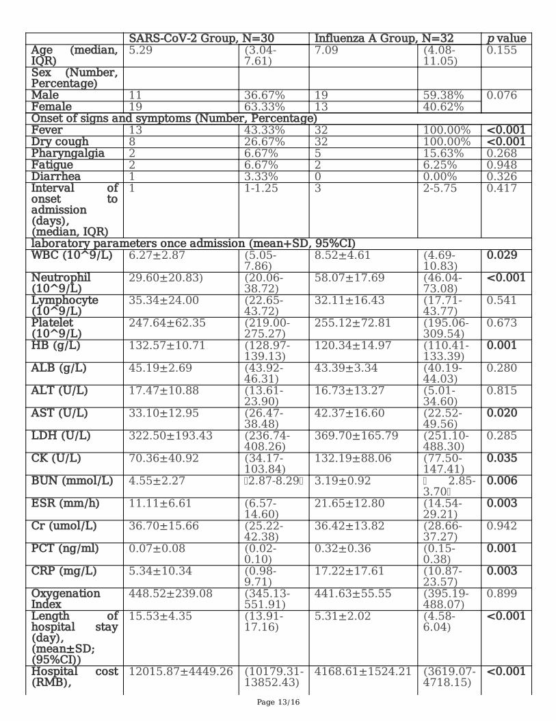
TablesTable 1. Baseline characteristics of SARS-CoV-2 and influenza-A-infected children.
Page 13/16
SARS-CoV-2 Group, N=30 Influenza A Group, N=32 p valueAge (median,IQR)
5.29 (3.04-7.61)
7.09 (4.08-11.05)
0.155
Sex (Number,Percentage)
Male 11 36.67% 19 59.38% 0.076 Female 19 63.33% 13 40.62%Onset of signs and symptoms (Number, Percentage)Fever 13 43.33% 32 100.00% <0.001Dry cough 8 26.67% 32 100.00% <0.001Pharyngalgia 2 6.67% 5 15.63% 0.268 Fatigue 2 6.67% 2 6.25% 0.948 Diarrhea 1 3.33% 0 0.00% 0.326 Interval ofonset toadmission(days),(median, IQR)
1 1-1.25 3 2-5.75 0.417
laboratory parameters once admission (mean+SD, 95%CI)WBC (10^9/L) 6.27±2.87 (5.05-
7.86)8.52±4.61 (4.69-
10.83)0.029
Neutrophil(10^9/L)
29.60±20.83) (20.06-38.72)
58.07±17.69 (46.04-73.08)
<0.001
Lymphocyte(10^9/L)
35.34±24.00 (22.65-43.72)
32.11±16.43 (17.71-43.77)
0.541
Platelet(10^9/L)
247.64±62.35 (219.00-275.27)
255.12±72.81 (195.06-309.54)
0.673
HB (g/L) 132.57±10.71 (128.97-139.13)
120.34±14.97 (110.41-133.39)
0.001
ALB (g/L) 45.19±2.69 (43.92-46.31)
43.39±3.34 (40.19-44.03)
0.280
ALT (U/L) 17.47±10.88 (13.61-23.90)
16.73±13.27 (5.01-34.60)
0.815
AST (U/L) 33.10±12.95 (26.47-38.48)
42.37±16.60 (22.52-49.56)
0.020
LDH (U/L) 322.50±193.43 (236.74-408.26)
369.70±165.79 (251.10-488.30)
0.285
CK (U/L) 70.36±40.92 (34.17-103.84)
132.19±88.06 (77.50-147.41)
0.035
BUN (mmol/L) 4.55±2.27 2.87-8.29 3.19±0.92 2.85-3.70
0.006
ESR (mm/h) 11.11±6.61 (6.57-14.60)
21.65±12.80 (14.54-29.21)
0.003
Cr (umol/L) 36.70±15.66 (25.22-42.38)
36.42±13.82 (28.66-37.27)
0.942
PCT (ng/ml) 0.07±0.08 (0.02-0.10)
0.32±0.36 (0.15-0.38)
0.001
CRP (mg/L) 5.34±10.34 (0.98-9.71)
17.22±17.61 (10.87-23.57)
0.003
OxygenationIndex
448.52±239.08 (345.13-551.91)
441.63±55.55 (395.19-488.07)
0.899
Length ofhospital stay(day),(mean±SD;(95%CI))
15.53±4.35 (13.91-17.16)
5.31±2.02 (4.58-6.04)
<0.001
Hospital cost(RMB),
12015.87±4449.26 (10179.31-13852.43)
4168.61±1524.21 (3619.07-4718.15)
<0.001
Page 14/16
(mean±SD;(95%CI))
Data of continuous variables are given as mean±SD with 95% CI. While for data notnormally distributed, median with interquartile range (IQR) is used. Categorical variablesare given as counts and percentages. Abbreviations: WBC, white blood cell; HB,hemoglobin; ALB, Albumin; ALT, alanine aminotransferase; AST, aminotransferase; LDH,lactate dehydrogenase; CK, creatine kinase; BUN, blood urea nitrogen; ESR, erythrocytesedimentation rate; Cr, creatinine; PCT, procalcitonin; CRP, C-reactive protein. P values inbold are considered to be significant.
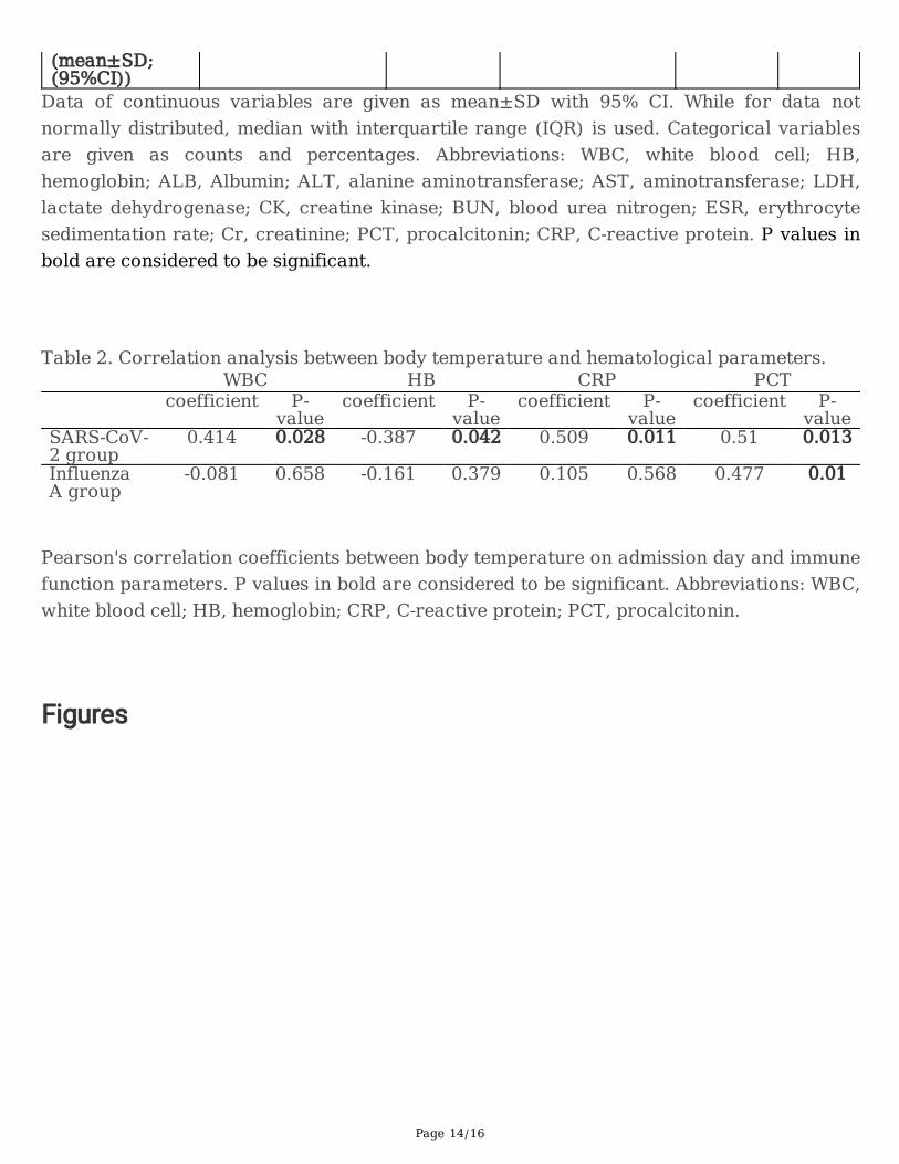
Table 2. Correlation analysis between body temperature and hematological parameters. WBC HB CRP PCT coefficient P-
valuecoefficient P-
valuecoefficient P-
valuecoefficient P-
valueSARS-CoV-2 group
0.414 0.028 -0.387 0.042 0.509 0.011 0.51 0.013
InfluenzaA group
-0.081 0.658 -0.161 0.379 0.105 0.568 0.477 0.01
Pearson's correlation coefficients between body temperature on admission day and immunefunction parameters. P values in bold are considered to be significant. Abbreviations: WBC,white blood cell; HB, hemoglobin; CRP, C-reactive protein; PCT, procalcitonin.
Figures
Page 15/16
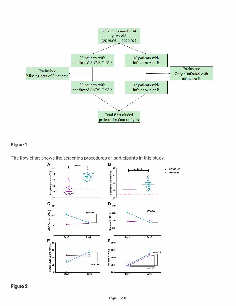
Figure 1
The �ow chart shows the screening procedures of participants in this study.
Figure 2
Page 16/16
Changes of body temperature and laboratory parameters in the peri-hospitalization period. Box plot withthe mean line and 95% CI of the body temperature observations showing comparisons between SARS-CoV-2 and in�uenza-A-infected children before hospital admission (A) and after 3-day of hospital stay(B). Only 5 children’s body temperature exceeded 37.5℃ while 23 patients remained continuous feverafter 3 days of hospital stay. Comparisons on counts of white blood cell (WBC) (C), neutrophil (D),lymphocyte (E), as well as platelet (F) showed signi�cant increase in in�uenza-A-infected children,represented with blue triangles. While those of SARS-CoV-2-infected children were represented with purpledot. Error bars: Mean ±SEM.