Embed Size (px)
Citation preview
Childhood Immune ThrombocytopenicPurpura: Diagnosis and Management
Victor Blanchette, FRCPa,*,Paula Bolton-Maggs, DM, FRCPb
aDivision of Hematology/Oncology, The Hospital for Sick Children, Department of Pediatrics,
University of Toronto, 555 University Avenue, Toronto, Ontario M5G 1X8, CanadabUniversity Department of Haematology, Manchester Royal Infirmary,
Oxford Road, Manchester M13 9WL, United Kingdom
Immune thrombocytopenic purpura (ITP) is an autoimmune disordercharacterized by a low circulating platelet count caused by destruction ofantibody-sensitized platelets in the reticuloendothelial system [1]. ITP canbe classified based on patient age (childhood versus adult), duration of ill-ness (acute versus chronic), and presence of an underlying disorder (primaryversus secondary). Persistence of thrombocytopenia, generally defined asa platelet count of less than 150 � 109/L for longer than 6 months, definesthe chronic form of the disorder. Secondary causes of ITP include collagenvascular disorders, such as systemic lupus erythematosus (SLE); immunedeficiencies, such as common variable immunodeficiency (CVID); andsome chronic infections (eg, HIV and hepatitis C).
This article focuses on the diagnosis and management of children (under18 years of age) who have acute and chronic ITP. Emphasis is placed onareas of controversy and new therapies.
Pediatr Clin N Am 55 (2008) 393–420
Pathophysiology
The pathophysiology of ITP increasingly is understood better (reviewedby Cines and Blanchette [1]). Not surprisingly, it is complex with involve-ment of many players in the human immune orchestra, including antibodies,cytokines, antigen-presenting cells, costimulatory molecules, and T and Blymphocytes (including T-helper, T-cytotoxic, and T-regulatory lympho-cytes). Current knowledge is summarized later.
* Corresponding author.
E-mail address: [email protected] (V. Blanchette).
0031-3955/08/$ - see front matter � 2008 Elsevier Inc. All rights reserved.
doi:10.1016/j.pcl.2008.01.009 pediatric.theclinics.com
394 BLANCHETTE & BOLTON-MAGGS
A key element in the pathophysiology of ITP is loss of self tolerance lead-ing to the production of autoantibodies directed against platelet antigens.Evidence for an ‘‘antiplatelet factor’’ in the plasma of subjects who haveITP was provided in a seminal report from Harrington and coworkers [2]in 1951. The investigators demonstrated that the infusion of plasma fromsubjects who had ITP into volunteers induced a rapid fall in platelet countand a clinical picture that mimics ITP. The ‘‘antiplatelet factor’’ subse-quently was confirmed as an immunoglobulin [3]. Now it is known thatthe autoantibodies in patients who have ITP mostly are of the IgG classwith specificity against platelet-specific antigens, in particular, glycoproteinsIIb/IIIa and Ib/IX. Unfortunately, accurate detection of platelet autoanti-bodies is difficult and not available routinely in most clinical hematologylaboratories; clinicians should be aware that indirect platelet autoantibodytests (tests that detect free autoantibodies in the plasma) are inferior todirect tests (tests that detect platelet-bound autoantibodies) and that evenwith the best direct tests performed in expert immunohematology laborato-ries, the positivity rate in patients who have well-characterized ITP does notexceed 80% [4]. A negative platelet antibody test, therefore, does notexclude a diagnosis of ITP. For this reason, platelet antibody testing isnot recommended as part of the routine diagnostic strategy [5].
It is increasingly clear that cellular immune mechanisms play a pivotalrole in ITP [1]. The production of antiplatelet antibodies by B cells requiresantigen-specific, CD4-postive, T-cell help (Fig. 1). It also is possible that insome ITP cases, cytotoxic T cells play a role in the destruction of platelets. Apossible sequence of events in ITP is as follows. A trigger, possibly an infec-tion or toxin, leads to the formation of antibodies/immune complexes thatattach to platelets. Antibody-coated platelets then bind to antigen-present-ing cells (macrophages or dendritic cells) through low-affinity Fcg receptors(Fcg RIIA/Fcg RIIIA) and are internalized and degraded. Activated anti-gen-presenting cells then expose novel peptides on the cell surface andwith costimulatory help facilitate the proliferation of platelet antigen-specific, CD4-positive, T-cell clones. These T-cell clones drive autoantibodyproduction by platelet antigen-specific B-cell clones. As part of the plateletdestructive process in ITP, cryptic epitopes from platelet antigens areexposed, leading to the formation of secondary platelet antigen-specificT-cell clones, with stimulation of new platelet antigen-specific B-cell clonesand broadening of the immune response. The autoantibody profile of indi-vidual patients who have ITP reflects activity of polyclonal autoreactiveB-cell clones derived by antigen-driven affinity selection and somaticmutation.
Although increased platelet destruction clearly plays a key role in thepathogenesis of ITP, it is now recognized that impaired platelet productionalso is important in many cases. In adults, as many as 40% of ITP cases mayhave reduced platelet turnover, reflecting the inhibitory effect of plateletautoantibodies on megakaryopoiesis [6]. Studies of platelet kinetics in
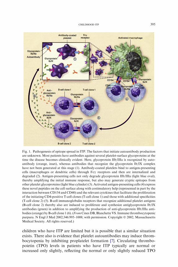
Fig. 1. Pathogenesis of epitope spread in ITP. The factors that initiate autoantibody production
are unknown. Most patients have antibodies against several platelet-surface glycoproteins at the
time the disease becomes clinically evident. Here, glycoprotein IIb/IIIa is recognized by auto-
antibody (orange, inset), whereas antibodies that recognize the glycoprotein Ib/IX complex
have not been generated at this stage (1). Antibody-coated platelets bind to antigen-presenting
cells (macrophages or dendritic cells) through Fcg receptors and then are internalized and
degraded (2). Antigen-presenting cells not only degrade glycoprotein IIb/IIIa (light blue oval),
thereby amplifying the initial immune response, but also may generate cryptic epitopes from
other platelet glycoproteins (light blue cylinder) (3). Activated antigen-presenting cells (4) express
these novel peptides on the cell surface along with costimulatory help (represented in part by the
interaction between CD154 and CD40) and the relevant cytokines that facilitate the proliferation
of the initiating CD4-positive T-cell clones (T-cell clone 1) and those with additional specificities
(T-cell clone 2) (5). B-cell immunoglobulin receptors that recognize additional platelet antigens
(B-cell clone 2) thereby also are induced to proliferate and synthesize antiglycoprotein Ib/IX
antibodies (green) in addition to amplifying the production of anti-glycoprotein IIb/IIIa anti-
bodies (orange) by B-cell clone 1 (6). (FromCinesDB, Blanchette VS. Immune thrombocytopenic
purpura. N Engl J Med 2002;346:995–1008; with permission. Copyright � 2002, Massachusetts
Medical Society. All rights reserved.)
395CHILDHOOD ITP
children who have ITP are limited but it is possible that a similar situationexists. There also is evidence that platelet autoantibodies may induce throm-bocytopenia by inhibiting proplatelet formation [7]. Circulating thrombo-poietin (TPO) levels in patients who have ITP typically are normal orincreased only slightly, reflecting the normal or only slightly reduced TPO
396 BLANCHETTE & BOLTON-MAGGS
receptor mass in this acquired platelet disorder. In contrast, TPO levels arehigh in inherited platelet production disorders, such as thrombocytopenia-absent radii or congenital amegakaryocytic thrombocytopenia [8]. TPO test-ing generally is not available, but these observations have led to the questionof whether or not TPO or molecules mimicking TPO may increase plateletproduction and be a new treatment strategy in ITP. Several such agentscurrently are in clinical trials.
Differential diagnosis
Primary ITP is a diagnosis of exclusion. The question, ‘‘When does a lowplatelet count not mean ITP?’’ is important, especially for atypical cases.When an unexpected low platelet count in a child is obtained, artifact orlaboratory error should be considered first and excluded. Pseudothrombocy-topenia is an example of spurious thrombocytopenia that is caused by plate-let aggregation and clumping in the presence of ethylenediamine tetraaceticacid (EDTA) anticoagulant [9]. Examination of well-stained blood smearsprepared from a venous blood sample collected separately into EDTAand 3.8% sodium citrate anticoagulant usually confirms or excludes pseudo-thrombocytopenia. A smear prepared from the collection tube with EDTAshould demonstrate platelet clumping, whereas a smear prepared from thetube with sodium citrate should not. Some patients, however, have plateletsthat also clump in citrate anticoagulant.
A detailed history, careful physical examination, and results of selectedtests confirm or eliminate common causes of secondary thrombocytopenia,such as SLE. A positive antinuclear antibody is common in children whohave ITP and, as an isolated finding, does not confirm or exclude SLE[10]; more specific tests, such as an anti–double-stranded DNA test, shouldbe ordered if a diagnosis of SLE-associated ITP is suspected. A transfusionhistory should be obtained in all cases and, depending on the age of thechild, the history should include questioning about drug use (prescriptionand nonprescription) and sexual activity. If relevant, testing for antibodiesto hepatitis C and HIV should be performed.
A detailed family history should be obtained in all cases. Especially inchildren who have apparent ‘‘chronic’’ ITP and isolated moderate thrombo-cytopenia, the possibility of an inherited thrombocytopenia should be consid-ered. The topic, ‘‘inherited thrombocytopenia: when a low platelet count doesnot mean ITP,’’ is the focus of an excellent review [11]. The inherited throm-bocytopenias can be classified based on platelet size (large, normal, and small)and gene mutations. They include conditions, such as the MYH9-relatedmacrothrombocytopenias, Wiskott-Aldrich syndrome (WAS), and rare con-ditions, such as gray platelet syndrome (Box 1). The pattern of inheritance(eg, X-linked in boys who have WAS) and abnormalities on peripheral bloodsmear (eg, Dohle-like inclusions in neutrophils of patients who have MYH9disorders or pale agranular platelets in gray platelet syndrome) may provide
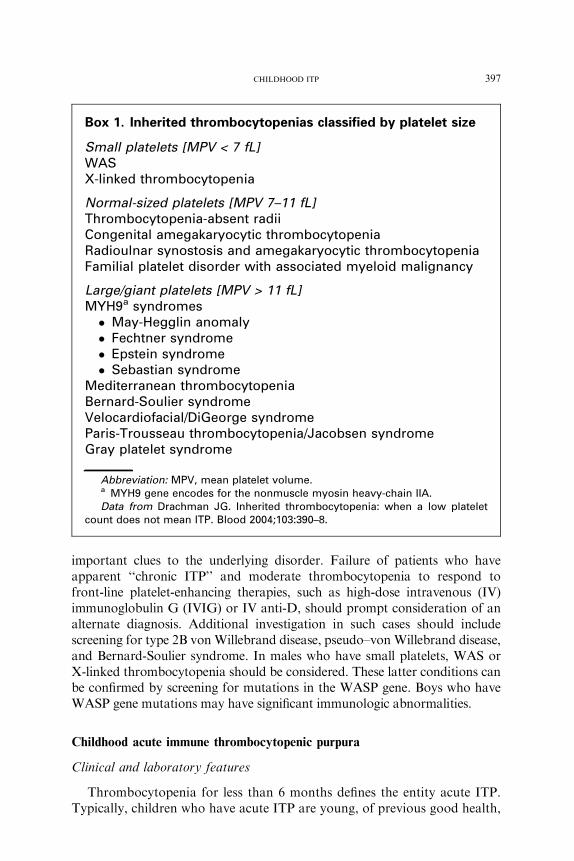
Box 1. Inherited thrombocytopenias classified by platelet size
Small platelets [MPV < 7 fL]WASX-linked thrombocytopenia
Normal-sized platelets [MPV 7–11 fL]Thrombocytopenia-absent radiiCongenital amegakaryocytic thrombocytopeniaRadioulnar synostosis and amegakaryocytic thrombocytopeniaFamilial platelet disorder with associated myeloid malignancy
Large/giant platelets [MPV > 11 fL]MYH9a syndromes� May-Hegglin anomaly� Fechtner syndrome� Epstein syndrome� Sebastian syndrome
Mediterranean thrombocytopeniaBernard-Soulier syndromeVelocardiofacial/DiGeorge syndromeParis-Trousseau thrombocytopenia/Jacobsen syndromeGray platelet syndrome
Abbreviation: MPV, mean platelet volume.a MYH9 gene encodes for the nonmuscle myosin heavy-chain IIA.Data from Drachman JG. Inherited thrombocytopenia: when a low platelet
count does not mean ITP. Blood 2004;103:390–8.
397CHILDHOOD ITP
important clues to the underlying disorder. Failure of patients who haveapparent ‘‘chronic ITP’’ and moderate thrombocytopenia to respond tofront-line platelet-enhancing therapies, such as high-dose intravenous (IV)immunoglobulin G (IVIG) or IV anti-D, should prompt consideration of analternate diagnosis. Additional investigation in such cases should includescreening for type 2B vonWillebrand disease, pseudo–vonWillebrand disease,and Bernard-Soulier syndrome. In males who have small platelets, WAS orX-linked thrombocytopenia should be considered. These latter conditions canbe confirmed by screening for mutations in the WASP gene. Boys who haveWASP gene mutations may have significant immunologic abnormalities.
Childhood acute immune thrombocytopenic purpura
Clinical and laboratory features
Thrombocytopenia for less than 6 months defines the entity acute ITP.Typically, children who have acute ITP are young, of previous good health,
398 BLANCHETTE & BOLTON-MAGGS
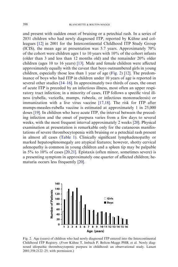
and present with sudden onset of bruising or a petechial rash. In a series of2031 children who had newly diagnosed ITP, reported by Kuhne and col-leagues [12] in 2001 for the Intercontinental Childhood ITP Study Group(ICIS), the mean age at presentation was 5.7 years. Approximately 70%of the cohort were children ages 1 to 10 years with 10% of the cohort infants(older than 3 and less than 12 months old) and the remainder 20% olderchildren (ages 10 to 16 years) [13]. Male and female children were affectedapproximately equally with the caveat that boys outnumbered girls in youngchildren, especially those less than 1 year of age (Fig. 2) [12]. The predom-inance of boys who had ITP in children under 10 years of age is reported inseveral other studies [14–16]. In approximately two thirds of cases, the onsetof acute ITP is preceded by an infectious illness, most often an upper respi-ratory tract infection; in a minority of cases, ITP follows a specific viral ill-ness (rubella, varicella, mumps, rubeola, or infectious mononucleosis) orimmunization with a live virus vaccine [17,18]. The risk for ITP aftermumps-measles-rubella vaccine is estimated at approximately 1 in 25,000doses [19]. In children who have acute ITP, the interval between the preced-ing infection and the onset of purpura varies from a few days to severalweeks, with the most frequent interval approximately 2 weeks [20]. Physicalexamination at presentation is remarkable only for the cutaneous manifes-tations of severe thrombocytopenia with bruising or a petechial rash presentin almost all cases (Table 1). Clinically significant lymphadenopathy ormarked hepatosplenomegaly are atypical features; however, shotty cervicaladenopathy is common in young children and a spleen tip may be palpablein 5% to 10% of cases [20,21]. Epistaxis (often minor, sometimes severe) isa presenting symptom in approximately one quarter of affected children; he-maturia occurs less frequently [20].
Fig. 2. Age (years) of children who had newly diagnosed ITP entered into the Intercontinental
Childhood ITP Registry. (From Kuhne T, Imbach P, Bolton-Maggs PHB, et al. Newly diag-
nosed idiopathic thrombocytopenic purpura in childhood: an observational study. Lancet
2001;358:2122–25; with permission.)
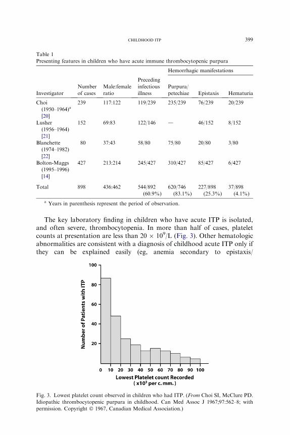
Table 1
Presenting features in children who have acute immune thrombocytopenic purpura
Hemorrhagic manifestations
Investigator
Number
of cases
Male:female
ratio
Preceding
infectious
illness
Purpura/
petechiae Epistaxis Hematuria
Choi
(1950–1964)a
[20]
239 117:122 119/239 235/239 76/239 20/239
Lusher
(1956–1964)
[21]
152 69:83 122/146 d 46/152 8/152
Blanchette
(1974–1982)
[22]
80 37:43 58/80 75/80 20/80 3/80
Bolton-Maggs
(1995–1996)
[14]
427 213:214 245/427 310/427 85/427 6/427
Total 898 436:462 544/892
(60.9%)
620/746
(83.1%)
227/898
(25.3%)
37/898
(4.1%)
a Years in parenthesis represent the period of observation.
399CHILDHOOD ITP
The key laboratory finding in children who have acute ITP is isolated,and often severe, thrombocytopenia. In more than half of cases, plateletcounts at presentation are less than 20 � 109/L (Fig. 3). Other hematologicabnormalities are consistent with a diagnosis of childhood acute ITP only ifthey can be explained easily (eg, anemia secondary to epistaxis/
Fig. 3. Lowest platelet count observed in children who had ITP. (From Choi SI, McClure PD.
Idiopathic thrombocytopenic purpura in childhood. Can Med Assoc J 1967;97:562–8; with
permission. Copyright � 1967, Canadian Medical Association.)
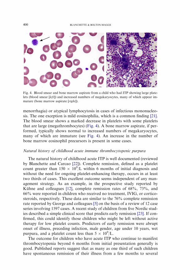
Fig. 4. Blood smear and bone marrow aspirate from a child who had ITP showing large plate-
lets (blood smear [left]) and increased numbers of megakaryocytes, many of which appear im-
mature (bone marrow aspirate [right]).
400 BLANCHETTE & BOLTON-MAGGS
menorrhagia) or atypical lymphocytosis in cases of infectious mononucleo-sis. The one exception is mild eosinophilia, which is a common finding [21].The blood smear shows a marked decrease in platelets with some plateletsthat are large (megathrombocytes) (Fig. 4). A bone marrow aspirate, if per-formed, typically shows normal to increased numbers of megakaryocytes,many of which are immature (see Fig. 4). An increase in the number ofbone marrow eosinophil precursors is present in some cases.
Natural history of childhood acute immune thrombocytopenic purpura
The natural history of childhood acute ITP is well documented (reviewedby Blanchette and Carcao [22]). Complete remission, defined as a plateletcount greater than 150 � 109/L within 6 months of initial diagnosis andwithout the need for ongoing platelet-enhancing therapy, occurs in at leasttwo thirds of cases. This excellent outcome seems independent of any man-agement strategy. As an example, in the prospective study reported byKuhne and colleagues [12], complete remission rates of 68%, 73%, and66% were reported in children who received no treatment, IVIG, or cortico-steroids, respectively. These data are similar to the 76% complete remissionrate reported by George and colleagues [5] on the basis of a review of 12 caseseries involving 1597 cases. A recent study of children from five Nordic stud-ies described a simple clinical score that predicts early remission [23]. If con-firmed, this could identify those children who might be left without activetherapy for low platelet counts. Predictors of early remission were abruptonset of illness, preceding infection, male gender, age under 10 years, wetpurpura, and a platelet count less than 5 � 109/L.
The outcome for children who have acute ITP who continue to manifestthrombocytopenia beyond 6 months from initial presentation generally isgood. Published reports suggest that as many as one third of such childrenhave spontaneous remission of their illness from a few months to several
401CHILDHOOD ITP
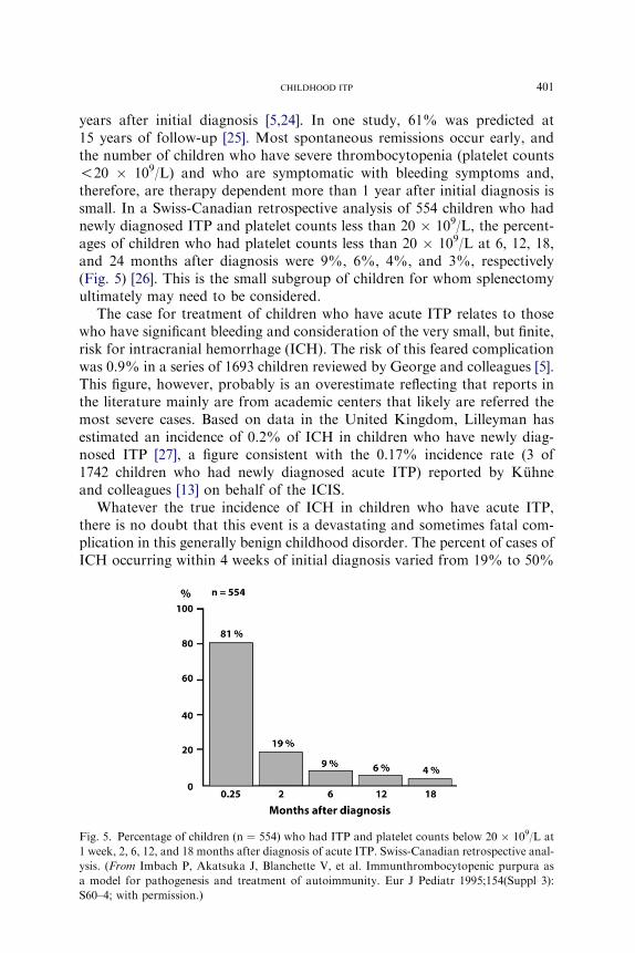
years after initial diagnosis [5,24]. In one study, 61% was predicted at15 years of follow-up [25]. Most spontaneous remissions occur early, andthe number of children who have severe thrombocytopenia (platelet counts!20 � 109/L) and who are symptomatic with bleeding symptoms and,therefore, are therapy dependent more than 1 year after initial diagnosis issmall. In a Swiss-Canadian retrospective analysis of 554 children who hadnewly diagnosed ITP and platelet counts less than 20 � 109/L, the percent-ages of children who had platelet counts less than 20 � 109/L at 6, 12, 18,and 24 months after diagnosis were 9%, 6%, 4%, and 3%, respectively(Fig. 5) [26]. This is the small subgroup of children for whom splenectomyultimately may need to be considered.
The case for treatment of children who have acute ITP relates to thosewho have significant bleeding and consideration of the very small, but finite,risk for intracranial hemorrhage (ICH). The risk of this feared complicationwas 0.9% in a series of 1693 children reviewed by George and colleagues [5].This figure, however, probably is an overestimate reflecting that reports inthe literature mainly are from academic centers that likely are referred themost severe cases. Based on data in the United Kingdom, Lilleyman hasestimated an incidence of 0.2% of ICH in children who have newly diag-nosed ITP [27], a figure consistent with the 0.17% incidence rate (3 of1742 children who had newly diagnosed acute ITP) reported by Kuhneand colleagues [13] on behalf of the ICIS.
Whatever the true incidence of ICH in children who have acute ITP,there is no doubt that this event is a devastating and sometimes fatal com-plication in this generally benign childhood disorder. The percent of cases ofICH occurring within 4 weeks of initial diagnosis varied from 19% to 50%
Fig. 5. Percentage of children (n ¼ 554) who had ITP and platelet counts below 20 � 109/L at
1 week, 2, 6, 12, and 18 months after diagnosis of acute ITP. Swiss-Canadian retrospective anal-
ysis. (From Imbach P, Akatsuka J, Blanchette V, et al. Immunthrombocytopenic purpura as
a model for pathogenesis and treatment of autoimmunity. Eur J Pediatr 1995;154(Suppl 3):
S60–4; with permission.)
402 BLANCHETTE & BOLTON-MAGGS
in different reports [5,27,28]; in one retrospective review, 10% (7/69) of casesof ICH occurred within 3 days of diagnosis of ITP [29]. Trauma to the headand use of antiplatelet drugs, such as aspirin, were identified as risk factorsfor ICH in children who had ITP and very low platelet counts [30].
Unfortunately, a prospective randomized controlled trial to determinedefinitively whether or not therapeutic intervention can decrease the inci-dence of ICH significantly in children who have newly diagnosed ITP andplatelet counts below 20 � 109/L is not feasible, because of the large num-bers of cases required to ensure a statistically significant outcome. Physi-cians who care for children who have acute ITP, therefore, must act inthe best interest of each child without the benefit of definitive data. Becauseof the significant morbidity and mortality associated with ICH and theavailability of highly effective platelet-enhancing therapies, some recom-mend that families of young children who have newly diagnosed acuteITP at risk for ICH (who have platelet counts !10 � 109/L) be offeredthe option of treatment using the minimum therapy necessary to increasethe platelet count rapidly to a safe, hemostatic level. There is no currentevidence, however, that such a management strategy significantly reducesthe incidence of ICH in children who have ITP, although intuitively thisseems probable.
In addition, there is evidence to suggest that the rate of platelet responseto frontline therapies (corticosteroids or IVIG) in the subset of children whohave ITP and clinically significant hemorrhage is suboptimal [31]. Discus-sion with parents and children, if of appropriate age, should include consid-eration of best available evidence with regard to the three key issues: (1) totreat or not to treat (2) to perform a bone marrow aspirate or not and (3) tohospitalize or not.
To treat or not to treat
Observation
The case for observation of children who have acute ITP rests with theknowledge that acute ITP is, for the majority of affected children, a benignself-limiting disorder, usually with mild clinical symptoms and has a low riskfor serious bleeding (approximately 3% with ICH being rare) and the factthat there are no prospective studies that clearly indicate a decrease in theincidence of ICH associated with treatment [32]. Several children who hadITP-associated ICH were receiving platelet-enhancing therapy at the timeof the hemorrhage [28]. In addition, all treatments suffer from the disadvan-tage of side effects, which can be severe.
Guidelines for initial management of children who have acute ITP havebeen published and reflect the ongoing debate, ‘‘to treat or not to treat’’[5,33–36]. Recommendations from the Working Party of the British Com-mittee for Standards in Haematology General Haematology Task Force
403CHILDHOOD ITP
state that treatment of children who have acute ITP should be decided onthe basis of clinical symptoms in addition to cutaneous signs, not the plateletcount alone [36]. The Working Party considered it appropriate to managechildren who have acute ITP and mild clinical disease expectantly, with sup-portive advice, and a 24-hour contact point irrespective of the platelet count.Based on these guidelines, intervention is reserved for the few children whohave overt hemorrhage and platelet counts below 20 � 109/L or those whohave organ- or life-threatening bleeding irrespective of the circulating plate-let count [34,36]. Many clinicians in Europe manage children who have ITPexpectantly (ie, without medication to increase the platelet count) because ofthe rapid remissions in most cases, the low risk for bleeding, and toxicities ofcurrently available medical therapies. Data are reported from the UnitedKingdom and Germany promoting the use of advice and support to childrenand their families during the usually short duration of the illness [15,32,37].
Corticosteroids
The corticosteroid treatment regimen used to treat children who havenewly diagnosed ITP in most reported studies, and worldwide, is oral pred-nisone at a dose of 1 to 2 mg/kg per day given in divided doses and continuedfor a fewweeks. Two randomized studies support the benefit of corticosteroidtherapy in children who have ITP. In the first study, conducted by Sartorius[38] and reported in 1984, 73 children ages 10 months to 14 years who hadnewly diagnosed ITP were randomized to receive oral prednisolone(60 mg/m2 per day for 21 days) or a placebo. Platelet responses were signifi-cantly faster in the corticosteroid-treated group, with 90% of children achiev-ing a platelet count of 30 � 109/L within the first 10 days of treatmentcompared with 45% of children in the placebo no-treatment group. TheRumpel-Leede test, which measures capillary resistance (blood vessel integ-rity), became negative sooner in the corticosteroid-treated group. In thesecond study, reported by Buchanan and Holtkamp [39] in 1984, 27 childrenwho had acute ITP were randomized to receive oral prednisone (2 mg/kg perday for 14 days, with tapering and discontinuation of corticosteroids by day21) or placebo. Although there was a definite trend in favor of corticoste-roids, only on day 7 of therapy did the prednisone-treated patients havesignificantly higher platelet counts, lower bleeding scores, and shorter bleed-ing times than children receiving placebo. Taken together, these two studiessuggest limited early benefit from conventional dose oral corticosteroidtherapy in children who have acute ITP.
The risks and benefits of high-dose corticosteroid therapy administeredorally or IV to children who have acute ITP merit discussion. In a studyof 20 children randomized to receive oral megadose methylprednisolone(30 mg/kg for 3 days followed by 20 mg/kg for 4 days) or IVIG (0.4 g/kg� 5 days), Ozsoylu and colleagues [40] reported that 80% of children inboth groups had platelet counts greater than 50 � 109/L by 72 hours after
404 BLANCHETTE & BOLTON-MAGGS
the start of treatment. Corticosteroids were given before 9:00 AM andadverse effects were not observed. In contrast, Suarez and colleagues [41]reported that hyperactivity and behavioral problems occurred in 5 of 9 chil-dren who had acute ITP given 6 to 8 mg/kg per day of oral prednisone for 3days or until platelet counts had increased to 20 � 109/L. Immediate plateletresponses with this regimen were impressive: the mean time to achievea platelet count of 20 � 109/L was 1.9 � 0.6 days (range 1–3 days).
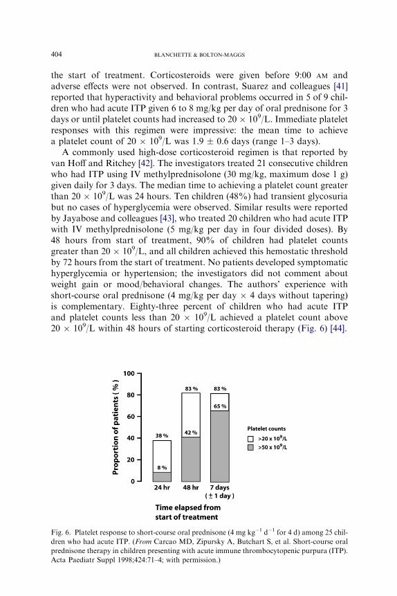
A commonly used high-dose corticosteroid regimen is that reported byvan Hoff and Ritchey [42]. The investigators treated 21 consecutive childrenwho had ITP using IV methylprednisolone (30 mg/kg, maximum dose 1 g)given daily for 3 days. The median time to achieving a platelet count greaterthan 20 � 109/L was 24 hours. Ten children (48%) had transient glycosuriabut no cases of hyperglycemia were observed. Similar results were reportedby Jayabose and colleagues [43], who treated 20 children who had acute ITPwith IV methylprednisolone (5 mg/kg per day in four divided doses). By48 hours from start of treatment, 90% of children had platelet countsgreater than 20 � 109/L, and all children achieved this hemostatic thresholdby 72 hours from the start of treatment. No patients developed symptomatichyperglycemia or hypertension; the investigators did not comment aboutweight gain or mood/behavioral changes. The authors’ experience withshort-course oral prednisone (4 mg/kg per day � 4 days without tapering)is complementary. Eighty-three percent of children who had acute ITPand platelet counts less than 20 � 109/L achieved a platelet count above20 � 109/L within 48 hours of starting corticosteroid therapy (Fig. 6) [44].
Fig. 6. Platelet response to short-course oral prednisone (4 mg kg�1 d�1 for 4 d) among 25 chil-
dren who had acute ITP. (From Carcao MD, Zipursky A, Butchart S, et al. Short-course oral
prednisone therapy in children presenting with acute immune thrombocytopenic purpura (ITP).
Acta Paediatr Suppl 1998;424:71–4; with permission.)
405CHILDHOOD ITP
On the basis of these studies, it can be concluded that a clinically signif-icant increment in platelet count can be achieved rapidly in the majority ofchildren who have acute ITP after the administration of high-doses of cor-ticosteroids (approximately 4 mg/kg per day of prednisone or an equivalentcorticosteroid preparation) administered orally or parenterally. The fre-quency and severity of corticosteroid toxicity relates to dose and durationof therapy and merits further study. If a decision is made to use corticoste-roid therapy for children who have acute ITP, it seems wise to use high-dosecorticosteroid regimens for as short a period of time as is necessary toachieve a clinically meaningful endpoint (eg, cessation of bleeding orachievement of a platelet count O20 � 109/L). This approach minimizesthe predictable, and sometimes serious, adverse effects of long-term cortico-steroid therapy (reviewed by Beck and colleagues [45]). A fall in plateletcount often occurs during the period of tapering corticosteroids but notusually to clinically significant levels.
Intravenous immunoglobulin G
Imbach and colleagues [46] first reported that IV infusion of a pooled,largely monomeric IgG preparation produced a rapid reversal of thrombo-cytopenia in children who had acute and chronic ITP. This landmark obser-vation was confirmed subsequently by several investigators (reviewed byBlanchette and Carcao [22]). Transient blockade of Fc receptors on macro-phages in the reticuloendothelial system, especially the spleen, is believed toplay a major role in the immediate, and often dramatic, platelet responsesobserved after treatment of children who have ITP using a high dose ofIVIG (1–2 g/kg). Two Canadian prospective randomized clinical trials areinstructive in the context of IVIG treatment of children who have acuteITP. In the first study, reported by Blanchette and colleagues [47] in 1993,53 children who had acute ITP and platelet counts less than 20 � 109/Lwere randomized to receive IVIG (1 g/kg on 2 consecutive days), oral pred-nisone (4 mg/kg per day � 7 days with tapering and discontinuation by day21), or expectant management (no treatment). The rate of platelet responsewas significantly faster in children who received treatment compared withthose managed expectantly; for the endpoint of time (days) taken to achievea platelet count greater than or equal to 20 � 109/L, IVIG and corticoste-roids were equivalent, whereas IVIG was superior to oral corticosteroidtherapy for the endpoint of time (days) taken to achieve a platelet countgreater than 50 � 109/L. Bleeding symptoms were not recorded in this study,however; the platelet count alone was used as a surrogate marker forresponse. The follow-up Canadian randomized trial compared two IVIGtreatment regimens (1 g/kg on 2 consecutive days and 0.8 g/kg once), oralprednisone (4 mg/kg per day for 7 days with tapering and discontinuationby day 21), and for the subset of children who were blood group rhesus(D) positive, IV anti-D (25 mg/kg on 2 consecutive days) [48]. The key
406 BLANCHETTE & BOLTON-MAGGS
findings from this second randomized trial in children who had newly diag-nosed ITP and platelet counts less than 20 � 109/L were (1) a single dose ofIVIG (0.8 g/kg) was as effective as the larger dose of IVIG 1 g/kg for 2 daysin raising the platelet count and (2) both IVIG regimens were superior to IVanti-D administered as 25 mg/kg for 2 days for the clinically importantendpoint of time (number of days) to achieve a platelet count greater thanor equal to 20 � 109/L. Bleeding symptoms were not recorded in the study.The choice of the 0.8 g/kg dose as a single infusion reflected the early obser-vation by Imbach and colleagues [49] that in children who had acute ITPtreated with 0.4 g/kg of IVIG daily for 5 consecutive days, platelet responsesoften were observed after the first two infusions. These studies show thattreatment with corticosteroids or IVIG can produce a rapid rise in the plate-let count of children who have ITP with the caveat that the effect on bleed-ing symptoms was not assessed. As a result of these observations, theauthors recommend that if a decision is made to treat children who havenewly diagnosed ITP with IVIG, the initial dose should be 0.8 to 1.0 g/kgadministered as a single infusion with subsequent IVIG doses given basedon the clinical situation and follow-up platelet counts. Reflex administrationof a second dose of IVIG (ie, a total dose of 2 g/kg) generally is not neces-sary and for the majority of children only leads to an increased frequency ofadverse side effects (eg, headache, nausea, or vomiting) and higher costs.
It generally is accepted that IVIG therapy in children who have ITP,although expensive, is safe. High doses (2 g/kg), however, frequently areassociated with side effects, principally fever and headache [47]. Other uncom-mon but clinically significant treatment-associated adverse effects includeneutropenia and hemolytic anemia caused by alloantibodies in the IVIGprep-arations and self-limiting aseptic meningitis that generally occurs a few daysafter IVIG therapy. This latter complication is characterized by severe head-ache and, for the subset of children who still are significantly thrombocytope-nic, often prompts investigation with a CT scan to rule out an ICH. Ona reassuring note, although IVIG is a human plasma–derived product, cur-rent commercially available IVIG preparations are treated with highly effec-tive measures to inactivate lipid-coated viruses, such as HIV and hepatitis C.
Intravenous anti-D
In 1983, Salama and colleagues [50] reported that the IV infusion of anti-Dresulted in the reversal of thrombocytopenia in patients who had ITP andwere rhesus (D) positive. The investigators speculated that the beneficialeffect of anti-D was due to the competitive inhibition of reticuloendothelialfunction by preferential sequestration of immunoglobulin-coated autologousred blood cells (RBCs). These observations subsequently were confirmed byseveral investigators. In a report that detailed experience with IV anti-D treat-ment in 272 subjects who had ITP, Scaradavou and colleagues [51] docu-mented several important findings, including (1) anti-D at conventional
407CHILDHOOD ITP
doses is ineffective in splenectomized subjects; (2) platelet responses are sig-nificantly better in children compared with adults; and (3) responders to IVanti-D generally respond on retreatment. There was a trend toward a higherplatelet count after therapy in patients who received 40 to 60 mg/kg of IV anti-D compared with those who received less than or equal to 40 mg/kg. The doseresponse to IV anti-D is of importance. A recent report by Tarantino andcolleagues [52], describing the results of a prospective randomized clinicaltrial of IV anti-D (50 mg/kg and 75 mg/kg) and IVIG (0.8 g/kg) in 101 childrenwho had acute ITP and platelet counts less than 20 � 109/L, clearly estab-lished that IV anti-D (75 mg/kg) is superior to IV anti-D (50 mg/kg) and equiv-alent to IVIG (0.8 g/kg) with respect to the numbers of cases with plateletcounts greater than 20 � 109/L at 24 hours after therapy.
Short-term adverse effects, such as fever, chills, and nausea/vomiting, aremore frequent with a 75-mg/kg than a 50-mg/kg dose and are likely related torelease of pro-inflammatory cytokines/chemokines after IV anti-D [53].These side effects can be ameliorated/prevented by premedication of patientswith acetaminophen/corticosteroids. The most predictable adverse effect ofanti-D therapy in subjects who are rhesus (D) positive is a fall in hemoglobinlevel due to RBC destruction by infused RBC alloantibodies. The fall inhemoglobin occurs within 1 week of the anti-D therapy with recovery gen-erally evident by day 21. In the Scaradavou study, the mean hemoglobindecrease was 0.8 g/dL at 7 days post IV anti-D treatment, and only 16%of cases had a hemoglobin decrease greater than 2.1 g/dL [51]. In occasionalcases, abrupt severe intravascular hemolysis is reported after therapy; themajority of these cases were in adults, some of whom had comorbid diseases[54]. This complication also is reported in rare cases after IVIG therapy.Physicians who treat children who have ITP using anti-D should be awareof this complication and advise parents and children to report symptomsand signs, such as excessive tiredness or pallor or passage of dark (tea-colored)urine, promptly. No clinically significant increase in treatment-related hemo-lysis has been reported with 75 versus 50 mg/kg of IV anti-D, and a singledose of 75 mg/kg of anti-D now can be recommended as standard dosingfor the treatment of children who have acute ITP and are rhesus (D) positive.
To perform a bone marrow aspirate or not
There is consensus that bone marrow aspiration is not necessary forchildren who have newly diagnosed typical acute ITP if managementinvolves observation or plasma based therapies, such as IVIG or anti-D.The contentious issue is whether or not a bone marrow aspirate should beperformed in children who have typical acute ITP before starting corticoste-roids to avoid missing, and therefore treating inappropriately, an underlyingleukemia. The results of a retrospective study of bone marrow aspiratesperformed in children who have suspected acute ITP are instructive inthis regard [55]. No children who had typical laboratory features, defined
408 BLANCHETTE & BOLTON-MAGGS
as a normal hemoglobin level and total white blood cell and neutrophilcount for age, had underlying leukemia; cases of leukemia, however, wereobserved in children who had atypical laboratory features. A bone marrowexamination, therefore, should be considered mandatory in atypical cases ofchildhood acute ITP, defined as those who have lassitude, protracted fever,bone or joint pain, and unexplained anemia, neutropenia, or macrocytosis.The diagnosis should be questioned, particularly in those children who failto remit. The most common diagnosis to emerge after isolated thrombocy-topenia in a well child is aplastic anemia.
To hospitalize or not
The majority of children who have newly diagnosed acute ITP and plate-let counts less than 20 � 109/L are hospitalized. The figure was 83% in thefirst United Kingdom National Survey [14] and 78% of 1995 children whohad newly diagnosed ITP reported by Kuhne and colleagues [12] on behalfof the ICIS. This high hospitalization rate is driven by the decision to treatand the perceived need for a bone marrow aspirate before starting cortico-steroid therapy. If a conservative management approach is used, with bonemarrow aspiration and treatment reserved for selected cases only (eg, thosewith atypical features or clinically significant bleeding), a low rate of hospi-talization can be achieved [37]. Outpatient infusion of IVIG or anti-D also isan option in selected cases.
Chronic immune thrombocytopenic purpura
Conventionally, chronic ITP is defined as thrombocytopenia (plateletcount less than 150 � 109/L) persisting for longer than 6 months from theonset of illness. Using this definition, approximately 20% to 25% of childrenmanifest chronic ITP at 6 months after the initial diagnosis of ITP. Manychildren who have platelet counts in the range of 30 to 150� 109/L, however,require no platelet-enhancing therapy and some enter a spontaneous com-plete remission in the 6 to 24 months after initial presentation [24]. The clini-cally important subgroup of children is those who have platelet counts lessthan or equal to 20� 109/L at 6months from initial diagnosis andwho requireongoing platelet-enhancing therapy because of bleeding symptoms. This is thesmall groupof children forwhom second-line therapies (eg, rituximab) or sple-nectomy may need to be considered, approximately 5% of children who haveacute ITP at the time point of 18 months after initial presentation [26].
Management
Presplenectomy management
Medical management is preferred over splenectomy for children whohave chronic ITP for less than 12 months. Treatment options include oral
409CHILDHOOD ITP
corticosteroids (including pulse oral dexamethasone), IVIG, and IV anti-D(reviewed by Blanchette and Price [56]). Avoidance of medications known toaffect platelet function adversely, especially aspirin, should be stressed andhigh-risk competitive or contact activities should be avoided during periodsof severe thrombocytopenia. The goal should be to maintain a hemostati-cally ‘‘safe’’ platelet count while avoiding the potential toxicities and costof overtreatment, in particular the well-known adverse effects of protractedcorticosteroid therapy. If treatment is recommended, the authors’ preferenceis to use short courses of relatively high-dose oral prednisone (4 mg/kg perday for 4 days, maximum daily dose 180 mg), IVIG (0.8 to 1.0 g/kg once),or, for children who are rhesus (D) positive, IV anti-D (75 mg/kg once), withall treatments given intermittently based on clinical need. Treatment is, inthe main, outpatient based and parents and children (if of an appropriateage) should be informed about the risk, benefits, and alternatives to treat-ment, including the remote risk for transfusion-transmitted infections withvirus-inactivated plasma-based therapies, such as IVIG and IV anti-D.The advantage of anti-D over IVIG in this clinical setting relates to theease of administration (anti-D can be infused over 5–10 minutes comparedwith several hours for IVIG), significantly lower cost in some countries, anda comparable platelet-enhancing effect.
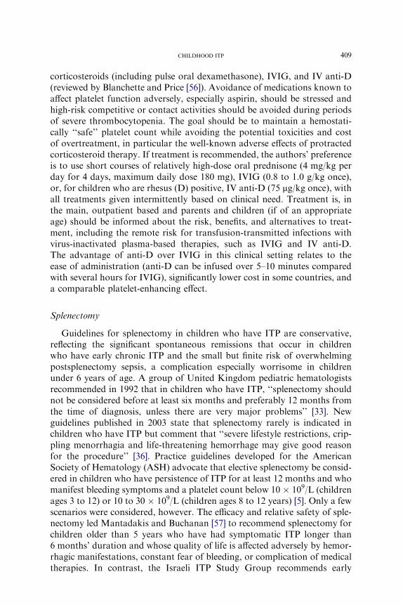
Splenectomy
Guidelines for splenectomy in children who have ITP are conservative,reflecting the significant spontaneous remissions that occur in childrenwho have early chronic ITP and the small but finite risk of overwhelmingpostsplenectomy sepsis, a complication especially worrisome in childrenunder 6 years of age. A group of United Kingdom pediatric hematologistsrecommended in 1992 that in children who have ITP, ‘‘splenectomy shouldnot be considered before at least six months and preferably 12 months fromthe time of diagnosis, unless there are very major problems’’ [33]. Newguidelines published in 2003 state that splenectomy rarely is indicated inchildren who have ITP but comment that ‘‘severe lifestyle restrictions, crip-pling menorrhagia and life-threatening hemorrhage may give good reasonfor the procedure’’ [36]. Practice guidelines developed for the AmericanSociety of Hematology (ASH) advocate that elective splenectomy be consid-ered in children who have persistence of ITP for at least 12 months and whomanifest bleeding symptoms and a platelet count below 10 � 109/L (childrenages 3 to 12) or 10 to 30 � 109/L (children ages 8 to 12 years) [5]. Only a fewscenarios were considered, however. The efficacy and relative safety of sple-nectomy led Mantadakis and Buchanan [57] to recommend splenectomy forchildren older than 5 years who have had symptomatic ITP longer than6 months’ duration and whose quality of life is affected adversely by hemor-rhagic manifestations, constant fear of bleeding, or complication of medicaltherapies. In contrast, the Israeli ITP Study Group recommends early
410 BLANCHETTE & BOLTON-MAGGS
splenectomy in children not responding rapidly to corticosteroid therapy[58]. This seems premature as many children likely remit spontaneouslygiven time.
If elective splenectomy is performed, the laparoscopic technique is pre-ferred; accessory spleens often are present and should be removed at thetime of surgical intervention. Preoperative treatment with corticosteroids,IVIG, or anti-D is considered appropriate for children who have plateletcounts less than 30 � 109/L. The outcome after splenectomy in childrenwho have primary ITP is good, and a complete remission rate of approxi-mately 70% can be expected after the procedure (Table 2). Some of thechildren reported in these series, however, may have entered a spontaneousremission over time without splenectomy. In adults, potential predictors ofsuccess after splenectomy include imaging studies to document the sites ofplatelet destruction and the historical response to medical therapies, such asIVIG and IV anti-D [63–65]. The results of imaging studies are insufficientlyspecific, however, and reports of the predictive value of prior responses tomedical therapies too conflicting to recommend that this information beused to determine reliably whether or not a splenectomy should be performedin children who have chronic ITP.
Protection against overwhelming postsplenectomy infection
Before elective splenectomy, children who have ITP should be immunizedwith the hemophilus influenza type b and pneumococcal vaccines; dependingon their age and immunization history, meningococcal vaccine also is recom-mended [66]. Because the protection provided after immunization is incom-plete (not all pneumococcal serotypes are included in the currently availablevaccines), daily prophylaxis with penicillin, or an equivalent antibiotic if thechild is allergic to penicillin, is recommended for children up to 5 years ofage and for at least 1 year after splenectomy to prevent pneumococcal sepsis,in particular. Some physicians recommend continuing antibiotic prophylaxisinto adulthood. All febrile episodes should be assessed carefully and the use ofparenteral antibiotics considered because overwhelming postsplenectomy
Table 2
Complete remission rates after splenectomy in children who had immune thrombocytopenic
purpura
Number of cases Complete remission (%)
ASH review [5] 271 72
Blanchette (1992) [59] 21 81
Ben Yehuda (1994) [58] 27 67
Mantadakis (2000) [57] 38 76
Aronis (2004) [60] 33 79
Kuhne (2006) [61] 134 67
Wang (2006) [62] 65 89
589 74
411CHILDHOOD ITP
infection can occur despite immunization and use of antibiotic prophylaxis.Children should wear a medical alert bracelet indicating that they have hada splenectomy and when traveling abroad should carry an explanatory letterand a supply of antibiotics to be started in the event of a febrile episode whilearranging for medical assessment. In the United Kingdom, patients are issuedwith a card stating that they are asplenic.
Emergency treatment
On rare occasions, children who have acute ITP and severe thrombocyto-peniamaymanifest symptoms or signs suggestive of organ- or life-threateninghemorrhage (eg, ICH). Management of such cases is challenging and shouldinvolve measures that have the potential to increase the circulating plateletcount rapidly. An approach commonly used involves the immediate IVadministration of methylprednisolone (30 mg/kg, maximum dose 1 g) over20 to 30 minutes plus a larger than usual (two- to threefold) infusion of donorplatelets in an attempt to boost the circulating platelet count temporarily.After administration of IV methylprednisolone and platelets, an infusion ofIVIG (1 g/kg) should be started with IVIG and methylprednisolone repeateddaily as indicated clinically, generally for at least 1 to 2 days. Survival of trans-fused donor platelets may be improved after IVIG therapy [67]. Depending onthe specific clinical circumstances, an emergency splenectomy may need to beconsidered. Continuous infusion of platelets may be beneficial in selectedcases. Experience with recombinant factor VIIa is limited but this hemostaticagent can be administered rapidly and should be considered in criticalsituations [68].
Combined cytopenias
The combination of ITP and clinically significant autoimmune hemolyticanemia (Evans’s syndrome) or autoimmune neutropenia occurs in a minorityof cases [69–73]. Affected children often are older than those who presentwith typical acute ITP. The clinical course is variable and often prolonged
Table 3
Retrospective reviews of patients who had Evans’s syndrome
Investigator
Number
of cases
Median age
at onset (y)
Male:female
ratio
Associated
neutropenia
Number
of deaths
Wang (1988) [69] 10 7.5 6:4 50% 3/10
Savasxan (1997) [70] 11 5.5 10:1 55% 4/11
Matthew (1997) [71] 42 7.7 22:20 38% 3/42
Blouin (2005) [72] 36 4.0 20:16 27% 3/36
99 58:41 37% 13/99
(13.1%)
412 BLANCHETTE & BOLTON-MAGGS
with significant morbidity and mortality reported in retrospective series(Table 3). Response to single-agent therapy or splenectomy often is poor[74]; combination immunosuppressive therapy may yield improved results[74–77]. Underlying causes for the combined cytopenias include SLE,CVID, and the autoimmune lymphoprolipherative syndrome (ALPS).Malignancies (eg, Hodgkin’s disease and lymphomas) and chronic infections(eg, HIV and hepatitis C) also need to be considered. The possibility of theseconditions should be kept in mind in children who have combined immunecytopenias and appropriate investigations performed.
Features of CVID include recurrent bacterial infections (especially sino-pulmonary), gastrointestinal disturbances similar to those seen in childrenwho have inflammatory bowel disease, and granulomatous disease, espe-cially affecting the lungs [78–82]. Laboratory features include low serumIgG levels and in some cases low serum IgA and IgM levels, absent or im-paired specific antibody responses to infection or vaccination, and variableabnormalities of the immune system (eg, decreased numbers or function ofT and B cells). Approximately 10% to 20% of subjects who have CVIDmanifest autoimmune cytopenias [79]. Treatment consists of regular IVIGreplacement therapy [82]. Caution should be exercised about performingsplenectomy in cases of CVID-associated ITP because of the risk for over-whelming postsplenectomy infection.
ALPS is a rare but important disorder because of defects in programmedcell death of lymphocytes [83–87]. Mutations in the Fas receptor, Fas ligand,and caspase genes are identified in approximately 70% of cases. Clinicalfeatures of the disorder include massive lymphadenopathy, most often inthe cervical and axillary areas, and hepatosplenomegaly. The laboratoryhallmark of ALPS is an increased number of double-negative (CD4-negativeand CD8-negative) T cells that express the a/b T-cell receptor. Defective invitro antigen-induced apoptosis in cultured lymphocytes can be demon-strated in affected cases. For accurate diagnosis of ALPS, these tests shouldbe performed by laboratories familiar with the test methods and in whichlocal normal values are established [88]. The best frontline treatment ofpatients who have ALPS is with mycophenolate mofetil (MMF); in the larg-est series of ALPS reported to date of treatment with this immunosuppres-sive agent, a response rate of 92% was observed [89]. Splenectomy should beavoided in ALPS cases because of the high risk for overwhelming postsple-nectomy sepsis.
New therapies
First-line therapies in children include corticosteroids, high-dose IVIG,and, for children who are rhesus positive, IV anti-D. Splenectomy is the tra-ditional second-line treatment of those children who have well-established,symptomatic chronic ITP who have failed or are intolerant of first-linetherapies. An array of third-line therapies is available for children in whom
413CHILDHOOD ITP
splenectomy is refused or contraindicated. Agents include azathioprine,cyclophosphamide, danazol, vinca alkaloids, dapsone, cyclosporine, MMF,or combination therapy. As with adults, current evidence supporting effec-tiveness and safety of these therapies in children who have severe chronicrefractory ITP is minimal [5,90]. The decision to choose one of these agentsor combinations usually is based on physician preferences and experience.A major difficulty with many of these third-line therapies is modest responserates and frequently a slow onset of action. In addition, bone marrow sup-pression and an increased incidence of infection complicate treatment withmany of the immunosuppressive agents. Before physicians can confidentlyknow the best management for their patients, these treatments, and perhapscombinations of agents and new approaches to treatment, must be evaluatedfor effectiveness and safety in prospective cohort studies of consecutivepatients or randomized controlled trials. Such trials should include measure-ment of relevant clinical outcomes (eg, bleeding manifestations and quality oflife) other than the platelet count alone [90].
Rituximab is a human murine (chimeric) monoclonal antibody directedagainst the CD20 antigen expressed on pre-B and mature B lymphocytes.Rituximab eliminates most circulating B cells with recovery of B-cell counts6 to 12 months after therapy. Rituximab currently is indicated for the treat-ment of lymphoma in adults. Because of its ability to deplete autoantibody-producing lymphocytes, it is used off-label to treat patients who have a varietyof autoimmune diseases. Experience with rituximab therapy for patients whohave ITP is greatest for adults. In a recent systematic review that involved313 patients from 19 studies, Arnold and colleagues [91] reported a completeresponse rate, defined as a platelet count greater than 150 � 109/L, in 43.6%of cases (95% CI, 29.5% to 57.7%); 62.5% of cases (95% CI, 52.6% to72.5%) achieved platelet counts greater than 50 � 109/L. The treatment reg-imen used most frequently was 375 mg/m2 administered weekly for 4 weeks.Themedian time to response was 5.5 weeks and themedian response duration10.5 months. Durable responses were more frequent in patients who achievedcomplete remission. The largest pediatric series reported data including 36 pa-tients, ages 2.6 to 18.3 years, six of whom had Evans’s syndrome [92].Responses, defined as a platelet count greater than 50 � 109/L during 4 con-secutive weeks starting in weeks 9 to 12 after 4 weekly doses of rituximab(375 mg/m2 per dose), were observed in 31% of cases (CI, 16% to 48%). Inadults who had chronic ITP, durable responses lasting longer than 1 yearwere more likely in complete responders, and these patients also were morelikely to respond to retreatment after relapse [93,94]. Although these resultsare promising, there is an urgent need for randomized control trials to definethe role of rituximab as a splenectomy-sparing strategy or as treatment ofpatients who fail splenectomy and who have severe, symptomatic ITP. Clini-cally severe, short- and medium-term adverse effects after rituximab therapyfor patients who have ITP fortunately are rare. They include therapy-associated serum sickness, immediate and delayed neutropenia, and
414 BLANCHETTE & BOLTON-MAGGS
reactivation of coexisting chronic infections (eg, hepatitis B) [95,96]. The re-cent report of two patients who had SLE who developed progressive multifo-cal leukoencephalopathy after rituximab therapy prompted an alert from theFood and Drug Administration’s MedWatch Program [96]. Althoughchanges in circulating immunoglobulin levels are observed in some childrenafter rituximab therapy, it seems that IVIG replacement therapy for other-wise healthy pediatric patients who have ITP and who do not have underlyingimmunodeficiency treated with rituximab is unnecessary [92].
TPO is the primary growth factor in regulation of platelet production[97]. Megakaryopoiesis is controlled by signaling through the c-Mpl recep-tor present on megakaryocytes and platelets. On the basis that platelet pro-duction is impaired in some patients who have ITP, studies evaluated the useof a pegylated, truncated form of human TPO (PEG-megakaryocyte growthand development factor [MGDF]) with encouraging results. PEG-MGDFwas immunogenic and induced production of neutralizing anti-TPO anti-bodies in some recipients, resulting in thrombocytopenia [98]. It waswithdrawn, therefore, from further clinical investigation. Recently, nonim-munogenic thrombopoietic peptides (AMG 531) and small nonpeptide mol-ecules (eltrombopag and AKR-501) have been developed [99] (reviewed byKuter [100]). AMG 531 consists of a peptide-binding domain, which stimu-lates megakaryopoiesis in the same way as TPO, and a carrier Fc domain.AMG 531 activates c-Mpl receptors to stimulate the growth and maturationof megakaryocytes and this effect ultimately results in increased productionof platelets. Preliminary studies with AMG 531 in adults who have ITP areencouraging [101,102]. A prospective pediatric study is underway. Eltrom-bopag and AKR-501 are small-molecule thrombopoietic receptor agonistsadministered orally [99]. Early results with eltrombopag in adults whohave ITP also are encouraging [103]. Apart from reversible marrow fibrosisin some adult patients treated with AMG 531, these novel platelet-enhanc-ing therapies seem remarkably nontoxic. Their true place in the manage-ment of children who have ITP remains to be determined throughprospective clinical trials. It should be borne in mind that, based on experi-ence in adults, recurrence of thrombocytopenia in cases of chronic, refrac-tory ITP is likely in most cases once these novel thrombopoiesis-stimulating agents are discontinued.
Future directions
Although much has been learned about the pathogenesis and treatment ofITP over the past 3 decades, many questions remain unanswered. Optimalmanagement of children who have newly diagnosed acute ITP and plateletcounts less than 20 � 109/L remains the subject of debate and there is anurgent need for a well-designed large trial to address the issues of to treator not, to perform a bone marrow aspirate or not, and whether or not tohospitalize such children. Experience from the United Kingdom suggests
415CHILDHOOD ITP
that promotion of conservative guidelines for management of childhoodacute ITP can result in a decrease in the frequency of treatment and invasiveprocedures, such as bone marrow aspirates [104]. The role of new therapies,such as rituximab and thrombopoietic agents, remains to be defined by well-designed, prospective clinical trials. All future clinical trials for childhoodITP should include outcome measures more than the platelet count alone(eg, bleeding scores, health-related quality-of-life assessments, and economicanalyses) [105–111]. Finally, exchange of information between adult andpediatric hematologists who care for patients who have ITP must be encour-aged, especially with regard to guidelines for investigation and management[112].
References
[1] Cines DB, Blanchette VS. Immune thrombocytopenic purpura. N Engl J Med 2002;346:
995–1008.
[2] HarringtonWJ, Minnich V, Hollingsworth JW, et al. Demonstration of a thrombocytope-
nic factor in the blood of patientswith thrombocytopenic purpura. J LabClinMed 1951;38:
1–10.
[3] Shulman NR, Marder VJ, Weinrach RS. Similarities between known antiplatelet anti-
bodies and the factor responsible for thrombocytopenia in idiopathic purpura. Physiologic,
serologic and isotopic studies. Ann N Y Acad Sci 1965;124:499–542.
[4] Berchtold P, Muller D, Beardsley D, et al. International study to compare antigen-specific
methods used for the measurement of antiplatelet autoantibodies. Br J Haematol 1997;96:
477–83.
[5] George JN,Woolf SH, RaskobGE, et al. Idiopathic thrombocytopenic purpura: a practice
guideline developed by explicit methods for the American Society of Hematology. Blood
1996;88:3–40.
[6] Louwes H, Lathori OAZ, Vellenga E, et al. Platelet kinetic studies in patients with
idiopathic thrombocytopenic purpura. Am J Med 1999;106:430–4.
[7] Takahashi R, Sekine N, Nakatake T. Influence of monoclonal antiplatelet glycoprotein
antibodies on in vitro human megakaryocyte colony formation and proplatelet formation.
Blood 1999;93:1951–8.
[8] Cremer M, Schulze H, Linthorst G, et al. Serum levels of thrombopoietin, IL-11, and IL-6
in pediatric thrombocytopenias. Ann Hematol 1999;78:401–7.
[9] Payne BA, Pierre RV. Pseudothrombocytopenia: a laboratory artifact with potentially
serious consequences. Mayo Clin Proc 1984;59:123–5.
[10] Lowe EJ, Buchanan GR. Idiopathic thrombocytopenic purpura diagnosed during the
second decade of life. J Pediatr 2002;141:253–8.
[11] Drachman JG. Inherited thrombocytopenia: when a low platelet count does not mean ITP.
Blood 2004;103:390–8.
[12] Kuhne T, Imbach P, Bolton-Maggs PHB, et al. Newly diagnosed idiopathic thrombocyto-
penic purpura in childhood: an observational study. Lancet 2001;358:2122–5.
[13] Kuhne T, Buchanan GR, Zimmerman S, et al. A prospective comparative study of 2540
infants and children with newly diagnosed idiopathic thrombocytopenic purpura (ITP)
from the Intercontinental Childhood ITP Study Group. J Pediatr 2003;143:605–8.
[14] Bolton-Maggs PHB, Moon I. Assessment of UK practice for management of acute child-
hood idiopathic thrombocytopenia purpura against published guidelines. Lancet 1997;350:
620–3.
[15] Sutor AH, Harms A, Kaufmehl K. Acute immune thrombocytopenia (ITP) in childhood:
retrospective and prospective survey in Germany. Semin ThrombHemost 2001;27:253–67.
416 BLANCHETTE & BOLTON-MAGGS
[16] Rosthoj S, Hedlund-Treutiger I, Rajantie J, et al. Duration and morbidity of newly diag-
nosed idiopathic thrombocytopenic purpura in children. A prospective Nordic study of
an unselected cohort. J Pediatr 2003;143:302–7.
[17] Oski FA, Naiman JL. Effect of live measles vaccine on the platelet count. N Engl J Med
1966;275:352–6.
[18] Miller E, Waight P, Farrington CP, et al. Idiopathic thrombocytopaenic purpura and
MMR vaccine. Arch Dis Child 2001;84:227–9.
[19] Black C, Kaye JA, Jick H. MMR vaccine and idiopathic thrombocytopenic purpura.
Br J Clin Pharmacol 2003;55:107–11.
[20] Choi SI,McClure PD. Idiopathic thrombocytopenic purpura in childhood.CanMedAssoc
J 1967;97:562–8.
[21] Lusher JM, Zuelzer WW. Idiopathic thrombocytopenic purpura in childhood. J Pediatr
1966;68:971–9.
[22] Blanchette VS, Carcao M. Childhood acute immune thrombocytopenic purpura: 20 years
later. Semin Thromb Hemost 2003;29:605–17.
[23] Edslev PW, Rosthøj S, Treutiger I, et al. A clinical score predicting a brief and uneventful
course of newly diagnosed idiopathic thrombocytopenic purpura in children. Br JHaematol
2007;138:513–6.
[24] Imbach P, Kuhne T, Muller D, et al. Childhood ITP: 12 months follow-up data from the
prospective Registry I of the Intercontinental Childhood ITP Study Group (ICIS). Pediatr
Blood Cancer 2006;46:351–6.
[25] Reid MM. Chronic idiopathic thrombocytopenic purpura: incidence, treatment and
outcome. Arch Dis Child 1995;72:125–8.
[26] Imbach P, Akatsuka J, Blanchette V, et al. Immunthrombocytopenic purpura as a model
for pathogenesis and treatment of autoimmunity. Eur J Pediatr 1995;154(Suppl 3):
S60–4.
[27] Lilleyman JS, on behalf of the Paediatric Haematology Forum of the British Society of
Haematology. Intracranial haemorrhage in idiopathic thrombocytopenic purpura. Arch
Dis Child 1994;71:251–3.
[28] Lee MS, Kim WC. Intracranial hemorrhage associated with idiopathic thrombocytopenic
purpura: report of seven patients and a meta-analysis. Neurology 1998;50:1160–3.
[29] ButrosLJ,Bussel JB. Intracranial hemorrhage in immune thrombocytopenic purpura: a ret-
rospective analysis. J Pediatr Hematol Oncol 2003;25:660–4.
[30] Woerner SJ, Abildgaard CF, French BN. Intracranial hemorrhage in children with idio-
pathic thrombocytopenic purpura. Pediatrics 1981;67:453–60.
[31] Medeiros D, Buchanan GR. Major hemorrhage in children with idiopathic thrombocyto-
penic purpura: immediate response to therapy and long-term outcome. J Pediatr 1998;133:
334–9.
[32] Bolton-Maggs PHB,DickerhoffR, VoraAJ. The non-treatment of childhood ITP (or ‘‘The
art of medicine consists of amusing the patient until nature cures the disease’’). Semin
Thromb Hemost 2001;27:269–75.
[33] EdenOB, Lilleyman JS, on behalf of the British PaediatricHaematologyGroup.Guidelines
for management of idiopathic thrombocytopenic purpura. ArchDis Child 1992;67:1056–8.
[34] Lilleyman JS. Management of childhood idiopathic thrombocytopenic purpura. Br
J Haematol 1999;105:871–5.
[35] DeMattia D, Del Principe D, Del Vecchio GC, et al. Acute childhood idiopathic thrombo-
cytopenic purpura: AIEOP concensus guidelines for diagnosis and treatment. Haematolog-
ica 2000;85:420–4.
[36] Provan D, Newland A, Norfolk D, et al. Working Party of the British Committee for
Standards inHaematologyGeneral Haematology Task Force. Guidelines for the investiga-
tion and management of idiopathic thrombocytopenic purpura in adults, children and in
pregnancy. Br J Haematol 2003;120:574–96.
417CHILDHOOD ITP
[37] DickerhoffR, vonRuecker A. The clinical course of immune thrombocytopenic purpura in
children who did not receive intravenous immunoglobulins or sustained prednisone treat-
ment. J Pediatr 2000;137:629–32.
[38] Sartorius JA.Steroid treatmentof idiopathic thrombocytopenicpurpura in children.Prelim-
inary results of a randomized cooperative study.AmJPediatrHematolOncol 1984;6:165–9.
[39] Buchanan GR, Holtkamp CA. Prednisone therapy for children with newly diagnosed
idiopathic thrombocytopenic purpura. A randomized clinical trial. Am J Pediatr Hematol
Oncol 1984;6:355–61.
[40] Ozsoylu S, Sayli TR, Ozturk G. Oral megadose methylprednisolone versus intravenous
immunoglobulin for acute childhood idiopathic thrombocytopenic purpura. Pediatr
Hematol Oncol 1993;10:317–21.
[41] Suarez CR, Rademaker D, Hasson A, et al. High-dose steroids in childhood acute
idiopathic thrombocytopenia purpura. Am J Pediatr Hematol Oncol 1986;8:111–5.
[42] van Hoff J, Ritchey AK. Pulse methylprednisolone therapy for acute childhood idiopathic
thrombocytopenic purpura. J Pediatr 1988;113:563–6.
[43] Jayabose S, Patel P, Inamdar S, et al. Use of intravenous methylprednisolone in acute
idiopathic thrombocytopenic purpura. Am J Pediatr Hematol Oncol 1987;9:133–5.
[44] Carcao MD, Zipursky A, Butchart S, et al. Short-course oral prednisone therapy in
children presenting with acute immune thrombocytopenic purpura (ITP). Acta Paediatr
Suppl 1998;424:71–4.
[45] Beck CE, Nathan PC, Parkin PC, et al. Corticosteroids versus intravenous immune globu-
lin for the treatment of acute immune thrombocytopenic purpura in children: a systematic
review and meta-analysis of randomized controlled trials. J Pediatr 2005;147:521–7.
[46] Imbach P, Barandun S, d’Apuzzo V, et al. High-dose intravenous gammaglobulin for
idiopathic thrombocytopenic purpura in childhood. Lancet 1981;1228–31.
[47] Blanchette VS, Luke B, Andrew M, et al. A prospective, randomized trial of high-dose
intravenous immune globulin G therapy, oral prednisone therapy, and no therapy in
childhood acute immune thrombocytopenic purpura. J Pediatr 1993;123:989–95.
[48] Blanchette V, Imbach P, Andrew M, et al. Randomised trial of intravenous immunoglob-
ulin G, intravenous anti-D and oral prednisone in childhood acute immune thrombocyto-
penic purpura. Lancet 1994;344:703–7.
[49] Imbach P, Wagner HP, Berchtold W, et al. Intravenous immunoglobulin versus oral corti-
costeroids in acute immune thrombocytopenic purpura in childhood. Lancet 1985;464–8.
[50] SalamaA,Mueller-Eckhardt C,Kiefel V. Effect of intravenous immunoglobulin in immune
thrombocytopenia. Competitive inhibition of reticuloendothelial system function by
sequestration of autologous red blood cells? Lancet 1983;193–5.
[51] Scaradavou A, Woo B, Woloski BMR, et al. Intravenous anti-D treatment of immune
thrombocytopenic purpura: experience in 272 patients. Blood 1997;89:2689–700.
[52] Tarantino MD, Young G, Bertolone SJ, et al. Single dose of anti-D immune globulin at 75
mg/kg is as effective as intravenous immune globulin at rapidly raising the platelet count in
newly diagnosed immune thrombocytopenic purpura in children. J Pediatr 2006;148:489–94.
[53] Newman GC, Novoa MV, Fodero EM, et al. A dose of 75 mg/kg/d of i.v. anti-D increases
the platelet count more rapidly and for a longer period of time than 50 mg/kg/d in adults
with immune thrombocytopenic purpura. Br J Haematol 2001;112:1076–8.
[54] Gaines AR. Disseminated intravascular coagulation associated with acute hemoglobine-
mia or hemoglobinuria following Rho(D) immune globulin intravenous administration
for immune thrombocytopenic purpura. Blood 2005;106:1532–7.
[55] Calpin C, Dick P, Poon A, et al. Is bone marrow aspiration needed in acute childhood id-
iopathic thrombocytopenic purpura to rule out leukemia? Arch Pediatr AdolescMed 1998;
152:345–7.
[56] Blanchette VS, Price V. Childhood chronic immune thrombocytopenic purpura: unre-
solved issues. J Pediatr Hematol Oncol 2003;25:S28–33.
418 BLANCHETTE & BOLTON-MAGGS
[57] Mantadakis E, Buchanan GR. Elective splenectomy in children with idiopathic thrombo-
cytopenic purpura. J Pediatr Hematol Oncol 2000;22:148–53.
[58] Ben-Yehuda D, Gillis S, Eldor A, et al. Clinical and therapeutic experience in 712 Israeli
patients with idiopathic thrombocytopenic purpura. Acta Haematol 1994;91:1–6.
[59] Blanchette VS, Kirby MA, Turner C. Role of intravenous immunoglobulin G in autoim-
mune hematologic disorders. Semin Hematol 1992;29:72–82.
[60] Aronis S, Platokouki H,AvgeriM, et al. Retrospective evaluation of long-term efficacy and
safety of splenectomy in chronic idiopathic thrombocytopenic purpura in children. Acta
Paediatr 2004;93:638–42.
[61] Kuhne T, Blanchette V, Buchanan GR, et al. Splenectomy in children with idiopathic
thrombocytopenic purpura: a prospective study of 134 children from the Intercontinental
Childhood ITP Study Group. Pediatr Blood Cancer 2007;49:829–34.
[62] Wang T, Xu M, Ji L, et al. Splenectomy for chronic idiopathic thrombocytopenic purpura
in children: a single centre study in China. Acta Haematol 2006;115:39–45.
[63] Najean Y, Rain J-D, Billotey C. The site of destruction of autologous 111 In-labelled plate-
lets and the efficiency of splenectomy in children and adults with idiopathic thrombocyto-
penic purpura: a study of 578 patients with 268 splenectomies. Br J Haematol 1997;97:
547–50.
[64] Holt D, Brown J, Terrill K, et al. Response to intravenous immunoglobulin predicts sple-
nectomy response in children with immune thrombocytopenic purpura. Pediatrics 2003;
111:87–90.
[65] Bussel JB, Kaufmann CP, Ware RE, et al. Do the acute platelet responses of patients with
immune thrombocytopenic purpura (ITP) to IV anti-D and to IV gammaglobulin predict
response to subsequent splenectomy? Am J Hematol 2001;67:27–33.
[66] Price VE, Dutta S, Blanchette VS, et al. The prevention and treatment of bacterial infec-
tions in children with asplenia or hyposplenia: practice considerations at the Hospital for
Sick Children, Toronto. Pediatr Blood Cancer 2006;46:597–603.
[67] Baumann MA, Menitove JE, Aster RH, et al. Urgent treatment of idiopathic thrombocy-
topenic purpura with single-dose gammaglobulin infusion followed by platelet transfusion.
Ann Intern Med 1986;104:808–9.
[68] Barnes C, Blanchette V, Canning P, et al. Recombinant FVIIa in the management of intra-
cerebral haemorrhage in severe thrombocytopenia unresponsive to platelet-enhancing
treatment. Transfus Med 2005;15:145–50.
[69] WangWC. Evans syndrome in childhood: pathophysiology, clinical course, and treatment.
Am J Pediatr Hematol Oncol 1988;10:330–8.
[70] Savasxan S, Warrier I, Ravindranath Y. The spectrum of Evans’ syndrome. Arch Dis Child
1997;77:245–8.
[71] Mathew P, Chen G, Wang W. Evans syndrome: results of a national survey. J Pediatr
Hematol Oncol 1997;19:433–7.
[72] Blouin P, Auvrignon A, Pagnier A, et al. [Evans’ syndrome: a retrospective study from the
ship (french society of pediatric hematology and immunology) (36 cases)]. Arch Pediatr
2005;12:1600–7 [in French].
[73] Calderwood S, Blanchette V, Doyle J, et al. Idiopathic thrombocytopenia and neutropenia
in childhood. Am J Pediatr Hematol Oncol 1994;16:95–101.
[74] Norton A, Roberts I. Management of Evans syndrome. Br J Haematol 2005;132:125–37.
[75] Scaradavou A, Bussel J. Evans syndrome: results of a pilot study utilizing a multiagent
treatment protocol. J Pediatr Hematol Oncol 1995;17:290–5.
[76] Ucar B, Akgun N, Aydogdu SD, et al. Treatment of refractory Evans’ syndrome with
cyclosporine and prednisone. Pediatr Int 1999;41:104–7.
[77] Williams JA, Boxer LA. Combination therapy for refractory idiopathic thrombocytopenic
purpura in adolescents. J Pediatr Hematol Oncol 2003;25:232–5.
[78] Cunningham-Rundles C. Hematologic complications of primary immune deficiencies.
Blood Rev 2002;16:61–4.
419CHILDHOOD ITP
[79] Michel M, Chanet V, Galicier L, et al. Autoimmune thrombocytopenic purpura and
common variable immunodeficiency. Analysis of 21 cases and review of the literature.
Medicine 2004;83:254–63.
[80] Knight AK, Cunningham-Rundles C. Inflammatory and autoimmune complications of
common variable immune deficiency. Autoimmun Rev 2006;5:156–9.
[81] Brandt D, Gershwin ME. Common variable immune deficiency and autoimmunity. Auto-
immun Rev 2006;5:465–70.
[82] Wang J, Cunningham-Rundles C. Treatment and outcome of autoimmune hematologic
disease in common variable immunodeficiency (CVID). J Autoimmun 2005;25:57–62.
[83] Sneller MC, Dale JK, Straus SE. Autoimmune lymphoproliferative syndrome. Curr Opin
Rheumatol 2003;15:417–21.
[84] Oliveira JB, Fleischer T. Autoimmune lymphoproliferative syndrome. Curr Opin Allergy
Clin Immunol 2004;4:497–503.
[85] Rao VK, Straus SE. Causes and consequences of the autoimmune lymphoproliferative
syndrome. Hematology 2006;11:15–23.
[86] Worth A, Thrasher AJ, Gaspar HB. Autoimmune lymphoproliferative syndrome: molecu-
lar basis of disease and clinical phenotype. Br J Haematol 2006;133:124–40.
[87] Savasxan S,Warrier I, Buck S, et al. Increased lymphocyte Fas expression and high incidence
of common variable immunodeficiency disorder in childhood Evans’ syndrome. Clin
Immunol 2007;125:224–9.
[88] TeacheyDT,MannoCS, AxsomKM, et al. Unmasking Evans syndrome: T-cell phenotype
and apoptotic response reveal autoimmune lymphoproliferative syndrome (ALPS). Blood
2005;105:2443–8.
[89] Rao VK, Dugan F, Dale JK, et al. Use of mycophenolate mofetil for chronic, refractory
immune cytopenias in children with autoimmune lymphoproliferative syndrome. Br
J Haematol 2005;129:534–8.
[90] Vesely SK, Perdue JJ, Rizvi MA, et al. Management of adult patients with persistent
idiopathic thrombocytopenic purpura following splenectomy. A systematic review. Ann
Intern Med 2004;140:112–20.
[91] Arnold DM, Dentali F, Crowther MA, et al. Systematic review: efficacy and safety of ritux-
imab for adultswith idiopathic thrombocytopenic purpura.Ann InternMed2007;146:25–33.
[92] Bennett CM,Rogers ZR, KinnamonDD, et al. Prospective phase 1/2 study of rituximab in
childhood and adolescent chronic immune thrombocytopenic purpura. Blood 2006;107:
2639–42.
[93] CooperN, Stasi R, Cunningham-Rundles S, et al. The efficacy and safety of B-cell depletion
with anti-CD20 monoclonal antibody in adults with chronic immune thrombocytopenic
purpura. Br J Haematol 2004;125:232–9.
[94] Perrotta AL. Re-treatment of chronic idiopathic thrombocytopenic purpura with Rituxi-
mab: literature review. Clin Appl Thromb Hemost 2006;12:97–100.
[95] Larrar S, Guitton C, Willems M, et al. Severe hematological side effects following Rituxi-
mab therapy in children. Haematologica 2006;91:101–2.
[96] Anonymous. Rituxan warning. FDA Consum 2007;41:2.
[97] Kaushansky K. Thrombopoietin. N Engl J Med 1998;339:746–54.
[98] Li J, Yang C, Xia Y, et al. Thrombocytopenia caused by the development of antibodies to
thrombopoietin. Blood 2001;98:3241–8.
[99] Erickson-Miller CL, DeLorme E, Tian SS, et al. Discovery and characterization of a selec-
tive, nonpeptidyl thrombopoietin receptor agonist. Exp Hematol 2005;33:85–93.
[100] Kuter DJ. New thrombopoietic growth factors. Blood 2007;109:4607–16.
[101] Newland A, CaulierMT, Kappers-KlunneM, et al. An open-label, unit dose-finding study
of AMG 531, a novel thrombopoiesis-stimulating peptibody, in patients with immune
thrombocytopenic purpura. Br J Haematol 2006;135:547–53.
[102] Bussel JB,KuterDJ,George JN, et al. AMG531, a thrombopoiesis-stimulating protein, for
chronic ITP. N Engl J Med 2006;355:1672–81.
420 BLANCHETTE & BOLTON-MAGGS
[103] Bussel JB, Cheng G, SalehMN, et al. Eltrombopag for the treatment of chronic idiopathic
thrombocytopenic purpura. N Engl J Med 2007;357:2237–47.
[104] Bolton-Maggs PHB. Management of immune thrombocytopenic purpura. Paediatr Child
Health 2007;17:305–10.
[105] Buchanan GR, Adix L. Outcome measures and treatment endpoints other than platelet
count in childhood idiopathic thrombocytopenic purpura. Semin Thromb Hemost 2001;
27:277–85.
[106] Barnard D, Woloski M, Feeny D, et al. Development of disease-specific health-related
quality of life instruments for children with immune thrombocytopenic purpura and their
parents. J Pediatr Hematol Oncol 2003;25:56–62.
[107] von Mackensen S, Nilsson C, Jankovic M, et al. Development of a disease-specific quality
of life questionnaire for children and adolescents with idiopathic thrombocytopenic
purpura (ITP-QoL). Pediatr Blood Cancer 2006;47:688–91.
[108] Klaassen RJ, Blanchette VS, Barnard D, et al. Validity, reliability and responsiveness of
a new measure of health-related quality of life in children with immune thrombocytopenic
purpura: the Kids’ ITP Tools. J Pediatr 2007;150:510–5.
[109] Buchanan GR, Adix L. Grading of hemorrhage in children with idiopathic thrombocyto-
penic purpura. J Pediatr 2002;141:683–8.
[110] PageKL, Psaila B, ProvanD, et al. The immune thrombocytopenic purpura (ITP) bleeding
score: assessment of bleeding in patients with ITP. Br J Haematol 2007;138:245–8.
[111] O’Brien SH,RitcheyAK, SmithKJ.A cost-utility analysis of treatment for acute childhood
idiopathic thrombocytopenic purpura (ITP). Pediatr Blood Cancer 2007;48:173–80.
[112] Cines DB, Bussel JB. How I treat idiopathic thrombocytopenic purpura (ITP). Blood 2005;
106:2244–51.