Embed Size (px)
Citation preview
Chest Tubes
by Charlotte Cooper RN, MSN, CNSmodified by Kelle Howard RN, MSN
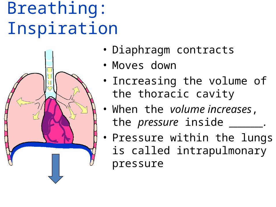
Breathing: Inspiration
• Diaphragm contracts • Moves down • Increasing the volume of the
thoracic cavity • When the volume increases, the
pressure inside ________.• Pressure within the lungs is called
intrapulmonary pressure
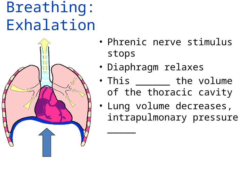
Breathing: Exhalation
• Phrenic nerve stimulus stops• Diaphragm relaxes• This ______ the volume of the
thoracic cavity• Lung volume decreases,
intrapulmonary pressure _____
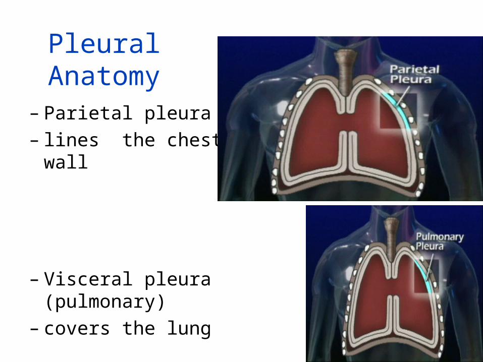
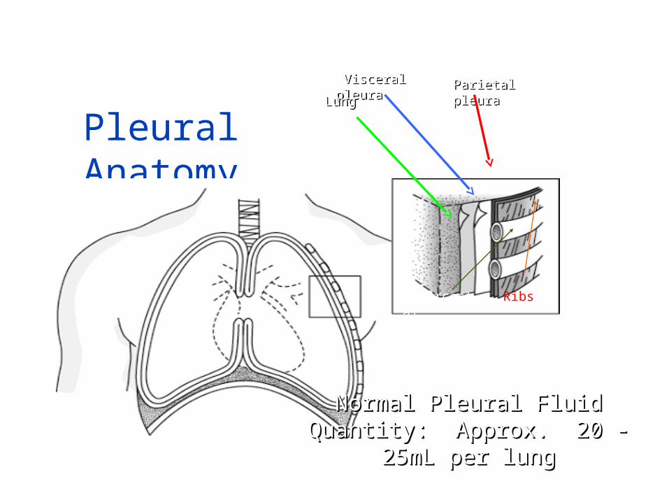
Pleural Anatomy
– Parietal pleura – lines the chest wall
– Visceral pleura (pulmonary) – covers the lung
Pleural Anatomy
Parietal pleuraParietal pleura Visceral pleura Visceral pleura
Normal Pleural Fluid Quantity: Normal Pleural Fluid Quantity: Approx. 20 - 25mL per lungApprox. 20 - 25mL per lung
Normal Pleural Fluid Quantity: Normal Pleural Fluid Quantity: Approx. 20 - 25mL per lungApprox. 20 - 25mL per lung
LungLung
RibsIntercostal muscles
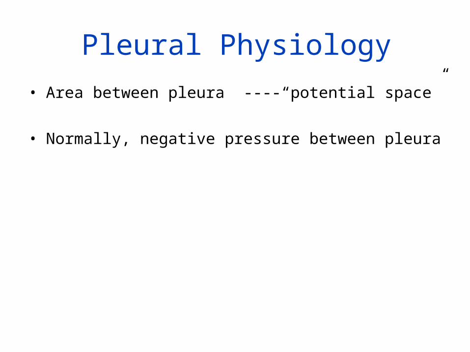
Pleural Physiology• Area between pleura ----“potential space”
• Normally, negative pressure between pleura
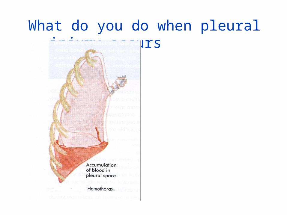
What do you do when pleural injury occurs
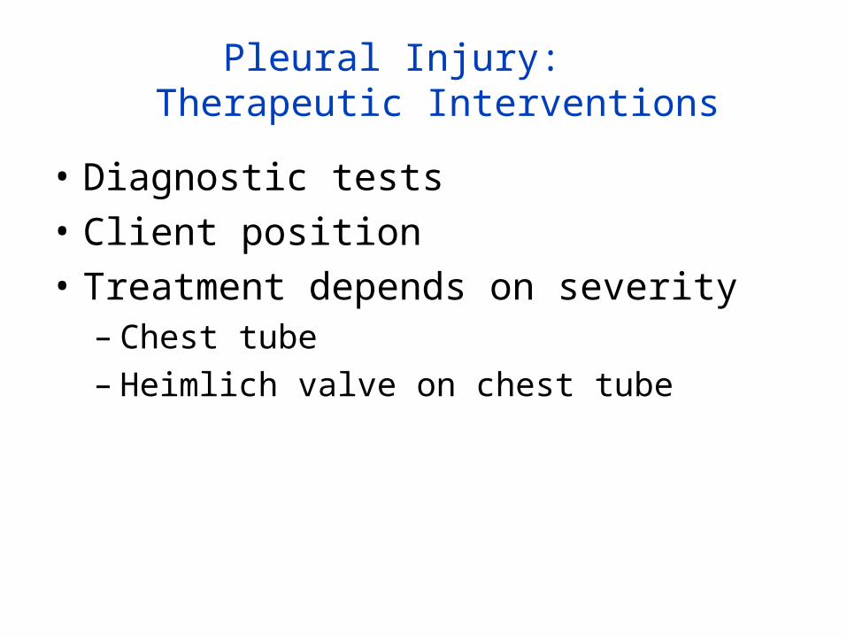
Pleural Injury: Therapeutic Interventions
• Diagnostic tests• Client position • Treatment depends on severity
– Chest tube– Heimlich valve on chest tube
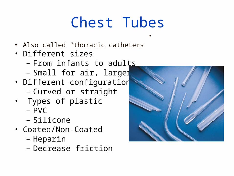
Chest Tubes• Also called “thoracic catheters”• Different sizes
– From infants to adults– Small for air, larger for fluid
• Different configurations– Curved or straight
• Types of plastic– PVC– Silicone
• Coated/Non-Coated– Heparin– Decrease friction
Chest Tube Placement
• In what setting/environment is a chest tube placed?
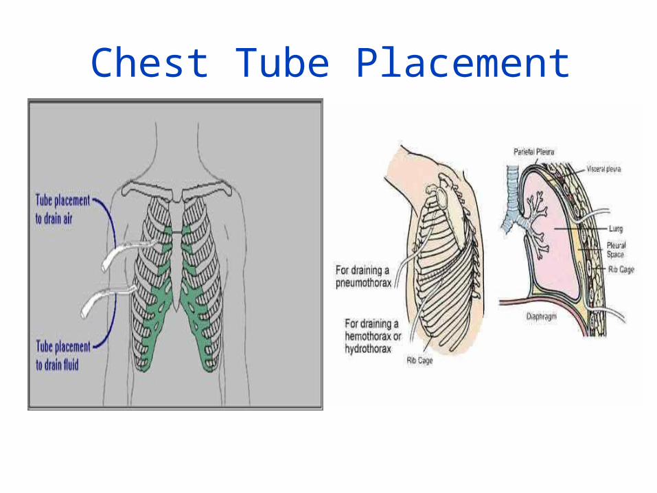
Chest Tube Placement
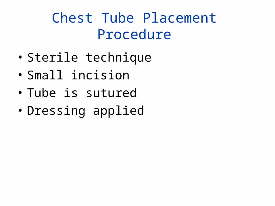
Chest Tube Placement Procedure
• Sterile technique• Small incision• Tube is sutured• Dressing applied
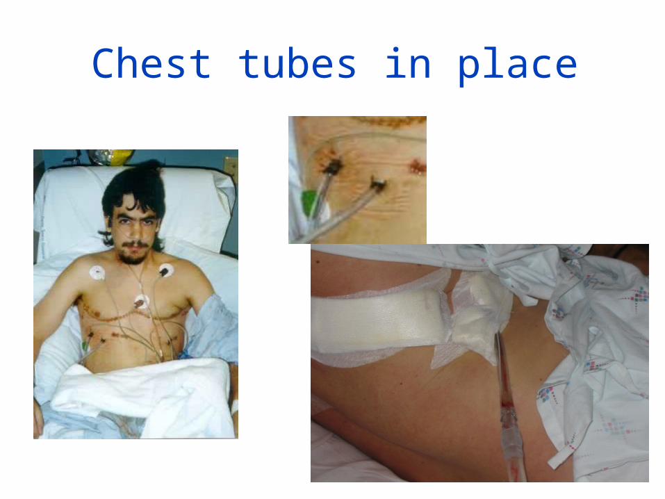
Chest tubes in place
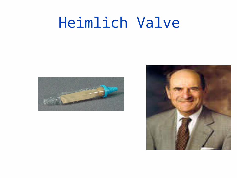
Heimlich Valve
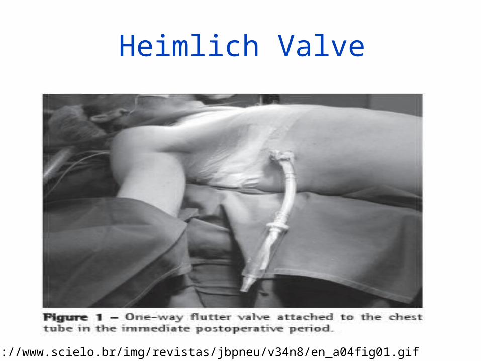
Heimlich Valve
http://www.scielo.br/img/revistas/jbpneu/v34n8/en_a04fig01.gif
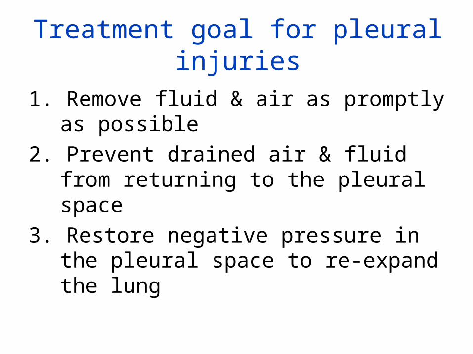
Treatment goal for pleural injuries
1. Remove fluid & air as promptly as possible2. Prevent drained air & fluid from returning to
the pleural space3. Restore negative pressure in the pleural space
to re-expand the lung
How a chest drainage system
works
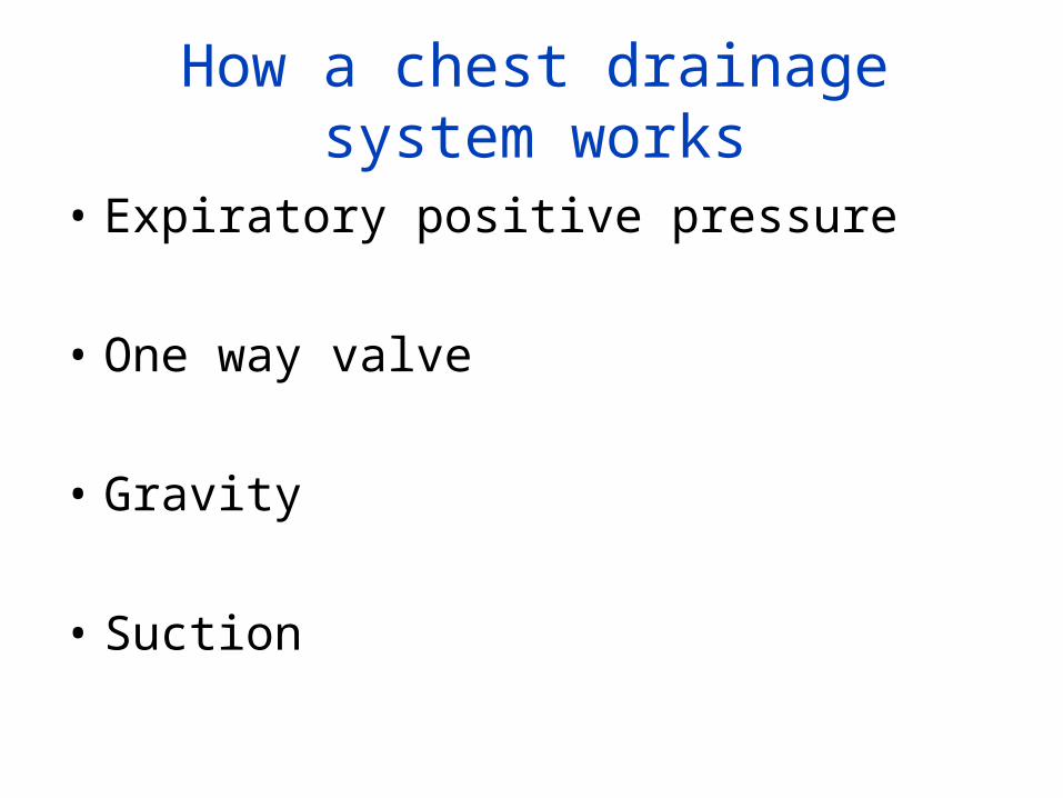
How a chest drainage system works
• Expiratory positive pressure
• One way valve
• Gravity
• Suction
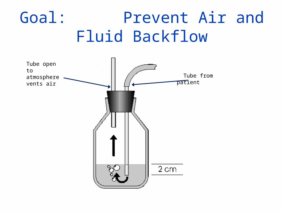
Goal: Prevent Air and Fluid Backflow
Tube open to atmosphere vents air Tube from patient
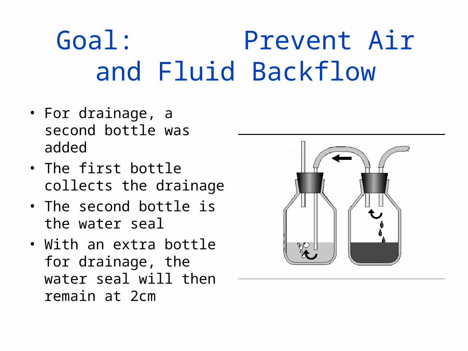
Goal: Prevent Air and Fluid Backflow
• For drainage, a second bottle was added
• The first bottle collects the drainage
• The second bottle is the water seal
• With an extra bottle for drainage, the water seal will then remain at 2cm
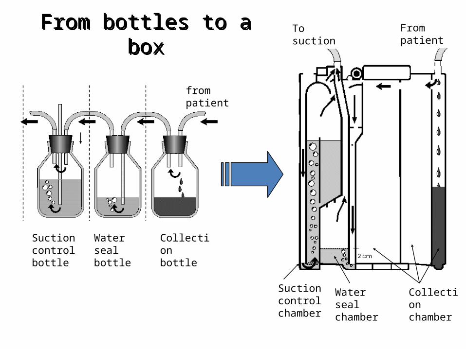
From bottles to a boxFrom bottles to a box
Collection chamber
Water seal chamber
Suction control chamber
from patient
Suction control bottle
Water seal bottle
Collection bottle
From patientTo suction
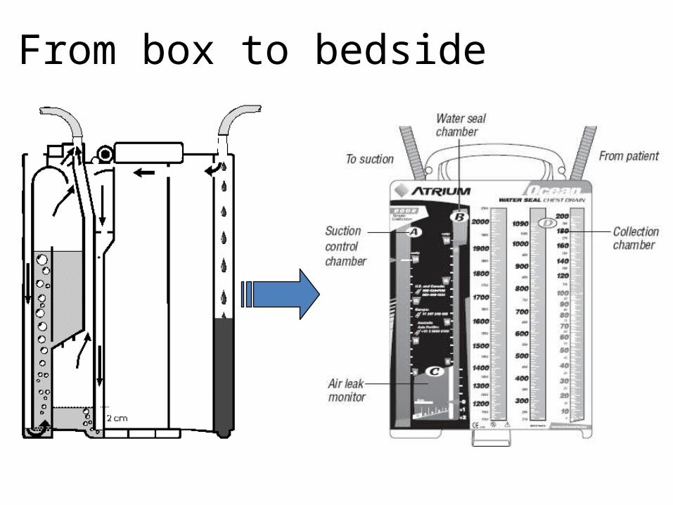
From box to bedside
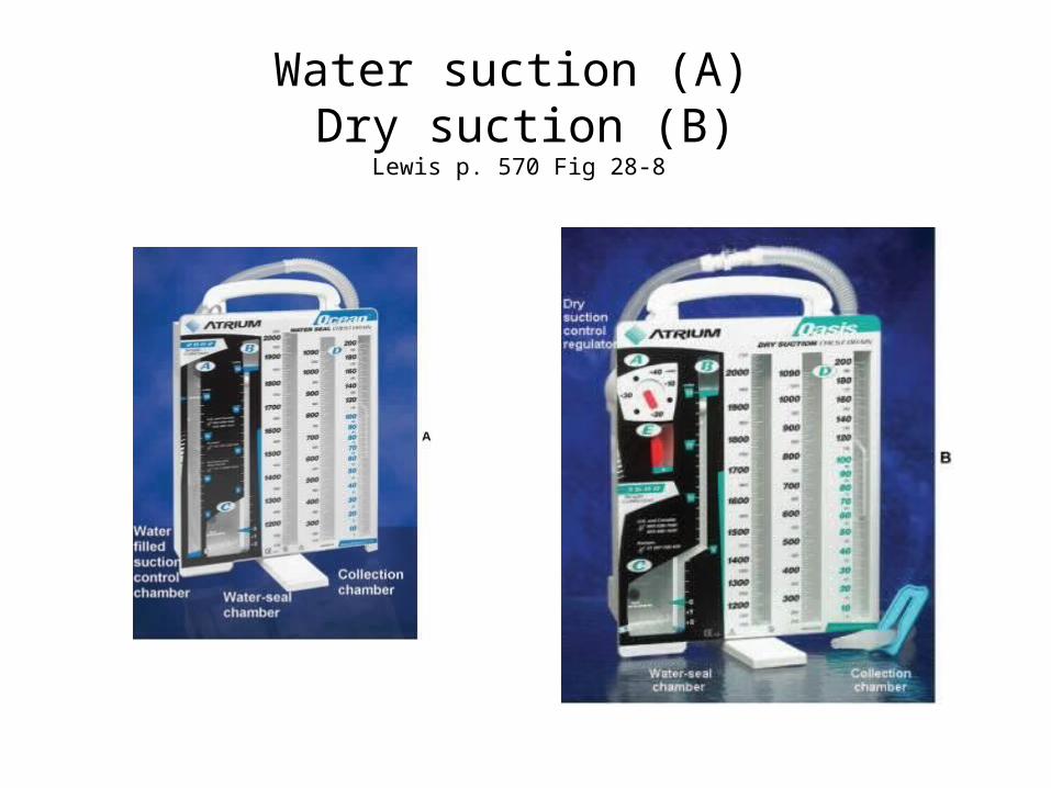
Water suction (A) Dry suction (B)
Lewis p. 570 Fig 28-8
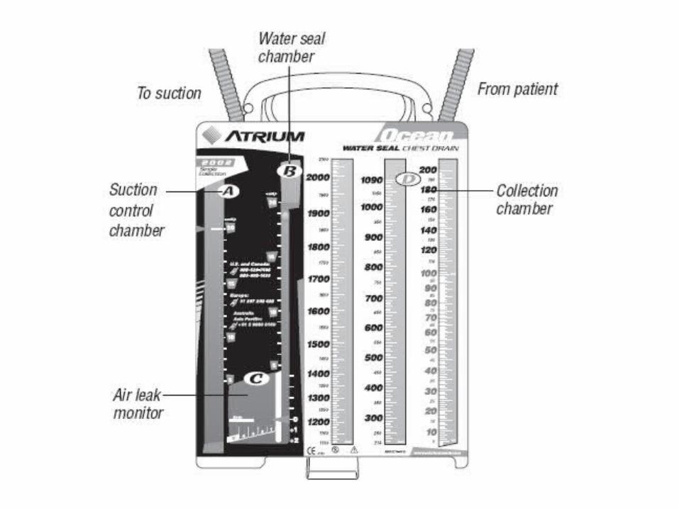
Atrium Chest Tube System• Chamber A
– Suction control chamber• Chamber B
– Water seal chamber• Chamber C
– Air leak monitor• Chamber D
– Collection chamber
Be sure you under stand how to set up the system, the function of each chamber and how to troubleshoot issues with each chamber.
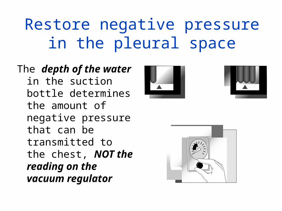
Restore negative pressure in the pleural space
The depth of the water in the suction bottle determines the amount of negative pressure that can be transmitted to the chest, NOT the reading on the vacuum regulator
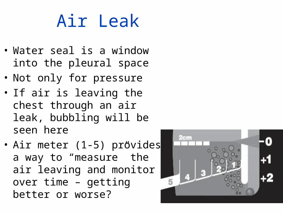
Air Leak
• Water seal is a window into the pleural space
• Not only for pressure• If air is leaving the chest through
an air leak, bubbling will be seen here
• Air meter (1-5) provides a way to “measure” the air leaving and monitor over time – getting better or worse?
Assessment • Focused respiratory assessment
– Breath sounds– Respiratory rate– Respiratory depth – SpO2– ABG– CXR
Assessment
• Cardiovascular assessment• Level of consciousness• Pain • Chest tube & Chest tube system
– Be sure you know what is to be assessed
Interventions
• System position
• Tubing position
• Connections to patient and system
• Monitoring & recording & reporting output
Interventions
• Dressing changes
• Oxygen therapy
• Analgesics
• IS and turn, cough, deep breathe
Complications
What are some common complications?
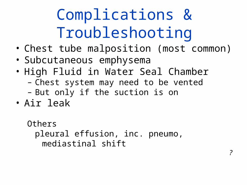
Complications & Troubleshooting• Chest tube malposition (most common)• Subcutaneous emphysema• High Fluid in Water Seal Chamber
– Chest system may need to be vented– But only if the suction is on
• Air leak
Otherspleural effusion, inc. pneumo,
mediastinal shift?
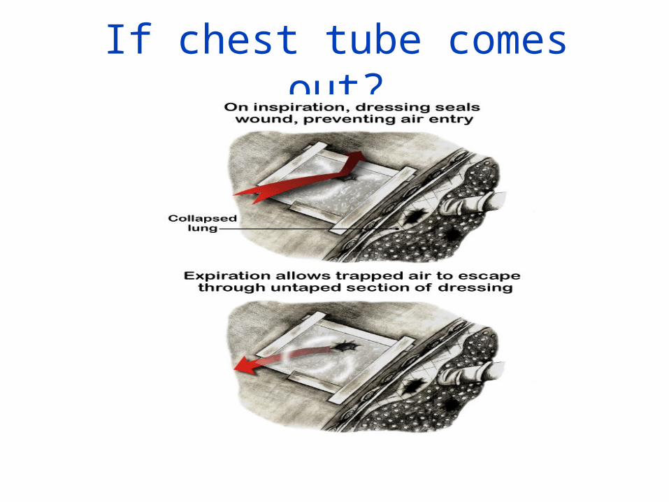
If chest tube comes out?
Review
• Check fluid level in suction chamber• Observe water seal chamber fluid level• Assess for tidaling in water seal chamber• Assess tubing – non dependent• Determine if the unit has been knocked over• Note the amount, color and consistency of drainage
What is most important?
• Monitor your client• Notify MD STAT if
– Significant drainage– Increasing shortness of breath– Pain– Absence of breath sounds
Management
• Do not remove suction without an order• Manage pain• When full - place in biohazard container• Do not change collection device on client with
an air leak without an order• When suction discontinued, must disconnect
from suction, not just turn off
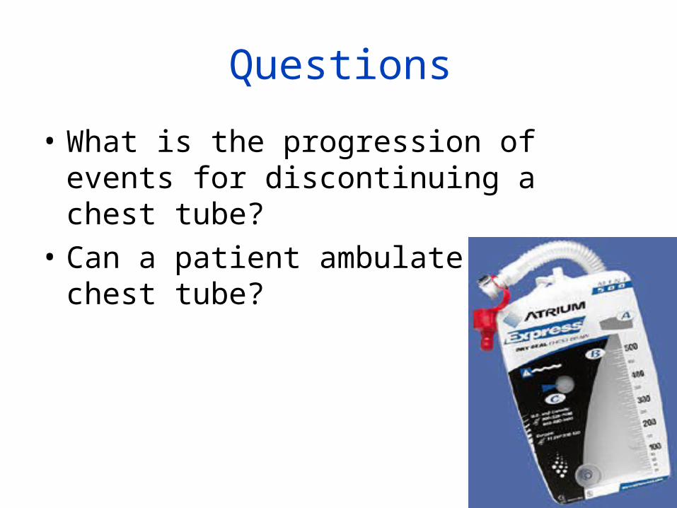
Questions
• What is the progression of events for discontinuing a chest tube?
• Can a patient ambulate with a chest tube?
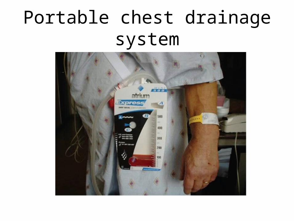
Portable chest drainage system
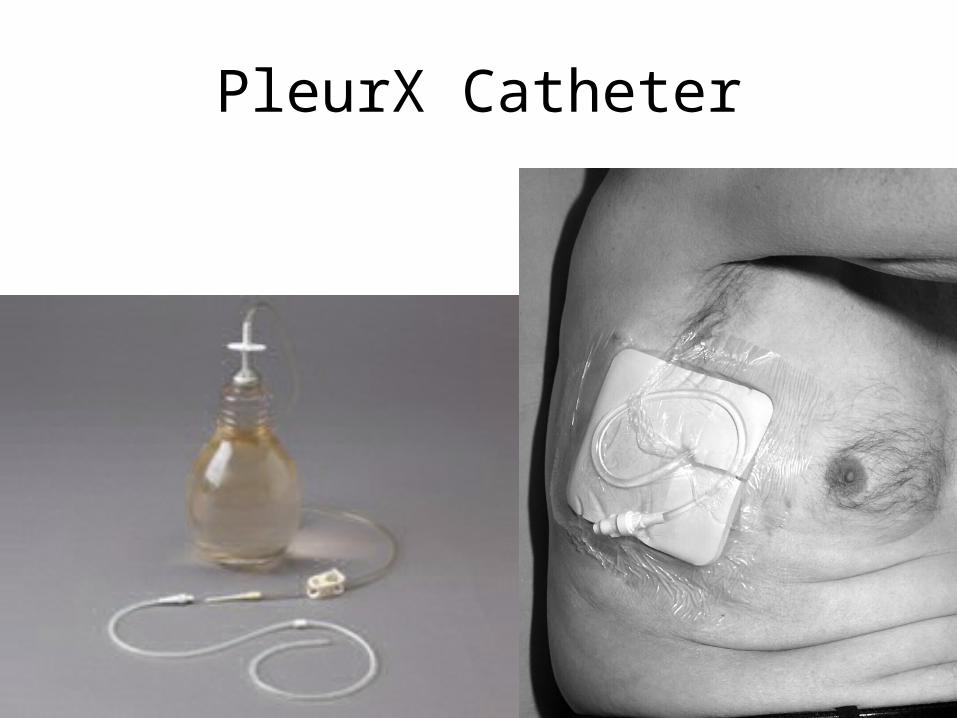
PleurX Catheter