Embed Size (px)
Citation preview
5
Chemotherapy-Induced Alopecia
Sudjit Luanpitpong and Yon Rojanasakul West Virginia University,
Department of Pharmaceutical Sciences, Morgantown, West Virginia, USA
1. Introduction
Chemotherapy-induced alopecia (CIA) is a frequent toxicity and arguably the most feared
side effect of cancer chemotherapy (Carelle et al., 2002). The incidence of CIA is
approximately 65% of all patients (Wang et al., 2006). CIA could be easily noticeable by self
and others in a relative short time, thus it is linked with having cancer and chemotherapy.
CIA compromises patient quality of life, especially for female and children, leading to poor
therapeutic outcome. Despite significant progresses and substantial efforts in CIA research
and development, no reliable and effective preventive treatment has become available. This
limitation has been attributed to the lack of basic understanding of CIA pathogenesis and
appropriate experimental models. This chapter will provide an overview of the basic and
clinical aspects of CIA including hair follicle biology, characteristics of CIA along with the
state-of-the-art experimental models and treatment strategies. Experimental approaches for
pharmacologic inhibition of CIA including drug-specific antibodies, hair growth cycle
modifiers, cytokines, growth factors, antioxidants, cell cycle modifiers, and apoptosis
inhibitors will be discussed. Current understanding in the molecular mechanisms of CIA
and the role of specific genes, e.g. p53 and Fas, in the process will also be discussed. The
chapter will conclude with the perspective on the prevention and management of CIA.
2. Hair follicle biology
Chemotherapy causes structural damage of human scalp hairs. The effects may vary from altered hair appearance, decreased rate of hair growth, partial or complete hair loss (alopecia). To discuss the advances in the pathogenesis of CIA, an overview of hair follicle biology is first covered.
2.1 Hair follicle structure
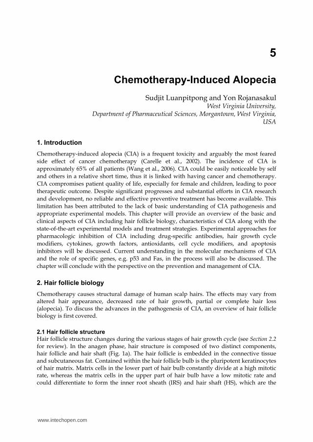
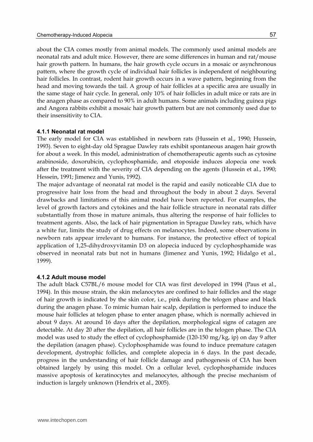
Hair follicle structure changes during the various stages of hair growth cycle (see Section 2.2 for review). In the anagen phase, hair structure is composed of two distinct components, hair follicle and hair shaft (Fig. 1a). The hair follicle is embedded in the connective tissue and subcutaneous fat. Contained within the hair follicle bulb is the pluripotent keratinocytes of hair matrix. Matrix cells in the lower part of hair bulb constantly divide at a high mitotic rate, whereas the matrix cells in the upper part of hair bulb have a low mitotic rate and could differentiate to form the inner root sheath (IRS) and hair shaft (HS), which are the
www.intechopen.com
Topics in Cancer Survivorship
54
middle and innermost layer of hair follicle, respectively. Outer root sheath (ORS), is the outermost layer of hair follicle that separates the whole organ from dermis and is believed to contain epithelial stem cells at its bulge region (Hardy, 1992; Krause and Foitzik, 2006; Alonso and Fuchs, 2006). Pigmentation of hair shaft depends on melanocytes, which reside in the hair matrix of hair follicle. Melanocytes transfer the melanin granule to keratinocytes of the growing hair shaft (Ohnemus et al., 2006). Besides the epithelial cells, hair follicle also contains the mass of mesenchymal dermal papilla (DP) cells at its base (Fig. 1b). The DP cells are connected to capillaries to derive nutrients from the blood and also function as a regulator of hair cycle (Sakita et al., 1995). Moreover, substantial evidence supports the correlation between DP cell number and the size of hair follicle and shaft (Elliot et al., 1993; Ishino et al., 1997).
Fig. 1. Diagrammatic representation of hair follicle structure in its mature anagen phase. (a) A full-length longitudinal view of hair follicle. (b) Hair follicle bulb. Abbreviations: APM, arector pili muscle; B, bulge; CTS, connective tissue sheath; CTX, cortex of hair shaft; CU, cuticle of hair shaft; DP, dermal papilla; E, epidermis; HM, hair matrix; HS, hair shaft; IRS, inner root sheath; M, melanocytes; ORS, outer root sheath; S, sebaceous gland.
2.2 Hair growth cycle
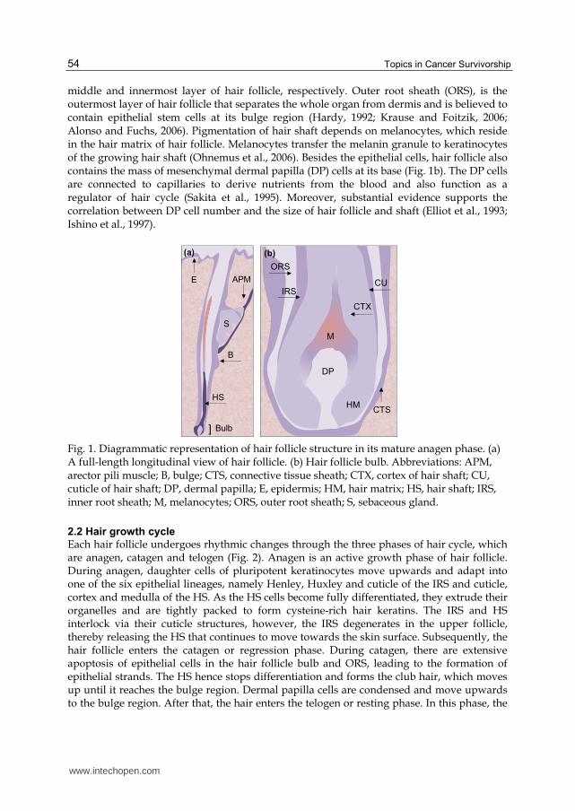
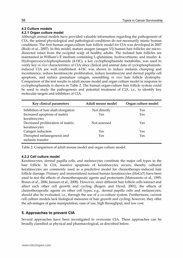
Each hair follicle undergoes rhythmic changes through the three phases of hair cycle, which are anagen, catagen and telogen (Fig. 2). Anagen is an active growth phase of hair follicle. During anagen, daughter cells of pluripotent keratinocytes move upwards and adapt into one of the six epithelial lineages, namely Henley, Huxley and cuticle of the IRS and cuticle, cortex and medulla of the HS. As the HS cells become fully differentiated, they extrude their organelles and are tightly packed to form cysteine-rich hair keratins. The IRS and HS interlock via their cuticle structures, however, the IRS degenerates in the upper follicle, thereby releasing the HS that continues to move towards the skin surface. Subsequently, the hair follicle enters the catagen or regression phase. During catagen, there are extensive apoptosis of epithelial cells in the hair follicle bulb and ORS, leading to the formation of epithelial strands. The HS hence stops differentiation and forms the club hair, which moves up until it reaches the bulge region. Dermal papilla cells are condensed and move upwards to the bulge region. After that, the hair enters the telogen or resting phase. In this phase, the
E
DP
IRS
ORS
HM
CTX
CTS
CU
M
B
APM
S
HS
Bulb]
(a) (b)
www.intechopen.com
Chemotherapy-Induced Alopecia
55
HS exhibits no significant proliferation, apoptosis or differentiation. The transition from telogen to anagen occurs when the bulge stem cells are activated (Cotsaleris and Millar, 2001; Krause and Foitzik, 2006; Alonso and Fuchs, 2006; Ohnemus et al., 2006).
Fig. 2. Hair growth cycle. A new hair shaft is produced during anagen, and the old hair is released from the follicle as the new shaft develops. Anagen VI (mature anagen) is the stage where new HS reaches the skin surface and continues to grow through the rest of anagen. During catagen, the lower two thirds of the epithelial follicle are regressed. The hair develops a club structure, which retains the hair in the follicle. Then, the follicle enters a telogen phase until a new growth cycle is activated. Abbreviations: B, bulge; DP, dermal papilla; HS, hair shaft.
3. Chemotherapy-induced alopecia
CIA or hair loss caused by chemotherapy is the most common cutaneous side effect of
chemotherapy. CIA ranks among patients as a severe side effect that affects their quality of
life.
3.1 Impact on cancer therapy
CIA has an enormous psychological and social impact on patients, which can be summarized
as: (i) symbol of cancer for self (constant reminder of their treatment) and others (outwardly
visible); (ii) personal confrontation of being ill or mortality; (iii) vulnerability; (iv)
powerlessness; (v) shame; (vi) loss of privacy; (vii) punishment, and (viii) change in self and
other perception (Freedman, 1994; Pozo-Kaderman et al., 1999). Female and children have
more difficulties coping with the CIA. Indeed, up to 8% of women are reported to reject
chemotherapy for fear of CIA (Mundstedt et al., 1997; McGarvey et al., 2001). CIA also results
in reduced social activities since hair partly plays a role in social and sexual communications
(Batchelor, 2001). Additionally, these negative impacts of CIA may contribute to poor
therapeutic outcome, as stress and depression lowers the body’s immune function and is
highly associated with cancer progression (Spiegel and Giese-Davis, 2003; O’Leary, 1990).
Anagen (VI) Catagen Telogen
DP
B B B
HS
Club] Activation
www.intechopen.com
Topics in Cancer Survivorship
56
3.2 Pathophysiology
The basic principle of chemotherapy is to impair the mitotic and metabolic process of cancer cells. Unfortunately, certain normal cells and tissues with rapid metabolic and mitotic rates such as the hair follicles are also affected by the chemotherapy. Up to 90% of hair follicles undergo anagen, an active growth phase, at a given time. The rapid hair growth as well as the high blood flow rate around the hair bulb leading to the accumulation of drugs is a key predisposing factor for rapid and extensive alopecia (Batchelor, 2001). In humans, CIA usually begins approximately 2 to 4 weeks and is complete at 1 to 2 months after the initiation of chemotherapy (Batchelor, 2001). Hair might be easily depilated as early as 1 to 2 weeks after the treatment due to the weakening and breakage of hair shaft. The hair would fall out upon combing and in the bedding area. The degree of CIA depends on the type of chemotherapy, dosage regimen and route of administration. Almost all chemotherapies cause alopecia but with varying degrees of severity and frequency (Apisanthanarax and Duvic, 2003) as summarized in Table 1.
More common or severe Less common or severe
Bleomycin Cytarabine Dacarbazine Docetaxel Etoposide Idarubicin Interferon-α Mechlorethamine Paclitaxel Topotecan Vincristine
Cyclophosphamide Cisplatin Dactinomycin Doxorubicin Fluorouracil Ifosfamide Irinotecan Nitroureas Thiotepa Vinblastine Vindesine
Amscarine Carmusine Carboplatin Gemcitabine Interleukin-2 Mercaptopurine Mitomycin Procarbazine Vinorelbine
Busulfan Chlorambucil Epirubicin Hydroxyurea Melphalan Methotrexate Mitoxantrone Teniposide
Table 1. Chemotherapeutic agents associated with alopecia.
A high-dose intravenous chemotherapy is commonly associated with more rapid and extensive alopecia. By contrast, oral therapy at lower doses on a weekly schedule tends to cause less alopecia even though the total dose may be large (Wilkes, 1996). Combination therapy consisting of two or more chemotherapeutic agents normally causes a higher incidence and more severe CIA compared to single agent therapy. Long-term chemotherapy may also result in the loss of pubic, axillary and facial hair. CIA is usually reversible with the hair regrowth generally occurring 3 to 6 months after the end of treatment. However, in most cases the new hair is grey or differs in color, representing the distortion of pigmentation process. Moreover, the new hair typically exhibits some changes in hair structure and texture, e.g. coarser, slow growth, and reduced density (Wang et al., 2006; Trueb, 2009). Permanent alopecia has been reported but rarely occurs (Betcheler, 2001).
4. Experimental models
4.1 Animal models
Due to the ethical problems in obtaining scalp biopsies from chemotherapy patients, little is known about the mechanisms of CIA in humans. In the last decade, important information
www.intechopen.com
Chemotherapy-Induced Alopecia
57
about the CIA comes mostly from animal models. The commonly used animal models are neonatal rats and adult mice. However, there are some differences in human and rat/mouse hair growth pattern. In humans, the hair growth cycle occurs in a mosaic or asynchronous pattern, where the growth cycle of individual hair follicles is independent of neighbouring hair follicles. In contrast, rodent hair growth occurs in a wave pattern, beginning from the head and moving towards the tail. A group of hair follicles at a specific area are usually in the same stage of hair cycle. In general, only 10% of hair follicles in adult mice or rats are in the anagen phase as compared to 90% in adult humans. Some animals including guinea pigs and Angora rabbits exhibit a mosaic hair growth pattern but are not commonly used due to their insensitivity to CIA.
4.1.1 Neonatal rat model
The early model for CIA was established in newborn rats (Hussein et al., 1990; Hussein,
1993). Seven to eight-day old Sprague Dawley rats exhibit spontaneous anagen hair growth
for about a week. In this model, administration of chemotherapeutic agents such as cytosine
arabinoside, doxorubicin, cyclophosphamide, and etoposide induces alopecia one week
after the treatment with the severity of CIA depending on the agents (Hussein et al., 1990;
Hessein, 1991; Jimenez and Yunis, 1992).
The major advantage of neonatal rat model is the rapid and easily noticeable CIA due to
progressive hair loss from the head and throughout the body in about 2 days. Several
drawbacks and limitations of this animal model have been reported. For examples, the
level of growth factors and cytokines and the hair follicle structure in neonatal rats differ
substantially from those in mature animals, thus altering the response of hair follicles to
treatment agents. Also, the lack of hair pigmentation in Sprague Dawley rats, which have
a white fur, limits the study of drug effects on melanocytes. Indeed, some observations in
newborn rats appear irrelevant to humans. For instance, the protective effect of topical
application of 1,25-dihydroxyvitamin D3 on alopecia induced by cyclophosphamide was
observed in neonatal rats but not in humans (Jimenez and Yunis, 1992; Hidalgo et al.,
1999).
4.1.2 Adult mouse model
The adult black C57BL/6 mouse model for CIA was first developed in 1994 (Paus et al.,
1994). In this mouse strain, the skin melanocytes are confined to hair follicles and the stage
of hair growth is indicated by the skin color, i.e., pink during the telogen phase and black
during the anagen phase. To mimic human hair scalp, depilation is performed to induce the
mouse hair follicles at telogen phase to enter anagen phase, which is normally achieved in
about 9 days. At around 16 days after the depilation, morphological signs of catagen are
detectable. At day 20 after the depilation, all hair follicles are in the telogen phase. The CIA
model was used to study the effect of cyclophosphamide (120-150 mg/kg, ip) on day 9 after
the depilation (anagen phase). Cyclophosphamide was found to induce premature catagen
development, dystrophic follicles, and complete alopecia in 6 days. In the past decade,
progress in the understanding of hair follicle damage and pathogenesis of CIA has been
obtained largely by using this model. On a cellular level, cyclophosphamide induces
massive apoptosis of keratinocytes and melanocytes, although the precise mechanism of
induction is largely unknown (Hendrix et al., 2005).
www.intechopen.com
Topics in Cancer Survivorship
58
4.2 Culture models 4.2.1 Organ culture model Although animal models have provided valuable information regarding the pathogenesis of CIA, the animal physiological and pathological conditions do not necessarily mimic human conditions. The first human organ-culture hair follicle model for CIA was developed in 2007 (Bodo et al., 2007). In this model, mature anagen (anagen VI) human hair follicles are micro-dissected intact from the occipital scalp of healthy adults. The isolated hair follicles are maintained in William’s E medium containing L-glutamine, hydrocortisone, and insulin. 4-Hydroperoxycyclophosphamide (4-HC), a key cyclophosphamide metabolite, was used to verify key in vivo characteristics of CIA since clinical and animal data of cyclophosphamide-induced CIA are well established. 4-HC was shown to induce melanin clumping and incontinence, reduce keratinocyte proliferation, induce keratinocyte and dermal papilla cell apoptosis, and induce premature catagen, resembling in vivo hair follicle dystrophy. Comparison of the test results in adult mouse model and organ culture model in response to cyclophosphamide is shown in Table 2. The human organ-culture hair follicle system could be used to study the pathogenesis and potential treatment of CIA, i.e., to identify key molecular targets and inhibitors of CIA.
Key clinical parameters Adult mouse model Organ culture model
Inhibition of hair shaft elongation Increased apoptosis of matrix keratinocytes Decreased proliferation of matrix keratinocytes Catagen induction Disrupted melanogenesis and melanin transfer
Not directly Yes
Not assessed
Yes Yes
Yes Yes
Yes
Yes Yes
Table 2. Comparison of adult mouse model and organ culture model.
4.2.2 Cell culture model
Keratinocytes, dermal papilla cells, and melanocytes constitute the major cell types in the hair follicle. In CIA, massive apoptosis of keratinocytes occurs; thereby cultured keratinocytes are commonly used as a predictive model for chemotherapy-induced hair follicle damage. Primary and immortalized normal human keratinocytes (HaCaT) have been used to test the effects of chemotherapeutic agents and protectants (Matsumoto et al., 1995; Braun et al., 2006; Janssen et al., 2008). However, since different hair follicle cells interact and affect each other cell growth and cycling (Rogers and Hynd, 2001), the effects of chemotherapeutic agents on other cell types, e.g., dermal papilla cells and melanocytes, should also be evaluated, i.e., through the use of a co-culture system. Furthermore, current cell culture models lack biological measures of hair growth and cycling; however, they offer the advantages of gene manipulation, ease of use, high throughput, and low cost.
5. Approaches to prevent CIA
Several approaches have been investigated to overcome CIA. These approaches can be broadly classified as physical and pharmacological, as described below.
www.intechopen.com
Chemotherapy-Induced Alopecia
59
5.1 Physical prevention 5.1.1 Scalp torniques
Scalp torniques are the application of bands around the head to occlude the superficial blood flow to scalp, thus reducing the amount of drugs delivered to the hair follicles. The torniques range from 10 mmHg above systolic pressure to 300 mmHg around the scalp. These torniques are applied 5-10 minutes prior to or at the time of chemotherapy until up to 30 minutes after the drug administration (Cline, 1984). Although reports described mild to moderate prevention of CIA induced by vincristine, cyclophosphamide, and doxorubicin, this technique is no longer recommended due to patient discomfort (Wang et al., 2006).
5.1.2 Scalp cooling
Scalp cooling or hypothermia is the application of cold to the scalp using a device (cap) that is pre-cooled in a freezer or exchanges coolant with reservoir. A period of cooling lasts from 5 minutes prior to chemotherapy until an hour or more after the drug administration. Many studies have shown that the efficacy of scalp cooling can range from 0-90% (for review, see Grevelman and Breed, 2005). A recent study reported that scalp cooling helps reduce major CIA in patients receiving doxorubicin (60 mg/m2), docetaxel (80 mg/m2), or combination of 5-fluorouracil (600 mg/m2), epirubicin (60 mg/m2), and cyclophosphamide (600 mg/m2) for 6 to 9 cycles (Auvinen et al., 2010). The current hypotheses of the protective effect are: (i) cooling reduces blood flow to hair follicles by vasoconstriction, resulting in a decrease in the amount of drugs available for uptake; and (ii) cooling decreases cellular metabolism and drug uptake. Scalp cooling to 20°C was shown to reduce blood flow to 20% of normal flow; however, further decrease in the temperature (<18°C) did not result in further decrease in scalp blood flow (Janssen et al., 2007). Recent in vitro studies indicate the significant role of temperature on keratinocyte cell viability upon doxorubicin chemotherapy; however, there is no difference in cell survival between 10°C and 22°C (Janssen et al., 2008). Based on these findings, it appears that there is an optimal temperature for scalp cooling (~20°C), and that increasing the cooling will only result in patient discomfort. Other factors affecting the effectiveness of this method include drug regimen, application and duration of cooling, and the cool conductivity (Betcheler, 2001). Scalp cooling is practically ineffective if the chemotherapeutic agent is administered as a
continuous infusion over a prolonged period. Additionally, scalp cooling increases the risk
of scalp metastasis, and is therefore contraindicated in patients with hematological
malignancies and cutaneous T-cell lymphoma (Dean et al., 1979; Apisanthanarax and Duvic,
2003).
5.1.3 Heat treatment Stress protein response is one of the cellular protective mechanisms against various adverse conditions. Enhanced expression of stress proteins such as Hsp90, Hsp70, and Hsp25 has been observed in response to certain physical and chemical stresses, which has been linked to increased stress tolerance. Previous in vitro studies have shown that heat treatment and overexpression of stress response proteins, e.g., Hsp70 and Hsp27, could protect against the cytotoxic effects of anticancer drugs such as doxorubicin, cyclophosphamide, etoposide, and taxol (Kampinga, 1995; Jaattela et al., 1998; Kwak et al., 1998; Ito et al., 1999; Xia et al., 1999), leading to the investigation of the protective effect of stress protein activation on CIA in neonatal rats (Jimenez et al., 2008). In this study, heat was locally applied to the skin with a copper cylinder through which heated water was circulated. Conducting gel (Vaseline) was
www.intechopen.com
Topics in Cancer Survivorship
60
applied to the skin to improve heat conductance. Heat treatment at 48-48.5°C for 20 minutes increases Hsp70 and subsequently protects against CIA in response to various treatments as summarized in Table 3. The protective effect of heat treatment was confirmed in an adult mouse model receiving cyclophosphamide. Additionally, localized heat treatment was shown not to interfere with the anti-tumor activity of drugs. These findings suggest that localized activation of stress proteins in the hair follicles might be an effective strategy against CIA without affecting the anti-tumor efficacy.
Chemotherapeutic agents Dosing Protective Frequency
Etoposide Cyclophosphamide Cyclophosphamide/doxorubicin Taxol
I.P. 2.5 µg/g twice I.P. 35.5 µg/g twice I.P. 20-30 µg/g once/2.5-4.5 µg/g twice S.C. 5 µg/animal, twice
0.94 (45/48) 0.97 (29/30) 1.0 (56/56) 1.0 (7/7)
Table 3. Localized, heat-induced protection against CIA in neonatal rats.
5.2 Pharmacological prevention
Currently, there are no FDA-approved drug treatments for CIA but several pharmacological
strategies have been proposed. Many of these strategies have shown promising results in
animals but their clinical use will require further investigations.
5.2.1 Tumor targeting delivery
Differences in the molecular machinery of normal cells and tumor cells as a result of cell
transformation dominate the tumor targeting delivery arena. Tumor-specific ligands and
antibodies have been used to provide targeting ability to drug carriers such as liposomes.
Accordingly, these liposomes can protect patients from the side effects of chemotherapy,
including hair loss. Examples of the targeting moieties are folate receptor (FR) for ovarian,
colorectal, and breast cancer; transferrin for pancreatic cancer; anti-HER2 antibody for breast
cancer; anti-CD19 for malignant B cells; anti-GD2 for neuroblastoma and melonoma; and
prostate-specific membrane antigen (PMSA) aptamer for prostate cancer and tumor vascular
endothelium (Huges et al., 2001; Yu et al., 2009).
5.2.2 Drug-specific antibodies
MAD11 monoclonal antibody (MAb) is an anti-anthracycline antibody that reacts with
doxorubicin and other anthracycline chemotherapeutics. Topical administration of
liposomes containing MAD11 MAb was shown to prevent CIA in doxorubicin-treated
neonatal rats at the frequency of 31 in 45 rats (Balsari et al., 1994). MAD11 MAb was
encapsulated into liposomes to facilitate absorption through the stratum corneum and to
delay systemic distribution of the antibody. Topical MAD11 MAb was found to be nontoxic
and does not induce systemic activation of cytokines. Thus, MAD11-loaded liposomes
might be an effective strategy in preventing anthracycline-induced alopecia in cancer
patients. However, the advantage of this strategy is limited in combination therapy since the
antibody could not react with the other drugs in combination.
www.intechopen.com
Chemotherapy-Induced Alopecia
61
5.2.3 Hair growth cycle modifiers
5.2.3.1 Cyclosporine A
Cyclosporine A is an immunosuppressive immunophilin ligand used in the treatment of autoimmune diseases and in post-organ transplantation to reduce patients’ graft rejection. In T-lymphocytes, cyclosporine A forms complex with cyclophilin and inhibits calcineurin, leading to the inhibition of Go to G1 cell cycle transition and proliferation. The use of cyclosporine A in alopecia originates from its common side effect of excessive hair growth called hypertrichosis. Cyclosporin A induces anagen and inhibits catagen of the hair cycle, leading to the promotion of hair growth under normal and pathologic conditions such as alopecia areata and androgenetic alopecia (Paus et al., 1989; Taylor et al., 1993; Lutz et al., 1994). The effect of cyclosporine A on CIA has been investigated in neonatal rat and adult mouse
models. In neonatal rats, topical administration of cyclosporine A prevents CIA induced by
cyclophosphamide, cytosine arabinoside and etoposide (Hussein et al., 1995). In adult mice
given cyclophosphamide, topical or systemic administration of cyclosporine A retards CIA,
prevents the progression of damaged hair into telogen, and thus induces faster hair
regrowth.
5.2.3.2 AS101
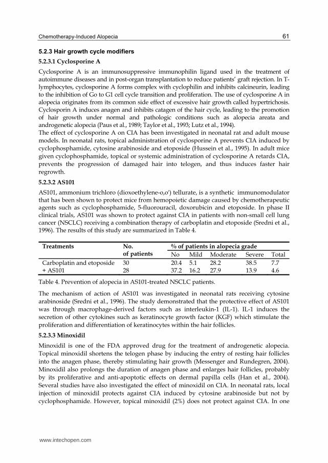
AS101, ammonium trichloro (dioxoethylene-o,o') tellurate, is a synthetic immunomodulator that has been shown to protect mice from hemopoietic damage caused by chemotherapeutic agents such as cyclophosphamide, 5-fluorouracil, doxorubicin and etoposide. In phase II clinical trials, AS101 was shown to protect against CIA in patients with non-small cell lung cancer (NSCLC) receiving a combination therapy of carboplatin and etoposide (Sredni et al., 1996). The results of this study are summarized in Table 4.
Treatments No. of patients
% of patients in alopecia grade
No Mild Moderate Severe Total
Carboplatin and etoposide + AS101
30 28
20.4 37.2
5.1 16.2
28.2 27.9
38.5 13.9
7.7 4.6
Table 4. Prevention of alopecia in AS101-treated NSCLC patients.
The mechanism of action of AS101 was investigated in neonatal rats receiving cytosine
arabinoside (Sredni et al., 1996). The study demonstrated that the protective effect of AS101
was through macrophage-derived factors such as interleukin-1 (IL-1). IL-1 induces the
secretion of other cytokines such as keratinocyte growth factor (KGF) which stimulate the
proliferation and differentiation of keratinocytes within the hair follicles.
5.2.3.3 Minoxidil
Minoxidil is one of the FDA approved drug for the treatment of androgenetic alopecia.
Topical minoxidil shortens the telogen phase by inducing the entry of resting hair follicles
into the anagen phase, thereby stimulating hair growth (Messenger and Rundegren, 2004).
Minoxidil also prolongs the duration of anagen phase and enlarges hair follicles, probably
by its proliferative and anti-apoptotic effects on dermal papilla cells (Han et al., 2004).
Several studies have also investigated the effect of minoxidil on CIA. In neonatal rats, local
injection of minoxidil protects against CIA induced by cytosine arabinoside but not by
cyclophosphamide. However, topical minoxidil (2%) does not protect against CIA. In one
www.intechopen.com
Topics in Cancer Survivorship
62
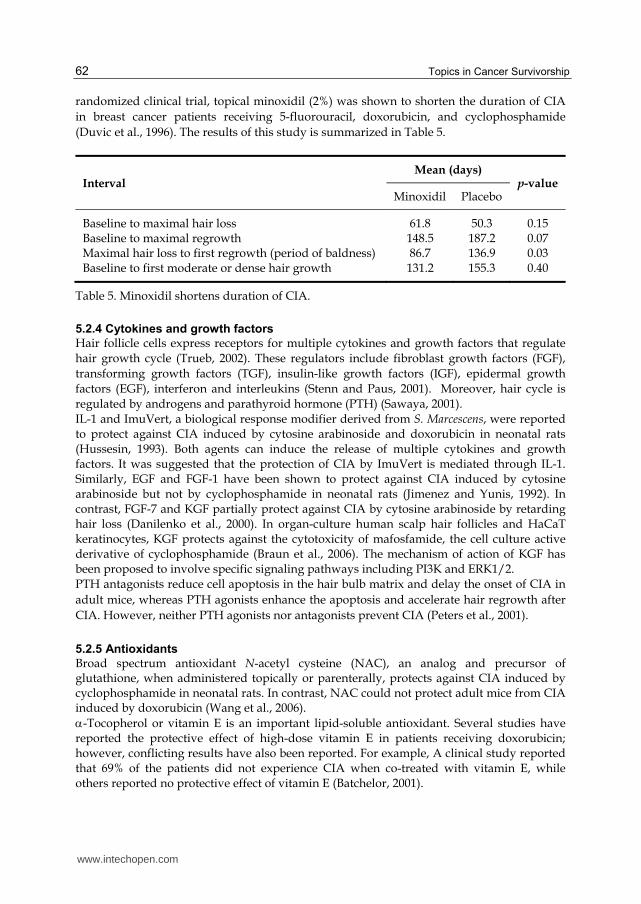
randomized clinical trial, topical minoxidil (2%) was shown to shorten the duration of CIA
in breast cancer patients receiving 5-fluorouracil, doxorubicin, and cyclophosphamide
(Duvic et al., 1996). The results of this study is summarized in Table 5.
Interval Mean (days)
p-value Minoxidil Placebo
Baseline to maximal hair loss Baseline to maximal regrowth Maximal hair loss to first regrowth (period of baldness) Baseline to first moderate or dense hair growth
61.8 148.5 86.7 131.2
50.3 187.2 136.9 155.3
0.15 0.07 0.03 0.40
Table 5. Minoxidil shortens duration of CIA.
5.2.4 Cytokines and growth factors
Hair follicle cells express receptors for multiple cytokines and growth factors that regulate
hair growth cycle (Trueb, 2002). These regulators include fibroblast growth factors (FGF),
transforming growth factors (TGF), insulin-like growth factors (IGF), epidermal growth
factors (EGF), interferon and interleukins (Stenn and Paus, 2001). Moreover, hair cycle is
regulated by androgens and parathyroid hormone (PTH) (Sawaya, 2001). IL-1 and ImuVert, a biological response modifier derived from S. Marcescens, were reported to protect against CIA induced by cytosine arabinoside and doxorubicin in neonatal rats (Hussesin, 1993). Both agents can induce the release of multiple cytokines and growth factors. It was suggested that the protection of CIA by ImuVert is mediated through IL-1. Similarly, EGF and FGF-1 have been shown to protect against CIA induced by cytosine arabinoside but not by cyclophosphamide in neonatal rats (Jimenez and Yunis, 1992). In contrast, FGF-7 and KGF partially protect against CIA by cytosine arabinoside by retarding hair loss (Danilenko et al., 2000). In organ-culture human scalp hair follicles and HaCaT keratinocytes, KGF protects against the cytotoxicity of mafosfamide, the cell culture active derivative of cyclophosphamide (Braun et al., 2006). The mechanism of action of KGF has been proposed to involve specific signaling pathways including PI3K and ERK1/2. PTH antagonists reduce cell apoptosis in the hair bulb matrix and delay the onset of CIA in
adult mice, whereas PTH agonists enhance the apoptosis and accelerate hair regrowth after
CIA. However, neither PTH agonists nor antagonists prevent CIA (Peters et al., 2001).
5.2.5 Antioxidants
Broad spectrum antioxidant N-acetyl cysteine (NAC), an analog and precursor of glutathione, when administered topically or parenterally, protects against CIA induced by cyclophosphamide in neonatal rats. In contrast, NAC could not protect adult mice from CIA induced by doxorubicin (Wang et al., 2006).
-Tocopherol or vitamin E is an important lipid-soluble antioxidant. Several studies have reported the protective effect of high-dose vitamin E in patients receiving doxorubicin; however, conflicting results have also been reported. For example, A clinical study reported that 69% of the patients did not experience CIA when co-treated with vitamin E, while others reported no protective effect of vitamin E (Batchelor, 2001).
www.intechopen.com
Chemotherapy-Induced Alopecia
63
5.2.6 Cell cycle or proliferation modifiers
Rapid proliferation of keratinocytes during the anagen phase of hair follicle is one the main
predisposing factors of CIA. Thus, one approach to protect against CIA is to arrest the cell
cycle and inhibit cell proliferation.
5.2.6.1 Calcitriol
Multiple effects of calcitriol (1,25-dihydroxyvitamin D3) on keratinocytes, i.e., inhibition
of DNA synthesis, Go/G1 cell cycle arrest, and induction of cell differentiation, have been
reported (Kobayashi et al., 1998; Wang et al., 2006). Thus, it is likely that calcitriol induces
changes in keratinocyte proliferation and/or terminal differentiation, subsequently
altering cellular susceptibility to apoptosis. In neonatal rats, topical administration of
calcitriol reduces CIA induced by cyclophosphamide, etoposide, and a combination
treatment of cyclophosphamide and doxorubicin (Jimenez and Yunis., 1992). In adult mice
receiving cyclophosphamide, topical calcitriol however fails to prevent or retard CIA, but
somehow reduces massive apoptosis of hair matrix keratinocytes, a key feature of CIA,
and enhances the regrowth of normal hair shaft. (Paus et al., 1996; Schilli et al., 1998). In
humans, calcitriol has a protective effect against CIA induced by paclitaxel (Jimenez and
Yunis, 1996), but not by a combination of 5-fluorouracil, doxorubicin and
cyclophosphamide (Hidalgo et al., 1999)
5.2.6.2 CDK2 inhibitor
Cyclin-dependent kinase 2 (CDK2) is a member of the serine/threonine protein kinase
family that plays a key role from late G1 to late G2 of the cell cycle. Potent small inhibitors of
CDK2 have been synthesized and tested for their effect on CIA. One of these synthetic
inhibitors was shown to inhibit the progression from late G1 into S phase in human diploid
fibroblasts and also inhibit apoptosis induced by etoposide, 5-fluorouracil, taxol, cisplatin
and doxorubicin. In neonatal rats, topical application of the inhibitor reduces hair loss at the
site of application in 50% of the rats having etoposide-induced CIA and in 33% of the rats
with CIA induced by cyclophosphamide and doxorubicin (Davis et al., 2001). Histological
examinations of the skin from etoposide-treated rats show that the inhibitor increases the
number of viable hair follicles and dermal papilla, reduces the level of inflammation and
amount of damage to epithelium, reduces the thickening of epidermis and decreases the
number of apoptotic cells in the hair follicle matrix. However, in subsequent studies the
authors reported that they were unable to reproduce the results in the neonatal rat model
(Davis et al., 2002), thus the use of this inhibitor in CIA becomes questionable, although the
idea of using CDK2 inhibitors is still ongoing.
5.2.7 Inhibitor of apoptosis
5.2.7.1 Caspase3 inhibitor
Various chemotherapeutic agents induce apoptosis of hair follicle cells and cause CIA,
although the underlying mechanisms are unclear. Caspase-3 is a key executor of apoptosis
and its activation is normally used as an indicator of caspase-dependent apoptosis (Porter
and Janicke, 1999). M50054, 2,2’-methylenebis, is an inhibitor of caspase-3 activation that
was shown to inhibit etoposide-induced apoptosis in human monocytes. In neonatal rats,
topical administration of M50054 reduces CIA induced by etoposide (Tsuda et al., 2001).
www.intechopen.com
Topics in Cancer Survivorship
64
5.2.7.2 Anti-death FNK protein
FNK protein constructed from rat Bcl-xL by site-directed mutagenesis (Y22F/Q26N/R165K) localizes to mitochondria and functions to maintain mitochondrial membrane potential (Aosh et al., 2000). Mitochondrial membrane potential regulates the release of cytochrome C, which once binds to caspase-activating proteins such as Apaf-1 initiates the intrinsic caspase cascade and apoptosis (Li et al., 1997). Recently, FNK protein has been fused to protein transduction domain (PTD) to improve its cellular entry. Subcutaneous injection of PTD-FNK protects against CIA induced by etoposide in the neonatal rat model. The fusion protein helps retain hair follicle structures, prevent hair follicle regression and maintain the anagen duration upon etoposide treatment (Nakashima-Kamimura et al., 2008). Indeed, its protective effect on CIA suggests that it could penetrate the epidermis and reach the dermal hair follicles. Localized administration of FNK fusion protein has been suggested as a potential protein therapy for CIA without affecting the chemotherapy efficacy.
6. Molecular mechanisms of CIA
Molecular mechanisms of CIA are not well understood, in part due to the lack of
appropriate experimental models that mimic human CIA. Much of our understanding on
CIA is based on animal and cell culture models, some of which are described below.
6.1 DNA damage
Most chemotherapeutic agents including cyclophosphamide, doxorubicin and cisplatin induce DNA damage and kill both normal and cancer cells by apoptosis (Muller et al., 1998). p53 is a transcription factor and tumor suppressor protein that plays a critical role in cell cycle progression and apoptosis. Activation of p53 in response to DNA damage is associated with the degradation of Mdm2/p53 complex, leading to increased availability of p53 to bind DNA and consequently transcriptional activation of p53 target genes. Many p53 target genes, including Fas, Bax, Bcl-2, insulin-growth factor receptor type I (IGFR1), and insulin-like growth factor binding protein 3 (IGF-BP3), are expressed in the hair follicles (Lindner et al., 1997). In the adult mouse model for CIA, p53 was shown to be essential in the hair follicle response to DNA damage induced by cyclophosphamide. Specifically, hair loss was not observed and hair follicle cells remained active in p53-deficient mice, as shown by a large volume of hair bulb and dermal papilla, and active keratinocyte proliferation in the hair matrix (Botchkarev et al., 2000).
6.2 Apoptosis
Chemotherapy-induced apoptosis of hair follicle cells is one of the major findings from CIA
animal studies. Although the mechanism of apoptosis is not well understood, p53 and Fas
signaling pathways are believed to play a key role.
In adult mice, cyclophosphamide-treated hair follicles show a strong up-regulation of p53 in
the hair matrix, particularly in TUNEL-positive apoptotic keratinocytes (Botchkarev et al.,
2000). By contrast, in p53-deficient mice, apoptosis in the matrix keratinocytes was not
detected after cyclophosphamide treatment, indicating the involvement of p53 in the
apoptotic process. The precise mechanism of p53-dependent apoptosis in the hair follicles
remains unclear, but likely involves several p53 target genes. Cyclophosphamide-treated
www.intechopen.com
Chemotherapy-Induced Alopecia
65
p53-deficient mice show strongly down-regulated Fas in the hair follicle keratinocytes and
highly up-regulated Bcl-2 in the dermal papilla as compared to wild-type mice. The role of
Fas in the control of cyclophosphamide-induced apoptosis in keratinocytes was also
investigated using Fas-deficient mice (Sharov et al., 2004). These mice show significantly
reduced CIA and a parallel decrease in apoptotic keratinocytes and FADD and caspase-8
expression. Similarly, anti-Fas ligand neutralizing antibody inhibits cyclophosphamide-
induced keratinocyte apoptosis. These studies indicate that Fas signaling is an important
pathway in mediating the apoptosis induced by cyclophosphamide and suggest the cross-
talk between p53 and Fas death signaling. However, the eventual hair loss observed in Fas-
deficient mice points to the lower resistance of hair follicles to cyclophosphamide as
compared to p53-deficient mice. Thus, it is likely that Fas signaling represents only a
component of the p53-dependent apoptosis machinery in the hair follicles and that other p53
targets are also involved. Cyclophosphamide treatment also alters the expression of
melanogenic proteins and causes apoptosis of hair follicle melanocytes (Sharov et al., 2003).
In contrast to matrix keratinocytes, the melanocytes undergo apoptosis primarily through
Fas signaling but not p53 signaling.
6.3 Reactive oxygen species
The observation that antioxidants such as NAC protect against CIA in animals suggest the involvement of reactive oxygen species (ROS) in CIA. Various chemotherapeutic agents induce oxidative stress through multiple mechanisms, i.e., activation of NADPH oxidase system and mitochondrial respiration chain. Agents that induce a high level of ROS include anthracyclines (e.g., doxorubicin, epirubicin, and daunorubicin), alkylating agents (e.g., cyclophosphamide), platinum coordination complexes (e.g., cisplatin, carboplatin, and oxaliplatin), and epipodophyllotoxins (e.g., etoposide) (Conklin, 2004). Interestingly, anthracyclines, alkylating agents, platinum complexes, and epipodophyllotoxins also induce CIA more frequently and more severely than most other agents, suggesting a relationship between ROS generation and CIA. The exact mechanism of how ROS induces or promotes CIA is unclear, but likely involves apoptosis regulation since apoptosis of hair follicles is a hallmark of CIA and since ROS generation is generally required for the induction of apoptosis by chemotherapeutic agents (Simon et al., 2000).
7. Perspectives
CIA is a major side effect that compromises patient quality of life, particularly for females
and children. Overcoming CIA remains a major challenge in the management of cancer
patients. Significant progresses in the pathobiology and molecular mechanisms of CIA have
been made during the past decade, and several physical and pharmacological approaches to
treat CIA have been attempted. However, effective treatment strategies have yet to be
developed. A key to this success is a better understanding of the human CIA mechanisms
which requires the development of more predictive experimental models. Animal models
have been useful but have limitations and may not be predictive of human CIA. The newly
developed organ culture system using human hair follicles is promising and could lead to
the development of more effective treatment strategies for CIA. The recent success in
combination chemotherapy also provides mechanistic insights to combating CIA through
the use of different combination strategies.
www.intechopen.com
Topics in Cancer Survivorship
66
Even if CIA cannot be completely prevented, it can be managed. Healthcare providers and
patient family could help patients prepare for the sudden loss of hair, thus minimizing the
negative impact on patients. Patients should receive the information regarding self-care
strategies to take control and cope with CIA. Patients with long hair should be encouraged
to try short hair style to make a better transition to total CIA. Patients are also advised to
avoid physical and chemical trauma to the hair (e.g. bleaching, coloring and perming) and
to shave their hair once the hair loss becomes prominent. Appropriate head covering may be
used, depending on individual preference (Bachelor, 2001; Trueb, 2010).
8. Acknowledgment
This work was supported by the NIH grants HL076340, HL076340-04S1, and HL095579.
9. References
Alonso, L. & Fuchs, E. (2006) The hair cycle. Journal of Cell Science Vol.119, No.3 (January
2006), pp. 391-393.
Aosh, S.; Ohtsu, T. & Ohta, S. (2000) The super anti-apoptotic factor Bcl-xFNK constructed
by disturbing intramolecular polar interactions in rat Bcl-xL. The Journal of Biological
Chemistry Vol.275, No.47, (November 2000), pp. 37240-37245.
Apisanthanarax, N. & Duvic, M. (2003) Dermatologic complications of cancer
chemotherapy, In: Frei Cancer Medicine, R. C. Bast; D. W. Kufe; R. E. Pollock; R. R.
Weichsellbaum; J. F. Holland & E. Frei, (5), 2271-2278, BC Decker, Hamilton,
London.
Batchelor, D. (2001). Hair and cancer chemotherapy: consequences and nursing care—a
literature study. European Journal of Cancer Care Vol.10, No.3, (September 2001), pp.
147-163.
Bodo, E.; Tobin, D. J.; Kamensich, Y.; Biro, T.; Berneburg, M.; Funk, W. & Paus, R. (2007)
Dissecting the impact of chemotherapy on the human hair follicle. A pragmatic in
vitro assay for studying the pathogenesis and potential management of hair follicle
dystrophy. The American Journal of Pathology Vol.171, No.4, (October 2007), pp.
1153-1167.
Botchkarev, V. A.; Komarova, E. V.; Siebenhaar, F.; Botchkareva, N. V.; Komarov, P. G.;
Maurer, M.; Gilchrest, B. A. & Gudkov, A. V. (2000) p53 is essential for
chemotherapy-induced hair loss. Cancer Research Vol.60, No.18, (September 2000),
pp. 5002-5006.
Braun, S.; Krampert, M.; Bodo, E.; Kumin, A.; Born-Berclaz, C.; Paus, R. & Werner, S. (2006)
Keratinocyte growth factor protects epidermis and hair follicles from cell death
induced by UV irradiation, chemotherapeutic or cytotoxic agents. Journal of Cell
Science Vol.110, No.Pt 23, (November 2006), pp. 4841-4849.
Balsari, A. L.; Morelli, D.; Menard, S.; Veronesi, U. & Colnagho, M. I. (1994) Protection
against doxorubicin-induced alopecia in rats by liposome-entrapped
monoclonal antibodies. The FASEB Journal Vol.8, No.2, (February 1994), pp.
226-230.
www.intechopen.com
Chemotherapy-Induced Alopecia
67
Carelle, N.; Piotto, E.; Bellanger, A.; Germanaud, J.; Thuillier, A.; & Khayat, D. (2002)
Changing patient perceptions of the side effects of cancer chemotherapy. Cancer
Vol.95, No.1, (July 2002), pp. 155-163.
Cline, B. W. (1984) Prevention of chemotherapy-induced alopecia: A review of the literature.
Cancer Nursing Vol.7, No.3, (June 1984), pp. 221-228.
Conklin, K. A. (2004) Chemotherapy-associated oxidative stress: impact on
chemotherapeutic effectiveness. Integrative Cancer Therapies Vol.3, No.4, (December
2004), pp. 294-300.
Corsarelis, G. and Millar, S. E. (2001) Towards a molecular understanding of hair loss
and its treatment. TRENDS in Molecular Medicine Vol.7, No.7, (July 2001), pp.
293-301.
Danilenko, D. M.; Ring, B. D.; Yanagihara, D.; Benson, W.; Wiemann, B.; Starnes, C. O. &
Pierce, G. F. (1995) Keratinocyte growth factor is an important endogenous
mediator of hair follicle growth, development, and differentiation. Normalization
of the nu/nu follicular differentiation defect and amelioration of chemotherapy-
induced alopecia. American Journal of Pathology Vol.147, No.1, (July 1995), pp. 145-
154.
Davis, S. T.; Benson, B. G.; Bramson, H. N.; Chapman, D. E.; Dickerson, S. H.; Dold, K. M.;
Eberwein, D. J.; Edelstein, M.; Frye, S. V.; Gampe Jr., R. T.; Griffin, R. J.; Harris, P.
A.; Hassel, A. M.; Holmes, W. D.; Hunter, R. N.; Knick, V. B.; Lackey, K.; Lovejoy,
B.; Luzzio, M. J.; Murray, D.; Parker, P.; Rocque, W. J.; Shewchuk, L.; Veal, J. M.;
Walker, D. H. & Kuyper, L. F. (2001) Prevention of chemotherapy-induced
alopecia in rats by CDK inhibitors. Sciences Vol.291, No.5501, (January 2001), pp.
134-137.
Davis, S. T.; Benson, B. G.; Bramson, H. N.; Chapman, D. E.; Dickerson, S. H.; Dold, K. M.;
Eberwein, D. J.; Edelstein, M.; Frye, S. V.; Gampe Jr., R. T.; Griffin, R. J.; Harris, P.
A.; Hassel, A. M.; Holmes, W. D.; Hunter, R. N.; Knick, V. B.; Lackey, K.; Lovejoy,
B.; Luzzio, M. J.; Murray, D.; Parker, P.; Rocque, W. J.; Shewchuk, L.; Veal, J. M.;
Walker, D. H. & Kuyper, L. F. (2002) Retraction. Sciences Vol.298, No.5602,
(December 2002), pp. 2327.
Dean, J. C.; Salmon, S. E. & Griffiith, K. S. (1979) Prevention of doxorubicin-induced hair loss
with scalp hypothermia. New England Journal of Medicine Vol.301, No.26, (December
27), pp. 1427-1429.
Duvic, M.; Lemak, N. A.; Valero, V.; Hymes, S. R.; Farmer, K. L.; Hortobagyi, G. N.; Trancik,
R. J.; Bandstra, B. A. & Compton, L. D. (1996) A randomized trial of minoxidil in
chemotherapy-induced alopecia. Journal of the American Academy of Dermatology
Vol.35, No.1, (July 1996), pp. 74-78.
Elliott, K.; Stephenson, T. J. & Messenger, A. G. (1999) Differences in hair follicle dermal
papilla volume are due to extracellular matrix volume and cell
number: implications for the control of hair follicle size and androgen responses.
The Journal of Investigative Dermatology Vol.113, No.6, (December 1999), pp.
873-877.
Freedman, T. G. (1994) Social and cultural dimensions of hair loss in women treated for
breast cancer. Cancer Nursing Vol.17, No.4, (August 1994), pp. 334-341.
www.intechopen.com
Topics in Cancer Survivorship
68
Grevelman, E. G. & Breed, W. P. M. (2005) Prevention of chemotherapy-induced hair
loss by scalp cooling. Annals of Oncology Vol.16, No.3, (January 2006), pp. 352-
358.
Han, J. H.; Kwon, O. S.; Chung, J. H.; Cho, K. H.; Eun, H. C. & Kim, K. H. (2004) Effect
of minoxidil on proliferation and apoptosis in dermal papilla cells of human
hair follicle. Journal of Dermatological Science Vol.34, No.2, (April 2004), pp. 91-
98.
Hardy, M. H. (1992) The secret life of the hair follicle. Trends in Genetics Vol.8, No.2,
(February 1992), pp. 55-61.
Hendrix, S.; Handjiski, B.; Peters, E. M. J. & Paus, R. (2005) A guide to assessing damage
response pathways of the hair follicle: Lessons from cyclophosphamide-induced
alopecia in mice. The Journal of Investigative Dermatology Vol.125, No.1, (July 2005),
pp. 42-51.
Hidalgo, M.; Rinaldi, M.; Medina, G.; Griffin, T.; Turner, J. & Van Hoff, D. D. (1999) A phase
I trial of topical topitriol (calcitriol, 1,25-dihydroxyvitamin D3) to prevent
chemotherapy-induced alopecia. Anticancer Drugs Vol.10, No.4, (April 1999), pp.
393-395.
Huges, M. D.; Hussain, M.; Nawaz, Q.; Sayyed, P. & Akhtar, S. (2001) The cellular delivery
of antisense oligonucleotides and ribozymes. Drug Discovery Today Vol.6, No.6,
(March 2001), pp. 305-315.
Hussein, A. M.; Jimenez, J. J.; McCall, C. A. & Yunis, A. A. (1990) Protection from
chemotherapy-induced alopecia in a rat model. Science Vol.249, No.4976,
(September 1990), pp.1564-1566.
Hussein, A. M. (1991) Interleukin-1 protects against 1-beta-D-Arabinofuranosylcytosine-
induced alopecia in the newborn rat model. Cancer Research Vol.51, No.12 , (June
1995), pp. 3329-3330.
Hussein, A. M. (1993) Chemotherapy-induced alopecia: New developments. Southern
Meidcal Journal Vol.86, No.5, (May 1993), pp. 489-496.
Hussein, A. M.; Stuart, A. & Peters, W. P. (1995) Protection against chemotherapy-induced
alopecia by cyclosporine A in the newborn rat animal model. Dermatology Vol.190,
No.3, (1995), pp. 192-196.
Ishino, A.; Uzuka, M.; Tsuji, Y.; Nakanishi, J.; Hanzawa, N. & Imamura S. (1997) Progressive
decrease in hair diameter in Japanese with male pattern baldness. Journal of
Dermatology Vol.24, No.12, (Dec 1997), pp. 758-764.
Ito, H.; Shimojo, T.; Fujisaki, H; Tamamori, M.l Ishiyama, S.; Adachi, S.; Abe, S.; Marumo, F.
& Hiroe, M. (1999) Thermal preconditioning protects rat cardiac muscle cells from
doxorubicin-induced apoptosis. Life Sciences Vol.64, No.9, (January 1999), pp. 755-
761.
Jaattela, M.; Wissing, D.; Kokholm, K.; Kallunki, T. & Egeblad, M. (1998) Hsp70 exerts its
anti-apoptotic function downstream of caspase-3-like proteases. The EMBO Journal
Vol.17, No.21, (November 1998), pp. 6124-6134.
Janssen, F. E. M.; Rajan, V.; Steenbergen, W.; van Leeuwen, G. M. J. & van Steenhoven, A. A.
(2007) The relationship between local scalp skin temperature and cutaneous
www.intechopen.com
Chemotherapy-Induced Alopecia
69
perfusion during scalp cooling. Physiological Measurement Vol.28, No.8, (August
2007), pp. 829-839.
Janssen, F-P. E. M.; Bouten, C. V. C.; van Leeuwen, G. M. J. & van Steenhoven, A. A. (2008)
Effects of temperature and doxorubicin exposure on keratinocyte damage in vitro.
In Vitro Cellular and Developemental Biology. Animal Vol.44, No.304, (March-April
2008), pp. 81-86.
Jimenez, J. J. & Yuniz A. A. (1992) Protection from chemotherapy-induced alopecia by 1,25-
dihydroxyvitamind D3. Cancer Research Vol.52, No.18, (September 1992), pp. 5123-
5125.
Jimenez, J. J. & Yunis, A. A. (1996) Vitamin D3 and chemotherapy-induced alopecia.
Nutrition Vol.12, No.6, (June 1996), pp. 448-449.
Jimenez, J. J.; Roberts, S. M.; Mejia, J.; Mauro, L. M.; Munson, J. W.; Elgart, G. W.; Connelly,
E. A.; Chen, Q.; Zou, J.; Goldenberg, C. & Voellmy, R. (2008) Prevention of
chemotherapy-induced alopecia in rodent models. Cell Stress and Chaperones
Vol.13, No.1, (February 2008), pp. 31-38.
Kampinga, H. H. (1995) Hyperthemia, thermotolerance and topoisomerase II inhibitors.
British Journal of Cancer Vol.72, No.2, (August 1995), pp. 333-338.
Kobayashi, T.; Okumura, H.; Hashimoto, K.; Asada, H.; Inui, S. & Yoshikawa, K. (1998)
Synchronization of normal human keratinocytes in culture: its application to the
analysis of 1,25-dihydroxyvitamin D3 effects on cell cycle. Journal of Dermatological
Science Vol.17, No.2, (June 1998), pp. 108-114.
Krause, K. & Foitzik, K. (2006) Biology of the hair follicle. Seminars in Cutaneous Medicine and
Surgery Vol.25, No.1, (March 2006), pp. 2-10.
Kwak, H. J.; Jun, C. D., Pae, H. P., Yoo, J. C.; Park, Y. C.; Choi, B. M.; Na, Y. G.; Park, R. K.;
Chung, H. T.; Chung, H. Y.; Park, W. Y. & Seo, J. S. (1998) The role of inducible 70-
kDa heat shock protein in cell cycle control, differentiation, and apoptotic cell
death of the human myeloid leukemic HL-60 cells. Cellular Immunology Vol.187,
No.1, (July 1998), pp. 1-12.
Li, P.; Nijhawan, D.; Budihardjo, I.; Srinivasula, S. M.; Ahmad, M.; Alnemn, E. S. & Wang, X.
(1997) Cytochrome c and dATP-dependent formation of Apaf-1/caspase-9 complex
initiates an apoptotic protease cascade. Cell Vol.91, No.4, (November 1997), pp. 479-
489.
Lindner, G.; Botchkarev, V. A.; Botchkareva, N. V.; Ling, G.; van der Veen, C. & Paus, R.
(1997) Analysis of apoptosis during hair follicle regression (catagen). American
Journal of Pathology Vol.151, No.6, (December 1997), pp. 1601-1617.
Lutz, G. (1994) Effects of cyclosporin A on hair. Skin Pharmacology Vol.7, No.1-2, (1994), pp.
101-104.
Matsumoto, Y.; Hayakawa, A.; Tamada, Y.; Mori, H. & Ohashi, M. (1996) Upregulated
expression of Fas antigen on cultured human keratinocytes with induction of
apoptosis by cisplatin. Archieves for Dermatological Research Vol.288, No.5-6, (May
1996), pp. 267-269.
McGravey, E. L.; Baum, L. D.; Pinkerton, R. C. & Rogers L. M. Psychological sequelae and
alopecia among women with cancer. Cancer Practise Vol.9, No.6,
(November/December 2001), pp. 283-289.
www.intechopen.com
Topics in Cancer Survivorship
70
Messenger, A. G. & Rundegren, J. (2004) Minoxidil: mechanisms of action on hair
growth. British Journal of Dermatology Vol.150, No.2, (February 2004), pp. 186-
194.
Muller, M.; Wilder, S.; Bannasch, D.; Israeli, D.; Lehlbach, K.; Li-Weber, M.; Friedman,
S. L.; Galle, P. R.; Stremmel, W.; Oren, M. & Krammer, P. H. (1998) p53
activates the CD95 (Apo-1/Fas) gene in response to DNA damage by
anticancer drugs. The Journal of Experimental Medicine Vol.188, No.11,
(December 1998), pp. 2033-2045.
Munstedt, K.; Manthey, N.; Sachsse, S. & Vahrson, H. (1997) Changes in self-concept and
body image during alopecia induced cancer chemotherapy. Support Care Cancer
Vol.5, No.2, (March 1997), pp. 139-143.
Nakashima-Kamimura, N.; Nishimaki, K.; Mori, T.; Asoh, S. & Ohta, S. (2008) Prevention of
hemotherapy-induced alopecia by anti-death FNK protein. Life Sciences Vol.82,
No.3-4, (January 2008), pp. 218-225.
O’Leary, A. (1990) Stress, emotion, and human immune function. Psychological Bulletin
Vol.108, No.3, (November 1990), pp. 363-382.
Ohnemus, U.; Uenalan, M.; Inzunza, J.; Gustafsson, J. A. & Paus, R. (2006) The hair follicle as
an estrogen target and source. Endocrine Reviews Vol.27, No.6, (July 2006), pp. 677-
706.
Paus, R.; Stenn, K. S. & Link, R. E. (1989) The induction of anagen hair growth in telogen
mouse skin by cyclosporine A administration. Laboratory Investigation Vol.60, No.3,
(March 1989), pp. 365-369.
Paus, R.; Handjiski, B.; Eichmuller, S. & Czarnetzki, B. M. (1994) Chemotherapy-induced
alopecia in mice induction by cyclophosphamide, inhibition by cyclosporine A, and
modulation by dexamethasone. American Journal of Pathology Vol.144, No.4, (April
1994), pp. 719-734.
Paus, R.; Schilli, M. B.; Handjiski, B.; Menrad, A.; Henz, B.M. & Plonka, P. (1996) Topical
calcitriol enhances normal hair regrowth but does not prevent chemotherapy-
induced alopecia in mice. Cancer Research Vol.56, No.19, (October 1996), pp. 4438-
4443.
Peters, E. M.; Foitzik, K.; Paus, R.; Ray, S. & Holick, M. F. (2001) A new strategy for
modulating chemotherapy-induced alopecia, using PTH/PTHrP receptor agonist
and antagonist. Journal of Investigative Dermatology Vol.117, No.2, (August 2001), pp.
171-178.
Porter, A. G. & Janicke, R. U. (1999) Emerging roles of caspase-3 in apoptosis. Cell Death and
Differentiation Vol.6, No.2, (February 1999), pp. 99-104.
Pozo-Kaderman, C.; Kaderman, R. A. & Toonkel R. (1999) The psychological aspects of
breast cancer. Nursing Practitioner Forum Vol.10, No.3, (September 1999), pp. 165-
174.
Sakita, S.; Ohtani, O. & Morohashi, M. (1995) Dynamic changes in the microvascular
architecture of rat hair follicles during the hair cycle. Medical Electron Microscopy
Vol.28, No.3-4, (November 1995), pp. 187-192.
Sawaya, M. E. (2001) Regulation of the human hair cycle. Current Problem in Dermatology
Vol.13, No.3, (May 2001), 206-210.
www.intechopen.com
Chemotherapy-Induced Alopecia
71
Schilli, M. B.; Paus, R. & Menrad, A. (1998) Reduction of intrafollicular apoptosis in
chemotherapy-induced alopecia by topical calcitriol-analogs. Journal of Investigative
Dermatology Vol.111, No.4, (October 1998), pp. 598-604.
Sharov, A. A.; Li, G. Z.; Palkina, T. N.; Sharova, T. Y.; Gilchrest, B. A. & Botchkarev, V. A.
(2003) Fas and c-kit are involved in the control of hair follicle melanocyte apoptosis
and migration in chemotherapy-induced hair loss. Journal of Investigative
Dermatology Vol.120, No.1, (January 2003), pp. 27-35.
Sharov, A. A.; Siebanhaar, F.; Sharova, T. Y.; Botchkareva, N. V.; Gilchrest, B. A. &
Botchkarev, V. A. (2004) Fas signalling is involved in the control of hair follicle
response to chemotherapy. Cancer Research Vol.64, No.17, (September 2004), pp.
6266-6270.
Simon, H. U.; Haj-Yehia, A. & Levi-Schaffer, F. (2000) Role of reactive oxygen species
(ROS) in apoptosis induction. Apoptosis Vol.5, No.5, (November 2000), pp. 415-
418.
Spiegel, D. & Giese-Davis, J. (2003) Depression and cancer: mechanisms and disease
progression. Biological Psychiatry Vol.54, No.3, (August 2003), pp. 269-282.
Sredeni, B.; Xu, R. H.; Albeck, M.; Gafter, U.; Gal, R.; Shani, A.; Tichler, T.; Shapira, J.;
Bruderman, I.; Catanae, R.; Kaufman, B.; Whisnant, J. K.; Mettinger, K. L. &
Kalechman, Y. (1996) The protective role of immunomodulator AS101 against
chemotherapy-induced alopecia studies on human and animal models.
International Journal f Cancer Vol.65, No.1, (January 1996), pp. 97-103.
Stenn, K.S. & Paus, R. (2001) Control of hair follicle cycling. Physiological Reviews Vol.81,
No.1, (January 2001), pp. 449-494.
Taylor, M.; Ashcroft, A. T. & Messenger, A G. (1993) Cyclosporin A prolongs human hair
growth in vitro. Journal of Investigative Dermatology Vol.100, No.3, (March 1993), pp.
365-369.
Trueb, R.M. (2002) Molecular mechanisms of androgenetic alopecia. Experimental Gerontology
Vol.37, No.8-9, (August-September 2002), pp. 981-990.
Tsuda, T.; Ohmori, Y.; Muramatsu, H.; Hosaka, Y.; Takigushi, K.; Saitoh, F.; Kato, K.;
Nakayama, K.; Nakamura, N.; Nagata, S. & Mochizuki, H. (2001) Inhibitory effect
of M50054, a novel inhibitor of apoptosis, on anti-Fas-antibody-induced hepatitis
and chemotherapy-induced alopecia. European Journal of Pharmacology Vol.433,
No.1, (December 2001), pp. 43337-43345.
Trueb, R. M. (2009) Chemotherapy-induced alopecia. Seminars in Cutaneous Medicine and
Surgery Vol.28, No.1, (March 2009), pp. 11-14.
Trueb, R. M. (2010) Chemotherapy-induced alopecia. Current Opinion in Supportive and
Palliative Care Vol.4, No.4, (December 2010), pp. 281-284.
Wang, J.; Lu, Z. & Au, J. L. S. (2006) Protection against chemotherapy-induced alopecia.
Pharmaceutical Research Vol.23, No.11, (November 2006), pp. 2505-2514.
Wilkes, G. M. (1996) Potential toxicities and nursing management, In: Cancer Chemotherapy: a
Nursing Process Approach, M. Barton Burke; G. M. Wilkes & K. Inguersen, (2), 130-
135, Jones & Bartlet, Boston.
www.intechopen.com
Topics in Cancer Survivorship
72
Xia, W.; Vilaboa, N.; Martin, J. L.; Mestril, R.; Guo, Y. & Voellmy, R. (1999) Modulation of
tolerance by mutant heat shcok transcription factors. Cell Stress and Chaperones
Vol.4, No.1, (February 1999), pp. 8-18.
Yu, B.; Zhao, X.; Lee, L. J. & Lee, R. J. (2009) Targeted delivery systems for oligonucleotide
therapeutics. The AAPS Journal Vol.11, No.11, (March 2009), pp. 195-203.
www.intechopen.com
Topics in Cancer SurvivorshipEdited by Prof. Ravinder Mohan
ISBN 978-953-307-894-6Hard cover, 290 pagesPublisher InTechPublished online 27, January, 2012Published in print edition January, 2012
InTech EuropeUniversity Campus STeP Ri Slavka Krautzeka 83/A 51000 Rijeka, Croatia Phone: +385 (51) 770 447 Fax: +385 (51) 686 166www.intechopen.com
InTech ChinaUnit 405, Office Block, Hotel Equatorial Shanghai No.65, Yan An Road (West), Shanghai, 200040, China
Phone: +86-21-62489820 Fax: +86-21-62489821
Cancer is now the leading cause of death in the world. In the U.S., one in two men and one in three women willbe diagnosed with a non-skin cancer in their lifetime. Cancer patients are living longer than ever before. Forinstance, when detected early, the five-year survival for breast cancer is 98%, and it is about 84% in patientswith regional disease. However, the diagnosis and treatment of cancer is very distressing. Cancer patientsfrequently suffer from pain, disfigurement, depression, fatigue, physical dysfunctions, frequent visits to doctorsand hospitals, multiple tests and procedures with the possibility of treatment complications, and the financialimpact of the diagnosis on their life. This book presents a number of ways that can help cancer patients tolook, feel and become healthier, take care of specific symptoms such as hair loss, arm swelling, and shortnessof breath, and improve their intimacy, sexuality, and fertility.
How to referenceIn order to correctly reference this scholarly work, feel free to copy and paste the following:
Sudjit Luanpitpong and Yon Rojanasakul (2012). Chemotherapy-Induced Alopecia, Topics in CancerSurvivorship, Prof. Ravinder Mohan (Ed.), ISBN: 978-953-307-894-6, InTech, Available from:http://www.intechopen.com/books/topics-in-cancer-survivorship/chemotherapy-induced-alopecia

© 2012 The Author(s). Licensee IntechOpen. This is an open access articledistributed under the terms of the Creative Commons Attribution 3.0License, which permits unrestricted use, distribution, and reproduction inany medium, provided the original work is properly cited.


![ing Innova ions t Chemotherapy-Induced Alopecia Clinical ... … · Alopecia makes patients aware of their own vulnerability [12,13] and serves as a constant reminder of illness and](https://img.pdfslide.us/doc/110x75/6011c5fa611cfd6d4f1faad1/ing-innova-ions-t-chemotherapy-induced-alopecia-clinical-alopecia-makes-patients.jpg)