Embed Size (px)
Citation preview
Case Reports
Chemotherapy-Associated Recurrent Pneumothoracesin Lymphangioleiomyomatosis
Emer Kelly MD, Elaine Ni Mhurchu MD, Sumainizah Sukor MD, Timothy J McDonnell MD,Dimitrios Tryfonopoulos MD, and Michael P Keane MD
Lymphangioleiomyomatosis is a rare cause of pneumothorax in women. We present the case of a48-year-old woman with lymphangioleiomyomatosis, who had never had a pneumothorax prior tocommencing chemotherapy for breast cancer. During chemotherapy she developed 3 pneumotho-races and 2 episodes of pneumomediastinum. We suggest that the pneumothoraces were caused bythe chemotherapy. To our knowledge, this is the first reported case of chemotherapy triggeringpneumothoraces in a woman with lymphangioleiomyomatosis. Key words: lymphangioleiomyomato-sis; lymphangiomyomatosis; pneumothorax; chemotherapy; pneumomediastinum. [Respir Care 2010;55(11):1491–1494. © 2010 Daedalus Enterprises]
Introduction
Pulmonary lymphangioleiomyomatosis (LAM) is a rarecause of pneumothorax in women. The diagnosis is usu-ally made between the ages of 30 and 50 years. LAM isassociated with dyspnea and a mixed obstructive/restric-tive pattern on pulmonary function testing. Extensive re-search has been carried out on LAM, but the trigger for theproliferation of LAM cells that eventually infiltrate thelungs and contribute to the cystic destruction remains un-known. Effective therapy remains elusive.
We present the case of a 48-year-old woman who wasdiagnosed with LAM and subsequently with metastaticbreast cancer. She had not had a pneumothorax previously,but within 4 months of starting chemotherapy she had 3pneumothoraces and 2 episodes of pneumomediastinum.We suggest that the chemotherapy caused the pneumotho-races.
Case Report
A 48-year-old woman initially presented with increas-ing shortness of breath on exertion. She was dyspneic onwalking 600 feet or ascending one flight of stairs. She hada 30-pack-year history of smoking but had quit 3 yearspreviously. Pulmonary function tests showed an FEV1 of0.81 L (28% of predicted), forced vital capacity of 2.31 L(68% of predicted), and a diffusion capacity of 37% ofpredicted, which is consistent with a severe obstructivedefect. High-resolution computed tomography (CT)showed diffuse cystic changes consistent with either se-vere emphysema or LAM. A 6-min-walk test showed de-saturation to 81% from walking 600 feet. The diagnosis ofLAM was confirmed via biopsy of a right kidney mass (anincidental finding on ultrasound) that was found to be anangiomyolipoma. She was prescribed domiciliary oxygenand followed up in the clinic.
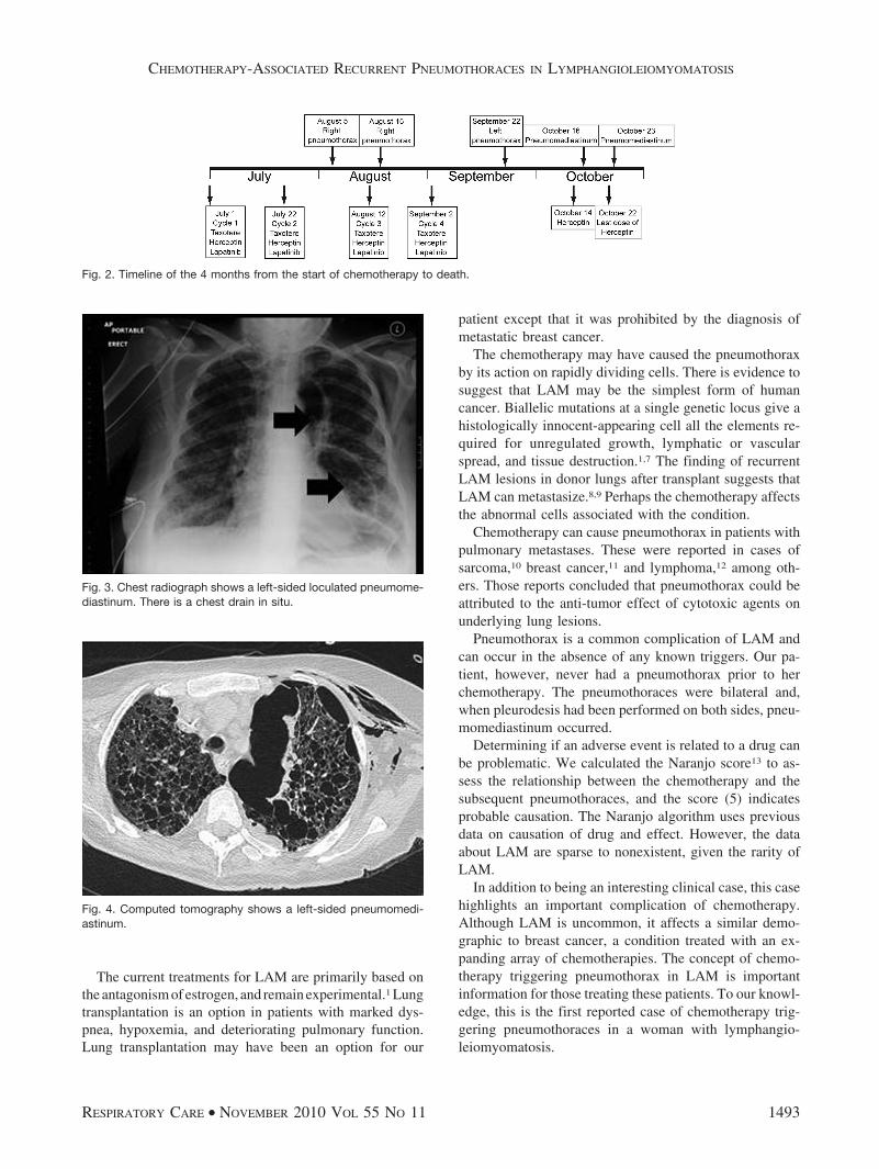
A year and a half after her initial presentation she wasdiagnosed with breast cancer, with a single metastasis inthe liver. She was started on chemotherapy with docetaxel,trastuzumab, and lapatinib. Each cycle of therapy com-menced with docetaxel given on day 1. Trastuzumab wasgiven once weekly for the 3-week cycle and lapatinib wasgiven orally daily. Chemotherapy began within 2 weeks ofthis diagnosis. Prior to commencing chemotherapy she hadno history of pneumothorax.
Shortly after starting chemotherapy she developed new-onset shortness of breath and oxygen saturation of 80% onroom air. Arterial blood gas analysis found pH 7.36, PaCO2
Emer Kelly MD, Elaine Ni Mhurchu MD, Timothy J McDonnell MD,and Michael P Keane MD are affiliated with the Department of Medicineand Therapeutics; and Sumainizah Sukor MD and Dimitrios Tryfono-poulos MD are affiliated with the Department of Oncology, UniversityCollege Dublin, St Vincent’s University Hospital, Elm Park, Dublin,Ireland.
The authors have disclosed no conflicts of interest.
Correspondence: Emer Kelly MD, Department of Medicine and Thera-peutics, University College Dublin, St Vincent’s University Hospital,Elm Park, Dublin 4, Ireland. E-mail: [email protected].
RESPIRATORY CARE • NOVEMBER 2010 VOL 55 NO 11 1491
55 mm Hg, PO241 mm Hg, and HCO3
– 30.4 mEq/L. CTshowed no evidence of pulmonary embolism, pneumotho-rax, or new infiltrates, but demonstrated multiple cystsconsistent with LAM. Following clinical improvement shewas discharged home and advised to continue her pre-scribed oxygen.
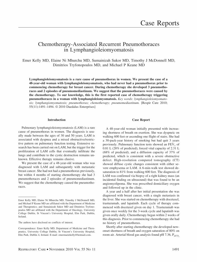
One month after commencing chemotherapy she wasadmitted with a 2-day history of worsening shortness ofbreath. A right-sided pneumothorax was found on chestradiograph and further evaluated via CT (Fig. 1). A chestdrain was inserted, and the pneumothorax resolved.
Chemotherapy continued (Fig. 2). Two weeks later shewas again admitted with a right-sided pneumothorax. Achest drain was inserted. As this was the second right-sided pneumothorax, we conducted a video-assisted tho-racoscopic surgical pleurodesis. Her postoperative coursewas complicated by hypercapnic respiratory failure, whichresponded to noninvasive ventilation.
On the day of her planned discharge, 1 month later, shesuddenly became unwell. She had chest pain and increaseddyspnea. A left-sided pneumothorax was diagnosed, a chestdrain was inserted, and she underwent another left-sidedvideo-assisted thoracoscopic surgical pleurodesis.
Once again, she had a complicated postoperative course,with type-2 respiratory failure that required noninvasiveventilation and a left-lower-lobe collapse/consolidation thatrequired intensive physiotherapy and antibiotics. She re-quired continuous noninvasive ventilation.
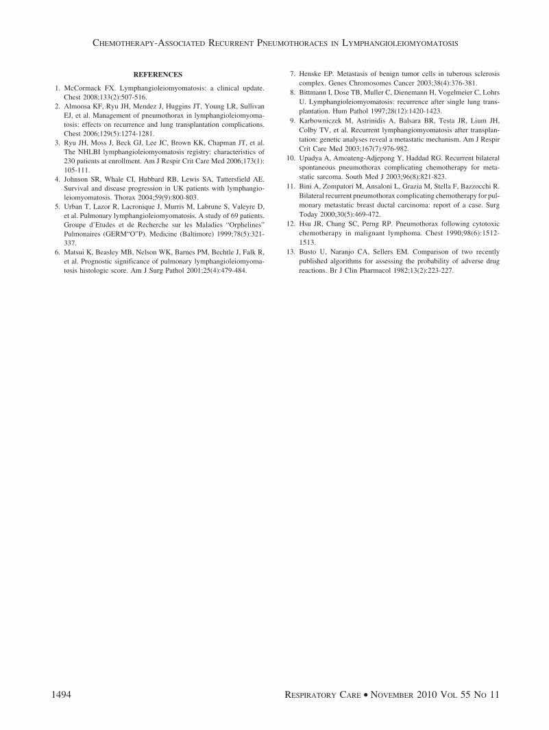
She subsequently developed 2 episodes of pneumome-diastinum, both within 48 h of trastuzumab administration(Figs. 3 and 4). The pneumomediastinums were treatedwith CT-guided insertion of chest drains, and resolved onsubsequent chest radiographs.
Unfortunately, she continued to deteriorate and finallysuccumbed to nosocomial pneumonia 4 months after shewas diagnosed with breast cancer.
Discussion
LAM is an uncommon disease that occurs predomi-nantly in women. It is characterized by smooth musclecell infiltration and cystic destruction of the lung.1 Clin-ically, LAM is characterized by progressive dyspnea onexertion, recurrent pneumothoraces, abdominal and tho-racic lymphadenopathy, and abdominal tumors, includ-ing angiomyolipomas and lymphangiomyomas. LAMoccurs in approximately 30% of women with the tuber-ous sclerosis complex, a genetic disorder associated withseizure, brain tumors, and cognitive impairment. It alsooccurs in women who do not have tuberous sclerosiscomplex (ie, sporadic LAM).
Women with LAM have symptoms for an average of3–5 years and experience an average of 2.2 pneumotho-races before the disease is diagnosed.2 Diagnosis of LAMrequires a high-resolution CT that demonstrates thin-walledcystic change, and either a positive tissue biopsy or acompatible clinical context, such as clinically confirmedtuberous sclerosis, angiomyolipomata, or chylothorax. Thediagnosis of LAM in our patient was confirmed via char-acteristic CT findings and a biopsy-proven renal angio-myolipoma. Approximately 93% of patients with tuberoussclerosis complex LAM, and 30–50% of patients withsporadic LAM, have renal angiomyolipomas, which arebenign renal tumors composed of dysplastic blood vessels,smooth muscle, and variable amounts of fat.1
The average age at diagnosis of LAM in multiple serieshas been approximately 35 years.1 Classically, LAM pre-sents with a mixed ventilatory defect on pulmonary func-tion testing. The National Heart, Lung, and Blood Insti-tute’s LAM registry reported that, in their large cohort of230 patients, 57% exhibited an obstructive pattern, theaverage FEV1 was 70% of predicted,3 and the mean rate ofFEV1 decline was 75 � 9 mL/y. LAM mortality is ap-proximately 10–20% at 10 years after the onset of symp-toms,4,5 and 30% at 10 years after lung biopsy,6 but thetime to death ranges widely.
Pneumothoraces occur in approximately 60 –70% ofpatients with LAM, and the rate of recurrence is � 70%,which is the highest among all chronic lung diseases.1
Although pneumothoraces are common in LAM, we sug-gest that the pneumothoraces in our patient were trig-gered by the chemotherapy, which was effectively con-trolling the breast cancer. Our patient had never had apneumothorax prior to chemotherapy, and the pneumo-thoraces had an immediate temporal relationship to thechemotherapy.
Fig. 1. Computed tomography shows diffuse cystic change con-sistent with lymphangioleiomyomatosis. A left-sided pneumotho-rax is evident.
CHEMOTHERAPY-ASSOCIATED RECURRENT PNEUMOTHORACES IN LYMPHANGIOLEIOMYOMATOSIS
1492 RESPIRATORY CARE • NOVEMBER 2010 VOL 55 NO 11
The current treatments for LAM are primarily based onthe antagonism of estrogen, and remain experimental.1 Lungtransplantation is an option in patients with marked dys-pnea, hypoxemia, and deteriorating pulmonary function.Lung transplantation may have been an option for our
patient except that it was prohibited by the diagnosis ofmetastatic breast cancer.
The chemotherapy may have caused the pneumothoraxby its action on rapidly dividing cells. There is evidence tosuggest that LAM may be the simplest form of humancancer. Biallelic mutations at a single genetic locus give ahistologically innocent-appearing cell all the elements re-quired for unregulated growth, lymphatic or vascularspread, and tissue destruction.1,7 The finding of recurrentLAM lesions in donor lungs after transplant suggests thatLAM can metastasize.8,9 Perhaps the chemotherapy affectsthe abnormal cells associated with the condition.
Chemotherapy can cause pneumothorax in patients withpulmonary metastases. These were reported in cases ofsarcoma,10 breast cancer,11 and lymphoma,12 among oth-ers. Those reports concluded that pneumothorax could beattributed to the anti-tumor effect of cytotoxic agents onunderlying lung lesions.
Pneumothorax is a common complication of LAM andcan occur in the absence of any known triggers. Our pa-tient, however, never had a pneumothorax prior to herchemotherapy. The pneumothoraces were bilateral and,when pleurodesis had been performed on both sides, pneu-momediastinum occurred.
Determining if an adverse event is related to a drug canbe problematic. We calculated the Naranjo score13 to as-sess the relationship between the chemotherapy and thesubsequent pneumothoraces, and the score (5) indicatesprobable causation. The Naranjo algorithm uses previousdata on causation of drug and effect. However, the dataabout LAM are sparse to nonexistent, given the rarity ofLAM.
In addition to being an interesting clinical case, this casehighlights an important complication of chemotherapy.Although LAM is uncommon, it affects a similar demo-graphic to breast cancer, a condition treated with an ex-panding array of chemotherapies. The concept of chemo-therapy triggering pneumothorax in LAM is importantinformation for those treating these patients. To our knowl-edge, this is the first reported case of chemotherapy trig-gering pneumothoraces in a woman with lymphangio-leiomyomatosis.
Fig. 2. Timeline of the 4 months from the start of chemotherapy to death.
Fig. 3. Chest radiograph shows a left-sided loculated pneumome-diastinum. There is a chest drain in situ.
Fig. 4. Computed tomography shows a left-sided pneumomedi-astinum.
CHEMOTHERAPY-ASSOCIATED RECURRENT PNEUMOTHORACES IN LYMPHANGIOLEIOMYOMATOSIS
RESPIRATORY CARE • NOVEMBER 2010 VOL 55 NO 11 1493
REFERENCES
1. McCormack FX. Lymphangioleiomyomatosis: a clinical update.Chest 2008;133(2):507-516.
2. Almoosa KF, Ryu JH, Mendez J, Huggins JT, Young LR, SullivanEJ, et al. Management of pneumothorax in lymphangioleiomyoma-tosis: effects on recurrence and lung transplantation complications.Chest 2006;129(5):1274-1281.
3. Ryu JH, Moss J, Beck GJ, Lee JC, Brown KK, Chapman JT, et al.The NHLBI lymphangioleiomyomatosis registry: characteristics of230 patients at enrollment. Am J Respir Crit Care Med 2006;173(1):105-111.
4. Johnson SR, Whale CI, Hubbard RB, Lewis SA, Tattersfield AE.Survival and disease progression in UK patients with lymphangio-leiomyomatosis. Thorax 2004;59(9):800-803.
5. Urban T, Lazor R, Lacronique J, Murris M, Labrune S, Valeyre D,et al. Pulmonary lymphangioleiomyomatosis. A study of 69 patients.Groupe d’Etudes et de Recherche sur les Maladies “Orphelines”Pulmonaires (GERM“O”P). Medicine (Baltimore) 1999;78(5):321-337.
6. Matsui K, Beasley MB, Nelson WK, Barnes PM, Bechtle J, Falk R,et al. Prognostic significance of pulmonary lymphangioleiomyoma-tosis histologic score. Am J Surg Pathol 2001;25(4):479-484.
7. Henske EP. Metastasis of benign tumor cells in tuberous sclerosiscomplex. Genes Chromosomes Cancer 2003;38(4):376-381.
8. Bittmann I, Dose TB, Muller C, Dienemann H, Vogelmeier C, LohrsU. Lymphangioleiomyomatosis: recurrence after single lung trans-plantation. Hum Pathol 1997;28(12):1420-1423.
9. Karbowniczek M, Astrinidis A, Balsara BR, Testa JR, Lium JH,Colby TV, et al. Recurrent lymphangiomyomatosis after transplan-tation: genetic analyses reveal a metastatic mechanism. Am J RespirCrit Care Med 2003;167(7):976-982.
10. Upadya A, Amoateng-Adjepong Y, Haddad RG. Recurrent bilateralspontaneous pneumothorax complicating chemotherapy for meta-static sarcoma. South Med J 2003;96(8):821-823.
11. Bini A, Zompatori M, Ansaloni L, Grazia M, Stella F, Bazzocchi R.Bilateral recurrent pneumothorax complicating chemotherapy for pul-monary metastatic breast ductal carcinoma: report of a case. SurgToday 2000;30(5):469-472.
12. Hsu JR, Chang SC, Perng RP. Pneumothorax following cytotoxicchemotherapy in malignant lymphoma. Chest 1990;98(6):1512-1513.
13. Busto U, Naranjo CA, Sellers EM. Comparison of two recentlypublished algorithms for assessing the probability of adverse drugreactions. Br J Clin Pharmacol 1982;13(2):223-227.
CHEMOTHERAPY-ASSOCIATED RECURRENT PNEUMOTHORACES IN LYMPHANGIOLEIOMYOMATOSIS
1494 RESPIRATORY CARE • NOVEMBER 2010 VOL 55 NO 11