Embed Size (px)
Citation preview
Chemokine CXCL12 and Its Receptors in theDeveloping Central Nervous System: EmergingThemes and Future Perspectives
Yan Zhu, Fujio Murakami
Graduate School of Frontier Biosciences, Osaka University, Yamadaoka 1-3, Suita, Osaka 565-0871,Japan
Received 8 March 2012; revised 25 May 2012; accepted 1 June 2012
ABSTRACT: Homeostatic chemokine CXCL12
(also known as SDF-1) and its receptor CXCR4 are in-
dispensable for the normal development of the nervous
system. This chemokine system plays a plethora of func-
tions in numerous neural developmental processes, from
which the underlying molecular and cellular mecha-
nisms are beginning to be unravelled. Recent identifica-
tion of CXCR7 as a second receptor for CXCL12 pro-
vides opportunities to gain deeper insights into how
CXCL12 operates in the nervous system. Here, we
review the diverse roles of CXCL12 in the developing
central nervous system, summarize the recent progress
in uncovering CXCR7 functions, and discuss the emerg-
ing common themes from these works and future
perspectives. ' 2012 Wiley Periodicals, Inc. Develop Neurobiol 00:
000–000, 2012
Keywords: chemokine; CXCL12; developing CNS;
neuronal migration; axon guidance
Chemokines are a large family of structurally related,
mostly small secreted polypeptides, whose signals are
transduced by 7-transmembrane G-protein coupled
receptors (GPCRs) (Rossi and Zlotnik, 2000). The
name \chemokine" is coined from Chemotactic Cyto-
kine, because these molecules were originally recog-
nized for their predominant role in controlling leuko-
cyte trafficking during inflammatory response and
immune surveillance (Rot and Von Andrian, 2004;
Moser et al., 2004). However, it has since been real-
ized that chemokines are not merely traffic controllers
in the immune system. They are in fact versatile
intercellular mediators, whose functions include regu-
lating cell migration, proliferation, survival and adhe-
sion, and whose terrain of action extends far beyond
the immune system, reaching the nervous system,
cancer biology, and other developmental and patho-
logical paradigms (Horuk, 2001; Tran and Miller
2003; Li and Ransohoff, 2008; Zlotnik et al., 2011).
The first evidence that chemokines are required for
the proper development of the nervous system
emerged in 1998, when it was found that chemokine
CXCL12 and its receptor CXCR4 regulate cerebellar
granule cell development (Ma et al., 1998; Zou et al.,
1998). In the decade or so that has followed, a large
amount of accumulating evidence points to a wide
involvement of chemokines and their receptors in a
range of processes during the normal development of
the nervous system, with CXCL12/CXCR4 dominat-
ing the scene. The recent identification of CXCR7 as
a second receptor of CXCL12 suggests that some of
the functions of CXCL12 may be mediated or modi-
fied by CXCR7. While the field continues to expand
rapidly, common themes and underlying principles
have begun to emerge. In this review, we will sum-
marize key studies that illustrate the diverse roles
CXCL12 and its two receptors CXCR4, CXCR7 in
Correspondence to: Y. Zhu ([email protected]).Contract grant sponsor: Grant-in-Aid for Scientific Research
from the Ministry of Education, Culture, Sports, Science and Tech-nology, Japan; contract grant number: 23570226 (to Y.Z.), and2222004 (to F.M.).
' 2012 Wiley Periodicals, Inc.Published online in Wiley Online Library (wileyonlinelibrary.com).DOI 10.1002/dneu.22041
1
the development of the central nervous system (CNS)
with focus on three main areas: neuronal migration,
axon guidance, and regulation of neural stem/progen-
itor cells. We will outline the emerging rules and
principles inferred from these studies and discuss
future challenges and perspectives as we go along.
Due to space limitations, this review does not extend
to discuss involvement of CXCL12 and its receptors
in CNS under pathological conditions, which is a vast
and exciting topic in its own right. Readers interested
in this topic may refer to some excellent reviews past
and recent (Tran and Miller, 2003; White et al, 2007;
Li and Ransohoff, 2008; Gross and Meier, 2009; Li
and Ransohoff, 2009; Rostene et al, 2011).
CONTROL OF NEURONAL MIGRATIONAND POSITIONING
Migration of Neuron Precursors
In 1998, two studies showed for the first time that
chemokines could be important for normal brain de-
velopment (Ma et al., 1998; Zou et al., 1998). By gen-
erating and analyzing CXCR4 knockout mice, these
reports showed defective development of the external
granule layer (EGL), which is a secondary prolifera-
tive zone that is positioned beneath the pial meninges
and stretches across the entire surface of the cerebel-
lar primordium. Precursors of cerebellar granule cells
born from the upper rhombic lip first migrate tangen-
tially and superficially into the cerebellar primordium
to form the EGL. These cells then stay and proliferate
within the EGL for a protracted period until an appro-
priate developmental time when they exit the cell
cycle and migrate radially into deep cerebellar cortex
to form the internal granule layer (IGL) (Hatten,
1999; Komuro and Yacubova, 2003, Fig. 1). Retain-
ing granule cell precursors in the EGL serves two
purposes: to ensure sufficient cell proliferation, and
to allow the descending of EGL cells only when the
cerebellar cortex is ready to receive them (Ma et al.,
1998; Zou et al., 1998; Choi et al., 2005). During the
formation of EGL, CXCR4 is expressed in EGL cells,
while CXCL12 is expressed in the overlying pial
meninges (Klein et al., 2001; Reiss et al., 2002; Zhu
et al., 2002). In CXCR4 mutants, granule cell precur-
sors prematurely depart the EGL and descend inter-
nally forming ectopias (Ma et al., 1998; Zou et al.,
1998). In vitro migration assays subsequently demon-
strated that either the pial meninges or recombinant
CXCL12 could chemoattract the cerebellar granule
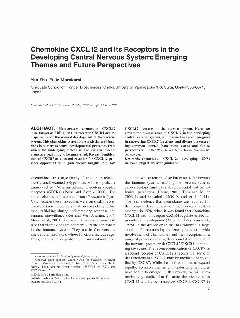
Figure 1 CXCL12/CXCR4 signaling regulates the migration and positioning of cerebellar gran-
ule cells, cortical interneurons and hindbrain pontine neurons. Note that migration of pontine neu-
rons is only depicted on the left half of hindbrain. At early stages in development (top panel), the
migrating neurons (red) expressing CXCR4, are compartmentalized within regions expressing high
level of CXCL12 (green). Later in development (bottom panel), CXCL12/CXCR4 signaling is
down-regulated, allowing neurons to decompartmentalize and move into their final positions. The
mechanism underlying this down-regulation is still little understood. CP: cortical plate; EGL: exter-
nal granule layer; IGL: internal granule layer; lRL: lower rhombic lip; MGE: medial ganglionic
eminence; ML: molecular layer; MNG: meninges; PCL: Purkinje cell layer; PMS: pontine migra-
tory stream; PN: pontine nucleus; SVZ/IZ: subventricular zone/intermediate zone.
2 Zhu and Murakami
Developmental Neurobiology

precursors (Klein et al., 2001; Lu et al., 2001; Reiss
et al., 2002; Zhu et al., 2002). Thus, it appears that
CXCL12 derived exclusively from the pial meninges
functions to anchor cerebellar granule precursors
within a favorable proliferative zone via mechanisms
of chemoattraction (Fig 1). Interestingly, CXCL12 in
addition facilitates cell proliferation in the EGL syn-
ergistically with Sonic Hedgehog (Shh), a known
mitogen for the cerebellar precursor cells (Klein et
al., 2001). This dual functionality of CXCL12 in the
developing cerebellum highly resonates with its origi-
nal roles established in the immune system in regulat-
ing the homing of hematopoetic cells to their prolifer-
ative niche, the bone marrow (Nagasawa et al., 1996;
Tachibana et al., 1998; Ma et al., 1998; Zou et al.,
1998) and facilitating their proliferation synergisti-
cally with interleukin-7 (IL-7) (Nagasawa et al.,
1994).
The constitutive expression of CXCR4 in the
developing CNS, and the ubiquitous expression of
CXCL12 in pial meninges overlying the entire neural
tube predicted widespread neural functions of this
chemokine pair (Jazin et al., 1997; McGrath et. al,
1999; Tissir et al., 2004; Stumm et al., 2007). These
predictions were verified by mounting evidence that
followed. For instance, in the hippocampus, defective
CXCL12/CXCR4 signaling causes a malformed den-
tate gyrus (DG) during development (Bagri et al.,
2002; Lu et al., 2002). The defect might be a com-
pound one, involving failed migration of both prolif-
erating progenitors as well as newly differentiated
granule cells. CXCL12 was again proposed to control
cell migration into the DG, as its expression aligns
the migratory path (Bagri et al., 2002; Lu et al.,
2002). More recently, it was shown that CXCL12
helps to relocate the DG neurogenic zone from the
hippocampal ventricular zone to a transient subpial
proliferative zone aligning the DG blades, which
eventually descend into the DG hilus to form the per-
manent subgranular zone (SGZ) as one of two sites of
adult neurogenesis. The role of CXCL12 appears to
get the CXCR4-expressing granule cell progenitors
into the DG and anchoring them beneath the pial
meninges to form the transient subpial proliferative
zone (Li et al., 2009).
Migration of Postmitotic Neurons
It soon became clear that CXCL12 is also a critical
regulator for the tangential migration of postmitotic
neurons. Tangential migration allows new born neu-
rons to be transported over long distances to destina-
tions far from their birthplaces, thus enabling neuro-
nal types from distant origins to interact and connect
efficiently. The invasion of the neocortex by cortical
interneurons born mostly from the medial ganglionic
eminence (MGE) in the subpallium is probably the
most well-known example, owing to the importance
of interneuron positioning for cortical function.
Therefore, that CXCL12 is implicated in this system
attracted much attention in the field. MGE-derived
interneurons enter into the pallium from the lateral to
medial direction following two distinct tangential mi-
gratory streams, namely the marginal zone (MZ) and
the subventricular zone/intermediate zone (SVZ/IZ)
streams, sparing the cortical plate until later stages
when the interneurons switch to migrate radially into
it (Marın and Rubenstein, 2003; Metin et al., 2006;
Tanaka et al., 2009, Fig. 1). In CXCR4 knockout cor-
tex, interneurons enter the pallium diffusely even
within the cortical plate, disrespecting the two migra-
tory streams (Stumm et al., 2003; Tiveron et al.,
2006; Li et al., 2008; Lopez-Bendito et al., 2008).
The expression pattern of CXCL12 in the developing
cortex shows stunning alignment with the interneuron
migratory paths in the pial meninges overlying the
MZ and the SVZ/IZ itself (Daniel et al., 2005;
Tiveron et al., 2006, Fig. 1). We and others have
shown that CXCL12 determines the migratory
streams for interneurons, both early in migration by
sorting them into the two pathways at the palium-sub-
palium boundary (Li et al., 2008), and later by keep-
ing the interneurons within the MZ for a substantial
period of time before their diving down into the corti-
cal plate (Tiveron et al., 2006; Li et al., 2008; Lopez-
Bendito et al., 2008; Tanaka et al., 2009). By doing
so, CXCL12 may serve two functions: to channel
interneurons into migratory corridors that are optimal
for their efficient dispersion (for example, MZ is a
relatively cell sparse region), and to prevent inter-
neurons from prematurely contacting their future syn-
aptic partners, pyramidal neurons. If these functions
are truly important, then disrupting them should have
lasting consequences on the final interneuron distri-
bution in the cortex. By generating conditional
CXCR4 knockout mice that survive postnatally (Li et
al., 2008; Tanaka et al., 2010), or using cell transplan-
tation (Lopez-Bendito et al., 2008), we and others
have shown that disrupting the interneuron migratory
paths indeed has long-term consequences, affecting
both the regional and laminar distributions of inter-
neurons. The reduction in the number of interneurons
or interneuron subtypes in certain cortical regions and
the presence of occasional clustered interneurons are
in accordance with the notion that interneurons may
disperse less efficiently outside their migratory paths
(Li et al., 2008; Tanaka et al., 2010). Laminar distri-
bution is also disrupted with defects specific to inter-
CXCL12 in Developing CNS 3
Developmental Neurobiology

neuron subtypes (Li et al., 2008; Tanaka et al., 2010).
The extent of functional consequences of disrupted
interneuron migratory pathways is still not fully
explored and would be of great interest to revisit once
further knowledge and new tools become available to
analyze in detail interneuron subtypes and their corre-
sponding local circuitries.
A chemoattractive role of CXCL12 has again been
proposed for the migrating cortical interneurons and
gained support from both in vitro and in vivo evi-
dence (Li et al., 2008; Lopez-Bendito et al., 2008;
Liapi et al., 2008; Tanaka et al., 2009). Cortical inter-
neurons in their migratory streams are clearly
attracted to ectopically expressed CXCL12 in vivo or
to CXCL12 in vitro. Both the initial sorting of new-
born interneurons into the MZ and SVZ/IZ streams as
well as the retention of interneurons within migratory
streams could be accounted for by chemoattraction
toward CXCL12. However, an alternative interpreta-
tion has also been suggested to explain the latter
phenomenon (Lysko and Golden, 2011). This study
showed that CXCL12 signaling affects the dynamic
morphology of migrating interneurons in the SVZ/IZ.
Interneurons migrate by frequently branching their
leading processes and subsequently translocating
their soma toward branch points (Bellion et al., 2005;
Martini et al. 2009). These branches may serve as
sensors to detect environmental guidance cues.
CXCL12 signaling appears to reduce branching fre-
quency and increase the speed of migrating interneur-
ons, thereby minimizing the chance of these neurons
to be distracted by cues outside the CXCL12-
enriched migratory path. Interestingly, this function
of CXCL12 depends only on the ability of CXCR4
coupled G protein to inhibit the cAMP pathway,
which alone does not lead to chemotaxis. Thus, the
authors argued that it was not necessary for CXCL12
to act as a chemoattractant to retain interneurons
within their migratory paths. Regardless of the exact
mechanisms, the ability of CXCL12 to direct the
course of migrating interneurons raises the question
of whether CXCL12 may also influence the lateral-
to-medial migratory direction of cortical interneur-
ons. Studies on the role of CXCL12 in the tangential
migration of Cajal-Retzius cells (C-R cells) provide
some interesting insight into this issue. Cortical hem-
derived C-R cells (Takiguchi-Hayashi et al., 2004)
also depend on meningeal CXCL12 for their tangen-
tial migration and dispersion within the MZ stream
(Borrell and Marın, 2006; Paredes et al., 2006), but in
this case from medial-to-lateral direction. This sug-
gests that the meninge-derived CXCL12 is unlikely
to provide cues for the tangential direction of migrat-
ing C-R cells and interneurons. Borrell and Marın
(2006) provide evidence suggesting C-R cells spread
by a mode of contact inhibition, which results in cells
migrating from high to low cell density area. It would
be interesting to see if contact inhibition may also
play a part in controlling the tangential direction of
cortical interneuron migration.
The expression of CXCL12 in the SVZ/IZ is in-
triguing, as it raises the question of which cells
express this chemokine there. Tiveron et al (2006)
showed that CXCL12 is expressed by intermediate
progenitors of future pyramidal neurons that undergo
amplification in the SVZ. An important implication
of this finding is that excitatory components of the
cortex might themselves be involved in the develop-
ment of inhibitory components. A recent study lends
further support to this notion (Sessa et al., 2010),
showing that when Tbr2, a gene expressed in and crit-
ical for the development of intermediate progenitors
in the SVZ, is force-expressed, it leads to the attrac-
tion of cortical interneurons toward the ectopic
expression site. This seems to be achieved by Tbr2-
dependent induction of CXCL12 expression, thus
linking an important intermediate progenitor marker
directly with the chemokine expression profile of
cortical cells.
CXCL12 also controls the tangential migration of
new born neurons in other brain regions. In the hind-
brain, pontine neurons, the most prominent subset of
precerebellar neurons, take a circuitous migratory
path from the lower rhombic lip to the anterior–ven-
tral hindbrain to form the pontine nucleus (Altman
and Bayer, 1997; Kawauchi et al., 2006). These neu-
rons migrate marginally beneath the pial surface and
move anteriorly for a fixed distance before turning
ventrally at a stereotypic position toward the midline
(Fig. 1). We have shown that disrupting CXCL12/
CXCR4 signaling causes a substantial portion of pon-
tine neurons to deviate from their marginal migratory
stream and consequentially turn ventrally without
traveling anteriorly (Zhu et al., 2009). We found that
meninge-derived CXCL12 again functions to retain
migrating pontine neurons within the marginal zone.
Curiously, a proportion of pontine neurons that
remain to migrate marginally still fail in their anterior
migration as they prematurely turn toward the ventral
midline before their supposed turning point, suggest-
ing that CXCL12 may directly control the anterior
migration of pontine neurons. How can CXCL12
ubiquitously expressed in the pial meninges provide
tangential directions for migrating pontine neurons?
If such guidance is provided by a gradient of extracel-
lular CXCL12 protein along the anteroposterior axis,
then how is such a gradient established in the first
place? To unambiguously visualize and quantify
4 Zhu and Murakami
Developmental Neurobiology

extracellular distribution of CXCL12 protein, or for
that matter any secreted molecules, is still technically
challenging, yet, will be greatly helpful to address
these questions.
Silencing CXCL12/CXCR4 Signaling
A common theme that has emerged from all these
studies is that CXCL12 functions to compartmental-
ize developmental processes within a specific lamina
zone of highly stratified tissue like neuroepithelium.
Such compartmentalization often is transient with the
subsequent decompartmentalization being carefully
timed to achieve fine coordination with other devel-
opmental processes (Fig. 1). This model begs an im-
portant question as to how the CXCL12/CXCR4 sig-
naling becomes inhibited at specific developmental
times to allow for proper decompartmentalization.
Despite its importance and biological relevance, we
presently know little of its answer. It does not seem
to be a case of simply down-regulating the transcrip-
tion of CXCL12 and CXCR4 as their expression per-
sist even after the presumed inhibition of CXCL12/
CXCR4 signaling (Klein et al., 2001; Reiss et al.,
2002; Stumm et al., 2003). The inhibition might
therefore be achieved by turning off the signaling
pathway downstream of CXCR4. An elegant mecha-
nism of this kind has been proposed for the cerebellar
granule precursors in the EGL, albeit only in vitro(Lu et al., 2001). It has been shown that reverse sig-
naling between EphB and ephrinB can down-regulate
CXCR4 signaling by binding to a PDZ-RGS protein
(RGS stands for Regulator of G protein Signaling),
and presumably bringing RGS close to CXCR4
receptors. Since RGS is essentially a GTPase Activat-
ing Protein (GAP) for the heterotrimeric G protein, it
leads to the hydrolysis of G protein and thus dampens
CXCR4 signaling. Whether such an RGS-based in-
hibitory mechanism operates in vivo and whether
similar or different mechanisms operate in the cases
of cortical interneurons, DG progenitors and pontine
neurons, these are important and imminent questions
awaiting future investigation.
CONTROL OFAXON GUIDANCE:ATTRACTANT OR MODULATOR?
CXCL12 clearly is critical for controlling the migra-
tion of neurons and their precursors by functioning at
least in part as a guidance cue that via CXCR4 signal-
ing leads to biased migration direction. Because the
guidance of migrating neurons and that of growth
cones of extending axons share similar signaling
mechanisms and chemical guidance cues (Guan and
Rao, 2003), it was expected that CXCL12/CXCR4
may also play a role in axon pathfinding. Xiang et al.
(2002) provided the first evidence that growth cones
of rat cerebellar granule neurons, when presented
with a steep gradient of CXCL12, could turn either
away or toward the source depending on the intracel-
lular cyclic GMP levels. A dual functionality of
CXCL12 on axons was also demonstrated by Ara-
kawa et al. (2003). They showed, using a dissociated
mouse cerebellar granule cell culture, that CXCL12
promoted axon elongation at low concentration but
inhibited it at high concentration. The authors further
probed into downstream signaling pathways for these
two opposing responses showing that high CXCL12
concentration selectively activate the Rho-ROCK
pathway which negatively regulates cytoskeleton dy-
namics, while low CXCL12 concentration activates
the Rho-mDia pathway without activating ROCK.
Considering that most reagents controlling axon
extension can cause axon turning when presented as a
point source, one implication from this study is that
CXCL12 can also guide these axons. In line with
these in vitro studies, it was shown in zebrafish that
ectopically expressed CXCL12 appears to aberrantly
attract retinal ganglion cell axons on their way to the
optic stalk (Li et al., 2005).
These initial works, which suggest that CXCL12
is a chemotactic cue for axons, were challenged by
emerging evidence advocating a permissive rather
than chemotropic role of CXCL12 for axons. In
zebrafish, Miyasaka et al. (2007) showed that ubiqui-
tously-expressed CXCL12 works equally effectively
as localized CXCL12 in guiding olfactory sensory
axons. In chick, Chalasani et al. (2003) found, using
collagen explant culture and growth cone collapse
assay, that CXCL12 acts as neither an attractant nor a
repellent on its own, but rather to reduce the repulsive
activity of other chemorepellents when present simul-
taneously. This repellent-reducing activity of
CXCL12 was demonstrated for cultured chicken reti-
nal ganglion cell (RGC) axons, dorsal root ganglion
(DRG) axons, and sympathetic ganglion axons for
their respective repellents, Slit-2, Sema3A, and
Sema3F. Intriguingly, this activity of CXCL12 seems
to solely depend on a CXCR4-mediated elevation of
cAMP levels. The antirepellent effect of CXCL12
has subsequently gained in vivo support in zebrafish
(Chalasani et al., 2007, Xu et al., 2010). Zebrafish
RGC axons frequently make pathfinding errors within
the optic stalk when Slit-Robo signaling is compro-
mised. Chalasani et al. (2007) showed that knocking
down CXCL12 or CXCR4 can partially rescue the
misprojection of RGC axons in a robo2 hypomorph
CXCL12 in Developing CNS 5
Developmental Neurobiology

which retains residual low level robo activities, but
cannot rescue a robo2 null mutant. The authors
argued that the absence of CXCL12/CXCR4
increases the sensitivity of RGC axons to the residual
robo activity in the hypomorph, thus rescuing the
phenotype. In a follow-up study, they showed in the
same system that the antirepellent activity of
CXCL12 is dependent on a calmodulin-activated ade-
nylate cyclase (ADCY8), an enzyme that facilitates
cAMP synthesis, and ADCY8 knockdown rescues the
robo2 hypomorph similarly as CXCL12 knockdown
(Xu et al., 2010).
A critical and rather puzzling question arose from
these studies. CXCR4 is traditionally thought to cou-
ple with the Gai/o subunit of the heterotrimeric G
protein that decreases cAMP levels upon CXCL12
binding. How then can CXCL12 stimulation in axons
lead to an increase in cAMP levels? This question is
particularly pertinent since elevation of cAMP upon
CXCL12 stimulation has been directly demonstrated
in cultured chicken RGC using a FRET-based cAMP
sensor (Xu et al., 2010). A recent study has provided
some clues as to how this might happen (Twery and
Raper, 2011). By selectively blocking various Gasubunits as well as Gbc in cultured embryonic
chicken DRG neurons, the authors showed that Gai,
Gaq, Gbc, and phospholipase C (PLC) all seem to
mediate the antirepellent activity of CXCL12. Taken
previous studies together, they proposed a model in
which downstream of CXCR4, Gai, Gaq, and Gbccooperatively activate PLC, which in turn activates
calcium-calmodulin activated ADCY8, thus resulting
in an increase of cAMP. This model, particularly its
in vivo relevance, awaits future validation. It should
be noted that the Twery and Raper report demon-
strates just how complex and poorly understood are
the downstream signaling pathways that GPCR recep-
tors like CXCR4 could evoke in different cellular
contexts. Much is to be explored on this front particu-
larly in in vivo systems.
Curiously, studies reporting axon guidance defects
in CXCL12- or CXCR4-deficient mice are few. Chala-
sani et al. (2003) analyzed the overall axon profiles in
the spinal cord of CXCR4 mutant mice and reported
a general hyperfasciculation of axon tracts and mis-
projected sensory afferents within the spinal cord.
Lieberam et al. (2005) demonstrated an involvement
of CXCL12 in guiding motor axons ventrally out of
the mouse spinal cord and hindbrain. They showed
that CXCR4 is transiently expressed in ventrally-pro-
jecting motor neurons, while CXCL12 is expressed in
the mesenchyme surrounding the ventral motor exit
point. Disruption of CXCL12/CXCR4 signaling
resulted in a failure of many motor axons to exit the
neural tube. The underlying mechanism of this phe-
nomenon is still unclear. Interestingly, the authors
noted that they could not detect any chemoattractive
activity of CXCL12 in motor axons growing out of
spinal cord explants, raising an alternative possibility
that CXCL12/CXCR4 signaling in these motor axons
may be to antagonize repulsion from repellents
located near the ventral motor exit points. However, a
recent study also focusing on motor axons but in ros-
tral hindbrains showed that CXCL12 could chemoat-
tract chicken oculomotor axons in a collagen explant
assay and that oculomotor axons exiting hindbrain
are reduced in CXCR4 mutant mice (Lerner et al.,
2010). Whether CXCL12 acts as an antirepellent
modulator or a chemotactic guidance cue for develop-
ing axons in vivo is still an unsettled issue. The differ-
ences in existing reports may partly reflect differen-
ces in the types of in vitro chemotaxis assays being
used and partly owe to different cellular contexts.
Nevertheless, these attempts to clarify the role of
CXCL12 in axon guidance highlight two important
issues: (1) the complexity and context-dependency of
downstream signaling of CXCL12/CXCR4; and (2)
the possibility of cross-talks between signaling path-
ways of CXCL12 and other axon guidance cues.
CXCR7, ANOTHER RECEPTOR OFCXCL12 THAT HAS MULTIFACETEDROLES
The near identical phenotypes between CXCR4- and
CXCL12-deficient mice have been interpreted to
mean a one-to-one relationship between the receptor
and the ligand, making them an exception in the gen-
erally promiscuous chemokine family. However, this
monogamous relationship was refuted following the
recent discovery of a second receptor for CXCL12,
namely CXCR7 (also known as RDC1) (Balabanian
et al., 2005; Burns et al., 2006). CXCR7, a prior
orphan GPCR, binds to CXCL12 with higher affinity
than CXCR4 and has a second ligand CXCL11 (also
known as ITAC). The addition of a third member to
CXCL12 chemokine system predicted a higher level
of complexity in the mechanisms underlying
CXCL12 functions and urged us to review existing
work in this new context.
Early assessments of CXCR7 already have
revealed signs that it may not be a typical chemokine
receptor. Although CXCR7 shares sequence homol-
ogy with 7-transmembrane GPCRs, the \DRYLAIV"amino acid motif in the second intracellular loop,
which is highly conserved among chemokine recep-
tors and considered to be necessary for G protein
6 Zhu and Murakami
Developmental Neurobiology

coupling (Colvin et al., 2004; Lagane et al., 2005), is
altered to \DRYLSIT" in CXCR7 (Sierro et al.,
2007; Thelen and Thelen, 2008). In line with this
sequence alternation, typical chemokine responses
such as calcium mobilization, chemotaxis, and activa-
tion of PI3K/AKT pathways, often fail to be detected
upon ligand stimulation of CXCR7 (Burns et al.,
2006; Sierro et al., 2007; Mazzinghi et al., 2008; Bol-
dajipour et al., 2008). These \oddities" set CXCR7
apart from classical chemokine receptors like
CXCR4, suggesting that the two are unlikely to be
functionally redundant.
Cellular and Molecular Functions
The cellular and molecular function of CXCR7 has
been the subject of heated pursuits ever since its dis-
covery, and mounting evidence emerged from which,
mostly biochemical but also in vivo in zebrafish, gave
rise to three major models (Fig. 2). (1) The first
model suggests that CXCR7 functions as a scavenger
receptor that predominantly mediates CXCL12 inter-
nalization and subjects it to degradation in lysosomes
(Dambly-Chaudiere et al., 2007; Boldajipour et al.,
2008; Zabel et al., 2009; Luker et al., 2010; Naumann
et al., 2010). Like other scavenger chemokine recep-
tors (Haraldsen and Rot, 2006), the capability of
CXCR7 to clear excess CXCL12 from the extracellu-
lar space may help keep the concentration of bioac-
tive CXCL12 within an optimal range, and/or shape
the distribution of CXCL12 protein at tissue levels.
Strong in vivo support of this model comes from an
elegant study in zebrafish on the migration of primor-
dial germ cells (PGCs) (Boldajipour et al.., 2008).
PGCs generated in the head region express CXCR4
and migrate posteriorly following an attractive signal
associated with a CXCL12 expression domain that
itself dynamically shifts toward the future gonad pos-
teriorly. Boldajipour et al. (2008) provided several
lines of complementary evidence by showing that
CXCR7 expressed in the somatic tissue mediates
CXCL12 endocytosis, whereby clearing CXCL12
protein from nontarget areas and sharpening the
localized CXCL12 protein distribution necessary for
precise PGCs guidance. (2) The second model sug-
gests that CXCR7 can form heterodimers with
CXCR4 when copresent in the same cell and as a
result modify CXCR4 downstream signaling (Sierro
et al., 2007; Levoye et al.., 2009; Decaillot et al.,
2011). While some reports have shown that the coex-
pression of CXCR4 and CXCR7 in cell lines enhan-
ces calcium mobilization (Sierro et al., 2007) and
chemotaxis (Decaillot et al., 2011), others have dem-
onstrated compromised CXCR4 signaling (Levoye et
al., 2009). (3) The third model suggests that CXCR7
can mediate signaling independently from CXCR4 in
response to CXCL12 (Balabanian et al., 2005; Valen-
tin et al., 2007; Zabel et al., 2009; Odemis et al.,
2010, 2012; Rajagopal et al., 2010). Most GPCRs can
signal via both G proteins and b-arrestins in a rather
balanced manner. Coupling with b-arrestins triggers
receptor desensitization and endocytosis, and also
activates downstream ERK1/2 pathway (Luttrell and
Lefkowitz, 2002). Whereas CXCR7 is considered
incapable of signaling via G proteins, a couple of
studies recently have provided direct evidence that
CXCR7 can recruit b-arrestin 2 and activate ERK1/2
pathway (Zabel et al., 2009; Rajagopal et al., 2010),
Figure 2 Three major models that have been proposed for the cellular and molecular functions
of CXCR7. Green: CXCL12; orange: CXCR7; blue: CXCR4.
CXCL12 in Developing CNS 7
Developmental Neurobiology

putting CXCR7 into the category of b-arrestin-biased
GPCRs. More recently, one report showed quite sur-
prisingly that CXCR7 could even signal via G protein
and trigger calcium mobilization in CXCR4-deficient
primary asctrocytes upon CXCL12 binding (Odemis
et al., 2012). However, caution should be taken as it
is still unclear in this case whether CXCR7 signals by
itself or by heterodimerizing with another as-of-yet
unknown receptor. One implication from models (2)
and (3) is that CXCR7 alone or the CXCR7/CXCR4
heterodimer can mediate different downstream sig-
naling and thus lead to different cellular responses
compared with CXCR4 alone. Indeed, several lines
of evidence suggest that while CXCR4 predomi-
nantly controls chemotaxis, CXCR7 is more impor-
tant for regulating cell-cell adhesion (Burns et al.,
2006; Hartmann et al, 2008; Mazzinghi et al., 2008;
Zabel et al., 2009). In summary, each of the above
three models has gathered its fair share of experimen-
tal evidence and none are mutually exclusive. For
example, coupling to b-arrestin 2 would enable
CXCR7 to trigger CXCL12 internalization as well as
b-arrestin 2-mediated ERK1/2 activation simultane-
ously in the same cell. The differential expression
patterns of CXCR7 and CXCR4 showing coexpres-
sion in some cells and nonoverlapping in others, and
the fact that CXCR7 and CXCL12 are often coex-
pressed in somatic tissues (Schonemeier et al., 2008;
Tiveron et al., 2010; Zhu et al., unpublished observa-
tion) suggest that all three models might apply in vivodependent on the cellular and tissue context.
THE ROLE OF CXCR7 FUNCTION IN THEDEVELOPING CNS
The high sequence conservation of CXCR7 across
mammalian species implies its functional importance,
and indeed two independent CXCR7-deficient mice
lines that have been generated showed defects in
heart development and lethality soon after birth
(Sierro et al., 2007; Gerrits et al., 2008). However, an
initial analysis of the developing nervous system
including the cerebellum, dentate gyrus, and spinal
cord, all of which are noted for showing defects in
CXCR4- or CXCL12-deficient mice, did not show
obvious defects in CXCR7 mutant (Sierro et al.,
2007). Nevertheless, recent in-depth analyzes of
CXCR7 mutant cortex uncovered its importance in
regulating the intracortical tangential migration of
interneurons (Sanchez-Alcaniz et al., 2011; Wang et
al.., 2011). Both studies showed that interneurons in
CXCR7-deficient mice failed to respect the stereo-
typic MZ and SVZ/IZ migratory streams and prema-
turely entered the cortical plate, a defect highly iden-
tical to that in CXCR4-deficient mice (see an earlier
section). CXCR7 and CXCR4 are coexpressed in the
majority of migrating cortical interneurons, and fur-
thermore interneuron specific conditional deletion of
CXCR7 causes similar phenotype as constitutive null
mutant, suggesting a cell-autonomous requirement.
So what role does CXCR7 play in cortical interneur-
ons? This is where the two reports begin to diverge.
Sanchez-Alcaniz et al. (2011) proposed a scavenger
role for CXCR7. They have shown an abnormally
high level of CXCL12 protein and a loss of CXCR4
protein in CXCR7 deficient cortex. They have further
suggested that the former causes the latter since
excess CXCL12 has previously been shown to over-
trigger desensitization and degradation of CXCR4
(Marchese and Benovic, 2001; Alsayed et al., 2007;
Kolodziej et al.., 2008). Thus, the authors conclude
that CXCR7, even when copresent with CXCR4 in
cortical interneurons, functions as a scavenger recep-
tor to prevent over-accumulation of CXCL12 in the
cortex, thereby ensuring a sufficient amount of
CXCR4 protein to be present to mediate chemokine
responses. Wang et al. (2011), on the other hand,
although not dismissing the scavenger role for
CXCR7, suggested that CXCR4 and CXCR7 each
plays distinct functions in regulating interneuron
migration with CXCR4 via G proteins and CXCR7
via the ERK1/2 pathway. How these distinct signal-
ing profiles relate to their in vivo functions is still
unclear, but the authors showed by time-lapse imag-
ing that CXCR7-deficient interneurons displayed
lower motility with shorter leading processes in con-
trast to higher motility with longer leading processes
of CXCR4-deficient interneurons. These two models
being proposed are not necessarily mutually exclu-
sive. For example, the reduced motility and shorter
leading processes seen in CXCR7-deficient interneur-
ons could be a consequence of an abnormally high
level of CXCL12 in the environment. This possibility
is supported by two previous observations. First,
migrating PGCs in zebrafish display reduced motility
when either CXCL12 is overexpressed or CXCR7 is
depleted (presumably via an increasing ambient
CXCL12 level), in contrast to normal motility when
CXCL12 is simply depleted (Boldajipour et al.,
2008). Second, in the context of axon growth, high
levels of CXCL12 was found to activate the Rho/
Rock pathway which negatively regulates cytoskele-
ton as discussed above (Arakawa et al., 2003). To
build a more coherent model in the future will require
better understanding of how signaling properties and
cellular responses are affected by varying chemokine
concentrations and receptor combinations. Neverthe-
8 Zhu and Murakami
Developmental Neurobiology

less, the two in vivo studies on migrating cortical
interneurons offer important insights, (1) CXCR7
may comprise a general mechanism employed to
keep CXCL12 at modest levels so as to prevent the
undesirable negative effect that occurs with excess
CXCL12; (2) CXCR7 may independently signal and
cause distinct cellular effects in vivo.
The role of CXCR7 in the developing nervous sys-
tem is bound to extend beyond the migrating cortical
interneurons. We have found that precerebellar neu-
ronal migration is affected in CXCR7-deficient mice
and the defect differs from that in CXCR4 mutant
(Zhu et al., unpublished observation). It is almost cer-
tain that the list will grow and new roles will be
assigned to this second receptor of CXCL12. The key
issue, however, is to have a clear and thorough under-
standing of the intricate mechanisms that have been
carried out through the interplay between CXCL12
and its two receptors in a complex and dynamic tissue
environment such as the developing brain.
Role of CXCL12 in Neural Stem andNeural Progenitor Cells
In the developing CNS, CXCR4 is expressed predom-
inantly in neural stem and neural progenitor cells
residing in the primary (e.g. ventricular zone) and
secondary (e.g. cerebellar EGL) proliferative zones
from the spinal cord to the forebrain (McGrath et al.,
1999; Tissir et al., 2004; Corti et al., 2005; Stumm et
al., 2007; Diotel et al., 2010). In postnatal brains,
CXCR4 expression continues in neural progenitor
cells in limited locations where adult neurogenesis
persists, such as the SGZ in dentate gyrus and the
SVZ of the lateral ventricle (Tissir et al., 2004; Tran
et al., 2007). As for the ligand, CXCL12 expression
in embryos is mainly confined to the pial meninges
(McGrath et al., 1999; Tissir et al., 2004), with which
neural progenitor cells usually taking on the form of
radial glia cells have contact via their basal endfeet.
In adult neurogenic regions, CXCL12 is found in
cells adjacent to the proliferating neural progenitors
(Banisadr et al., 2003; Kokovay et al., 2010). These
expression patterns suggest that neural stem/progeni-
tor cells may develop and function under the influ-
ence of CXCL12. In vitro, CXCR4 expression is
almost a hallmark of neural progenitor cells or neuro-
spheres isolated and grown from embryonic or adult
brains (Tran et al., 2004; Peng et al., 2004; Dziem-
bowska et al., 2005). What then are the effects of
CXCL12 on neural progenitor cells? There are sev-
eral occasions where neural progenitor cells need to
be relocated from their primary sites of residence to
secondary proliferative zones within which neurogen-
esis takes place. CXCL12 seems critically involved
in this process by mobilizing neural progenitor cells
and then retaining them in their second niches, as
exemplified by the cases of cerebellar EGL and den-
tate gyrus SGZ (discussed in an earlier section). A
similar scenario was recently described in adult SVZ
where CXCL12 regulates the lineage progression of
SVZ progenitors by transporting them from their rela-
tively quiescent ependymal niche to a basal vascula-
ture niche where these cells undergo active amplifica-
tion (Kokovay et al., 2010). In all these scenarios, it
is thought that CXCL12 fulfils its function by regulat-
ing neural progenitor cell migration as a chemoattrac-
tant. Indeed, CXCL12 can attract and enhance migra-
tion of in vitro cultured neural progenitor cells pre-
pared from a variety of regions in the CNS
(Dziembowska et al., 2005; Imitola et al., 2004; Car-
bajal et al., 2010, Luo et al., 2005). Perhaps the most
important and relevant implication of this observation
is that CXCL12 may be a key chemoattractant capa-
ble of directing either endogenous or transplanted
neural progenitor cells to sites of CNS injury and
engaging them for tissue repair, a possibility that has
already gained support from several mouse models of
brain pathology (Imitola et al., 2004; Corti et al.,
2005; Carbajal et al., 2010).
It should be noted, however, that most CXCR4-
expressing neural progenitor cells reside in the ven-
tricular zone where neurogenesis takes place on site,
and thus do not undergo migration during embryonic
development. This suggests that CXCL12 may regu-
late some other aspects of these cells: conceivably,
proliferation and survival. In fact, CXCL12 was orig-
inally cloned owing to its ability to stimulate prolifer-
ation of pre-B cells synergistically with IL-7 (Naga-
sawa et al., 1994). So far, several studies have
reported that CXCL12 can stimulate proliferation of
in vitro cultured neural progenitor cells from both
rodent and human origins (Imitola et al., 2004; Pritch-
ett et al., 2007; Wu et al., 2009; Li et al., 2011),
although contradictory results show that CXCL12
induces human neural progenitor cells to enter quies-
cence (Krathwohl and Kaiser, 2004). What accounts
for the discrepancy is still unclear. Similarly,
CXCL12 also appears to promote survival of neural
progenitor cells cultured in vitro (Dziembowska et
al., 2005; Prichett et al., 2007). However, beyond
these in vitro studies, an effect of CXCL12 on prolif-
eration and survival of neural progenitor cells in the
ventricular zones of CXCR4- or CXCL12-deficient
mice has not yet been reported. One possibility could
be redundancy resulting from the presence of a num-
ber of growth promoting factors operating in this
CXCL12 in Developing CNS 9
Developmental Neurobiology
system. Li et al. (2011) recently suggested that
CXCL12 may only stimulate neural progenitor prolif-
eration in synergy with epidermal growth factor
(EGF) and fibroblast growth factor (FGF). Further-
more, neural progenitor cells appear to express sev-
eral other chemokine receptors besides CXCR4
(Peng et al., 2004; Tran et al., 2007), raising the pos-
sibility of involvement of other chemokine molecules
in regulating the proliferation and survival of neural
progenitor cells. An alternative possibility that should
be considered is that CXCL12/CXCR4 may regulate
some aspects of neural progenitor cells in vivo other
than migration, proliferation, or survival. Indeed, we
recently found that CXCL12 signaling is required to
maintain the integrity of radial glial processes of neu-
ral progenitor cells in the caudal spinal cord (Zhu et
al., unpublished observation).
CONCLUDING REMARKS
Among the large chemokine family, the CXCL12
chemokine receptor system is best known for its
involvement in the normal development of CNS. This
may partly be due to the vast amount of studies focus-
ing on this chemokine system in comparison with the
others. A few other chemokine receptors have also
been found to be expressed in the developing CNS
and may have roles in glial cell development (Bajetto
et al., 2001; Tsai et al., 2002; Tran and Miller, 2003;
Ambrosini and Aloisi, 2004; Tran et al., 2007),
although little in vivo function has so far been attrib-
uted to them. Future effort may uncover new players
from the chemokine family for the normal brain de-
velopment. On the other hand, the predominant role
of CXCL12 and its receptors in CNS development
may have its roots in the fact that CXCL12 and
CXCR4 are evolutionarily more ancient than most
other chemokine members, and that their ancestral
role could very well be in the development of the
CNS which predates the emergence of the adaptive
immune system in vertebrates (Huising et al., 2003).
The plethora of functions CXCL12 and its receptors
play during the development of CNS is impressive
and the list will continue to grow. Yet we still do not
have a coherent view of how this chemokine system
operates mechanistically in the complex and dynamic
tissue environment like the developing brain. Impor-
tant questions remain unanswered. How CXCL12
signaling is turned off for the developmental process
to move onto the next step? What different down-
stream effectors are activated in different cell types
or even different compartment of one cell? How
CXCL12 is distributed and how this distribution is
maintained or reshaped as tissue morphogenesis takes
place? How CXCL12 signaling interacts with other
signaling pathways to influence cell behaviors? To
answer these questions, particularly in vivo, poses
future challenges for us developmental neurobiolo-
gists who work on chemokines in the nervous system.
The authors thank Dr. Peter Karagiannis for editing the
manuscript.
REFERENCES
Altman J, Bayer SA. 1997. Development of the Cerebellar
System in Relation to its Evolution, Structure, and Func-
tions. Boca Raton, FL: CRC Press.
Alsayed Y, Ngo H, Runnels J, Leleu X, Singha UK, Pitsil-
lides CM, Spencer JA, et al. 2007. Mechanisms of regu-
lation of CXCR4/SDF-1 (CXCL12)-dependent migration
and homing in multiple myeloma. Blood 109:
2708–2717.
Ambrosini E, Aloisi F. 2004. Chemokines and glial cells: A
complex network in the central nervous system. Neuro-
chem Res 29:1017–1038.
Arakawa Y, Bito H, Furuyashiki T, Tsuji T, Takemoto-
Kimura S, Kimura K, Nozaki K, et al. 2003. Control of
axon elongation via an SDF-1alpha/Rho/mDia pathway
in cultured cerebellar granule neurons. J Cell Biol
161:381–391.
Bagri A, Gurney T, He X, Zou YR, Littman DR, Tessier-
Lavigne M, Pleasure SJ. 2002. The chemokine SDF1 reg-
ulates migration of dentate granule cells. Development
129:4249–4260.
Bajetto A, Barbero S, Bonavia R, Piccioli P, Pirani P, Florio
T, Schettini G. 2001. Stromal cell-derived factor-1alpha
induces astrocyte proliferation through the activation of
extracellular signal-regulated kinases 1/2 pathway.
J Neurochem 77:1226–1236.
Balabanian K, Lagane B, Infantino S, Chow KY, Harriague
J, Moepps B, Arenzana-Seisdedos F, et al. 2005. The
chemokine SDF-1/CXCL12 binds to and signals through
the orphan receptor RDC1 in T lymphocytes. J Biol
Chem 280:35760–35766.
Banisadr G, Skrzydelski D, Kitabgi P, Rostene W, Parsada-
niantz SM. 2003. Highly regionalized distribution of stro-
mal cell-derived factor-1/CXCL12 in adult rat brain: Con-
stitutive expression in cholinergic, dopaminergic and vas-
opressinergic neurons. Eur J Neurosci 18:1593–1606.
Bellion A, Baudoin JP, Alvarez C, Bornens M, Metin C.
2005. Nucleokinesis in tangentially migrating neurons
comprises two alternating phases: Forward migration of
the Golgi/centrosome associated with centrosome split-
ting and myosin contraction at the rear. J Neurosci
25:5691–5699.
Boldajipour B, Mahabaleshwar H, Kardash E, Reichman-
Fried M, Blaser H, Minina S, Wilson D, et al. 2008. Con-
trol of chemokine-guided cell migration by ligand
sequestration. Cell 132:463–473.
10 Zhu and Murakami
Developmental Neurobiology

Borrell V, Marın O. 2006. Meninges control tangential
migration of hem-derived Cajal-Retzius cells via
CXCL12/CXCR4 signaling. Nat Neurosci 9:1284–1293.
Burns JM, Summers BC, Wang Y, Melikian A, Berahovich
R, Miao Z, Penfold ME, et al. 2006. A novel chemokine
receptor for SDF-1 and I-TAC involved in cell survival,
cell adhesion, and tumor development. J Exp Med
203:2201–2213.
Carbajal KS, Schaumburg C, Strieter R, Kane J, Lane TE.
2010. Migration of engrafted neural stem cells is medi-
ated by CXCL12 signaling through CXCR4 in a viral
model of multiple sclerosis. Proc Natl Acad Sci USA
107:11068–11073.
Chalasani SH, Sabelko KA, Sunshine MJ, Littman DR,
Raper JA. 2003. A chemokine, SDF-1, reduces the effec-
tiveness of multiple axonal repellents and is required for
normal axon pathfinding. J Neurosci 23:1360–1371.
Chalasani SH, Sabol A, Xu H, Gyda MA, Rasband K, Gran-
ato M, Chien CB, et al. 2007. Stromal cell-derived fac-
tor-1 antagonizes slit/robo signaling in vivo. J Neurosci
27:973–980.
Choi Y, Borghesani PR, Chan JA, Segal RA. 2005. Migra-
tion from a mitogenic niche promotes cell-cycle exit.
J Neurosci 25:10437–10445.
Colvin RA, Campanella GS, Sun J, Luster AD. 2004. Intra-
cellular domains of CXCR3 that mediate CXCL9,
CXCL10, and CXCL11 function. J Biol Chem
279:30219–30227.
Corti S, Locatelli F, Papadimitriou D, Donadoni C, Del Bo
R, Fortunato F, Strazzer S, et al. 2005. Multipotentiality,
homing properties, and pyramidal neurogenesis of CNS-
derived LeX(ssea-1)+/CXCR4+ stem cells. FASEB J
19:1860–1862.
Dambly-Chaudiere C, Cubedo N, Ghysen A. 2007. Control
of cell migration in the development of the posterior lat-
eral line: Antagonistic interactions between the chemo-
kine receptors CXCR4 and CXCR7/RDC1. BMC Dev
Biol 7:23.
Daniel D, Rossel M, Seki T, Konig N. 2005. Stromal cell-
derived factor-1 (SDF-1) expression in embryonic mouse
cerebral cortex starts in the intermediate zone close to
the pallial-subpallial boundary and extends progressively
towards the cortical hem. Gene Expr Patterns 5:317–322.
Diotel N, Vaillant C, Gueguen MM, Mironov S, Anglade I,
Servili A, Pellegrini E, et al. 2010. Cxcr4 and Cxcl12
expression in radial glial cells of the brain of adult zebra-
fish. J Comp Neurol 518:4855–4876.
Dziembowska M, Tham TN, Lau P, Vitry S, Lazarini F,
Dubois-Dalcq M. 2005. A role for CXCR4 signaling in
survival and migration of neural and oligodendrocyte
precursors. Glia 50:258–269.
Decaillot FM, Kazmi MA, Lin Y, Ray-Saha S, Sakmar TP,
Sachdev P. 2011. CXCR7/CXCR4 heterodimer constitu-
tively recruits beta-arrestin to enhance cell migration.
J Biol Chem 286:32188–32197.
Gerrits H, van Ingen Schenau DS, Bakker NE, van Dissel-
dorp AJ, Strik A, Hermens LS, Koenen TB, et al. 2008.
Early postnatal lethality and cardiovascular defects in
CXCR7-deficient mice. Genesis 46:235–245.
Gross N, Meier R. 2009. Chemokines in neuroectodermal
cancers: The crucial growth signal from the soil. Semin
Cancer Biol 19:103–110.
Guan KL, Rao Y. 2003. Signalling mechanisms mediating
neuronal responses to guidance cues. Nat Rev Neurosci
4:941–956.
Haraldsen G, Rot A. 2006. Coy decoy with a new ploy: In-
terceptor controls the levels of homeostatic chemokines.
Eur J Immunol 36:1659–1661.
Hartmann TN, Grabovsky V, Pasvolsky R, Shulman Z,
Buss EC, Spiegel A, Nagler A, et al. 2008. A crosstalk
between intracellular CXCR7 and CXCR4 involved in
rapid CXCL12-triggered integrin activation but not in
chemokine-triggered motility of human T lymphocytes
and CD34+ cells. J Leukoc Biol 84:1130–1140.
Hatten ME. 1999. Central nervous system neuronal migra-
tion. Annu Rev Neurosci 22:511–539.
Horuk R. 2001. Chemokine receptors. Cytokine Growth
Factor Rev 12:313–335.
Huising MO, Stet RJ, Kruiswijk CP, Savelkoul HF, Lidy
Verburg-van Kemenade BM. 2003. Molecular evolution
of CXC chemokines: extant CXC chemokines originate
from the CNS. Trends Immunol 24:307–313.
Imitola J, Raddassi K, Park KI, Mueller FJ, Nieto M, Teng
YD, Frenkel D, et al. 2004. Directed migration of neural
stem cells to sites of CNS injury by the stromal cell-
derived factor 1alpha/CXC chemokine receptor 4 path-
way. Proc Natl Acad Sci USA 101:18117–18122.
Jazin EE, Soderstrom S, Ebendal T, Larhammar D. 1997.
Embryonic expression of the mRNA for the rat homo-
logue of the fusin/CXCR-4 HIV-1 co-receptor. J Neuro-
immunol 79:148–154.
Kawauchi D, Taniguchi H, Watanabe H, Saito T, Murakami
F. 2006. Direct visualization of nucleogenesis by precere-
bellar neurons: Involvement of ventricle-directed, radial
fibre-associated migration. Development 133:1113–1123.
Klein RS, Rubin JB, Gibson HD, DeHaan EN, Alvarez-
Hernandez X, Segal RA, Luster AD. 2001. SDF-1 alpha
induces chemotaxis and enhances Sonic hedgehog-
induced proliferation of cerebellar granule cells. Devel-
opment 128:1971–1981.
Kokovay E, Goderie S, Wang Y, Lotz S, Lin G, Sun Y,
Roysam B, et al. 2010. Adult SVZ lineage cells home to
and leave the vascular niche via differential responses to
SDF1/CXCR4 signaling. Cell Stem Cell 7:163–173.
Kolodziej A, Schulz S, Guyon A, Wu DF, Pfeiffer M, Ode-
mis V, Hollt V, et al. 2008. Tonic activation of CXC che-
mokine receptor 4 in immature granule cells supports
neurogenesis in the adult dentate gyrus. J Neurosci
28:4488–4500.
Komuro H, Yacubova E. 2003. Recent advances in cerebel-
lar granule cell migration. Cell Mol Life Sci 60:
1084–1098.
Krathwohl MD, Kaiser JL. 2004. Chemokines promote qui-
escence and survival of human neural progenitor cells.
Stem Cells 22:109–118.
Lagane B, Ballet S, Planchenault T, Balabanian K, Le Poul
E, Blanpain C, Percherancier Y, et al. 2005. Mutation of
the DRY motif reveals different structural requirements
CXCL12 in Developing CNS 11
Developmental Neurobiology

for the CC chemokine receptor 5-mediated signaling and
receptor endocytosis. Mol Pharmacol 67:1966–1976.
Lerner O, Davenport D, Patel P, Psatha M, Lieberam I,
Guthrie S. 2010. Stromal cell-derived factor-1 and hepa-
tocyte growth factor guide axon projections to the extra-
ocular muscles. Dev Neurobiol 70:549–564.
Levoye A, Balabanian K, Baleux F, Bachelerie F, Lagane
B. 2009. CXCR7 heterodimerizes with CXCR4 and regu-
lates CXCL12-mediated G protein signaling. Blood
113:6085–6093.
Li G, Adesnik H, Li J, Long J, Nicoll RA, Rubenstein JL,
Pleasure SJ. 2008. Regional distribution of cortical inter-
neurons and development of inhibitory tone are regulated
by Cxcl12/Cxcr4 signaling. J Neurosci 28:1085–1098.
Li G, Kataoka H, Coughlin SR, Pleasure SJ. 2009. Identifi-
cation of a transient subpial neurogenic zone in the
developing dentate gyrus and its regulation by Cxcl12
and reelin signaling. Development 136:327–335.
Li M, Chang CJ, Lathia JD, Wang L, Pacenta HL, Cotleur
A, Ransohoff RM. 2011. Chemokine receptor CXCR4
signaling modulates the growth factor-induced cell cycle
of self-renewing and multipotent neural progenitor cells.
Glia 59:108–118.
Li M, Ransohoff RM. 2008. Multiple roles of chemokine
CXCL12 in the central nervous system: A migration
from immunology to neurobiology. Prog Neurobiol
84:116–131.
Li M, Ransohoff RM. 2009. The roles of chemokine
CXCL12 in embryonic and brain tumor angiogenesis.
Semin Cancer Biol 19:111–115.
Li Q, Shirabe K, Thisse C, Thisse B, Okamoto H, Masai I,
Kuwada JY. 2005. Chemokine signaling guides axons
within the retina in zebrafish. J Neurosci 25:1711–1717.
Liapi A, Pritchett J, Jones O, Fujii N, Parnavelas JG, Nadar-
ajah B. 2008. Stromal-derived factor 1 signalling regu-
lates radial and tangential migration in the developing
cerebral cortex. Dev Neurosci 30:117–131.
Lieberam I, Agalliu D, Nagasawa T, Ericson J, Jessell TM.
2005. A Cxcl12-CXCR4 chemokine signaling pathway
defines the initial trajectory of mammalian motor axons.
Neuron 47:667–679.
Lu M, Grove EA, Miller RJ. 2002. Abnormal development
of the hippocampal dentate gyrus in mice lacking the
CXCR4 chemokine receptor. Proc Natl Acad Sci USA
99:7090–7095.
Lu Q, Sun EE, Klein RS, Flanagan JG. 2001. Ephrin-B
reverse signaling is mediated by a novel PDZ-RGS pro-
tein and selectively inhibits G protein-coupled chemoat-
traction. Cell 105:69–79.
Luker KE, Steele JM, Mihalko LA, Ray P, Luker GD.
2010. Constitutive and chemokine-dependent internaliza-
tion and recycling of CXCR7 in breast cancer cells to de-
grade chemokine ligands. Oncogene 29:4599–4610.
Luo Y, Cai J, Xue H, Miura T, Rao MS. 2005. Functional
SDF1 alpha/CXCR4 signaling in the developing spinal
cord. J Neurochem 93:452–462.
Luttrell LM, Lefkowitz RJ. 2002. The role of beta-arrestins
in the termination and transduction of G-protein-coupled
receptor signals. J Cell Sci 115:455–465.
Lysko DE, Putt M, Golden JA. 2011. SDF1 regulates lead-
ing process branching and speed of migrating interneur-
ons. J Neurosci 31:1739–1745.
Lopez-Bendito G, Sanchez-Alcaniz JA, Pla R, Borrell V,
Pico E, Valdeolmillos M, Marın O. 2008. Chemokine sig-
naling controls intracortical migration and final distribution
of GABAergic interneurons. J Neurosci 28:1613–1624.
Ma Q, Jones D, Borghesani PR, Segal RA, Nagasawa T,
Kishimoto T, Bronson RT, et al. 1998. Impaired B-lym-
phopoiesis, myelopoiesis, and derailed cerebellar neuron
migration in CXCR4- and SDF-1-deficient mice. Proc
Natl Acad Sci USA 95:9448–9453.
Marchese A, Benovic JL. 2001. Agonist-promoted ubiquiti-
nation of the G protein-coupled receptor CXCR4 medi-
ates lysosomal sorting. J Biol Chem 276:45509–45512.
Martini FJ, Valiente M, Lopez Bendito G, Szabo G, Moya
F, Valdeolmillos M, Marın O. 2009. Biased selection of
leading process branches mediates chemotaxis during
tangential neuronal migration. Development 136:41–50.
Marın O, Rubenstein JL. 2003. Cell migration in the fore-
brain. Annu Rev Neurosci 26:441–483.
Mazzinghi B, Ronconi E, Lazzeri E, Sagrinati C, Ballerini
L, Angelotti ML, Parente E, et al. 2008. Essential but dif-
ferential role for CXCR4 and CXCR7 in the therapeutic
homing of human renal progenitor cells. J Exp Med
205:479–490.
McGrath KE, Koniski AD, Maltby KM, McGann JK, Palis
J. 1999. Embryonic expression and function of the che-
mokine SDF-1 and its receptor, CXCR4. Dev Biol
213:442–456.
Miyasaka N, Knaut H, Yoshihara Y. 2007. Cxcl12/Cxcr4
chemokine signaling is required for placode assembly
and sensory axon pathfinding in the zebrafish olfactory
system. Development 134:2459–2468.
Moser B, Wolf M, Walz A, Loetscher P. 2004. Chemo-
kines: Multiple levels of leukocyte migration control.
Trends Immunol 25:75–84.
Metin C, Baudoin JP, Rakic S, Parnavelas JG. 2006. Cell
and molecular mechanisms involved in the migration of
cortical interneurons. Eur J Neurosci 23:894–900.
Nagasawa T, Hirota S, Tachibana K, Takakura N, Nishi-
kawa S, Kitamura Y, Yoshida N, et al. 1996. Defects of
B-cell lymphopoiesis and bone-marrow myelopoiesis in
mice lacking the CXC chemokine PBSF/SDF-1. Nature
382:635–638.
Nagasawa T, Kikutani H, Kishimoto T. 1994. Molecular
cloning and structure of a pre-B-cell growth-stimulating
factor. Proc Natl Acad Sci USA 91:2305–2309.
Naumann U, Cameroni E, Pruenster M, Mahabaleshwar H,
Raz E, Zerwes HG, Rot A, et al. 2010. CXCR7 functions
as a scavenger for CXCL12 and CXCL11. PLoS One
5:e9175.
Odemis V, Boosmann K, Heinen A, Kury P, Engele J.
2010. CXCR7 is an active component of SDF-1 signal-
ling in astrocytes and Schwann cells. J Cell Sci
123:1081–1088.
Odemis V, Lipfert J, Kraft R, Hajek P, Abraham G, Hatter-
mann K, Mentlein R, et al. 2012. The presumed atypical
chemokine receptor CXCR7 signals through G(i/o) pro-
12 Zhu and Murakami
Developmental Neurobiology
teins in primary rodent astrocytes and human glioma
cells. Glia 60:372–381.
Paredes MF, Li G, Berger O, Baraban SC, Pleasure SJ.
2006. Stromal-derived factor-1 (CXCL12) regulates lam-
inar position of Cajal-Retzius cells in normal and dys-
plastic brains. J Neurosci 26:9404–9412.
Peng H, Huang Y, Rose J, Erichsen D, Herek S, Fujii N,
Tamamura H, et al. 2004. Stromal cell-derived factor 1-
mediated CXCR4 signaling in rat and human cortical
neural progenitor cells. J Neurosci Res 76:35–50.
Pritchett J, Wright C, Zeef L, Nadarajah B. 2007. Stromal
derived factor-1 exerts differential regulation on distinct
cortical cell populations in vitro. BMC Dev Biol 7:31.
Rajagopal S, Kim J, Ahn S, Craig S, Lam CM, Gerard NP,
Gerard C, et al. 2010. Beta-arrestin- but not G protein-
mediated signaling by the \decoy" receptor CXCR7.
Proc Natl Acad Sci USA 107:628–632.
Reiss K, Mentlein R, Sievers J, Hartmann D. 2002. Stromal
cell-derived factor 1 is secreted by meningeal cells and
acts as chemotactic factor on neuronal stem cells of the
cerebellar external granular layer. Neuroscience
115:295–305.
Rossi D, Zlotnik A. 2000. The biology of chemokines and
their receptors. Annu Rev Immunol 18:217–242.
Rostene W, Dansereau MA, Godefroy D, Van Steenwinckel
J, Reaux-Le Goazigo A, Melik-Parsadaniantz S, Apartis
E, et al. 2011. Neurochemokines: A menage a trois pro-
viding new insights on the functions of chemokines in
the central nervous system. J Neurochem 118:680–694.
Rot A, von Andrian UH. 2004. Chemokines in innate and
adaptive host defense: basic chemokinese grammar for
immune cells. Annu Rev Immunol 22:891–928.
Schonemeier B, Kolodziej A, Schulz S, Jacobs S, Hoellt V,
Stumm R. 2008. Regional and cellular localization of the
CXCl12/SDF-1 chemokine receptor CXCR7 in the
developing and adult rat brain. J Comp Neurol 510:207–
220.
Sessa A, Mao CA, Colasante G, Nini A, Klein WH, Broc-
coli V. 2010. Tbr2-positive intermediate (basal) neuronal
progenitors safeguard cerebral cortex expansion by con-
trolling amplification of pallial glutamatergic neurons
and attraction of subpallial GABAergic interneurons.
Genes Dev 24:1816–1826.
Sierro F, Biben C, Martınez-Munoz L, Mellado M, Ransoh-
off RM, Li M, Woehl B, et al. 2007. Disrupted cardiac
development but normal hematopoiesis in mice deficient
in the second CXCL12/SDF-1 receptor, CXCR7. Proc
Natl Acad Sci USA 104:14759–14764.
Stumm R, Kolodziej A, Schulz S, Kohtz JD, Hollt V. 2007.
Patterns of SDF-1alpha and SDF-1gamma mRNAs,
migration pathways, and phenotypes of CXCR4-express-
ing neurons in the developing rat telencephalon. J Comp
Neurol 502:382–399.
Stumm RK, Zhou C, Ara T, Lazarini F, Dubois-Dalcq M,
Nagasawa T, Hollt V, et al. 2003. CXCR4 regulates
interneuron migration in the developing neocortex.
J Neurosci 23:5123–5130.
Sanchez-Alcaniz JA, Haege S, Mueller W, Pla R, Mackay
F, Schulz S, Lopez-Bendito G, et al. 2011. Cxcr7
controls neuronal migration by regulating chemokine
responsiveness. Neuron 69:77–90.
Tachibana K, Hirota S, Iizasa H, Yoshida H, Kawabata K,
Kataoka Y, Kitamura Y, et al. 1998. The chemokine re-
ceptor CXCR4 is essential for vascularization of the gas-
trointestinal tract. Nature 393:591–594.
Takiguchi-Hayashi K, Sekiguchi M, Ashigaki S, Takamatsu
M, Hasegawa H, Suzuki-Migishima R, Yokoyama M, et
al. 2004. Generation of reelin-positive marginal zone
cells from the caudomedial wall of telencephalic
vesicles. J Neurosci 24:2286–2295.
Tanaka DH, Mikami S, Nagasawa T, Miyazaki J, Nakajima
K, Murakami F. 2010. CXCR4 is required for proper re-
gional and laminar distribution of cortical somatostatin-,
calretinin-, and neuropeptide Y-expressing GABAergic
interneurons. Cereb Cortex 20:2810–2817.
Tanaka DH, Yanagida M, Zhu Y, Mikami S, Nagasawa T,
Miyazaki J, Yanagawa Y, et al. 2009. Random walk
behavior of migrating cortical interneurons in the mar-
ginal zone: Time-lapse analysis in flat-mount cortex.
J Neurosci 29:1300–1311.
Thelen M, Thelen S. 2008. CXCR7, CXCR4 and CXCL12:
An eccentric trio? J Neuroimmunol 198:9–13.
Tissir F, Wang CE, Goffinet AM. 2004. Expression of the
chemokine receptor Cxcr4 mRNA during mouse brain
development. Brain Res Dev Brain Res 149:63–71.
Tiveron MC, Boutin C, Daou P, Moepps B, Cremer H.
2010. Expression and function of CXCR7 in the mouse
forebrain. J Neuroimmunol 224:72–79.
Tiveron MC, Rossel M, Moepps B, Zhang YL, Seidenfaden
R, Favor J, Konig N, et al. 2006. Molecular interaction
between projection neuron precursors and invading inter-
neurons via stromal-derived factor 1 (CXCL12)/CXCR4
signaling in the cortical subventricular zone/intermediate
zone. J Neurosci 26:13273–13278.
Tran PB, Banisadr G, Ren D, Chenn A, Miller RJ. 2007.
Chemokine receptor expression by neural progenitor
cells in neurogenic regions of mouse brain. J Comp Neu-
rol 500:1007–1033.
Tran PB, Miller RJ. 2003. Chemokine receptors: Signposts
to brain development and disease. Nat Rev Neurosci
4:444–455.
Tran PB, Ren D, Veldhouse TJ, Miller RJ. 2004. Che-
mokine receptors are expressed widely by embryonic
and adult neural progenitor cells. J Neurosci Res
76:20–34.
Tsai HH, Frost E, To V, Robinson S, Ffrench-Constant C,
Geertman R, Ransohoff RM, et al. 2002. The chemokine
receptor CXCR2 controls positioning of oligodendrocyte
precursors in developing spinal cord by arresting their
migration. Cell 110:373–383.
Twery EN, Raper JA. 2011. SDF1-induced antagonism of
axonal repulsion requires multiple G-protein coupled sig-
naling components that work in parallel. PLoS One
6:e18896.
Valentin G, Haas P, Gilmour D. 2007. The chemokine
SDF1a coordinates tissue migration through the spatially
restricted activation of Cxcr7 and Cxcr4b. Curr Biol
17:1026–1031.
CXCL12 in Developing CNS 13
Developmental Neurobiology

Wang Y, Li G, Stanco A, Long JE, Crawford D, Potter GB,
Pleasure SJ, et al. 2011. CXCR4 and CXCR7 have dis-
tinct functions in regulating interneuron migration. Neu-
ron 69:61–76.
White FA, Jung H, Miller RJ. 2007. Chemokines and the
pathophysiology of neuropathic pain. Proc Natl Acad Sci
USA 104:20151–20158.
Wu Y, Peng H, Cui M, Whitney NP, Huang Y, Zheng JC.
2009. CXCL12 increases human neural progenitor cell
proliferation through Akt-1/FOXO3a signaling pathway.
J Neurochem 109:1157–1167.
Xiang Y, Li Y, Zhang Z, Cui K, Wang S, Yuan XB, Wu
CP, et al. 2002. Nerve growth cone guidance mediated
by G protein-coupled receptors. Nat Neurosci 5:
843–848.
Xu H, Leinwand SG, Dell AL, Fried-Cassorla E, Raper JA.
2010. The calmodulin-stimulated adenylate cyclase
ADCY8 sets the sensitivity of zebrafish retinal axons to
midline repellents and is required for normal midline
crossing. J Neurosci 30:7423–7433.
Zabel BA, Wang Y, Lewen S, Berahovich RD, Penfold
ME, Zhang P, Powers J, et al. 2009. Elucidation of
CXCR7-mediated signaling events and inhibition of
CXCR4-mediated tumor cell transendothelial migration
by CXCR7 ligands. J Immunol 183:3204–3211.
Zhu Y, Matsumoto T, Mikami S, Nagasawa T, Murakami
F. 2009. SDF1/CXCR4 signalling regulates two distinct
processes of precerebellar neuronal migration and its
depletion leads to abnormal pontine nuclei formation.
Development 136:1919–1928.
Zhu Y, Yu T, Zhang XC, Nagasawa T, Wu JY, Rao Y. 2002.
Role of the chemokine SDF-1 as the meningeal attractant
for embryonic cerebellar neurons. Nat Neurosci 5:719–720.
Zlotnik A, Burkhardt AM, Homey B. 2011. Homeostatic
chemokine receptors and organ-specific metastasis. Nat
Rev Immunol 11:597–606.
Zou YR, Kottmann AH, Kuroda M, Taniuchi I, Littman
DR. 1998. Function of the chemokine receptor CXCR4
in haematopoiesis and in cerebellar development. Nature
393:595–599.
14 Zhu and Murakami
Developmental Neurobiology