Embed Size (px)
Citation preview

Chemistry and Imaging

Body Chemistry In order to be an
effective health care professional, an individual must have an understanding of basic chemistry and biochemistry.
What is Chemistry?

Basic Chemistry
• CHEMISTRY: study of structure of matter and composition of substances, properties, and chemical reactions– Many chemical reactions occur in the body
• EX: digestion of piece of food
• BIOCHEMISTRY: study of chemical reactions of living things– Chemical reactions necessary for life occur in
cells

Matter
• MATTER: anything that has weight (mass) and takes up space– solid, liquid, gas, and plasma
• EX: bone, blood, oxygen

Matter
Matter can’t be created or destroyed, but it can change form
– Form change occurs through physical and chemical means• EX: physical change: chew food and it breaks into pieces;
chemical change: that food is acted upon by enzymes to become molecules of fat and glucose
– Many times the body can use what it changes matter into as a source of energy

Energy
• ENERGY: the ability to do work• 2 types of energy in the body
1) potential energy: energy stored in cells waiting to be releasedEX: lying in bed
2) kinetic energy: work resulting in motion
EX: getting out of bed

Atoms
• Atoms: smallest piece of an element. They are made up of subatomic particles.
– Protons: + charge– Neutrons: no charge– Electrons: - charge
• Arrangement of these subatomic particles is how elements differ from each other.


Isotopes• Isotopes: atoms of a specific element that
have the same number of protons but a different number of neutrons– All isotopes of a particular element have the
same number of electrons

Radioactive Isotopes
• Radioactive Isotopes: isotopes that are unstable and may decay (come apart)– As they decay, they give off energy in the form of
radiation
• Certain detectors can pick up this radiation– With a computer, an image of the distribution of this
radioactive energy within the body can be made• This allows us to use radioactive isotopes to study
structure and function of particular tissues

Medical Imaging
Medical imaging: non-invasive techniques and processes used to create images of the human body for clinical purposes Some of these techniques use radioactive
isotopes! There are 5 main imaging techniques we will
discuss in this class:CAT scan, MRI, PET scan, Specific organ scan, and
sonography/ultrasound
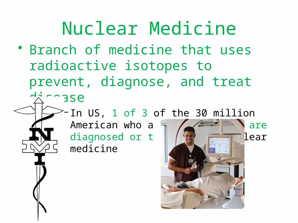
Nuclear Medicine• Branch of medicine that uses radioactive
isotopes to prevent, diagnose, and treat disease
– In US, 1 of 3 of the 30 million American who are hospitalized are diagnosed or treated with nuclear medicine

X Rays• X Rays: Rays are passed through the body part
desired– As they pass through the body part, the rays
are absorbed in different amounts by the different density of tissues encountered• EX: calcium in bone is very dense, so it absorbs a
lot of the rays; flesh is not very dense, so it does not absorb the rays as much
– There is a film behind the body part• Essentially, a picture of the different absorbencies
is taken because more or less ray is exposed to the film; this picture is developed much like a normal photograph


Computer Axial Tomography(CAT or CT scan)
• X-Ray procedure that uses ionizing radiation to produce cross-section images of the body– Computer detects radiation absorption and variations
in tissue density– Produces a series of anatomic pictures
• End up with a 3-D view of the tissue being examined
• Great because they have mostly eliminated the need for exploratory surgery!
• Uses: brain, abdominal, and lymphoid tissue


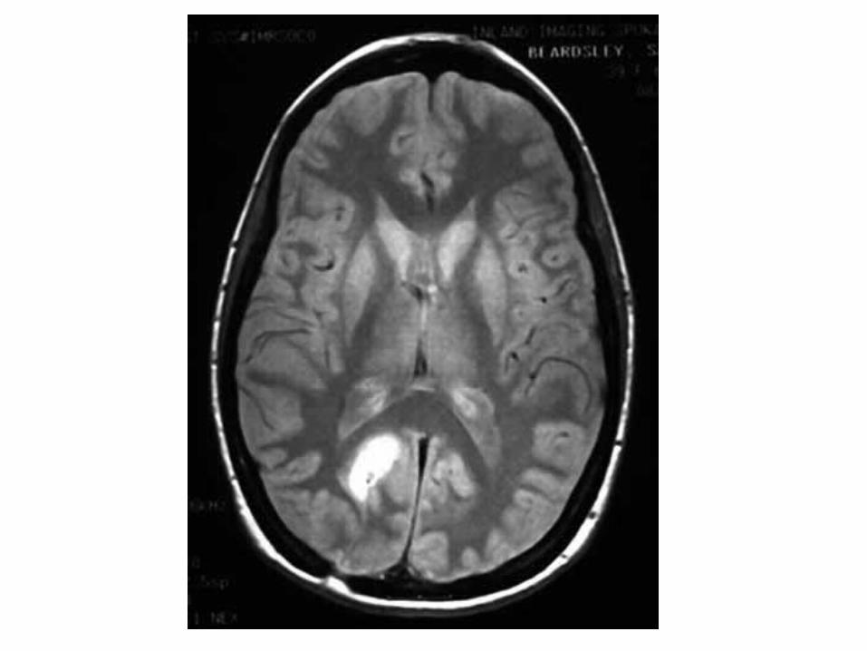
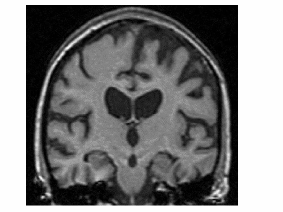
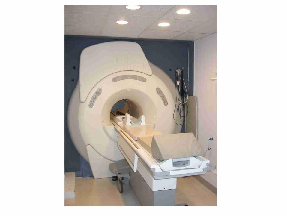
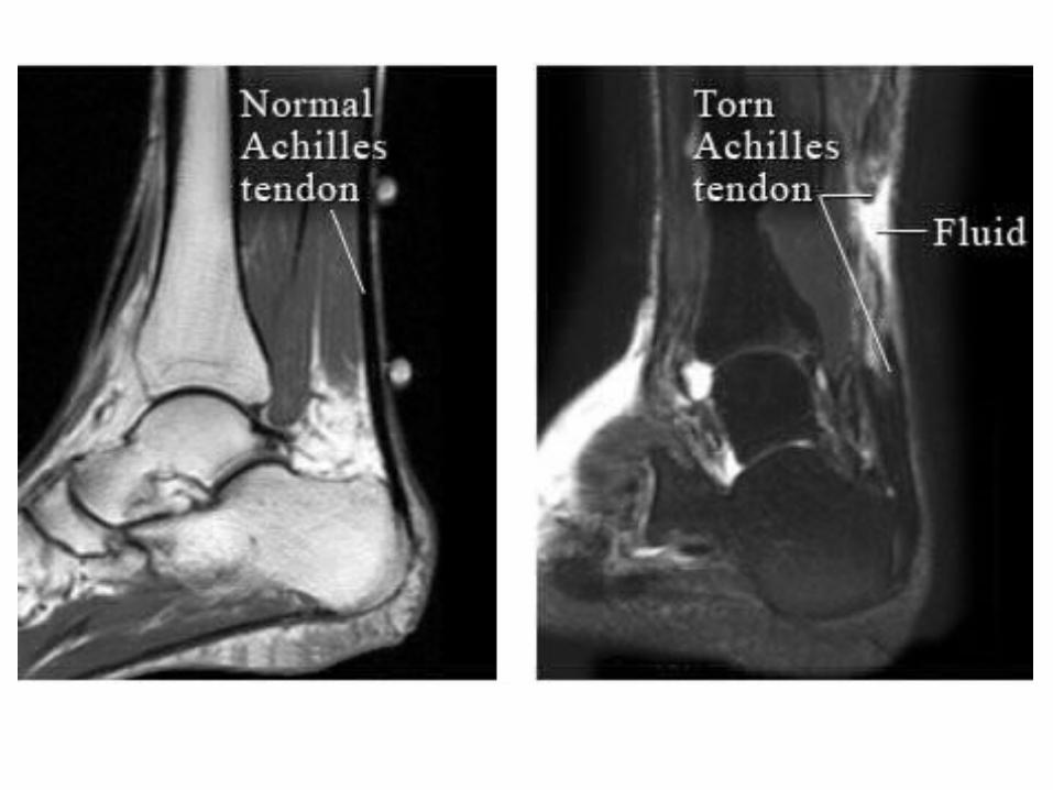
Magnetic Resonance Imaging(MRI)
• Entire person placed inside electromagnetic tube• Specific radio signals and very strong magnetic fields are
generated• These signals change the alignment of hydrogen atoms
in the body very briefly• When the signal stops, the hydrogen atoms drop back
down to where they are supposed to be– When they drop, they emit energy– It is this energy that the MRI records and is able to make into
pictures on a screen• Much higher detail than CT scan• Visualization of fluid, very precise detector of blood flow,
soft tissue, and bony structures• No radiation
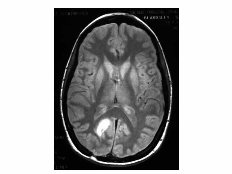

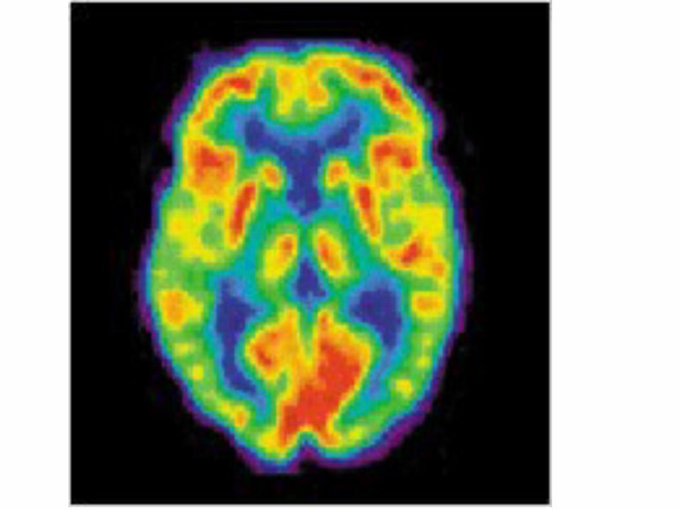
Positron Emission Tomography(PET) scan
• Patient is given with a short-lived radioactive isotope (usually injected)
• These isotopes allow us to see metabolic activity of the evaluated structure
• Patient can be awake within the scanner and can answer questions to see how metabolic activity changes
• Most useful in diagnosing brain tumors, cerebral palsy, stroke, and heart disease


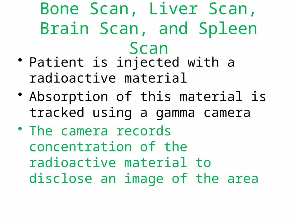
Bone Scan, Liver Scan, Brain Scan, and Spleen Scan
• Patient is injected with a radioactive material
• Absorption of this material is tracked using a gamma camera
• The camera records concentration of the radioactive material to disclose an image of the area


Bone scan

Sonography/Ultrasound
• Uses high frequency sound waves• Sound waves sent through body using a
transducer• Transducer also receives returning echoes that
bounce off internal structures– These returning sound waves are converted into
electrical signals that are fed into a computer to produce a picture
– Use to see fetus in womb