Embed Size (px)
Citation preview
Anion sensing by small molecules and molecular ensembles
Journal: Chemical Society Reviews
Manuscript ID: CS-REV-05-2014-000179.R1
Article Type: Tutorial Review
Date Submitted by the Author: 19-May-2014
Complete List of Authors: Gale, Philip; University of Southampton, School of Chemistry Caltagirone, Claudia; Universit� degli Studi di Cagliari, Dipartimento di Chimica Inorganica ed Analitica
Chemical Society Reviews
Journal Name RSCPublishing
ARTICLE
This journal is © The Royal Society of Chemistry 2013 J. Name., 2013, 00, 1-3 | 1
Cite this: DOI: 10.1039/x0xx00000x
Received 00th January 2012,
Accepted 00th January 2012
DOI: 10.1039/x0xx00000x
www.rsc.org/
Anion sensing by small molecules and molecular
ensembles
Philip A. Gale*,acd
and Claudia Caltagirone*,b
This tutorial review provides a short survey of anion sensing by small molecule anion
receptors, molecular ensembles and chemodosimeters. The review highlights the many
different sensing mechanisms and approaches employed by supramolecular chemists and the
wide structural variety present in these systems.
Introduction
The development of small molecule anion sensors has been an
area attracting significant attention over the last 25 years. This
has been driven by the important roles anions play in biology
and industrial processes, in addition to the need to produce new
methods of sensing anionic pollutants in the environment.
Three main approaches have been used. Early systems from
pioneers such as Paul Beer consisted of an anion-binding site
formed from hydrogen bond donor groups that are arranged
close to a redox-active ‘reporter’ group such ferrocene or a
fluorescent group such as ruthenium trisbipyridyl. When an
anion bound to the hydrogen bond donor array, the electronic
properties of the reporter group were perturbed resulting in a
change in the redox or fluorescent properties of the receptor are
perturbed so allowing the anion to be detected.1 Many sensors
for anions have been subsequently developed using these
principles.
Another important approach used in anion sensing is to
employ a displacement assay. This method, pioneered by Eric
Anslyn,2 involves forming a complex between an indicator and
a receptor via non-covalent interactions. The target anionic
guest binds to the receptor and so displaces the indicator. This
changes the microenvironment around the indicator resulting in
perturbations to its fluorescent properties and/or colour
allowing the anion to be detected.
The other important mechanism by which anionic species
can be sensed by small molecules is by chemical reaction
generating a new species with different properties. So-called
chemodosimeters can give very selective responses to particular
anionic guests.3
As interest in this area has grown, the selectivity of sensors
has improved and sensors have moved out of the laboratory
finding application in a number of areas including in sensing
anionic species in vivo. In this tutorial review we will examine
a range of different small molecule anion sensors and the
mechanisms by which they operate.
Hydrogen bonding sensors
As mentioned in the introduction coupling a reporter group
to a hydrogen-bonding array is an effective strategy in
designing an anion sensor.1 Gunnlaugsson and co-workers have
designed and synthesised a wide variety of colorimetric and
fluorescent anion sensors using this approach4 and particularly,
recently, based upon the 1,8-naphthalimide subunit.5 For
example, compound 1 was designed as a fluorescent anion
sensor that functions by a photoinduced-electron transfer (PET)
mechanism. This compound contains a fluorescent 1,8-
nathphalimide group tethered to an anion-binding thiourea.6
Upon addition of fluoride or acetate anions in DMSO solution
the fluorescence of compound 1 is switched off. Gunnlaugsson
proposed that this is due to photoelectron transfer from the
receptor unit to the fluorophore due to anion complexation
increasing the reduction potential of the receptor and so making
PET more favourable. When methanol was added to the
solution it disrupted the hydrogen bonds between the thiourea
group and the anionic guest resulting in the fluorescence being
restored. Interestingly when multiple equivalents of fluoride
were added the colour of the solution in DMSO changed from a
light yellow to deep purple colour. The authors showed that
this was due to deprotonation of the 4-amino moiety on the
Page 1 of 17 Chemical Society Reviews
ARTICLE Journal Name
2 | J. Name., 2012, 00, 1-3 This journal is © The Royal Society of Chemistry 2012
naphthalimide group (due to formation of HF2-). Thus this
sensor can operate via a dual sensing action – at low
equivalents of anions the fluorescence is quenched but at high
fluoride concentration there is a colorimetric response.
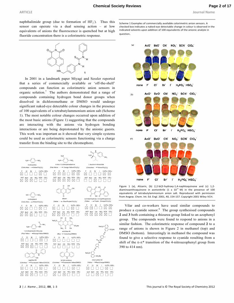
In 2001 in a landmark paper Miyagi and Sessler reported
that a series of commercially available or ‘off-the-shelf’
compounds can function as colorimetric anion sensors in
organic solution.7 The authors demonstrated that a range of
compounds containing hydrogen bond donor groups when
dissolved in dichloromethane or DMSO would undergo
significant naked-eye detectable colour changes in the presence
of 100 equivalents of a tetrabutylammonium anion salt (Scheme
1). The most notable colour changes occurred upon addition of
the most basic anions (Figure 1) suggesting that the compounds
are interacting with the anions via hydrogen bonding
interactions or are being deprotonated by the anionic guests.
This work was important as it showed that very simple systems
could be used as colorimetric sensors functioning via a charge
transfer from the binding site to the chromophore.
Scheme 1 Examples of commercially available colorimetric anion sensors. A
checked box indicates a naked-eye detectable change in colour is observed in the
indicated solvents upon addition of 100 equivalents of the anionic analyte in
question.
Figure 1 (a), Alizarin, (b) 2,2-bi(3-hydroxy-1,4-naphtoquinone and (c) 1,2-
diaminoanthraquinone in acetonitrile (1 x 10⁻⁴
M) in the presence of 100
equivalents of tetrabutylammonium anion salt. Reproduced with permission
from Angew. Chem. Int. Ed. Engl. 2001, 40, 154-157. Copyright 2001 Wiley-VCH.
Vilar and co-workers have used similar compounds to
produce a cyanide sensor.8 The group synthesised compounds
2 and 3 both containing a thiourea group linked to an azophenyl
group. The compounds were found to respond to anions in a
similar fashion. The colorimetric response of compound 2 to a
range of anions is shown in Figure 2 in methanol (top) and
DMSO (bottom). Interestingly in methanol the compound was
found to give a selective response to cyanide resulting from a
shift of the π-π* transition of the 4-nitroazophenyl group from
390 to 414 nm).
Page 2 of 17Chemical Society Reviews
Journal Name ARTICLE
This journal is © The Royal Society of Chemistry 2012 J. Name., 2012, 00, 1-3 | 3
Scheme 2 The synthesis of thioureas 2 and 3.
Figure 2 Solutions of compound 2 (0.5 mM) with different anions (30 equiv.) in
methanol (top) and DMSO (bottom). Reproduced with permission from Chem.
Eur. J. 2008, 14, 3006-3012. Copyright 2008 Wiley-VCH.
Mesoporous Al2O3 films were prepared and loaded with
compound 2. Below pH 9 the surface of film is positively
charged and hence can interact strongly with the carboxylate
moiety present in compound 2. The film was immersed in
5mM aqueous solutions of different anions and once again a
selective response to cyanide was observed (Figure 3) with a
detection limit of 2.6 ppm.
Figure 3 Normalised absorption spectra of 2/Al2O3 films immersed in 5 mM
aqueous solutions of different anions. Reproduced with permission from Chem.
Eur. J. 2008, 14, 3006-3012. Copyright 2008 Wiley-VCH.
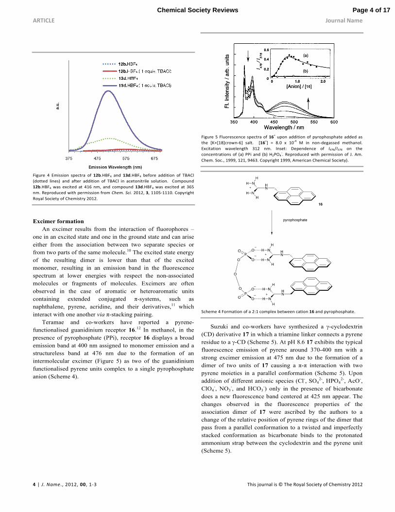
Johnson, Haley and co-workers have explored the
fluorescent properties of a series of sixteen differently
substituted 2,6-ethynylpyridine bisphenylurea compounds
containing a range of electron withdrawing and electron
donating substituents (Scheme 3).9 From the library of
compounds prepared, it was found that compound 13d
containing electron-withdrawing pentafluorophenyl substituents
was not emissive in its unbound state but in the presence of 1
equivalent of chloride the fluorescence was “switched on”
(Figure 4). The authors are continuing to investigate the cause
of this fluorescence.
Scheme 3 Synthesis of sixteen differentially substituted 2,6-ethynylpyridine
bisphenylurea scaffolds.
Page 3 of 17 Chemical Society Reviews
ARTICLE Journal Name
4 | J. Name., 2012, 00, 1-3 This journal is © The Royal Society of Chemistry 2012
Figure 4 Emission spectra of 12b.HBF4 and 13d.HBF4 before addition of TBACl
(dotted lines) and after addition of TBACl in acetonitrile solution. Compound
12b.HBF4 was excited at 416 nm, and compound 13d.HBF4 was excited at 365
nm. Reproduced with permission from Chem. Sci. 2012, 3, 1105-1110. Copyright
Royal Society of Chemistry 2012.
Excimer formation
An excimer results from the interaction of fluorophores –
one in an excited state and one in the ground state and can arise
either from the association between two separate species or
from two parts of the same molecule.10 The excited state energy
of the resulting dimer is lower than that of the excited
monomer, resulting in an emission band in the fluorescence
spectrum at lower energies with respect the non-associated
molecules or fragments of molecules. Excimers are often
observed in the case of aromatic or heteroaromatic units
containing extended conjugated π-systems, such as
naphthalene, pyrene, acridine, and their derivatives,11 which
interact with one another via π-stacking pairing.
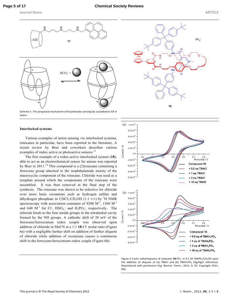
Teramae and co-workers have reported a pyrene-
functionalised guanidinium receptor 16.12 In methanol, in the
presence of pyrophosphate (PPi), receptor 16 displays a broad
emission band at 400 nm assigned to monomer emission and a
structureless band at 476 nm due to the formation of an
intermolecular excimer (Figure 5) as two of the guanidinium
functionalised pyrene units complex to a single pyrophosphate
anion (Scheme 4).
Figure 5 Fluorescence spectra of 16
+ upon addition of pyrophosphate added as
the [K+[18]crown-6] salt. [16+] = 8.0 x 10
-4 M in non-degassed methanol.
Excitation wavelength 312 nm. Inset: Dependence of I476/I376 on the
concentrations of (a) PPi and (b) H2PO4-. Reproduced with permission of J. Am.
Chem. Soc., 1999, 121, 9463. Copyright 1999, American Chemical Society).
Scheme 4 Formation of a 2:1 complex between cation 16 and pyrophosphate.
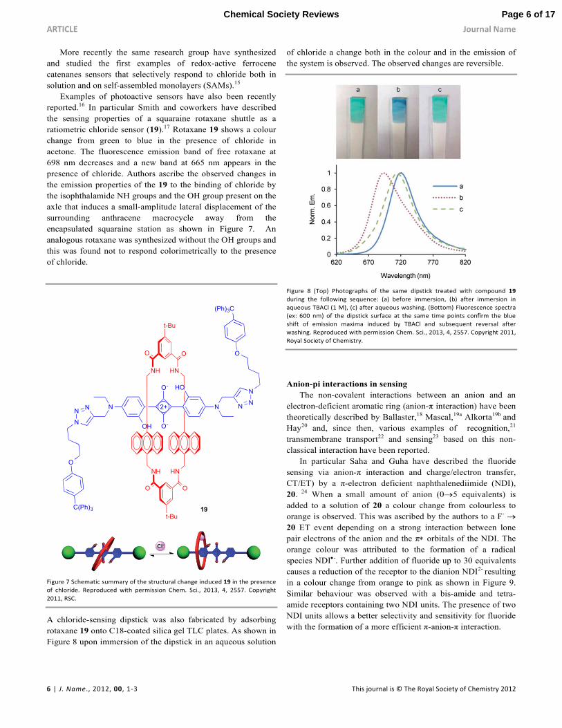
Suzuki and co-workers have synthesized a γ-cyclodextrin
(CD) derivative 17 in which a triamine linker connects a pyrene
residue to a γ-CD (Scheme 5). At pH 8.6 17 exhibits the typical
fluorescence emission of pyrene around 370-400 nm with a
strong excimer emission at 475 nm due to the formation of a
dimer of two units of 17 causing a π-π interaction with two
pyrene moieties in a parallel conformation (Scheme 5). Upon
addition of different anionic species (Cl-, SO42-, HPO4
2-, AcO-,
ClO4-, NO3
-, and HCO3-) only in the presence of bicarbonate
does a new fluorescence band centered at 425 nm appear. The
changes observed in the fluorescence properties of the
association dimer of 17 were ascribed by the authors to a
change of the relative position of pyrene rings of the dimer that
pass from a parallel conformation to a twisted and imperfectly
stacked conformation as bicarbonate binds to the protonated
ammonium strap between the cyclodextrin and the pyrene unit
(Scheme 5).
Page 4 of 17Chemical Society Reviews
Journal Name ARTICLE
This journal is © The Royal Society of Chemistry 2012 J. Name., 2012, 00, 1-3 | 5
HN
HN
HN
17γ-CD
_
_
_
HCO3- =
Scheme 5 The proposed mechanism of bicarbonate sensing by cyclodextrin 17 in
water.
Interlocked systems
Various examples of anion sensing via interlocked systems,
rotaxanes in particular, have been reported in the literature. A
recent review by Beer and coworkers describes various
examples of redox active or photoactive sensors.13
The first example of a redox-active interlocked system (18),
able to act as an electrochemical sensor for anions was reported
by Beer in 2011.14 This compound is a [2]rotaxane containing a
ferrocene group attached to the isophthalamide moiety of the
macrocyclic component of the rotaxane. Chloride was used as a
template around which the components of the rotaxane were
assembled. It was then removed in the final step of the
synthesis. The rotaxane was shown to be selective for chloride
over more basic oxoanions such as hydrogen sulfate and
dihydrogen phosphate in CDCl3:CD3OD (1:1 v/v) by 1H NMR
spectroscopy with association constants of 4200 M-1, 1560 M-1
and 640 M-1 for Cl-, HSO4-, and H2PO4
-, respectively. The
chloride binds to the four amide groups in the tetrahedral cavity
formed by the NH groups. A cathodic shift of 20 mV of the
ferrocene/ferrocenium redox couple was observed upon
addition of chloride in MeCN at a 1:1 18:Cl- molar ratio (Figure
6a) with a negligible further shift on addition of further aliquots
of chloride while addition of oxoanions causes a continuous
shift in the ferrocene/ferrocenium redox couple (Figure 6b).
NH
OO
O
HN O O O
OOFe
HN
N+
O
HN
O
PF6-
18
Figure 6 Cyclic voltamograms of rotaxane 18+PF6
- in 0.1 M TBAPF6/CH3CN upon
the addition of aliquots of (a) TBACl and (b) TBAH2PO4 (Ag/AgCl reference).
Reproduced with permission Org. Biomol. Chem., 2011, 9, 92. Copyright 2011,
RSC.
Page 5 of 17 Chemical Society Reviews
ARTICLE Journal Name
6 | J. Name., 2012, 00, 1-3 This journal is © The Royal Society of Chemistry 2012
More recently the same research group have synthesized
and studied the first examples of redox-active ferrocene
catenanes sensors that selectively respond to chloride both in
solution and on self-assembled monolayers (SAMs).15
Examples of photoactive sensors have also been recently
reported.16 In particular Smith and coworkers have described
the sensing properties of a squaraine rotaxane shuttle as a
ratiometric chloride sensor (19).17 Rotaxane 19 shows a colour
change from green to blue in the presence of chloride in
acetone. The fluorescence emission band of free rotaxane at
698 nm decreases and a new band at 665 nm appears in the
presence of chloride. Authors ascribe the observed changes in
the emission properties of the 19 to the binding of chloride by
the isophthalamide NH groups and the OH group present on the
axle that induces a small-amplitude lateral displacement of the
surrounding anthracene macrocycle away from the
encapsulated squaraine station as shown in Figure 7. An
analogous rotaxane was synthesized without the OH groups and
this was found not to respond colorimetrically to the presence
of chloride.
N NN
N
O
(Ph)3C
HO
NNN
N
O
C(Ph)3
OH O-
O-
2+
t-Bu
NH HN
O O
NH HN
t-Bu
O O
19
Figure 7 Schematic summary of the structural change induced 19 in the presence
of chloride. Reproduced with permission Chem. Sci., 2013, 4, 2557. Copyright
2011, RSC.
A chloride-sensing dipstick was also fabricated by adsorbing
rotaxane 19 onto C18-coated silica gel TLC plates. As shown in
Figure 8 upon immersion of the dipstick in an aqueous solution
of chloride a change both in the colour and in the emission of
the system is observed. The observed changes are reversible.
Figure 8 (Top) Photographs of the same dipstick treated with compound 19
during the following sequence: (a) before immersion, (b) after immersion in
aqueous TBACl (1 M), (c) after aqueous washing. (Bottom) Fluorescence spectra
(ex: 600 nm) of the dipstick surface at the same time points confirm the blue
shift of emission maxima induced by TBACl and subsequent reversal after
washing. Reproduced with permission Chem. Sci., 2013, 4, 2557. Copyright 2011,
Royal Society of Chemistry.
Anion-pi interactions in sensing
The non-covalent interactions between an anion and an
electron-deficient aromatic ring (anion-π interaction) have been
theoretically described by Ballaster,18 Mascal,19a Alkorta19b and
Hay20 and, since then, various examples of recognition,21
transmembrane transport22 and sensing23 based on this non-
classical interaction have been reported.
In particular Saha and Guha have described the fluoride
sensing via anion-π interaction and charge/electron transfer,
CT/ET) by a π-electron deficient naphthalenediimide (NDI),
20. 24 When a small amount of anion (0→5 equivalents) is
added to a solution of 20 a colour change from colourless to
orange is observed. This was ascribed by the authors to a F- →
20 ET event depending on a strong interaction between lone
pair electrons of the anion and the π∗ orbitals of the NDI. The
orange colour was attributed to the formation of a radical
species NDI•-. Further addition of fluoride up to 30 equivalents
causes a reduction of the receptor to the dianion NDI2- resulting
in a colour change from orange to pink as shown in Figure 9.
Similar behaviour was observed with a bis-amide and tetra-
amide receptors containing two NDI units. The presence of two
NDI units allows a better selectivity and sensitivity for fluoride
with the formation of a more efficient π-anion-π interaction.
Page 6 of 17Chemical Society Reviews
Journal Name ARTICLE
This journal is © The Royal Society of Chemistry 2012 J. Name., 2012, 00, 1-3 | 7
Figure 9 (Top) An illustration of proposed anion-π and CT interactions between F
-
and receptor 20 generating a colorimetric sensing (bottom). (Reproduced with
permission of JACS, 2010, 132, 17674, Copyright 2010, ACS).
Metal complexes
One of the most useful strategies for binding anions and that
allows anion sensing in competitive solvent media is the use of
metal complexes. The metal can play two main different roles:
it can induce a geometrical pre-organisation of the complex that
results in a better host-guest complementarity, or it can act as
binding site for the anion forming a strong coordinate bond.25
An example of a colorimetric sensor for iodide and cyanide
based on a coordinatively unsaturated copper(II) complex
containing the tetradentate ligand 1-(2-quinolinylmethyl)-1,4,7-
triazacyclononane) (21) was reported by Caltagirone and
Lippolis.26 This complex is able to recognise the presence of
iodide and cyanide in MeCN because the anion is able to bind
to the unsaturated copper centre and cause a colour change
from light blue to dark blue, pink and green upon addition of 1
equivalent of CN-, two equivalents of CN- and 1 equivalent of I-
, respectively (Figure 10). Interestingly, in water only addition
of cyanide results in a colour change.
Figure 10 a) The structure of receptor 21, b) the single crystal X-Ray crystal
structure of the complex cation [Cu(21)CN]+ (hydrogen atoms and BF4
- counter
ion have been omitted for clarity) and c) Colour change of the copper complex
(1.00 x 10⁻³
M) after addition of different anions in MeCN. From left to right: 21,
21 + 1.0 equiv. F-, 21 + 1.0 equiv. Cl
-, 21 + 1 equiv. Br
-, 21 + 1 eq of CN
-, 21 + 2 eq
of CN-, 21 + 1 eq of I
-. Reproduced with permission from Chem. Commun. 2011,
47, 3805. Copyright 2011 RSC.
Lanthanide complexes are very effective platforms for
anion sensing in water. Anions can displace the water
molecules that normally occupy the vacant coordination site on
the lanthanide in heptadentate ligands. If the ligand bears a
chromophore, anion binding to the lanthanide can cause a
change in the spectral properties of the system that can be used
to sense the anion.
Various examples of anion sensing via binding to lanthanide
complexes have been recently reviewed by Parker.27 For
example, Parker and co-workers have described the behaviour
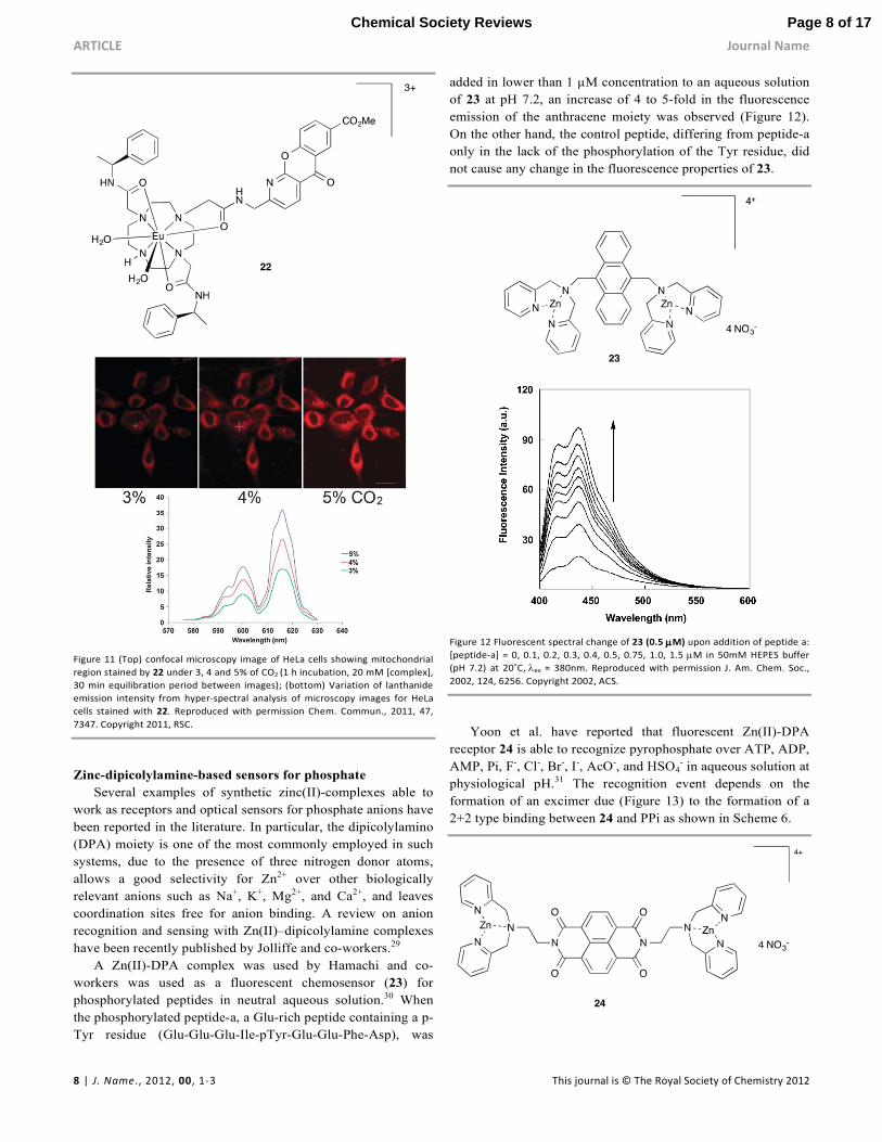
of the europium complex 22 in sensing bicarbonate in water.28
At pH 7.4 complex 22 is able to bind HCO3- with an apparent
stability constant of log K = 3.85. Moreover, complex 22 is able
to selectively stain the mitochondrial region of HeLa cells and
to increase the image intensity upon increasing percentage of
CO2 as shown in Figure 11. The observed increase of Eu
emission intensity with pCO2 has been attributed to an increase
in the steady state bicarbonate concentration in the
mitochondrial region of the cells.
Page 7 of 17 Chemical Society Reviews
ARTICLE Journal Name
8 | J. Name., 2012, 00, 1-3 This journal is © The Royal Society of Chemistry 2012
Figure 11 (Top) confocal microscopy image of HeLa cells showing mitochondrial
region stained by 22 under 3, 4 and 5% of CO2 (1 h incubation, 20 mM [complex],
30 min equilibration period between images); (bottom) Variation of lanthanide
emission intensity from hyper-spectral analysis of microscopy images for HeLa
cells stained with 22. Reproduced with permission Chem. Commun., 2011, 47,
7347. Copyright 2011, RSC.
Zinc-dipicolylamine-based sensors for phosphate
Several examples of synthetic zinc(II)-complexes able to
work as receptors and optical sensors for phosphate anions have
been reported in the literature. In particular, the dipicolylamino
(DPA) moiety is one of the most commonly employed in such
systems, due to the presence of three nitrogen donor atoms,
allows a good selectivity for Zn2+ over other biologically
relevant anions such as Na+, K+, Mg2+, and Ca2+, and leaves
coordination sites free for anion binding. A review on anion
recognition and sensing with Zn(II)–dipicolylamine complexes
have been recently published by Jolliffe and co-workers.29
A Zn(II)-DPA complex was used by Hamachi and co-
workers was used as a fluorescent chemosensor (23) for
phosphorylated peptides in neutral aqueous solution.30 When
the phosphorylated peptide-a, a Glu-rich peptide containing a p-
Tyr residue (Glu-Glu-Glu-Ile-pTyr-Glu-Glu-Phe-Asp), was
added in lower than 1 µM concentration to an aqueous solution
of 23 at pH 7.2, an increase of 4 to 5-fold in the fluorescence
emission of the anthracene moiety was observed (Figure 12).
On the other hand, the control peptide, differing from peptide-a
only in the lack of the phosphorylation of the Tyr residue, did
not cause any change in the fluorescence properties of 23.
Figure 12 Fluorescent spectral change of 23 (0.5 µµµµM) upon addition of peptide a:
[peptide-a] = 0, 0.1, 0.2, 0.3, 0.4, 0.5, 0.75, 1.0, 1.5 µM in 50mM HEPES buffer
(pH 7.2) at 20˚C, λex = 380nm. Reproduced with permission J. Am. Chem. Soc.,
2002, 124, 6256. Copyright 2002, ACS.
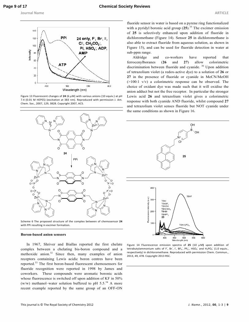
Yoon et al. have reported that fluorescent Zn(II)-DPA
receptor 24 is able to recognize pyrophosphate over ATP, ADP,
AMP, Pi, F-, Cl-, Br-, I-, AcO-, and HSO4- in aqueous solution at
physiological pH.31 The recognition event depends on the
formation of an excimer due (Figure 13) to the formation of a
2+2 type binding between 24 and PPi as shown in Scheme 6.
Page 8 of 17Chemical Society Reviews
Journal Name ARTICLE
This journal is © The Royal Society of Chemistry 2012 J. Name., 2012, 00, 1-3 | 9
Figure 13 Fluorescent changes of 24 (6 µM) with various anions (10 equiv.) at pH
7.4 (0.01 M HEPES) (excitation at 383 nm). Reproduced with permission J. Am.
Chem. Soc., 2007, 129, 3828. Copyright 2007, ACS.
Scheme 6 The proposed structure of the complex between of chemosensor 24
with PPi resulting in excimer formation.
Boron-based anion sensors
In 1967, Shriver and Biallas reported the first chelate
complex between a chelating bis-boron compound and a
methoxide anion.32 Since then, many examples of anion
receptors containing Lewis acidic boron centres have been
reported.33 The first boron-based fluorescent chemosensors for
fluoride recognition were reported in 1998 by James and
coworkers. These compounds were aromatic boronic acids
whose fluorescence is switched off upon addition of KF in 50%
(w/w) methanol–water solution buffered to pH 5.5.34 A more
recent example reported by the same group of an OFF-ON
fluoride sensor in water is based on a pyrene ring functionalized
with a pyridyl boronic acid group (25).35 The excimer emission
of 25 is selectively enhanced upon addition of fluoride in
dichloromethane (Figure 14). Sensor 25 in dichloromethane is
also able to extract fluoride from aqueous solution, as shown in
Figure 15), and can be used for fluoride detection in water at
sub-ppm range.
Aldridge and co-workers have reported that
ferrocenylboranes (26 and 27) allow colorimetric
discrimination between fluoride and cyanide. 36 Upon addition
of tetrazolium violet (a redox-active dye) to a solution of 26 or
27 in the presence of fluoride or cyanide in MeCN/MeOH
(>100:1 v/v) a colorimetric response can be observed. The
choice of oxidant dye was made such that it will oxidise the
anion adduct but not the free receptor. In particular the stronger
Lewis acid 26 and tetrazolium violet gives a colorimetric
response with both cyanide AND fluoride, whilst compound 27
and tetrazolium violet senses fluoride but NOT cyanide under
the same conditions as shown in Figure 16.
Figure 14 Fluorescence emission spectra of 25 (10 µM) upon addition of
tetrabutylammonium salts of F-, Br
-, I
-, BF4
-, PF6
-, HSO4
- and H2PO4
- (1.0 equiv.,
respectively) in dichloromethane. Reproduced with permission Chem. Commun.,
2013, 49, 478. Copyright 2013 RSC.
Page 9 of 17 Chemical Society Reviews
ARTICLE Journal Name
10 | J. Name., 2012, 00, 1-3 This journal is © The Royal Society of Chemistry 2012
Figure 15 Biphasic extraction experiment of 25 (50mM) in dichloromethane with
increasing concentration of NaF (0.10-3.80 ppm) in water. (a) The fluorescence
profiles of 25. (b) The relative fluorescence intensity at 495 nm plotted against
fluoride anion concentration. Inset: Photograph of vials containing sensor 25 (50
µM) exposed to aqueous solutions of NaF (52.6 µM, 1.0 ppm) before (A) and
after shaking (B). Reproduced with permission Chem. Commun., 2013, 49, 478.
Copyright 2013 RSC.
Fe
Me5
B
Mes
Mes
Fe
Me5
B
O
O Ph
Ph
26 27
Figure 16 UV/Vis spectra of 26 and 27 and tetrazolium violet in the presence
(black trace) or in the absence (grey trace) of fluoride or cyanide in MeCN/MeOH
(>100;1 v/v): a) 26 + F-; b) 26 + CN
-; c) 27 + F
-; d) 27 + CN
-. Reproduced with
permission Chem. Eur. J., 2008, 14, 7525. Copyright 2008 Wiley-VCH.
Halogen bonding
Halogen bonding is increasing recognized as a
fundamentally important interaction in supramolecular
chemistry and has been employed by Beer and co-workers in a
series of novel fluorescent anion sensors 28 that employ a
combination of halogen bonding and electrostatic interactions
to bind anionic guests.37 Beer found that bromo-derivative 28c2+
and the syn conformation of the rotationally restricted iodo-
derivative 28d2+ form strong 1:1 complexes with iodide and
bromide anions in CD3OD/D2O (9:1). Binding was
accompanied by a significant fluorescence enhancement
making these compounds the first examples of halogen bond
chemosensors to function in aqueous solvent mixtures. By
Page 10 of 17Chemical Society Reviews
Journal Name ARTICLE
This journal is © The Royal Society of Chemistry 2012 J. Name., 2012, 00, 1-3 | 11
contrast no evidence of anion sensing was observed by either
the protic or chloro-analogues 28a2+ or 28b2+.
Chemodosimeters
The “chemodosimeter approach” for the design of
chemosensors takes advantage of usually specific anion-
induced reactions coupled with selective changes in
fluorescence or colour.38 When an anionic substrate reacts with
a chemodosimeter it can remain covalently bound to it or the
anion can catalyze a chemical reaction. In both cases the
product formed is different from the starting material with
changes in its optical properties allowing the anion to be
detected.
Ahn and coworkers have synthesized an ortho-TFADA
compound (29). The fluorescence of this compound is
selectively switched on in MeCN in the presence of cyanide
(Figure 15b) due to the formation of an intramolecular
hydrogen bond in the host-guest adduct (Figure 17).39 The
authors propose the mechanism shown in Scheme 7 in which
the reaction of CN- with 29 causes the formation of the
negatively charged carbonyl adduct stabilized by an
intramolecular hydrogen bond between the carbonyl and the
proton of the sulfonamide. This results in rigidification of the
sensor and consequently a fluorescence enhancement.
N
SO2N
CF3
O
H
CN-
N
SO2N
CF3
O-
H
CN
29
OFF ON
Scheme 7
Figure 17 Changes in the fluorescent emission of 29 in the presence of different
anions (equimolar mixture each component 20 µM). Reproduced with
permission Chem. Commun., 2006, 186. Copyright 2006 RSC.
A simple colorimetric and fluorimetric chemodosimeter
(30) for fluoride recognition containing a quinoline
chromophore has been reported by Bai and co-workers.40 A
colour change from colourless to yellowish-green is observed
upon addition of fluoride in THF and in wet organic solution. In
the same solvent the blue fluorescence of 30 turns yellowish-
green and it is switched on selectively by this anion. The
addition of fluoride causes a desilylation of 30 (Scheme 8). An
excited state proton transfer (ESPT) from the receptor to
fluoride occurs that causes the optical changes observed. In the
presence of other anionic guests no changes were observed
(Figure 18).
A further example of a chemodosimeter (31a) for fluoride
recognition containing a 1,8-naphthalimide moiety was
reported more recently by Wu and co-workers.41 In this case, as
shown in Scheme 9 when a moderate amount of fluoride is
added to an acetonitrile solution of 31a the adduct [31a-F]- is
formed. A concomitant colour change from colourless to deep
blue and the switching on of the fluorescence is observed. The
addition of a large excess of fluoride causes the deprotonation
of 31a.
Scheme 8 Desilylation of compound 30 by fluoride.
Page 11 of 17 Chemical Society Reviews
ARTICLE Journal Name
12 | J. Name., 2012, 00, 1-3 This journal is © The Royal Society of Chemistry 2012
Figure 18 a) Effect of reaction time on the fluorescence intensity of the sensor
system by F-: [30] = [F
-]= 20 mM. (b) Fluorescence intensity of the sensor system
at 520 nm in the presence of 10 equivalents of F-, Cl
-, Br
-, HSO4
-, ClO4
- (TBA
+ salts),
NO3-, AcO
-, H2PO4
- (Na
+ salts) in THF: [30] = 20 µM; λex = 335 nm. Reproduced
with permission Chem. Commun., 2011, 47, 3957. Copyright 2011 RSC.
At this point an in situ autoxidation occurs with the subsequent
release of CN-. A new species 31b is formed which is
colourless and not fluorescent. Thus, chemodosimeter 31a
works as an OFF-ON-OFF ratiometric chromofluorogenic
sensor for fluoride.
Scheme 9 Proposed mechanism for the spectroscopic changes of 31a in the
presence of fluoride.
Displacement assays
Another strategy for anion sensing is the use of a
displacement assay. In this approach the anion binding site is
occupied by the signaling unit (such as a fluorescent or
coloured dye) forming a molecular ensemble. The addition of
the target anion to the solution containing the molecular
ensemble causes the signaling unit to be displaced from the
receptor. Consequently the signaling unit recovers its non-
coordinating spectroscopic behaviour in solution. In order to
design an efficient system the spectroscopic characteristics of
the signaling unit should be different to those in its non-
coordinating state; moreover the stability constant for the
formation of the complex between the binding site and the
signaling unit should be lower than that between the binding
site and the analyte anion. This approach offers advantages
such as the possibility to use a great variety of signaling units,
depending on the purpose, as they are non-covalently attached
to the binding site so a further functionalisation is not required.
Moreover, they work well both in aqueous and organic solvents
giving the possibility to tune the solvent media in order to
obtain the desired Ka values for the signaling subunit and the
analyte with the receptor. However, this type of sensor cannot
be used to imaging tissues or cells because the signaling unit is
present everywhere in the solution as it is not covalently
attached to the receptor.2 One of the first examples of a
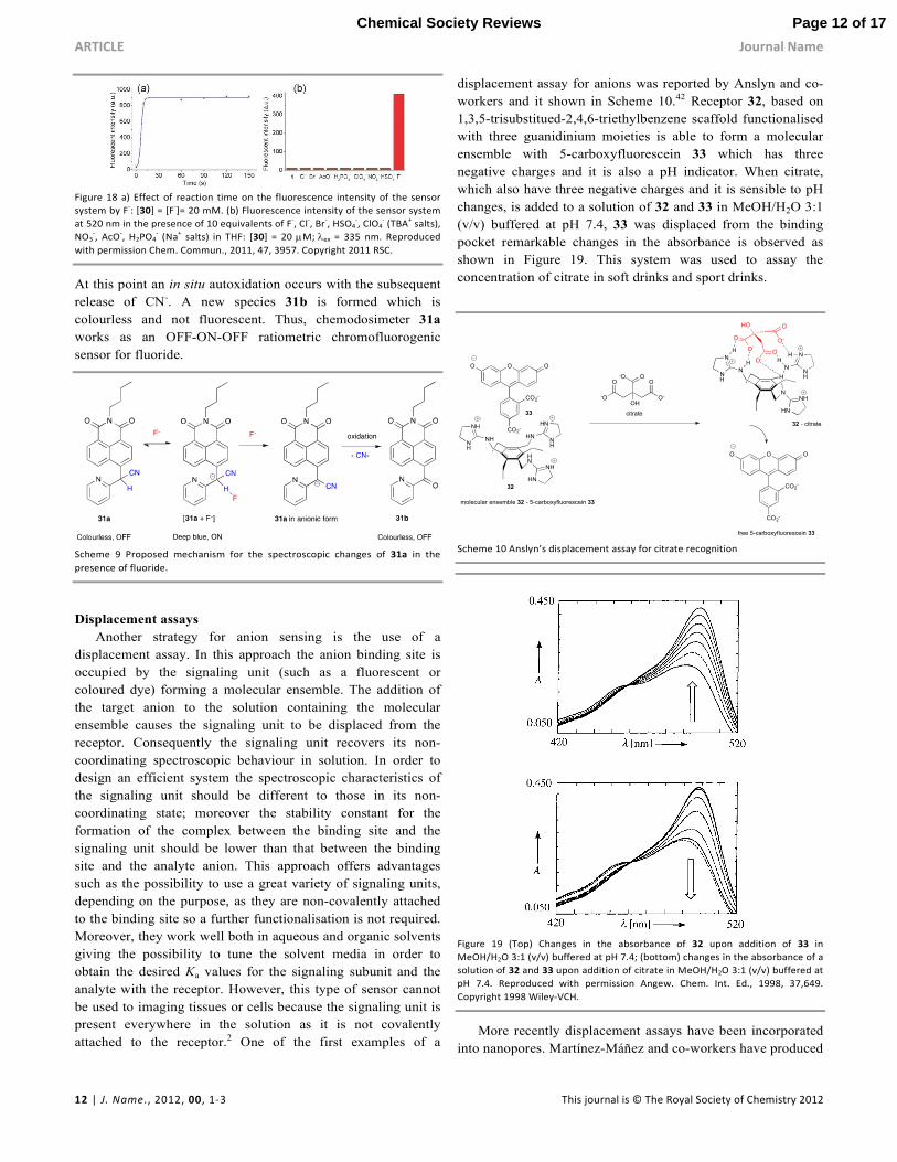
displacement assay for anions was reported by Anslyn and co-
workers and it shown in Scheme 10.42 Receptor 32, based on
1,3,5-trisubstitued-2,4,6-triethylbenzene scaffold functionalised
with three guanidinium moieties is able to form a molecular
ensemble with 5-carboxyfluorescein 33 which has three
negative charges and it is also a pH indicator. When citrate,
which also have three negative charges and it is sensible to pH
changes, is added to a solution of 32 and 33 in MeOH/H2O 3:1
(v/v) buffered at pH 7.4, 33 was displaced from the binding
pocket remarkable changes in the absorbance is observed as
shown in Figure 19. This system was used to assay the
concentration of citrate in soft drinks and sport drinks.
HNNH
HN
NH
HN
HN
NH
NH
NH
-O
O
-O O
OH
O
O-
O OO
CO2-
CO2-
32
H
NN
N
NH
N
HN
NH
N
NH
H
H
HH
O
HO
O-
O
O-
O
O-
O OO
CO2-
CO2-
citrate
free 5-carboxyfluorescein 33
molecular ensemble 32 - 5-carboxyfluorescein 33
33
32 - citrate
Scheme 10 Anslyn’s displacement assay for citrate recognition
Figure 19 (Top) Changes in the absorbance of 32 upon addition of 33 in
MeOH/H2O 3:1 (v/v) buffered at pH 7.4; (bottom) changes in the absorbance of a
solution of 32 and 33 upon addition of citrate in MeOH/H2O 3:1 (v/v) buffered at
pH 7.4. Reproduced with permission Angew. Chem. Int. Ed., 1998, 37,649.
Copyright 1998 Wiley-VCH.
More recently displacement assays have been incorporated
into nanopores. Martínez-Máñez and co-workers have produced
Page 12 of 17Chemical Society Reviews
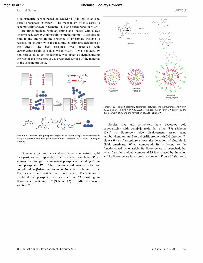
Journal Name ARTICLE
This journal is © The Royal Society of Chemistry 2012 J. Name., 2012, 00, 1-3 | 13
a colorimetric sensor based on MCM-41 (34) that is able to
detect phosphate in water.43 The mechanism of this assay is
schematically shown in Scheme 11. Nano-sized pores in MCM-
41 are functionalized with an amine and loaded with a dye
(methyl red, carboxyfluorescein or methylthymol Blue) able to
bind to the amine. In the presence of phosphate the dye is
released in solution with the resulting colorimetric detection of
the guest. The best response was observed with
carboxyfluorescein as a dye. When MCM-41 was replaced by
non-porous silica gel no response was observed demonstrating
the role of the mesoporous 3D organized surface of the material
in the sensing protocol.
Scheme 11 Protocol for phosphate signaling in water using the displacement
assay 34. Reproduced with permission Chem. Commun, 2008, 3639. Copyright
2008 RSC.
Gunnlaugsson and co-workers have synthesised gold
nanoparticles with appended Eu(III) cyclen complexes 35 as
sensors for biologically important phosphates including flavin
monophosphate 37. The functionalised nanoparticles are
complexed to β-diketone antennas 36 which is bound to the
Eu(III) centre and switches on fluorescence. The antenna is
displaced by phosphate species such as 37 resulting in
fluorescence switching off (Scheme 12) in buffered aqueous
solution.44
Scheme 12 The self-assembly formation between the nonluminescent AuNP-
35.Eu and 36 to give AuNP-35.Eu.36. The sensing of flavin 37 occurs by the
displacement of 36 and the formation of AuNP-35.Eu-37.
Sessler, Lee and co-workers have decorated gold
nanoparticles with calix[4]pyrrole derivative (38) (Scheme
13).45 A fluorescent dye displacement assay using
tetrabutylammonium-2-oxo-4-(trifluoromethyl)-2H-chromen-7-
olate (39) as fluorophore allows the detection of fluoride in
dichloromethane. When compound 39 is bound to the
functionalized nanoparticle its fluorescence is quenched, but
when fluoride is added, compound 39 is displaced by the anion
and its fluorescence is restored, as shown in Figure 20 (bottom).
Page 13 of 17 Chemical Society Reviews
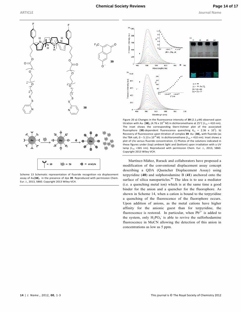
ARTICLE Journal Name
14 | J. Name., 2012, 00, 1-3 This journal is © The Royal Society of Chemistry 2012
Scheme 13 Schematic representation of fluoride recognition via displacement
assay of Au[38]n in the presence of dye 39. Reproduced with permission Chem.
Eur. J., 2013, 5860. Copyright 2013 Wiley-VCH.
Figure 20 a) Changes in the fluorescence intensity of 39 (2.1 µM) observed upon
titration with Au⋅ [38]n (4.76 x 10⁻⁹
M) in dichloromethane at 25˚C (λex = 410 nm).
The inset shows the corresponding Stern-Volmer plot of the associated
fluorophore (39)-dependent fluorescence quenching Ksv = 2.36 x 10⁷). b)
Recovery of fluorescence upon titration of complex 39⋅ Au⋅ [38]n with fluoride (as
the TBA salt, 0 – 5.13 x 10⁻⁸
M) in dichloromethane (λex = 410 nm). Inset shows a
plot of I/Io versus fluoride concentration. C) Photos of the solutions indicated in
these figures under (top) ambient light and (bottom) upon irradiation with a UV
lamp (λex =365 nm). Reproduced with permission Chem. Eur. J., 2013, 5860.
Copyright 2013 Wiley-VCH.
Martínez-Máñez, Rurack and collaborators have proposed a
modification of the conventional displacement assay concept
describing a QDA (Quencher Displacement Assay) using
terpyridine (40) and sulphorodamine B (41) anchored onto the
surface of silica nanoparticles.46 The idea is to use a mediator
(i.e. a quenching metal ion) which is at the same time a good
binder for the anion and a quencher for the fluorophore. As
shown in Scheme 14, when a cation is bound to the terpyridine
a quenching of the fluorescence of the fluorophore occurs.
Upon addition of anions, as the metal cations have higher
affinity for the anionic guest than for terpyridine, the
fluorescence is restored. In particular, when Pb2+ is added to
the system, only H2PO4- is able to revive the sulforhodamine
fluorescence in MeCN allowing the detection of this anion in
concentrations as low as 5 ppm.
Page 14 of 17Chemical Society Reviews
Journal Name ARTICLE
This journal is © The Royal Society of Chemistry 2012 J. Name., 2012, 00, 1-3 | 15
Scheme 14 A representation of the quencher displacement assay (QDA) based on
terpyridine (40) and sulforhodamine (41). Reproduced with permission Chem.
Commun, 2011, 47, 10599. Copyright 2011 RSC.
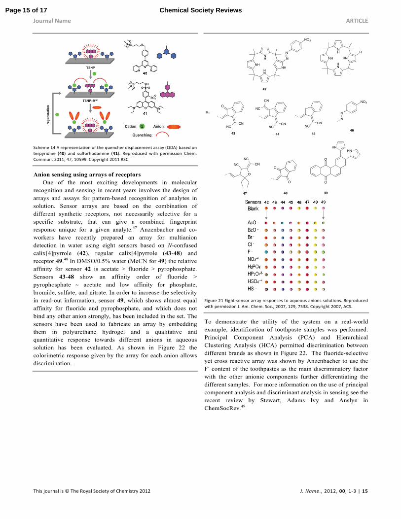
Anion sensing using arrays of receptors
One of the most exciting developments in molecular
recognition and sensing in recent years involves the design of
arrays and assays for pattern-based recognition of analytes in
solution. Sensor arrays are based on the combination of
different synthetic receptors, not necessarily selective for a
specific substrate, that can give a combined fingerprint
response unique for a given analyte.47 Anzenbacher and co-
workers have recently prepared an array for multianion
detection in water using eight sensors based on N-confused
calix[4]pyrrole (42), regular calix[4]pyrrole (43-48) and
receptor 49.48 In DMSO/0.5% water (MeCN for 49) the relative
affinity for sensor 42 is acetate > fluoride > pyrophosphate.
Sensors 43-48 show an affinity order of fluoride >
pyrophosphate ∼ acetate and low affinity for phosphate,
bromide, sulfate, and nitrate. In order to increase the selectivity
in read-out information, sensor 49, which shows almost equal
affinity for fluoride and pyrophosphate, and which does not
bind any other anion strongly, has been included in the set. The
sensors have been used to fabricate an array by embedding
them in polyurethane hydrogel and a qualitative and
quantitative response towards different anions in aqueous
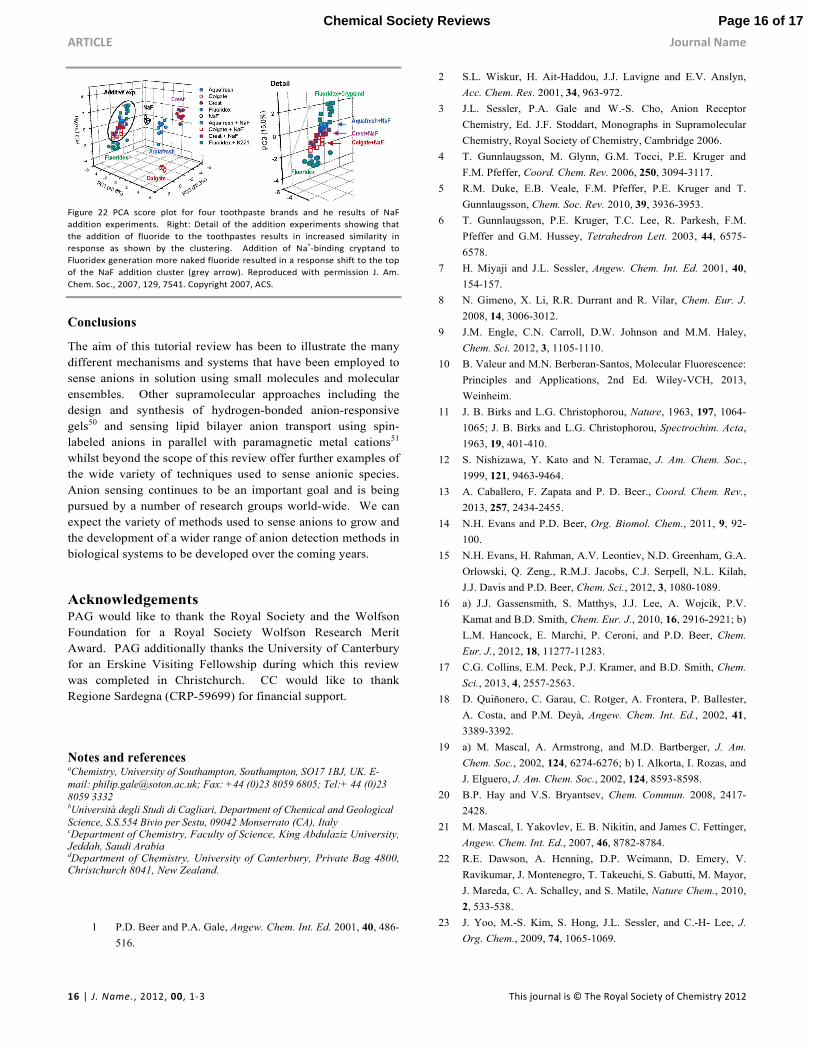
solution has been evaluated. As shown in Figure 22 the
colorimetric response given by the array for each anion allows
discrimination.
Figure 21 Eight-sensor array responses to aqueous anions solutions. Reproduced
with permission J. Am. Chem. Soc., 2007, 129, 7538. Copyright 2007, ACS.
To demonstrate the utility of the system on a real-world
example, identification of toothpaste samples was performed.
Principal Component Analysis (PCA) and Hierarchical
Clustering Analysis (HCA) permitted discrimination between
different brands as shown in Figure 22. The fluoride-selective
yet cross reactive array was shown by Anzenbacher to use the
F- content of the toothpastes as the main discriminatory factor
with the other anionic components further differentiating the
different samples. For more information on the use of principal
component analysis and discriminant analysis in sensing see the
recent review by Stewart, Adams Ivy and Anslyn in
ChemSocRev.49
Page 15 of 17 Chemical Society Reviews
ARTICLE Journal Name
16 | J. Name., 2012, 00, 1-3 This journal is © The Royal Society of Chemistry 2012
Figure 22 PCA score plot for four toothpaste brands and he results of NaF
addition experiments. Right: Detail of the addition experiments showing that
the addition of fluoride to the toothpastes results in increased similarity in
response as shown by the clustering. Addition of Na+-binding cryptand to
Fluoridex generation more naked fluoride resulted in a response shift to the top
of the NaF addition cluster (grey arrow). Reproduced with permission J. Am.
Chem. Soc., 2007, 129, 7541. Copyright 2007, ACS.
Conclusions
The aim of this tutorial review has been to illustrate the many
different mechanisms and systems that have been employed to
sense anions in solution using small molecules and molecular
ensembles. Other supramolecular approaches including the
design and synthesis of hydrogen-bonded anion-responsive
gels50 and sensing lipid bilayer anion transport using spin-
labeled anions in parallel with paramagnetic metal cations51
whilst beyond the scope of this review offer further examples of
the wide variety of techniques used to sense anionic species.
Anion sensing continues to be an important goal and is being
pursued by a number of research groups world-wide. We can
expect the variety of methods used to sense anions to grow and
the development of a wider range of anion detection methods in
biological systems to be developed over the coming years.
Acknowledgements PAG would like to thank the Royal Society and the Wolfson
Foundation for a Royal Society Wolfson Research Merit
Award. PAG additionally thanks the University of Canterbury
for an Erskine Visiting Fellowship during which this review
was completed in Christchurch. CC would like to thank
Regione Sardegna (CRP-59699) for financial support.
Notes and references aChemistry, University of Southampton, Southampton, SO17 1BJ, UK. E-
mail: [email protected]; Fax: +44 (0)23 8059 6805; Tel:+ 44 (0)23
8059 3332 bUniversità degli Studi di Cagliari, Department of Chemical and Geological
Science, S.S.554 Bivio per Sestu, 09042 Monserrato (CA), Italy cDepartment of Chemistry, Faculty of Science, King Abdulaziz University, Jeddah, Saudi Arabia dDepartment of Chemistry, University of Canterbury, Private Bag 4800, Christchurch 8041, New Zealand.
1 P.D. Beer and P.A. Gale, Angew. Chem. Int. Ed. 2001, 40, 486-
516.
2 S.L. Wiskur, H. Ait-Haddou, J.J. Lavigne and E.V. Anslyn,
Acc. Chem. Res. 2001, 34, 963-972.
3 J.L. Sessler, P.A. Gale and W.-S. Cho, Anion Receptor
Chemistry, Ed. J.F. Stoddart, Monographs in Supramolecular
Chemistry, Royal Society of Chemistry, Cambridge 2006.
4 T. Gunnlaugsson, M. Glynn, G.M. Tocci, P.E. Kruger and
F.M. Pfeffer, Coord. Chem. Rev. 2006, 250, 3094-3117.
5 R.M. Duke, E.B. Veale, F.M. Pfeffer, P.E. Kruger and T.
Gunnlaugsson, Chem. Soc. Rev. 2010, 39, 3936-3953.
6 T. Gunnlaugsson, P.E. Kruger, T.C. Lee, R. Parkesh, F.M.
Pfeffer and G.M. Hussey, Tetrahedron Lett. 2003, 44, 6575-
6578.
7 H. Miyaji and J.L. Sessler, Angew. Chem. Int. Ed. 2001, 40,
154-157.
8 N. Gimeno, X. Li, R.R. Durrant and R. Vilar, Chem. Eur. J.
2008, 14, 3006-3012.
9 J.M. Engle, C.N. Carroll, D.W. Johnson and M.M. Haley,
Chem. Sci. 2012, 3, 1105-1110.
10 B. Valeur and M.N. Berberan-Santos, Molecular Fluorescence:
Principles and Applications, 2nd Ed. Wiley-VCH, 2013,
Weinheim.
11 J. B. Birks and L.G. Christophorou, Nature, 1963, 197, 1064-
1065; J. B. Birks and L.G. Christophorou, Spectrochim. Acta,
1963, 19, 401-410.
12 S. Nishizawa, Y. Kato and N. Teramae, J. Am. Chem. Soc.,
1999, 121, 9463-9464.
13 A. Caballero, F. Zapata and P. D. Beer., Coord. Chem. Rev.,
2013, 257, 2434-2455.
14 N.H. Evans and P.D. Beer, Org. Biomol. Chem., 2011, 9, 92-
100.
15 N.H. Evans, H. Rahman, A.V. Leontiev, N.D. Greenham, G.A.
Orlowski, Q. Zeng., R.M.J. Jacobs, C.J. Serpell, N.L. Kilah,
J.J. Davis and P.D. Beer, Chem. Sci., 2012, 3, 1080-1089.
16 a) J.J. Gassensmith, S. Matthys, J.J. Lee, A. Wojcik, P.V.
Kamat and B.D. Smith, Chem. Eur. J., 2010, 16, 2916-2921; b)
L.M. Hancock, E. Marchi, P. Ceroni, and P.D. Beer, Chem.
Eur. J., 2012, 18, 11277-11283.
17 C.G. Collins, E.M. Peck, P.J. Kramer, and B.D. Smith, Chem.
Sci., 2013, 4, 2557-2563.
18 D. Quiñonero, C. Garau, C. Rotger, A. Frontera, P. Ballester,
A. Costa, and P.M. Deyà, Angew. Chem. Int. Ed., 2002, 41,
3389-3392.
19 a) M. Mascal, A. Armstrong, and M.D. Bartberger, J. Am.
Chem. Soc., 2002, 124, 6274-6276; b) I. Alkorta, I. Rozas, and
J. Elguero, J. Am. Chem. Soc., 2002, 124, 8593-8598.
20 B.P. Hay and V.S. Bryantsev, Chem. Commun. 2008, 2417-
2428.
21 M. Mascal, I. Yakovlev, E. B. Nikitin, and James C. Fettinger,
Angew. Chem. Int. Ed., 2007, 46, 8782-8784.
22 R.E. Dawson, A. Henning, D.P. Weimann, D. Emery, V.
Ravikumar, J. Montenegro, T. Takeuchi, S. Gabutti, M. Mayor,
J. Mareda, C. A. Schalley, and S. Matile, Nature Chem., 2010,
2, 533-538.
23 J. Yoo, M.-S. Kim, S. Hong, J.L. Sessler, and C.-H- Lee, J.
Org. Chem., 2009, 74, 1065-1069.
Page 16 of 17Chemical Society Reviews
Journal Name ARTICLE
This journal is © The Royal Society of Chemistry 2012 J. Name., 2012, 00, 1-3 | 17
24 S. Guha, and S. Saha, J. Am. Chem. Soc., 2010, 132, 17674-
17677.
25 A. Bencini, V. Lippolis, and B. Valtancoli, Inorg. Chim. Acta.,
2014, DOI:10.1016/j.ica.2014.01.003
26 M. A. Tetilla, M. C. Aragoni, M. Arca, C. Caltagirone, C.
Bazzicalupi, A. Bencini, A. Garau, F. Isaia, A. Laguna, V.
Lippolis, and V. Meli, Chem. Commun., 2011, 47, 3805-3807.
27 S.J- Butler, and D. Parker, Chem. Soc. Rev., 2013, 42, 1652-
1666.
28 D.G. Smith, G.-L. Law, B.S. Murray, R. Pal, D. Parker, and
K.L. Wong, Chem. Commun., 2011, 47, 7347-7349.
29 H.T. Ngo, X. Liu, and K.A. Jolliffe, Chem. Soc. Rev., 2012,
41, 4928–4965.
30 A. Ojida, Y. Mito-oka, M. Inoue, and I. Hamachi, J. Am.
Chem. Soc., 2002, 124, 6256-6258.
31 H. N. Lee, Z. Xu, S.K. Kim, K.M.K. Swamy, Y. Kim, S.-J.
Kim, and J. Yoon, J. Am. Chem. Soc., 2007, 129, 3828-3829.
32 D.F. Shriver, and M.J. Biallas, J. Am. Chem. Soc., 1967, 89,
1078-1081.
33 E. Gailbraith, and T. D. James, Chem. Soc. Rev., 2010, 39,
3831-3842.
34 C. R. Cooper, N. Spencer, and T.D. James, Chem. Commun.,
1998, 1365-1366.
35 T. Nishimura, S.-Y. Xu, Y.-B. Jiang, J. S. Fossey, K. Sakurai,
S. D. Bull, T. D. James, Chem. Commun., 2013, 49, 478-780.
36 A. E. J. Broomsgrove, D. A. Addy, C. Bresner, I. A. Fallis, A.
L. Thompson, and S. Aldridge, Chem. Eur. J., 2008, 14, 7525-
7529.
37 F. Zapata, A. Caballero, N.G. White, T.D.W. Claridge, P.J.
Costa, V. Félix and P.D. Beer, J. Am. Chem. Soc., 2012, 134,
11533-11541.
38 R.Martínez-Máñez, and F. Sancenon, Chem. Rev. 2003, 103,
4419-4476; R. Martínez-Máñez, and F. Sancenon, Coord.
Chem. Rev. 2006, 250, 3081-3093.
39 Y. M. Chung, B. Raman, D.-S. Kim, and K. H. Ahn, Chem.
Commun., 2006, 186-188.
40 Y. Bao, B. Liu, H. Wang, J. Tian, and R. Bai, Chem.
Commun., 2011, 47, 3957-3959.
41 J. Chen, C. Liu, J. Zhang, W. Ding, M. Zhou, and F. Wu,
Chem. Commun., 2013, 49, 10814-10816.
42 A. Metzger, E.V. Anslyn, Angew. Chem. Ed. Engl., 1998, 37,
649-652.
43 M. Comes, M.D. Marcos, R. Martínez-Máñez, F. Sancenón, J.
Soto, L.A. Villaescusa, P. Amorós, Chem. Commun., 2008,
3639-3641.
44 J. Massue, S.J. Quinn and T. Gunnlaugsson, J. Am. Chem. Soc.
2008, 130, 6900-6901.
45 P. Sokkalingam, S.-J. Hong, A. Aydogan, J.L. Sessler, and C.-
H. Lee, Chem. Eur. J., 2013, 19, 5860-5867.
46 P. Calero, M. Hecht, R. Martínez-Máñez, F. Sancenón, J. Soto,
J.L. Vivancos, and K. Rurack, Chem. Commun., 2011, 47,
10599-10601.
47 A.T. Wright, E.V. Anslyn, Chem. Soc. Rev. 2006, 35, 14-28.
48 M.A. Palacios, R. Nishiyabu, M. Marquez, and P.
Anzenbacher, J. Am. Chem. Soc., 2007, 129, 7538-7544.
49 S. Stewart, M. Adams Ivy and E.V. Anslyn, Chem. Soc. Rev.
2014, 43, 70-84.
50 G.O. Lloyd and J.W. Steed, Nature Chem. 2009, 1, 437-442.
51 N. Busschaert, L.E. Karagiannidis, M. Wenzel, C.J.E. Haynes,
N.J. Wells, P.G. Young, D. Makuc, J. Plavec, K.A. Jolliffe and
P.A. Gale, Chem. Sci. 2014, 5, 1118-1127.
Key learning points
• Appreciate the mechanisms by which anions can trigger colour
changes in small molecule receptors containing hydrogen bond
donor groups
• Understand how excimer formation can be used to sense
anionic guests by bringing about conformational change or
aggregation in fluorescent receptors
• Understand the different roles metals and Lewis acids can play
in selective anion sensing
• Appreciate how the displacement assay approach allows
receptors that do not contain signalling groups to act as sensors
for anions.
• Understand that receptors can be used in arrays together with
pattern recognition techniques to distinguish mixtures of
anions.
Page 17 of 17 Chemical Society Reviews