Embed Size (px)
Citation preview
Biochemistry 1991,
Schleif, R. (1988) Science 240, 127-128. Sinden, R. R., & Hagerman, P. J. (1984) Biochemistry 23,
Stellwagen, N. C. (1983) Biochemistry 22, 6186-6193. ThEveny, B., Coulaud, D., Le Bret, M., & RCvet, B. (1988)
in Structure and Expression Vol. 3: DNA Bending & Curuature, pp 39-55, Adenine Press, Guilderland, NY.
Trifonov, E. N., & Sussman, J. L. (1980) Proc. Natl. Acad. Sci. U.S.A. 77, 3816-3820.
Trifonov, E. N., & Ulanovsky, L. E. (1988) in Unusual DNA Structures (Wells, R. D., & Harvey, S. C., Eds.) Spring- er-Verlag, New York.
6299-6303.
30, 4873-4879 4873
Van Dyke, M. W., Hertzberg, R. P., & Dervan, P. B. (1982)
Van Dyke, M. W., & Dervan, P. B. (1983) Biochemistry 22,
Van Dyke, M. M., & Dervan, P. B. (1984) Science 225,
Wang, J. C., & Giaever, G. N. (1 988) Nature 240,300-304. Wakelin, L. P. G. (1986) Med. Res. Rev. 6, 275-340. Waring, M. J. (1981) Annu. Rev. Biochem. 50, 159-192. Wu, H. M., & Crothers, D. M. (1984) Nature 308,509-513. Zimmer, C., & Wahnert, U. (1986) Prog. Biophys. Mol. Biol.
Proc. Natl. Acad. Sci. U S A . 79, 5470-5474.
2373-2377.
1122-1127.
47, 31-112.
Chemical Nature of DNA-Protein Cross-Links Produced in Mammalian Chromatin by Hydrogen Peroxide in the Presence of Iron or Copper Ionst
Zeena Nackerdien,**e Govind Rao,ll Marco A. Cacciuttolo,ll Ewa Gajewski,t and Miral Dizdaroglu*it Chemical Science & Technology Laboratory, National Institute of Standards and Technology, Gaithersburg, Maryland 20899, Department of Radiotherapy, University of Stellenbosch, Tygerberg, South Africa, and Chemical and Biochemical Engineering,
University of Maryland Baltimore County, and Medical Biotechnology Center, Maryland Biotechnology Institute, Baltimore, Maryland 21 228
Received January 2, 1991
A": We report on the elucidation of DNA-protein cross-links formed in isolated mammalian chromatin upon treatment with H202 in the presence of iron or copper ions. Analysis of chromatin samples by gas chromatography/mass spectrometry after hydrolysis and derivatization showed the presence of 3- [ (1,3- dihydro-2,4-dioxopyrimidin-5-yl)methyl]-~-tyrosine (thymine-tyrosine cross-link) on the basis of the gas chromatographic and mass spectrometric characteristics of the trimethylsilylated authentic compound. Other DNA-protein cross-links involving thymine and the aliphatic amino acids and cytosine and tyrosine, which were known to occur in nucleohistone y-irradiated under anoxic conditions, were not observed. This was due to inhibition by oxygen as clearly shown by experiments that were carried out using ionizing radiation under both oxic and anoxic conditions instead of using H202 and metal ions. However, oxygen did not inhibit formation of the thymine-tyrosine cross-link in y-irradiated chromatin or in chromatin treated with H 2 0 2 and metal ions. The yield of the thyminetyrosine cross-link was higher upon treatment with H202/chelated Fe3+ ions than with H202/unchelated Fe3+ ions. By contrast, H202/unchelated Cu2+ ions produced a higher yield than H202/chelated Cu2+ ions. Almost complete inhibition of cross-link formation was provided by the hydroxyl radical scavengers mannitol and dimethyl sulfoxide when H202/chelated metal ions were used. On the other hand, scavengers only partially inhibited formation of cross-links when H202/unchelated metal ions were used, possibly indicating the site-specific nature of cross-linking. Superoxide dismutase afforded partial inhibition only when chelated ions were used. The mechanism underlying formation of this DNA- protein cross-link is thought to involve addition of the hydroxyl radical generated allyl radical of thymine to carbon-3 of tyrosine followed by subsequent oxidation of the adduct radical.
F r e e radicals produced in vivo have been implicated in the occurrence of a number of biological processes including mutagenesis and carcinogenesis [for a review see Halliwell and Gutteridge (1989)l. Excess generation of free radicals in living cells by endogenous or exogenous sources may result in damage to biological molecules including DNA. Oxygen-derived species such as superoxide radical (0;)' and H202 are gen- erated in mammalian cells as a result of aerobic metabolism
This work was supported in part by the Office of Health and Envi- ronmental Research, Office of Energy Research, U.S. Department of Energy, Washington, DC. Z.N. acknowledges support from the South African Medical Reserach Council. G.R. acknowledges support from the National Science Foundation, Washington, DC, Grant BE-8808775.
* To whom correspondence should be addressed.
8 University of Stellenbosch. National Institute of Standards and Technology.
University of Maryland Baltimore County and Maryland Biotech- nology Institute.
and also by exogenous sources such as redox-cycling drugs [for reviews see Halliwell and Gutteridge (1989) and Fridovich (1986)l. However, neither 0,- nor H202 appears to produce strand breaks in DNA or to cause modification of bases in DNA (Lesko et al., 1980; Rowley & Halliwell, 1983; Sag- ripanti & Kraemer, 1989; Aruoma et al., 1989a,b; Blakely et al., 1990). Thus much of the toxicity of 0; and H202 in vivo is thought to arise from their metal ion dependent conversion into hydroxyl radical ('OH) (Halliwell & Gutteridge, 1989, 1988; Mello-Filho & Meneghini, 1985; Nassi-Calo et al.,
I Abbreviations: 0 , superoxide radical; 'OH, hydroxyl radical; D E , DNA-protein cross-link; NTA, nitrilotriacetic acid; SOD, copper-zinc superoxide dismutase; Phe-Phe, phenylalanylphenylalanine; asc, ascorbic acid; BSTFA, bis(trimethylsily1)trifluoroacetamide; GC/MS-SIM, gas chromatography/mass spectrometry with selected-ion monitoring; Thy, thymine; Cyt, cytosine; Thy-Tyr cross-link, 3 4 1,3-dihydro-2,4-dioxo- pyrimidin-5-yl)methyl]-~-tyrosine; chr, chromatin; Gy, gray (J/kg).
0006-2960/91/0430-4873$02.50/0 0 1991 American Chemical Society
4874 Biochemistry, Vol. 30, No. 20, 1991
1989), which reacts with most organic compounds and DNA bases at diffusion-controlled rates [for a review see von Sonntag (1 987) J . Evidence for 'OH production is also supported by the fact that DNA base products that are typically produced by ionizing radiation in aqueous solution are also formed in DNA upon exposure to 02--producing systems and to H202 in the presence of metal ions (Aruoma et al., 1989a,b, 1991; Blakely et al., 1990; Jackson et al., 1989; Dizdaroglu et al., 1991). Iron and copper ions appear to be the best candidates for a role in generating 'OH in vivo by the metal ion catalyzed Haber-Weiss reaction (Halliwell & Gutteridge, 1989).
DNA-protein cross-links (DPCs) are one of the major types of DNA damage produced in isolated chromatin and in mammalian cells by ionizing and UV radiations, and by a number of carcinogenic and chemotherapeutic chemicals (Smith, 1976; Mee & Adelstein, 1981; Fornace & Little, 1979; Moule et al., 1982; Ireland et al., 1982; Cress & Bowden, 1983; Banjar et al., 1983; Patierno & Costa, 1985; Oleinick et al., 1987). Formation of DPCs in isolated chromatin by H202 and Fe2+-EDTA has also been demonstrated (Lesko et al., 1982). Evidence indicates that chemical bonds involved in DPCs are of a covalent nature (Mee & Adelstein, 1981; Cress & Bow- den, 1983; Oleinick et al., 1987; Lesko et al., 1982). Hydroxyl radical appears to be the main species responsible for the formation of ionizing radiation induced and H202/Fe2+- EDTA-induced DPCs in isolated chromatin and in mammalian cells (Mee & Adelstein, 1981; Oleinick et al., 1987; Lesko et al., 1982). Despite past extensive research, the chemical nature of DPCs in living cells and their mechanism of formation remained elusive. Knowledge of the chemical nature of DPCs is necessary for an understanding of their mechanism of for- mation, and for an assessment of their role in free radical induced biological end points such as cell lethality, muta- genesis, and carcinogenesis.
Recently, we reported on the chemical nature of various DPCs involving thymine (Thy), cytosine (Cyt), and a number of amino acids in calf thymus nucleohistone exposed to ionizing radiation in aqueous solution (Gajewski et al., 1988; Diz- daroglu et al., 1989; Dizdaroglu & Gajewski, 1989; Gajewski & Dizdaroglu, 1990). In the present work, we have investi- gated the chemical nature of DPCs formed in isolated mam- malian chromatin in aqueous suspension upon exposure to H202 in the presence of various forms of iron or copper ions.
EXPERIMENTAL PROCEDURES Materials.2 Nitrilotriacetic acid (NTA), EDTA, dimethyl
sulfoxide (Me,SO), mannitol, ascorbic acid (asc), phenyl- alanylphenylalanine (Phe-Phe), copper-zinc superoxide dis- mutase (SOD), and fetal bovine serum were purchased from Sigma Chemical Co. Units of SOD were as defined by the cytochrome c assay (McCord & Fridovich, 1969). Histones H1, H2A, H2B, H3, and H4 were purchased from Boehringer Mannheim. Reagents for electrophoresis and Chelex 100 resin (200-400 mesh, sodium form) were obtained from Bio-Rad. Dialysis membranes with a molecular weight cutoff of 3500 were purchased from Fisher Scientific Co. Constant-boiling HCI (6 M), acetonitrile, and bis(trimethylsily1)trifluoro- acetamide (BSTFA) containg 1% trimethylchlorosilane were obtained from Pierce Chemical Co.
Nackerdien et al.
Certain commercial equipment or materials are identified in this paper in order to specify adequately the experimental procedure. Such identification does not imply recommendation or endorsement by the National Institute of Standards and Technology, nor does it imply that the materials or equipment identified are necessarily the best available for the purpose.
Cell Culture and Isolation of Chromatin. The cells used for chromatin isolation were SPZ/O-derived murine hybrido- mas. The cell line has been designated HyHEL-10 and pro- duces IgG antibodies against hen egg white lysozyme (courtesy of Dr. S. J. Smith-Gill, National Cancer Institute, Bethesda, MD). Cells were cultured at 37 O C in a bioreactor in 2 L of a 1/1 (v/v) mixture of DMEM/F-12 medium supplemented with 4% fetal bovine serum. Cells were harvested at midex- ponential phase at a density of 6 X lo5 cells/mL. Viability as determined by trypan blue exclusion was 90%. The dis- solved oxygen level was maintained at 20% of air saturation. The culture pH was 7.0 and decreased to 6.9 at harvest time. Isolation of chromatin from cells was performed as described previously (Gajewski et al., 1990). Chromatin, which was obtained in 1 mM Tris buffer (pH 7.4), was dialyzed exten- sively against 1 mM phosphate buffer (pH 7.4) treated with Chelex resin. All operations were carried out at 4 OC. After dialysis, chromatin was homogenized briefly with a few strokes in a glass homogenizer.
Characterization of Chromatin. Chromatin was charac- terized as described previously (Gajewski et al., 1990). The ratio of the amount of protein to that of DNA was 1.9 (w/w). The RNA content of chromatin was 1 5 % of the amount of DNA. The chromatin exhibited the following spectral char- acteristics: Azss/Azso = 1.65; A258/A230 = 1.17; A2,, /A3~ = 14.2; A(maximum)/A(minimum) = 1.43.
The protein components of chromatin were analyzed by gel electrophoresis essentially as described by Laemmli (1970), with a modification in the ratio of acrylamide to bis(acry1- amide). Separating gel (18%) and stacking gel (4%) were prepared from a stock solution of 29.8% acrylamide and 0.2% bis(acry1amide). Electrophoresis was carried out in a 15 X 18 cm slab gel at 40 mA for 2 h. Gels were stained with Coomassie blue.
Treatment of Chromatin. Reaction mixtures contained the following compounds, where appropriate, in a final volume of 4 mL of 1 mM phosphate buffer (pH 7.4): chromatin dialyzed against phosphate buffer (0.12 mg of DNA/mL), H202 (2.8 mM), FeC13 (25 pM), CuSOl (25 pM), EDTA (100 pM), NTA (100 pM), ascorbic acid (100 pM), mannitol (50 mM), Me2S0 (50 mM), and SOD (200 units/mL). Where indi- cated, FeCl, and CuS04 were mixed with EDTA or NTA prior to addition to the reaction mixture. Chelex-treated phosphate buffer (1 mM, pH 7.4) was used for all dilutions. Mixtures were incubated at 37 OC for 1 h. After incubation, aliquots of chromatin samples containing 0.5 mg of DNA were immediately frozen in liquid nitrogen and lyophilized.
Irradiations. Aliquots of chromatin samples (0.12 mg of DNA/mL) dialyzed against phosphate buffer (pH 7.4) were bubbled separately with N 2 0 and N 2 0 / 0 2 (4/1) for 20 min prior to and then throughout the irradiations, which were done in a 6oCo y-source (dose rate 124 Gy/min). After irradiation, samples were lyophilized.
Hydrolysis and Derivatization. Lyophilized chromatin samples (0.5 mg of DNA in each) were hydrolyzed with 1 mL of 6 M HCI in evacuated and sealed tubes for 6 h at 120 OC. After cooling to room temperature, an aliquot of Phe-Phe was added as an internal standard to hydrolyzed samples. Fol- lowing the addition of the internal standard, samples were frozen immediately in liquid nitrogen and then lyophilized. Dried samples were trimethylsilylated with 0.15 mL of a mixture of BSTFA and acetonitrile (2/1 v/v) by heating for 30 min at 130 OC.
Gas ChromatographylMass Spectrometry (CC/MS). An aliquot (4 pL) of each derivatized sample was injected without
DNA-Protein Cross-Links
Ion 218
70000 A 60000
50000
40000
30000
20000
10000
0 6 . 6 7 . 0
T t m r C m t n . )
Ion 448 7 0 0 0 0 ~
Biochemistry, Vol. 30, No. 20, 1991 4875 -
Ion 21f
70000 B 60000
50000
5 40000 U 5 30000 n = 20000
10600
0
0 . 6 .6 7 . 0
R
T t m r C m t n . )
Ion 444 700007
1 2
~~~ 1000 & 600
4 00
200
0 6 . 6 7 . 0
T l m r C m t n . )
+
E a U C 3 n
FIGURE 1 : Gel electrophoresis of chromatin suspended in phosphate buffer (lane 1) and of commercial histones H1, H3, H2B, H2A, and H4 (lane 2).
any further treatment onto the injection port of the gas chromatograph using the split mode of injection. Separations were carried out on a fused silica capillary column (8 m X 0.2 mm i.d.) coated with cross-linked 5% phenylmethylsilicone gum (film thickness 0.1 1 pm) (Hewlett-Packard). The amount of DNA in chromatin samples injected onto the column for each analysis was approximately 1 pg. Equipment and other details of analyses were as described previously (Gajewski et al., 1990).
RESULTS The objective of this work was to elucidate the chemical
nature of DPCs formed in isolated mammalian chromatin in aqueous suspension upon exposure to H202 in the presence of Fe3+ or Cu2+ ions. First, the characterization of the isolated chromatin was undertaken. The authenticity of the protein components in isolated chromatin was determined by sodium dodecyl sulfate-polyacrylamide gel electrophoresis using commercially available histones as reference compounds. The electrophoretic patterns of isolated chromatin (lane 1) and of commercial histones (lane 2) are illustrated in Figure 1. These patterns of histones are similar to those published previously (Panyim & Chalkley, 1969; Biihm et al., 1973; Mee & Adelstein, 1981; Gajewski et al., 1990). Histones H1, H3, H2B, H2A, and H4 were present in isolated chromatin sus- pended in phosphate buffer, as Figure 1 clearly illustrates. The absorption spectrum of isolated chromatin (not shown) re- sembled absorption spectra characteristic of mammalian chromatin (Bonner et al., 1968).
Elucidation of DPCs in Chromatin. Previously, we de- scribed the gas chromatographic and mass spectrometric characteristics of DPCs involving the DNA bases Thy and Cyt and various amino acids (Margolis et al., 1988; Gajewski et al., 1988; Dizdaroglu et al., 1989; Dizdaroglu & Gajewski, 1989; Gajewski & Dizdaroglu, 1990). With that information, the GC/MS-SIM technique was used in the present work to search for those DPCs in trimethylsilylated hydrolysates of
I ' 20000 = 20000
10000
6 . 6 7 . 0 6 . 6 7 . 0 T l m r C m l n . ) T l m r ( m t n . )
Ion 548 3500
3000 I
, 2s00
5 2000 C
5 1s00 C
E 1000
0 J u
a 3 U
3 n
5 00
0 6 . 6 7 . 0
T l m r Cmcn. )
1 8001 Ion 6581
Ion 54
6 . 6 7 . 0 T I N C m t n . )
Ion 6SQ 10007
Ion 54 3508
3000
2s00
2 000
1 s00
1000
Y .
6 . 6 7 :e T I N C m t n . )
Ion 6SQ 10007
~~~ ~
1000
600
4 00
200
0 6 . 6 7 . 0
T l m r ( m l n . )
FIGURE 2: Ion-current profiles of the ions at m/z 218,448,548, and 650 obtained during GC/MS-SIM analysis of trimethylsilylated hydrolysates of chromatin. (A) Chromatin treated with H202/Cu2+; (B) untreated chromatin. The GC column was programmed from 190 to 270 "C at 10 "C/min after 1 min at 190 "C. Other exper- imental details are given under Experimental Procedures.
chromatin. Among the DPCs known to occur in calf thymus nucleohistone upon exposure to ionizing radiation under anoxic conditions, only 3- [ ( 1,3-di hydr0-2,4-dioxopyrimidine-5-y1)- methyl]- tyrosine (Thy-Tyr cross-link) was detected in the present work in chromatin treated with H202 in the presence of Fe3+ or Cu2+ ions. As an example, Figure 2 illustrates identification of this cross-link in chromatin exposed to H202 in the presence of Cu2+ ions. Parts A and B of Figure 2 illustrate the ion-current profiles of the m / z 218,448, 548, and 650 ions obtained during GC/MS-SIM analysis of treated and untreated chromatin samples, respectively. A number of other characteristic ions from the known mass spectrum of the trimethylsilyl (Me,Si) derivative of the Thy-Tyr cross-link (Margolis et al., 1988; Dizdaroglu, 1990) were also monitored simultaneously in the expected retention time region for an unequivocal identification. However, only profiles of four ions are plotted in Figure 2 for practical reasons. Signals of the monitored ions occurred at the expected retention time of the Me3Si derivative of the Thy-Tyr cross-link. Subsequently, a
4876 Biochemistry, Vol. 30, No. 20, 1991 Nackerdien et al.
1
10:
20 301 520 =,,?E 447
\ \ 6?2 'FO
218
i
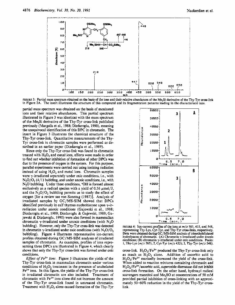
\ 448
partial mass spectrum was obtained on the basis of monitored ions and their relative abundances. This partial spectrum illustrated in Figure 3 was identical with the mass spectrum of the Me3Si derivative of the Thy-Tyr cross-link published previously (Margolis et al., 1988; Dizdaroglu, 1990), meaning the unequivocal identification of this DPC in chromatin. The insert in Figure 3 illustrates the chemical structure of the Thy-Tyr cross-link. Quantitative measurements of the Thy- Tyr cross-link in chromatin samples were performed as de- scribed in an earlier paper (Dizdaroglu et al., 1989).
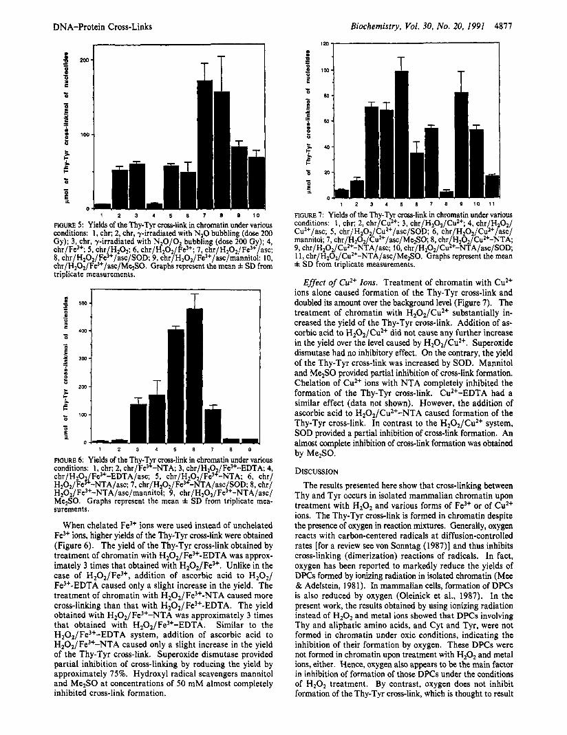
Since only the Thy-Tyr cross-link was found in chromatin treated with H202 and metal ions, efforts were made in order to find out whether inhibition of formation of other DPCs was due to the presence of oxygen in the system. For this purpose, parallel experiments were carried out using ionizing radiation instead of using H202 and metal ions. Chromatin samples were y-irradiated separately under oxic conditions, Le., with N20/02 (4/ 1) bubbling, and under anoxic conditions, i.e., with N 2 0 bubbling. Under these conditions, 'OH is formed almost exclusively as a radical species with a yield of 0.56 pmol/J, and the N 2 0 / 0 2 bubbling permits us to study the effect of oxygen [for a review see von Sonntag (1987)l. Analysis of irradiated samples by GC/MS-SIM showed that DPCs identified previously in calf thymus nucleohistone upon y-ir- radiation under anoxic conditions (Gajewski et al., 1988; Dizdaroglu et al., 1989; Dizdaroglu & Gajewski, 1989; Ga- jewski & Dizdaroglu, 1990) were also formed in mammalian chromatin y-irradiated under anoxic conditions (with N 2 0 bubbling). However, only the Thy-Tyr cross-link was detected in chromatin y-irradiated under oxic conditions (with N20/02 bubbling). Figure 4 illustrates representative ion-current profiles obtained during GC/MS-SIM analyses of irradiated samples of chromatin. As examples, profiles of ions repre- senting three DPCs are illustrated in Figure 4, which clearly shows that only the Thy-Tyr cross-link was formed under oxic conditions.
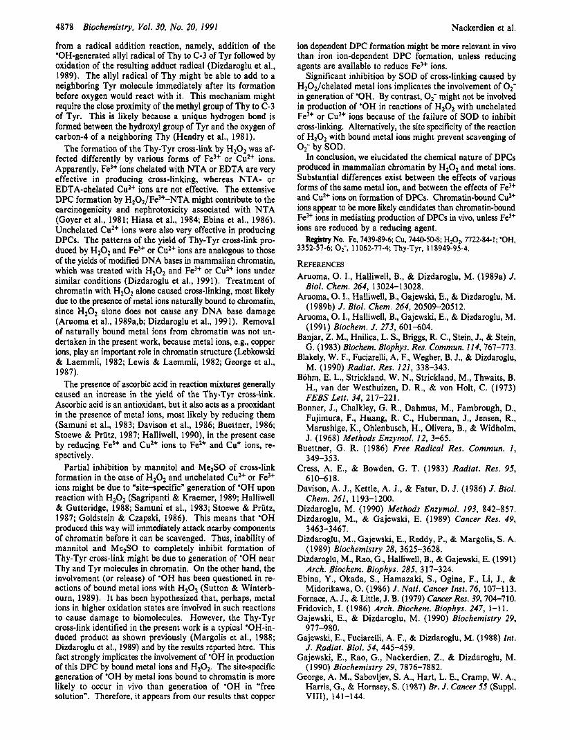
Effect of Fe3+ Zons. Figure 5 illustrates the yields of the Thy-Tyr cross-link in mammalian chromatin under various conditions of H202 treatment in the presence of unchelated Fe3+ ions. In this figure, the yields of the Thy-Tyr cross-link in irradiated chromatin are also included. Treatment of chromatin with Fe3+ ions alone did not increase the amount of the Thy-Tyr cross-link found in untreated chromatin. Treatment with H202 alone caused formation of the Thy-Tyr
-
P) u C
-0 C J P U
""""1 3 1 A
50001 I/ 4000i
2
0- 6.5 7.0
T i m e ( m f n . )
B
1000],-y, JL. , 0
6 . 5 7 . 0 T i m e ( m l n . )
FIGURE 4: Ion-current profiles of the ions at m / z 585,433, and 548, representing Thy-Lys, Cyt-Tyr, and Thy-Tyr cross-links, respectively. Data were obtained during GC/MS-SIM analysis of trimethylsilylated hydrolysates of chromatin. (A) Chromatin y-irradiated under anoxic conditions; (B) chromatin y-irradiated under oxic conditions. Peaks: 1, Thy-Lys (mlz 585); 2, Cyt-Tyr (mlz 433); 3 , Thy-Tyr ( m / z 548).
cross-link. H202/Fe3+ produced the Thy-Tyr cross-link only as much as H202 alone. Addition of ascorbic acid to H202/Fe3+ markedly increased the yield of the cross-link. When added to reaction mixtures containing chromatin and H202/Fe3+/ascorbic acid, superoxide dismutase did not inhibit cross-link formation. On the other hand, hydroxyl radical scavengers mannitol and Me2S0 at concentrations of 50 mM provided partial inhibition of cross-linking with an approxi- mately 50-60s reduction in the yield of the Thy-Tyr cross- link.
DNA-Protein Cross-Links Biochemistry, Vol. 30, No. 20, 1991 4871
T T
n . . . 1 2 3 4 5 6 7 8 0 10
FIGURE 5: Yields of the Thy-Tyr cross-link in chromatin under various conditions: 1, chr; 2, chr, y-irradiated with NzO bubbling (dose 200 Gy); 3, chr, y-irradiated with NzO/Oz bubbling (dose 200 Gy); 4, chr/Fe”; 5 , chr HzOz; 6, chr/HZO2 Fe”; 7, chr/H2O2/Fe3+/asc; 8, chr/HzOz/Fei+/asc/SOD; 9, chr/HZO2/Fe3+/asc/mannitol; 10, chr/H202/Fe3+/asc/MezS0. Graphs represent the mean f SD from triplicate measurements.
1
1 2 3 4 5 6 7 8 9
FIGURE 6: Yields of the Thy-T cross-link in chromatin under various conditions: 1, chr; 2, C ~ ~ / F ~ ) ~ N T A ; 3, chr/HzO /Fe3+-EDTA 4, chr/HzO Fe3+-EDTA/asc; 5 , chr/H O2/Fe3?-NTA; 6 , chr/ Hz02/Fe~‘-NTA/asc: 7, ~hr/H,0~/Fe~~-NTA/asc SOD; 8, chr/ HZO2/Fe3+-NTA/asc/mannito1; 9, chr/H2O2/F&+-NTA/asc/ MezSO. Graphs represent the mean f SD from triplicate mea- surements.
When chelated Fe3+ ions were used instead of unchelated Fe3+ ions, higher yields of the Thy-Tyr cross-link were obtained (Figure 6 ) . The yield of the Thy-Tyr cross-link obtained by treatment of chromatin with H202/Fe3+-EDTA was approx- imately 3 times that obtained with H202/Fe3+. Unlike in the case of H202/Fe3+, addition of ascorbic acid to H20z/ Fe3+-EDTA caused only a slight increase in the yield. The treatment of chromatin with HzOz/Fe3+-NTA caused more cross-linking than that with H202/Fe3+-EDTA. The yield obtained with H202/Fe3+-NTA was approximately 3 times that obtained with H202/Fe3+-EDTA. Similar to the H202/ Fe3+-EDTA system, addition of ascorbic acid to H202/Fe3+-NTA caused only a slight increase in the yield of the Thy-Tyr cross-link. Superoxide dismutase provided partial inhibition of cross-linking by reducing the yield by approximately 75%. Hydroxyl radical scavengers mannitol and Me2S0 at concentrations of 50 mM almost completely inhibited cross-link formation.
T
1 2 3 4 5 8 7 8 9 1 0 1 1
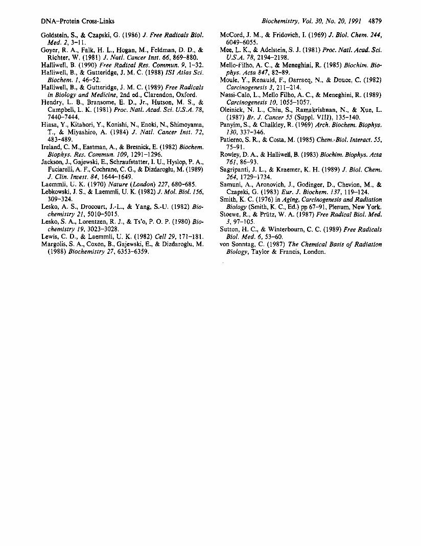
FIGURE 7: Yields of the Thy-Tyr cross-link in chromatin under various conditions: 1, chr; 2, chr/Cu2+; 3, chr/HZO2/CuZ+; 4, chr/HZOz/ Cu2+/asc; 5 , chr/H202/Cu2+/asc/SOD; 6 , chr/H202/Cuz+/asc/ mannitol; 7, chr/H~0z/CuZ+/asc/Me2S0 8, chr HZOz/Cu2+-NTA; 9, chr/HzO2/Cu2+-NTA/asc; 10, chr/H202/C&+-NTA/asc/SOD* 1 1, chr/Hz02/Cu2+-NTA/asc/Me2S0. Graphs represent the mea; f SD from triplicate measurements.
Effect of Cu2+ Zons. Treatment of chromatin with Cu2+ ions alone caused formation of the Thy-Tyr cross-link and doubled its amount over the background level (Figure 7). The treatment of chromatin with H20z/Cu2+ substantially in- creased the yield of the Thy-Tyr cross-link. Addition of as- corbic acid to H2O2/CuZ+ did not cause any further increase in the yield over the level caused by Hz02/Cu2+. Superoxide dismutase had no inhibitory effect. On the contrary, the yield of the Thy-Tyr cross-link was increased by SOD. Mannitol and Me2S0 provided partial inhibition of cross-link formation. Chelation of Cu2+ ions with NTA completely inhibited the formation of the Thy-Tyr cross-link. CuZ+-EDTA had a similar effect (data not shown). However, the addition of ascorbic acid to H202/Cu2+-NTA caused formation of the Thy-Tyr cross-link. In contrast to the H202/Cu2+ system, SOD provided a partial inhibition of cross-link formation. An almost complete inhibition of cross-link formation was obtained by Me2S0.
DISCUSSION The results presented here show that cross-linking between
Thy and Tyr occurs in isolated mammalian chromatin upon treatment with H202 and various forms of Fe3+ or of Cu2+ ions. The Thy-Tyr cross-link is formed in chromatin despite the presence of oxygen in reaction mixtures. Generally, oxygen reacts with carbon-centered radicals at diffusion-controlled rates [for a review see von Sonntag (1987)J and thus inhibits cross-linking (dimerization) reactions of radicals. In fact, oxygen has been reported to markedly reduce the yields of DPCs formed by ionizing radiation in isolated chromatin (Mee & Adelstein, 1981). In mammalian cells, formation of DPCs is also reduced by oxygen (Oleinick et al., 1987). In the present work, the results obtained by using ionizing radiation instead of H202 and metal ions showed that DPCs involving Thy and aliphatic amino acids, and Cyt and Tyr, were not formed in chromatin under oxic conditions, indicating the inhibition of their formation by oxygen. These DPCs were not formed in chromatin upon treatment with H2O2 and metal ions, either. Hence, oxygen also appears to be the main factor in inhibition of formation of those DPCs under the conditions of H202 treatment. By contrast, oxygen does not inhibit formation of the Thy-Tyr cross-link, which is thought to result
4878 Biochemistry, Vol. 30, No. 20, 1991
from a radical addition reaction, namely, addition of the 'OH-generated allyl radical of Thy to C-3 of Tyr followed by oxidation of the resulting adduct radical (Dizdaroglu et al., 1989). The allyl radical of Thy might be able to add to a neighboring Tyr molecule immediately after its formation before oxygen would react with it. This mechanism might require the close proximity of the methyl group of Thy to C-3 of Tyr. This is likely because a unique hydrogen bond is formed between the hydroxyl group of Tyr and the oxygen of carbon-4 of a neighboring Thy (Hendry et al., 1981).
The formation of the Thy-Tyr cross-link by H202 was af- fected differently by various forms of Fe3+ or Cu2+ ions. Apparently, Fe3+ ions chelated with NTA or EDTA are very effective in producing cross-linking, whereas NTA- or EDTA-chelated Cu2+ ions are not effective. The extensive DPC formation by H202/Fe3+-NTA might contribute to the carcinogenicity and nephrotoxicity associated with NTA (Goyer et al., 1981; Hiasa et al., 1984; Ebina et al., 1986). Unchelated Cu2+ ions were also very effective in producing DPCs. The patterns of the yield of Thy-Tyr cross-link pro- duced by H202 and Fe3+ or Cu2+ ions are analogous to those of the yields of modified DNA bases in mammalian chromatin, which was treated with H 2 0 2 and Fe3+ or Cu2+ ions under similar conditions (Dizdaroglu et al., 1991). Treatment of chromatin with H202 alone caused cross-linking, most likely due to the presence of metal ions naturally bound to chromatin, since H202 alone does not cause any DNA base damage (Aruoma et al., 1989a,b; Dizdaroglu et al., 1991). Removal of naturally bound metal ions from chromatin was not un- dertaken in the present work, because metal ions, e.g., copper ions, play an important role in chromatin structure (Lebkowski & Laemmli, 1982; Lewis & Laemmli, 1982; George et al., 1987).
The presence of ascorbic acid in reaction mixtures generally caused an increase in the yield of the Thy-Tyr cross-link. Ascorbic acid is an antioxidant, but it also acts as a prooxidant in the presence of metal ions, most likely by reducing them (Samuni et al., 1983; Davison et al., 1986; Buettner, 1986; Stoewe & Priitz, 1987; Halliwell, 1990), in the present case by reducing Fe3+ and Cu2+ ions to Fe2+ and Cu+ ions, re- spectively.
Partial inhibition by mannitol and Me2S0 of cross-link formation in the case of H202 and unchelated Cu2+ or Fe3+ ions might be due to "sitespecific" generation of 'OH upon reaction with H202 (Sagripanti & Kraemer, 1989; Halliwell & Gutteridge, 1988; Samuni et al., 1983; Stoewe & Priitz, 1987; Goldstein & Czapski, 1986). This means that 'OH produced this way will immediately attack nearby components of chromatin before it can be scavenged. Thus, inability of mannitol and Me2S0 to completely inhibit formation of Thy-Tyr cross-link might be due to generation of 'OH near Thy and Tyr molecules in chromatin. On the other hand, the involvement (or release) of *OH has been questioned in re- actions of bound metal ions with H202 (Sutton & Winterb- ourn, 1989). It has been hypothesized that, perhaps, metal ions in higher oxidation states are involved in such reactions to cause damage to biomolecules. However, the Thy-Tyr cross-link identified in the present work is a typical *OH-in- duced product as shown previously (Margolis et al., 1988; Dizdaroglu et al., 1989) and by the results reported here. This fact strongly implicates the involvement of 'OH in production of this DPC by bound metal ions and H202. The site-specific generation of 'OH by metal ions bound to chromatin is more likely to occur in vivo than generation of *OH in "free solution". Therefore, it appears from our results that copper
Nackerdien et al.
ion dependent DPC formation might be more relevant in vivo than iron ion-dependent DPC formation, unless reducing agents are available to reduce Fe3+ ions.
Significant inhibition by SOD of cross-linking caused by H202/chelated metal ions implicates the involvement of 02- in generation of 'OH. By contrast, 0,- might not be involved in production of 'OH in reactions of H202 with unchelated Fe3+ or Cu2+ ions because of the failure of SOD to inhibit cross-linking. Alternatively, the site specificity of the reaction of H202 with bound metal ions might prevent scavenging of
In conclusion, we elucidated the chemical nature of DPCs produced in mammalian chromatin by H202 and metal ions. Substantial differences exist between the effects of various forms of the same metal ion, and between the effects of Fe3+ and Cu2+ ions on formation of DPCs. Chromatin-bound Cu2+ ions appear to be more likely candidates than chromatin-bound Fe3+ ions in mediating production of DPCs in vivo, unless Fe3+ ions are reduced by a reducing agent. Registry No. Fe, 7439-89-6; Cu, 7440-50-8; H202, 7722-84-1; 'OH,
3352-57-6; OF, 11062-77-4; Thy-Tyr, 118949-95-4.
0 2 - by SOD.
REFERENCES Aruoma, 0. I., Halliwell, B., & Dizdaroglu, M. (1989a) J.
Biol. Chem. 264, 13024-1 3028. Aruoma, 0. I., Halliwell, B., Gajewski, E., & Dizdaroglu, M.
(1989b) J. Biol. Chem. 264, 20509-20512. Aruoma, 0. I., Halliwell, B., Gajewski, E., & Dizdaroglu, M.
(1991) Biochem. J . 273, 601-604. Banjar, Z. M., Hnilica, L. S., Bnggs, R. C., Stein, J., & Stein,
G. (1983) Biochem. Biophys. Res. Commun. 114,767-773. Blakely, W. F., Fuciarelli, A. F., Wegher, B. J., & Dizdaroglu,
M. (1990) Radiat. Res. 121, 338-343. Bohm, E. L., Strickland, W. N., Strickland, M., Thwaits, B.
H., van der Westhuizen, D. R., & von Holt, C. (1973) FEBS Lett. 34, 217-221.
Bonner, J., Chalkley, G. R., Dahmus, M., Fambrough, D., Fujimura, F., Huang, R. C., Huberman, J., Jensen, R., Marushige, K., Ohlenbusch, H., Olivera, B., & Widholm, J. (1968) Methods Enzymol. 12, 3-65.
Buettner, G. R. (1986) Free Radical Res. Commun. 1, 349-3 5 3.
Cress, A. E., & Bowden, G. T. (1983) Radiat. Res. 95, 6 10-6 1 8.
Davison, A. J., Kettle, A. J., & Fatur, D. J. (1986) J. Biol. Chem. 261, 1193-1200.
Dizdaroglu, M. (1990) Methods Enzymol. 193, 842-857. Dizdaroglu, M., & Gajewski, E. (1989) Cancer Res. 49,
Dizdaroglu, M., Gajewski, E., Reddy, P., & Margolis, S . A. (1989) Biochemistry 28, 3625-3628.
Dizdaroglu, M., Rao, G., Halliwell, B., & Gajewski, E. (1991) Arch. Biochem. Biophys. 285, 317-324.
Ebina, Y., Okada, S., Hamazaki, S., Ogina, F., Li, J., & Midorikawa, 0. (1986) J. Natl. Cancer Znst. 76, 107-113.
Fornace, A. J., & Little, J. B. (1979) Cancer Res. 39, 704-710. Fridovich, I. (1986) Arch. Biochem. Biophys. 247, 1-1 1. Gajewski, E., & Dizdaroglu, M. (1990) Biochemistry 29,
Gajewski, E., Fuciarelli, A. F., & Dizdaroglu, M. (1988) In?. J. Radiat. Biol. 54, 445-459.
Gajewski, E., Rao, G., Nackerdien, Z., & Dizdaroglu, M. (1 990) Biochemistry 29, 7876-7882.
George, A. M., Sabovljev, S. A., Hart, L. E., Cramp, W. A,, Harris, G., & Hornsey, S. (1987) Br. J. Cancer 55 (Suppl.
3463-3467.
977-980.
VIII), 141-144.
DNA-Protein Cross-Links
Goldstein, S., & Czapski, G. (1986) J. Free Radicals Biol. Med. 2, 3-1 1.
Goyer, R. A,, Falk, H. L., Hogan, M., Feldman, D. D., & Richter, W, (1981) J. Natl. Cancer Inst. 66, 869-880.
Halliwell, B. (1990) Free Radical Res. Commun. 9, 1-32. Halliwell, B., & Gutteridge, J. M. C. (1988) ISZ Atlas Sci.
Biochem. 1 , 4 6 4 2 . Halliwell, B., & Gutteridge, J. M. C. (1989) Free Radicals
in Biology and Medicine, 2nd ed., Clarendon, Oxford. Hendry, L. B., Bransome, E. D., Jr., Hutson, M. S . , &
Campbell, L. K. (1981) Proc. Natl. Acad. Sci. U.S.A. 78,
Hiasa, Y., Kitahori, Y., Konishi, N., Enoki, N., Shimoyama, T., & Miyashiro, A. (1984) J. Natl. Cancer Inst. 72, 483-489.
Ireland, C. M., Eastman, A., & Bresnick, E. (1982) Biochem. Biophys. Res. Commun. 109, 1291-1296.
Jackson, J., Gajewski, E., Schraufstatter, I. U., Hyslop, P. A., Fuciarelli, A. F., Cochrane, C. G., & Dizdaroglu, M. (1989) J. Clin. Invest. 84, 1644-1649.
7440-7444.
Laemmli, U. K. (1970) Nature (London) 227, 680-685. Lebkowski, J. S., & Laemmli, U. K. (1982) J. Mol. Biol. 156,
Lesko, A. S. , Drocourt, J.-L., & Yang, S.-U. (1982) Bio-
Lesko, S. A., Lorentzen, R. J., & Ts’o, P. 0. P. (1980) Bio-
Lewis, C. D., & Laemmli, U. K. (1982) Cell 29, 171-181. Margolis, S. A,, Coxon, B., Gajewski, E., & Dizdaroglu, M.
309-3 24.
chemistry 21, 5010-5015.
chemistry 19, 3023-3028.
(1988) Biochemistry 27, 6353-6359.
Biochemistry, Vol. 30, No. 20, 1991 4879
McCord, J. M., & Fridovich, I. (1969) J. Biol. Chem. 244,
Mee, L. K., & Adelstein, S. J. (1981) Proc. Nutl. Acad. Sci.
Mello-Filho, A. C., & Meneghini, R. (1985) Biochim. Bia-
Mode, Y. , Renauld, F., Darracq, N., & Douce, C. (1982)
Nassi-Calo, L., Mello Filho, A. C., & Meneghini, R. (1989)
Oleinick, N. L., Chiu, S., Ramakrishnan, N., & Xue, L.
Panyim, S., & Chalkley, R. (1969) Arch. Biochem. Biophys.
Patierno, S . R., & Costa, M. (1985) Chem.-Biol. Interact. 55,
Rowley, D. A., & Halliwell, B. (1983) Biochim. Biophys. Acta
Sagripanti, J. L., & Kraemer, K. H. (1989) J. Biol. Chem.
Samuni, A., Aronovich, J., Godinger, D., Chevion, M., & Czapski, G. (1983) Eur. J. Biochem. 137, 119-124.
Smith, K. C. (1976) in Aging, Carcinogenesis and Radiation Biology (Smith, K. C., Ed.) pp 67-91, Plenum, New York.
Stoewe, R., & Priitz, W. A. (1987) Free Radical Biol. Med.
Sutton, H. C., & Winterbourn, C. C. (1989) Free Radicals
von Sonntag, C. (1987) The Chemical Basis of Radiation
6049-6055.
U.S.A. 78, 2194-2198.
phys. Acta 847, 82-89.
Carcinogenesis 3, 2 1 1-2 14.
Carcinogenesis 10, 1055-1057.
(1987) Br. J . Cancer 55 (Suppl. VIII), 135-140.
130, 337-346.
75-91.
761, 86-93.
264, 1729-1734.
3, 97-105.
Biol. Med. 6, 53-60.
Biology, Taylor & Francis, London.


![Long Noncoding RNAs, Chromatin, and Developmentdownloads.hindawi.com/journals/tswj/2010/180798.pdf · active chromatin modifications and a more open chromatin conformation[26,39,40,41,42]](https://img.pdfslide.us/doc/110x75/5f8885d811957319d07a36bf/long-noncoding-rnas-chromatin-and-active-chromatin-modifications-and-a-more-open.jpg)