Embed Size (px)
Citation preview
CHEMICAL LABELING STRATEGIES FOR MASS SPECTROMETRY-BASED BIOMOLECULAR IDENTIFICATION, CHARACTERIZATION AND QUANTIFICATION
By
Shuai Nie
A DISSERTATION
Submitted to Michigan State University
in partial fulfillment of the requirements for the degree of
Chemistry – Doctor of Philosophy
2015
ABSTRACT
CHEMICAL LABELING STRATEGIES FOR MASS SPECTROMETRY-BASED BIOMOLECULAR IDENTIFICATION, CHARACTERIZATION AND QUANTIFICATION
By
Shuai Nie
Advances in the development of mass spectrometry (MS) and tandem mass
spectrometry (MS/MS) instrumentation have made this technique a versatile analytical
tool to identify, characterize and quantify biomolecules including peptides, proteins,
lipids, nucleic acids, oligosaccharides and other metabolites. However, based on the
individual physicochemical properties of various biomolecules, biomolecular MS or
MS/MS on its own may not necessarily give the desired analytical information.
Therefore, chemical labeling strategies which alter the behavior of analytes with respect
to their ionization, fragmentation and mass analysis are commonly used to facilitate MS-
based analysis of biomolecules. This dissertation focuses on the development of
biomolecular chemical labeling strategies for lipids, peptides and proteins, to provide
improved capabilities for MS-based qualitative and quantitative analysis.
Structural labeling via gas phase ion chemistry provides a convenient and rapid
modification method for structural and reactivity characterization of modified
biomolecular ions. Here, a novel photo-induced inter-molecular gas-phase cross-linking
reaction has been developed to investigate the cross-linking reactivity of individual
triacylglyceride (TG) molecules as a function of their structures. Ultraviolet
photodissociation tandem mass spectrometry (UVPD-MS/MS) of non-covalent complex
ions consisting of TG dimers and protonated diiodoaniline resulted in the formation of
multiple cross-linked TG products via homolysis of carbon-iodine bonds, hydrogen
abstraction and radical recombination. The efficiency of the UVPD reaction depended
on the number of unsaturation sites present within the TG lipids.
For MS-based quantification, an approach for the multiplexed relative
quantification of aminophospholipids from within two different crude lipid extracts was
developed. Relative quantification at the ‘sum composition’ and/or ‘molecular lipid’
levels was achieved using high resolution/accurate mass MS/MS by ratiometric
measurement of pairs of ‘reporter’ ions formed via the neutral loss from isobaric stable
isotope-labeled d6-‘heavy’ and d6-‘light’ S,S′-dimethylthiobutanoylhydroxysuccinimide
and iodine/methanol derivatized aminophospholipid ions.
In addition, absolute quantification of full length parathyroid hormone (PTH 1-84),
a clinical protein biomarker of secondary hyperparathyroidism, and its in vivo oxidized
and truncated variants was achieved using a dual stable isotope-labeled internal
standard approach coupled with immunocapture and high resolution LC-MS and MS/MS.
Analysis of clinical PTH samples using this strategy revealed that no oxidation or PTH
7-84 occurred in vivo. However, several novel sites of in vivo PTH truncation were
discovered. At last, stable isotope-containing dimethyl labeling and multi-dimensional
LC-MS/MS were applied for proteomic profiling of human RPMI-8226 cells treated with
competitive (i.e., Bortezomib) and non-competitive (i.e., TCH-013) proteasome inhibitors
to evaluate their distinct mechanisms of action. Four proteins closely related to the
regulation of mitochondrial functions and growth and division of cancer cells were
observed to be selectively down-regulated after TCH-013 treatment compared to
Bortezomib or vehicle control treatment.
iv
ACKNOWLEDGEMENTS
The experience of study and research in the methodology and application
development of bioanalytical mass spectrometry and related chemical derivatization
strategies in the past four and half years is the most exciting and challenging adventure
I’ve ever had in my life so far. I would like to thank all the people mentioned below for
their generous help and support during the life-changing experience.
First, I want to give special thanks to my advisor, Dr. Gavin Reid, who introduced
me to the amazing world of bioanalytical mass spectrometry and provided me the
guidance toward becoming an independent analytical chemist. Your enthusiasms for
scientific discoveries, broad knowledge and meticulous thinking have been always
inspiring me. Also, thanks for letting me have the opportunity to conduct my research
and collaborate with other talented scientists at both Michigan State University and the
University of Melbourne. For my previous and current committee members, Dr. Merlin
Bruening, Dr. Marcos Dantus, Dr. Jetze Tepe and Dr. Xuefei Huang, I sincerely
appreciate your help to my research projects, especially for providing professional
advices in fields that I was not familiar with before.
I greatly appreciate my collaborators, Evert Njomen and Dr. Jetze Tepe at
Michigan State University, for providing the cell lysates of drug-treated RPMI-8226 cells
for the TCH-013 drug action mechanism study. Also, thank DiaSorin for providing the
clinical patient samples for the quantitative analysis of PTH proteins.
I would like to greatly thank the current and former Reid lab members, Dr. Eileen
Ryan, Dr. Todd Lydic and Dr. Li Cui for supporting me and proofreading my dissertation.
I also thank all the other Reid lab members who have helped me with my experiments,

v
scientific writing and exams. Thanks for Dr. Cassie Fhaner who taught me the sample
handling and derivatization techniques for lipids and lipidomic analysis. I deeply
appreciate all the help I received from the Dr. Nicholas Williamson, Dr. Ching-Seng Ang
and Dr. David Perkins during my one-year visit in the Bio21 Molecular Science &
Biotechnology Institute at the University of Melbourne. Thanks for helping me with the
instrument setup and data analysis using bioinformatic tools.
Finally, I want to thank my parents. I can always feel their unconditional love and
support even though most of the time I was thousands of miles away from them. Thanks
also to my girlfriend for supporting and understanding me when I was busy doing
experiments or writing. Finally, thanks to all my other friends who will always give me
warm encouragement or just simply listen to me when I was having a bad day.
vi
TABLE OF CONTENTS
LIST OF TABLES ............................................................................................................ x
LIST OF FIGURES ..........................................................................................................xi
KEY TO ABBREVIATIONS ............................................................................................xv
CHAPTER ONE An Overview of Chemical Labeling Strategies for Mass Spectrometry Analysis of Biomolecules ................................................................................................................... 1 1.1 Introduction to Mass Spectrometry Analysis of Natural Biomolecules ...................... 1
1.1.1 Ionization Techniques ................................................................................................ 1
1.1.2 Mass Analyzers .......................................................................................................... 2
1.1.3 Fragmentation Techniques ........................................................................................ 5
1.1.3.1 Collision-Induced Dissociation .............................................................................. 5
1.1.3.2 Electron Capture Dissociation and Electron Transfer Dissociation ................... 6
1.1.3.3 Photodissociation ................................................................................................... 7
1.1.4 Applications for Identification and Quantification of Natural Biomolecules ........... 7
1.1.4.1 Peptides and Proteins............................................................................................ 8
1.1.4.2 Lipids ......................................................................................................................10
1.1.4.3 Nucleic Acids .........................................................................................................14
1.1.4.4 Oligosaccharides and Other Small Molecule Metabolites .................................14
1.2 Chemical Labeling Strategies for Improved Identification, Characterization and Quantification of Biomolecules Using Mass Spectrometry ........................................15
1.2.1 Applications of Structural Labeling for Improved Identification and Structural Characterization ........................................................................................................16
1.2.1.1 Ionization Efficiency ..............................................................................................16
1.2.1.2 Structural Characterization of Peptides and Proteins ........................................19
1.2.1.2.1 Sequence of Amino Acid Residues .................................................................19
1.2.1.2.2 Post-Translational Modifications .....................................................................21
1.2.1.2.3 Tertiary and Quaternary Structures of Proteins .............................................22
1.2.1.3 Structural Characterization of Lipids ...................................................................24
1.2.1.3.1 Lipid Classes and Sum Compositions ............................................................24
1.2.1.3.2 Identities and Linkage Positions of Glycerol Backbone Substituents ..........25
1.2.1.3.3 Positions and Stereochemistry of Carbon-Carbon Double Bonds ...............27
1.2.1.4 Intra- and Inter-Molecular Interactions of Nucleic Acids ....................................28
1.2.1.5 Structural Characterization of Oligosaccharides ................................................28
1.2.2 Applications of Stable Isotope Labeling for Improved Structural Characterization and Quantification .....................................................................................................29
1.2.2.1 Structures of Large Biomolecules and Mechanisms of Gas Phase Biomolecular Ion Fragmentation ..........................................................................29
1.2.2.2 Quantification of Biomolecules ............................................................................31
1.2.2.2.1 Stable Isotope-Labeled Analogue Internal Standards ..................................32
1.2.2.2.2 Metabolic Labeling ............................................................................................33
vii
1.2.2.2.3 Chemical Labeling ............................................................................................35
1.3 Aims of this Dissertation................................................................................................37 CHAPTER TWO Photo-Induced Inter-Molecular Cross-Linking of Gas Phase Triacylglycerol Lipids Ions ...................................................................................................................................... 39 2.1 Introduction .....................................................................................................................39
2.2 Results and Discussion .................................................................................................42
2.2.1 Photo-Induced Dissociation of Protonated Diiodoaniline-TG Dimer Complex Ions ....................................................................................................................................42
2.2.2 Proposed Mechanisms and Structures of Covalently Cross-linked TG Dimer Product Ions ...............................................................................................................45
2.2.3 Proposed Fragmentation Mechanisms of TG Dimer Product Ion Structures 1a, 1b and 1c ...................................................................................................................48
2.2.4 Effects of Structures of the Cross-Linker and TG Lipids on the Formation of Cross-Linked TG Dimer Product Ions .....................................................................59
2.2.5 Solution Phase Cross-Linking of TG lipids .............................................................60
2.3 Conclusions ....................................................................................................................62
CHAPTER THREE Characterization and Multiplexed Quantification of Derivatized Aminophospholipids ... 63 3.1 Introduction .....................................................................................................................63
3.2 Results and Discussion .................................................................................................66
3.2.1 Characterization of the Gas Phase Fragmentation Reactions of DMBNHS Derivatized Aminophospholipids ..............................................................................66
3.2.1.1 Diacyl-Aminophospholipids ..................................................................................66
3.2.1.2 Alkyl Ether-Aminophospholipids ..........................................................................74
3.2.1.3 Plasmalogen-Aminophospholipids ......................................................................76
3.2.2 Characterization and Quantification of Isobaric Stable Isotope Containing DMBNHS Derivatized Aminophospholipid Standards ...........................................77
3.2.3 Multiplexed Quantification of Selected Aminophospholipid Ion Abundance Changes Between a Metastatic Colorectal Cancer Cell Line, SW620 and its AlkyGlycerone Phosphate Synthase (AGPS) siRNA Knockdown ........................81
3.3 Conclusions ....................................................................................................................91
CHAPTER FOUR Experimental Methods for Chapters Two and Three ..................................................... 92 4.1 Materials .........................................................................................................................92
4.2 siRNA Knockdown of Alkyglycerone Phosphate Synthase (AGPS) in SW620 Colorectal Cancer Cells and Lipid Extraction ..............................................................92
4.3 Derivatization of Lipid Standards and Crude Lipid Extracts ......................................93
4.3.1 Synthesis of Saturated TG Lipids from Unsaturated TG Lipid Standards ...........93
4.3.2 Photo-Induced Solution Phase Cross-linking of TG Lipid Standards ...................93
4.3.3 Derivatization of Aminophospholipids Using d6-Heavy/light DMBNHS and Iodine/Methanol Methods .........................................................................................94
4.4 Mass Spectrometry ........................................................................................................95
viii
4.4.1 Photo-Induced Gas Phase Cross-linking MS/MS and CID-MSn Structural Analysis of TG Dimer Complex Ions ........................................................................95
4.4.2 ESI-MS, -MS/MS and -MS3 Analysis of Derivatized Aminophospholipid Standards ...................................................................................................................96
4.4.3 ESI-MS and -MS/MS Analysis of Derivatized Crude Lipid Extracts from Combined Control and AGPS siRNA Knockdown of the SW620 Colorectal Cancer Cells ..............................................................................................................97
CHAPTER FIVE Quantitative Immuno LC-MS/MS of Parathyroid Hormone and Its In Vivo Heterogeneous Post-Translational Protein Modifications: Oxidation and Truncation .... 99 5.1 Introduction .....................................................................................................................99
5.2 Results and Discussion ............................................................................................... 105
5.2.1 Quantitative Immuno LC-MS/MS Workflow with Optimized Sample Preparation, High Resolution/Accurate Mass HCD-MS/MS and Near Full Sequence Coverage .................................................................................................................. 105
5.2.2 Quantitative Analysis of In Vivo Full Length, Oxidized and Truncated PTH Tryptic Peptides in Patient Samples ...................................................................... 112
5.2.3 Identification of Novel Truncation Sites of PTH Proteins ..................................... 120
5.3 Conclusions .................................................................................................................. 122
CHAPTER SIX Quantitative Proteomic Analysis Using Dimethyl Labeling and Two Dimensional LC-MS/MS to Study the Mechanism of Action of the Non-Competitive Proteasome Inhibitor, TCH-013 ...................................................................................................................... 123 6.1 Introduction ................................................................................................................... 123
6.2 Results and Discussion ............................................................................................... 126
6.2.1 Quantitative Proteomic Analysis Workflow ........................................................... 126
6.2.2 Coverage of Protein Identification and Selection Criteria During Protein Quantification ........................................................................................................... 129
6.2.3 Protein Regulation in Human RPMI-8226 Cells Induced by TCH-013 ............... 132
6.3 Conclusions .................................................................................................................. 136
CHAPTER SEVEN Experimental Methods for Chapters Five and Six ....................................................... 137 7.1 Materials ....................................................................................................................... 137
7.2 Biological Samples ...................................................................................................... 138
7.2.1 Patient Information and Immuno-Affinity Enrichment of PTH Proteins .............. 138
7.2.2 Cell Culture and Treatment Using TCH-013, Bortezomib and DMSO ............... 138
7.3 Protein Digestion ......................................................................................................... 139
7.3.1 Trypsin Digestion of PTH Standards and Immuno-Enriched Samples from Patient Plasmas ....................................................................................................... 139
7.3.2 Trypsin Digestion of Proteins in Lysates of TCH-013, Bortezomib and DMSO Treated RPMI-8226 Cells ....................................................................................... 140
7.4 Dimethyl Labeling of Digested Proteins from TCH-013, Bortezomib and DMSO Treated RPMI-8226 Cells ........................................................................................... 140
ix
7.5 Basic pH Reversed-Phase Fractionation of Digested and Dimethyl Labeled Proteins ......................................................................................................................... 141
7.6 Mass Spectrometry ...................................................................................................... 141
7.6.1 LC-MS/MS Analysis of Digested PTH standards and Immuno-Affinity Enriched Samples from Patient Plasmas .............................................................................. 141
7.6.2 LC-MS/MS Analysis of Digested, Dimethyl Labeled and Fractionated Proteins from TCH-013, Bortezomib and DMSO Treated RPMI-8226 Cells .................... 143
7.7 Data Analysis ............................................................................................................... 144
7.7.1 Quantification of Full Length, Oxidized and Truncated PTH Variants in Patient Plasma ............................................................................................................ 144
7.3.3 Bioinformatic Analysis for Proteomic Profiling of Proteins in TCH-013, Bortezomib and DMSO Treated RPMI-8226 Cells .......................................... 145
BIBLIOGRAPHY ......................................................................................................... 147
x
LIST OF TABLES
Table 5.1 HCD-MS/MS precursor/fragment ion transitions, optimized HCD normalized collision energy (NCE) and retention times for quantitatively monitored PTH peptides. .................................................................................................... 109
Table 5.2 Concentration of full length, oxdizied and truncated PTH tryptic peptides in patient plasma determined by quantitative immuno LC-MS/MS with near full sequence coverage. Concentrations of oxidized PTH peptides were corrected to remove ex vivo oxidation. ND indicates that the peptide was not detectable. ................................................................................................. 118
Table 6.1 Protein down-regulation (near 2 folds) expressed in log2 heavy/light protein ratios in three biological replicates of TCH-013 (heavy) treated RPMI-8226 cells compared to that treated with Bortezomib (light) and DMSO control (light). ......................................................................................................... 135
xi
LIST OF FIGURES
Figure 1.1 Structural complexity of glycerophospholipids lipids. PC, PE, PS, PA, PG, CL and PI indicate glycerophosphocholine, glycerophosphoethanolamine, glycerophosphoserine, glycerophosphate, glycerophosphoglycerol, glycerophosphoglycerophosphoglycerol and glycerophosphoinositol, respectively. ............................................................................................... 13
Figure 2.1 Ion trap 266 nm UVPD-MS/MS of mono-unsaturated and saturated TG homodimer complex ions with protonated diIA. (A) 2TG(14:0/16:1/14:0) + 3,4-diIA (complex 1), (B) 2TG(14:0/16:0/14:0) + 3,4-diIA (complex 2) and (C) 2TG(14:0/16:1/14:0) + 2,4-diIA (complex 3). Note, that high mass ion complexes are fragile ions and as such exhibit an apparent mass-shift to lower m/z (see explanation provided in the text). ................................................................ 44
Figure 2.2 Proposed mechanisms for the photo-induced cross-linking reactions of TG dimer complex ions [2TG(14:0/16:1/14:0) + 3,4-diIA + H]+ (complex 1). ............. 47
Figure 2.3 Ion trap CID-MS3 analysis of cross-linked mono-unsaturated and saturated TG homodimer complex ions with protonated 3,4-diIA or 2,4-diIA, formed by 266 nm UVPD-MS/MS. (A) [complex 1 - 2I]+ from Figure 2.1A, (B) [complex 2 - 2I]+ from Figure 2.1B, and (C) [complex 3 - 2I]+ from Figure 2.1C. The major product ions formed from proposed structures 1a, 1b and 1c in Figure 2.2 are labeled in Figure 2.3A. ................................................................... 49
Figure 2.4 Ion trap CID-MS4 analysis of cross-linked mono-unsaturated TG homodimer complex ions from Figure 2.3A. (A) [complex 1 - 2I - aniline]+, (B) [complex 1 - 2I - TG(14:0/16:1/14:0)]+, and (C) [complex 1 - 2I - 14:0 FA]+. ......................... 52
Figure 2.5 Proposed mechanisms for the CID-MS3 and -MS4 fragmentation reactions of [complex 1 - 2I]+ ions corresponding to structure 1a in Figure 2.2. ......... 53
Figure 2.6 Proposed mechanisms for the CID-MS3 and -MS4 fragmentation reactions of [complex 1 - 2I]+ ions corresponding to structure 1b in Figure 2.2. ......... 55
Figure 2.7 Ion trap CID-MS3 analysis of the cross-linked saturated/mono-unsaturated TG heterodimer complex ion [complex 4 - 2I]+, formed by 266 nm UVPD-MS/MS. ....................................................................................................... 56
Figure 2.8 Proposed mechanisms for the CID-MS3 and -MS4 fragmentation reactions of [complex 1 - 2I]+ ions corresponding to structure 1c in Figure 2.2. MG denotes monoacylglyceride. ....................................................................... 58
Figure 2.9 Ion trap MS and MS/MS analysis of products in solution phase inter-molecular cross-linking reactions of TG lipids. (A) MS analysis of products in
xii
the solution containing TG(14:0/16:1/14:0) and 3,5-diiodoaniline after 9 min of irradiation with 266 nm UV photons; (B) CID-MS/MS analysis of the product ions at m/z 1496 from Figure 2.9A. ............................................................. 61
Figure 3.1 HCD-MS/MS analysis of DMBNHS derivatized (A) PE(18:0/18:1) and (B) PS(16:0/18:1). .................................................................................................. 69
Figure 3.2 Proposed CID-MS/MS fragmentation mechanisms for DMBNHS derivatized PE(18:0/18:1) and PS(16:0/18:1). .......................................................................... 70
Figure 3.3 HCD-MS3 analysis of the S(CH3)2 neutral loss (NL) product ions formed by linear ion trap CID-MS/MS of the DMBNHS derivatized (A) PE(18:0/18:1) and (B) PS(16:0/18:1). ............................................................................................ 72
Figure 3.4 Proposed HCD-MS3 fragmentation mechanisms for DMBNHS derivatized PE(18:0/18:1) after the neutral loss of 271 Da. ................................................ 73
Figure 3.5 CID-MS/MS analysis of a DMBNHS derivatized monoalkyl-ether PE lipid PE(O-16:0_18:1) from a crude lipid extract of SW620 colorectal cancer cells. The inset shows the HCD-MS3 spectrum of the S(CH3)2 neutral loss (NL) product ion. ................................................................................................. 75
Figure 3.6 CID-MS/MS, HCD-MS3 and HCD-MS/MS of a 1:1 mixture of d6-heavy/light DMBNHS and iodine/methanol derivatized PE lipids. (A) CID-MS/MS of pooled d6-heavy/light DMBNHS derivatized PE(14:0/14:0). The inset shows the HCD-MS3 spectrum of the S(CD3)2 neutral loss product ion from the d6-heavy DMBNHS derivatized lipid as well as the structures and initial fragmentation sites for the d6-heavy/light DMBNHS derivatized lipid species (B) HCD-MS/MS of pooled d6-heavy/light DMBNHS and iodine/methanol derivatized PE(P-18:0/22:6). ............................................................................. 80
Figure 3.7 Ultra-high resolution/accurate mass ESI-MS of a 1:1 mixture (normalized to protein concentration) of isobaric d6-heavy/light DMBNHS and iodine/methanol derivatized crude lipid extracts from a metastatic colorectal cancer cell line, SW620 and its alkyglycerone phosphate synthase (AGPS) siRNA knockdown. The insets show expanded m/z regions containing the lipids PE(35:1) and PE(O-36:1), PS(O-28:2), and PE(P-35:1). The ion labeled with a # corresponds to a diisooctyl phthalate contaminant ion. .............................. 83
Figure 3.8 HCD-MS/MS of the isobaric d6-heavy/light DMBNHS derivatized PE(35:1) and PE(O-36:1) lipids from Figure 3.5. The inset shows expanded m/z regions of the spectrum containing the characteristic S(CH3)2/S(CD3)2 neutral loss product ions. ............................................................................................... 85
Figure 3.9 HCD-MS/MS of the isobaric d6-heavy/light DMBNHS derivatized PS(O-28:2)
lipid from Figure 3.7. The inset shows an expanded m/z region of the
xiii
spectrum containing the characteristic S(CH3)2/S(CD3)2 neutral loss product ions. ............................................................................................................ 87
Figure 3.10 HCD-MS/MS of the isobaric d6-heavy/light DMBNHS and iodine/methanol derivatized PE(P-35:1) lipid from Figure 3.7. The inset shows an expanded m/z region of the spectrum containing the characteristic O-alkenyl-chain neutral loss product ions. ........................................................................................ 90
Figure 5.1 Summary of (A) previously identified full length PTH 1-84, N-terminally truncated PTH X-84 (X=7, 28, 34, 37, 38, 45 and 48) and mid-molecular fragment PTH X-77 (X= 34, 37 and 38) [248-250]; (B) full length, oxidized and truncated PTH protein-unique tryptic peptides monitored quantitatively during LC-MS/MS in this study; (C) truncated PTH protein-unique tryptic peptides monitored qualitatively during LC-MS/MS in this study. The dashed lines represent the variable lengths of truncated forms of PTH. ............... 103
Figure 5.2 Oxidation percentages of PTH in different sets of patient samples [264]. The percentage of oxidation was calculated by taking the sum concentration of PTH 1-13 M(O), PTH 7-13 M(O) and PTH 14-20 M(O) divided by the sum concentration of all full length, truncated and oxidized tryptic peptides quantified. There were 41 blood samples in set A collected from site 1 and stored at least a week before sample analysis, and 29 samples in set B and 4 samples in set C collected from site 2 and stored at most 2 days before sample analysis. ....................................................................................... 104
Figure 5.3 Quantitative immuno LC-MS/MS workflow with optimized sample preparation, high resolution/accurate mass HCD-MS/MS and near full sequence coverage. Pmp indicates paramagnetic particles. .................... 107
Figure 5.4 HCD-MS/MS spectra of (A)15N-labeled PTH 1-13 (z = +3 ); (B)13C615N1Leu-
labeled PTH 1-13 M(O) (z = +3); (C)15N-labeled PTH 7-13 (z = +3); (D)13C6
15N1Leu-labeled PTH 7-13 M(O) (z = +3); (E)15N-labeled PTH 14-20 (z = +2); (F)13C6
15N1Leu-labeled PTH 14-20 M(O) (z = +2) with optimized charge states of precursor ions and HCD normalized collision energy (NCE) for sensitivity. ............................................................................................ 108
Figure 5.5 Resolving monitored fragment ions from artefact ions with isobaric m/z in a patient sample using high resolution/accurate mass HCD-MS/MS (A) extracted ion current (XIC) chromatogram of y11
2+ (m/z 643.3320) from PTH 1-13 M(O) (m/z 491.2576); (B) XIC chromatogram of y11
2+ (m/z 651.3080) from 15N-labeled PTH 1-13 M(O) (m/z 497.2396); (C) XIC chromatogram of y11
2+ (m/z 646.8404) from 13C615N1Leu-labeled PTH 1-13 M(O) (m/z
493.5968). Resolution for Figure 5.5A-C was at 17,500 (at m/∆m = 200). The inset shows expanded m/z region of the HCD-MS/MS of PTH 1-13 M(O) averaged across the XIC peak in Figure 5.5A at resolution of 17,500 (dotted line) and 140,000 (solid line) .................................................................... 111
xiv
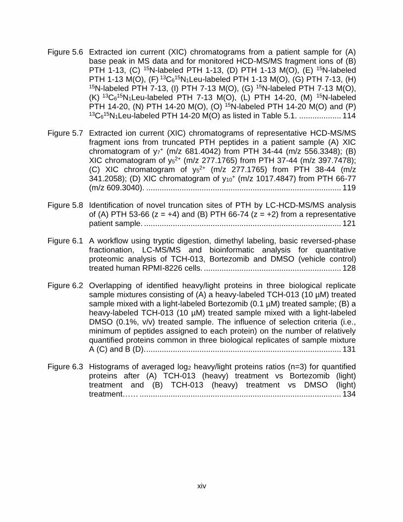
Figure 5.6 Extracted ion current (XIC) chromatograms from a patient sample for (A) base peak in MS data and for monitored HCD-MS/MS fragment ions of (B) PTH 1-13, (C) 15N-labeled PTH 1-13, (D) PTH 1-13 M(O), (E) 15N-labeled PTH 1-13 M(O), (F) 13C6
15N1Leu-labeled PTH 1-13 M(O), (G) PTH 7-13, (H) 15N-labeled PTH 7-13, (I) PTH 7-13 M(O), (G) 15N-labeled PTH 7-13 M(O), (K) 13C6
15N1Leu-labeled PTH 7-13 M(O), (L) PTH 14-20, (M) 15N-labeled PTH 14-20, (N) PTH 14-20 M(O), (O) 15N-labeled PTH 14-20 M(O) and (P)
13C615N1Leu-labeled PTH 14-20 M(O) as listed in Table 5.1. ................... 114
Figure 5.7 Extracted ion current (XIC) chromatograms of representative HCD-MS/MS fragment ions from truncated PTH peptides in a patient sample (A) XIC chromatogram of y7
+ (m/z 681.4042) from PTH 34-44 (m/z 556.3348); (B) XIC chromatogram of y5
2+ (m/z 277.1765) from PTH 37-44 (m/z 397.7478); (C) XIC chromatogram of y5
2+ (m/z 277.1765) from PTH 38-44 (m/z 341.2058); (D) XIC chromatogram of y10
+ (m/z 1017.4847) from PTH 66-77 (m/z 609.3040). ........................................................................................ 119
Figure 5.8 Identification of novel truncation sites of PTH by LC-HCD-MS/MS analysis of (A) PTH 53-66 (z = +4) and (B) PTH 66-74 (z = +2) from a representative patient sample. ......................................................................................... 121
Figure 6.1 A workflow using tryptic digestion, dimethyl labeling, basic reversed-phase fractionation, LC-MS/MS and bioinformatic analysis for quantitative proteomic analysis of TCH-013, Bortezomib and DMSO (vehicle control) treated human RPMI-8226 cells. .............................................................. 128
Figure 6.2 Overlapping of identified heavy/light proteins in three biological replicate sample mixtures consisting of (A) a heavy-labeled TCH-013 (10 µM) treated sample mixed with a light-labeled Bortezomib (0.1 µM) treated sample; (B) a heavy-labeled TCH-013 (10 µM) treated sample mixed with a light-labeled DMSO (0.1%, v/v) treated sample. The influence of selection criteria (i.e., minimum of peptides assigned to each protein) on the number of relatively quantified proteins common in three biological replicates of sample mixture A (C) and B (D). ........................................................................................ 131
Figure 6.3 Histograms of averaged log2 heavy/light proteins ratios (n=3) for quantified proteins after (A) TCH-013 (heavy) treatment vs Bortezomib (light) treatment and (B) TCH-013 (heavy) treatment vs DMSO (light) treatment…… ........................................................................................... 134
xv
KEY TO ABBREVIATIONS
AGPS: alkyglycerone phosphate synthase
CI: chemical ionization
CID: collision-induced dissociation
CL: glycerophosphoglycerophosphoglycerol
DAG: diacylglyceride
diIA: diiodoaniline
DMBNHS: S,S’-dimethylthiobutanoylhydroxysuccinimide ester iodide
DNA: deoxyribonucleic acid
ECD: electron capture dissociation
EI: electron ionization
ESI: electrospray ionization
ETD: electron transfer dissociation
FA: fatty acid
FT-ICR: Fourier transform ion cyclotron resonance
GC: gas chromatography
HCD: higher-energy collisional dissociation
HDX: hydrogen-deuterium exchange
HPLC: high performance liquid chromatography
ICAT: isotope-coded affinity tags
IDMS: isotope dilution mass spectrometry
iTRAQ: isobaric tags for relative and absolute quantification
MALDI: matrix-assisted laser desorption/ionization
MM: multiple myeloma
xvi
MRM: multiple reaction monitoring
MS: mass spectrometry
MS/MS: tandem mass spectrometry
MSn: multi-stage tandem mass spectrometry
m/∆m: the m/z value of a peak divided by the full width of the peak at half its maximum
height
NHS: N-hydroxysuccinimide
PA: glycerophosphate
PC: glycerophosphocholine
PE: glycerophosphoethanolamine
PG: glycerophosphoglycerol
PI: glycerophosphoinositol
PS: glycerophosphoserine
PTH: parathyroid hormone
PTHrR: PTH related protein
PTM: post-translational modification
RDD: radical-directed dissociation
RNA: ribonucleic acid
SILAC: stable isotope labeling with amino acid in cell culture
siRNA: silencing RNA
TG: triglyceride
TMTs: tandem mass tags
TOF: time-of-flight
UHPLC: ultra-high performance liquid chromatography
UVPD: ultraviolet photodissociation

xvii
XIC: extracted ion current
1
CHAPTER ONE
An Overview of Chemical Labeling Strategies for Mass Spectrometry Analysis of
Biomolecules
1.1 Introduction to Mass Spectrometry Analysis of Natural Biomolecules
1.1.1 Ionization Techniques
Mass spectrometry (MS) represents a key analytical method for the analysis of
biomolecules, including peptides, proteins [1], lipids [2], nucleic acids [3],
oligosaccharides [4] and other small molecule metabolites [5] and is based on the
intrinsic and most basic property of any molecule, its molecular weight. Before the
advent of MS, which measures the mass to charge ratios (m/z) of ions, molecular
weights were indirectly assigned based on molecular size or sedimentation coefficient
with low resolution and throughput, using size exclusion chromatography [6],
electrophoresis [7] and ultracentrifugation [8]. In a mass spectrometer, electric and/or
magnetic fields are used to control the trajectories of ionized molecules. Physical
parameters related to analyte ions can be measured by the instrument for calculation of
their molecular weight based on rigorous equations of ion motion, with high resolution
and accuracy [9]. At the early stage of MS-based biomolecule analysis, electron
ionization (EI) and chemical ionization (CI) were the two most widely used ionization
techniques which required the analytes to be gasified before ionization and therefore
limited the applications to volatile and thermally stable small biomolecules (e.g.,
oligopeptides and esterified fatty acids) [9]. With the introduction of ‘soft’ ionization
techniques including electrospray ionization (ESI) [10] and matrix-assisted laser
desorption ionization (MALDI) [11] in the late 1980s, MS analysis of large biomolecules
2
became practical. During the process of these two ‘soft’ ionization techniques, non-
volatile and thermally unstable large biomolecules can be directly extracted from
solution phase or a mixture of sample and matrix into the gas phase as intact structures
due to the negligible amount of internal energy deposited [1-5]. ESI produces multiply
or singly charged analyte ions [10]. Due to the very gentle ionization process, even
weakly bound complex ions consisting of multiple biomolecules can be preserved and
introduced into the mass spectrometer for analysis of the inter-molecular interaction
after ESI [12]. MALDI primarily produces singly charged analyte ions and has inherent
advantages for application to two dimensional [13] or even three dimensional [14]
imaging of various biomolecules in tissue samples.
1.1.2 Mass Analyzers
After ionization, a mixture of analyte ions can be transferred to various types of
mass analyzers to directly determine their m/z, or may be further subjected to gas
phase ion reactions (e.g., fragmentation) before mass analysis. The functioning
mechanisms and applications of commonly used mass analyzers and other related
techniques are discussed below.
The earliest types of mass analyzers, magnetic sector and double focusing
electromagnetic analyzers, employ magnetic fields alone or magnetic and electric fields
in tandem, respectively, to separate analyte ions with different m/z values in space
before detection [9]. With a typical source acceleration voltage around 10,000 V,
electromagnetic instruments can achieve high energy (~thousands of eV) collision-
induced fragmentation of analyte ions which is especially useful in characterization of
subtle structural features of biomolecules [15]. Time-of-flight (TOF) analyzers determine
3
the m/z values of analyte ions by their m/z dependent-flight times after acceleration by
high voltage. Operating with moderate m/z resolving power, high energy collision-
induced fragmentation and compatibility with pulsed ionization sources can all be
achieved using time-of-flight instruments [16]. Quadrupole type analyzers determine the
m/z values of analyte ions by their m/z dependent-trajectory stability in an applied
oscillating electric field [9]. The triple quadrupole mass spectrometer is widely used for
tandem mass spectrometry (MS/MS)-based quantitative analysis [17]. For analysis of
complex mixtures containing multiple analytes with isobaric (i.e., same nominal mass)
or isomeric (i.e., same exact mass) mass values, MS/MS identification and
quantification with high specificity, sensitivity and throughput can be achieved by
selection of precursor ions, fragmentation and detection of characteristic product ions
[18]. Ion trap analyzers (i.e., three dimensional and linear ion traps) store ions in an
oscillating electric field and separate them based on their m/z dependent-resonance
frequencies of motion after applying a supplemental radial frequency voltage [9].
Relying on the unique ability of ion trapping, resonance activation and ion ejection,
MS/MS and multistage MS/MS (MSn) can be performed in a single ion trap analyzer in a
time-dependent manner. Also, ion trap analyzers are frequently used as a reaction
vessel and mass analyzer for the study of gas phase ion chemistry [19]. Fourier
transform ion cyclotron resonance (FT-ICR) [20] and Orbitrap analyzers [21] function by
trapping ions in an static magnetic or electric field, respectively, and then measuring the
m/z dependent-resonance frequency of circulating or oscillating analyte ions. The most
unique feature of these two types of mass analyzers is their high resolving power,
represented by the m/z value of a peak divided by the full width of the peak at half its
4
maximum height (i.e., m/∆m). Currently, m/∆m up to 2,000,000 for FT-ICR [22] and
450,000 for Orbitrap [23] can be achieved. During analysis of complex mixtures,
overlapping analyte ions at low resolution can be considerably resolved for more
confident identification [24] and quantification [25] as long as the exact masses of those
analyte ions are different. With the ability to trap low energy electrons, electron-induced
activation can be performed in FT-ICR instruments for structural characterization of
large biomolecule ions and their fragile post-translational modifications (PTM) [26].
Providing comparable mass resolving power as that of FT-ICR, Orbitrap type
instruments have more modest size and cost.
The sensitivity of MS-based analysis has improved considerably due to ongoing
technological advancements in ion source/MS interfaces for improved sample
desolvation and ion transfer [27-30]. In addition, other platforms are often used in
combination with mass spectrometry to further expand its analytical capabilities. For
instance, without any enrichment steps, analytes at ng/mL level in blood or tissue can
be identified and quantified using a combined ultra-high performance liquid
chromatography (UHPLC) - tandem mass spectrometry workflow [31]. For isobaric or
isomeric analyte ions which have same nominal or exact m/z value, high performance
liquid chromatography (HPLC) and ion mobility spectrometry can be easily coupled to a
mass spectrometer to resolve and detect those analyte ions if they have different
hydrophobicity and ion mobility [32]. For high throughput analysis, various types of
ambient sampling/ionization methods have enabled rapid identification and
quantification of individual analytes in complex samples with minimal sample
preparation [33]. With the rapid advancement in mass analyzer instrumentation and
5
related MS technologies as discussed above, MS has become not only a device to
separate and analyze gas phase ions, but also a powerful platform to solve various
challenging analytical problems with high specificity, sensitivity and throughput.
1.1.3 Fragmentation Techniques
Soft ionization techniques maintain the atomic connectivity of neutral analytes
after ionization and thus molecular weight can be easily interpreted from an MS
spectrum. However, considering the possible structural isomers and the limited mass
resolution of many mass spectrometers, the m/z of precursor ions alone, is usually
insufficient for unequivocal biomolecule identification. Thus, one of the methods
commonly used for MS-based identification involves activation and fragmentation of the
analyte ions, followed by interpretation of the product ions to re-assemble the analyte
structures. Various fragmentation techniques which activate ions differently have been
developed to provide complementary product ions for comprehensive characterization
of analyte structures.
1.1.3.1 Collision-Induced Dissociation
Collision-induced dissociation (CID), one of the most commonly used
fragmentation techniques, activates ions by converting kinetic energy to internal energy
during the collision of analyte ions and neutral gas molecules in a collision cell such as
an ion trap and quadrupole [34]. The amount of energy deposited into ions as internal
energy during collision can considerably affect the subsequent fragmentation reactions
[35]. Low energy CID, usually with less than 100 eV of available collision energy for
conversion into internal energy, breaks the weakest bonds and can have more diverse
fragmentation pathways including intra-molecular rearrangements [36]. Commonly used
6
techniques of low energy CID include resonance excitation in linear and three
dimensional ion traps and beam type CID in quadrupoles and collision cells of higher-
energy collisional dissociation (HCD). High energy CID with a few thousand eV of
available collision energy gives more direct bond cleavage [35]. While low energy CID
and HCD are compatible with almost all of the current ionization sources and mass
analyzers, high energy CID is usually limited to applications using electromagnetic and
time of flight (TOF) instruments in which analyte ions with high kinetic energy can be
produced.
1.1.3.2 Electron Capture Dissociation and Electron Transfer Dissociation
Randomization of internal energy in low energy CID usually leads to less bond
cleavage or exclusive cleavage of specific weak bonds in large or labile modification-
containing analyte ions, resulting in spectra with relatively limited information for
structural interpretation [37]. To overcome this drawback, electron capture dissociation
(ECD) [38] and electron transfer dissociation (ETD) [37] were developed to provide
more random and extensive fragmentation via direct bond cleavage prior to
randomization of the internal energy. In both ECD and ETD, an electron is captured by
or transferred to a multiply charged cation (charge state >1) to form a distonic cation
with a positive charge and a radical. The fast process of electron recombination induces
activation resulting in direct bond cleavage and complementary product ions compared
to those of CID. ECD is performed in ICR instruments where trapping of low-energy
electrons is possible while ETD can be applied in more widely used ion trap instruments
through ion/ion reactions between analyte cations and singly charged anthracene
anions. ECD and ETD have been widely applied for structural characterization of intact
7
proteins and labile PTM of peptides, including phosphorylation, glycosylation,
sulfonation, and nitrosylation [37, 39]. However, due to the reduced charge state after
electron recombination, both ECD and ETD cannot be applied to analysis of singly
charged analyte ions.
1.1.3.3 Photodissociation
Another emerging ion activation method is photodissociation (PD), especially
ultraviolet photodissociation (UVPD) which has less dependency on charge state of
analyte ions due to the photon-based activation and has been employed for structural
characterization of a wide range of biomolecular classes [40-44]. Selective activation
can also be achieved since only ions (with or without chemical derivatization) that
absorb the excitation wavelength can be activated [45]. Various types of lasers have
been coupled to mass spectrometers to perform gas phase PD induced by photons with
different wavelengths, including 157 nm (F2 excimer laser), 193 nm (ArF excimer laser),
266 nm (the fourth harmonic of a Nd:YAG laser), 351 nm (XF excimer laser), 355 nm
(the third harmonic of a Nd:YAG laser), 800 nm (femtosecond titanium sapphire laser),
a tunable range of 205-2550 nm (optical parametric oscillator-Nd:YAG lasers) and 10.6
µm (CO2 laser) [46].
1.1.4 Applications for Identification and Quantification of Natural Biomolecules
The varied structures and abundances of biomolecules including peptides,
proteins, lipids, nucleic acids, oligosaccharides and other metabolites in a living
organism at different physiological and pathological states are closely related to their
biological activities and functions [47]. Qualitative and quantitative analysis of
biomolecules in complex mixtures are critical to understand their functional roles and
8
further decipher mechanisms associated with both normal cell functioning and the onset
and progression of disease, thus aiding in the discovery of novel biomarkers and
potential therapeutic drug targets [1, 5, 48, 49]. The merits that MS provides as an
analytical tool have been described above. From here the identification and
quantification of natural (i.e., without chemical labeling) biomolecules using MS-based
methods is discussed while highlighting the limitations of MS-based analysis without
chemical labeling for each class of biomolecule.
1.1.4.1 Peptides and Proteins
Proteins or polypeptides are biopolymers of amino acids and frequently modified
after their translation. They are responsible for near all cellular functions (e.g.,
enzymatic catalysis, signaling and transport of molecules) [47]. In MS analysis of
proteins, proteolytic peptide-based structural characterization and quantitative analysis,
namely ‘bottom-up’ workflows, are typically performed [50]. Relying on enzymatic
digestion, separation techniques, soft ionization methods (i.e., ESI and MALDI), well-
understood gas phase fragmentation mechanisms, advanced instrumentation of mass
spectrometry and automated data processing softwares, characterization of the primary
structure (i.e., sequences of amino acid residues) and PTMs of peptides derived from
complex protein mixtures has become much easier compared to using other available
methods [50]. Various enzymes (e.g., trypsin, Lys-C, Arg-C, Asp-N, and Glu-C) having
different proteolytic specificities are commercially available for cleaving proteins into
peptides that have appropriate m/z ranges and adequate ionization efficiency for
subsequent MS analysis [51]. Separation techniques including liquid chromatography,
electrophoresis and ion mobility separation are often necessary to resolve peptides
9
based on their hydrophobicity, size and mobility before introducing them into mass
spectrometers [50, 52]. Data acquisition during MS/MS analysis may be non-targeted or
targeted depending on the desired outcome. For instance, for comprehensive profiling
of all peptides and proteins in a complex mixture, non-targeted strategies including
data-dependent [53] or data-independent [54] acquisition are usually selected to
achieve broad coverage of identification. For targeted analysis of specific peptides or
proteins, pre-determined multiple reaction monitoring (MRM) transitions can be applied
to achieve optimal sensitivity for detection of peptides even at pg/mL level [55]. Finally,
software can be employed for peptide and protein identification during data
interpretation using database searching or de novo sequencing algorithms [56]. In
addition to the classic proteolysis-based ‘bottom-up’ workflows, ‘top-down’ approaches
employing the gas phase fragmentations of natural intact protein ions have also been
developed for the characterization of their primary structures and PTMs [39, 40].
Identities of and interactions (e.g., binding sites and stoichiometry) between
components in a natural protein complex at a near native state can also be
characterized by ‘native mass spectrometry’ where non-denaturing buffers and
instruments capable of high m/z measurement are employed [39, 57]. For the label free
quantification of peptides and proteins, either spectra counting-based or signal intensity-
based methods have been developed with data processing softwares (e.g., MaxQuant,
Skyline and Progenesis) [58, 59]. These methods are especially advantageous when
comparative quantification between large sets of experimental data is required [60].
However, successful identification, characterization and quantification of
individual peptides and proteins as well as their PTMs in complex mixtures from
10
biological samples are still problematic. For example, with an estimated concentration
dynamic range of cellular proteome of at least 109, direct MS analysis of low abundance
proteins without any enrichment or derivatization to improve the ionization efficiency is
very challenging [61]. Also, the chemical properties of peptides derived from cellular
proteomes, including hydrophobicity and the ability to acquire charge, have significant
effects on their ionization efficiency during ESI and thus affect MS sensitivity [62]. A
typical example is the decreased ionization efficiency of peptides with acidic
phosphorylated amino acid residues under positive mode ESI compared to their
unmodified counterparts [63]. Localization of PTMs on peptides or intact proteins is
complex due to their low stoichiometry and possible structural lability depending on the
specific modification [64]. As for the quantification of unlabeled peptide and proteins as
well as their PTMs, a robust LC-MS/MS setup and complicated data processing
workflow are commonly required to ensure reproducibility of quantitative results [60].
Also, since all samples are processed and analyzed through LC-MS separately,
systematic errors that may compromise the accuracy of quantitative results are
unavoidable through the entire sample preparation and data acquisition stages.
1.1.4.2 Lipids
Lipids play critical roles as structural and functional components of biological
membranes, as signaling and targeting molecules, and as a cellular energy resource
[47]. The qualitative and quantitative analysis of lipidome profiles under different cellular
states has emerged as a complementary alternative to proteomics and genomics to
address challenging cell biological questions [49]. Based on the International Lipid
Classification and Nomenclature Committee nomenclature, lipids are divided into eight
11
major categories including fatty acyls, glycerolipids, glycerophospholipids, sphingolipids,
sterol lipids, prenol lipids, saccharolipids, and polyketides [65]. Of these, fatty acyls,
glycerolipids, glycerophospholipids, sphingolipids and sterol lipids are most often
targeted in lipidomic studies [49]. Similar to proteins, structural information of lipids can
be hierarchically categorized as different levels, including lipid class, ‘sum composition’,
‘molecular lipids’ and ‘structurally defined molecular lipids’ as proposed in a recent
review by Ekroos [66]. Lipids in each of eight major categories can be further divided
into lipid subclasses based on the similarity of chemical structures. Then, the ‘sum
compositions’ of lipids in each class or subclass are defined with additional information
including the number of carbon elements and carbon-carbon double bonds. Further
structural confirmation of the ‘molecular lipids’ with at least two glycerol backbone
substituents requires the determined length and number of unsaturation of the
substituents. Finally, ‘structurally defined molecular lipids’ can be defined with more
detailed structural information including the position and stereochemistry of unsaturation
on glycerol backbone substituents. To date, identification and quantification at the lipid
class, sum composition and molecular lipids levels can be achieved through high
resolution mass spectrometry or MS/MS coupled with prior LC separation, or direct
infusion [67-70]. For instance, through a combination of spiking in exogenous internal
standards, high resolution mass spectrometry and MS/MS, 20 major lipid classes and
~250 molecular lipid species from yeast cells have been absolutely quantified without
any chromatography separation and chemical derivatization in order to study effects of
external and internal stimuli on the molecular composition profile of yeast lipidome [70].
12
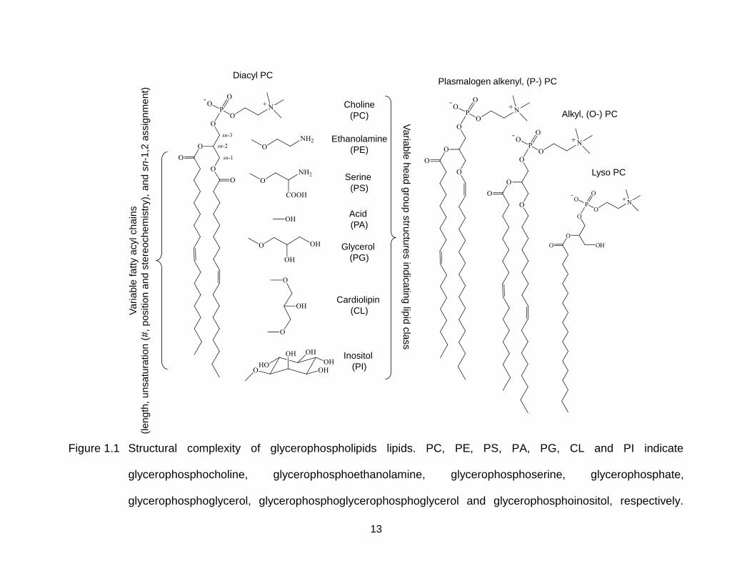
However, owing to their structural diversity (over 40,000 biologically relevant
molecular lipid structures recorded in the LIPID MAPS Structure Database,
http://www.lipidmaps.org/data/structure), accurate identification and quantification of
entire molecular lipids or even the structurally defined molecular lipids in a cellular
lipidome with an estimated dynamic range of at least 106 is still challenging [71]. For
example, the structural complexity of glycerolphospholipids is highlighted in Figure 1.1
[65]. Different lipid classes are differentiated based on the chemical structure of the
head group. Identity of the sn substituent on the glycerol backbone (i.e., acyl,
plasmalogen alkenyl and alkyl chain or no substituent) indicates the specific subclass.
Other structural features of the sn substituent are also variable (e.g., the length, the
unsaturation (number, sites and stereochemistry) and the site of linkage to the glycerol
backbone of the fatty acyl chains in the diacyl glycerophosphocholine (PC)). Although
methods employing high energy CID [15] and MSn [72-74] have been developed to
locate the site of unsaturation and the linkage site of sn substituent for a range of
glycerolipids and fatty acids with no chemical derivatization, complicated spectra for
interpretation and low sensitivity make it difficult to apply those methods for analysis of
complex lipid mixtures. The accuracy of quantitative analysis of cellular lipidomes is also
limited by the lack of enough commercially available exogenous lipids as internal
standards to represent the structural diversity of endogenous lipids, as well as possible
error that can be introduced during separate analysis of a given lipid mixture at different
polarities (i.e., both positive and negative ionization mode) during mass spectrometry
analysis.
13
Figure 1.1 Structural complexity of glycerophospholipids lipids. PC, PE, PS, PA, PG, CL and PI indicate
glycerophosphocholine, glycerophosphoethanolamine, glycerophosphoserine, glycerophosphate,
glycerophosphoglycerol, glycerophosphoglycerophosphoglycerol and glycerophosphoinositol, respectively.
Choline
(PC)
Ethanolamine
(PE)
Serine
(PS)
Acid
(PA)
Glycerol
(PG)
Cardiolipin
(CL)
Inositol
(PI)
Va
riab
le h
ead
gro
up
stru
ctu
res in
dic
atin
g lip
id c
lass
Va
ria
ble
fa
tty a
cyl ch
ain
s
(le
ng
th,
un
sa
tura
tio
n (
#,
positio
n a
nd s
tere
och
em
istr
y),
and s
n-1
,2 a
ssig
nm
en
t)
Alkyl, (O-) PC
Lyso PC
O
O
O
P
OO
ON+
-
OH
Plasmalogen alkenyl, (P-) PCDiacyl PC
14
1.1.4.3 Nucleic Acids
Nucleic acids including deoxyribonucleic acids (DNA) and ribonucleic acids (RNA)
are biopolymers consisting of nucleotides [47]. Similar to the amino acids as basic
building blocks of peptides and proteins, different nucleotides consisting of five-carbon
sugars, phosphate group(s) and nucleobases are polymerized in various sequences as
the primary structure of nucleic acids and the coded genetic information. To date, MS
has played an important role in qualitative and quantitative analysis of nucleic acids,
especially for structural characterization of post-transcriptionally modified transfer RNA
[75], quantitative analysis of DNA damage [76] and stoichiometry of the nucleic
acid/ligand complex [77]. However, without any functional modification to the naturally
synthesized nucleic acids, direct analysis of their secondary or tertiary structures and
interactions between components in a nucleic acid/ligand complex are still challenging
[3].
1.1.4.4 Oligosaccharides and Other Small Molecule Metabolites
Oligosaccharides are biopolymers of simple sugars responsible for various
important cellular events including the modulation of protein functions, cell adhesion and
cell signaling [78]. They can exit alone or be covalently bonded to proteins or lipids as a
modification (i.e., glycosylation) [47]. Although the methodologies and technologies of
MS-based analysis of oligosaccharides keep evolving, it remains a challenge to
structurally characterize and quantify this type of molecules. Natural oligosaccharides
have low ionization efficiency in positive ionization mode due to the lack of basic sites
[4]. Also, unlike proteins and nucleic acids that have linear primary structures,
oligosaccharides can have extremely high structural complexity due to the possible
15
branching of the simple sugar monomer, isomeric positions of glycosidic bonds and
stereochemical properties [47]. These two facts lead to the limited sensitivity and
complicated MS/MS spectra during MS-based analysis of natural oligosaccharides.
Other biological relevant small molecule metabolites including but not limited to
nucleotides, amino acids, fatty acids, organic acids and their derivatives are also targets
of MS-based analysis [5].
Given the limitations of direct analysis of natural biomolecules using MS, it is
clear that functional modification (e.g., chemical labeling) of natural biomolecules for the
purposes of improved separation before MS analysis, enhanced ionization efficiency,
controlled fragmentation behaviors to investigate specific structural features of analytes
and accurate quantitative analysis is necessary along with continuous development of
sample separation techniques, instrumentation, ion fragmentation methods and related
data processing software [79]. Herein, a detailed overview of chemical labeling
strategies for improved MS-based analysis of biomolecules is described.
1.2 Chemical Labeling Strategies for Improved Identification, Characterization
and Quantification of Biomolecules Using Mass Spectrometry
Most of the chemical labeling strategies used for improved MS-based analysis of
biomolecules can be classified into one or both of the categories discussed below. The
first involves linking functional groups, covalently or non-covalently, to the targeted
biomolecules to facilitate their MS-based identification and structural elucidation. The
second requires stable isotope labeling through chemical or metabolic incorporation of
stable isotopic elements (e.g., D, 13C, 15N and 18O) to enable MS-based characterization
16
of tertiary/quaternary structures of large biomolecules, studies on the mechanisms of
their gas phase ion fragmentations and most importantly, quantitative analysis.
1.2.1 Applications of Structural Labeling for Improved Identification and
Structural Characterization
1.2.1.1 Ionization Efficiency
Since MS is an analytical tool to study gas phase ions, the ionization efficiency of
analytes is closely related to MS sensitivity, limit of detection and limit of quantification.
The chemical labeling methods for ESI-based analysis are primarily discussed here
since ESI is the most widely employed ionization technique for both qualitative and
quantitative mass spectrometry analysis. During ESI, the chemical properties of
analytes play important roles in the processes of desolvation and charge acquisition;
thus chemical labeling to facilitate desolvation (e.g., increasing the surface activity of
analytes by modifying their hydrophobicity) and/or incorporation of fixed charges are the
main directions to enhance ionization efficiency [62, 80].
For improved ionization efficiency of peptides, structural labeling to incorporate
hydrophobic chemical structures (e.g., alkyl groups with long methylene chains) and/or
fixed charges (mainly quaternary ammonium and sulfonium group) mainly targets the
primary amine (N-terminus) and carboxylic (C-terminus) groups as well as side chains
of specific amino acid residues. Acylation- and alkylation-based chemistries are
commonly used for derivatization of peptide primary amine groups. For example,
Mirzaei and Regnier developed an N-hydroxysuccinimide (NHS) ester-based labeling
reagent containing a quaternary ammonium group linked to a straight alkyl chain with 8
carbons [81]. After acylation of primary amine groups in peptides under 500 Da, the
17
ionization efficiency increased (up to 500 fold) and a 10 fold enhancement of ESI
response was generally observed for most of the peptides studied. Another NHS ester-
based reagent, S,S′-dimethylthiobutanoylhydroxysuccinimide ester iodide (DMBNHS)
containing a sulfonium group linked to an alkyl chain has been employed to derivatize
phosphopeptides by Lu et al. [82]. A 2.5 fold increase in ionization efficiency in general
was demonstrated and more ETD-friendly phosphopeptides with multiple charges
(i.e., >+3) were observed. A method employing alkylation chemistry was also reported
by Kulevich et al. [83]. Tryptic peptides from selected protein digests were dialkylated
using aldehydes with varying length (i.e., 4, 6 and 8 carbons) through a simple one step
alkylation reaction. Both ionization efficiency and sequence coverage of selected
proteins were improved after combined analysis of alkylated and unlabeled tryptic
peptides. Studies describing esterification of carboxylic groups [84], guanidation of
lysine residues [85] and alkylation of cysteine residues [86] for improved ionization
efficiency of peptides were also reported.
For lipids, chemical labeling strategies to improve ionization efficiency similar to
that of peptides have also been developed, mainly through incorporating or uncovering
a fixed charge after chemical labeling. A typical example is the application of the
primary amine specific DMBNHS reagent containing sulfonium group to both
phosphopeptides (as discussed in the last paragraph) and aminophospholipids for
enhanced ESI response [24]. After a 30 min acylation reaction using deuterium labeled
DMBNHS reagent at room temperature, the average fold changes in intensity of
glycerophosphoethanolamine (PE) and glycerophosphoserine (PS) lipid ions in positive
ionization mode ESI-MS were around 3 and 6 fold, respectively. Methods employing
18
incorporation of quaternary amine groups to fatty acids and diacylglycerols [87, 88],
phosphonium group to hydroxyl group-containing lipids [89] and sulfate group to free
cholesterol [90] to improve their ionization efficiency were also reported. Another
method to uncover inherent charges in lipids through converting the zwitterionic lipids to
anionic species was reported by Han et al. [91]. After a one-step in situ acylation
reaction of primary amine groups in zwitterionic PE and lyso PE lipids using 9-
fluorenylmethoxylcarbonyl chloride, the detection sensitivity of the derivatized anionic
lipids was significantly improved (at picomolar level) under negative mode ESI. Instead
of covalent chemical labeling, non-covalent complexation has also been used to
enhance the ionization efficiency of lipids. Pham and Julian reported an approx. 10 fold
increase in detection sensitivity of PE species by non-covalent complexation with 18-
crown-6 ether during positive ion mode ESI [92], possible due to the increased surface
activity of ions complexed with 18-crown-6 ether [93].
In comparison to peptides and lipids, oligosaccharides are inherently difficult to
ionize due to their hydrophilicity and lack of basic sites to be protonated. To improve the
ionization efficiency of oligosaccharides, many methods have been developed to
increase their hydrophobicity, mainly targeting the hydroxyl groups through
permethylation and peracetylation reactions [94, 95]. However, the wider application of
permethylation and peracetylation-based labeling strategies was limited by their time-
consuming sample preparations and ‘non-quantitative’ labeling. Alternatively, hydrazone
labeling methods targeting carbonyl groups in the open-ring aldehyde form of
oligosaccharides is gaining popularity due to the absence of cleanup after labeling and
quantitative labeling efficiency [96]. The effects of permanent charge and hydrophobicity
19
of various forms of hydrazide labeling reagents on ionization efficiency of derivatized
oligosaccharides were also studied, suggesting that increased hydrophobicity enhanced
the ESI response while the incorporated permanent charge decreased the MS
abundance of derivatized oligosaccharides [97].
Similarly, incorporating hydrophobic groups or fixed charges into nucleobases,
nucleotides, oligonucleotides [98-100] and amino acids [101] have been reported to
enhance their ESI response for improved qualitative and quantitative analysis of nucleic
acids and metabolites.
1.2.1.2 Structural Characterization of Peptides and Proteins
1.2.1.2.1 Sequence of Amino Acid Residues
The structures of proteins, including their primary structures (i.e., the sequence of
amino acid residues), are closely related to their biological functions and therefore
sequencing of proteins is necessary to further understand their functioning mechanisms
[47]. To improve MS-based sequencing of peptides derived from proteins, the
fragmentation behaviors of peptide ions have been modified through structural labeling
with functional groups in order to simplify the MS/MS spectra or provide more abundant
and/or diverse fragment ions. Most of the reported labeling approaches follows the
principles including 1) modifying the charge (e.g., charge state, mobility and polarity) of
gas phase peptide ions to change their fragmentation behaviors in CID or to enable
ETD; 2) modifying the proton mobility of peptide ions in order to produce more abundant
and/or diverse sequence-specific fragment ions after activation and 3) incorporating
functional groups to enable fragmentation upon specific ion activation methods (e.g., PD
and radical-directed disassociation (RDD)).
20
Incorporating fixed charges to peptides not only increases their ionization
efficiency as discussed in Chapter 1, section 1.2.1.1, but also facilitates sequencing of
peptides by resembling the directing influence of the positions of inherently charged
basic amino acid residues on CID fragmentation of protonated peptides [82, 102] or
increasing the abundance of ETD-friendly multiply charged (i.e., >+3) peptide cations
[82, 103]. Methods employing solution phase labeling of quaternary ammonium [103,
104], phosphonium [105] and sulfonium [82] have been reported. Instead of performing
a labeling reaction in solution phase, gas phase charge polarity inversion of peptide ions
with the advantages of minimal sample preparation has been reported to improve
sequence coverage by acquiring complementary CID fragment ions in both polarities
[106] or enabling ETD fragmentation [107]. To modify the proton mobility of peptide ions,
both solution phase [108] and gas phase modification [109] methods have been
developed to either decrease the proton mobility of tryptic peptide anions or increase
the proton mobility of tryptic peptide cations to produce more diverse sequence-specific
fragment ions during CID-MS/MS analysis. For complementary sequence-specific
fragmentation of peptide ions induced by other ion activation methods compared to that
in CID, various forms of chromophore labeling reagents have been developed to enable
or improve PD (mainly at 351 nm [110-112], 355 nm [113] and 10.6 µm [114]) of
derivatized peptide ions. RDD has also been used to aid the sequencing of peptides
incorporated with radical precursors containing ultraviolet (UV)-labile covalent bonds
(e.g., carbon-iodine [115], carbon-sulfur [116] and carbon-oxygen bonds [117]) or weak
bonds which undergo homolysis upon collisional activation [118].
21
Recently, two papers described gas phase synthesis of linear [119, 120] or
branched [120] peptide ions with controlled sequences in a mass spectrometer. Without
time-consuming condensed-phase synthesis and separation, gas phase ultraviolet
photoexcitation or ion/ion reactions were employed to induce the formation of an amide
bond in a peptide dimer complex ion followed by facile mass-based separation and
MS/MS-based structural analysis of the products. Although the quantity of synthesized
peptides in the gas phase is unlikely to be sufficient for additional solution phase
modification, this type of method provides an approach to rapidly generate the library of
peptide MS/MS data to facilitate sequencing of unknown peptides.
1.2.1.2.2 Post-Translational Modifications
After biosynthesis, proteins are often post-translationally modified in potentially
hundreds of different ways [121]. Although PTMs occur at low stoichiometry and
abundance, they perform more precise regulations of cellular functions compared to
gene expression [122]. As a powerful analytical tool for structural elucidation, MS has
been widely used for analysis of PTMs [64], however for certain PTMs, sample
preparation including structural labeling for improved enrichment and favored gas phase
fragmentation is necessary for their improved identification and localization [79]. Biotin
tag labeling at sites of phosphorylation [123] and O-linked glycosylation [124] followed
by affinity chromatography are typical examples of enrichment strategies achieved by
structural labeling. For identification and localization of multiple PTMs (e.g.,
phosphorylation) in the same peptide, or very labile PTMs (e.g., glycosylation) during
gas phase fragmentation, ETD [37] and RDD [116] have been proved to be
advantageous compared to the conventional low energy CID fragmentation technique.
22
Therefore, labeling methods to enable ETD or RDD through increasing the percentage
and abundance of highly charged (>3) peptide ions or incorporating UV-labile radical
precursors have been developed [82, 125].
1.2.1.2.3 Tertiary and Quaternary Structures of Proteins
Preliminary structural labeling can also be applied for mass spectrometric
analysis of protein tertiary (e.g., protein conformations) and quaternary (i.e.,
protein/protein or protein/ligand interactions) structures. The basic principle is that after
changing the masses of the proteins via chemical labeling at the side chains of amino
acid residues in a structure-dependent manner, structural information can be derived
from mass spectrometry-based identification and relative quantification of peptides
derived from the labeled proteins. For example, information on protein conformational
changes can be derived from the changed surface residue accessibility characterized
by ‘bottom-up’ mass spectrometric analysis of proteolytic peptides of the labeled
proteins since surface residues exposed to solvent can more readily react with the
labeling reagent while other residues buried in the proteins react slower or not at all
[126]. Various covalent labeling reagents targeting side chains of specific amino acid
residues have been developed for this purpose [127-131]. For instance, Zhou et al.
developed the protein surface lysine accessibility mapping strategy using the fixed
charge containing DMBNHS reagent combined with data-dependent multistage tandem
mass spectrometry analysis [127]. By monitoring the changed lysine modification rates
between native and oxidized proteins using the developed strategy, it was found that
the functional domains within an important protein phosphatase, calcineurin became
more accessible to the DMBNHS labeling upon H2O2 oxidation-induced conformational
23
perturbation and suggested the phenomenon as a novel mechanism explaining the
inactivation of calcineurin caused by H2O2 oxidation [128]. Other reagents including
iodoacetamide [129], vicinal dicarbonyl compounds [130], and diethylpyrocarbonate
[131] have also been commonly used to map the accessibility of protein surface
cysteine, arginine, and histidine residues, respectively, but to a lesser extent compared
to lysine-specific reagents due to the wide range of available reagents targeting primary
amine group and the high frequency of lysine residue in proteins. In addition, as a
promising method to acquire broad sequence coverage of labeled proteins and with no
need for enzymatic digestion as in the ‘bottom-up’ approach, a ‘top-down’ strategy
probing conformational changes during denaturation of myoglobin and domain C of
PARP-1 proteins by gas phase UVPD fragmentation of intact protein ions after lysine-
specific labeling using S-methylacetimidate was reported by Cammarata et al. [132].
However, more efforts on the elucidation of gas phase UVPD fragmentation
mechanisms and the development of diverse ion activation methods for intact protein
ions are required for broader applications of the ‘top-down’ approach.
To characterize the interaction (e.g., distance and/or sites) of mutual interacting
sites within proteins or between protein/protein and protein/ligand complexes, cross-
linking reactions combined with mass spectrometric analysis can be employed [133].
Typically, a cross-linker reagent with reactive sites on two ends and a ‘spacer’ with fixed
length in the middle is used to covalently link two nearby sites in a protein or between
two components in a protein complex. Cross-linking reagents having two amine-specific
reactive sites targeting lysine residue were again used most frequently due to the
availability of reagents and the occurrence frequency of lysine in proteins as described
24
above. Photo-reactive cross-linkers containing diazirine, aryl azide and benzophenone
as reactive sites were also used as non-specific cross-linking reagents [134].
Subsequent ‘bottom-up’ mass spectrometric analysis can provide identities of cross-
linking sites and low resolution maximum distances between cross-linked sites [135].
Interestingly, employing the ion/ion reaction between ubiquitin protein cations and
homobifunctional sulfo-N-hydroxysulfosuccinimide ester reagent anions in an ion trap
reaction vessel, Webb et al. demonstrated the protein intra-molecular cross-linking
reactions in gas phase and characterized the cross-linked structures based on product
ions from CID experiments in a ‘top-down’ manner [136]. Although it is still not clear that
whether the mass spectrometric analysis of gas phase cross-linked proteins ions can
provide useful solution phase structural information, this method represents a creative
direction toward high throughput protein interaction study with minimal time for sample
preparation.
1.2.1.3 Structural Characterization of Lipids
1.2.1.3.1 Lipid Classes and Sum Compositions
For high throughput profiling of complex lipid mixtures or cellular lipidomes, it is
meaningful to separate and collectively study lipids based on their classes or
subclasses or sum compositions considering the similar physiochemical properties
affecting related cellular events of lipids with the same class, subclass or sum
composition [47]. However, many challenges exist due to the structural diversity and
complexity of lipid mixtures within crude cellular extractions. For example, during
shotgun lipid analysis with no LC separation, lipids of the same class but different
subclasses may not be able to be resolved based on mass due to their isomeric
25
structures and resultant same exact mass (e.g., plasmalogen lipids containing one less
site of unsaturation compared to alkyl-ether containing lipids, both with the same head
group (e.g., [PC(P-36:0) + H]+ and [PC(O-36:1) + H]+ both have the elemental composition of
C44H89NO7P). Also, lipids in different classes may suffer from the same situation (e.g.,
[PE(32:0) + H]+ and [PC(29:0) + H]+ both have the elemental composition of C37H75NO8P).
To overcome this limitation, Fhaner et al. developed a sequential structural labeling
strategy for aminophospholipids (i.e., PE and PS lipids ) and the O-alkenyl ether double
bond of plasmalogen lipids, using a ‘fixed charge’ sulfonium ion-containing stable
isotope-labeled-S,S-dimethylthiobutanoylhydroxysuccinimide ester reagent (DMBNHS),
and iodine and methanol, respectively, to eliminate potential overlap of these lipids with
other lipid classes or subclasses [24, 137]. Using the structural labeling strategy
combined with high resolution/accurate mass MS and “targeted” MS/MS analysis, >600
molecular lipid species from 36 lipid classes and sub-classes have been identified from
various colorectal cancer cell lines and their secreted exosomes, without need for
extensive sample handling or chromatographic fractionation prior to analysis [24, 138].
Similarly, Han et al. noted that identification and further structural characterization of the
interested lipids were potentially improved by shifting the m/z range of a labeled lipid
class to a region with much less interference with other lipid classes after using the 9-
fluorenylmethoxylcarbonyl chloride reagent targeting at aminophospholipids including
PE and lyso PE lipids [91].
1.2.1.3.2 Identities and Linkage Positions of Glycerol Backbone Substituents
Analysis of X-ray protein crystal structure data have suggested that the identities
(e.g., length) and linkage positions of glycerol backbone substituents of
26
glycerophospholipids are likely to affect their binding affinity to nuclear receptor SF-1,
which can be regulated by glycerophospholipid second messengers [139]. To further
understand the functional roles of lipids in cellular events, a comprehensive
characterization of lipid structure including subtle structural features is necessary [140].
Although MS-based identification of the length of the glycerol backbone substituents for
most glycerolipids is straightforward and does not require structural labeling, the
identification of other lipids is still challenging. For instance, glycerophosphocholine
lipids, containing an endogenous fixed positive charge quaternary amine, ionize poorly
in negative mode and yield generally limited structural information under positive
ionization mode CID-MS/MS conditions [2]. Methods describing solution phase
hydrolysis for analysis of resultant fatty acids [141] or addition of high concentration of
salts to enable acquisition of glycerol backbone substituent-specific fragment ions at
both positive [142] and negative polarities [143] have been reported. However, their
wider applications are limited by the absence of molecular structure information or by
low sensitivity. For this, Stutzman et al. converted the gas phase protonated PC lipid
ions, [PC+H]+ into demethylated PC anions, [PC-CH3]- through non-covalent labeling of
doubly deprotonated 1,4-phenylenedipropionic acid followed by CID-induced ion/ion
reaction [144]. Subsequent CID analysis of the [PC-CH3]- anions produced abundant
fatty acid carboxylate anions indicating the length and unsaturation degree of glycerol
backbone substituents.
Another more challenging task is to characterize the subtle structural features of
glycerophospholipids (i.e. the linkage position (sn position) of their glycerol back bone
substituents). Pham et al. have demonstrated the existence and differentiation of sn
27
positional isomers for six major glycerophospholipids classes through gas phase
ozonolysis (i.e., addition of ozone to carbon-carbon double bonds) of product ions
arising from CID of sodium adduct of glycerophospholipids [145]. After the
CID/ozonolysis experiments, abundant and predictable product ions that are
characteristic to the specific sn position of glycerol backbone substituents were
observed.
1.2.1.3.3 Positions and Stereochemistry of Carbon-Carbon Double Bonds
Location and stereochemistry (i.e., cis or trans) of carbon-carbon double bonds
(C=C) in lipids are also subtle structural features of lipids that can change their
biochemical properties [146-148]. Differentiation of C=C bond positional isomers in
lipids were traditionally conducted by separation through gas chromatography (GC) or
HPLC followed by mass spectrometric or other detection methods [141]. However,
direct determination of positions and stereochemistry of C=C bonds in underivatized
lipids using MS is problematic since the C=C bond isomers of lipids give identical low
energy CID-MS/MS spectra. Although methods employing high energy CID [15, 16] and
MSn [72, 73, 149] are available, low sensitivity and complicated spectra interpretation
limit their broader applications. To facilitate the localization of C=C double bonds in
lipids, structural labeling strategies have been developed to selectively induce more
abundant fragmentation cleavages within their unsaturated aliphatic chains. Several
methods exploiting selective gas phase ion/molecule reactions (e.g., covalent adduct
chemical ionization [150], ozonolysis [145], Paternò-Büchi reactions [151]) targeting at
C=C double bonds were demonstrated to modify the fragmentation behaviors of lipid
ions and product abundant fragment ions indicating the position of C=C double bonds.
28
RDD has also been employed to induce C=C bond position-specific fragmentations
occurring within the unsaturated aliphatic chains of lipid ions after introduction of lipid
ions, which are covalently [152] or non-covalently [92, 153] linked to a UV-labile radical
precursor, into the mass spectrometer via ESI followed by activation of UV photons to
release the radicals. Finally, although still very challenging, MS-based differentiation of
lipid isomers only differing in stereochemistry of C=C double bonds was demonstrated
to be feasible by Pham et al. without prior separation [154]. Relative abundance of CID
product ions of proton bound complex ions consisting of fatty acid methyl esters and
iodoaniline were proved to be sensitive to the stereochemistry (i.e., cis or trans) of C=C
double bonds.
1.2.1.4 Intra- and Inter-Molecular Interactions of Nucleic Acids
Although the role of MS in genome-wide sequencing analysis was superseded by
other high-throughput methods in the late 1990s [155], structural labeling combined with
mass spectrometric analysis is still advantageous as a rapid and sensitive method to
characterize the intra- and inter-molecular interactions of nucleic acids, mainly using
alkylating cross-linkers based on the same principles applied to probe protein intra- and
inter-molecular interactions, discussed in Chapter 1, section 1.2.1.1.2 [3].
1.2.1.5 Structural Characterization of Oligosaccharides
In addition to enhancing the ionization efficiency, structural labeling of
oligosaccharides is helpful in obtaining characteristic tandem mass spectra allowing
identification of their sequence, branching and interglycosidic linkages [79].
Permethylation and peracetylation of the hydroxyl groups of oligosaccharides are
traditionally used to modify the fragmentation behaviors as well as increase ionization
29
efficiency [4]. Labeling strategies enabling RDD and UVPD of oligosaccharides are
gaining attention due to the ability to acquire complementary fragment ions indicating
extra structural information compared to that from CID. For example, both 355 nm
UVPD of chromophore-incorporated oligosaccharides [156] and photo-induced RDD of
oligosaccharides labeled with iodoaniline [157] were reported to provide fragment ions
distinguishing positional oligosaccharide isomers.
1.2.2 Applications of Stable Isotope Labeling for Improved Structural
Characterization and Quantification
1.2.2.1 Structures of Large Biomolecules and Mechanisms of Gas Phase
Biomolecular Ion Fragmentation
Stable isotope labeling is widely used for MS-based structural characterization of
large biomolecules (i.e., proteins and their complexes) [158] and mechanistic studies of
gas phase fragmentation of other smaller biomolecular ions (i.e., peptide [159-162], lipid
[163], nucleic acid [164], oligosaccharide [165] and other small molecule metabolite ions
[166]). After replacement of specific elements in selected regions of biomolecules with
corresponding stable isotopes, the masses of stable isotope-labeled regions are
changed while covalently and non-covalently connected structures and resultant gas
phase ion fragmentation behaviors are kept the same or near identical. Thus, by
comparative study of labeled and unlabeled analytes, the identity of ions derived from
the stable isotope-labeled regions which are characterized by MS can be linked back to
their spatial positions in the original biomolecules.
Hydrogen-deuterium exchange (HDX) MS is a typical example and a key
technique employing stable isotope labeling to characterize the intactness of hydrogen
30
bonding networks of proteins and their complexes in solution [158]. The principle of this
method is that when exposed to deuterium-labeled water (D2O), amide bond hydrogens
in the disordered or highly dynamic regions of proteins undergo rapid exchange with
surrounded deuterium due to lack of stable hydrogen-bonding [158]. The ‘bottom-up’
mass spectrometric analysis workflows are typically used to analyze structure-
dependent HDX patterns of proteolytic peptides from proteins under different
experimental conditions for structural characterization [167]. However, attention needs
to be given during selection of an appropriate ion activation method for MS/MS-based
identification of residue-specific HDX patterns of peptides. Consistent with the mobile
proton model explaining the CID fragmentation mechanisms of protonated peptides, it
was demonstrated that intra-molecular migration of hydrogen and deuterium during the
collision-induced vibrational activation process could lead to complete randomization
(i.e., ‘scrambling’) of the HDX patterns [168]. Therefore, ion activation methods not
involving the vibrational excitation (e.g., ETD [169] and ECD [170]) are more
advantageous in acquiring residue-specific HDX patterns of peptides. Also,
experimental procedures need to be optimized to minimize back exchange from
deuterium to hydrogen during proteolysis and followed LC separation of deuterium-
labeled proteins. Keeping the solution temperature close to 0 °C after HDX and
minimizing the time for sample preparation can effectively preserve much of the HDX
patterns [158]. Alternatively, the back exchange in proteolysis step can be avoided in
‘top-down’ analysis where the gas phase fragmentation of deuterium-labeled intact
protein ions directly generates peptide ions for HDX pattern study and sequencing. The
31
‘scrambling’ processes in “top-down” analysis can also be minimized by using ECD [171]
and ETD [172] instead of CID.
Understanding gas phase fragmentation mechanisms of biomolecular ions is
essential for accurate interpretation of mass spectra data and structural elucidation.
Direct comparison between fragmentation spectra of stable isotope-labeled and
unlabeled biomolecules provides a straightforward approach to investigate
fragmentation mechanisms. For this purpose, various stable isotopes (e.g., D, 18O, 15N
and 13C) have been incorporated into peptides [159-162], lipids [163], nucleic acids
[164], oligosaccharides [165] and other small molecule metabolites through HDX or
chemical synthesis.
1.2.2.2 Quantification of Biomolecules
Unlike quantitative analysis based on the Beer-Lambert law, there is no fixed
response coefficient between an MS signal and the concentration of an individual
analyte. Therefore, for accurate and absolute MS-based quantification, ratiometric
measurement of simultaneously determined intensities of the analyte ions and their
stable isotopic analogue ions as internal standards having the same or near identical
MS signal responses is the ideal requirement [60]. For targeted analysis of a limited
number of analytes, this approach, referred to as isotope dilution mass spectrometry
(IDMS) is applicable with the advantage of providing absolute quantities of targeted
biomolecules [90, 173-175]. However, during a typical quantitative ‘-omics’ study, the
requirement to quantify hundreds to thousands of individual analytes makes the
preparation of the corresponding stable isotope-labeled standards impossible.
Alternatively, metabolic labeling and chemical labeling can incorporate stable isotope
32
labels to biomolecules at large scale, enabling relative quantification of individual
biomolecule in cells or tissues under various stimuli [4, 60, 71]. Details of isotope
labeling strategies developed and their application for quantitative analysis of
biomolecules are discussed below.
1.2.2.2.1 Stable Isotope-Labeled Analogue Internal Standards
For absolute quantification of targeted peptides using IDMS, most isotopically
labeled internal standards can be easily synthesized using commercially available
isotopically labeled amino acids and automated peptide synthesis [176]. For proteins,
either their unique peptide sequences or the proteins themselves can be isotopically
labeled through chemical or biological synthesis as internal standards. Currently,
although still relatively expensive, isotope-labeled peptides are widely employed as
surrogates for absolute quantification of proteins using MRM analysis [173, 177].
However, since those isotopically labeled peptides are usually spiked into the samples
at a late stage of the sample preparation, systematic errors (e.g., sample loss and
sample handling-induced modification of targeted proteins) prior to standard spiking can
affect the accuracy of quantification if no correction is made [178, 179]. Alternatively,
several methods using various types of isotopically labeled protein internal standards
were developed to overcome this limitation. Caroll et al. used recombinant DNA
techniques to construct an isotopically labeled protein containing 59 tryptic internal
standard peptides for absolute quantification of 27 enzymes using IDMS [180].
Systematic errors arising from tryptic digestion were minimized since targeted proteins
and the biologically synthesized protein standards were mixed before digestion.
Methods describing biosynthesis of stable isotopic analogues of targeted proteins in
33
cells or cell-free systems were also reported and applied to considerably minimize
sample handling-related errors in IDMS analysis by spiking these protein standards into
samples at the earliest stage of sample handling (e.g., cell lysis or collection of biofluids)
[178, 179, 181].
Due to the structural complexity of lipids, especially for glycerolipids and
glycerophospholipids, synthesizing a large array of their stable isotopic analogues is
very challenging [66]. To date, quantitative IDMS analysis of lipids using stable isotopic
analogue internal standards mainly target biologically relevant ‘simple’ lipids (e.g.,
cholesterol [90], fatty acid [174] and ceramide [182]). For the similar reason related to
lack of enough stable isotopic analogue standards, methods for quantitative IDMS
analysis of oligosaccharides were less developed. For nucleic acids, IDMS analysis for
quantification of DNA damage is continuously gaining popularity [3]. Due to the often
low abundance and stoichiometry of DNA damage or modification, DNA is usually
hydrolyzed to produce nucleobase monomers instead of the whole biopolymer as
targets of MS analysis [3]. Correspondingly, stable isotopic analogues of the targeted
modified nucleobase monomer can be spiked in before downstream sample preparation
and analysis. For other small molecule metabolites, IDMS is now the gold standard for
quantitative analysis [183].
1.2.2.2.2 Metabolic Labeling
Metabolic labeling for MS-based quantitative analysis of biomolecules is mainly
achieved through replacing the monomers of biopolymers (e.g., proteins [60] and
saccharides [184]) with corresponding stable isotopic analogues during biosynthesis.
For MS-based quantitative proteomics, metabolic labeling can incorporate stable
34
isotope labels into proteins at the earliest stages (i.e., biosynthesis) to maximally avoid
the sample handling-related errors [60]. Currently, the most widely applied metabolic
labeling method for protein samples derived from cultured cells is stable isotope labeling
with amino acid in cell culture (SILAC) developed by Ong et al.. Heavy stable isotope
(i.e., 13C and 15N) labeled arginine and lysine added in the cell medium ensure that,
after digestion of the extracted proteins, most of the tryptic peptides having arginine or
lysine residues at their C-terminus will contain at least one stable isotope labeled amino
acid residue [185]. By adding arginine and lysine incorporated with various numbers or
identities of stable isotopes during cell culture to differentially label multiple sets of
samples, intensities of isotopic clusters of the differentially labeled and pooled peptides
can be detected through mass spectrometric analysis and used as surrogates for
protein expression levels. More modified versions of the SILAC strategy were also
developed to quantitatively study protein synthesis and degradation (i.e., pulsed SILAC)
[186] or enable quantitative analysis of proteins between animals [187].
Mass spectrometry-based quantitative analysis of metabolically labeled
oligosaccharides has also been demonstrated using murine embryonic stem cells
cultured with and without addition of 15N-labeled glutamine as the only nitrogen source
of hexosamines [184]. One of the disadvantages for metabolic labeling strategies is that
most of the methods are only applicable to cultured cells instead of samples from
animals or human where the stable isotope incorporation during biosynthesis of
analytes is unlikely. Another disadvantage is the possibility of overlapping peaks in the
mass spectra of multiplexed (>3) metabolically labeled samples, which can compromise
the accuracy of quantification [60]. High resolution MS (m/∆m > 200,000 at m/z 400)
35
combined with neutron-coded SILAC represents a promising quantitative strategy
employing subtle mass differences of stable isotopes (e.g., 36 mDa between 13C615N2-
labeled lysine and D8-labeled lysine) to theoretically enable highly multiplexed
quantitative analysis but has not been practically applied [25]. An alternative solution is
chemical labeling as discussed below.
1.2.2.2.3 Chemical Labeling
In chemical labeling strategies for stable isotope incorporation and subsequent
MS-based quantitative analysis, samples under different experimental conditions are
separately derivatized by a set of stable isotope-encoded reagents so that the same
labeled biomolecules in different samples become stable isotopic analogues. After
pooling of separately derivatized samples, MS analysis can provide quantitative
information based on ratiometric measurement of 1) the intensities of differentially
labeled biomolecular ions in mass spectra if they have different m/z or 2) the intensities
of reporter ions derived from differentially labeled and isobaric biomolecular ions in
MS/MS or MS3 spectra [60].
For quantitative analysis of peptides and proteins, chemical labeling methods
employing MS-based quantification include isotope-coded affinity tags (ICAT) and
dimethyl labeling [188, 189]. ICAT reagents target the thiol group-containing side chains
of cysteine residue. A biotin group is also incorporated in each ICAT reagent to enable
sample enrichment through affinity chromatography. Dimethyl labeling selectively reacts
primary amine groups on the N-terminus and side chains of lysine residues with
formaldehyde and its stable isotopic analogues followed by reduction using
cyanoborohydride. This method is gaining its popularity due to the inexpensive reagents
36
and applicability to a broad quantity range of samples (sub-micrograms to milligrams)
[189]. However, due to the incorporated deuterium in both reagents, these two methods
suffer from the deuterium isotope effect-induced retention time shift during reverse
phase LC [190]. The ability to perform highly multiplexed quantification is also limited to
pools of at most three differentially labeled samples due to the increasing complexity of
the mass spectra when combining multiple labeled samples together [60].
Alternatively, isobaric stable isotope-coded reagents employing MS/MS or MS3-
based quantification can overcome these limitations by measuring the peak intensities
of reporter ions in the low mass range of MS/MS or MS3 data where the spectra are
relatively simple compared to MS data [60]. Retention time shifts during reverse phase
LC separation can be avoided since isobarically labeled peptides co-elute precisely [60].
Tandem mass tags (TMTs) (up to 10 plex) [191] and isobaric tags for relative and
absolute quantification (iTRAQ) (up to 8 plex) [192] are the two most popular methods
for this purpose and both types of reagents are commercially available. Similar to
dimethyl labeling, TMTs and iTRAQ reagents both target the primary amine groups of
peptides. Even higher orders of multiplexed quantification (12 plex) have been
demonstrated by a combination of ICAT, dimethyl labeling and iodoacetyl-TMT methods
[193].
For lipids, iTRAQ and other NHS ester-based reagents were also developed for
MS/MS-based quantification of primary amine-containing lipids. For example, Berry et al.
have previously reported results from iTRAQ and isotope labelled 4-
(dimethylamino)benzoic acid NHS ester derivatization of primary amino groups for the
quantitative analysis of diacyl-, ether- and plasmalogen-PE lipids in complex biological
37
mixtures [194-196]. Nabetani et al. also recently applied the iTRAQ labeling strategy for
the quantitative analysis of hydrolyzed ceramide lipids [197]. For quantitative analysis of
oligosaccharides, a modified TMT reagent, stable isotope labeled carbonyl-reactive
tandem mass tags (glyco-TMTs) was also developed and applied to study N-linked
protein glycosylation profiles of the isogenic primary colorectal cancer cell line, SW480
and its malignant cell line, SW620 [198]. It should be noted that depending on the
mixture complexity and prior chromatographic fraction that may be used, these current
isobaric stable isotope tagging methods for quantification of biomolecules using low
mass reporter ions may suffer from interference in the reporter ion abundances due to
the presence of co-isolated and fragmented precursor ions present within the isolation
window that is used prior to dissociation [199, 200]. MS3 dissociation [201], gas phase
purification using proton transfer reaction [202] and ion mobility separation [203] have
been proposed to minimize the described limitations for quantitative proteomic studies.
A method employing neutral loss product ions of derivatized aminophospholipids for
relative quantification after isobaric stable isotope labeling is also described in Chapter
three to avoid the possible interference in low mass reporter ions-based quantification.
1.3 Aims of this Dissertation
The aims of this dissertation include:
1. Development of a novel gas-phase cross-linking reaction for saturated and
unsaturated TG lipid ions using di-iodoaniline reagents and photo-induced
radical chemistry in a modified ion trap mass spectrometer enabling UVPD
experiments.
38
2. Characterization of the structures and formation mechanisms of cross-linked
TG lipid ions and elucidation of the effects of various cross-linkers and TG
lipid structures using multistage CID-MS/MS.
3. Characterization of CID and HCD fragmentation behaviors of S,S′-
dimethylthiobutanoylhydroxysuccinimide ester iodide (DMBNHS) derivatized
aminophospholipids.
4. Development of a novel strategy for characterization and multiplexed
quantification of deuterium-coded DMBNHS derivatized aminophospholipids
in complex biological samples using high resolution/accurate mass ESI-MS,
CID-MS/MS, HCD-MS/MS and -MS3.
5. Development of a quantitative immuno LC-MS/MS method for parathyroid
hormone (PTH) and its in vivo oxidation and truncation.
6. Characterization of novel post-translationally modified PTH in patient samples
using the immuno LC-MS/MS workflow.
7. Quantitative proteomic analysis to study the drug action mechanism of a non-
competitive proteasome inhibitor, TCH-013 using dimethyl labeling and two
dimensional LC-MS/MS.
39
CHAPTER TWO
Photo-Induced Inter-Molecular Cross-Linking of Gas Phase Triacylglycerol Lipids
Ions
2.1 Introduction
Plant derived oils have widespread use as renewable ‘green’ feedstock
components in a diverse range of industrial applications, including food, cosmetics,
pharmaceuticals, antioxidants, biofuels, surfactants, lubricants, and polymers. For
example, the cross-linking of triglycerol (TG) lipids or their derivatives is a common
process associated with the industrial scale synthesis of plant oil-based polymers [204],
as well as being an unwanted reaction associated with long term storage [205] and high
temperature cooking (e.g., frying) [206]. However, the physical and chemical properties
of the polymeric products from these reactions are known to be affected by many
factors, including the structures of the TG monomers (i.e., the length of the fatty acyl
chains, the number and positions of unsaturation sites, and their stereochemistry) [207],
the mechanism of the cross-linking reaction [208], and other experimental conditions.
Thus, a detailed understanding of these factors, particularly those relating to the
structure and intrinsic reactivity of the TG monomers under a given set of cross-linking
conditions, may provide important insights for the improved manufacture of polymers
with desired properties.
Traditionally, the cross-linking reactions of TG lipids or their derivatives are
conducted in the solution phase, then the products are analyzed by Fourier transform
The results described in this chapter were published in: Nie, S., Pham, H.T.,
Blanksby, S.J. and Reid, G.E. “Photo-induced inter-molecular cross-linking of gas
phase triacylglycerol lipid ions.” Eur. J. Mass Spectrom. 2015, 21, 287.
40
infrared spectroscopy, Fourier transform Raman spectroscopy [209], and/or by reverse-
phase high-performance liquid chromatography [210] or gel permeation
chromatography [211]. However, these methods are often limited in their ability to obtain
detailed molecular information regarding the individual structural features of the TG
lipids that may be present within a complex mixture, that may affect their cross-linking
reactivity and the structure of their polymerized products. Thus, there remains a need
for the development of alternative approaches that enable monitoring and
characterization of the intrinsic intra- and/or inter-molecular interactions and chemical
reactivity of individual lipids within a given mixture.
As discussed in chapter one, MS and MS/MS-based methods are increasingly
being used to analyze the solution phase products of cross-linked biomolecular
polymers [212], as well as to characterize the products of cross-linking reactions
performed in the gas phase, using a variety of activation methods including collision
induced dissociation (CID) [213] and ultraviolet photo-dissociation (UVPD) [214].
Interestingly, Muller et al. recently reported the formation of an inter-molecular cross-
linking reaction within a non-covalent dimer of a diacylglycerol (DAG) anion and a
neutral DAG molecule upon performing CID in negative ionization mode [215]. However,
the SN2 nucleophilic substituent mechanism associated with this reaction cannot readily
be directly applied to the formation and analysis of cross-linked TG lipids, which may
contain carbon-carbon (C=C) double bonds as the only reactive sites, and are usually
observed in positive ionization mode.
Considering that the C=C bond of unsaturated TG lipids is one type of reactive
site commonly involved in the solution phase radical polymerization of plant oils [216], it
41
is interesting to study whether similar reactions could be achieved in the gas phase, in
which radical addition and recombination drive the cross-linking process. Radicals can
be easily generated by UVPD of precursors containing carbon-iodine [115] or carbon-
sulfur bonds [116], via the incorporation of weak bonds that easily undergo homolysis
during CID [118, 217] and through CID-induced one-electron oxidative dissociation of
metal ion/biomolecule complex ions [218, 219]. An example of the formation of a
covalent linkage between a radical precursor and the site of unsaturation within a TG
lipid via radical addition, using 266 nm UVPD, was recently reported by Pham et al. [153]
In that work, a non-covalent complex consisting of protonated 4-iodoaniline ion and a
neutral unsaturated TG lipid was introduced into a modified ion trap mass spectrometer
via electrospray ionization (ESI), then isolated and subjected to activation with 266 nm
UV photons. This resulted in homolysis of the carbon-iodine bond and the formation of
an anilinyl radical that subsequently underwent radical addition to yield a covalent bond
between the protonated aniline and the TG molecule. The presence of the radical within
this product ion was then shown, using CID-MS3, to direct the dissociation toward novel
analytically useful fragmentation pathways (e.g., to enable determination of the site(s) of
unsaturation within the acyl chain(s)). In this chapter, it was demonstrated that using
UVPD and multistage CID-MSn experiments, similar reactions involving a protonated
diiodoaniline ion non-covalently associated with two neutral saturated or unsaturated
TG lipid molecules can occur, resulting in the formation of multiple cross-linked TG
products.
42
2.2 Results and Discussion
2.2.1 Photo-Induced Dissociation of Protonated Diiodoaniline-TG Dimer Complex
Ions
Diiodoaniline (diIA) was selected as a potential cross-linker for TG lipids for
several reasons. First, the protonated amine group of diIA could be an effective ionizing
adduct for a TG dimer since the detection of an ammonium cation complexed with a TG
dimer after ESI has been reported [220]. Second, the phenyl ring of diIA is a
chromophore, which increases the cross section for photon absorption. Finally, the
biradical product that is generated upon cleavage of the two carbon-iodine bonds upon
activation with UV photons has the potential to form covalent linkages by reaction at
sites of unsaturation (as well as at other sites – see below) within the TG lipids [153]. To
first establish the feasibility of using diIA as a photo-induced inter-molecular cross-
linking reagent for TG lipids, sample solutions containing either mono-unsaturated
TG(14:0/16:1/14:0) or saturated TG(14:0/16:0/14:0) with 3,4-diIA, 2,4-diIA or 3,5-diIA were
introduced into the modified linear ion trap mass spectrometer via ESI. In the resultant
mass spectra (data not shown), abundant ions were observed at m/z 1841 and 1845,
respectively, with broad peak widths, that were found to correspond to the presence of
[2TG(14:0/16:1/14:0) + diIA + H]+ and [2TG(14:0/16:0/14:0) + diIA + H]+ with calculated m/z values
of 1843.3 and 1847.3, respectively. The broadened peak widths and shifted m/z values
compared to the theoretical values for these non-covalent complexes can be explained
as being due to dissociation occurring during resonance ejection [221, 222]. The
phenomenon of mass shifts and broadened peak widths associated with the presence
of non-covalently linked TG dimer complex precursor and product ions were observed
43
consistently in the experiments described herein, but did not negatively affect
interpretation of the results. Figure 2.1A and 2.1B show the product ion spectra
obtained following 266 nm UVPD-MS/MS spectra of the precursor ions of
[2TG(14:0/16:1/14:0) + 3,4-diIA + H]+ (complex 1) and [2TG(14:0/16:0/14:0) + 3,4-diIA + H]+
(complex 2) containing 3,4-diIA, respectively, while Figure 2.1C shows the UVPD-
MS/MS spectrum obtained from the [2TG(14:0/16:1/14:0) + 2,4-diIA + H]+ (complex 3)
precursor ion containing 2,4-diIA. In each case, product ions corresponding to the
sequential neutral losses of one and two iodine radicals were observed, as well as,
additional products corresponding to the further losses of intact neutral TG molecules.
The anilinyl biradical-containing product ions that were formed via the loss of two iodine
radicals (i.e., m/z 1588.7 in Figure 2.1A, 1592.7 in Figure 2.1B and 1588.9 in Figure
2.1C) can then potentially undergo subsequent reactions via either radical addition or
hydrogen abstraction. Radical addition at unsaturation site(s) within the unsaturated
TG(14:0/16:1/14:0) containing dimers in Figure 2.1A and 2.1C would be analogous to
chemistry previously reported by Pham et al. [153]. Hydrogen abstraction is at least
thermodynamically feasible [223] at some sites along the fatty acyl chains and on the
glycerol backbone of the TG molecules within both the unsaturated TG(14:0/16:1/14:0) and
the saturated TG(14:0/16:0/14:0) dimers.
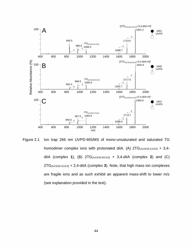
44
Figure 2.1 Ion trap 266 nm UVPD-MS/MS of mono-unsaturated and saturated TG
homodimer complex ions with protonated diIA. (A) 2TG(14:0/16:1/14:0) + 3,4-
diIA (complex 1), (B) 2TG(14:0/16:0/14:0) + 3,4-diIA (complex 2) and (C)
2TG(14:0/16:1/14:0) + 2,4-diIA (complex 3). Note, that high mass ion complexes
are fragile ions and as such exhibit an apparent mass-shift to lower m/z
(see explanation provided in the text).
966.91094.0
1588.7-I
-I
-TG(14:0/16:1/14:0)
[2TG(14:0/16:1/14:0)+3,4-diIA+H]+
-I
-I
400 600 800 1000 1200 1400 1600 1800 2000
1095.9842.4
1592.7
-I
-TG(14:0/16:0/14:0)
[2TG(14:0/16:0/14:0)+3,4-diIA+H]+
-I
400 600 800 1000 1200 1400 1600 1800 2000
-I-I
1713.7967.5 1093.9
840.51588.9
-I-TG(14:0/16:1/14:0)
[2TG(14:0/16:1/14:0)+2,4-diIA+H]+
-I
400 600 800 1000 1200 1400 1600 1800 2000
-I
-I
Rela
tive A
bun
dance (
%)
840.5
100
100
100
A
B
C
1714.0
1841.2
1717.0
1844.9
968.8
1840.6
m/z
1841UVPD
1845UVPD
1841UVPD
45
2.2.2 Proposed Mechanisms and Structures of Covalently Cross-linked TG Dimer
Product Ions
Figure 2.2 shows proposed mechanisms for the initial photo-dissociation reaction
and subsequent formation of cross-linked TGs as well as unlinked products from
complex 1. After homolysis of the carbon-iodine bonds, and formation of the protonated
anilinyl biradical ion, H-abstraction at an aliphatic C-H bond followed by radical
recombination may occur to yield the inter-molecular cross-linked product ion structure
1a. Alternatively, radical addition to a C=C bond followed by radical recombination, or H-
abstraction at an aliphatic C-H bond followed by radical recombination, would yield
structures 1b and 1c, respectively. It should be noted that while H-abstraction may be
possible from C-H bonds at a number of positions on each of the fatty acyl chains of the
two saturated or unsaturated TG molecules in the complex ion, the thermodynamically
preferred H-abstraction positions are expected to involve activated C-H bonds adjacent
to the C=C, C=O and C-O bonds [223]. Cross-linking could also take place at sites
remote to the site of the initial H-abstraction, after radical site migration. Thus,
structures 1a and 1c should be considered representative of the many possible inter-
molecular cross-linked structures that may result from the mechanisms illustrated in
Figure 2.2.
Interestingly, a significant increase in the relative abundance of the 266 nm
UVPD-MS/MS cross-linked TG dimer product ions, [2TG + diIA - 2I + H]+ under the
same experimental conditions was observed as a function of the number of C=C bonds
within the TG lipids. For example, the % relative abundance of the product ions from
complex 1, and from [TG(14:0/16:1/14:0) + TG(16:0/18:1/16:0) + 3,5-diIA - 2I + H]+ (data not
46
shown), both containing two C=C bonds, were found to be 5.6% ± 0.3 (n=3) and 4.7% ±
0.1, respectively, whereas the yield of [TG(14:0/16:0/14:0) + TG(16:0/18:1/16:0) + 3,5-diIA - 2I +
H]+ (complex 4) containing one C=C bond and complex 2 containing no C=C bonds
were only 3.2% ± 0.1 and 1.9% ± 0.3, respectively. This suggests that TG lipids
containing a greater number of sites of unsaturation are more easily cross-linked with
diiodoaniline in the gas phase.
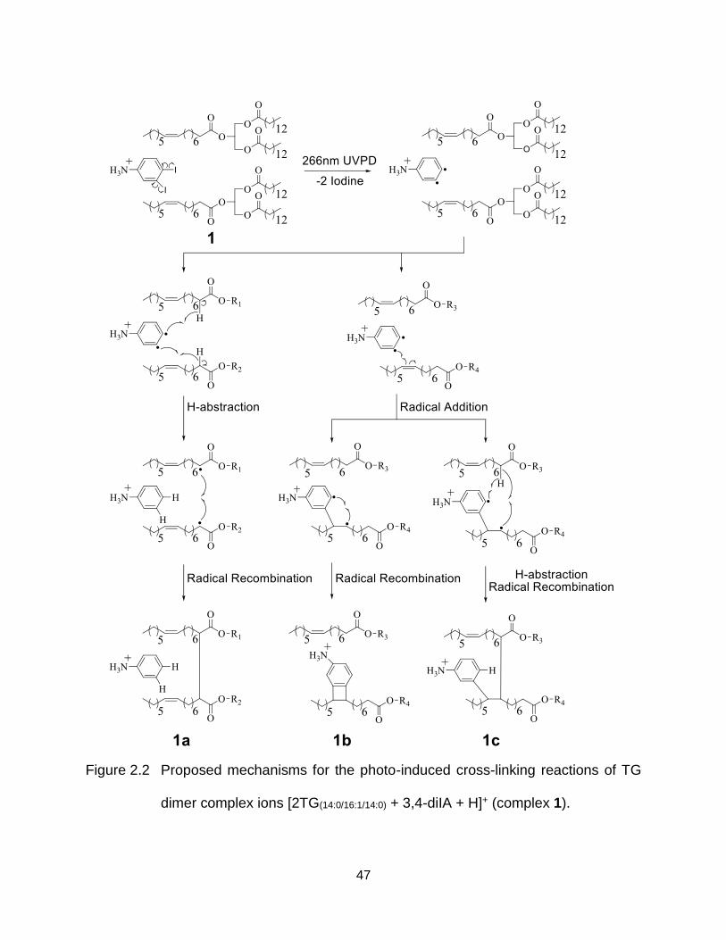
47
Figure 2.2 Proposed mechanisms for the photo-induced cross-linking reactions of TG
dimer complex ions [2TG(14:0/16:1/14:0) + 3,4-diIA + H]+ (complex 1).
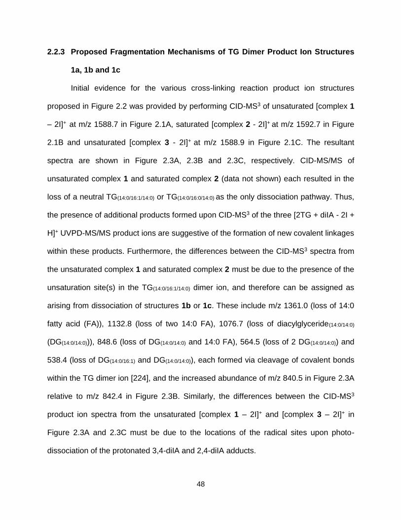
48
2.2.3 Proposed Fragmentation Mechanisms of TG Dimer Product Ion Structures
1a, 1b and 1c
Initial evidence for the various cross-linking reaction product ion structures
proposed in Figure 2.2 was provided by performing CID-MS3 of unsaturated [complex 1
– 2I]+ at m/z 1588.7 in Figure 2.1A, saturated [complex 2 - 2I]+ at m/z 1592.7 in Figure
2.1B and unsaturated [complex 3 - 2I]+ at m/z 1588.9 in Figure 2.1C. The resultant
spectra are shown in Figure 2.3A, 2.3B and 2.3C, respectively. CID-MS/MS of
unsaturated complex 1 and saturated complex 2 (data not shown) each resulted in the
loss of a neutral TG(14:0/16:1/14:0) or TG(14:0/16:0/14:0) as the only dissociation pathway. Thus,
the presence of additional products formed upon CID-MS3 of the three [2TG + diIA - 2I +
H]+ UVPD-MS/MS product ions are suggestive of the formation of new covalent linkages
within these products. Furthermore, the differences between the CID-MS3 spectra from
the unsaturated complex 1 and saturated complex 2 must be due to the presence of the
unsaturation site(s) in the TG(14:0/16:1/14:0) dimer ion, and therefore can be assigned as
arising from dissociation of structures 1b or 1c. These include m/z 1361.0 (loss of 14:0
fatty acid (FA)), 1132.8 (loss of two 14:0 FA), 1076.7 (loss of diacylglyceride(14:0/14:0)
(DG(14:0/14:0))), 848.6 (loss of DG(14:0/14:0) and 14:0 FA), 564.5 (loss of 2 DG(14:0/14:0)) and
538.4 (loss of DG(14:0/16:1) and DG(14:0/14:0)), each formed via cleavage of covalent bonds
within the TG dimer ion [224], and the increased abundance of m/z 840.5 in Figure 2.3A
relative to m/z 842.4 in Figure 2.3B. Similarly, the differences between the CID-MS3
product ion spectra from the unsaturated [complex 1 – 2I]+ and [complex 3 – 2I]+ in
Figure 2.3A and 2.3C must be due to the locations of the radical sites upon photo-
dissociation of the protonated 3,4-diIA and 2,4-diIA adducts.
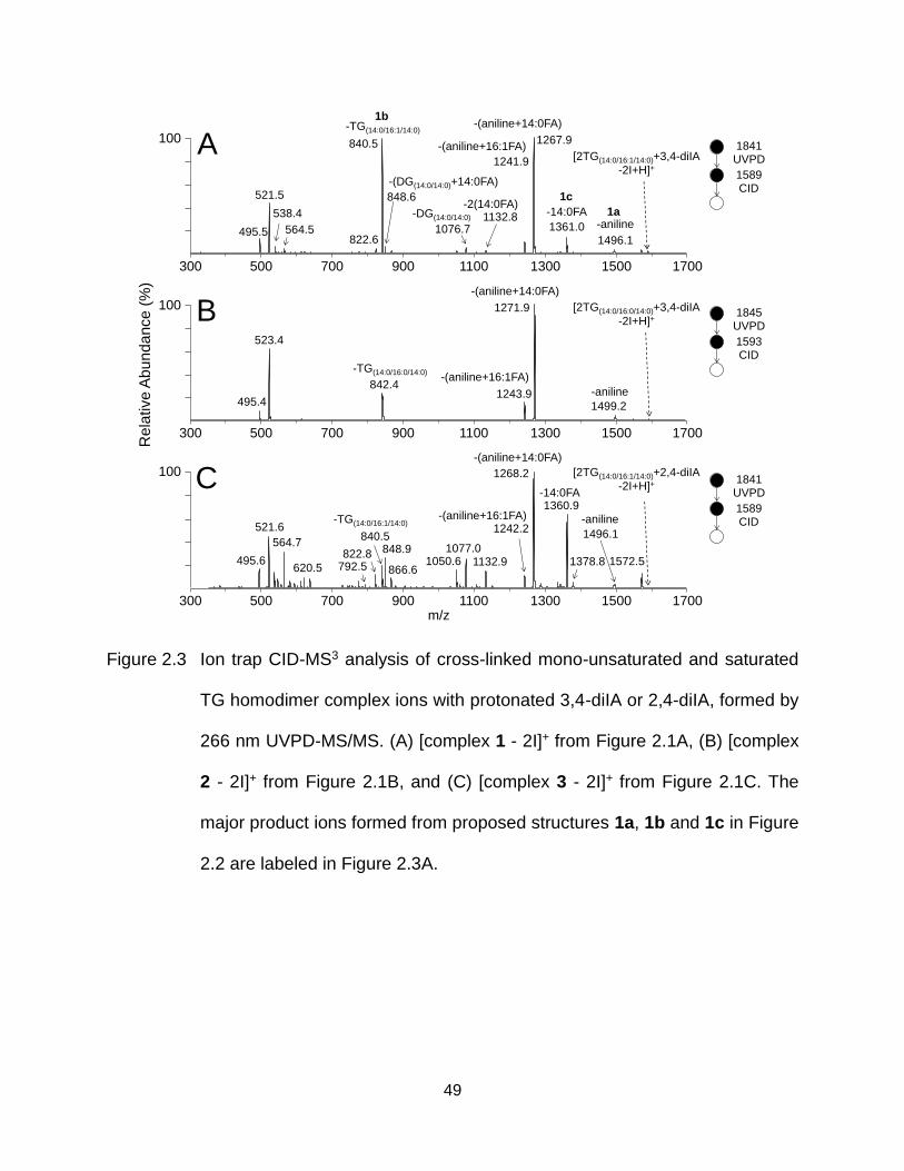
49
Figure 2.3 Ion trap CID-MS3 analysis of cross-linked mono-unsaturated and saturated
TG homodimer complex ions with protonated 3,4-diIA or 2,4-diIA, formed by
266 nm UVPD-MS/MS. (A) [complex 1 - 2I]+ from Figure 2.1A, (B) [complex
2 - 2I]+ from Figure 2.1B, and (C) [complex 3 - 2I]+ from Figure 2.1C. The
major product ions formed from proposed structures 1a, 1b and 1c in Figure
2.2 are labeled in Figure 2.3A.
[2TG(14:0/16:1/14:0)+3,4-diIA
-2I+H]+
1267.9
1361.01496.1
1241.9
1076.7
840.5
495.5
521.5
538.4
564.5
848.6
1132.8
300 500 700 900 1100 1300 1500 1700
-TG(14:0/16:1/14:0)
822.6
-aniline
-(aniline+14:0FA)
-14:0FA-DG(14:0/14:0)
-(DG(14:0/14:0)+14:0FA)
-2(14:0FA)
-(aniline+16:1FA)
1271.9
1499.21243.9
842.4
495.4
523.4
300 500 700 900 1100 1300 1500 1700
-TG(14:0/16:0/14:0)
-aniline
-(aniline+14:0FA)
1268.2
1360.9
1496.1840.5
495.6
521.6
620.5
564.7
300 500 700 900 1100 1300 1500 1700
-TG(14:0/16:1/14:0) -aniline
-(aniline+14:0FA)
-14:0FA
-(aniline+16:1FA)
848.91572.5
1242.2
792.5 866.61378.8
1077.01050.6 1132.9
822.8
Rela
tive A
bun
dance (
%)
100
100
100
m/z
[2TG(14:0/16:0/14:0)+3,4-diIA
-2I+H]+
[2TG(14:0/16:1/14:0)+2,4-diIA
-2I+H]+
-(aniline+16:1FA)
1b
1c
A
B
C
1845UVPD
1593CID
1841UVPD
1589CID
1841UVPD
1589CID
1a
50
To obtain further direct evidence for the proposed 266 nm UVPD-MS/MS product
ion structures 1a, 1b and 1c, each of the major product ions observed in the CID-MS3
spectra shown in Figure 2.3 were then subjected to further dissociation. For example,
the CID-MS4 spectra of m/z 1496.1 [complex 1 - 2I - aniline]+, m/z 840.5 [complex 1 - 2I
- TG(14:0/16:1/14:0)]+ and m/z 1361.0 [complex 1 - 2I - 14:0 FA]+ from Figure 2.3A,
representing first generation products formed from proposed structures 1a, 1b and 1c,
respectively, are shown in Figure 2.4A, 2.4B and 2.4C.
It is reasonable to expect that structure 1a, containing an inter-molecular cross-
link directly between the two TG monomers, would undergo the facile loss of neutral
aniline upon CID-MS3 activation. Thus, CID-MS4 of the [complex 1 - 2I - aniline]+
product was performed (Figure 2.4A), resulting in the dominant loss of a neutral 14:0
fatty acid at m/z 1267.9, that notably was also observed as the dominant product ion in
the CID-MS3 spectra in Figure 2.3A. This ion subsequently underwent further
dissociation (as determined by MS5, and by MS4 of the m/z 1267.9 in Figure 2.3A), to
yield the products at m/z 495.5 and m/z 521.5 in Figure 2.4A (and in Figure 2.3A). The
proposed structures of the m/z 1267.9 and m/z 495.5 ions, and mechanisms for their
formation, are shown in Figure 2.5. The product ion at m/z 521.5, containing a 16:1 fatty
acyl chain, would be formed by a mechanism similar to that for m/z 495.5, but involving
an initial UVPD-MS/MS product ion cross-linked between 16:1 and 14:0 fatty acyl
chains rather than the two 16:1 chains as shown in Figure 2.5. Notably, the spectrum
obtained by CID-MS4 of the m/z 1499.2 and m/z 1496.1 product ions in Figures 2.3B
and 2.3C (i.e., saturated [complex 2 - 2I - aniline]+ with 3,4-diIA and unsaturated
[complex 3 - 2I - aniline]+ with 2,4-diIA) were both similar to that in Figure 2.4A (the
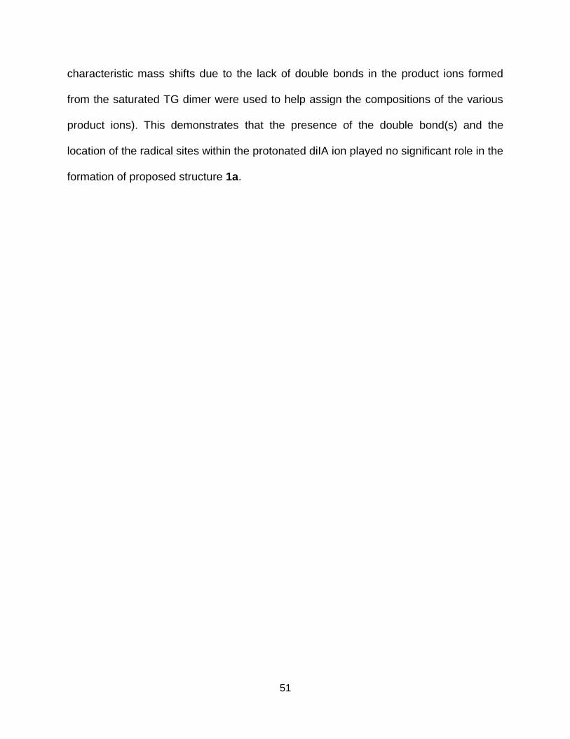
51
characteristic mass shifts due to the lack of double bonds in the product ions formed
from the saturated TG dimer were used to help assign the compositions of the various
product ions). This demonstrates that the presence of the double bond(s) and the
location of the radical sites within the protonated diIA ion played no significant role in the
formation of proposed structure 1a.
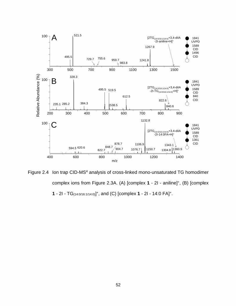
52
Figure 2.4 Ion trap CID-MS4 analysis of cross-linked mono-unsaturated TG homodimer
complex ions from Figure 2.3A. (A) [complex 1 - 2I - aniline]+, (B) [complex
1 - 2I - TG(14:0/16:1/14:0)]+, and (C) [complex 1 - 2I - 14:0 FA]+.
1841UVPD
1589CID1361
CID
300 500 700 900 1100 1500
1267.9
983.8959.7
755.6729.7
521.5
495.5
1300
1241.8
400 600 800 1000 1200 1400
1360.9
1343.1
1304.81150.7
1132.8
1106.9
1076.7904.7
878.7848.7
822.7620.6594.5
200 300 400 500 600 700 800 900
328.3
495.5 519.5
612.5
840.6
822.6384.3285.2235.1 538.5
Rela
tive A
bun
dance (
%)
m/z
100
100
100
[2TG(14:0/16:1/14:0)+3,4-diIA
-2I-aniline+H]+
[2TG(14:0/16:1/14:0)+3,4-diIA
-2I-TG(14:0/16:1/14:0)+H]+
[2TG(14:0/16:1/14:0)+3,4-diIA
-2I-14:0FA+H]+
A
B
C
1841UVPD
1589CID1496
CID
1841UVPD
1589CID840
CID
53
Figure 2.5 Proposed mechanisms for the CID-MS3 and -MS4 fragmentation reactions of [complex 1 - 2I]+ ions
corresponding to structure 1a in Figure 2.2.
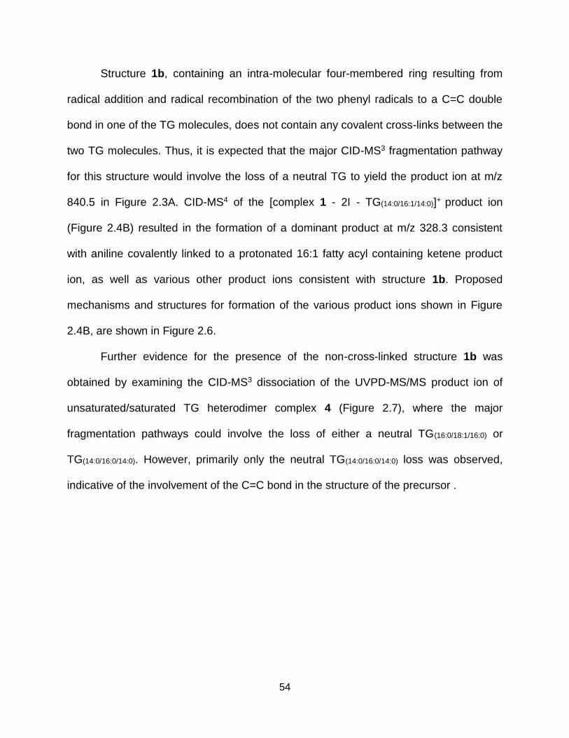
54
Structure 1b, containing an intra-molecular four-membered ring resulting from
radical addition and radical recombination of the two phenyl radicals to a C=C double
bond in one of the TG molecules, does not contain any covalent cross-links between the
two TG molecules. Thus, it is expected that the major CID-MS3 fragmentation pathway
for this structure would involve the loss of a neutral TG to yield the product ion at m/z
840.5 in Figure 2.3A. CID-MS4 of the [complex 1 - 2I - TG(14:0/16:1/14:0)]+ product ion
(Figure 2.4B) resulted in the formation of a dominant product at m/z 328.3 consistent
with aniline covalently linked to a protonated 16:1 fatty acyl containing ketene product
ion, as well as various other product ions consistent with structure 1b. Proposed
mechanisms and structures for formation of the various product ions shown in Figure
2.4B, are shown in Figure 2.6.
Further evidence for the presence of the non-cross-linked structure 1b was
obtained by examining the CID-MS3 dissociation of the UVPD-MS/MS product ion of
unsaturated/saturated TG heterodimer complex 4 (Figure 2.7), where the major
fragmentation pathways could involve the loss of either a neutral TG(16:0/18:1/16:0) or
TG(14:0/16:0/14:0). However, primarily only the neutral TG(14:0/16:0/14:0) loss was observed,
indicative of the involvement of the C=C bond in the structure of the precursor .
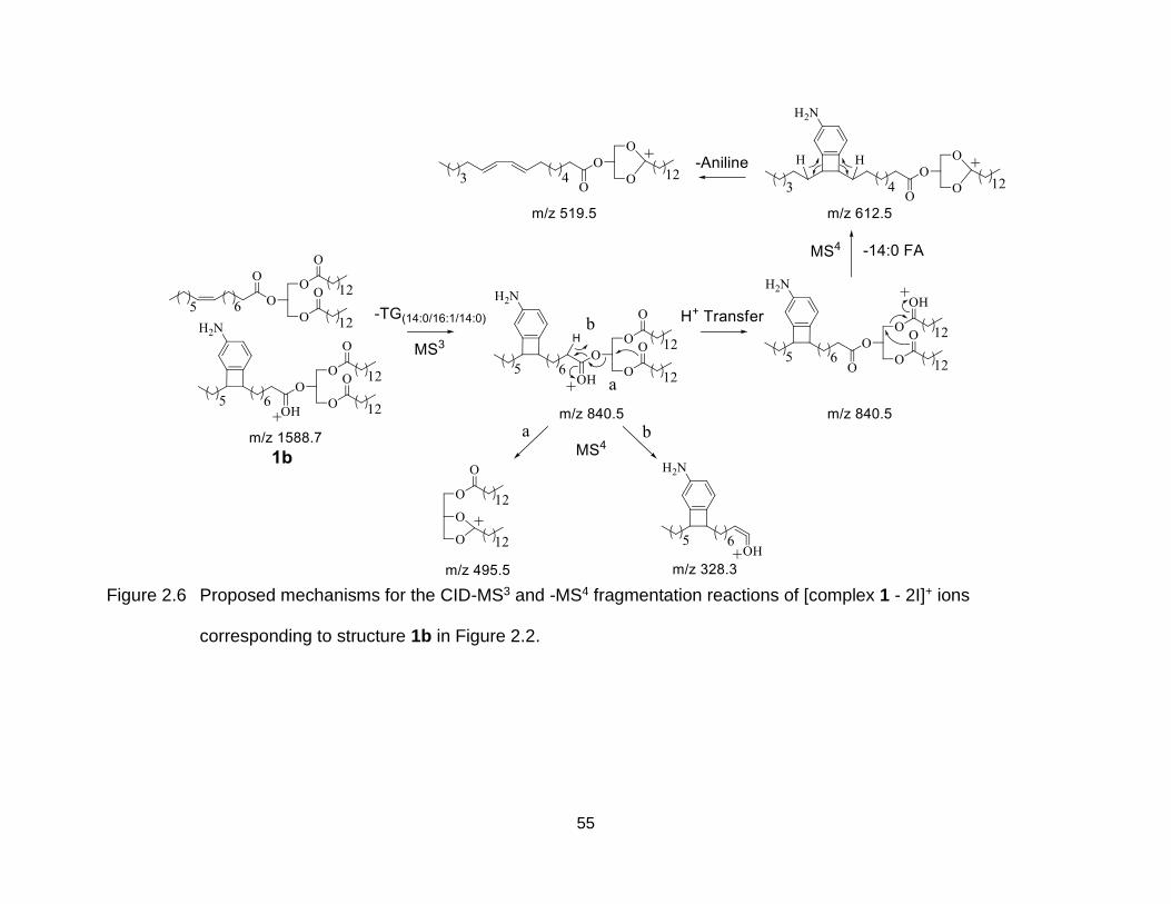
55
Figure 2.6 Proposed mechanisms for the CID-MS3 and -MS4 fragmentation reactions of [complex 1 - 2I]+ ions
corresponding to structure 1b in Figure 2.2.
56
Figure 2.7 Ion trap CID-MS3 analysis of the cross-linked saturated/mono-unsaturated
TG heterodimer complex ion [complex 4 - 2I]+, formed by 266 nm UVPD-
MS/MS.
1000400 600 800 1200 1400 1600
1447.0
1419.0
1581.1
1354.0
1326.0
1106.7
1162.9
1300.0
1134.8
878.7
924.6
842.5523.5
594.5495.5
577.5
-TG(14:0/16:0/14:0)
-TG(16:0/18:1/16:0)
Rela
tive A
bun
dance (
%)
m/z
100
[TG(14:0/16:0/14:0)+TG(16:0/18:1/16:0)
+3,4-diIA-2I+H]+
1927UVPD
1675CID
57
Evidence for structure 1c was obtained by CID-MS4 of the [complex 1 - 2I - 14:0
FA]+ product ion at m/z 1361.0 from Figure 2.3A (Figure 2.4C), in which the dominant
product observed at m/z 1132.8 corresponded to the neutral loss of a second 14:0 fatty
acid molecule (as well as m/z 1106.9 corresponding to the loss of a 16:1 fatty acid), and
various other products consistent with the presence of the anilinyl moiety covalently
linked at the site of unsaturation in the 16:1 fatty acyl chain. Proposed structures and
mechanisms for the formation of these ions are shown in Figure 2.8. Inter-molecular
cross-linking between the two sites of unsaturation (i.e., via sequential radical addition
reactions of the anilinyl biradical with the two separate C=C bonds within the
2TG(14:0/16:1/14:0) homodimer) cannot be ruled out. However, the data obtained from
control experiments involving the TG heterodimer complex 4 consisting of a saturated
TG(14:0/16:0/14:0) and a mono-unsaturated TG(16:0/18:1/16:0) (whose CID-MS3 spectrum is
shown in Figure 2.7) resulted in similar fragmentation behaviors, indicating that
complexes with two versus only one C=C bond have similar cross-linked structures. In
other words, cross-linking between two sites of C=C bond cannot be a dominant
process.
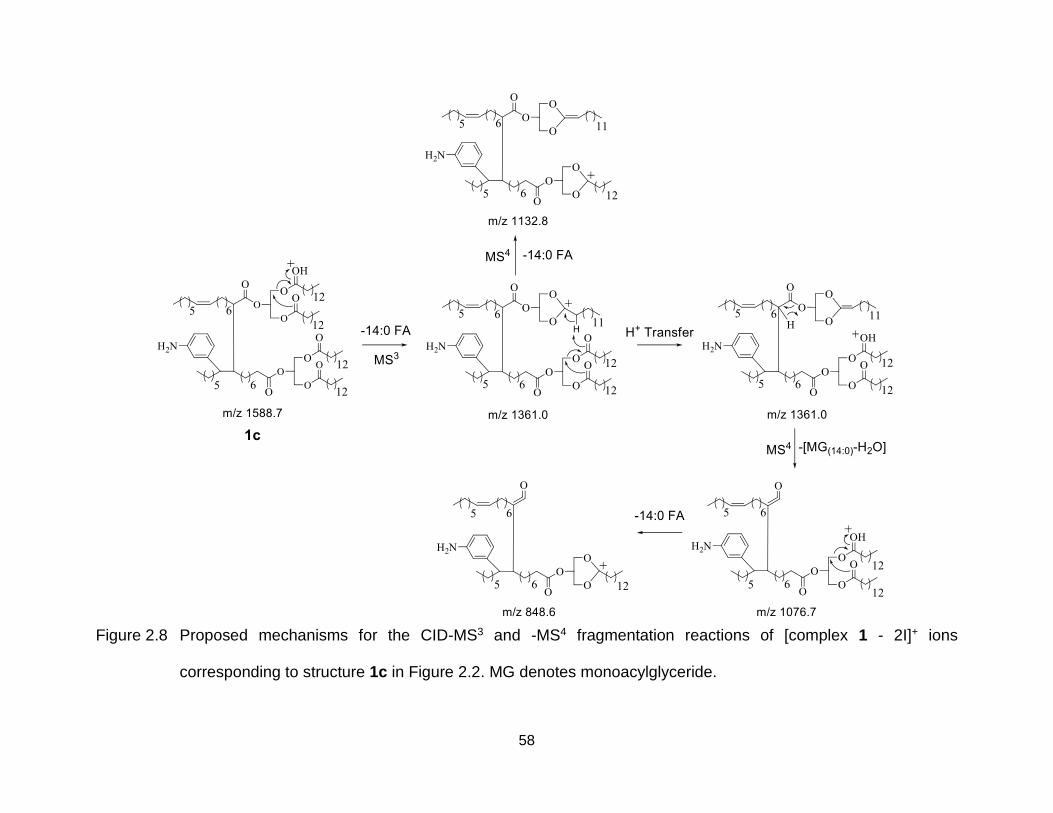
58
Figure 2.8 Proposed mechanisms for the CID-MS3 and -MS4 fragmentation reactions of [complex 1 - 2I]+ ions
corresponding to structure 1c in Figure 2.2. MG denotes monoacylglyceride.
59
2.2.4 Effects of Structures of the Cross-Linker and TG Lipids on the Formation of
Cross-Linked TG Dimer Product Ions
Next, the effect of the structure of the cross-linker on the reactivity and formation
of product ion structures of cross-linked TG dimer product ions was investigated by
using 2,4-diIA versus 3,4-diIA as the cross-linker. No difference in the initial 266 nm
UVPD-MS/MS reaction efficiency was observed as a function of the identity of the
diiodoaniline adduct (e.g., compare the relative abundance of the [2TG(14:0/16:1/14:0) + diIA
- 2I + H]+ product ions in Figures 2.1A and 2.1C). However, a significant decrease in the
formation of the non-cross-linked product ion structure 1b was observed when using
2,4-diIA, as evidenced by the low relative abundance of the ion at m/z 840.5 in Figures
2.1C and 2.3C corresponding to [complex 3 - 2I - TG(14:0/16:1/14:0)]+ compared to Figures
2.1A and 2.3A. A reasonable explanation is that after UVPD-MS/MS, the 3,4-biradical
can readily undergo radical addition and recombination with the C=C bond of the TG
molecule to form a stable four-membered ring. Furthermore, the 3,4-biradical has aryne-
character and thus may undergo concerted [2+2]-cycloaddition at sites of unsaturation
[72, 225]. This mechanism would also result in product 1b and may compete favorably
with the desired cross-linking biradical reactions that yield product types 1a and 1c. In
contrast, the yield of the characteristic product ions for structure 1c were observed to
significantly increase when using the 2,4-diIA adduct compared to 3,4-diIA. This
observation is consistent with the thermochemistry of H-abstraction for the 2,4- and 3,4-
biradicals based on the known bond energies of the benzyne archetype. That is,
experimental measurements find ortho-benzyne to be some 16 kcal mol-1 more stable
that its meta-isomer which translates to an enhanced H-atom affinity in the latter of the
60
same magnitude [223, 226]. While the impact of the local ammonium moiety on the
benzyne energetics is unknown, the stability trend is likely to be preserved and thus
predicts that the 2,4-biradical formed from 2,4-diIA is more likely to initiate H-abstraction
chemistries than its 3,4-isomer, consistent with the observations made here.
2.2.5 Solution Phase Cross-Linking of TG lipids
The reactivity of TG lipids toward cross linking reaction upon activation of 266 nm
UV photon and addition of diiodoaniline cross linker in solution phase was also
investigated. After irradiation of 266 nm UV photons to the sample solution containing
TG(14:0/16:1/14:0) and 3,5-diIA for 9 min, product ions at m/z 1495.9 corresponding to m/z
of [2TG(14:0/16:1/14:0) + 3,5-diIA – aniline - 2I + H]+ were observed in the MS analysis in
Figure 2.9A. In Figure 2.9B, further CID-MS/MS analysis of the product ions at m/z
1495.9 from Figure 2.9A produced similar fragmentation patterns compared to that of
the major cross-linked product ions at m/z 1496 from structure 1a in Figure 2.4A. The
observed similarity suggested that same cross-linked TG lipids were synthesized in both
gas phase and solution phase. Compared to Figure 2.1A, the missing product ions of
[2TG(14:0/16:1/14:0) + 3,5-diIA – aniline - 2I + H]+ in Figure 2.9A was possibly due to the fact
that the required irradiation time (~minutes) of solution phase TG cross-linking reactions
in order to observe abundant product ions was much longer than that in gas phase (<=
30 ms). Thus most of the [2TG(14:0/16:1/14:0) + 3,5-diIA -2I + H]+ ions were activated by UV
photons and further fragmented to loss an aniline.
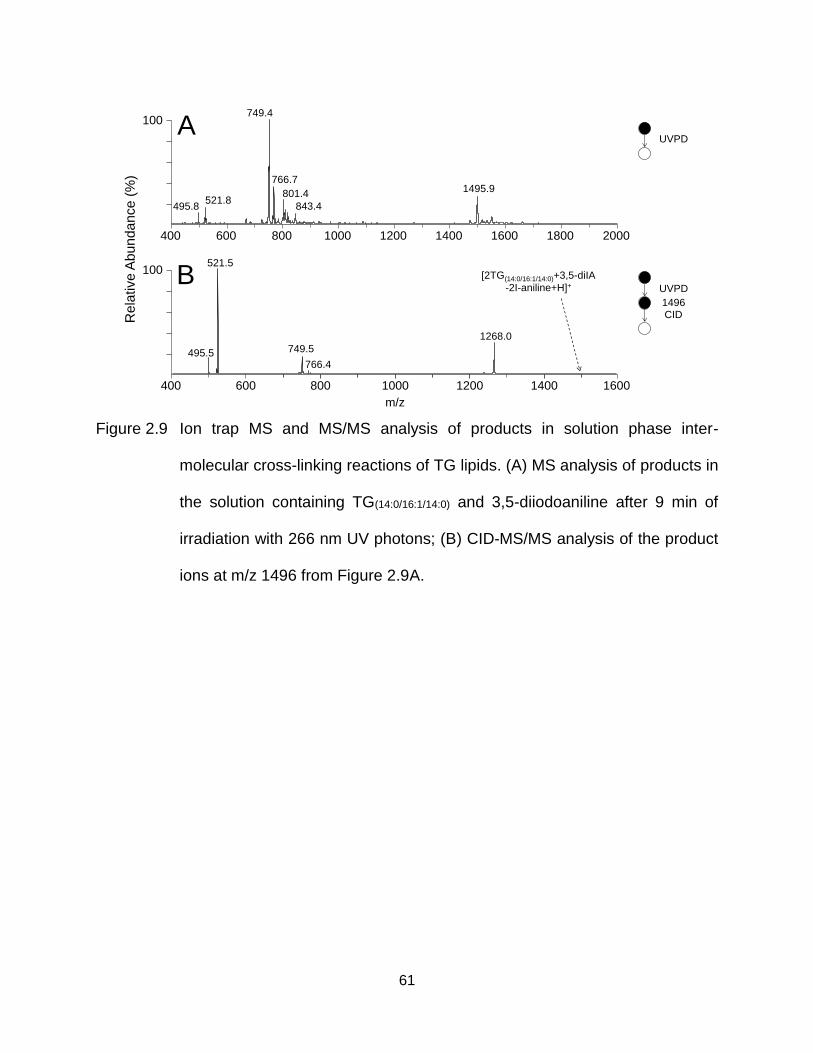
61
Figure 2.9 Ion trap MS and MS/MS analysis of products in solution phase inter-
molecular cross-linking reactions of TG lipids. (A) MS analysis of products in
the solution containing TG(14:0/16:1/14:0) and 3,5-diiodoaniline after 9 min of
irradiation with 266 nm UV photons; (B) CID-MS/MS analysis of the product
ions at m/z 1496 from Figure 2.9A.
400 600 800 1000 1200 1400 1600 1800 2000
400 600 800 1000 1200 1400 1600
Rela
tive A
bun
dance (
%)
100
100
A
B
UVPD
749.4
1495.9766.7
521.8843.4495.8
801.4
521.5
1268.0
749.5495.5766.4
[2TG(14:0/16:1/14:0)+3,5-diIA
-2I-aniline+H]+ UVPD
1496CID
m/z
62
2.3 Conclusions
The inter-molecular gas phase cross-linking of TG lipid dimers has been
demonstrated using anilinyl biradical ions formed via 266 nm UVPD-MS/MS of
protonated diiodoaniline adducts. Using multistage CID-MS3, -MS4 and –MS5, cross-
linking was shown to occur via sequential combinations of H-abstraction, radical
addition and recombination reactions. The efficiency of the UVPD reactions, and the
competition between the various inter-molecular cross-linking and non-cross-linking
reaction pathways, were shown to be dependent on the number of unsaturation sites
present within the TG lipids, as well as on the locations of the biradical sites within the
protonated aniline adduct ion. The reactivity of cross linking reactions in the gas phase
were observed to be identical to those obtained upon photo irradiation of the TG lipids in
the presence of 3,5-diiodoaniline in solution. Importantly, while there are many possible
inter-molecular cross-linked structures that may result due to the large number of sites
at which H-abstraction can occur (and in which the aniline adduct may or may not be
covalently incorporated), this type of novel gas phase cross-linking reaction provides a
method to covalently link two molecules without the requirement of any specific
functional group. Thus, the same approach could be applied to perform gas phase
cross-linking within a wide range of biomolecular classes.
63
CHAPTER THREE
Characterization and Multiplexed Quantification of Derivatized
Aminophospholipids
3.1 Introduction
Glycerophosphoethanolamine (PE) and glycerophosphoserine (PS) lipids are
subclasses of glycerophospholipids containing a primary amine functional group
attached to the terminal end of the phosphoglycerol backbone. Under normal
physiological conditions in eukaryotic cells, anionic PE and PS lipids reside
predominantly or exclusively in the inner leaflet of the plasma membrane, respectively,
where they play important roles in regulating various biological processes including
intra- and inter-cellular signaling, phagocytic recognition, apoptotic cell clearance, cell
division, angiogenesis and vascular remodeling [227-230]. Abnormalities in the
metabolic regulation of aminophospholipid compositions and distributions therefore
have established roles in human diseases including cancer, diabetes, obesity and
neurodegeneration [231-235].
Significant advances in lipidomics over the past ten years have largely been
driven by developments in mass spectrometry instrumentation and associated methods
for the comprehensive identification, structural characterization and quantification of the
array of structurally diverse lipid classes and molecular lipid species that may be
present within a cell, tissue or body fluid (e.g., plasma) sample of interest [66]. To date
however, most of the developed quantification strategies have required the separate
The results described in this chapter were published in: Nie, S., Fhaner, C.J.,
Liu, S., Peake, D., Kiyonami, R., Huang, Y. and Reid, G.E. Characterization and
multiplexed quantification of derivatized aminophospholipids. Int. J. Mass Spectrom.
2015, doi:10.1016/j.ijms.2015.07.002.
64
analysis of each sample, with appropriate technical replicates and corrections based on
the inclusion of internal standards, in order to be able to draw any conclusions regarding
the biological significance of changes observed between the samples. This is a
particular challenge for certain lipid classes, e.g., PS lipids that are observed as only
minor components of eukaryotic membranes. Improved sensitivity for the positive
ionization mode MS detection of low abundant aminophospholipids has been
demonstrated using various chemical labeling methods (i.e., incorporation of fixed
charge and/or hydrophobic groups) [24, 91, 92].
For mass spectrometry-based absolute quantification of molecular lipid
concentrations, isotopically labeled internal standards that are chemically identical to
the lipid molecule of interest are required. However, this is often impractical due to the
limited number of lipid internal standards that are commercially available, and the
estimated thousands of possible lipid structures that may be present. Alternatively,
albeit with its own potential limitations, a single internal standard for a lipid class can be
used [23]. Thus, many groups chose to report quantification results using relative, rather
than absolute, methods which require internal standards only for normalization of
differences in technical variances between the samples [24, 138, 236-238].
An emerging method for improving the accuracy of mass spectrometry-based
relative quantification of lipids involves the use of isobaric mass stable isotope labeling
derivatization reagents, coupled with MS/MS analysis, analogous to those used in
contemporary quantitative proteomics e.g., iTRAQ or TMTs as described in Chapter one,
section 1.2.1.3.3 [239]. For example, primary amine specific iTRAQ and isotope-labeled
4-(dimethylamino)benzoic acid NHS ester derivatization have been employed for
65
quantification of aminophospholipids [194-196] and hydrolyzed ceramide lipids [197].
Quantitative information can be readily obtained by ratiometric measurement of the
abundances of isotopically encoded low mass reporter ions from each sample, upon
MS/MS of their isobaric mass precursor ions. Using these methods, run-to-run or even
scan-to-scan variance in lipid ion abundances can be avoided since different samples
are ionized and mass analyzed simultaneously after separate derivatization and mixing.
However, special attentions need to be paid to avoid the interference in the intensities of
low mass reporter ions caused by the presence of co-isolated and fragmented precursor
ions present within the isolation window that is used prior to dissociation. For example,
diacyl-PE or -PS lipids have only a Δm of 0.0363 Da compared to ether-PE or -PS lipids
which contain the same degree of unsaturation but with one more carbon on their acyl,
alkyl or alkenyl chains. Although MS3 dissociation, gas phase purification using proton
transfer reaction and ion mobility separation methods have been developed to
overcome the similar limitation for quantitative proteomic studies, they are not
necessarily applicable to lipid analysis due to the decreased sensitivity [201] or the need
for multiply charged precursor ions [202] or specific instrumentation [203]. Moreover,
structural information on the acyl, alkyl or alkenyl chain compositions of
aminophospholipid ions cannot be acquired in positive ionization mode using current
isotope tagging methods, which compromises the throughput of analysis if structural
characterization of lipid molecular species is desired.
Lu et al. have previously reported the development and application of an isobaric
mass stable isotopic labeled derivatization strategy using d6-‘heavy’ and d6-‘light’
variants of the DMBNHS reagent described above, coupled with high
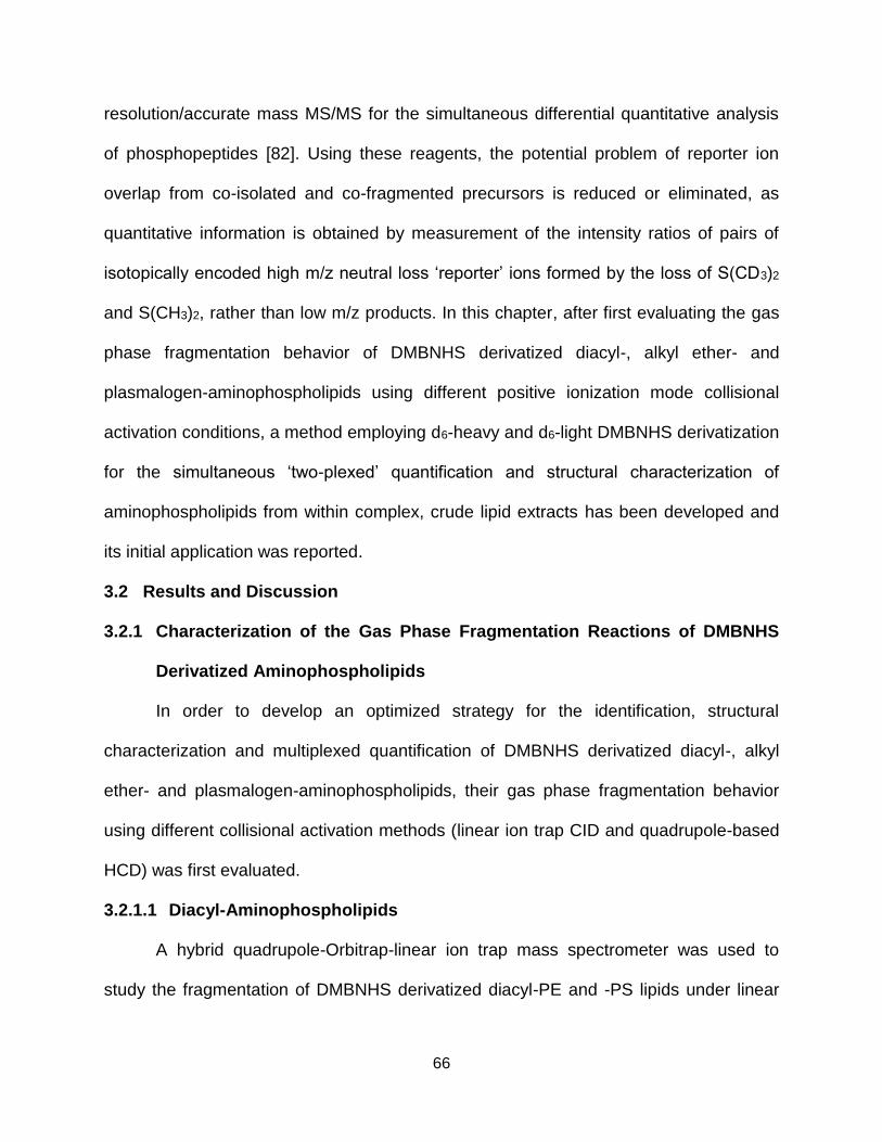
66
resolution/accurate mass MS/MS for the simultaneous differential quantitative analysis
of phosphopeptides [82]. Using these reagents, the potential problem of reporter ion
overlap from co-isolated and co-fragmented precursors is reduced or eliminated, as
quantitative information is obtained by measurement of the intensity ratios of pairs of
isotopically encoded high m/z neutral loss ‘reporter’ ions formed by the loss of S(CD3)2
and S(CH3)2, rather than low m/z products. In this chapter, after first evaluating the gas
phase fragmentation behavior of DMBNHS derivatized diacyl-, alkyl ether- and
plasmalogen-aminophospholipids using different positive ionization mode collisional
activation conditions, a method employing d6-heavy and d6-light DMBNHS derivatization
for the simultaneous ‘two-plexed’ quantification and structural characterization of
aminophospholipids from within complex, crude lipid extracts has been developed and
its initial application was reported.
3.2 Results and Discussion
3.2.1 Characterization of the Gas Phase Fragmentation Reactions of DMBNHS
Derivatized Aminophospholipids
In order to develop an optimized strategy for the identification, structural
characterization and multiplexed quantification of DMBNHS derivatized diacyl-, alkyl
ether- and plasmalogen-aminophospholipids, their gas phase fragmentation behavior
using different collisional activation methods (linear ion trap CID and quadrupole-based
HCD) was first evaluated.
3.2.1.1 Diacyl-Aminophospholipids
A hybrid quadrupole-Orbitrap-linear ion trap mass spectrometer was used to
study the fragmentation of DMBNHS derivatized diacyl-PE and -PS lipids under linear
67
ion trap CID and quadrupole-based HCD ion activation conditions. Representative
examples of the HCD-MS/MS fragmentation of DMBNHS derivatized PE(18:0/18:1) and
PS(16:0/18:1) lipids are shown in Figure 3.1A and 3.1B, respectively. Proposed
mechanisms for the observed fragmentation reactions are shown in Figure 3.2.
Alternative mechanisms involving SN2 nucleophilic attack by one of the acyl chain
carbonyl groups onto the glycerol sn-3 position are also possible, similar to that
previously demonstrated for the fragmentation of protonated PC lipids [35]. Further
experiments using deuterium labeled lipids would be required to determine the actual
mechanism that is responsible. However, both mechanisms yield product ion structures
with the same elemental compositions, so the specific mechanism that is responsible
does not negatively affect their assignment. In both spectra, a characteristic ‘reporter’
neutral loss of dimethylsulfide (S(CH3)2, 62 Da) was observed as the initial
fragmentation channel, similar to that previously described for other DMBNHS
derivatized primary amine-containing biomolecules [82, 127]. The product ions at m/z
605 in Figure 3.1A and m/z 577 in Figure 3.1B both correspond to the sequential loss of
an additional 209 Da (total neutral loss of 271 Da) or 253 Da (total neutral loss of 315
Da) from the initial [M-S(CH3)2]+ product ions. These ions are analogous to the neutral
losses of 141 Da and 185 Da observed from non-derivatized protonated PE and PS
lipids, and were accompanied by complementary ‘reporter’ protonated product ions at
m/z 210 and 254 [2]. The observation of these low m/z ‘reporter’ ions is most likely due
to the higher proton affinity of the phosphoethanolaminohydrofuran containing
structures in these products compared to the phosphoethanolamine or phosphoserine
containing structures formed from the non-derivatized lipids. The decreased relative
68
abundance of the m/z 254 product ion in the spectrum from the derivatized PS lipid
compared to m/z 210 for the PE lipid, and the corresponding increase in its
complementary m/z 605 neutral loss product, is likely due to its decreased proton
affinity resulting from the presence of the carboxylic acid group. Linear ion trap CID-
MS/MS spectra from the DMBNHS derivatized diacyl PE(18:0/18:0) and PS(16:0/18:1) lipids
were also acquired (data now shown). The exclusive neutral loss of S(CH3)2 (62 Da)
was observed as the only product for both the PE(18:0/18:0) and PS(16:0/18:1) lipids. CID-MS3
of these neutral loss product ions (data not shown) resulted in the formation of
secondary products and relative abundances similar to those seen in Figure 3.1A and
3.1B, thereby confirming the sequential nature of the fragmentation reactions proposed
in Figure 3.2.
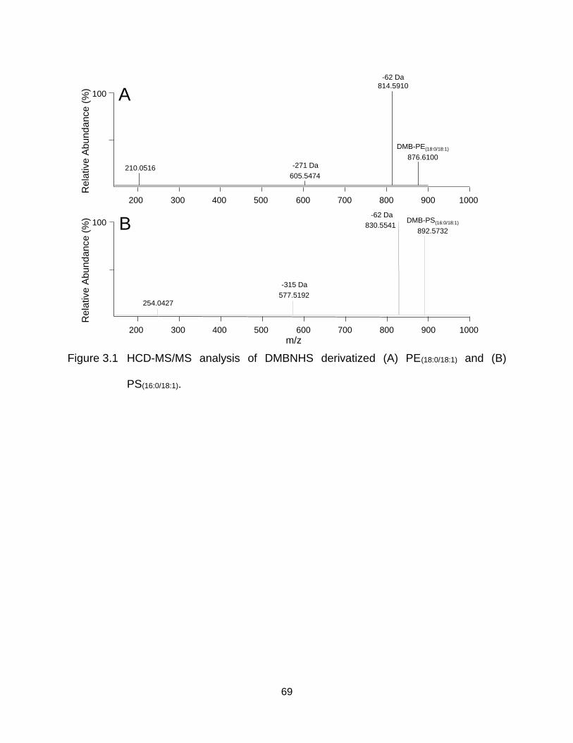
69
Figure 3.1 HCD-MS/MS analysis of DMBNHS derivatized (A) PE(18:0/18:1) and (B)
PS(16:0/18:1).
100 A
m/z
Re
lative
Ab
un
da
nce
(%)
Re
lative
Ab
un
da
nce
(%)
100 B
DMB-PE(18:0/18:1)
876.6100
892.5732
DMB-PS(16:0/18:1)830.5541
577.5192254.0427
814.5910
210.0516605.5474
200 300 400 500 600 700 800 900 1000
200 300 400 500 600 700 800 900 1000
-62 Da
-62 Da
-271 Da
-315 Da
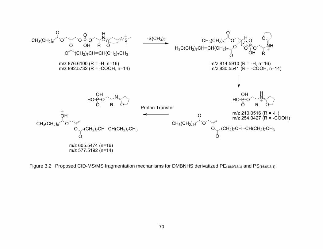
70
Figure 3.2 Proposed CID-MS/MS fragmentation mechanisms for DMBNHS derivatized PE(18:0/18:1) and PS(16:0/18:1).
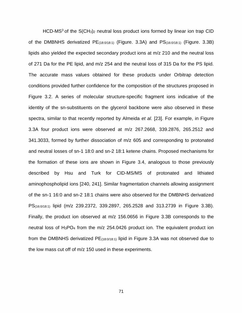
71
HCD-MS3 of the S(CH3)2 neutral loss product ions formed by linear ion trap CID
of the DMBNHS derivatized PE(18:0/18:1) (Figure. 3.3A) and PS(16:0/18:1) (Figure. 3.3B)
lipids also yielded the expected secondary product ions at m/z 210 and the neutral loss
of 271 Da for the PE lipid, and m/z 254 and the neutral loss of 315 Da for the PS lipid.
The accurate mass values obtained for these products under Orbitrap detection
conditions provided further confidence for the composition of the structures proposed in
Figure 3.2. A series of molecular structure-specific fragment ions indicative of the
identity of the sn-substituents on the glycerol backbone were also observed in these
spectra, similar to that recently reported by Almeida et al. [23]. For example, in Figure
3.3A four product ions were observed at m/z 267.2668, 339.2876, 265.2512 and
341.3033, formed by further dissociation of m/z 605 and corresponding to protonated
and neutral losses of sn-1 18:0 and sn-2 18:1 ketene chains. Proposed mechanisms for
the formation of these ions are shown in Figure 3.4, analogous to those previously
described by Hsu and Turk for CID-MS/MS of protonated and lithiated
aminophospholipid ions [240, 241]. Similar fragmentation channels allowing assignment
of the sn-1 16:0 and sn-2 18:1 chains were also observed for the DMBNHS derivatized
PS(16:0/18:1) lipid (m/z 239.2372, 339.2897, 265.2528 and 313.2739 in Figure 3.3B).
Finally, the product ion observed at m/z 156.0656 in Figure 3.3B corresponds to the
neutral loss of H3PO4 from the m/z 254.0426 product ion. The equivalent product ion
from the DMBNHS derivatized PE(18:0/18:1) lipid in Figure 3.3A was not observed due to
the low mass cut off of m/z 150 used in these experiments.
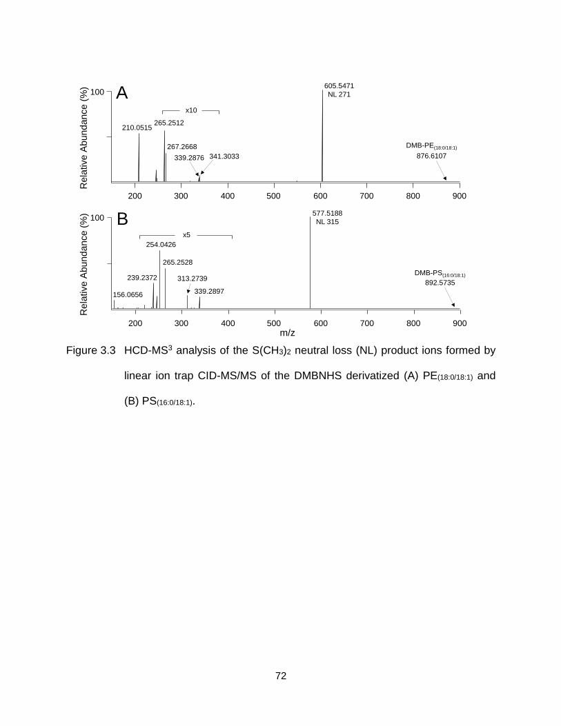
72
Figure 3.3 HCD-MS3 analysis of the S(CH3)2 neutral loss (NL) product ions formed by
linear ion trap CID-MS/MS of the DMBNHS derivatized (A) PE(18:0/18:1) and
(B) PS(16:0/18:1).
100
200 300 400 500 600 700 800 900
x10
339.2876 341.3033
265.2512
267.2668
210.0515
605.5471
NL 271A
m/z
Re
lative
Ab
un
da
nce
(%)
Re
lative
Ab
un
da
nce
(%)
100
200 300 400 500 600 700 800 900
x5
265.2528
239.2372
254.0426
339.2897
313.2739
156.0656
577.5188
NL 315B
DMB-PE(18:0/18:1)
876.6107
892.5735
DMB-PS(16:0/18:1)
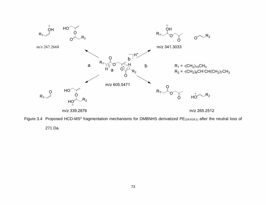
73
Figure 3.4 Proposed HCD-MS3 fragmentation mechanisms for DMBNHS derivatized PE(18:0/18:1) after the neutral loss of
271 Da.
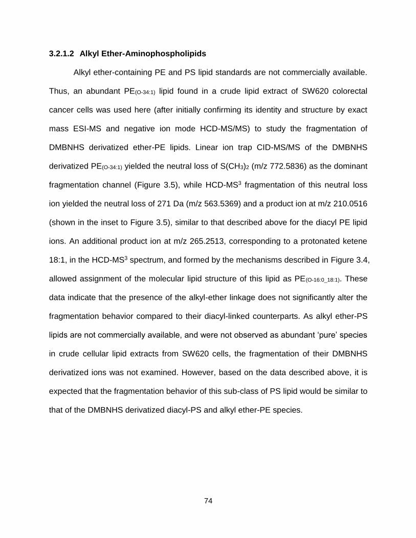
74
3.2.1.2 Alkyl Ether-Aminophospholipids
Alkyl ether-containing PE and PS lipid standards are not commercially available.
Thus, an abundant PE(O-34:1) lipid found in a crude lipid extract of SW620 colorectal
cancer cells was used here (after initially confirming its identity and structure by exact
mass ESI-MS and negative ion mode HCD-MS/MS) to study the fragmentation of
DMBNHS derivatized ether-PE lipids. Linear ion trap CID-MS/MS of the DMBNHS
derivatized PE(O-34:1) yielded the neutral loss of S(CH3)2 (m/z 772.5836) as the dominant
fragmentation channel (Figure 3.5), while HCD-MS3 fragmentation of this neutral loss
ion yielded the neutral loss of 271 Da (m/z 563.5369) and a product ion at m/z 210.0516
(shown in the inset to Figure 3.5), similar to that described above for the diacyl PE lipid
ions. An additional product ion at m/z 265.2513, corresponding to a protonated ketene
18:1, in the HCD-MS3 spectrum, and formed by the mechanisms described in Figure 3.4,
allowed assignment of the molecular lipid structure of this lipid as PE(O-16:0_18:1). These
data indicate that the presence of the alkyl-ether linkage does not significantly alter the
fragmentation behavior compared to their diacyl-linked counterparts. As alkyl ether-PS
lipids are not commercially available, and were not observed as abundant ‘pure’ species
in crude cellular lipid extracts from SW620 cells, the fragmentation of their DMBNHS
derivatized ions was not examined. However, based on the data described above, it is
expected that the fragmentation behavior of this sub-class of PS lipid would be similar to
that of the DMBNHS derivatized diacyl-PS and alkyl ether-PE species.
75
Figure 3.5 CID-MS/MS analysis of a DMBNHS derivatized monoalkyl-ether PE lipid
PE(O-16:0_18:1) from a crude lipid extract of SW620 colorectal cancer cells.
The inset shows the HCD-MS3 spectrum of the S(CH3)2 neutral loss (NL)
product ion.
100772.5836
-S(CH3)2
834.5978
816.6112
Rela
tive A
bun
dance
(%)
m/z
[DMB-PE(O-16:0_18:1)]+
200 300 400 500 600 700 800 900
200 300 400 500 600 700
265.2513 508.3399
563.5369
NL 271210.0516
76
3.2.1.3 Plasmalogen-Aminophospholipids
Fhaner et al. have previously reported the HCD-MS/MS fragmentation behavior
of plasmalogen-PE lipids following sequential DMBNHS derivatization of the amine
head group and iodine/methanol derivatization of the plasmalogen O-alkenyl-ether
double bond [137]. From this prior study, the neutral loss of dimethylsulfide was
observed as the primary fragmentation pathway, similar to that described above for the
DMBNHS derivatized diacyl- and alkyl ether- PE lipids. In contrast to these lipid sub-
classes however, this product then underwent further dissociation to yield a dominant
product corresponding to neutral loss of a characteristic RCICH(OCH3) alkene (i.e., the
derivatized plasmalogen O-alkenyl chain), that directly enables assignment of the
molecular lipid identity. Albeit lower in abundance relative to the above mentioned
fragmentation channel, the initial neutral loss product ion also underwent further
dissociation (following the loss of methanol) to yield the PE headgroup specific ion at
m/z 210 and its complementary neutral loss product, identical to those described above
for the DMBNHS derivatized diacyl- and alkyl ether- PE species.
Systematic examination of the fragmentation behavior of DMBNHS and
iodine/methanol derivatized plasmalogen PS lipids could not be performed here as
plasmalogen PS lipid standards are not commercially available, and are not observed
as abundant ‘pure’ species in crude cellular lipid extracts from SW620 cells. However,
similar fragmentation behavior is expected to that described above for plasmalogen-PE
lipids, given that the differences in head group structures do not play a significant role in
the observed fragmentation reactions.
77
3.2.2 Characterization and Quantification of Isobaric Stable Isotope Containing
DMBNHS Derivatized Aminophospholipid Standards
Lu et al. have previously reported the application of d6-‘heavy’ and d6-‘light’
DMBNHS reagents as an isobaric stable isotope-labeled derivatization strategy for the
enhanced electrospray ionization and simultaneous (multiplexed) quantitative analysis
of primary amine-containing phosphopeptides [82]. Here, using sequential amine
specific d6-heavy/light DMBNHS and plasmalogen O-alkenyl-ether specific
iodine/methanol derivatization, and the unique gas phase fragmentation behaviors
discussed above, a ‘targeted’ high resolution/accurate mass ESI-MS and -MS/MS
method for simultaneous (multiplexed) aminophospholipid quantification at the sum
composition or molecular lipid levels [66], and further molecular lipid structural
characterization, was developed. This method was first evaluated by derivatization of a
diacyl-PE(14:0/14:0) lipid standard with either d6-heavy or d6-light DMBNHS reagents, then
mixing them together at a 1:1 ratio. Following ESI-MS, isolation of the isobaric mass
precursor ions and linear ion trap CID-MS/MS, the exclusive formation of a pair of
product ions differing by 6 Da were observed, corresponding to the neutral losses of
S(CD3)2 and S(CH3)2 (m/z 704.4823 and 710.5194) from the ‘heavy’ and ‘light’ labeled
lipids (Figure 3.6A). After correction for the isotope purity of the d6-heavy/light DMBNHS
reagents (99% and 83%, respectively), the intensities of these ‘reporter’ ions can be
directly used for sum composition level relative quantification of the concentrations of
the lipids between the two samples. A linear dynamic range of quantitation was
observed across more than 3 orders of magnitude (R2 = 0.99) for this measurement, by
monitoring the intensity ratio’s of the CID-MS/MS S(CD3)2 and S(CH3)2 neutral loss
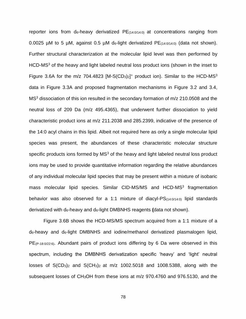
78
reporter ions from d6-heavy derivatized PE(14:0/14:0) at concentrations ranging from
0.0025 µM to 5 µM, against 0.5 µM d6-light derivatized PE(14:0/14:0) (data not shown).
Further structural characterization at the molecular lipid level was then performed by
HCD-MS3 of the heavy and light labeled neutral loss product ions (shown in the inset to
Figure 3.6A for the m/z 704.4823 [M-S(CD3)2]+ product ion). Similar to the HCD-MS3
data in Figure 3.3A and proposed fragmentation mechanisms in Figure 3.2 and 3.4,
MS3 dissociation of this ion resulted in the secondary formation of m/z 210.0508 and the
neutral loss of 209 Da (m/z 495.4365), that underwent further dissociation to yield
characteristic product ions at m/z 211.2038 and 285.2399, indicative of the presence of
the 14:0 acyl chains in this lipid. Albeit not required here as only a single molecular lipid
species was present, the abundances of these characteristic molecular structure
specific products ions formed by MS3 of the heavy and light labeled neutral loss product
ions may be used to provide quantitative information regarding the relative abundances
of any individual molecular lipid species that may be present within a mixture of isobaric
mass molecular lipid species. Similar CID-MS/MS and HCD-MS3 fragmentation
behavior was also observed for a 1:1 mixture of diacyl-PS(14:0/14:0) lipid standards
derivatized with d6-heavy and d6-light DMBNHS reagents (data not shown).
Figure 3.6B shows the HCD-MS/MS spectrum acquired from a 1:1 mixture of a
d6-heavy and d6-light DMBNHS and iodine/methanol derivatized plasmalogen lipid,
PE(P-18:0/22:6). Abundant pairs of product ions differing by 6 Da were observed in this
spectrum, including the DMBNHS derivatization specific ‘heavy’ and ‘light’ neutral
losses of S(CD3)2 and S(CH3)2 at m/z 1002.5018 and 1008.5388, along with the
subsequent losses of CH3OH from these ions at m/z 970.4760 and 976.5130, and the
79
DMBNHS derivatized PE head group specific pair of low m/z ‘reporter’ ions at 210.0515
and 216.0891. However, via the fragmentation pathways previously reported [137],
formation of a pair of isotopically encoded product ions at m/z 594.3157 and m/z
600.3530, via neutral loss of the derivatized plasmalogen O-alkenyl chain, directly
enabled simultaneous quantification and structural characterization at the molecular
lipid level.
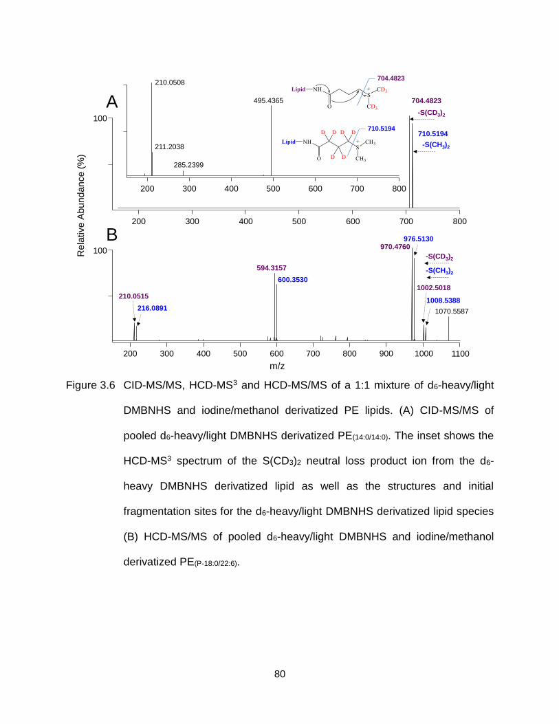
80
Figure 3.6 CID-MS/MS, HCD-MS3 and HCD-MS/MS of a 1:1 mixture of d6-heavy/light
DMBNHS and iodine/methanol derivatized PE lipids. (A) CID-MS/MS of
pooled d6-heavy/light DMBNHS derivatized PE(14:0/14:0). The inset shows the
HCD-MS3 spectrum of the S(CD3)2 neutral loss product ion from the d6-
heavy DMBNHS derivatized lipid as well as the structures and initial
fragmentation sites for the d6-heavy/light DMBNHS derivatized lipid species
(B) HCD-MS/MS of pooled d6-heavy/light DMBNHS and iodine/methanol
derivatized PE(P-18:0/22:6).
100
710.5194
704.4823
-S(CD3)2
-S(CH3)2
A
200 300 400 500 600 700 800 900 1000
216.0891
210.0515
1002.5018
970.4760
1008.5388
976.5130
594.3157
600.3530
1070.5587
100
-S(CH3)2
-S(CD3)2
m/z
1100
200 300 400 500 600 700 800
200 300 400 500 600 700 800
210.0508
495.4365
285.2399
211.2038
B
Re
lative A
bu
nd
an
ce
(%)
704.4823
710.5194
81
3.2.3 Multiplexed Quantification of Selected Aminophospholipid Ion Abundance
Changes Between a Metastatic Colorectal Cancer Cell Line, SW620 and its
AlkyGlycerone Phosphate Synthase (AGPS) siRNA Knockdown
To demonstrate the ‘proof of principle’ utility of the d6-heavy/light DMBNHS and
iodine/methanol derivatization strategy coupled with high resolution/accurate mass ESI-
MS and ‘targeted’ HCD-MS/MS for multiplexed quantification/characterization of
aminophospholipids within complex lipid mixtures, a 1:1 mixture of crude lipid extracts
(normalized to protein concentration) from a metastatic colorectal cancer cell line,
SW620 and its alkyglycerone phosphate synthase (AGPS) silencing RNA (siRNA)
knockdown, was analyzed. AGPS is the rate limiting enzyme involved in the
biosynthesis of cellular ether lipids. Fhaner et al. have previously found that the levels of
several ether-containing lipid sub classes (both alkyl-ether and plasmalogens) are
significantly enriched in the SW620 cell line compared to its isogenic primary colorectal
cancer cell line, SW480 [24]. Thus, siRNA knockdown of these lipids by AGPS is
expected to be useful for subsequent functional studies to elucidate the role of ether-
lipid metabolism in malignancy and metastatic progression.
Figure 3.7 shows the full scan ESI mass spectrum obtained from the 1:1 crude
lipid extract mixture of d6-heavy/light DMBNHS and iodine/methanol derivatized SW620
cells and its AGPS knockdown, with insets showing selected expanded m/z regions of
the mass spectrum. After database searching of the high resolution/accurate mass
values, 125 total PE and PS lipid ions were assigned at the sum composition level.
Then, a non-redundant inclusion list of 101 m/z values (multiple PE and PS lipids were
observed with the same nominal mass but different exact mass values), was submitted
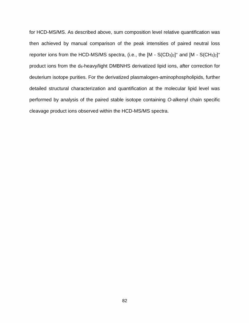
82
for HCD-MS/MS. As described above, sum composition level relative quantification was
then achieved by manual comparison of the peak intensities of paired neutral loss
reporter ions from the HCD-MS/MS spectra, (i.e., the [M - S(CD3)2]+ and [M - S(CH3)2]+
product ions from the d6-heavy/light DMBNHS derivatized lipid ions, after correction for
deuterium isotope purities. For the derivatized plasmalogen-aminophospholipids, further
detailed structural characterization and quantification at the molecular lipid level was
performed by analysis of the paired stable isotope containing O-alkenyl chain specific
cleavage product ions observed within the HCD-MS/MS spectra.
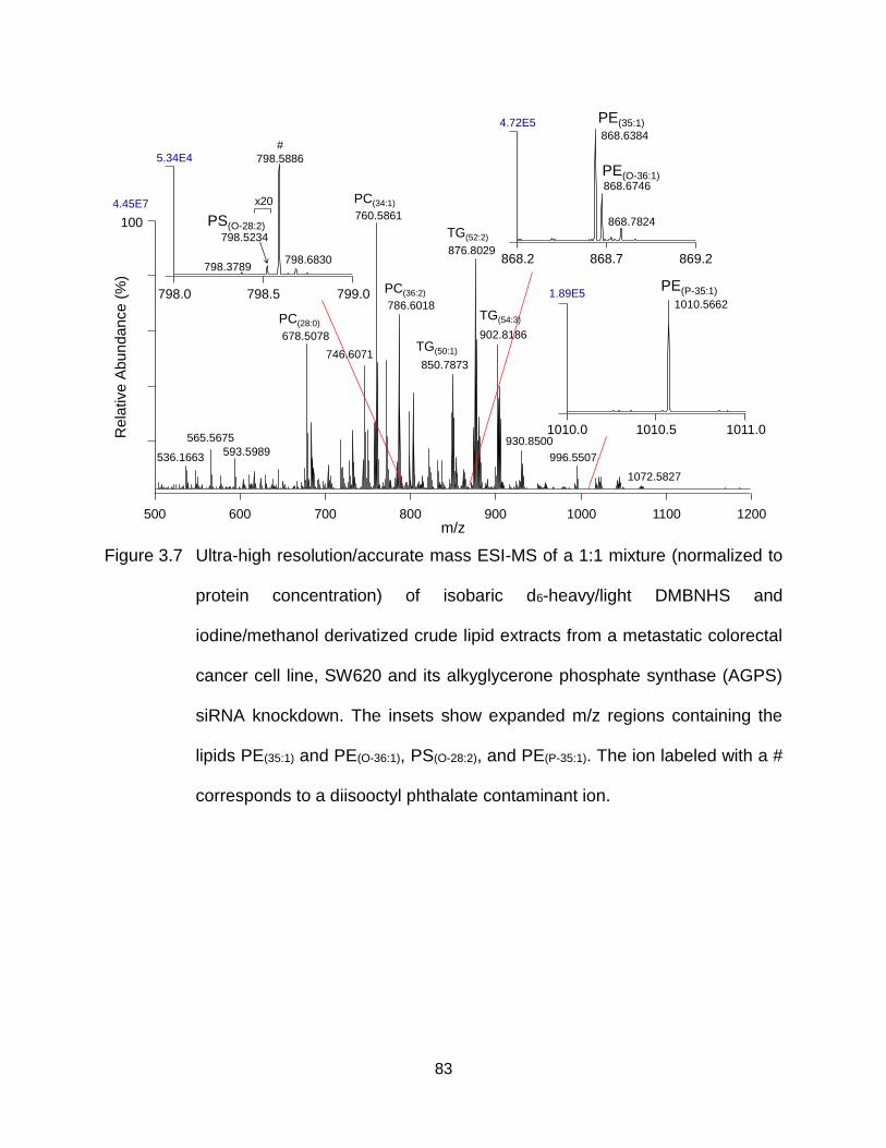
83
Figure 3.7 Ultra-high resolution/accurate mass ESI-MS of a 1:1 mixture (normalized to
protein concentration) of isobaric d6-heavy/light DMBNHS and
iodine/methanol derivatized crude lipid extracts from a metastatic colorectal
cancer cell line, SW620 and its alkyglycerone phosphate synthase (AGPS)
siRNA knockdown. The insets show expanded m/z regions containing the
lipids PE(35:1) and PE(O-36:1), PS(O-28:2), and PE(P-35:1). The ion labeled with a #
corresponds to a diisooctyl phthalate contaminant ion.
868.2 868.7 869.2
PE(35:1)
PE(O-36:1)
4.72E5
868.6746
868.6384
868.7824
798.0 798.5 799.0
5.34E4
798.3789
798.5234
PS(O-28:2)
#
798.5886
798.6830
x20
1010.0 1010.5 1011.0
1010.56621.89E5
PE(P-35:1)
100
500 600 700 800 900 1000 1100 1200
760.5861
876.8029
786.6018
902.8186678.5078
746.6071850.7873
565.5675 930.8500
536.1663593.5989
1072.5827
PC(34:1)
PC(36:2)
PC(28:0)
TG(52:2)
TG(54:3)
TG(50:1)
996.5507
4.45E7
m/z
Rela
tive A
bun
dance
(%)
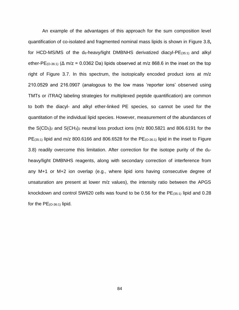
84
An example of the advantages of this approach for the sum composition level
quantification of co-isolated and fragmented nominal mass lipids is shown in Figure 3.8,
for HCD-MS/MS of the d6-heavy/light DMBNHS derivatized diacyl-PE(35:1) and alkyl
ether-PE(O-36:1) (Δ m/z = 0.0362 Da) lipids observed at m/z 868.6 in the inset on the top
right of Figure 3.7. In this spectrum, the isotopically encoded product ions at m/z
210.0529 and 216.0907 (analogous to the low mass ‘reporter ions’ observed using
TMTs or iTRAQ labeling strategies for multiplexed peptide quantification) are common
to both the diacyl- and alkyl ether-linked PE species, so cannot be used for the
quantitation of the individual lipid species. However, measurement of the abundances of
the S(CD3)2 and S(CH3)2 neutral loss product ions (m/z 800.5821 and 806.6191 for the
PE(35:1) lipid and m/z 800.6166 and 806.6528 for the PE(O-36:1) lipid in the inset to Figure
3.8) readily overcome this limitation. After correction for the isotope purity of the d6-
heavy/light DMBNHS reagents, along with secondary correction of interference from
any M+1 or M+2 ion overlap (e.g., where lipid ions having consecutive degree of
unsaturation are present at lower m/z values), the intensity ratio between the APGS
knockdown and control SW620 cells was found to be 0.56 for the PE(35:1) lipid and 0.28
for the PE(O-36:1) lipid.
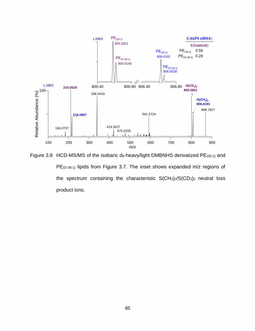
85
Figure 3.8 HCD-MS/MS of the isobaric d6-heavy/light DMBNHS derivatized PE(35:1) and
PE(O-36:1) lipids from Figure 3.7. The inset shows expanded m/z regions of
the spectrum containing the characteristic S(CH3)2/S(CD3)2 neutral loss
product ions.
100 200 300 400 500 600 700 800 900
m/z
100 800.5821
868.7827
806.6191
210.0529
338.3419
591.5724
184.0737
216.0907
419.3527
475.4155
1.28E5
-S(CH3)2
-S(CD3)2800.40 800.80 806.40 806.80
806.6528
800.6166
806.6191
800.5821
1.09E5
PE(O-36:1)
PE(35:1)
PE(O-36:1)
PE(35:1)PE(35:1)
PE(O-36:1)
0.56
0.28
Re
lative
Ab
un
da
nce
(%)
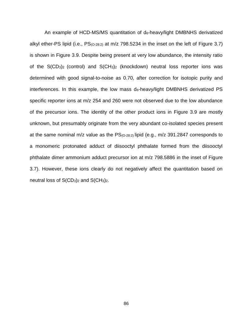
86
An example of HCD-MS/MS quantitation of d6-heavy/light DMBNHS derivatized
alkyl ether-PS lipid (i.e., PS(O-28:2) at m/z 798.5234 in the inset on the left of Figure 3.7)
is shown in Figure 3.9. Despite being present at very low abundance, the intensity ratio
of the S(CD3)2 (control) and S(CH3)2 (knockdown) neutral loss reporter ions was
determined with good signal-to-noise as 0.70, after correction for isotopic purity and
interferences. In this example, the low mass d6-heavy/light DMBNHS derivatized PS
specific reporter ions at m/z 254 and 260 were not observed due to the low abundance
of the precursor ions. The identity of the other product ions in Figure 3.9 are mostly
unknown, but presumably originate from the very abundant co-isolated species present
at the same nominal m/z value as the PS(O-28:2) lipid (e.g., m/z 391.2847 corresponds to
a monomeric protonated adduct of diisooctyl phthalate formed from the diisooctyl
phthalate dimer ammonium adduct precursor ion at m/z 798.5886 in the inset of Figure
3.7). However, these ions clearly do not negatively affect the quantitation based on
neutral loss of S(CD3)2 and S(CH3)2.
87
Figure 3.9 HCD-MS/MS of the isobaric d6-heavy/light DMBNHS derivatized PS(O-28:2)
lipid from Figure 3.7. The inset shows an expanded m/z region of the
spectrum containing the characteristic S(CH3)2/S(CD3)2 neutral loss product
ions.
1003.60E6
100 200 300 400 500 600 700 800
391.2847
149.0236
279.1594221.1754
167.0342
250.1781
413.2668362.3034
467.3739 577.5203
m/z
Rela
tive A
bu
nda
nce
(%)
5.20E4 731.6200
730.4669
736.5044
733.6348
728 730 732 734 736 738
PS(O-28:2) 0.70
88
Finally, an example of secondary multiplexed quantification and molecular lipid
structural characterization of a mixture of isomeric plasmalogen-PE lipids was
demonstrated by HCD-MS/MS of the m/z 1010.5662 ion (i.e., d6-heavy/light DMBNHS
and iodine/methanol derivatized PE(P-35:1)) shown in the inset on the bottom right of
Figure 3.7 (see Figure 3.10). Ratiometric measurement of the -(S(CD3)2+CH3OH) and -
(S(CH3)2+CH3OH) neutral loss reporter ions at m/z 910.4837 and 916.5211, as well as
the low mass reporter ions at m/z 210.0530 and 216.0907, yielded SW620 APGS
knockdown/control ratios at the sum composition level for this lipid of 0.49 and 0.53,
respectively. However, these ratios do not allow differences in abundance to be
separately determined between the knockdown and control SW620 cell samples, for
isomeric lipids that may be present (i.e., different combinations of acyl/O-alkenyl chain
lengths) within the sum composition of PE(P-35:1) lipid. However, as described above in
Figure 3.6B, HCD-MS/MS of d6-heavy/light DMBNHS and iodine/methanol derivatized
plasmalogen lipids yields pairs of isotopically encoded product ions via neutral loss of
the derivatized plasmalogen O-alkenyl chains, directly enabling simultaneous
quantification and structural characterization at the molecular lipid level. In the inset to
Figure 3.10, four pairs of these characteristic product ions were observed,
corresponding to the presence of PE(P-19:0_16:1), PE(P-18:0_17:1), PE(P-17:0_18:1) and PE(P-
16:0_19:1) lipids. Three of these odd-numbered carbon chain length containing molecular
plasmalogen-PE lipid species have been identified previously from an underivatized
lipid extract of SW620 cells, by HCD-MS/MS in negative ion mode [14]. After corrections,
the intensity ratios between the siRNA knockdown and control samples for these
molecular lipid species was determined to be 0.34, 0.34, 0.51 and 0.39. These different
89
ratios suggested that silencing of the RNA responsible for expression of AGPS may
affect plasmalogen-PE lipids to a different extent depending on the identities of their
acyl/alkenyl chains.
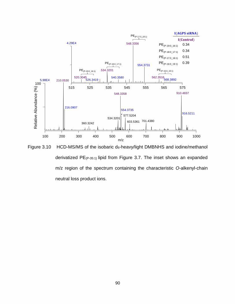
90
Figure 3.10 HCD-MS/MS of the isobaric d6-heavy/light DMBNHS and iodine/methanol
derivatized PE(P-35:1) lipid from Figure 3.7. The inset shows an expanded
m/z region of the spectrum containing the characteristic O-alkenyl-chain
neutral loss product ions.
548.3358
1005.98E4 210.0530
216.0907
910.4837
360.3242701.4380
534.3201
554.3735
603.5361
916.5211577.5204
100 200 300 400 500 600 700 800 900 1000m/z
Rela
tive A
bun
dance
(%)
PE(P-19:0_16:1)
PE(P-17:0_18:1)
PE(P-18:0_17:1)
0.34
0.51
0.34
PE(P-16:0_19:1) 0.39
4.29E4
515 525 535 545 555 565
548.3356
554.3731
520.3045526.3419
PE(P-19:0_16:1) 534.3201
540.3580
PE(P-18:0_17:1)
PE(P-17:0_18:1)
562.3516568.3892
PE(P-16:0_19:1)
575
91
3.3 Conclusions
The gas phase fragmentation behavior of DMBNHS derivatized diacyl- and alkyl
ether-PE and -PS aminophospholipids, as well as sequential isotope-labeled DMBNHS
and iodine/methanol derivatized plasmalogen-PE aminophospholipids, have been
studied using linear ion trap CID-MS/MS and -MS3, HCD-MS/MS and CID-MS/MS with
HCD-MS3. Importantly, the multiplexed quantification of derivatized PE and PS lipids
within complex biological mixtures using d6-heavy and d6-light DMBNHS isobaric mass
stable isotope labeling, and high resolution/accurate mass ESI-MS and targeted HCD-
MS/MS, eliminates the possibility of interference from co-isolated and fragmented
precursor ions with the same nominal mass values, that is inherent to quantification
methods based on detection using low m/z reporter ions. With the additional benefits of
obtaining molecular lipid level quantification and structural characterization of
plasmalogen aminophospholipids from the MS/MS spectra, or by MS3 of the initial
neutral loss product ions for diacyl- and alkyl ether-linked PE and PS lipids, this method
is expected to be an effective lipidomics workflow for the comprehensive quantitative
analysis of aminophospholipids within crude biological lipid extracts.
92
CHAPTER FOUR
Experimental Methods for Chapters Two and Three
4.1 Materials
Methanol (CH3OH), chloroform (CHCl3) and ACS grade isopropyl alcohol (IPA)
were purchased from Macron Chemicals (Center Valley, PA). 99% formic acid (FA) was
from Spectrum Scientific (Irvine, CA). 3,5-diiodoaniline (diIA), 2,4-diIA and 3,4-diIA were
purchased from Spectra Group Limited, Inc. (Millbury, OH). 3-iodoaniline (IA) was
purchased from Santa Cruz Biotechnology, Inc. (Dallas, TX). Aniline and Pd(OH)2 on
carbon (20 wt. %) were from Sigma-Aldrich (St. Louis, MO). Synthetic lipid standards
were from Avanti Polar Lipids, Inc. (Alabaster, AL). Iodine, N,N-dimethylformamide
(DMF) and triethylamine (TEA) were from Jade Scientific (Westland, MI). Acetonitrile
(CH3CN) was purchased from EMD Chemicals (San Diego, CA). Ammonium formate
was obtained from Alfa Aesar (Ward Hill, MA). Ammonium bicarbonate was from J.T.
Baker (Phillipsburg, NJ). SW620 colorectal cancer cells were purchased from the
American Type Culture Collection (ATCC) (Manassas, VA). siGENOME SMARTpool
siRNA was obtained from Thermo Fisher Scientific (Waltham, MA).
4.2 siRNA Knockdown of Alkyglycerone Phosphate Synthase (AGPS) in SW620
Colorectal Cancer Cells and Lipid Extraction
Alkylglycerone phosphate synthase (AGPS) knockdown of SW620 colorectal
cancer cells and followed lipid extraction were performed by Cassie J. Fhaner in Reid
Lab at Michigan State University (East Lansing, MI). Electroporation with an Amaxa
Nucleofector II Device (Lonza, Basel, Switzerland) and siGENOME SMARTpool siRNA
were employed for the knockdown experiments. Control siGENOME SMART-pool
93
siRNA was used for control experiments. Transfected cells were seeded in 6-well plates
(5 × 10⁵ cells / well) and cultured with RPMI 1640 growth medium with 10% FBS and 1%
penicillin/streptomycin, for four days. Cells were harvested by scraping and then
immediately extracted using a monophasic lipid extraction protocol as previously
described [238].
4.3 Derivatization of Lipid Standards and Crude Lipid Extracts
4.3.1 Synthesis of Saturated TG Lipids from Unsaturated TG Lipid Standards
Pd(OH)2 on carbon (1 mg) was added to 500 μL of a 1 mM solution of mono-
unsaturated TG(14:0/16:1/14:0) in 4 : 2 : 1 (v/v/v) IPA/CH3OH/CHCl3 and the mixture was
stirred for 24 hours under H2 at room temperature. After filtration, a solution of saturated
TG(14:0/16:0/14:0) was obtained. The purity of the resultant saturated TG(14:0/16:0/14:0) was
determined as 99% by MS analysis of its ammonium adduct ion.
4.3.2 Photo-Induced Solution Phase Cross-linking of TG Lipid Standards
Sample solution containing 5 mM TG lipids, 5 mM 3,5-diiodoaniline and 10%
formic acid in 4 : 2 : 1 (v/v/v) IPA/CH3OH/CHCl3 were prepared and then irradiated in a
short NMR tube (inner diameter = 4 mm) using a flash-lamp pumped Nd:YAG laser
(Continuum, Santa Clara, CA) with a beam diameter of 3 mm, UV photons of 266 nm, a
frequency of 15 Hz and a pulse energy of 7 mJ. After reacting for 9 min, 5 µL of sample
solution was diluted 200 fold in 4 : 2 : 1 (v/v/v) IPA/CH3OH/CHCl3 for mass
spectrometric analysis as described in Chapter 4, section 4.4.3 without photo-activation.
94
4.3.3 Derivatization of Aminophospholipids Using d6-Heavy/light DMBNHS and
Iodine/Methanol Methods
S,S’-Dimethylthiobutanoylhydroxysuccinimide ester iodide (DMBNHS) and
isobaric mass stable isotope containing d6-‘heavy’ and d6-‘light’ DMBNHS reagents
were synthesized as previously described [82]. The deuterium isotope purities of the d6-
heavy/light DMBNHS reagents were determined to be 99% and 83%, respectively. For
characterization of gas phase CID- or HCD-MS/MS and -MSn fragmentation behaviors,
the standard aminophospholipids PE(14:0/14:0), PE(18:0/18:0), PE(18:0/18:1), PE(18:1/18:1),
PE(18:1/20:4), PE(22:6/22:6), PS(14:0/14:0) and PS(18:0/18:1) were derivatized with DMBNHS as
previously described [24, 127, 137, 138]. DMBNHS derivatized monoalkyl-ether PE(O-
16:0/18:1) lipid was obtained from a crude lipid extract of the control SW620 colorectal
cancer cells. For characterization of CID-MS/MS, HCD-MS3 and HCD-MS/MS
quantitation performance, mixtures of standard lipids PE(14:0/14:0), PS(14:0/14:0) and PE(P-
18:0/22:6) were derivatized separately with d6-heavy and d6-light DMBNHS, followed by
further derivatization of the plasmalogen lipid with iodine/methanol as previously
described [137], then combined at a 1:1 ratio. For determination of the linear dynamic
range of lipid quantification, DMBNHS derivatized PE(14:0/14:0) (0.0005 µM to 5µM) was
mixed with 0.5 µM d6-light DMBNHS derivatized PE(14:0/14:0). Aminophospholipids within
the crude lipid extracts from control and AGPS siRNA knockdown experiments were
derivatized separately with d6-heavy and d6-light DMBNHS reagents, respectively,
followed by derivatization of plasmalogen lipids with iodine/methanol, as previously
described. Samples were then combined in a 1:1 ratio based on their normalized protein
95
concentrations. All samples were resuspended in 4 : 2 : 1 (v/v/v) IPA/CH3OH/CHCl3
containing 20 mM ammonium formate immediately prior to mass spectrometry analysis.
4.4 Mass Spectrometry
4.4.1 Photo-Induced Gas Phase Cross-linking MS/MS and CID-MSn Structural
Analysis of TG Dimer Complex Ions
Solutions of 100 μM of each TG lipid, 100 μM diiodoaniline in 4:2:1 (v:v:v)
IPA/CH3OH/CHCl3 containing 0.1% formic acid were prepared then loaded into
individual wells of a Whatman multichem 96-well plate (Sigma Aldrich, St. Louis, MO)
and sealed with Teflon Ultra Thin Sealing Tape (Analytical Sales and Services,
Prompton Plains, NJ). The samples were then introduced into an Thermo Scientific LTQ
linear ion trap mass spectrometer (San Jose, CA) using an Advion Triversa Nanomate
nano-electrospray ionization (nESI) source (Advion Ithaca, NY) with a spray voltage of
1.4 kV and a gas pressure of 0.3 psi. For MS/MS experiments, an isolation window of
30 m/z was used to provide sufficient signal for isolation of the non-covalent protonated
diiodoaniline-TG dimer complex ions. For UVPD-MS/MS, the mass spectrometer was
modified to enable usage of a flash-lamp pumped Nd:YAG laser (Continuum, Santa
Clara, CA) with a beam diameter of 3 mm, UV photons of 266 nm, a frequency of 15 Hz
and a pulse energy of 7 mJ. Laser pulses were introduced into the ion trap through a
quartz window installed on the back plate of the mass spectrometer. Synchronization
between the laser pulses and the beginning of the activation step of a typical MSn
experiment was achieved by feeding an amplified transistor-transistor logic (TTL) trigger
signal from the mass spectrometer (± 3.2 V) to the laser (± 5.0 V) via a pulse/function
generator (Hewlett-Packard, Palo Alto, CA). The laser was triggered from the Diagnostic
96
menu within the Tune Plus window of the Xcalibur software (Thermo Fisher Scientific,
San Jose, CA). CID-MS3, -MS4 and -MS5 were performed to analyze the products from
the UVPD-MS/MS experiments. Automated Gain Control (AGC) target numbers were
optimized to maximize the signal to noise ratio of the spectra without lowering the
resolution. Both electron multiplier voltages were set at -1350 V. Spectra shown are the
average of at least 100 scans.
4.4.2 ESI-MS, -MS/MS and -MS3 Analysis of Derivatized Aminophospholipid
Standards
Derivatized standard lipid samples were introduced for mass spectrometry
analysis via the Advion Triversa Nanomate nESI source operating under the same
conditions as described in Chapter 4, section 4.4.1. Samples were analyzed in positive
ionization mode using a Thermo Scientific LTQ Orbitrap Velos mass spectrometer
equipped with a linear ion trap and a HCD multipole collision cell (San Jose, CA). The
inlet temperature, S-lens values and mass resolving power were set at 100 oC, 50 %
and 100,000, respectively. Linear ion trap CID-MS/MS spectra were acquired using an
isolation window of m/z 3, a q-value of 0.17 and an activation time of 10 ms, while HCD-
MS3 spectra were collected with activation times of 0.1 ms and a low mass cut off of m/z
150. The HCD normalized collision energy (NCE) was optimized for each product ion of
interest.
97
4.4.3 ESI-MS and -MS/MS Analysis of Derivatized Crude Lipid Extracts from
Combined Control and AGPS siRNA Knockdown of the SW620 Colorectal
Cancer Cells
Derivatized crude lipid extracts from the combined control and AGPS siRNA
knockdown SW620 colorectal cancer cells were introduced into a Thermo Scientific Q
Exactive High Field Orbitrap mass spectrometer equipped with a HCD cell, using the
Advion Triversa Nanomate nESI source operating under the same conditions as
described in Chapter 4, section 4.4.1. The inlet temperature, S-lens value and mass
resolving power were set at 200 oC, 55 % and 240,000 (for MS scans) and 120,000 (for
HCD-MS/MS scans), respectively. For MS scans, a maximum injection time of 50 ms
and an AGC target of 1x10⁶ was used. For HCD-MS/MS scans, an isolation window of
m/z 1, a maximum injection time of 1000 ms, an AGC target of 1x10⁵ and a HCD NCE
of 15% were employed. Throughout this dissertation, the shorthand notation used for
lipid classification and structural representation is that proposed by Liebisch et al. as an
extension of the accepted International Lipid Classification and Nomenclature
Committee nomenclature [242]. Initial assignment of derivatized PE and PS lipid ions, at
the sum composition level (i.e., identity of the head group and fatty acyl/alkyl/alkenyl
linkages, and the total carbon-carbon double bonds), was achieved from the ESI-MS
accurate mass data by searching against a user-defined database of lipid species using
the LIpid Mass Spectrum Analysis (LIMSA v.1.0) software [243], with automated peak
finding and correction of 13C isotope effects, as previously described [138]. Then, a non-
redundant inclusion list of 101 m/z values (from 125 total PE and PS lipids) was
submitted for HCD-MS/MS. 6 scans were acquired for each m/z value in the inclusion
98
list, with a total acquisition time of 8.5 minutes. Quantification at sum composition levels
was achieved from the HCD-MS/MS spectra by manual comparison of the peak
intensities of paired neutral loss reporter ions, (i.e., [M - S(CD3)2]+ and [M - S(CH3)2]+)
from the d6-heavy/light DMBNHS derivatized lipid ions, after correction for deuterium
isotope purities. For the derivatized plasmalogen-aminophospholipids, further detailed
structural characterization and secondary quantification at the molecular lipid level was
performed by analysis of the paired stable isotope containing characteristic product ions
observed within the HCD-MS/MS spectra.
99
CHAPTER FIVE
Quantitative Immuno LC-MS/MS of Parathyroid Hormone and Its In Vivo
Heterogeneous Post-Translational Protein Modifications: Oxidation and
Truncation
5.1 Introduction
Parathyroid hormone is a protein produced in parathyroid glands and consists of
84 amino acids (PTH 1-84) in its full length active form [244]. The full length PTH 1-84
plays a critical role in the regulation of circulating calcium in plasma whose changing
concentration is closely related to many physiological and pathological processes,
including cell signaling, bone metabolism and reduction of the glomerular filtration
rate [245, 246]. In response to decreased concentration of circulating calcium, more
PTH is secreted into the circulatory system to stimulate the calcium reabsorption in
kidney and calcium resorption from bones into blood [247]. Excessive secretion of PTH
can result from abnormalities in the regulation system, which was defined as
hyperparathyroidism [246].
Studies have reported that PTH 1-84 can be post-translationally modified to
various truncated PTH variants through metabolic processes. A summary of previously
reported fragments of PTH is presented in Figure 5.1A [248-250]. In fact, the major
forms of the circulating PTH in human plasma are the amino (N)-terminally truncated
fragments of full length PTH 1-84 (e.g., PTH 7-84) [248]. However, these fragments
have no biologically active N-terminal residues to interact with type 1 PTH/PTH related
protein (PTHrP) receptor and therefore do not regulate calcium levels in the same way
as intact PTH 1-84 [251, 252]. Recent data have suggested that N-terminally truncated
100
PTH may have biological functions opposite to that of PTH 1-84 through interaction with
a not yet cloned receptor [248, 253]. Other truncated forms including mid-molecular
fragments having neither the N- or carboxyl (C)-terminal of PTH 1-84 have also been
reported [249].
Two methionine (M) residues at positions 8 and 18 of PTH 1-84 are possible
sites of oxidation when subjected to oxidative stress [254]. It has been reported that
methionine oxidation of PTH 1-84 results in greatly reduced potency in stimulating the
production of cyclic adenosine monophosphate (cAMP) through interaction with type 1
PTH/PTHrP receptor [255, 256]. Interestingly, a recent study in patients with late-stage
renal disease and on dialysis reported that only 7%-34% of PTH in patient plasmas was
free of oxidation [257]. In patients with renal impairment and parathyroid cancer,
phosphorylation of serine (S) at residue 17 of PTH 1-84 was also identified and
quantified with a large concentration range of 32-767 pg/mL [247].
Accurate quantification of the biologically active forms of PTH is a prerequisite to
the successful diagnosis and appropriate treatment of hyperparathyroidism. Several
generations of immunoassays have been developed for the measurement of PTH 1-84
in serum or plasma samples [258]. Current generation PTH assays use a ‘sandwich
type’ antibody pair targeting at the C- and N-terminal of PTH 1-84 for capture and
detection, respectively [259, 260]. However, due to the micro-heterogeneity within the
PTH proteins (e.g., the structural similarity between PTH 1-84, 7-84 and their oxidized
variants) and the cross reactivity of immunoassays, accurate measurement of PTH 1-84
is still problematic [257, 261, 262].
101
An alternate PTH assay or analysis strategy combining immuno-affinity
enrichment with liquid chromatography-tandem mass spectrometry (LC-MS/MS) has
been recently introduced as a possible reference level method to accurately quantify the
full length PTH 1-84 and its post-translationally modified forms [247]. Generally, PTH
proteins are first enriched using antibody, then digested and finally analyzed by LC-
MS/MS. The method developed by Kumar et al. employed a 15N-labeled PTH 1-84 as
internal standard before digestion to correct errors from sample loss and targeted the
PTH 1-13 peptide as a surrogate for full length PTH 1-84 while Lopez et al. spiked
isotope-labeled PTH 1-13, 7-13 as well as 34-44 internal standards in processed patient
samples before LC-MS/MS analysis, for quantitative analysis of both full length PTH 1-
84 and truncated PTH 7-84 and 34-84 [249, 263]. A method for more comprehensive
quantitative analysis of circulating PTH including full length, truncated and oxidized
forms was previously developed in the Reid lab at Michigan State University [264]. The
oxidation profile of PTH 1-84 and 7-84 during H2O2 oxidation was studied first to
investigate the major oxidized variants of PTH that should be targeted for quantitative
analysis. HPLC purification and ‘top-down’ mass spectrometric analysis of the oxidized
products of PTH showed that PTH 1-84 and 7-84 with methionine sulfoxide at residue 8
and/or 18 were the major products. Therefore, a dual-internal standard approach
employing 15N-labeled PTH 1-84 and 7-84 as well as 13C615N1-labeled lysine-containing
PTH 1-13 , 7-13 and 14-20 with methionine sulfoxide at residue 8 or 18 was applied to
quantify PTH 1-84, 7-84 and their oxidized variants. The linear dynamic range was 5-
2000 pg/mL and no significant biases in relative recovery yield among different oxidized
variants of PTH 1-84 or 7-84 was observed. The recovery yield of the developed
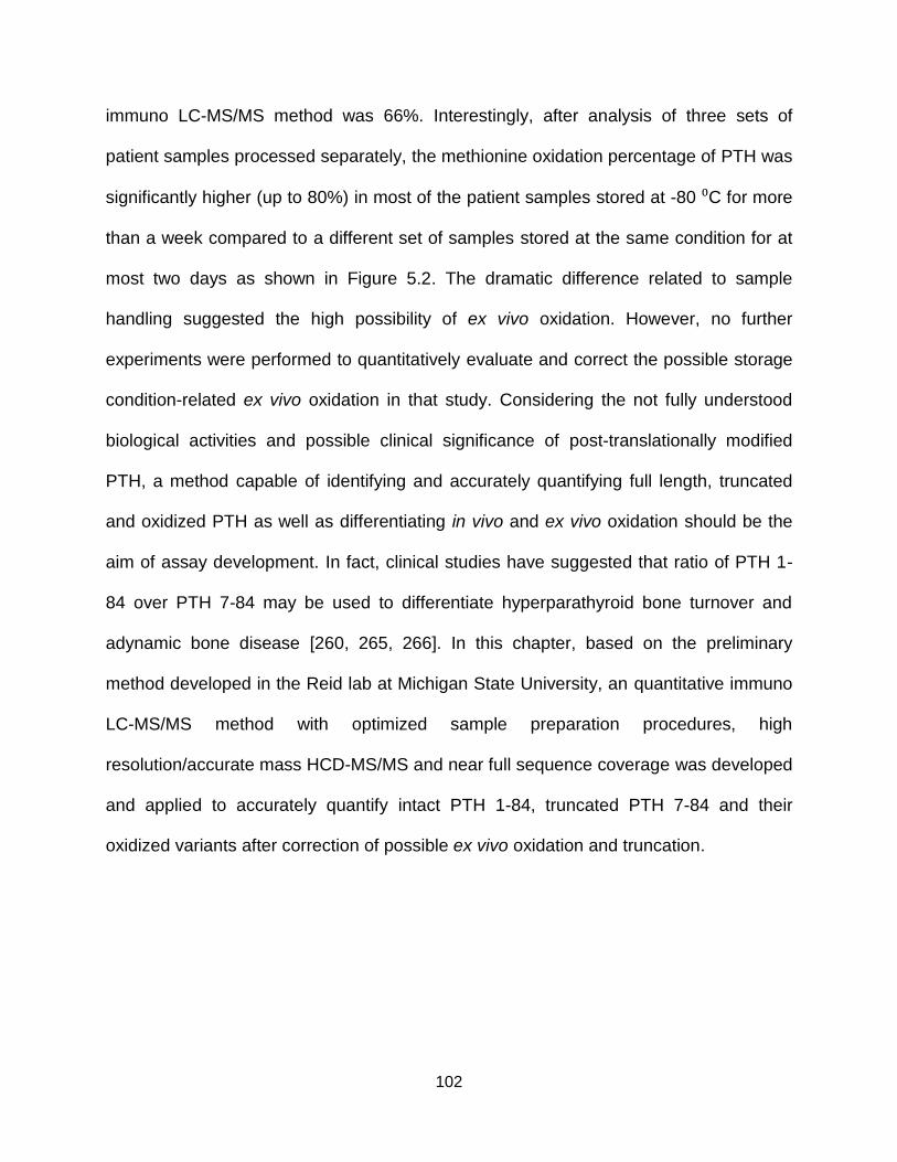
102
immuno LC-MS/MS method was 66%. Interestingly, after analysis of three sets of
patient samples processed separately, the methionine oxidation percentage of PTH was
significantly higher (up to 80%) in most of the patient samples stored at -80 ⁰C for more
than a week compared to a different set of samples stored at the same condition for at
most two days as shown in Figure 5.2. The dramatic difference related to sample
handling suggested the high possibility of ex vivo oxidation. However, no further
experiments were performed to quantitatively evaluate and correct the possible storage
condition-related ex vivo oxidation in that study. Considering the not fully understood
biological activities and possible clinical significance of post-translationally modified
PTH, a method capable of identifying and accurately quantifying full length, truncated
and oxidized PTH as well as differentiating in vivo and ex vivo oxidation should be the
aim of assay development. In fact, clinical studies have suggested that ratio of PTH 1-
84 over PTH 7-84 may be used to differentiate hyperparathyroid bone turnover and
adynamic bone disease [260, 265, 266]. In this chapter, based on the preliminary
method developed in the Reid lab at Michigan State University, an quantitative immuno
LC-MS/MS method with optimized sample preparation procedures, high
resolution/accurate mass HCD-MS/MS and near full sequence coverage was developed
and applied to accurately quantify intact PTH 1-84, truncated PTH 7-84 and their
oxidized variants after correction of possible ex vivo oxidation and truncation.
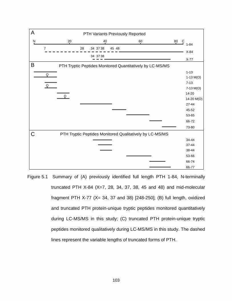
103
Figure 5.1 Summary of (A) previously identified full length PTH 1-84, N-terminally
truncated PTH X-84 (X=7, 28, 34, 37, 38, 45 and 48) and mid-molecular
fragment PTH X-77 (X= 34, 37 and 38) [248-250]; (B) full length, oxidized
and truncated PTH protein-unique tryptic peptides monitored quantitatively
during LC-MS/MS in this study; (C) truncated PTH protein-unique tryptic
peptides monitored qualitatively during LC-MS/MS in this study. The dashed
lines represent the variable lengths of truncated forms of PTH.
PTH Variants Previously Reported
1-13
7-13
14-20
27-44
45-52
53-65
66-72
73-80
PTH Tryptic Peptides Monitored Quantitatively by LC-MS/MS
1-13 M(O)O
7-13 M(O)O
14-20 M(O)O
1-84
X-77
N 20 40 60 80 C
PTH Tryptic Peptides Monitored Qualitatively by LC-MS/MS
7 28 34 37 38 45 48X-84
34 37 38
34-44
37-44
38-44
53-66
66-74
66-77
A
B
C
104
Figure 5.2 Oxidation percentages of PTH in different sets of patient samples [264]. The
percentage of oxidation was calculated by taking the sum concentration of
PTH 1-13 M(O), PTH 7-13 M(O) and PTH 14-20 M(O) divided by the sum
concentration of all full length, truncated and oxidized tryptic peptides
quantified. There were 41 blood samples in set A collected from site 1 and
stored at least a week before sample analysis, and 29 samples in set B and
4 samples in set C collected from site 2 and stored at most 2 days before
sample analysis.
0
10
20
30
40
50
60
70
80
90P
erc
en
t O
xid
ize
d P
TH
Set A (41) Set B (29) Set C (4)
105
5.2 Results and Discussion
5.2.1 Quantitative Immuno LC-MS/MS Workflow with Optimized Sample
Preparation, High Resolution/Accurate Mass HCD-MS/MS and Near Full
Sequence Coverage
A brief summary of the designed workflow for quantitative immuno LC-MS/MS
analysis of PTH 1-84, 7-84 and their oxidized variants is presented in Figure 5.3. 15N-
labeled PTH 1-84 and 7-84 protein internal standards were spiked in patient plasma
immediately after blood draw and centrifugation for quantitative analysis of PTH 1-84
and 7-84 as well as correction of possible ex vivo oxidation and truncation and sample
loss. Also, all patient plasma samples were stored at -80 ⁰C at most 2 days before
analysis to further minimize the possibility of ex vivo modification. Then, a C-terminal
antibody (against PTH 39-84) conjugated on paramagnetic particles (pmp) was selected
to unbiasedly enrich all targeted analytes. After immuno-affinity enrichment and elution,
tryptic digestion was performed to convert PTH proteins into peptides toward which LC-
MS/MS analysis has higher sensitivity [247]. In Figure 5.1B, tryptic peptides providing
near full sequence coverage of PTH 1-84, 7-84 and their oxidized variants as candidate
monitored peptides quantitatively representing various forms of PTH proteins in this
study are listed. Tryptic peptides representing other truncated PTH proteins with
previously reported truncation sites at residue 34, 37, 38 and 77 as well as new
truncation sites discovered in this study at residue 66 and 74 were also monitored
qualitatively and listed in Figure 5.1C [249, 250]. Sequences of PTH tryptic peptides
which are not unique to PTH 1-84 or 7-84 in human proteome were excluded. For
quantification of oxidized PTH peptides, 13C615N1Leu-labeled PTH 1-13 M(O), 7-13 M(O)
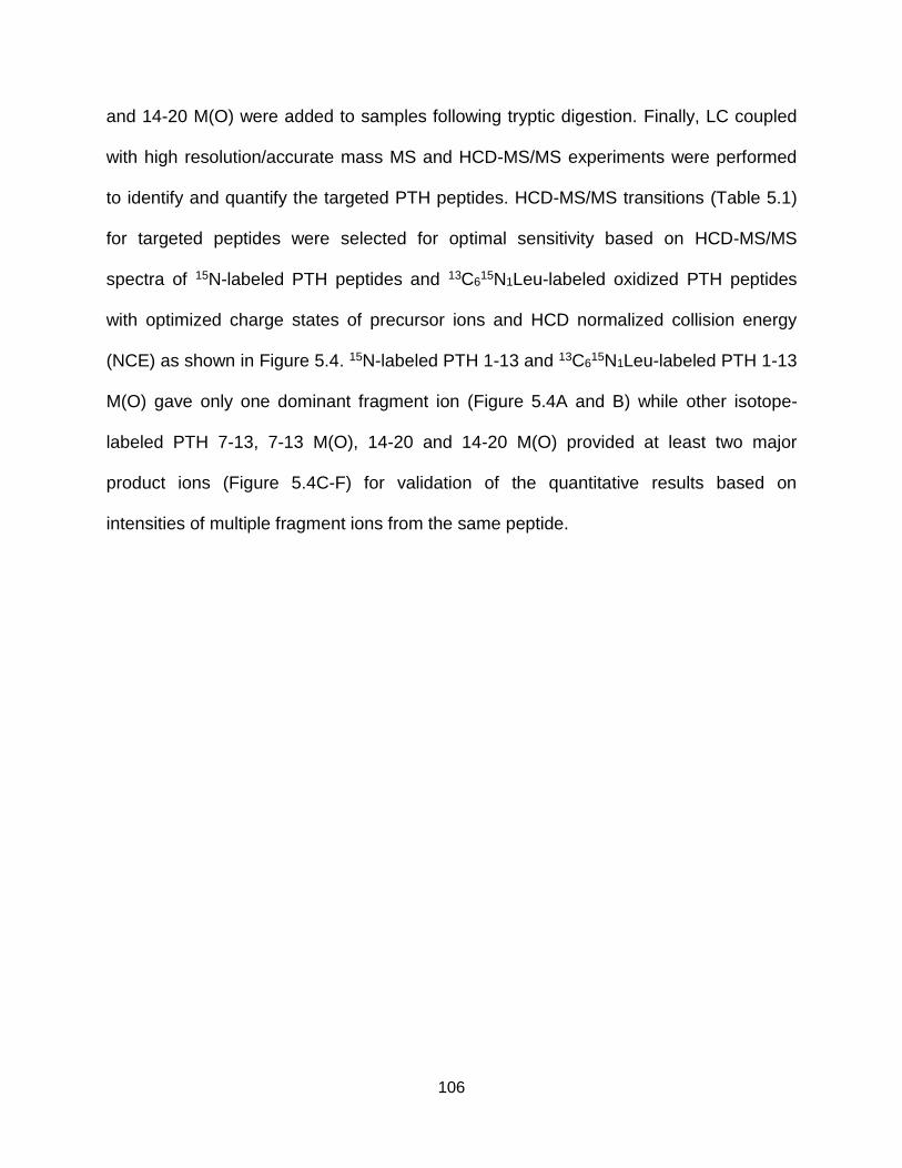
106
and 14-20 M(O) were added to samples following tryptic digestion. Finally, LC coupled
with high resolution/accurate mass MS and HCD-MS/MS experiments were performed
to identify and quantify the targeted PTH peptides. HCD-MS/MS transitions (Table 5.1)
for targeted peptides were selected for optimal sensitivity based on HCD-MS/MS
spectra of 15N-labeled PTH peptides and 13C615N1Leu-labeled oxidized PTH peptides
with optimized charge states of precursor ions and HCD normalized collision energy
(NCE) as shown in Figure 5.4. 15N-labeled PTH 1-13 and 13C615N1Leu-labeled PTH 1-13
M(O) gave only one dominant fragment ion (Figure 5.4A and B) while other isotope-
labeled PTH 7-13, 7-13 M(O), 14-20 and 14-20 M(O) provided at least two major
product ions (Figure 5.4C-F) for validation of the quantitative results based on
intensities of multiple fragment ions from the same peptide.
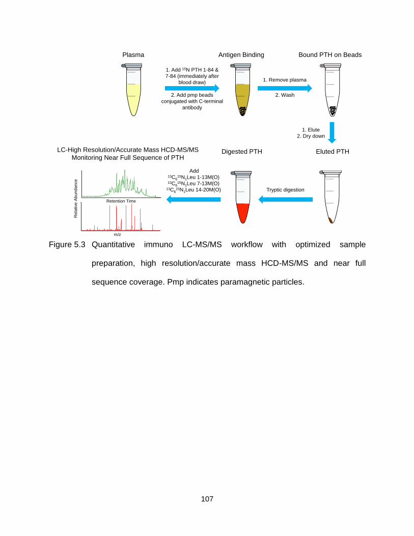
107
Figure 5.3 Quantitative immuno LC-MS/MS workflow with optimized sample
preparation, high resolution/accurate mass HCD-MS/MS and near full
sequence coverage. Pmp indicates paramagnetic particles.
Plasma
1. Add 15N PTH 1-84 &
7-84 (immediately after
blood draw)
2. Add pmp beads
conjugated with C-terminal
antibody
Antigen Binding
1. Remove plasma
2. Wash
1. Elute
2. Dry down
Tryptic digestion
Add 13C6
15N1Leu 1-13M(O)13C6
15N1Leu 7-13M(O)13C6
15N1Leu 14-20M(O)
LC-High Resolution/Accurate Mass HCD-MS/MS
Monitoring Near Full Sequence of PTH
Bound PTH on Beads
Eluted PTHDigested PTH
Retention Time
m/z
Re
lative
Ab
un
da
nce
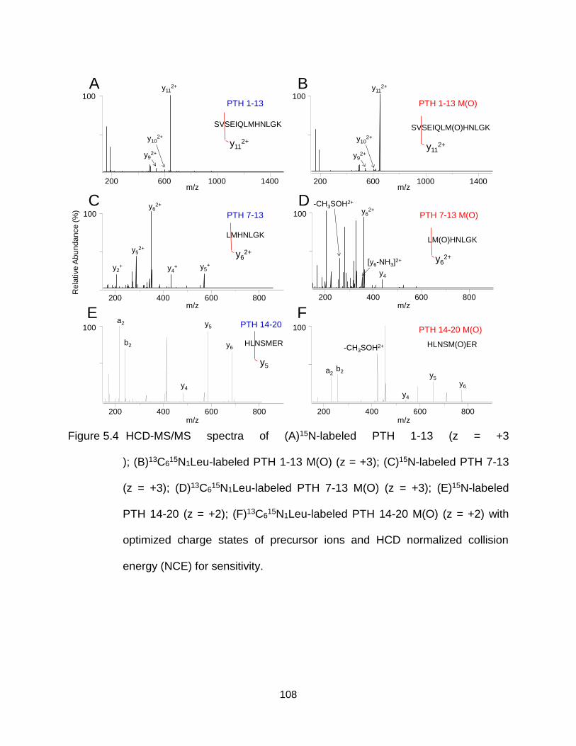
108
Figure 5.4 HCD-MS/MS spectra of (A)15N-labeled PTH 1-13 (z = +3
); (B)13C615N1Leu-labeled PTH 1-13 M(O) (z = +3); (C)15N-labeled PTH 7-13
(z = +3); (D)13C615N1Leu-labeled PTH 7-13 M(O) (z = +3); (E)15N-labeled
PTH 14-20 (z = +2); (F)13C615N1Leu-labeled PTH 14-20 M(O) (z = +2) with
optimized charge states of precursor ions and HCD normalized collision
energy (NCE) for sensitivity.
Rela
tive
Ab
un
da
nce (
%)
100 100PTH 1-13 PTH 1-13 M(O)
SVSEIQLMHNLGK SVSEIQLM(O)HNLGK
y112+
y112+
100100
b2
-CH3SOH2+
y5
y6
y4
y5
y4
b2 y6
PTH 14-20 M(O)PTH 14-20
HLNSMER
y5
HLNSM(O)ER
100 100y6
2+
y52+
y62+
y4
PTH 7-13 M(O)
LM(O)HNLGK
PTH 7-13
LMHNLGK
y62+
y62+
y112+
y92+
y102+
y112+
y92+
y102+
200 600 1000 1400 200 600 1000 1400
200 400 600 800 200 400 600 800
y5+
y4+y2
+
-CH3SOH2+
[y6-NH3]2+
200 400 600 800 200 400 600 800
a2
a2
A B
C D
E F
m/z
m/z
m/zm/z
m/z
m/z
109
Table 5.1 HCD-MS/MS precursor/fragment ion transitions, optimized HCD normalized
collision energy (NCE) and retention times for quantitatively monitored PTH
peptides.
PTH Peptide
Precursor Ion
PTH Peptide
Sequence
Inclusion List
Window
(Retention Time)
Precursor Ion m/z Optimized
HCD
NCE (%)
Monitored Most Abundant MS/MS
Fragment Ion m/z
Native 15N 13C615N1Leu Native 15N 13C6
15N1Leu
PTH 1-13
[M+3H]3+ SVSEIQLMHNLGK(12.0-13.0min)
12.5 min485.9260 491.9080 NA 16
y112+ y11
2+
NA635.3346 643.3106
PTH 1-13M(O)
[M+3H]3+ SVSEIQLM(O)HNLGK(10.4-11.4min)
10.9 min491.2576 497.2396 493.5968 16
y112+ y11
2+ y112+
643.3320 651.3080 646.8404
PTH 7-13LMHNLGK
(8.0-9.0min)
8.5 min271.4868 275.1424 NA 15
y62+ y6
2+
NA[M+3H]3+ 350.1840 355.1690
PTH 7-13M(O)
[M+3H]3+ LM(O)HNLGK(7.0-8.1min)
7.5 min276.8184 280.4741 279.1575 20
y62+ y6
2+ y62+
358.1814 363.1664 358.1814
PTH 14-20
[M+2H]2+ HLNSMER(7.0-8.1min)
7.6 min443.7139 450.1944 NA 27
y5+ y5
+
NA636.2770 645.2500
PTH 14-20M(O)
[M+2H]2+ HLNSM(O)ER(6.0-7.0min)
6.5 min451.7114 458.1919 455.2201 24
-CH3SOH2+ -CH3SOH2+ -CH3SOH2+
419.7119 426.1924 423.2203
PTH 27-44
[M+4H]4+KLQDVHNFVALGAPL
APR
(14.1-17.0min)
15.5min487.2797 493.7602 NA 11
b132+ b13
2+
NA697.3830 706.3560
PTH 45-52
[M+3H]3+ DAGSQRPR(4.0-11.5min)
5.5min296.1545 301.1395 NA 10
y62+ y6
2+
NA350.6961 357.1766
PTH 53-65
[M+4H]4+ KKEDNVLVESHEK(4.0-11.5min)
10.2min389.4585 394.1942 NA 23
y5+ y5
+
NA629.2889 637.2649
PTH 66-72
[M+2H]2+ SLGEADK(4.0-11.5min)
9.2min360.1821 364.1701 NA 17
y5+ y5
+
NA519.2409 525.2229
PTH 73-80
[M+2H]2+ ADVNVLTK(11.5-12.8min)
12.3min430.2478 435.2328 NA 20
y6+ y6
+
NA673.4243 681.4003
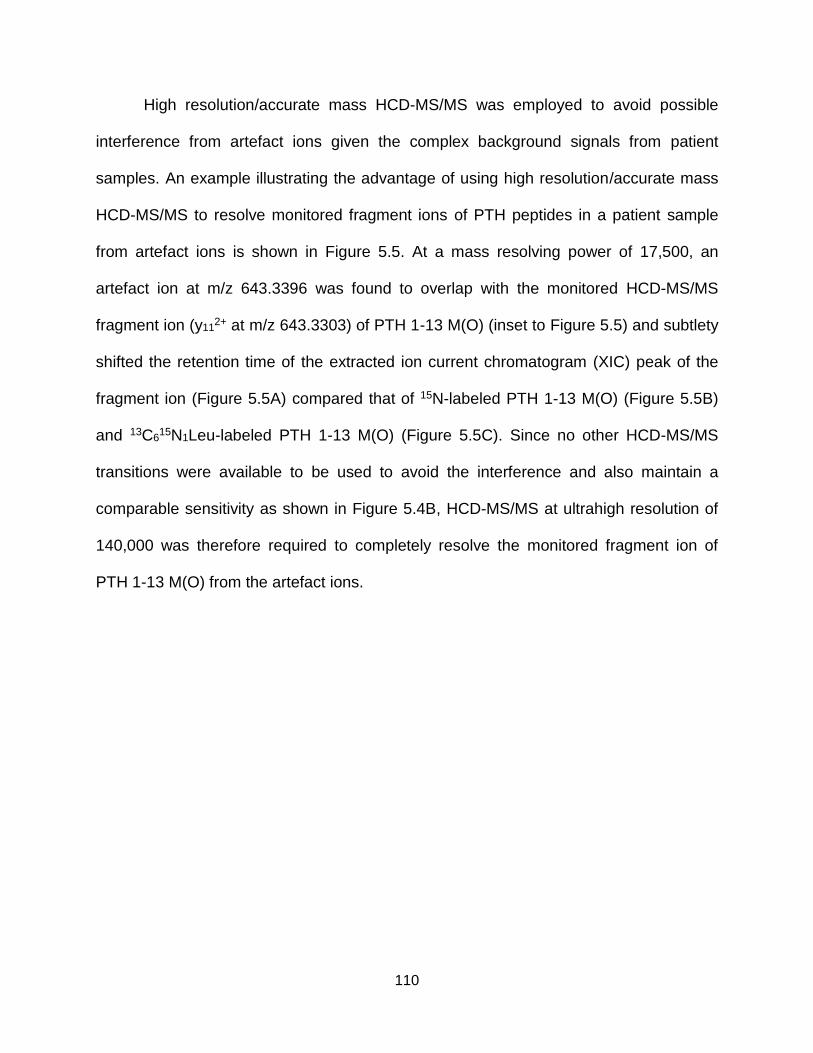
110
High resolution/accurate mass HCD-MS/MS was employed to avoid possible
interference from artefact ions given the complex background signals from patient
samples. An example illustrating the advantage of using high resolution/accurate mass
HCD-MS/MS to resolve monitored fragment ions of PTH peptides in a patient sample
from artefact ions is shown in Figure 5.5. At a mass resolving power of 17,500, an
artefact ion at m/z 643.3396 was found to overlap with the monitored HCD-MS/MS
fragment ion (y112+ at m/z 643.3303) of PTH 1-13 M(O) (inset to Figure 5.5) and subtlety
shifted the retention time of the extracted ion current chromatogram (XIC) peak of the
fragment ion (Figure 5.5A) compared that of 15N-labeled PTH 1-13 M(O) (Figure 5.5B)
and 13C615N1Leu-labeled PTH 1-13 M(O) (Figure 5.5C). Since no other HCD-MS/MS
transitions were available to be used to avoid the interference and also maintain a
comparable sensitivity as shown in Figure 5.4B, HCD-MS/MS at ultrahigh resolution of
140,000 was therefore required to completely resolve the monitored fragment ion of
PTH 1-13 M(O) from the artefact ions.
111
Figure 5.5 Resolving monitored fragment ions from artefact ions with isobaric m/z in a
patient sample using high resolution/accurate mass HCD-MS/MS (A)
extracted ion current (XIC) chromatogram of y112+ (m/z 643.3320) from PTH
1-13 M(O) (m/z 491.2576); (B) XIC chromatogram of y112+ (m/z 651.3080)
from 15N-labeled PTH 1-13 M(O) (m/z 497.2396); (C) XIC chromatogram of
y112+ (m/z 646.8404) from 13C6
15N1Leu-labeled PTH 1-13 M(O) (m/z
493.5968). Resolution for Figure 5.5A-C was at 17,500 (at m/∆m = 200).
The inset shows expanded m/z region of the HCD-MS/MS of PTH 1-13 M(O)
averaged across the XIC peak in Figure 5.5A at resolution of 17,500 (dotted
line) and 140,000 (solid line)
642.9 643.3 643.7m/z
0
100
642.5 644.1
642.8476
643.3396
643.8395
643.3486
643.8319
y112+
643.3303
643.8499
HCD-MS/MS
Res. = 17.5K (dotted line)
Res. = 140K (solid line)
Rela
tive A
bundance
0
100
A 1.15E5
491.2576 643.3320
PTH 1-13 M(O)
13.36
0
100
Re
lative
Ab
un
da
nce (
%)
B 4.40E5
497.2396 651.3080
15N PTH 1-13 M(O)
13.47
12.0 12.2 12.4 12.6 12.8 13.0 13.2 13.4 13.6 13.8 14.0 14.2 14.4 14.6 14.8 15.0
Time (min)
0
100
C 3.28E6
493.5968 646.8404
13C615N1Leu PTH 1-13 M(O)
13.47
112
5.2.2 Quantitative Analysis of In Vivo Full Length, Oxidized and Truncated PTH
Tryptic Peptides in Patient Samples
The practical performance of the quantitative immuno LC-MS/MS method using
conditions described above was tested using a series of patient samples. An example of
LC-MS and LC-MS/MS raw data for the various forms of monitored PTH 1-13 in a
patient sample is shown in Figure 5.6A and Figure 5.6B-F, which include the unlabeled
and 15N-labeled PTH 1-13 (Figure 5.6B and C) as well as unlabeled, 15N-labeled and
13C615N1Leu-labelled PTH 1-13 M(O) (Figure 5.6D to F). LC-MS/MS raw data for the
various forms of monitored PTH 7-13 and 14-20 are shown in Figure 5.6G-P. During the
concentration calculation, to correct the effects from incomplete stable isotope
incorporation in the stable isotope-labeled internal standards, the intensity of the
isotopic cluster of representative fragment ion instead of that of the monoisotopic peak
was used to represent the amount of the corresponding peptide. For each unoxidized
peptide, the concentration (Cnon_ox) was calculated using the equation (1),
Cnon_ox =Inon_ox
I15N_non_ox
C15N
V (1)
where Inon_ox and I15N_non_ox are intensities of isotopic cluster of the representative
product ion of the unoxidized PTH peptide and unoxidized 15N-labeled PTH peptide
internal standard. C15N is concentration of 15N-labeled PTH 1-84 or 7-84 internal
standard spiked in plasma and V is the volume of original plasma solution. Possible ex
vivo truncation and sample loss were already corrected by using the fragment ion
intensities of 15N-labeled peptides as digest of 15N-labeled PTH 1-84 spiked in
immediately after blood draw and centrifugation for plasma. Concentrations of in vivo
oxidized PTH peptides (Cox) were determined using the equation (2),
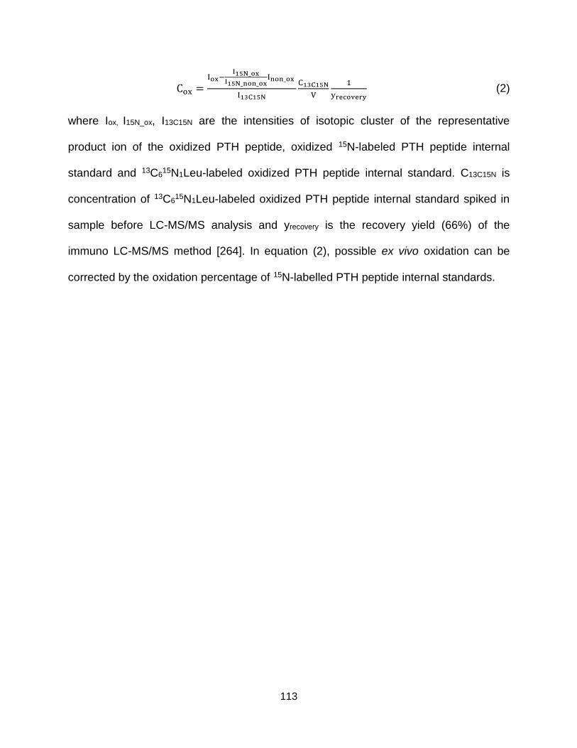
113
Cox =Iox−
I15N_oxI15N_non_ox
Inon_ox
I13C15N
C13C15N
V
1
yrecovery (2)
where Iox, I15N_ox, I13C15N are the intensities of isotopic cluster of the representative
product ion of the oxidized PTH peptide, oxidized 15N-labeled PTH peptide internal
standard and 13C615N1Leu-labeled oxidized PTH peptide internal standard. C13C15N is
concentration of 13C615N1Leu-labeled oxidized PTH peptide internal standard spiked in
sample before LC-MS/MS analysis and yrecovery is the recovery yield (66%) of the
immuno LC-MS/MS method [264]. In equation (2), possible ex vivo oxidation can be
corrected by the oxidation percentage of 15N-labelled PTH peptide internal standards.
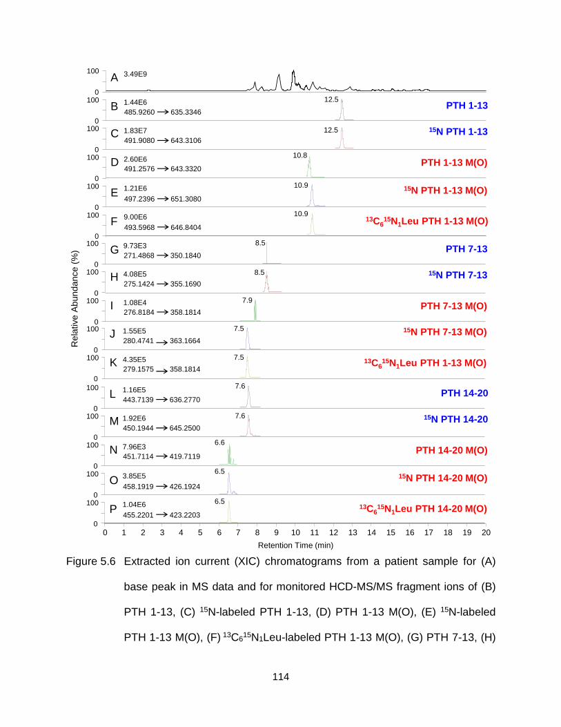
114
Figure 5.6 Extracted ion current (XIC) chromatograms from a patient sample for (A)
base peak in MS data and for monitored HCD-MS/MS fragment ions of (B)
PTH 1-13, (C) 15N-labeled PTH 1-13, (D) PTH 1-13 M(O), (E) 15N-labeled
PTH 1-13 M(O), (F) 13C615N1Leu-labeled PTH 1-13 M(O), (G) PTH 7-13, (H)
Rela
tive A
bundance (
%)
100
0
0
100
0
100
0
100
0
100
0
100
0
100
0
100
0
100
0
100
0
100
0
1000
100
0
100
0
100
0
100
A
PTH 1-13
15N PTH 1-13 M(O)
13C615N1Leu PTH 1-13 M(O)
B
PTH 1-13 M(O)
15N PTH 1-13C
D
E
F
PTH 7-13
PTH 7-13 M(O)
15N PTH 7-13
G
H
I
Retention Time (min)
PTH 14-20
PTH 14-20 M(O)
15N PTH 14-20
15N PTH 14-20 M(O)
13C615N1Leu PTH 14-20 M(O)
1 2 3 4 5 6 7 8 9 10 11 12 13 14 15 16 17 18 19 200
L
M
N
O
P
13C615N1Leu PTH 1-13 M(O)
15N PTH 7-13 M(O)J
K
3.49E9
485.9260 635.3346
497.2396 651.3080
1.44E6
1.21E6
9.00E6
493.5968 646.8404
491.9080 643.3106
491.2576 643.3320
1.83E7
2.60E6
271.4868 350.1840
275.1424 355.1690
276.8184 358.1814
9.73E3
4.08E5
1.08E4
443.7139 636.2770
450.1944 645.2500
458.1919 426.1924
451.7114 419.7119
1.16E5
1.92E6
3.85E5
7.96E3
1.04E6
455.2201 423.2203
280.4741 363.1664
279.1575 358.1814
1.55E5
4.35E5
12.5
12.5
10.8
10.9
10.9
8.5
8.5
7.9
7.5
7.5
7.6
7.6
6.6
6.5
6.5
115
15N-labeled PTH 7-13, (I) PTH 7-13 M(O), (G) 15N-labeled PTH 7-13 M(O),
(K) 13C615N1Leu-labeled PTH 7-13 M(O), (L) PTH 14-20, (M) 15N-labeled
PTH 14-20, (N) PTH 14-20 M(O), (O) 15N-labeled PTH 14-20 M(O) and (P)
13C615N1Leu-labeled PTH 14-20 M(O) as listed in Table 5.1.
116
By employing the quantitative immuno LC-MS/MS strategy described above, the
resultant concentrations of PTH peptides in five samples from patients with high
concentration of circulating PTH 1-84 (500 to 1500 pg/mL) previously measured by PTH
immunoassay were shown in Table 5.2. PTH 1-13 M(O) and 1-13 represent forms of
PTH 1-84 with and without oxidation within residue 1-13 while PTH 7-13 M(O) and 7-13
represent forms of PTH 7-84 with and without oxidation within residue 7-13. PTH 14-20
M(O) and 14-20 represent forms of residue 14-84 containing-PTH (i.e., PTH 1-84 and 7-
84) with and without oxidation within residue 14-20. 15N-labeled PTH 7-84 internal
standard was not spiked in these five samples since the PTH 7-13 and 7-13 M(O) were
not detected in preliminary experiments. The result is consistent with previous studies
using other immuno LC-MS/MS methods [249, 267]. Surprisingly, it was found that no in
vivo oxidized PTH peptides existed in the patient samples after correction for the ex vivo
oxidation. High concentrations of PTH 1-13 were detected as expected. The generally
higher concentrations of PTH 14-20 compared to that of PTH 1-13 (except in patient 1)
indicated other possible truncation sites between residue 1 and 13 of PTH 1-84 except
residue 7. Truncations at residues 4 and 10 have been suggested by D’Amour et al.
[248]. However, no PTH X-13 peptides (X=2-12 except 7) were detected within these 5
patient samples, partially due to the decreasing ionization efficiency of shortening
peptides if any truncation exists between residue 7 and 13. The concentrations of PTH
tryptic peptides from the mid-molecular and C-terminal region of PTH 1-84 were also
monitored. Despite that their measured concentration (up to 20 ng/mL) is out of the
determined linear range of the method (5-2000 pg/mL as previously described [264]),
the high concentration was consistent with the widely agreed knowledge that major
117
forms of the circulating PTH are N-terminally truncated fragments of PTH 1-84 [11]. The
much lower concentrations of PTH 27-44 and 73-80 compared to that of PTH 45-52, 53-
65 and 66-72 were also consistent with the identified PTH 34-44, 37-44, 38-44 and 66-
77 with high abundance (Figure 5.7A-D). 15N-labeled PTH 34-44, 37-44, 38-44 and 66-
77 were not observed in the five patient samples which confirmed that those identified
non-isotope-labeled truncated PTH peptides were produced in vivo instead of ex vivo.
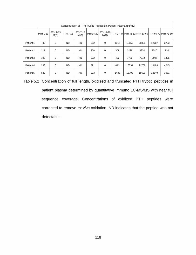
118
Table 5.2 Concentration of full length, oxidized and truncated PTH tryptic peptides in
patient plasma determined by quantitative immuno LC-MS/MS with near full
sequence coverage. Concentrations of oxidized PTH peptides were
corrected to remove ex vivo oxidation. ND indicates that the peptide was not
detectable.
Concentration of PTH Tryptic Peptides in Patient Plasma (pg/mL)
PTH 1-13 PTH 1-13
M(O) PTH 7-13
PTH7-13
M(O)PTH14-20
PTH14-20
M(O)PTH 27-44 PTH 45-52 PTH 53-65 PTH 66-72 PTH 73-80
Patient 1 432 0 ND ND 382 0 1018 18853 20335 12787 3763
Patient 2 211 0 ND ND 250 0 309 3228 3334 2515 736
Patient 3 196 0 ND ND 292 0 486 7788 7272 5097 1405
Patient 4 283 0 ND ND 391 0 811 18731 21758 19483 4245
Patient 5 682 0 ND ND 923 0 1438 15798 18620 13540 3971
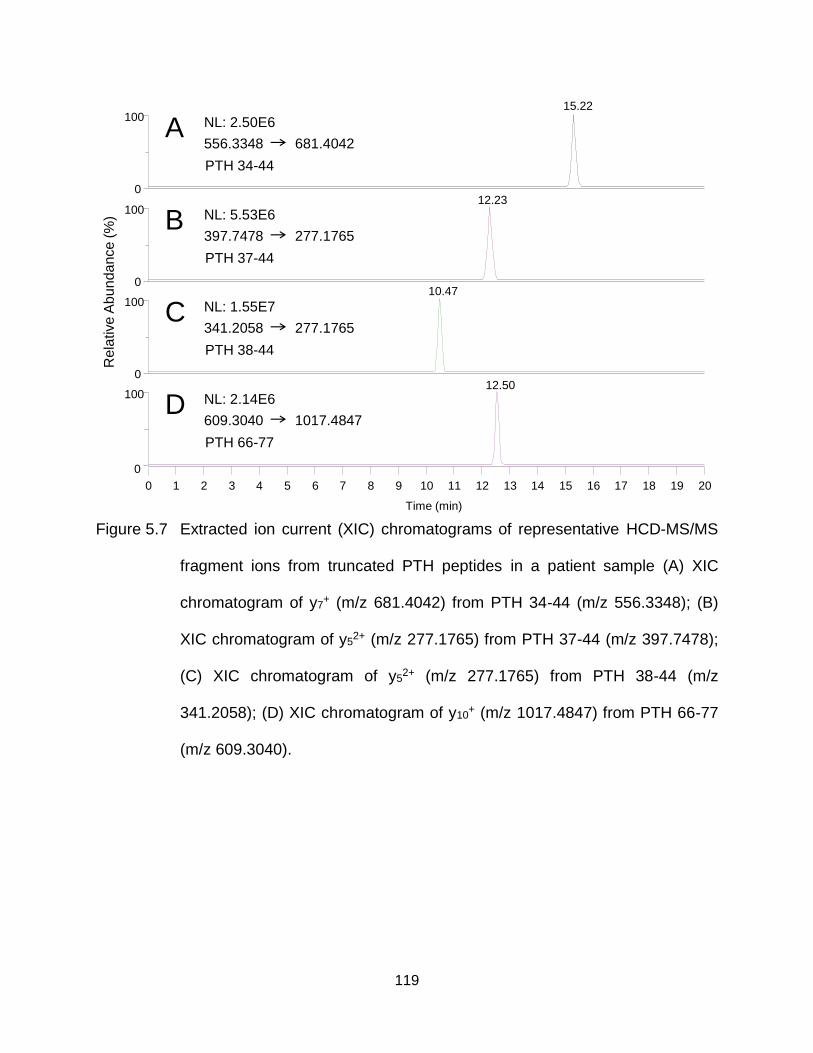
119
Figure 5.7 Extracted ion current (XIC) chromatograms of representative HCD-MS/MS
fragment ions from truncated PTH peptides in a patient sample (A) XIC
chromatogram of y7+ (m/z 681.4042) from PTH 34-44 (m/z 556.3348); (B)
XIC chromatogram of y52+ (m/z 277.1765) from PTH 37-44 (m/z 397.7478);
(C) XIC chromatogram of y52+ (m/z 277.1765) from PTH 38-44 (m/z
341.2058); (D) XIC chromatogram of y10+ (m/z 1017.4847) from PTH 66-77
(m/z 609.3040).
A NL: 2.50E6
556.3348 681.4042
PTH 34-44
B NL: 5.53E6
397.7478 277.1765
PTH 37-44
C NL: 1.55E7
341.2058 277.1765
PTH 38-44
D NL: 2.14E6
609.3040 1017.4847
PTH 66-77
Time (min)
Rela
tive
Ab
un
da
nce (
%)
0
0
10015.22
0
10012.23
0
10010.47
100
1 2 3 4 5 6 7 8 9 10 11 12 13 14 15 16 17 18 19 200
12.50
120
5.2.3 Identification of Novel Truncation Sites of PTH Proteins
Novel truncation sites at residue 66 and 74 of PTH 1-84 were also discovered
and evidenced by HCD-MS/MS analysis of PTH-specific peptides including PTH 53-66
(KKEDNVLVESHEKS) and PTH 66-74 (SLGEADKAD) (Figure 5.8A and B) found in
patient samples. Absence of 15N-labeled PTH 53-66 and 66-74 in patient samples
confirmed that the truncation at residue 66 and 74 occurred in vivo. Phosphorylation at
serine 17 of PTH 1-84 was not identified in these patient samples.
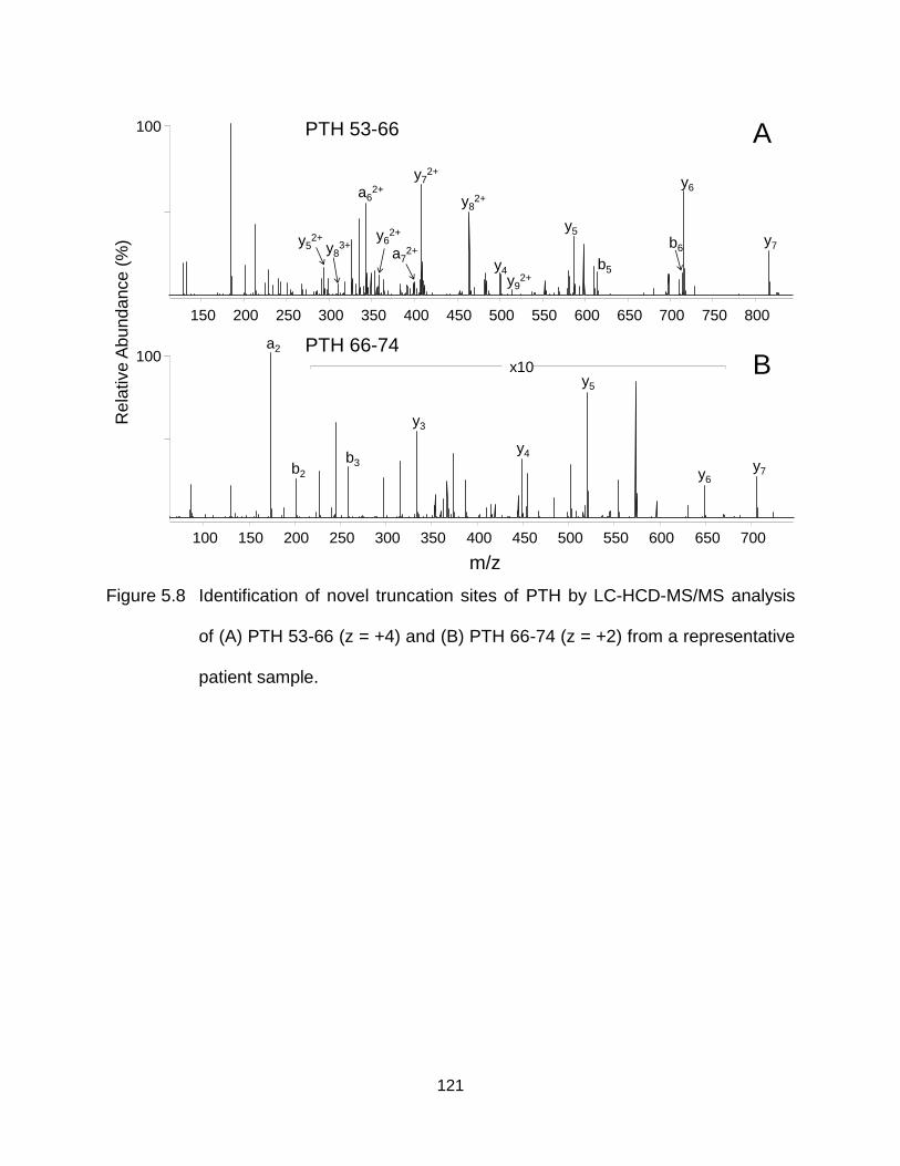
121
Figure 5.8 Identification of novel truncation sites of PTH by LC-HCD-MS/MS analysis
of (A) PTH 53-66 (z = +4) and (B) PTH 66-74 (z = +2) from a representative
patient sample.
Rela
tive A
bundance (
%)
150 200 250 300 350 400 450 500 550 600 650 700 750 800
100
b6
y92+
y7
y6
y5
y82+
y72+
a62+
y4 b5
a72+
y62+
y83+y5
2+
y7y6
y5
y4
y3
b3b2
100 150 200 250 300 350 400 450 500 550 600 650 700
m/z
100x10
a2
A
B
PTH 53-66
PTH 66-74
122
5.3 Conclusions
An immuno LC-MS/MS method enabling simultaneous identification and
quantification of in vivo full length, truncated and oxidized PTH was developed. Different
heavy isotope-labeled PTH protein and oxidized peptide internal standards were
employed for accurate quantitative analysis of various forms of PTH after correction of
possible ex vivo post-translational modification (i.e., oxidation and truncation) occurring
during sample handling. Transitions with near full sequence coverage were coupled with
high resolution/accurate mass tandem mass spectrometry to avoid possible interference
from complex background signals in patient samples and unambiguously quantify the
targeted PTH peptides. After analysis of five patient samples using the developed
method, no in vivo oxidation was detected after correction for ex vivo oxidation. Also, no
notable quantities of N-terminally truncated PTH 7-84 was detected as previously
reported. The inconsistent concentration of PTH 1-13 and 14-20 in patient samples
indicated the existence of other previously unidentified truncated PTH except PTH 7-84.
Novel in vivo truncation site at residue 66 and 77 of PTH 1-84 were also identified. It is
expected that more post-translationally modified forms, mainly truncated forms of PTH
will be discovered, identified and quantified in future study of PTH using immuno mass
spectrometric methods for the purpose of studying their not fully understood biological
activities and possible clinical utilities.
123
CHAPTER SIX
Quantitative Proteomic Analysis Using Dimethyl Labeling and Two Dimensional
LC-MS/MS to Study the Mechanism of Action of the Non-Competitive Proteasome
Inhibitor, TCH-013
6.1 Introduction
The 26S proteasome, consisting of a 20S catalytic core particle and 19S
regulatory particle is the central protease responsible for maintaining cellular
homoeostasis through the ubiquitin-proteasome pathway [268]. Inhibition of this
pathway using Bortezomib has emerged as a rational strategy for the chemotherapeutic
treatment of multiple myeloma (MM), a cancer of plasma cells located within bone
marrow [269]. Through covalent binding to the catalytic sites at N-terminal threonine of
several 20S β-subunits of the 26S proteasome, Bortezomib competitively blocks the
ubiquitin-proteasome pathway of protein degradation [270]. Via competitive inhibition,
Bortezomib is effective in inducing death of cells (e.g., multiple myeloma cells) where
the 26S proteasome is constantly under a high burden to degrade rapidly produced
toxic misfolded proteins [271], however, >97% of patients become resistant or intolerant
to treatment in the first few years, after which survival is typically less than a year [272].
Bortezomib resistance is, in part, due to the overexpressed mutated forms of catalytic
subdomains of 26S proteasome at the original drug binding sites which inhibit the
effectiveness of protein/Bortezomib binding [273, 274].
Alternatively, drugs enabling non-competitive inhibition of proteasomes have
been suggested to overcome treatment resistance and can present lower toxicities
[275-277]. Both peptide- and small molecules-based non-competitive proteasome
inhibitors have been reported [275-280]. For example, a proline- and arginine-rich
124
peptide named PR-39 can inhibit degradation of specific proteins including IκB after
binding to the non-catalytic α-subunit of 20S proteasome and changing the structure of
the proteasome [275]. As small molecule-based proteasome inhibitors, chloroquine and
the secondary fungal metabolite, gliotoxin, were shown to non-competitively inhibit the
activity of 20S proteasome, but both have issues of toxicity [278, 279]. Although 5-
amino-8-hydroxyquinoline (5AHQ) was also reported to selectively inhibit the
proteasome in myeloma and leukemia cells, no direct evidence for interaction between
5AHQ and the proteasome was provided [280]. Recently, Tepe and co-workers
described the substrate selectivity of proteasome mediated proteolysis induced by small
molecule imidazoline scaffolds, TCH-013 and its derivatives, via a non-competitive
protein/ligand interaction and with nanomolar potency [276, 277]. This class of small
molecules represents a promising direction for the development of novel non-
competitive proteasome inhibitors which overcome the limitations caused by competitive
proteasome inhibition. However, little is known about the site(s) at which the non-
competitive inhibitor binds to the proteasome, or their mechanism(s) of biological activity.
MS-based quantitative proteomic strategies are widely used to elucidate the
mode of action by which a drug exerts its pharmacological effect by providing
information on drug-induced regulation of functional proteins and/or their PTMs [281].
For example, the mechanism of action of Lenalidomide, another effective drug for MM,
is not fully understood [282]; through large scale quantitative proteomic analysis, Krönke
et al. reported that two essential transcription factors of MM cells, IKZF1 and IKZF3,
were selectively ubiquitinated and degraded by CRBN-CRL4 ubiquitin ligase, providing
a novel mode of action for lenalidomide [283]. For any successful proteomics study, a
125
broad coverage of identification and sufficient depth of quantification is critical for
unbiased searching of stimuli-induced changes in protein and PTM abundance [50, 64].
Therefore, reducing the sample complexity through enrichment and/or fractionation
techniques is usually required [50]. Basic pH reversed-phase fractionation is gaining
popularity due to the availability of reversed-phase columns tolerable to a wide pH
range, better resolving power of hydrophobicity-dependent separation compared to that
of other separation techniques (e.g., strong cation ion exchange) and more affordable
cost compared to antibody-based enrichment [284]. To enable large scale MS-based
quantitative proteomic analysis of animal or human samples where spiking stable
isotope labeled analogue standards and metabolic labeling of stable isotopes are not
applicable, chemical labeling with isotope-coded tags followed by sample pooling before
down-stream analysis is commonly used as discussed in Chapter 1, section 1.2.1.3.3.
Relying on the advantages of inexpensive reagents, fast reactions and applicability to a
broad range of sample quantities, dimethyl labeling is one of the frequently used
chemical labeling methods for quantitative proteomic studies [188]. In this chapter, a
strategy involving stable isotope-containing dimethyl labeling, basic reversed-phase
fractionation and quantitative LC-MS/MS was employed for quantitative proteomic
analysis of TCH-013 (imidazoline scaffold)-, Bortezomib- and vehicle control- (DMSO)
treated human RPMI-8226 cells, to gain insights into the mechanism of non-competitive
proteasome inhibition.
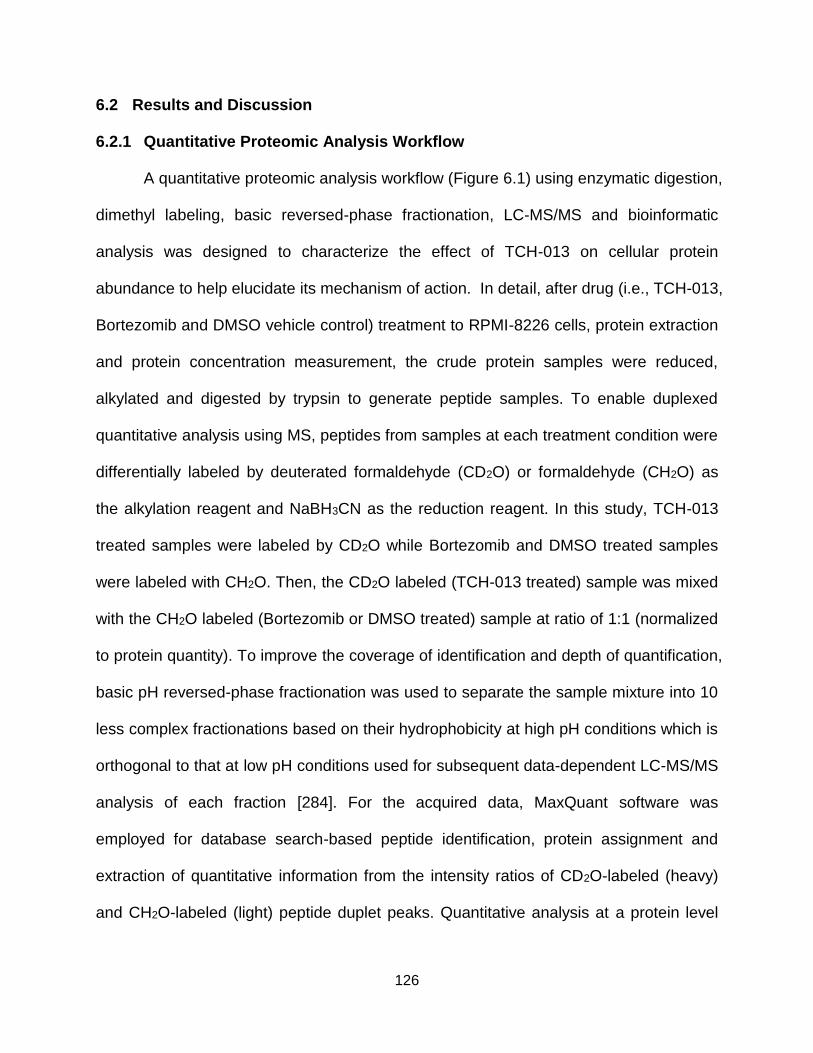
126
6.2 Results and Discussion
6.2.1 Quantitative Proteomic Analysis Workflow
A quantitative proteomic analysis workflow (Figure 6.1) using enzymatic digestion,
dimethyl labeling, basic reversed-phase fractionation, LC-MS/MS and bioinformatic
analysis was designed to characterize the effect of TCH-013 on cellular protein
abundance to help elucidate its mechanism of action. In detail, after drug (i.e., TCH-013,
Bortezomib and DMSO vehicle control) treatment to RPMI-8226 cells, protein extraction
and protein concentration measurement, the crude protein samples were reduced,
alkylated and digested by trypsin to generate peptide samples. To enable duplexed
quantitative analysis using MS, peptides from samples at each treatment condition were
differentially labeled by deuterated formaldehyde (CD2O) or formaldehyde (CH2O) as
the alkylation reagent and NaBH3CN as the reduction reagent. In this study, TCH-013
treated samples were labeled by CD2O while Bortezomib and DMSO treated samples
were labeled with CH2O. Then, the CD2O labeled (TCH-013 treated) sample was mixed
with the CH2O labeled (Bortezomib or DMSO treated) sample at ratio of 1:1 (normalized
to protein quantity). To improve the coverage of identification and depth of quantification,
basic pH reversed-phase fractionation was used to separate the sample mixture into 10
less complex fractionations based on their hydrophobicity at high pH conditions which is
orthogonal to that at low pH conditions used for subsequent data-dependent LC-MS/MS
analysis of each fraction [284]. For the acquired data, MaxQuant software was
employed for database search-based peptide identification, protein assignment and
extraction of quantitative information from the intensity ratios of CD2O-labeled (heavy)
and CH2O-labeled (light) peptide duplet peaks. Quantitative analysis at a protein level
127
was achieved by an in-house C++ script. A heavy/light ratio for each protein was
expressed as the median value of the normalized, log2 transformed and grouped
heavy/light ratio of each peptide assigned to that protein. As mentioned in Chapter 1,
section 1.2.1.3.3, dimethyl labeling can lead to peptide retention time shifts caused by
deuterium isotope effects [190]. The accuracy of quantifying based on the intensity
ratios of heavy/light peptide duplet peaks may be compromised by this phenomenon
which occurred during first and second dimensional separations in the designed
workflow [190]. To correct this type of systematic error, peptides identified in multiple
fractions were not used for quantification. Also, the intensity ratios of heavy/light peptide
duplet peaks were analyzed collectively over the whole chromatographic peak by
plotting the peak intensity of heavy peptide over that of light peptide and calculating the
slope (i.e., heavy/light ratio) using a linear fit model [285].
128
Figure 6.1 A workflow using tryptic digestion, dimethyl labeling, basic reversed-phase
fractionation, LC-MS/MS and bioinformatic analysis for quantitative
proteomic analysis of TCH-013, Bortezomib and DMSO (vehicle control)
treated human RPMI-8226 cells.
TCH-013 treated
RPMI-8226 cells
Bortezomib or DMSO treated
RPMI-8226 cells
(1)Reduction
(2)Alkylation
(3)Trypsin Digestion
Dimethyl Labeling 1:1 Mixture
Normalized to Protein Quantity
Basic Reversed-Phase Fractionation
HCD-MS/MS for identification
MS for quantification
m/z
LC-MS/MS Analysis
m/z
Bioinformatic Analysis through
MaxQuant and In-house C++ script
10 Fractions
Retention time
LC for separation
CH2O-labeled
CD2O-labeled
129
6.2.2 Coverage of Protein Identification and Selection Criteria During Protein
Quantification
Proteomic analysis of TCH-013-, Bortezomib- and DMSO-treated RPMI-8226
cells using the workflow described above revealed >1200 proteins with at least 2
peptides identified in each of the three treatments and in all three biological replicates.
The coverage of protein identities acquired by this workflow is illustrated in Figure 6.2A-
B. From TCH-013-treated (heavy) and Bortezomib-treated (light) samples (Figure 6.2A),
1721 proteins were identified in total from three biological replicates in which 78.4%
(1350) of them are common in at least two biological replicates and 59.4% (1022) of
them are common for all three biological replicates. Similarly, from TCH-013 treated
(heavy) and DMSO treated (light) cells (Figure 6.2B), 1788 proteins were identified in all
three biological replicated where 81.5% (1457) and 65.8% (1176) were identified in two
and three biological replicates, respectively. The good overlap of identified proteins
between biological replicates demonstrates that the designed workflow has a broad
enough coverage of identification for subsequent large scale quantitative analysis. The
influence of selection criteria (i.e., minimum number of peptides for each protein) on the
number of quantified proteins common in all three biological replicates was shown in
Figure 6.2C and D for the two sample mixtures. Although more heavy/light peptide pairs
assigned to each protein is desired to acquire a more reproducible heavy/light protein
ratio, the number of relatively quantified proteins decreases at the same time, which
limits the ability to find possible drug-regulated proteins with less peptides assigned. In
this study, stringent criteria was applied i.e. only proteins with >=4 peptides from each of
the three treatments and in all three biological replicates were used for a more
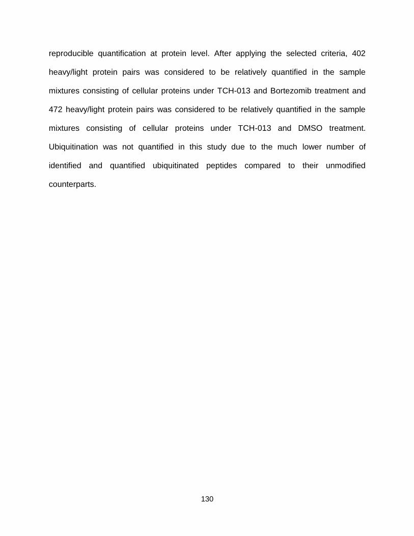
130
reproducible quantification at protein level. After applying the selected criteria, 402
heavy/light protein pairs was considered to be relatively quantified in the sample
mixtures consisting of cellular proteins under TCH-013 and Bortezomib treatment and
472 heavy/light protein pairs was considered to be relatively quantified in the sample
mixtures consisting of cellular proteins under TCH-013 and DMSO treatment.
Ubiquitination was not quantified in this study due to the much lower number of
identified and quantified ubiquitinated peptides compared to their unmodified
counterparts.
131
Figure 6.2 Overlapping of identified heavy/light proteins in three biological replicate
sample mixtures consisting of (A) a heavy-labeled TCH-013 (10 µM) treated
sample mixed with a light-labeled Bortezomib (0.1 µM) treated sample; (B) a
heavy-labeled TCH-013 (10 µM) treated sample mixed with a light-labeled
DMSO (0.1%, v/v) treated sample. The influence of selection criteria (i.e.,
minimum of peptides assigned to each protein) on the number of relatively
quantified proteins common in three biological replicates of sample mixture
A (C) and B (D).
10 µM TCH-013 (heavy)
+
0.1% DMSO (light)
Rep 1 Rep 2
Rep 3
1176
75
95111
89122
120
10 µM TCH-013 (heavy)
+
0.1 µM bortezomib (light)
Rep 1 Rep 2
Rep 3
150143 131
1022
84 94
97
0
100
200
300
400
500
600
700
800
900
1000
1 2 3 4 5 6 7 8
0
200
400
600
800
1000
1200
1 2 3 4 5 6 7 8
Min. # of peptides assigned to each protein
# o
f pro
tein
s c
om
mon in 3
bio
logic
al re
plic
ate
s o
f
10
µM
TC
H-0
13
(h
ea
vy)
+ 0
.1 µ
M b
ort
ezo
mib
(lig
ht)
Min. # of peptides assigned to each protein
A B
DC
# o
f p
rote
ins c
om
mo
n in
3 b
iolo
gic
al re
plic
ate
s o
f
10
µM
TC
H-0
13
(h
ea
vy)
+ 0
.1%
DM
SO
(lig
ht)
132
6.2.3 Protein Regulation in Human RPMI-8226 Cells Induced by TCH-013
Following relative quantification of the proteins from human RPMI-8226 cells
subjected to different treatments, the histograms of averaged log2 heavy/light proteins
ratios (n=3) for quantified proteins after TCH-013 (heavy) treatment vs Bortezomib
(light) treatment or TCH-013 (heavy) treatment vs DMSO (light) treatment were plotted
(Figure 6.3A and B). In general, protein abundance appeared to be unaffected by the
selective proteasome inhibitor, TCH-013 as well as the non-selective proteasome
inhibitor, Bortezomib. This steady-state level of proteins was consistent with previous
large scale proteomic studies involving human 293T and HCT116 cells treated with
Bortezomib and human Jurkat cells treated with another competitive proteasome
inhibitor, MG-132 [286, 287]. In addition, although both Bortezomib and TCH-013 have
been demonstrated to induce accumulation of ubiquitinated proteins as the substrates
of the ubiquitin-proteasome protein degradation pathway, no proteins were significantly
(near 2 folds) up-regulated in TCH-013 treated samples compared to those treated with
Bortezomib or DMSO (control), possibly due to the low stoichiometry of ubiquitination
[286, 287]. However, in the present study TCH-013, but not Bortezomib or DMSO,
treatment was associated with significant (near 2 folds) down-regulation of four proteins
(Table 6.1) namely prohibitin-2, NAD(P) transhydrogenase, NADH-ubiquinone
oxidoreductase and ADP/ATP translocate 2. Surprisingly, they are all closely involved in
regulation of the mitochondrial respiration for production of ATP or the detoxification of
reactive oxygen species (ROS), both of which are usually increasingly demanded for
growth and division of cancer cells having higher rates of metabolism [288-291]. For
example, knockdown of prohibition-1 and prohibition-2 together or prohibition-2 alone
133
have been reported to suppress tumor growth [288, 292, 293]. Also, the key roles of
mitochondrial ADP/ATP translocate 2 in the almost exclusive metabolism pathway (i.e.,
glycolysis) in many cancer cells were also summarized in a recent review [290]. To
further elucidate the mechanism of observed selective protein down-regulation induced
by TCH-013 and how it is related to TCH-induced activity changes of proteasome
and/or cell apoptosis, more targeted biological studies on protein ubiquitination and
protein interactions are required.
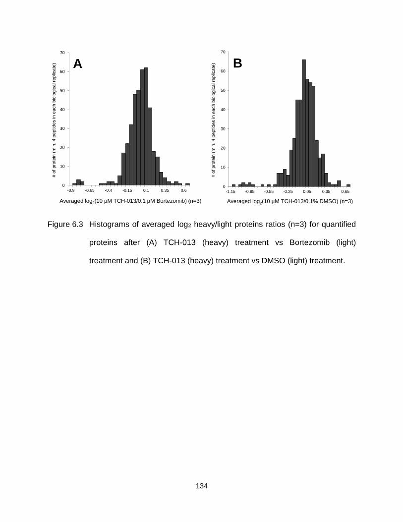
134
Figure 6.3 Histograms of averaged log2 heavy/light proteins ratios (n=3) for quantified
proteins after (A) TCH-013 (heavy) treatment vs Bortezomib (light)
treatment and (B) TCH-013 (heavy) treatment vs DMSO (light) treatment.
0
10
20
30
40
50
60
70
-1.15 -0.85 -0.55 -0.25 0.05 0.35 0.65
# o
f p
rote
in (
min
. 4 p
eptid
es in e
ach
bio
logic
al re
plic
ate
)
Averaged log2(10 µM TCH-013/0.1% DMSO) (n=3)
0
10
20
30
40
50
60
70
-0.9 -0.65 -0.4 -0.15 0.1 0.35 0.6
# o
f p
rote
in (
min
. 4
peptides in
each b
iolo
gic
al re
plic
ate
)
Averaged log2(10 µM TCH-013/0.1 µM Bortezomib) (n=3)
A B
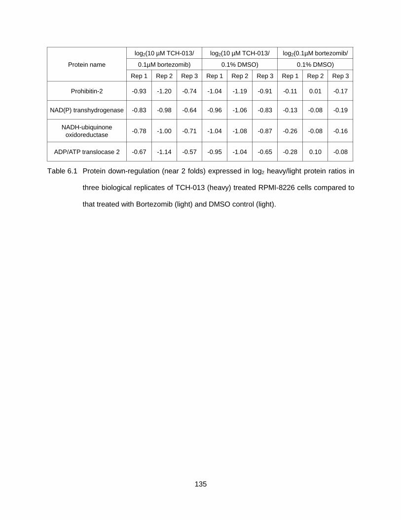
135
Table 6.1 Protein down-regulation (near 2 folds) expressed in log2 heavy/light protein ratios in
three biological replicates of TCH-013 (heavy) treated RPMI-8226 cells compared to
that treated with Bortezomib (light) and DMSO control (light).
Protein name
log2(10 µM TCH-013/ log2(10 µM TCH-013/ log2(0.1µM bortezomib/
0.1µM bortezomib) 0.1% DMSO) 0.1% DMSO)
Rep 1 Rep 2 Rep 3 Rep 1 Rep 2 Rep 3 Rep 1 Rep 2 Rep 3
Prohibitin-2 -0.93 -1.20 -0.74 -1.04 -1.19 -0.91 -0.11 0.01 -0.17
NAD(P) transhydrogenase -0.83 -0.98 -0.64 -0.96 -1.06 -0.83 -0.13 -0.08 -0.19
NADH-ubiquinone
oxidoreductase-0.78 -1.00 -0.71 -1.04 -1.08 -0.87 -0.26 -0.08 -0.16
ADP/ATP translocase 2 -0.67 -1.14 -0.57 -0.95 -1.04 -0.65 -0.28 0.10 -0.08
136
6.3 Conclusions
A quantitative proteomics strategy comprising human RPMI-8226 cells treated
with either the non-competitive proteasome inhibitor, TCH-013, the competitive
proteasome inhibitor, Bortezomib or DMSO (vehicle control), tryptic digestion, dimethyl
labeling, basic pH reversed-phase fractionation, LC-MS/MS and bioinformatic tools
were employed to study the mechanism of action of TCH-013. To minimize the errors of
quantification caused by retention time shifts of CD2O-labeled peptides compared to
their CH2O-labeled counterparts, peptides identified in multiple fractions were excluded
for quantitative analysis and the integral intensity ratios of CD2O/CH2O-labeled peptide
duplet peaks were calculated collectively over their chromatographic peaks. In each of
three treatments and in all three biological replicates >1200 proteins with at least two
peptides were identified and >400 proteins with at least 4 peptides were quantified.
Although no significantly up-regulated proteins were observed, 4 proteins including
prohibitin-2, NAD(P) transhydrogenase, NADH-ubiquinone oxidoreductase and
ADP/ATP translocate 2 were found to be down-regulated by at least two fold.
Interestingly, down-regulated proteins were all closely related to the regulation of
mitochondrial functions which are critical to growth and division of many cancer cells
[288-293]. While the exact mechanism of selective protein down-regulation induced by
TCH-013 in the present study remains unclear, this work provides candidate protein
targets for future more targeted TCH-013 mechanistic studies.
137
CHAPTER SEVEN
Experimental Methods for Chapters Five and Six
7.1 Materials
Acetic acid (99%) and formic acid (FA, 99%) were purchased from Thermo
Fisher Scientific (San Jose, CA), trifluoroacetic acid (TFA, LC grade) from Applied
Biosystems (Waltham, MA) and acetonitrile (ACN, HPLC grade) and 25% ammonia
solution from EMD Millipore (Billerica, MA). Trypsin (sequencing grade), iodoacetamide,
ammonium formate, triethylammonium bicarbonate (TEAB) buffer solution, 37%
formaldehyde (CH2O) in water (H2O) and 20% deuterium-labeled formaldehyde (CD2O)
in deuterium-labeled water (D2O) were purchased from Sigma-Aldrich (St. Louis, MO).
Urea, methanol, chloroform and tris(2-carboxyethyl)phosphine (TCEP) solution were
purchased from Thermo Fisher Scientific (San Jose, CA). HPLC grade H2O was
provided by a Milli-Q A10 water purification system (EMD Millipore, Billerica, MA).
Sodium bicarbonate (NaHCO3) was purchased from Chem Supply (Gillman, Australia).
Oasis solid phase extraction (SPE) cartridges (10 mg sorbent, 30 µM particle size) were
from Waters Corporation (Milford, MA). 15N-labeled recombinant human parathyroid
hormone (PTH) 1-84 internal standard was purchased from Cell Sciences (Canton, MA)
and its concentration was determined by amino acid analysis. World health organization
(WHO) international standard human recombinant PTH 1-84 was from National Institute
for Biological Standards and Control (NIBSC, Potters Bar, UK). Isotope-labeled oxidized
PTH 1-20 and 7-20 internal standard peptides, both with methionine sulfoxide (M(O)) at
residue 8 and 18 as well as 13C615N1-labeled leucine (13C6
15N1Leu) at residue 7 and 15
were purchased from Sigma-Aldrich (St. Louis, MO). Bortezomib was purchased from
138
Selleck Biochemicals (Houston, TX). TCH-013 was synthesized by the Tepe lab at
Michigan State University (East Lansing, MI) as previously described [294]. All other
reagents were commercially available and used without further purification.
7.2 Biological Samples
7.2.1 Patient Information and Immuno-Affinity Enrichment of PTH Proteins
Plasmas samples were collected and prepared from patients with high
concentrations of PTH 1-84 (500 to 1500 pg/mL as previously measured by
chemiluminescent immunoassay) by the Martin lab at Saint Louis University (St. Louis,
MO). For 1 mL of patient plasma, 20 µL of PTH-depleted plasma containing 25 pg/µL
15N-labeled PTH 1-84 was added immediately after blood draw and centrifuging
separation for plasma. Then patient plasmas were frozen, shipped, stored at -80 ⁰C and
thawed at room temperature before immuno-affinity enrichment at DiaSorin (Stillwater,
MN). As previously described [264], 350 µg of magnetic beads coated with goat anti-
PTH (39-84) antibody were added in 1 mL of plasma before incubation at room
temperature with gentle rotation for immuno-affinity enrichment. The plasma was then
removed using a DynaMag-2 Magnet (Life Technologies, Grand Island, NY) to capture
the beads and washed with 1 mL each of phosphate-buffered saline (PBS) buffer, 35%
ACN and PBS buffer followed by a final wash with 1 mL of 10 mM NH4HCO3. Bound
PTH was then eluted with 100 µL of 3.5% ACN/0.9% FA after 10 min incubation with
gentle rotation. The eluent was dried down under vacuum.
7.2.2 Cell Culture and Treatment Using TCH-013, Bortezomib and DMSO
Cell lysates from Human RPMI-8226 cells treated with TCH-013, Bortezomib or
DMSO were provided by Tepe lab at Michigan State University (East Lansing, MI). The
139
conditions for cell culture were described previously [276]. RPMI-8226 cells were
incubated with DMSO, Bortezomib or TCH-013 to final a concentrations of 0.1% (v/v),
0.1 μM and 10 μM, respectively, for 1 hour at 37 ⁰C and 5% CO2 before harvest and
measurement of total protein concentration using BCA reagent.
7.3 Protein Digestion
7.3.1 Trypsin Digestion of PTH Standards and Immuno-Enriched Samples from
Patient Plasmas
To each dried sample containing eluted PTH from 1 mL of patient plasma, 47.5
µL of 10 mM NH4HCO3 and 500 ng trypsin dissolved in 2 µL of H2O were added. The
solution was incubated at 37 ⁰C for 4 h, and the digestion was quenched by adding 0.5
µL of acetic acid. 13C615N1Leu-labeled PTH 1-20 M8(O)M18(O) mixed with equal molar
amount of 13C615N1Leu-labeled PTH 7-20 M8(O)M18(O) was digested using the same
conditions described above (trypsin : peptide = 1 : 1) to prepare a stock solution of
internal standards containing 1 µM 13C615N1Leu-labeled PTH 1-13 M(O) and 7-13 M(O)
as well as 2 µM 13C615N1Leu-labeled PTH 14-20 M(O). After dilution with H2O, 5 nM
13C615N1Leu-labeled PTH 1-13 M(O) and 7-13 M(O) as well as 10 nM 13C6
15N1Leu-
labeled PTH 14-20 M(O) in 10 µL H2O was spiked into the digested patient sample.
After drying down under vacuum, the patient sample was reconstituted in 50 μL of 0.1%
TFA/2% ACN in H2O and 5 μL of each sample solution was used for LC-MS/MS
analysis (described below). WHO PTH 1-84 standard was also digested with the same
conditions to facilitate optimization of MS parameters for PTH peptides as described
below.
140
7.3.2 Trypsin Digestion of Proteins in Lysates of TCH-013, Bortezomib and
DMSO Treated RPMI-8226 Cells
Following a methanol/chloroform precipitation, 50 µg of TCH-013, Bortezomib or
DMSO treated samples from each of three biological replicates were dried under
vacuum, separately. Thereafter, each sample was dissolved in 8 M urea in 50 mM
TEAB buffer (pH=8) and incubated at 37 °C for 30 min followed by reduction in 10 mM
TCEP at 37 °C for 45 min and alkylation in 55 mM iodoacetamide at 37 °C for 45 min.
Samples were then diluted to 1 M urea with 25 mM TEAB (pH=8) and digested for 18
hours at 37 °C with trypsin (1:40). Following digestion, samples were acidified to 1% FA,
desalted using an Oasis SPE cartridge and then dried under vacuum.
7.4 Dimethyl Labeling of Digested Proteins from TCH-013, Bortezomib and
DMSO Treated RPMI-8226 Cells
Each of the digested or desalted samples was re-dissolved in 200 µL of 100 mM
TEAB buffer (pH=8). TCH-013 treated samples were labeled by adding 8 µL of 4% (v/v)
CD2O in 100 mM TEAB buffer (pH=8) while Bortezomib and DMSO treated samples
were labeled by adding 8 µL of 4% (v/v) CH2O in 100 mM TEAB buffer (pH=8),
separately. 8 µL of 0.6 M NaBH3CN was added to each sample before incubation for 1
hour at room temperature while mixing. Thereafter 32 µL of 1% (v/v) ammonia solution
was added, vortexed, and 16 µL of formic acid was added to further quench the reaction.
Each sample was dried under vacuum and reconstituted with 100 µL of 5mM
ammonium formate/2% ACN, after which 50 µL of CD2O labeled TCH-013 treated
sample was mixed with either 50 µL of CH2O labeled Bortezomib treated sample or 50
µL of CH2O labeled DMSO treated sample.
141
7.5 Basic pH Reversed-Phase Fractionation of Digested and Dimethyl Labeled
Proteins
Off-line basic reversed-phase fractionation was completed using an Alltima HP
AQ C18 column (2.1 X 100 mm, 3 µM, Grace Corporate, Columbia, MD) and an Agilent
1100 series HPLC system (Agilent Technologies, Santa Clara, CA). Solvent A is 5mM
ammonium formate/2% ACN (pH=10) and solvent B is 5mM ammonium formate/90%
ACN (pH=10). The flow rate of the LC system is 0.1 mL/min. 100 µL of sample mixture
consisting of equal amount of TCH-013 treated sample with either Bortezomib or DMSO
treated sample in solvent A was injected. Then the percentage of solvent B in the
mobile phase was increased from 0% with a gradient of 1.2% B/min for 5 min, 1% B/min
for 2 min, 0.5% B/min for 19 min, 1% B/min for 4 min, 2% B/min for 8 min and 3% B/min
for 7 min. Finally, the percentage of solvent B was increased to 95% within 1 min, kept
for 4 min, decreased to 0% within 1 min and kept for 24 min for column cleaning and re-
equilibration. Across the entire gradient, the first fraction was collected from 0 - 17.5 min.
From 17.5 to 57.5 min, fractions were collected every 5 min. From 57.5 to 77.5 min, the
last fraction was collected. Each of the total 10 fractions for each injected sample was
dried under vacuum.
7.6 Mass Spectrometry
7.6.1 LC-MS/MS Analysis of Digested PTH standards and Immuno-Affinity
Enriched Samples from Patient Plasmas
LC-ESI-MS and HCD-MS/MS analyses were performed using a Thermo
Scientific Q Exactive Plus Orbitrap mass spectrometer (Thermo Fisher Scientific, San
Jose, CA) coupled to an Agilent 1200 Infinity Binary LC System (Agilent Technologies,
142
Santa Clara, CA). Analyses were performed using automated methods created by the
Xcalibur software (Thermo Fisher Scientific, San Jose, CA). Samples in 0.1 % TFA/2%
ACN were loaded onto an IntegraFrit Sample Trap Magic AQ C18 Column (3 µm, 200
Å , 150 µm I.D. x 25 mm in length, New Objective, Woburn, MA) at a flow rate of 2
μL/min for 5 min using 0.1% FA/2% ACN as the loading buffer. The peptides
concentrated on the trap column were eluted onto a PicoChip Magic AQ C18 Column (3
μm, 200 Å, 150 μm I.D. x 105 mm in length, New Objective, Woburn, MA) at a flow rate
of 800 nL/min using a linear 20 min gradient from 2% to 60% solvent B (Solvent A is 0.1%
FA and solvent B is 0.1% FA/95% ACN). After gradient separation, the columns were
cleaned with 90% solvent B for 6 min followed by re-equilibrium with 98% solvent A for
10 min. The spray voltage of the Q Exactive Plus Orbitrap mass spectrometer was
maintained at 2.7 kV. The ion transfer tube of the mass spectrometer was set at 300 °C,
and the S-lens was at 60%. Full MS scans were acquired at m/z 250 – 2000, a resolving
power of 70,000, an auto gain control (AGC) target value of 1.0 × 10⁶ and a maximum
injection time of 100 ms. HCD-MS/MS scans were taken at resolving power of 17,500,
AGC target value of 5.0 × 10⁴; maximum injection time of 50 ms and an isolation
window of m/z 1.5. The resolving power of HCD-MS/MS scans was increased to
140,000 from 12.5 min to 13.9 min within the 20 min gradient to distinguish monitored
fragment ions from artefact ions in patient samples. All spectra were recorded in profile
mode. Targeted HCD-MS/MS analysis was performed based on the information listed in
Table 5.1 where charge states of precursor ions, normalized collision energy (NCE) and
monitored fragment ions have been optimized for optimal sensitivity by LC-ESI-HCD-
143
MS/MS analysis of PTH peptides from digested WHO PTH 1-84, 13C615N1Leu-labeled
PTH 1-20 M8(O)M18(O) and 13C615N1Leu-labeled PTH 7-20 M8(O)M18(O).
7.6.2 LC-MS/MS Analysis of Digested, Dimethyl Labeled and Fractionated
Proteins from TCH-013, Bortezomib and DMSO Treated RPMI-8226 Cells
Each of 10 fractions from each sample reconstituted in 120 µL 0.1% FA/2% ACN
was analyzed by LC-MS/MS using a Q Exactive Plus mass spectrometer coupled to an
Ultimate 3000 UHPLC (Thermo Fisher Scientific, San Jose, CA). Solvent A is 0.1%
FA/2% ACN and solvent B is 0.1% FA in ACN. 6.4 µL of each fraction was injected onto
a PepMap C18 trap column (75 µM X 2 cm, 3 µM, 100 Å, Thermo Fisher Scientific, San
Jose, CA) at 2 µL/min for 15 min using 2% ACN and then separated through a PepMap
C18 analytical column (75 µM X 15 cm, 2 µM, 100 Å, Thermo Fisher Scientific, San
Jose, CA) at a flow rate of 300 nL/min. During separation, the percentage of solvent B in
mobile phase was increased from 2% to 10% in 1 min, from 10% to 35% in 50 min, from
35% to 60% in 1 min and from 60% to 90% in 1 min. Then the columns were cleaned at
90% B for 7 min before decreasing the % B to 2% in 1min and re-equilibrating for 6 min.
The spray voltage, temperature of ion transfer tube and S-lens of the Q Exactive Plus
Orbitrap mass spectrometer was set at 1.8 kV, 250 °C and 50%, respectively. The full
MS scans were acquired at m/z 300 – 2000, a resolving power of 70,000, an AGC
target value of 3.0 × 10⁶ and a maximum injection time of 30 ms. HCD-MS/MS scans
were acquired at a resolving power of 17,500, AGC target value of 5.0 × 10⁴, maximum
injection time of 120 ms, isolation window of m/z 1.4 and NCE of 25% for the top 12
most abundant ions in the MS spectra. All spectra were recorded in profile mode.
144
7.7 Data Analysis
7.7.1 Quantification of Full Length, Oxidized and Truncated PTH Variants in
Patient Plasma
PTH 1-13 M(O) and PTH 1-13 represent any form of PTH 1-84 with and without
oxidation within residue 1-13, respectively, while PTH 7-13 M(O) and PTH 7-13
represent any form of PTH 7-84 with and without oxidation, respectively, within residue
7-13. PTH 14-20 M(O) and PTH 14-20 represent any form of residue 14-84 containing-
PTH (i.e., PTH 1-84 and 7-84) with and without oxidation within residue 14-20. PTH 27-
44, 45-52, 53-65, 66-72 and 73-80 were also quantified using targeted HCD-MS/MS
and 15N-labeled standards from digestion of 15N-labeled PTH 1-84.
For each of the PTH peptides listed in Table 5.1, its HCD-MS/MS spectra were
averaged across the extracted ion current (XIC) chromatogram peak of the
corresponding representative fragment ion. For each unoxidized peptide, the
concentration (Cnon_ox) was calculated using the equation (1),
Cnon_ox =Inon_ox
I15N_non_ox
C15N
V (1)
where Inon_ox and I15N_non_ox are intensities of isotopic clusters of the representative
product ion of the unoxidized PTH peptide and unoxidized 15N-labeled PTH peptide
internal standard. C15N is concentration of 15N-labeled PTH 1-84 or 7-84 internal
standard spiked in plasma and V is the volume of original plasma sample.
Concentrations of in vivo oxidized PTH peptides (Cox) were determined using the
equation (2),
Cox =Iox−
I15N_oxI15N_non_ox
Inon_ox
I13C15N
C13C15N
V
1
yrecovery (2)
145
where Iox, I15N_ox, I13C15N are the intensities of isotopic clusters of the representative
product ion of the oxidized PTH peptide, oxidized 15N-labeled PTH peptide internal
standard and 13C615N1Leu-labeled oxidized PTH peptide internal standard. C13C15N is
concentration of 13C615N1Leu-labeled oxidized PTH peptide internal standard spiked in
sample before LC-MS/MS analysis and yrecovery is the recovery yield (66%) of the
immuno LC-MS/MS method [264].
7.3.3 Bioinformatic Analysis for Proteomic Profiling of Proteins in TCH-013,
Bortezomib and DMSO Treated RPMI-8226 Cells
Peptides and proteins were identified and quantified using the MaxQuant
software package, version 1.5.3.30 [285, 295]. The terminology described below is
specific to this software. Data was searched against human Uniprot protein database
containing 78,909 entries. For searching, the enzyme specificity was set to be trypsin
with the maximum number of missed cleavages set to be 2. The MS and MS/MS
tolerance were set to be 4.5 ppm and 20 ppm, respectively. Fixed modifications were
set as two-plex dimethyl labeling of lysine residues and peptide N-termini and
carbamidomethylation of cysteine residues. Variable modifications were set as oxidation
of methionine residues, acetylation of protein N termini and glycine-glycine addition
(Gly-Gly) on the side chains of lysine residues. The false discovery rate (FDR) for
peptide and protein identification was set to 1%. The minimum peptide length was set to
be 6 and peptide re-quantification functions were enabled. Following a MaxQuant
search, reverse and potential contaminant peptides and proteins were removed. Only
proteins identified with at least 2 unique/razor peptides were considered as identified.
In-house C++ script was written by David Perkins in the Bio21 Mass Spectrometry and
146
Proteomics Facility at the University of Melbourne and used to process the data in
MaxQuant Evidence table for quantitative analysis. Briefly, for each experiment,
peptides with same ‘Mod. Peptide ID’ but having different ‘Fraction’ numbers were
removed to avoid incorrect H/L peptide ratios caused by shifted retention times of CH2O
and CD2O labeled peptides in the basic reversed-phase fractionation. Then, to correct
errors from sample mixing, H/L ratios of the remaining peptides were normalized so that
the median ratio was 1. The normalized peptide H/L ratios were log2 transformed and
the median values were calculated for peptides with the same sequence as their
grouped H/L ratios. A H/L ratio of protein group was derived by calculating a median
value over normalized, log2 transformed and grouped H/L ratio of peptides assigned to
that protein group.

147
BIBLIOGRAPHY
148
BIBLIOGRAPHY
1. Bensimon, A., Heck, A.J.R., Aebersold, R., Mass spectrometry–based proteomics and network biology. Annual Review of Biochemistry 2012, 81, 379.
2. Murphy, R.C., Tandem mass spectrometry of lipids: Molecular analysis of complex lipids, Royal Society of Chemistry, Cambridge, UK, 2014.
3. Gabelica, V., Nucleic acids in the gas phase, in: Physical Chemistry in Action, Springer-Verlag Berlin Heidelberg, 2014.
4. Kailemia, M.J., Ruhaak, L.R., Lebrilla, C.B., Amster, I.J., Oligosaccharide analysis by mass spectrometry: A review of recent developments. Analytical Chemistry 2014, 86, 196.
5. Theodoridis, G., Gika, H.G., Wilson, I.D., Mass spectrometry-based holistic analytical approaches for metabolite profiling in systems biology studies. Mass Spectrometry Reviews 2011, 30, 884.
6. Piez, K.A., Molecular weight determination of random coil polypeptides from collagen by molecular sieve chromatography. Analytical Biochemistry 1968, 26, 305.
7. Grefrath, S.P., Reynolds, J.A., The molecular weight of the major glycoprotein from the human erythrocyte membrane. Proceedings of the National Academy of Sciences of the United States of America 1974, 71, 3913.
8. Siegel, L.M., Monty, K.J., Determination of molecular weights and frictional ratios of proteins in impure systems by use of gel filtration and density gradient centrifugation. Application to crude preparations of sulfite and hydroxylamine reductases. Biochimica et Biophysica Acta 1966, 112, 346.
9. Hoffmann, E.D., Stroobant, V., Mass spectrometry: Priciples and applications, Third edition, John Wiley & Sons Ltd, The Atrium, Southern Gate, Chichester, West Sussex, England, 2007.
10. Fenn, J.B., Mann, M., Meng, C.K., Wong, S.F., Whitehouse, C.M., Electrospray ionization for mass-spectrometry of large biomolecules. Science 1989, 246, 64.
11. Tanaka, K., Waki, H., Ido, Y., Akita, S., Yoshida, Y., Yoshida, T., Matsuo, T., Protein and polymer analyses up to m/z 100 000 by laser ionization time-of-flight mass spectrometry. Rapid Communications in Mass Spectrometry 1988, 2, 151.
12. Hilton, G.R., Benesch, J.L.P., Two decades of studying non-covalent biomolecular assemblies by means of electrospray ionization mass spectrometry. Journal of the Royal Society Interface 2012, 9, 801.
149
13. Seeley, E.H., Schwamborn, K., Caprioli, R.M., Imaging of intact tissue sections: Moving beyond the microscope. Journal of Biological Chemistry 2011, 286, 25459.
14. Seeley, E.H., Caprioli, R.M., 3D imaging by mass spectrometry: A new frontier. Analytical Chemistry 2012, 84, 2105.
15. Cheng, C., Gross, M.L., Pittenauer, E., Complete structural elucidation of triacylglycerols by tandem sector mass spectrometry. Analytical Chemistry 1998, 70, 4417.
16. Pittenauer, E., Allmaier, G., The renaissance of high-energy CID for structural elucidation of complex lipids: MALDI-TOF/RTOF-MS of alkali cationized triacylglycerols. Journal of the American Society for Mass Spectrometry 2009, 20, 1037.
17. Shushan, B., A review of clinical diagnostic applications of liquid chromatography-tandem mass spectrometry. Mass Spectrometry Reviews 2010, 29, 930.
18. Parker, C., Domanski, D., Percy, A., Chambers, A., Camenzind, A., Smith, D., Borchers, C., Mass spectrometry in high-throughput clinical biomarker assays: Multiple reaction monitoring in: L.S. Tang, N., Poon, T. (Eds.) Chemical Diagnostics, Springer Berlin Heidelberg, 2014, 117.
19. Xia, Y., McLuckey, S.A., Evolution of instrumentation for the study of gas-phase ion/ion chemistry via mass spectrometry. Journal of the American Society for Mass Spectrometry 2008, 19, 173.
20. Marshall, A.G., Chen, T., 40 years of Fourier transform ion cyclotron resonance mass spectrometry. International Journal of Mass Spectrometry 2015, 377, 410.
21. Eliuk, S., Makarov, A., Evolution of Orbitrap mass spectrometry instrumentation. Annual Review of Analytical Chemistry 2015, 8, 61.
22. Hendrickson, C., Quinn, J., Kaiser, N., Smith, D., Blakney, G., Chen, T., Marshall, A., Weisbrod, C., Beu, S., 21 tesla Fourier transform ion cyclotron resonance mass spectrometer: A national resource for ultrahigh resolution mass analysis. Journal of the American Society for Mass Spectrometry 2015, 26, 1626.
23. Almeida, R., Pauling, J., Sokol, E., Hannibal-Bach, H., Ejsing, C., Comprehensive lipidome analysis by shotgun lipidomics on a hybrid quadrupole-Orbitrap-linear ion trap mass spectrometer. Journal of the American Society for Mass Spectrometry 2015, 26, 133.
24. Fhaner, C.J., Liu, S., Ji, H., Simpson, R.J., Reid, G.E., Comprehensive lipidome profiling of isogenic primary and metastatic colon adenocarcinoma cell lines. Analytical Chemistry 2012, 84, 8917.
150
25. Hebert, A.S., Merrill, A.E., Bailey, D.J., Still, A.J., Westphall, M.S., Strieter, E.R., Pagliarini, D.J., Coon, J.J., Neutron-encoded mass signatures for multiplexed proteome quantification. Nature Methods 2013, 10, 332.
26. Zhurov, K.O., Fornelli, L., Wodrich, M.D., Laskay, U.A., Tsybin, Y.O., Principles of electron capture and transfer dissociation mass spectrometry applied to peptide and protein structure analysis. Chemical Society Reviews 2013, 42, 5014.
27. Cox, J., Marginean, I., Kelly, R., Smith, R., Tang, K., Improving the sensitivity of mass spectrometry by using a new sheath flow electrospray emitter array at subambient pressures. Journal of the American Society for Mass Spectrometry 2014, 25, 2028.
28. Kim, T., Udseth, H.R., Smith, R.D., Improved ion transmission from atmospheric pressure to high vacuum using a multicapillary inlet and electrodynamic ion funnel interface. Analytical Chemistry 2000, 72, 5014.
29. Krutchinsky, A., Padovan, J., Cohen, H., Chait, B., Maximizing ion transmission from atmospheric pressure into the vacuum of mass spectrometers with a novel electrospray interface. Journal of the American Society for Mass Spectrometry 2015, 26, 649.
30. Krutchinsky, A., Padovan, J., Cohen, H., Chait, B., Optimizing electrospray interfaces using slowly diverging conical duct (ConDuct) electrodes. Journal of the American Society for Mass Spectrometry 2015, 26, 659.
31. Jäntti, S., Kivilompolo, M., Öhrnberg, L., Pietiläinen, K., Nygren, H., Orešič, M., Hyötyläinen, T., Quantitative profiling of bile acids in blood, adipose tissue, intestine, and gall bladder samples using ultra high performance liquid chromatography-tandem mass spectrometry. Analytical and Bioanalytical Chemistry 2014, 406, 7799.
32. Damen, C.W.N., Isaac, G., Langridge, J., Hankemeier, T., Vreeken, R.J., Enhanced lipid isomer separation in human plasma using reversed-phase UPLC with ion-mobility/high-resolution MS detection. Journal of Lipid Research 2014, 55, 1772.
33. Harris, G.A., Galhena, A.S., Fernández, F.M., Ambient sampling/ionization mass spectrometry: Applications and current trends. Analytical Chemistry 2011, 83, 4508.
34. McLuckey, S.A., Goeringer, D.E., Slow heating methods in tandem mass spectrometry. Journal of Mass Spectrometry 1997, 32, 461.
35. Sleno, L., Volmer, D.A., Ion activation methods for tandem mass spectrometry. Journal of Mass Spectrometry 2004, 39, 1091.
151
36. Palumbo, A.M., Reid, G.E., Evaluation of gas-phase rearrangement and competing fragmentation reactions on protein phosphorylation site assignment using collision induced dissociation-MS/MS and MS3. Analytical Chemistry 2008, 80, 9735.
37. Mikesh, L.M., Ueberheide, B., Chi, A., Coon, J.J., Syka, J.E.P., Shabanowitz, J., Hunt, D.F., The utility of ETD mass spectrometry in proteomic analysis. Biochimica et Biophysica Acta (BBA) - Proteins and Proteomics 2006, 1764, 1811.
38. Zubarev, R.A., Kelleher, N.L., McLafferty, F.W., Electron capture dissociation of multiply charged protein cations. A nonergodic process. Journal of the American Chemical Society 1998, 120, 3265.
39. Zhang, H., Cui, W., Wen, J., Blankenship, R.E., Gross, M.L., Native electrospray and electron-capture dissociation FTICR mass spectrometry for top-down studies of protein assemblies. Analytical Chemistry 2011, 83, 5598.
40. Shaw, J.B., Li, W., Holden, D.D., Zhang, Y., Griep-Raming, J., Fellers, R.T., Early, B.P., Thomas, P.M., Kelleher, N.L., Brodbelt, J.S., Complete protein characterization using top-down mass spectrometry and ultraviolet photodissociation. Journal of the American Chemical Society 2013, 135, 12646.
41. O’Brien, J.P., Needham, B.D., Henderson, J.C., Nowicki, E.M., Trent, M.S., Brodbelt, J.S., 193 nm ultraviolet photodissociation mass spectrometry for the structural elucidation of lipid A compounds in complex mixtures. Analytical Chemistry 2014, 86, 2138.
42. Ko, B.J., Brodbelt, J.S., 193 nm ultraviolet photodissociation of deprotonated sialylated oligosaccharides. Analytical Chemistry 2011, 83, 8192.
43. Smith, S.I., Brodbelt, J.S., Characterization of oligodeoxynucleotides and modifications by 193 nm photodissociation and electron photodetachment dissociation. Analytical Chemistry 2010, 82, 7218.
44. O’Brien, J.P., Brodbelt, J.S., Structural characterization of gangliosides and glycolipids via ultraviolet photodissociation mass spectrometry. Analytical Chemistry 2013, 85, 10399.
45. Aponte, J.R., Vasicek, L., Swaminathan, J., Xu, H., Koag, M.C., Lee, S., Brodbelt, J.S., Streamlining bottom-up protein identification based on selective ultraviolet photodissociation (UVPD) of chromophore-tagged histidine- and tyrosine-containing peptides. Analytical Chemistry 2014, 86, 6237.
46. Brodbelt, J.S., Photodissociation mass spectrometry: New tools for characterization of biological molecules. Chemical Society Reviews 2014, 43, 2757.
47. Lodish, H.F., Molecular cell biology, 6th edition., W.H. Freeman, New York, 2008.
152
48. Feng, X., Liu, X., Luo, Q., Liu, B., Mass spectrometry in systems biology: An overview. Mass Spectrometry Reviews 2008, 27, 635.
49. Brugger, B., Lipidomics: Analysis of the lipid composition of cells and subcellular organelles by electrospray ionization mass spectrometry. Annual Review of Biochemistry 2014, 83, 79.
50. Angel, T.E., Aryal, U.K., Hengel, S.M., Baker, E.S., Kelly, R.T., Robinson, E.W., Smith, R.D., Mass spectrometry based proteomics: Existing capabilities and future directions. Chemical Society Reviews 2012, 41, 3912.
51. Swaney, D.L., Wenger, C.D., Coon, J.J., The value of using multiple proteases for large-scale mass spectrometry-based proteomics. Journal of Proteome Research 2010, 9, 1323.
52. Zhong, X., Zhang, Z., Jiang, S., Li, L., Recent advances in coupling capillary electrophoresis based separation techniques to ESI and MALDI MS. Electrophoresis 2014, 35, 1214.
53. Kalli, A., Smith, G.T., Sweredoski, M.J., Hess, S., Evaluation and optimization of mass spectrometric settings during data-dependent acquisition mode: Focus on LTQ-Orbitrap mass analyzers. Journal of Proteome Research 2013, 12, 3071.
54. Law, K.P., Lim, Y.P., Recent advances in mass spectrometry: Data independent analysis and hyper reaction monitoring. Expert Review of Proteomics 2013, 10, 551.
55. Shi, T., Su, D., Liu, T., Tang, K., Camp, D.G., Qian, W.-J., Smith, R.D., Advancing the sensitivity of selected reaction monitoring-based targeted quantitative proteomics. Proteomics 2012, 12, 1074.
56. McHugh, L., Arthur, J.W., Computational methods for protein identification from mass spectrometry data. PLoS Computational Biology 2008, 4, e12.
57. Heck, A.J.R., Native mass spectrometry: A bridge between interactomics and structural biology. Nature Methods 2008, 5, 927.
58. Nahnsen, S., Bielow, C., Reinert, K., Kohlbacher, O., Tools for label-free peptide quantification. Molecular & Cellular Proteomics 2013, 12, 549.
59. Schilling, B., Rardin, M.J., MacLean, B.X., Zawadzka, A.M., Frewen, B.E., Cusack, M.P., Sorensen, D.J., Bereman, M.S., Jing, E., Wu, C.C., Verdin, E., Kahn, C.R., MacCoss, M.J., Gibson, B.W., Platform-independent and label-free quantitation of proteomic data using MS1 extracted ion chromatograms in Skyline: Application to protein acetylation and phosphorylation. Molecular & Cellular Proteomics 2012, 11, 202.
153
60. Bantscheff, M., Lemeer, S., Savitski, M., Kuster, B., Quantitative mass spectrometry in proteomics: Critical review update from 2007 to the present. Analytical and Bioanalytical Chemistry 2012, 404, 939.
61. Fonslow, B.R., Carvalho, P.C., Academia, K., Freeby, S., Xu, T., Nakorchevsky, A., Paulus, A., Yates, J.R., Improvements in proteomic metrics of low abundance proteins through proteome equalization using proteoMiner prior to MudPIT. Journal of Proteome Research 2011, 10, 3690.
62. Rundlett, K.L., Armstrong, D.W., Mechanism of signal suppression by anionic surfactants in capillary electrophoresis−electrospray ionization mass spectrometry. Analytical Chemistry 1996, 68, 3493.
63. Gropengiesser, J., Varadarajan, B.T., Stephanowitz, H., Krause, E., The relative influence of phosphorylation and methylation on responsiveness of peptides to MALDI and ESI mass spectrometry. Journal of Mass Spectrometry 2009, 44, 821.
64. Olsen, J.V., Mann, M., Status of large-scale analysis of post-translational modifications by mass spectrometry. Molecular & Cellular Proteomics 2013, 12, 3444.
65. Fahy, E., Subramaniam, S., Brown, H.A., Glass, C.K., Merrill, A.H., Murphy, R.C., Raetz, C.R.H., Russell, D.W., Seyama, Y., Shaw, W., Shimizu, T., Spener, F., van Meer, G., VanNieuwenhze, M.S., White, S.H., Witztum, J.L., Dennis, E.A., A comprehensive classification system for lipids. Journal of Lipid Research 2005, 46, 839.
66. Ekroos, K., Lipidomics perspective: From molecular lipidomics to validated clinical diagnostics, in, Wiley-VCH Verlag GmbH & Co. KGaA, Weinheim, Germany, 2012.
67. Rainville, P.D., Stumpf, C.L., Shockcor, J.P., Plumb, R.S., Nicholson, J.K., Novel application of reversed-phase UPLC-oaTOF-MS for lipid analysis in complex biological mixtures: A new tool for lipidomics. Journal of Proteome Research 2007, 6, 552.
68. Schuhmann, K., Almeida, R., Baumert, M., Herzog, R., Bornstein, S.R., Shevchenko, A., Shotgun lipidomics on a LTQ Orbitrap mass spectrometer by successive switching between acquisition polarity modes. Journal of Mass Spectrometry 2012, 47, 96.
69. Han, X., Yang, K., Gross, R.W., Multi-dimensional mass spectrometry-based shotgun lipidomics and novel strategies for lipidomic analyses. Mass Spectrometry Reviews 2012, 31, 134.
70. Ejsing, C.S., Sampaio, J.L., Surendranath, V., Duchoslav, E., Ekroos, K., Klemm, R.W., Simons, K., Shevchenko, A., Global analysis of the yeast lipidome by quantitative shotgun mass spectrometry. Proceedings of the National Academy of Sciences 2009,
154
71. Murphy, R.C., Gaskell, S.J., New applications of mass spectrometry in lipid analysis. Journal of Biological Chemistry 2011, 286, 25427.
72. Hsu, F., Turk, J., Electrospray ionization multiple-stage linear ion-trap mass spectrometry for structural elucidation of triacylglycerols: Assignment of fatty acyl groups on the glycerol backbone and location of double bonds. Journal of the American Society for Mass Spectrometry 2010, 21, 657.
73. Hsu, F., Turk, J., Structural characterization of unsaturated glycerophospholipids by multiple-stage linear ion-trap mass spectrometry with electrospray ionization. Journal of the American Society for Mass Spectrometry 2008, 19, 1681.
74. Hsu, F., Turk, J., Toward total structural analysis of cardiolipins: Multiple-stage linear ion-trap mass spectrometry on the [M − 2H + 3Li]+ ions. Journal of the American Society for Mass Spectrometry 2010, 21, 1863.
75. Taucher, M., Breuker, K., Characterization of modified RNA by top-down mass spectrometry. Angewandte Chemie International Edition 2012, 51, 11289.
76. Tretyakova, N., Goggin, M., Sangaraju, D., Janis, G., Quantitation of DNA adducts by stable isotope dilution mass spectrometry. Chemical Research in Toxicology 2012, 25, 2007.
77. Beck, J.L., Developments in electrospray ionization mass spectrometry of non-covalent DNA–ligand complexes. Australian Journal of Chemistry 2011, 64, 705.
78. Varki, A., Biological roles of oligosaccharides: All of the theories are correct. Glycobiology 1993, 3, 97.
79. Zaikin, V., Halket, J., A handbook of derivatives for mass spectrometry, in, IM Publications LLP, Charlton, Chichester, UK, 2009.
80. Oss, M., Kruve, A., Herodes, K., Leito, I., Electrospray ionization efficiency scale of organic compounds. Analytical Chemistry 2010, 82, 2865.
81. Mirzaei, H., Regnier, F., Enhancing electrospray ionization efficiency of peptides by derivatization. Analytical Chemistry 2006, 78, 4175.
82. Lu, Y., Zhou, X., Stemmer, P.M., Reid, G.E., Sulfonium ion derivatization, isobaric stable isotope labeling and data dependent CID- and ETD-MS/MS for enhanced phosphopeptide quantitation, identification and phosphorylation site characterization. Journal of the American Society for Mass Spectrometry 2012, 23, 577.
83. Kulevich, S.E., Frey, B.L., Kreitinger, G., Smith, L.M., Alkylating tryptic peptides to enhance electrospray ionization mass spectrometry analysis. Analytical Chemistry 2010, 82, 10135.
155
84. Lecchi, P., Olson, M., Brancia, F.L., The role of esterification on detection of protonated and deprotonated peptide ions in matrix assisted laser desorption/Ionization (MALDI) mass spectrometry (MS). Journal of the American Society for Mass Spectrometry 2005, 16, 1269.
85. Brancia, F.L., Oliver, S.G., Gaskell, S.J., Improved matrix-assisted laser desorption/ionization mass spectrometric analysis of tryptic hydrolysates of proteins following guanidination of lysine-containing peptides. Rapid Communications in Mass Spectrometry 2000, 14, 2070.
86. Frahm, J.L., Bori, I.D., Comins, D.L., Hawkridge, A.M., Muddiman, D.C., Achieving augmented limits of detection for peptides with hydrophobic alkyl tags. Analytical Chemistry 2007, 79, 3989.
87. Yang, W., Adamec, J., Regnier, F.E., Enhancement of the LC/MS analysis of fatty acids through derivatization and stable isotope coding. Analytical Chemistry 2007, 79, 5150.
88. Li, Y.L., Su, X., Stahl, P.D., Gross, M.L., Quantification of diacylglycerol molecular species in biological samples by electrospray ionization mass spectrometry after one-step derivatization. Analytical Chemistry 2007, 79, 1569.
89. Woo, H., Go, E.P., Hoang, L., Trauger, S.A., Bowen, B., Siuzdak, G., Northen, T.R., Phosphonium labeling for increasing metabolomic coverage of neutral lipids using electrospray ionization mass spectrometry. Rapid Communications in Mass Spectrometry 2009, 23, 1849.
90. Sandhoff, R., Brügger, B., Jeckel, D., Lehmann, W.D., Wieland, F.T., Determination of cholesterol at the low picomole level by nano-electrospray ionization tandem mass spectrometry. Journal of Lipid Research 1999, 40, 126.
91. Han, X., Yang, K., Cheng, H., Fikes, K.N., Gross, R.W., Shotgun lipidomics of phosphoethanolamine-containing lipids in biological samples after one-step in situ derivatization. Journal of Lipid Research 2005, 46, 1548.
92. Pham, H.T., Julian, R.R., Mass shifting and radical delivery with crown ether attachment for separation and analysis of phosphatidylethanolamine lipids. Analytical Chemistry 2014, 86, 3020.
93. Julian, R.R., Beauchamp, J.L., Site specific sequestering and stabilization of charge in peptides by supramolecular adduct formation with 18-crown-6 ether by way of electrospray ionization. International Journal of Mass Spectrometry 2001, 210–211, 613.
94. Dell, A., Preparation and desorption mass spectrometry of permethyl and peracetyl derivatives of oligosaccharides in: Methods in Enzymology, Academic Press, 1990, 647.
156
95. Lamari, F.N., Kuhn, R., Karamanos, N.K., Derivatization of carbohydrates for chromatographic, electrophoretic and mass spectrometric structure analysis. Journal of Chromatography B 2003, 793, 15.
96. Bereman, M.S., Comins, D.L., Muddiman, D.C., Increasing the hydrophobicity and electrospray response of glycans through derivatization with novel cationic hydrazides. Chemical Communications 2010, 46, 237.
97. Walker, S.H., Papas, B.N., Comins, D.L., Muddiman, D.C., Interplay of permanent charge and hydrophobicity in the electrospray ionization of glycans. Analytical Chemistry 2010, 82, 6636.
98. Nordström, A., Tarkowski, P., Tarkowska, D., Dolezal, K., Åstot, C., Sandberg, G., Moritz, T., Derivatization for LC-electrospray ionization-MS: A tool for improving reversed-phase separation and ESI responses of bases, ribosides, and intact nucleotides. Analytical Chemistry 2004, 76, 2869.
99. Flarakos, J., Xiong, W., Glick, J., Vouros, P., A deoxynucleotide derivatization methodology for improving LC-ESI-MS detection. Analytical Chemistry 2005, 77, 2373.
100. Null, A.P., Nepomuceno, A.I., Muddiman, D.C., Implications of hydrophobicity and free energy of solvation for characterization of nucleic acids by electrospray ionization mass spectrometry. Analytical Chemistry 2003, 75, 1331.
101. Yang, W., Mirzaei, H., Liu, X., Regnier, F.E., Enhancement of amino acid detection and quantification by electrospray ionization mass spectrometry. Analytical Chemistry 2006, 78, 4702.
102. Biemann, K., Contributions of mass spectrometry to peptide and protein structure. Biological Mass Spectrometry 1988, 16, 99.
103. Ko, B., Brodbelt, J., Enhanced electron transfer dissociation of peptides modified at C-terminus with fixed charges. Journal of the American Society for Mass Spectrometry 2012, 23, 1991.
104. Setner, B., Rudowska, M., Klem, E., Cebrat, M., Szewczuk, Z., Peptides derivatized with bicyclic quaternary ammonium ionization tags. Sequencing via tandem mass spectrometry. Journal of Mass Spectrometry 2014, 49, 995.
105. Sadagopan, N., Watson, J.T., Investigation of the tris(trimethoxyphenyl)phosphonium acetyl charged derivatives of peptides by electrospray ionization mass spectrometry and tandem mass spectrometry. Journal of the American Society for Mass Spectrometry 2000, 11, 107.
106. Stutzman, J.R., McLuckey, S.A., Ion/Ion reactions of MALDI-derived peptide ions: Increased sequence coverage via covalent and electrostatic modification upon charge inversion. Analytical Chemistry 2012, 84, 10679.
157
107. Gunawardena, H.P., Emory, J.F., McLuckey, S.A., Phosphopeptide anion characterization via sequential charge inversion and electron-transfer dissociation. Analytical Chemistry 2006, 78, 3788.
108. Gao, X., Wu, H., Lee, K.-C., Liu, H., Zhao, Y., Cai, Z., Jiang, Y., Stable isotope N-phosphorylation labeling for peptide de novo sequencing and protein quantification based on organic phosphorus chemistry. Analytical Chemistry 2012, 84, 10236.
109. Stutzman, J.R., Hassell, K.M., McLuckey, S.A., Dissociation behavior of tryptic and intramolecular disulfide-linked peptide ions modified in the gas phase via ion/ion reactions. International Journal of Mass Spectrometry 2012, 312, 195.
110. Robotham, S.A., Kluwe, C., Cannon, J.R., Ellington, A., Brodbelt, J.S., De Novo sequencing of peptides using selective 351 nm ultraviolet photodissociation mass spectrometry. Analytical Chemistry 2013, 85, 9832.
111. O’Brien, J.P., Pruet, J.M., Brodbelt, J.S., Chromogenic chemical probe for protein structural characterization via ultraviolet photodissociation mass spectrometry. Analytical Chemistry 2013, 85, 7391.
112. Cotham, V.C., Wine, Y., Brodbelt, J.S., Selective 351 nm photodissociation of cysteine-containing peptides for discrimination of antigen-binding regions of IgG fragments in bottom-up liquid chromatography–tandem mass spectrometry workflows. Analytical Chemistry 2013, 85, 5577.
113. Ko, B., Brodbelt, J., Ultraviolet photodissociation of carboxylate-derivatized peptides in a quadrupole ion trap. Journal of the American Society for Mass Spectrometry 2011, 22, 49.
114. Vasicek, L., Wilson, J., Brodbelt, J., Improved infrared multiphoton dissociation of peptides through N-terminal phosphonite derivatization. Journal of the American Society for Mass Spectrometry 2009, 20, 377.
115. Ly, T., Julian, R.R., Residue-specific radical-directed dissociation of whole proteins in the gas phase. Journal of the American Chemical Society 2008, 130, 351.
116. Diedrich, J.K., Julian, R.R., Site-specific radical directed dissociation of peptides at phosphorylated residues. Journal of the American Chemical Society 2008, 130, 12212.
117. Marshall, D.L., Hansen, C.S., Trevitt, A.J., Oh, H.B., Blanksby, S.J., Photodissociation of TEMPO-modified peptides: new approaches to radical-directed dissociation of biomolecules. Physical Chemistry Chemical Physics 2014, 16, 4871.
118. Hodyss, R., Cox, H.A., Beauchamp, J.L., Bioconjugates for tunable peptide fragmentation: Free radical lnitiated peptide sequencing (FRIPS). Journal of the American Chemical Society 2005, 127, 12436.
158
119. Lee, S., Glover, M.S., Reilly, J.P., Clemmer, D.E., Photosynthesis of a combinatorial peptide library in the gas phase. Analytical Chemistry 2015, 87, 9384.
120. McGee, W.M., McLuckey, S.A., Efficient and directed peptide bond formation in the gas phase via ion/ion reactions. Proceedings of the National Academy of Sciences 2014, 111, 1288.
121. Krishna, R.G., Wold, F., Identification of common post-translational modifications, in: Protein structure: A practical approach, Oxford University Press, Oxford, 1997, 91.
122. Seo, J., Lee, K.J., Post-translational modifications and their biological functions: proteomic analysis and systematic approaches. Journal of Biochemistry Molecular Biology 2004, 37, 35.
123. Oda, Y., Nagasu, T., Chait, B.T., Enrichment analysis of phosphorylated proteins as a tool for probing the phosphoproteome. Nature Biotechnology 2001, 19, 379.
124. Wells, L., Gao, Y., Mahoney, J.A., Vosseller, K., Chen, C., Rosen, A., Hart, G.W., Dynamic O-glycosylation of nuclear and cytosolic proteins: Future characterization of the nucleocytoplasmic β-N-acetylglucosaminidase, O-GlcNAcase. Journal of Biological Chemistry 2002, 277, 1755.
125. Diedrich, J.K., Julian, R.R., Facile identification of phosphorylation sites in peptides by radical directed dissociation. Analytical Chemistry 2011, 83, 6818.
126. Mendoza, V.L., Vachet, R.W., Probing protein structure by amino acid-specific covalent labeling and mass spectrometry. Mass Spectrometry Reviews 2009, 28, 785.
127. Zhou, X., Lu, Y., Wang, W., Borhan, B., Reid, G.E., ‘Fixed charge' chemical derivatization and data dependant multistage tandem mass spectrometry form mapping rrotein surface residue accessibility. Journal of the American Society for Mass Spectrometry 2010, 21, 1339.
128. Zhou, X., Mester, C., Stemmer, P.M., Reid, G.E., Oxidation-induced conformational changes in calcineurin determined by covalent labeling and tandem mass spectrometry. Biochemistry 2014, 53, 6754.
129. Whitehurst, C.B., Soderblom, E.J., West, M.L., Hernandez, R., Goshe, M.B., Brown, D.T., Location and role of free cysteinyl residues in the sindbis virus E1 and E2 glycoproteins. Journal of Virology 2007, 81, 6231.
130. Liu, Y., Kvaratskhelia, M., Hess, S., Qu, Y., Zou, Y., Modulation of replication protein A function by its hyperphosphorylation-induced conformational change involving DNA binding domain B. Journal of Biological Chemistry 2005, 280, 32775.
159
131. Mendoza, V.L., Vachet, R.W., Protein surface mapping using diethylpyrocarbonate with mass spectrometric detection. Analytical Chemistry 2008, 80, 2895.
132. Cammarata, M., Lin, K.-Y., Pruet, J., Liu, H.-w., Brodbelt, J., Probing the unfolding of myoglobin and domain C of PARP-1 with covalent labeling and top-down ultraviolet photodissociation mass spectrometry. Analytical Chemistry 2014, 86, 2534.
133. Rappsilber, J., The beginning of a beautiful friendship: cross-linking/mass spectrometry and modelling of proteins and multi-protein complexes. Journal of structural biology 2011, 173, 530.
134. Pham, N.D., Parker, R.B., Kohler, J.J., Photocrosslinking approaches to interactome mapping. Current Opinion in Chemical Biology 2013, 17, 90.
135. Liu, F., Rijkers, D.T.S., Post, H., Heck, A.J.R., Proteome-wide profiling of protein assemblies by cross-linking mass spectrometry. Nature Methods 2015, advance online publication,
136. Webb, I., Mentinova, M., McGee, W., McLuckey, S., Gas-phase intramolecular protein crosslinking via ion/ion reactions: Ubiquitin and a homobifunctional sulfo-NHS ester. Journal of the American Society for Mass Spectrometry 2013, 24, 733.
137. Fhaner, C.J., Liu, S., Zhou, X., Reid, G.E., Functional group selective derivatization and gas-phase fragmentation reactions of plasmalogen glycerophospholipids. Mass Spectrom (Tokyo) 2013, 2, S0015.
138. Lydic, T.A., Townsend, S., Adda, C.G., Collins, C., Mathivanan, S., Reid, G.E., Rapid and comprehensive 'shotgun' lipidome profiling of colorectal cancer cell derived exosomes. Methods 2015,
139. Brown, S.H.J., Mitchell, T.W., Blanksby, S.J., Analysis of unsaturated lipids by ozone-induced dissociation. Biochimica et Biophysica Acta (BBA) - Molecular and Cell Biology of Lipids 2011, 1811, 807.
140. Lemmon, M.A., Membrane recognition by phospholipid-binding domains. Nature Reviews Molecular Cell Biology 2008, 9, 99.
141. Mitchell, T.W., Pham, H., Thomas, M.C., Blanksby, S.J., Identification of double bond position in lipids: From GC to OzID. Journal of Chromatography B 2009, 877, 2722.
142. Hsu, F., Bohrer, A., Turk, J., Formation of lithiated adducts of glycerophosphocholine lipids facilitates their identification by electrospray ionization tandem mass spectrometry. Journal of the American Society for Mass Spectrometry 1998, 9, 516.
160
143. Ekroos, K., Ejsing, C.S., Bahr, U., Karas, M., Simons, K., Shevchenko, A., Charting molecular composition of phosphatidylcholines by fatty acid scanning and ion trap MS3 fragmentation. Journal of Lipid Research 2003, 44, 2181.
144. Stutzman, J.R., Blanksby, S.J., McLuckey, S.A., Gas-phase transformation of phosphatidylcholine cations to structurally informative anions via ion/ion chemistry. Analytical Chemistry 2013, 85, 3752.
145. Pham, H.T., Maccarone, A.T., Thomas, M.C., Campbell, J.L., Mitchell, T.W., Blanksby, S.J., Structural characterization of glycerophospholipids by combinations of ozone- and collision-induced dissociation mass spectrometry: The next step towards "top-down" lipidomics. Analyst 2014, 139, 204.
146. Huang, C., Lin, H., Li, S., Wang, G., Influence of the positions of cis double bonds in the sn-2-acyl chain of phosphatidylethanolamine on the bilayer’s melting behavior. Journal of Biological Chemistry 1997, 272, 21917.
147. Lin, Y., Schuurbiers, E., Van der Veen, S., De Deckere, E.A.M., Conjugated linoleic acid isomers have differential effects on triglyceride secretion in Hep G2 cells. Biochimica et Biophysica Acta (BBA) - Molecular and Cell Biology of Lipids 2001, 1533, 38.
148. Heipieper, H.J., Meinhardt, F., Segura, A., The cis–trans isomerase of unsaturated fatty acids in Pseudomonas and Vibrio: Biochemistry, molecular biology and physiological function of a unique stress adaptive mechanism. FEMS Microbiology Letters 2003, 229, 1.
149. Hsu, F., Turk, J., Elucidation of the double-bond position of long-chain unsaturated fatty acids by multiple-stage linear ion-trap mass spectrometry with electrospray ionization. Journal of the American Society for Mass Spectrometry 2008, 19, 1673.
150. Xu, Y., Brenna, J.T., Atmospheric pressure covalent adduct chemical ionization tandem mass spectrometry for double bond localization in monoene- and diene-containing triacylglycerols. Analytical Chemistry 2007, 79, 2525.
151. Ma, X., Xia, Y., Pinpointing double bonds in lipids by Paternò-Büchi reactions and mass spectrometry. Angewandte Chemie International Edition 2014, 53, 2592.
152. Pham, H.T., Trevitt, A.J., Mitchell, T.W., Blanksby, S.J., Rapid differentiation of isomeric lipids by photodissociation mass spectrometry of fatty acid derivatives. Rapid Communications in Mass Spectrometry 2013, 27, 805.
153. Pham, H.T., Ly, T., Trevitt, A.J., Mitchell, T.W., Blanksby, S.J., Differentiation of complex lipid isomers by radical-directed dissociation mass spectrometry. Analytical Chemistry 2012, 84, 7525.
154. Pham, H.T., Prendergast, M.B., Dunstan, C.W., Trevitt, A.J., Mitchell, T.W., Julian, R.R., Blanksby, S.J., Dissociation of proton-bound complexes reveals geometry
161
and arrangement of double bonds in unsaturated lipids. International Journal of Mass Spectrometry 2015, doi:10.1016/j.ijms.2015.07.006,
155. Shendure, J., Ji, H., Next-generation DNA sequencing. Nature Biotechnology 2008, 26, 1135.
156. Wilson, J.J., Brodbelt, J.S., Ultraviolet photodissociation at 355 nm of fluorescently labeled oligosaccharides. Analytical Chemistry 2008, 80, 5186.
157. Zhang, X., Julian, R.R., Radical mediated dissection of oligosaccharides. International Journal of Mass Spectrometry 2014, 372, 22.
158. Konermann, L., Pan, J., Liu, Y.-H., Hydrogen exchange mass spectrometry for studying protein structure and dynamics. Chemical Society Reviews 2011, 40, 1224.
159. Reid, G.E., Simpson, R.J., O’Hair, R.A.J., Probing the fragmentation reactions of protonated glycine oligomers via multistage mass spectrometry and gas phase ion molecule hydrogen/deuterium exchange. International Journal of Mass Spectrometry 1999, 190–191, 209.
160. Polce, M.J., Ren, D., Wesdemiotis, C., Dissociation of the peptide bond in protonated peptides. Journal of Mass Spectrometry 2000, 35, 1391.
161. Cui, L., Yapici, I., Borhan, B., Reid, G.E., Quantification of competing H3PO4 Versus HPO3 + H2O neutral losses from regioselective 18O-labeled phosphopeptides. Journal of the American Society for Mass Spectrometry 2014, 25, 141.
162. Riba Garcia, I., Giles, K., Bateman, R.H., Gaskell, S.J., Studies of peptide a- and b-type fragment ions using stable isotope labeling and integrated ion mobility/tandem mass spectrometry. Journal of the American Society for Mass Spectrometry 2008, 19, 1781.
163. Zhang, X., Fhaner, C.J., Ferguson-Miller, S.M., Reid, G.E., Evaluation of ion activation strategies and mechanisms for the gas-phase fragmentation of sulfoquinovosyldiacylglycerol lipids from Rhodobacter sphaeroides. International Journal of Mass Spectrometry 2012, 316-318, 100.
164. Andersen, T.E., Kirpekar, F., Haselmann, K.F., RNA fragmentation in MALDI mass spectrometry studied by H/D-exchange: Mechanisms of general applicability to nucleic acids. Journal of the American Society for Mass Spectrometry 2006, 17, 1353.
165. Saad, O.M., Leary, J.A., Delineating mechanisms of dissociation for isomeric heparin disaccharides using isotope labeling and ion trap tandem mass spectrometry. Journal of the American Society for Mass Spectrometry 2004, 15, 1274.
162
166. Dookeran, N.N., Yalcin, T., Harrison, A.G., Fragmentation reactions of protonated α-amino acids. Journal of Mass Spectrometry 1996, 31, 500.
167. Chalmers, M.J., Busby, S.A., Pascal, B.D., He, Y., Hendrickson, C.L., Marshall, A.G., Griffin, P.R., Probing protein ligand interactions by automated hydrogen/deuterium exchange mass spectrometry. Analytical Chemistry 2006, 78, 1005.
168. Jørgensen, T.J.D., Gårdsvoll, H., Ploug, M., Roepstorff, P., Intramolecular migration of amide hydrogens in protonated peptides upon collisional activation. Journal of the American Chemical Society 2005, 127, 2785.
169. Rand, K.D., Zehl, M., Jensen, O.N., Jørgensen, T.J.D., Protein hydrogen exchange measured at single-residue resolution by electron transfer dissociation mass spectrometry. Analytical Chemistry 2009, 81, 5577.
170. Rand, K.D., Adams, C.M., Zubarev, R.A., Jørgensen, T.J.D., Electron capture dissociation proceeds with a low degree of intramolecular migration of peptide amide hydrogens. Journal of the American Chemical Society 2008, 130, 1341.
171. Pan, J., Han, J., Borchers, C.H., Konermann, L., Conformer-specific hydrogen exchange analysis of Aβ(1–42) oligomers by top-down electron capture dissociation mass spectrometry. Analytical Chemistry 2011, 83, 5386.
172. Abzalimov, R.R., Kaplan, D.A., Easterling, M.L., Kaltashov, I.A., Protein conformations can be probed in top-down HDX MS experiments utilizing electron transfer dissociation of protein ions without hydrogen scrambling. Journal of the American Society for Mass Spectrometry 2009, 20, 1514.
173. Kirkpatrick, D.S., Gerber, S.A., Gygi, S.P., The absolute quantification strategy: A general procedure for the quantification of proteins and post-translational modifications. Methods 2005, 35, 265.
174. Krötz, F., Riexinger, T., Buerkle, M.A., Nithipatikom, K., Gloe, T., Sohn, H.Y., Campbell, W.B., Pohl, U., Membrane potential-dependent inhibition of platelet adhesion to endothelial cells by expoxyeicosatrienoic acids. Arteriosclerosis, Thrombosis, and Vascular Biology 2004, 24, 595.
175. Chan, W., Chen, B., Wang, L., Taghizadeh, K., Demott, M.S., Dedon, P.C., Quantification of the 2-deoxyribonolactone and nucleoside 5′-aldehyde products of 2-deoxyribose oxidation in DNA and cells by isotope-dilution gas chromatography mass spectrometry: Differential effects of γ-radiation and Fe2+−EDTA. Journal of the American Chemical Society 2010, 132, 6145.
176. Merrifield, R.B., Solid phase synthesis (Nobel Lecture). Angewandte Chemie International Edition 1985, 24, 799.
163
177. Gerber, S.A., Rush, J., Stemman, O., Kirschner, M.W., Gygi, S.P., Absolute quantification of proteins and phosphoproteins from cell lysates by tandem MS. Proceedings of the National Academy of Sciences 2003, 100, 6940.
178. Brun, V., Dupuis, A., Adrait, A., Marcellin, M., Thomas, D., Court, M., Vandenesch, F., Garin, J., Isotope-labeled protein standards: Toward absolute quantitative proteomics. Molecular & Cellular Proteomics 2007, 6, 2139.
179. Hanke, S., Besir, H., Oesterhelt, D., Mann, M., Absolute SILAC for accurate quantitation of proteins in complex mixtures down to the attomole level. Journal of Proteome Research 2008, 7, 1118.
180. Carroll, K.M., Simpson, D.M., Eyers, C.E., Knight, C.G., Brownridge, P., Dunn, W.B., Winder, C.L., Lanthaler, K., Pir, P., Malys, N., Kell, D.B., Oliver, S.G., Gaskell, S.J., Beynon, R.J., Absolute quantification of the glycolytic pathway in yeast: Deployment of a complete QconCAT approach. Molecular & Cellular Proteomics 2011, 10, M111.007633.
181. Singh, S., Springer, M., Steen, J., Kirschner, M.W., Steen, H., FLEXIQuant: A novel tool for the absolute quantification of proteins, and the simultaneous identification and quantification of potentially modified peptides. Journal of Proteome Research 2009, 8, 2201.
182. Gold, H., Mirzaian, M., Dekker, N., Joao Ferraz, M., Lugtenburg, J., Codée, J.D.C., van der Marel, G.A., Overkleeft, H.S., Linthorst, G.E., Groener, J.E.M., Aerts, J.M., Poorthuis, B.J.H.M., Quantification of globotriaosylsphingosine in plasma and urine of fabry patients by stable isotope ultraperformance liquid chromatography–Tandem mass spectrometry. Clinical Chemistry 2013, 59, 547.
183. Noack, S., Wiechert, W., Quantitative metabolomics: A phantom? Trends in Biotechnology 2014, 32, 238.
184. Orlando, R., Lim, J.-M., Atwood, J.A., Angel, P.M., Fang, M., Aoki, K., Alvarez-Manilla, G., Moremen, K.W., York, W.S., Tiemeyer, M., Pierce, M., Dalton, S., Wells, L., IDAWG: Metabolic incorporation of stable isotope labels for quantitative glycomics of cultured cells. Journal of Proteome Research 2009, 8, 3816.
185. Ong, S., Blagoev, B., Kratchmarova, I., Kristensen, D.B., Steen, H., Pandey, A., Mann, M., Stable isotope labeling by amino acids in cell culture, SILAC, as a simple and accurate approach to expression proteomics. Molecular & Cellular Proteomics 2002, 1, 376.
186. Cambridge, S.B., Gnad, F., Nguyen, C., Bermejo, J.L., Krüger, M., Mann, M., Systems-wide proteomic analysis in mammalian cells reveals conserved, functional protein turnover. Journal of Proteome Research 2011, 10, 5275.
164
187. Geiger, T., Wisniewski, J.R., Cox, J., Zanivan, S., Kruger, M., Ishihama, Y., Mann, M., Use of stable isotope labeling by amino acids in cell culture as a spike-in standard in quantitative proteomics. Nature Protocols 2011, 6, 147.
188. Boersema, P.J., Raijmakers, R., Lemeer, S., Mohammed, S., Heck, A.J.R., Multiplex peptide stable isotope dimethyl labeling for quantitative proteomics. Nature Protocols 2009, 4, 484.
189. Shiio, Y., Aebersold, R., Quantitative proteome analysis using isotope-coded affinity tags and mass spectrometry. Nature Protocols 2006, 1, 139.
190. Zhang, R., Sioma, C.S., Wang, S., Regnier, F.E., Fractionation of isotopically labeled peptides in quantitative proteomics. Analytical Chemistry 2001, 73, 5142.
191. Werner, T., Sweetman, G., Savitski, M.F., Mathieson, T., Bantscheff, M., Savitski, M.M., Ion coalescence of neutron encoded TMT 10-plex reporter ions. Analytical Chemistry 2014, 86, 3594.
192. Pottiez, G., Wiederin, J., Fox, H.S., Ciborowski, P., Comparison of 4-plex to 8-plex iTRAQ quantitative measurements of proteins in human plasma samples. Journal of Proteome Research 2012, 11, 3774.
193. Gu, L., Evans, A., Robinson, R.S., Sample multiplexing with cysteine-selective approaches: cysDML and cPILOT. Journal of the American Society for Mass Spectrometry 2015, 26, 615.
194. Berry, K.A.Z., Murphy, R.C., Analysis of cell membrane aminophospholipids as isotope-tagged derivatives. Journal of Lipid Research 2005, 46, 1038.
195. Berry, K.A.Z., Murphy, R.C., Analysis of polyunsaturated aminophospholipid molecular species using isotope-tagged derivatives and tandem mass spectrometry/mass spectrometry/mass spectrometry. Analytical Biochemistry 2006, 349, 118.
196. Berry, K.A.Z., Turner, W.W., VanNieuwenhze, M.S., Murphy, R.C., Stable isotope labeled 4-(dimethylamino)benzoic acid derivatives of glycerophosphoethanolamine lipids. Analytical Chemistry 2009, 81, 6633.
197. Nabetani, T., Makino, A., Hullin-Matsuda, F., Hirakawa, T.A., Takeoka, S., Okino, N., Ito, M., Kobayashi, T., Hirabayashi, Y., Multiplex analysis of sphingolipids using amine-reactive tags (iTRAQ). Journal of Lipid Research 2011, 52, 1294.
198. Hahne, H., Neubert, P., Kuhn, K., Etienne, C., Bomgarden, R., Rogers, J.C., Kuster, B., Carbonyl-reactive tandem mass tags for the proteome-wide quantification of N-linked glycans. Analytical Chemistry 2012, 84, 3716.
165
199. Ow, S.Y., Salim, M., Noirel, J., Evans, C., Rehman, I., Wright, P.C., iTRAQ underestimation in simple and complex Mixtures: "The good, the bad and the ugly". Journal of Proteome Research 2009, 8, 5347.
200. Karp, N.A., Huber, W., Sadowski, P.G., Charles, P.D., Hester, S.V., Lilley, K.S., Addressing Accuracy and Precision Issues in iTRAQ Quantitation. Molecular and Cellular Proteomics 2010, 9, 1885.
201. Ting, L., Rad, R., Gygi, S.P., Haas, W., MS3 eliminates ratio distortion in isobaric multiplexed quantitative proteomics. Nature Methods 2011, 8, 937.
202. Wenger, C.D., Lee, M.V., Hebert, A.S., McAlister, G.C., Phanstiel, D.H., Westphall, M.S., Coon, J.J., Gas-phase purification enables accurate, multiplexed proteome quantification with isobaric tagging. Nature Methods 2011, 8, 933.
203. Sturm, R.M., Lietz, C.B., Li, L.J., Improved isobaric tandem mass tag quantification by ion mobility mass spectrometry. Rapid Communications in Mass Spectrometry 2014, 28, 1051.
204. Lligadas, G., Ronda, J.C., Galia, M., Cadiz, V., Renewable polymeric materials from vegetable oils: a perspective. Materials Today 2013, 16, 337.
205. Martin-Polvillo, M., Marquez-Ruiz, G., Dobarganes, M.C., Oxidative stability of sunflower oils differing in unsaturation degree during long-term storage at room temperature. Journal of the American Oil Chemists Society 2004, 81, 577.
206. Aladedunye, F.A., Przybylski, R., Protecting oil during frying: A comparative study. European Journal of Lipid Science and Technology 2009, 111, 893.
207. Zlatanic, A., Lava, C., Zhang, W., Petrovic, Z.S., Effect of structure on properties of polyols and polyurethanes based on different vegetable oils. Journal of Polymer Science Part B-Polymer Physics 2004, 42, 809.
208. Shogren, R.L., Petrovic, Z., Liu, Z.S., Erhan, S.Z., Biodegradation behavior of some vegetable oil-based polymers. Journal of Polymers and the Environment 2004, 12, 173.
209. Mallegol, J., Gardette, J.L., Lemaire, J., Long-term behavior of oil-based varnishes and paints I. Spectroscopic analysis of curing drying oils. Journal of the American Oil Chemists Society 1999, 76, 967.
210. Neff, W.E., List, G.R., Oxidative stability of natural and randomized high-palmitic- and high-stearic-acid oils from genetically modified soybean varieties. Journal of the American Oil Chemists Society 1999, 76, 825.
211. Asadauskas, S., Erhan, S.Z., Thin-film test to investigate liquid oxypolymerization of nonvolatile analytes: Assessment of vegetable oils and biodegradable lubricants. Journal of the American Oil Chemists Society 2001, 78, 1029.
166
212. Wesdemiotis, C., Solak, N., Polce, M.J., Dabney, D.E., Chaicharoen, K., Katzenmeyer, B.C., Fragmentation pathways of polymer ions. Mass Spectrometry Reviews 2011, 30, 523.
213. Mentinova, M., McLuckey, S.A., Intra- and inter-molecular cross-linking of peptide ions in the gas phase: Reagents and conditions. Journal of the American Society for Mass Spectrometry 2011, 22, 912.
214. Lee, S., Julian, R.R., Valentine, S.J., Reilly, J.P., Clemmer, D.E., Biomolecular condensation via ultraviolet excitation in vacuo. International Journal of Mass Spectrometry 2012, 316, 6.
215. Muller, C., Kanawati, B., Rock, T.M., Forcisi, S., Moritz, F., Schmitt-Kopplin, P., Dimer ion formation and intermolecular fragmentation of 1,2-diacylglycerols revealed by electrospray ionization Fourier transform ion cyclotron resonance mass spectrometry for more comprehensive lipid analysis. Rapid Communications in Mass Spectrometry 2014, 28, 1735.
216. Meier, M.A.R., Metzger, J.O., Schubert, U.S., Plant oil renewable resources as green alternatives in polymer science. Chemical Society Reviews 2007, 36, 1788.
217. Osburn, S., O'Hair, R.A.J., Unleashing radical sites in non-covalent complexes: The case of the protonated S-nitrosocysteine/18-crown-6 complex. Rapid Communications in Mass Spectrometry 2013, 27, 2783.
218. Barlow, C.K., McFadyen, W.D., O'Hair, R.A.J., Formation of cationic peptide radicals by gas-phase redox reactions with trivalent chromium, manganese, iron, and cobalt complexes. Journal of the American Chemical Society 2005, 127, 6109.
219. Kong, R.P.W., Quan, Q., Hao, Q., Lai, C.K., Siu, C.K., Chu, I.K., Formation and dissociation of phosphorylated peptide radical cations. Journal of the American Society for Mass Spectrometry 2012, 23, 2094.
220. Bird, S.S., Marur, V.R., Sniatynski, M.J., Greenberg, H.K., Kristal, B.S., Serum lipidomics profiling using LC-MS and high-energy collisional dissociation fragmentation: Focus on triglyceride detection and characterization. Analytical Chemistry 2011, 83, 6648.
221. McClellan, J.E., Murphy, J.P., Mulholland, J.J., Yost, R.A., Effects of fragile ions on mass resolution and on isolation for tandem mass spectrometry in the quadrupole ion trap mass spectrometer. Analytical Chemistry 2002, 74, 402.
222. Wells, J.M., Plass, W.R., Patterson, G.E., Zheng, O.Y., Badman, E.R., Cooks, R.G., Chemical mass shifts in ion trap mass spectrometry: Experiments and simulations. Analytical Chemistry 1999, 71, 3405.
223. Blanksby, S.J., Ellison, G.B., Bond dissociation energies of organic molecules. Accounts of Chemical Research 2003, 36, 255.
167
224. McAnoy, A.M., Wu, C.C., Murphy, R.C., Direct qualitative analysis of triacylglycerols by electrospray mass spectrometry using a linear ion trap. Journal of the American Society for Mass Spectrometry 2005, 16, 1498.
225. Pellissier, H., Santelli, M., The use of arynes in organic synthesis. Tetrahedron 2003, 59, 701.
226. Wenthold, P.G., Squires, R.R., Lineberger, W.C., Ultraviolet photoelectron spectroscopy of the o-, m-, and p-benzyne negative ions. Electron affinities and singlet-triplet splittings for o-, m-, and p-benzyne. Journal of the American Chemical Society 1998, 120, 5279.
227. Fadeel, B., Xue, D., The ins and outs of phospholipid asymmetry in the plasma membrane: Roles in health and disease. Critical Reviews in Biochemistry and Molecular Biology 2009, 44, 264.
228. Vance, J.E., Tasseva, G., Formation and function of phosphatidylserine and phosphatidylethanolamine in mammalian cells. Biochimica Et Biophysica Acta-Molecular and Cell Biology of Lipids 2013, 1831, 543.
229. Chaurio, R.A., Janko, C., Munoz, L.E., Frey, B., Herrmann, M., Gaipl, U.S., Phospholipids: Key players in apoptosis and immune regulation. Molecules 2009, 14, 4892.
230. Zhang, W., Tsan, R., Schroit, A.J., Fidler, I.J., Apoptotic cells initiate endothelial cell sprouting via electrostatic signaling. Cancer Research 2005, 65, 11529.
231. Min, H.K., Lim, S., Chung, B.C., Moon, M.H., Shotgun lipidomics for candidate biomarkers of urinary phospholipids in prostate cancer. Analytical and Bioanalytical Chemistry 2011, 399, 823.
232. Zhu, C., Liang, Q., Hu, P., Wang, Y., Luo, G., Phospholipidomic identification of potential plasma biomarkers associated with type 2 diabetes mellitus and diabetic nephropathy. Talanta 2011, 85, 1711.
233. Donovan, E.L., Pettine, S.M., Hickey, M.S., Hamilton, K.L., Miller, B.F., Lipidomic analysis of human plasma reveals ether-linked lipids that are elevated in morbidly obese humans compared to lean. Diabetology & Metabolic Syndrome 2013, 5,
234. Han, X., Multi-dimensional mass spectrometry-based shotgun lipidomics and the altered lipids at the mild cognitive impairment stage of Alzheimer's disease. Biochimica Et Biophysica Acta-Molecular and Cell Biology of Lipids 2010, 1801, 774.
235. Wang, X., Li, N., Liu, B., Sun, H., Chen, T., Li, H.Z., Qiu, J., Zhang, L., Wan, T., Cao, X., A novel human phosphatidylethanolamine-binding protein resists tumor necrosis factor alpha-induced apoptosis by inhibiting mitogen-activated protein
168
kinase pathway activation and phosphatidylethanolamine externalization. Journal of Biological Chemistry 2004, 279, 45855.
236. Rappley, I., Myers, D.S., Milne, S.B., Ivanova, P.T., LaVoie, M.J., Brown, H.A., Selkoe, D.J., Lipidomic profiling in mouse brain reveals differences between ages and genders, with smaller changes associated with alpha-synuclein genotype. Journal of Neurochemistry 2009, 111, 15.
237. Hein, E.M., Blank, L.M., Heyland, J., Baumbach, J.I., Schmid, A., Hayen, H., Glycerophospholipid profiling by high-performance liquid chromatography/mass spectrometry using exact mass measurements and multi-stage mass spectrometric fragmentation experiments in parallel. Rapid Communications in Mass Spectrometry 2009, 23, 1636.
238. Lydic, T.A., Busik, J.V., Reid, G.E., A monophasic extraction strategy for the simultaneous lipidome analysis of polar and nonpolar retina lipids. Journal of Lipid Research 2014, 55, 1797.
239. Christoforou, A.L., Lilley, K.S., Isobaric tagging approaches in quantitative proteomics: The ups and downs. Analytical and Bioanalytical Chemistry 2012, 404, 1029.
240. Hsu, F., Turk, J., Characterization of phosphatidylethanolamine as a lithiated adduct by triple quadrupole tandem mass spectrometry with electrospray ionization. Journal of Mass Spectrometry 2000, 35, 596.
241. Hsu, F., Turk, J., Electrospray ionization with low-energy collisionally activated dissociation tandem mass spectrometry of glycerophospholipids: Mechanisms of fragmentation and structural characterization. Journal of Chromatography B-Analytical Technologies in the Biomedical and Life Sciences 2009, 877, 2673.
242. Liebisch, G., Vizcaino, J.A., Kofeler, H., Trotzmuller, M., Griffiths, W.J., Schmitz, G., Spener, F., Wakelam, M.J., Shorthand notation for lipid structures derived from mass spectrometry. Journal of Lipid Research 2013, 54, 1523.
243. Haimi, P., Uphoff, A., Hermansson, M., Somerharju, P., Software tools for analysis of mass spectrometric lipidome data. Analytical Chemistry 2006, 78, 8324.
244. Isaia, G., Marchetti, M., PTH and PTH-related peptides in: Brandi, M. L., Brown, E. M. (Eds.) Hypoparathyroidism, Springer Milan, 2015, pp. 19.
245. Bushinsky, D.A., Monk, R.D., Electrolyte quintet: Calcium. Lancet 1998, 352, 306.
246. Fraser, W.D., Hyperparathyroidism. Lancet 2009, 374, 145.
247. Couchman, L., Taylor, D.R., Krastins, B., Lopez, M.F., Moniz, C.F., LC-MS candidate reference methods for the harmonisation of parathyroid hormone (PTH)
169
measurement: A review of recent developments and future considerations. Clinical Chemistry and Laboratory Medicine 2014, 52, 1251.
248. D'Amour, P., Acute and chronic regulation of circulating PTH: Significance in health and in disease. Clinical Biochemistry 2012, 45, 964.
249. Lopez, M.F., Rezai, T., Sarracino, D.A., Prakash, A., Krastins, B., Athanas, M., Singh, R.J., Barnidge, D.R., Oran, P., Borges, C., Nelson, R.W., Selected reaction monitoring-mass spectrometric immunoassay responsive to parathyroid hormone and related variants. Clinical Chemistry 2010, 56, 281.
250. Zhang, C., Weber, B.V., Thammavong, J., Grover, T.A., Wells, D.S., Identification of carboxyl-terminal peptide fragments of parathyroid hormone in human plasma at low-picomolar levels by mass spectrometry. Analytical Chemistry 2006, 78, 1636.
251. Pines, M., Adams, A.E., Stueckle, S., Bessalle, R., Rashti-Behar, V., Chorev, M., Rosenblatt, M., Suva, L.J., Generation and characterization of human kidney cell lines stably expressing recombinant human PTH/PTHrP receptor: Lack of interaction with a C-terminal human PTH peptide. Endocrinology 1994, 135, 1713.
252. Segre, G.V., Habener, J.F., Powell, D., Tregear, G.W., Potts, J.T., Parathyroid hormone in human plasma: Immunochemical characerization and biological implications. Journal of Clinical Investigation 1972, 51, 3163.
253. Murray, T.M., Rao, L.G., Divieti, P., Bringhurst, F.R., Parathyroid hormone secretion and action: Evidence for discrete receptors for the carboxyl-terminal region and related biological actions of carboxyl- terminal ligands. Endocrine Reviews 2005, 26, 78.
254. Ji, J.A., Zhang, B., Cheng, W., Wang, Y.J., Methionine, tryptophan, and histidine oxidation in a model protein, PTH: Mechanisms and stabilization. Journal of Pharmaceutical Sciences 2009, 98, 4485.
255. Frelinger, A.L., 3rd, Zull, J.E., The role of the methionine residues in the structure and function of parathyroid hormone. Archives of Biochemistry and Biophysics 1986, 244, 641.
256. Zull, J.E., Smith, S.K., Wiltshire, R., Effect of methionine oxidation and deletion of amino-terminal residues on the conformation of parathyroid hormone. Circular dichroism studies. Journal of Biological Chemistry 1990, 265, 5671.
257. Hocher, B., Armbruster, F.P., Stoeva, S., Reichetzeder, C., Gron, H.J., Lieker, I., Khadzhynov, D., Slowinski, T., Roth, H.J., Measuring parathyroid hormone (PTH) in patients with oxidative stress - do we need a fourth generation parathyroid hormone assay? PLoS One 2012, 7, e40242.
258. Vieira, J.G., PTH assays: Understanding what we have and forecasting what we will have. Journal of Osteoporosis 2012, 2012, 523246.
170
259. Gao, P., Scheibel, S., D'Amour, P., John, M.R., Rao, S.D., Schmidt-Gayk, H., Cantor, T.L., Development of a novel immunoradiometric assay exclusively for biologically active whole parathyroid hormone 1-84: Implications for improvement of accurate assessment of parathyroid function. Journal of Bone and Mineral Research 2001, 16, 605.
260. John, M.R., Goodman, W.G., Gao, P., Cantor, T.L., Salusky, I.B., Juppner, H., A novel immunoradiometric assay detects full-length human PTH but not amino-terminally truncated fragments: implications for PTH measurements in renal failure. Journal of Clinical Endocrinology & Metabolism 1999, 84, 4287.
261. Brossard, J.H., Lepage, R., Cardinal, H., Roy, L., Rousseau, L., Dorais, C., D'Amour, P., Influence of glomerular filtration rate on non-(1-84) parathyroid hormone (PTH) detected by intact PTH assays. Clinical Chemistry 2000, 46, 697.
262. Lafferty, F.W., Hamlin, C.R., Corrado, K.R., Arnold, A., Shuck, J.M., Primary hyperparathyroidism with a low-normal, atypical serum parathyroid hormone as shown by discordant immunoassay curves. Journal of Clinical Endocrinology & Metabolism 2006, 91, 3826.
263. Kumar, V., Barnidge, D.R., Chen, L.S., Twentyman, J.M., Cradic, K.W., Grebe, S.K., Singh, R.J., Quantification of serum 1-84 parathyroid hormone in patients with hyperparathyroidism by immunocapture in situ digestion liquid chromatography-tandem mass spectrometry. Clinical Chemistry 2010, 56, 306.
264. Cui, L., Evaluation and improvement of mass spectrometry based strategies for protein post-translational modification analysis, Michigan State University, Ann Arbor, 2014, 243.
265. Monier-Faugere, M.C., Geng, Z., Mawad, H., Friedler, R.M., Gao, P., Cantor, T.L., Malluche, H.H., Improved assessment of bone turnover by the PTH-(1-84)/large C-PTH fragments ratio in ESRD patients. Kidney International 2001, 60, 1460.
266. Salusky, I.B., Goodman, W.G., Adynamic renal osteodystrophy: is there a problem? Journal of the American Society of Nephrology 2001, 12, 1978.
267. Singh, R.J., Hines, J.M., Lopez, M.F., Krastins, B., Hoofnagle, A.N., Mass spectrometric immunoassay raises doubt for the existence of parathyroid hormone fragment 7-84. Clinical Chemistry 2015, 61, 558.
268. Murata, S., Yashiroda, H., Tanaka, K., Molecular mechanisms of proteasome assembly. Nature Reviews Molecular Cell Biology 2009, 10, 104.
269. Kisselev, A.F., van der Linden, W.A., Overkleeft, H.S., Proteasome inhibitors: An expanding army attacking a unique target. Chemistry & biology 2012, 19, 99.
270. Beck, P., Dubiella, C., Groll, M., Covalent and non-covalent reversible proteasome inhibition. Biological Chemistry 2012, 393, 1101.
171
271. Chapman, M.A., Lawrence, M.S., Keats, J.J., Cibulskis, K., Sougnez, C., Schinzel, A.C., Harview, C.L., Brunet, J.-P., Ahmann, G.J., Adli, M., Anderson, K.C., Ardlie, K.G., Auclair, D., Baker, A., Bergsagel, P.L., Bernstein, B.E., Drier, Y., Fonseca, R., Gabriel, S.B., Hofmeister, C.C., Jagannath, S., Jakubowiak, A.J., Krishnan, A., Levy, J., Liefeld, T., Lonial, S., Mahan, S., Mfuko, B., Monti, S., Perkins, L.M., Onofrio, R., Pugh, T.J., Rajkumar, S.V., Ramos, A.H., Siegel, D.S., Sivachenko, A., Stewart, A.K., Trudel, S., Vij, R., Voet, D., Winckler, W., Zimmerman, T., Carpten, J., Trent, J., Hahn, W.C., Garraway, L.A., Meyerson, M., Lander, E.S., Getz, G., Golub, T.R., Initial genome sequencing and analysis of multiple myeloma. Nature 2011, 471, 467.
272. Alexanian, R., Delasalle, K., Wang, M., Thomas, S., Weber, D., Curability of multiple myeloma. Bone Marrow Research 2012, 2012, 916479.
273. de Wilt, L.H.A.M., Jansen, G., Assaraf, Y.G., van Meerloo, J., Cloos, J., Schimmer, A.D., Chan, E.T., Kirk, C.J., Peters, G.J., Kruyt, F.A.E., Proteasome-based mechanisms of intrinsic and acquired bortezomib resistance in non-small cell lung cancer. Biochemical Pharmacology 2012, 83, 207.
274. Franke, N.E., Niewerth, D., Assaraf, Y.G., van Meerloo, J., Vojtekova, K., van Zantwijk, C.H., Zweegman, S., Chan, E.T., Kirk, C.J., Geerke, D.P., Schimmer, A.D., Kaspers, G.J.L., Jansen, G., Cloos, J., Impaired bortezomib binding to mutant β5 subunit of the proteasome is the underlying basis for bortezomib resistance in leukemia cells. Leukemia 2012, 26, 757.
275. Anbanandam, A., Albarado, D.C., Tirziu, D.C., Simons, M., Veeraraghavan, S., Molecular basis for proline- and arginine-rich peptide inhibition of proteasome. Journal of molecular biology 2008, 384, 219.
276. Lansdell, T.A., Hurchla, M.A., Xiang, J., Hovde, S., Weilbaecher, K.N., Henry, R.W., Tepe, J.J., Noncompetitive modulation of the proteasome by imidazoline scaffolds overcomes bortezomib resistance and delays MM tumor growth in vivo. ACS Chemical Biology 2013, 8, 578.
277. Azevedo, L.M., Lansdell, T.A., Ludwig, J.R., Mosey, R.A., Woloch, D.K., Cogan, D.P., Patten, G.P., Kuszpit, M.R., Fisk, J.S., Tepe, J.J., Inhibition of the human proteasome by imidazoline scaffolds. Journal of Medicinal Chemistry 2013, 56, 5974.
278. Sprangers, R., Li, X., Mao, X., Rubinstein, J.L., Schimmer, A.D., Kay, L.E., TROSY-based NMR evidence for a novel class of 20S proteasome inhibitors. Biochemistry 2008, 47, 6727.
279. Kroll, M., Arenzana-Seisdedos, F., Bachelerie, F., Thomas, D., Friguet, B., Conconi, M., The secondary fungal metabolite gliotoxin targets proteolytic activities of the proteasome. Chemistry & biology 1999, 6, 689.
172
280. Li, X., Wood, T.E., Sprangers, R., Jansen, G., Franke, N.E., Mao, X., Wang, X., Zhang, Y., Verbrugge, S.E., Adomat, H., Li, Z.H., Trudel, S., Chen, C., Religa, T.L., Jamal, N., Messner, H., Cloos, J., Rose, D.R., Navon, A., Guns, E., Batey, R.A., Kay, L.E., Schimmer, A.D., Effect of noncompetitive proteasome inhibition on bortezomib resistance. Journal of the National Cancer Institute 2010, 102, 1069.
281. Schirle, M., Bantscheff, M., Kuster, B., Mass spectrometry-based proteomics in preclinical drug discovery. Chemistry & biology 2012, 19, 72.
282. Rajkumar, S.V., Hayman, S.R., Lacy, M.Q., Dispenzieri, A., Geyer, S.M., Kabat, B., Zeldenrust, S.R., Kumar, S., Greipp, P.R., Fonseca, R., Lust, J.A., Russell, S.J., Kyle, R.A., Witzig, T.E., Gertz, M.A., Combination therapy with lenalidomide plus dexamethasone (Rev/Dex) for newly diagnosed myeloma. Blood 2005, 106, 4050.
283. Krönke, J., Udeshi, N.D., Narla, A., Grauman, P., Hurst, S.N., McConkey, M., Svinkina, T., Heckl, D., Comer, E., Li, X., Ciarlo, C., Hartman, E., Munshi, N., Schenone, M., Schreiber, S.L., Carr, S.A., Ebert, B.L., Lenalidomide causes selective degradation of IKZF1 and IKZF3 in multiple myeloma cells. Science 2014, 343, 301.
284. Batth, T.S., Francavilla, C., Olsen, J.V., Off-line high-pH reversed-phase fractionation for in-depth phosphoproteomics. Journal of Proteome Research 2014, 13, 6176.
285. Cox, J., Mann, M., MaxQuant enables high peptide identification rates, individualized p.p.b.-range mass accuracies and proteome-wide protein quantification. Nature Biotechnology 2008, 26, 1367.
286. Udeshi, N.D., Mani, D.R., Eisenhaure, T., Mertins, P., Jaffe, J.D., Clauser, K.R., Hacohen, N., Carr, S.A., Methods for quantification of in vivo changes in potein uiquitination following poteasome and dubiquitinase Inhibition. Molecular & Cellular Proteomics 2012, 11, 148.
287. Kim, W., Bennett, E.J., Huttlin, E.L., Guo, A., Li, J., Possemato, A., Sowa, M.E., Rad, R., Rush, J., Comb, M.J., Harper, J.W., Gygi, S.P., Systematic and quantitative assessment of the ubiquitin modified proteome. Molecular cell 2011, 44, 325.
288. Bavelloni, A., Piazzi, M., Raffini, M., Faenza, I., Blalock, W.L., Prohibitin 2: At a communications crossroads. IUBMB Life 2015, 67, 239.
289. Rydström, J., Mitochondrial NADPH, transhydrogenase and disease. Biochimica et Biophysica Acta (BBA) - Bioenergetics 2006, 1757, 721.
290. Chevrollier, A., Loiseau, D., Reynier, P., Stepien, G., Adenine nucleotide translocase 2 is a key mitochondrial protein in cancer metabolism. Biochimica et Biophysica Acta (BBA) - Bioenergetics 2011, 1807, 562.
173
291. Bridges, Hannah R., Birrell, James A., Hirst, J., The mitochondrial-encoded subunits of respiratory complex I (NADH:ubiquinone oxidoreductase): Identifying residues important in mechanism and disease. Biochemical Society Transactions 2011, 39, 799.
292. Polier, G., Neumann, J., Thuaud, F., Ribeiro, N., Gelhaus, C., Schmidt, H., Giaisi, M., Köhler, R., Müller, Wolfgang W., Proksch, P., Leippe, M., Janssen, O., Désaubry, L., Krammer, Peter H., Li-Weber, M., The natural anticancer compounds rocaglamides inhibit the Raf-MEK-ERK pathway by targeting prohibitin 1 and 2. Chemistry & biology 2012, 19, 1093.
293. Cheng, J., Gao, F., Chen, X., Wu, J., Xing, C., Lv, Z., Xu, W., Xie, Q., Wu, L., Ye, S., Xie, H., Zheng, S., Zhou, L., Prohibitin-2 promotes hepatocellular carcinoma malignancy progression in hypoxia based on a label-free quantitative proteomics strategy. Molecular Carcinogenesis 2014, 53, 820.
294. Kahlon, D.K., Lansdell, T.A., Fisk, J.S., Hupp, C.D., Friebe, T.L., Hovde, S., Jones, A.D., Dyer, R.D., Henry, R.W., Tepe, J.J., Nuclear factor-kappaB mediated inhibition of cytokine production by imidazoline scaffolds. Journal of Medicinal Chemistry 2009, 52, 1302.
295. Cox, J., Neuhauser, N., Michalski, A., Scheltema, R.A., Olsen, J.V., Mann, M., Andromeda: A peptide search engine integrated into the MaxQuant environment. Journal of Proteome Research 2011, 10, 1794.