Embed Size (px)
Citation preview
Chemical-Induced Chemical-Induced
CarcinogenesisCarcinogenesis
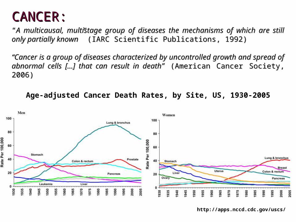
CANCER:CANCER:““A multicausal, multistage group of diseases the mechanisms of which are still only A multicausal, multistage group of diseases the mechanisms of which are still only partially knownpartially known” (IARC Scientific Publications, 1992)” (IARC Scientific Publications, 1992)
““Cancer is a group of diseases characterized by uncontrolled growth and spread of Cancer is a group of diseases characterized by uncontrolled growth and spread of abnormal cells […] that can result in death”abnormal cells […] that can result in death” (American Cancer Society, 2006) (American Cancer Society, 2006)
Age-adjusted Cancer Death Rates, by Site, US, 1930-2005
http://apps.nccd.cdc.gov/uscs/
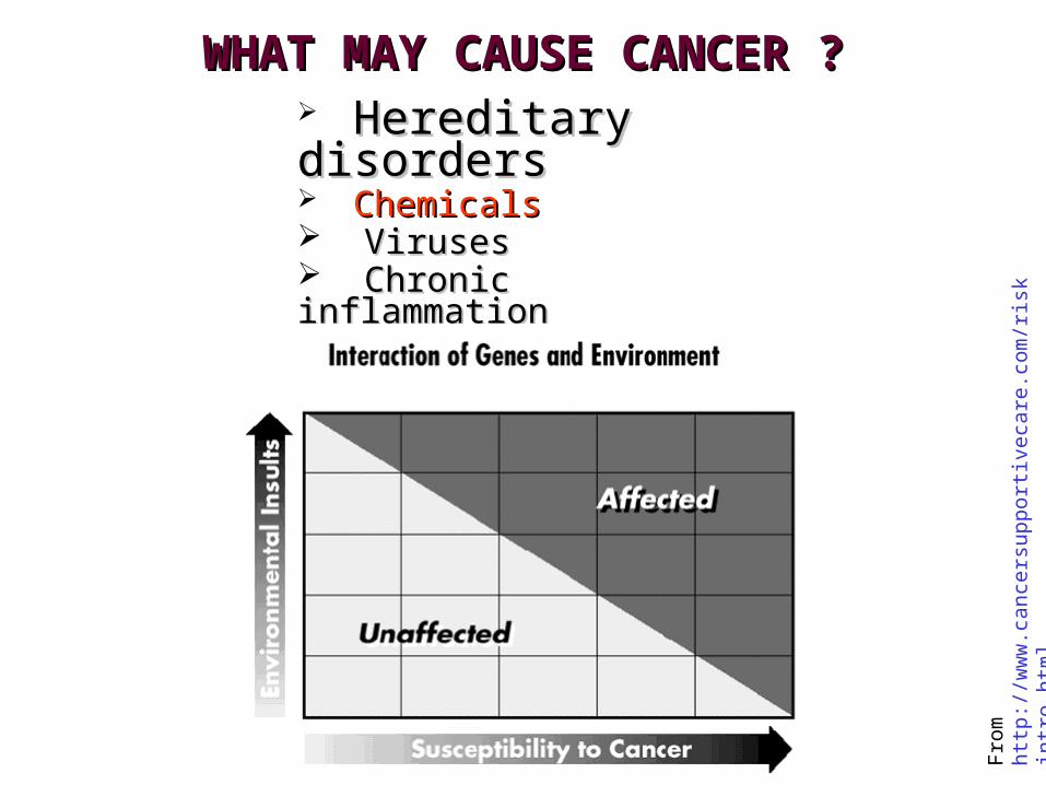
WHAT MAY CAUSE CANCER ?WHAT MAY CAUSE CANCER ? Hereditary Hereditary disordersdisorders ChemicalsChemicals VirusesViruses Chronic inflammationChronic inflammation ??????
Fro
m h
ttp:
//w
ww
.can
cers
uppo
rtiv
ecar
e.co
m/r
iski
ntro
.htm
l
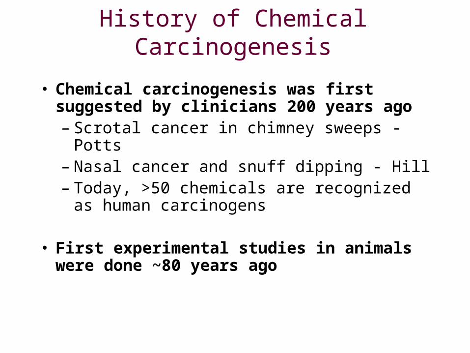
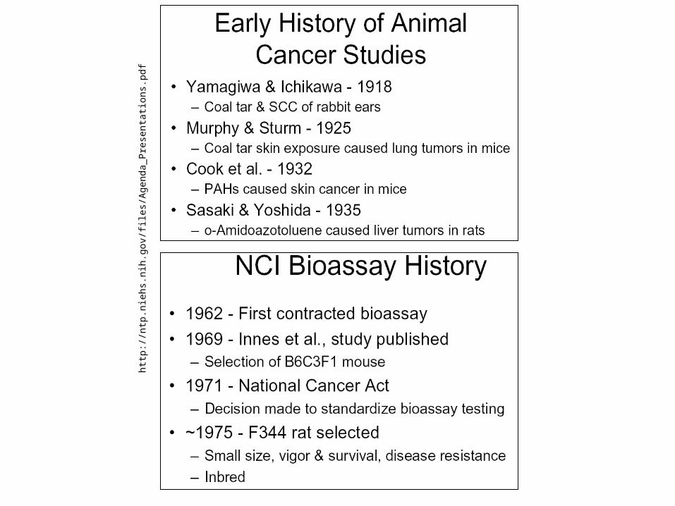
History of Chemical Carcinogenesis
• Chemical carcinogenesis was first suggested by clinicians 200 years ago– Scrotal cancer in chimney sweeps - Potts– Nasal cancer and snuff dipping - Hill– Today, >50 chemicals are recognized as
human carcinogens
• First experimental studies in animals were done ~80 years ago
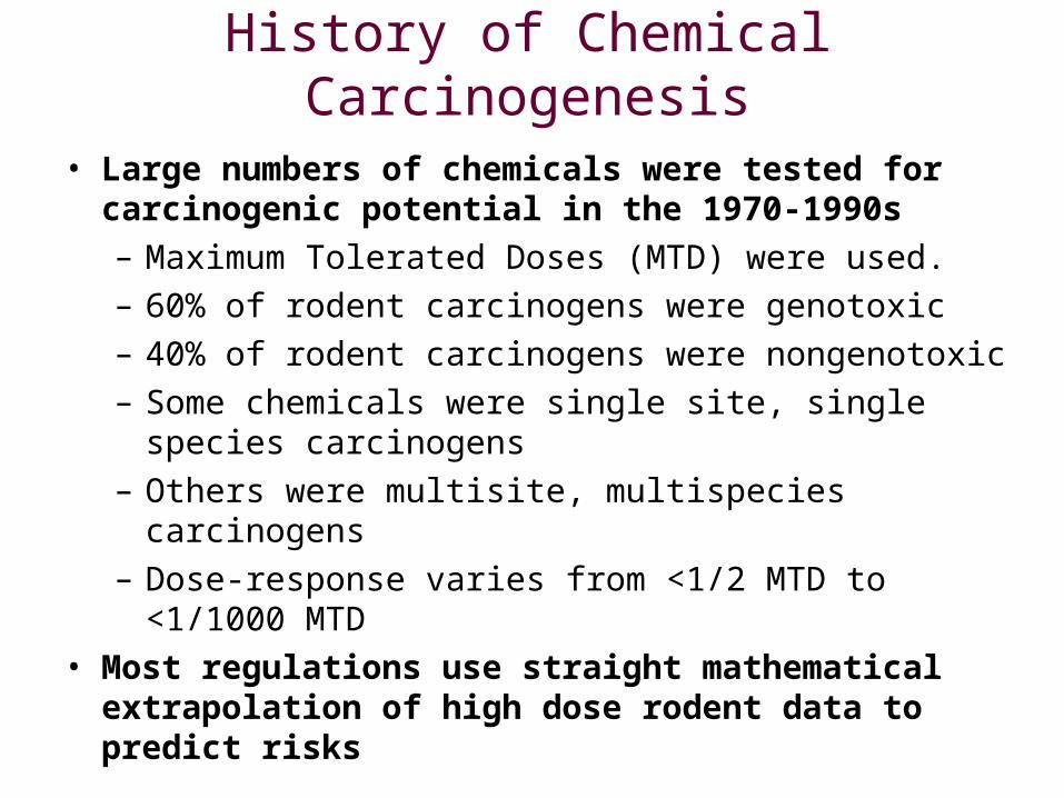
• Large numbers of chemicals were tested for carcinogenic potential in the 1970-1990s– Maximum Tolerated Doses (MTD) were used.– 60% of rodent carcinogens were genotoxic– 40% of rodent carcinogens were nongenotoxic– Some chemicals were single site, single species
carcinogens– Others were multisite, multispecies carcinogens– Dose-response varies from <1/2 MTD to
<1/1000 MTD• Most regulations use straight mathematical
extrapolation of high dose rodent data to predict risks
History of Chemical Carcinogenesis
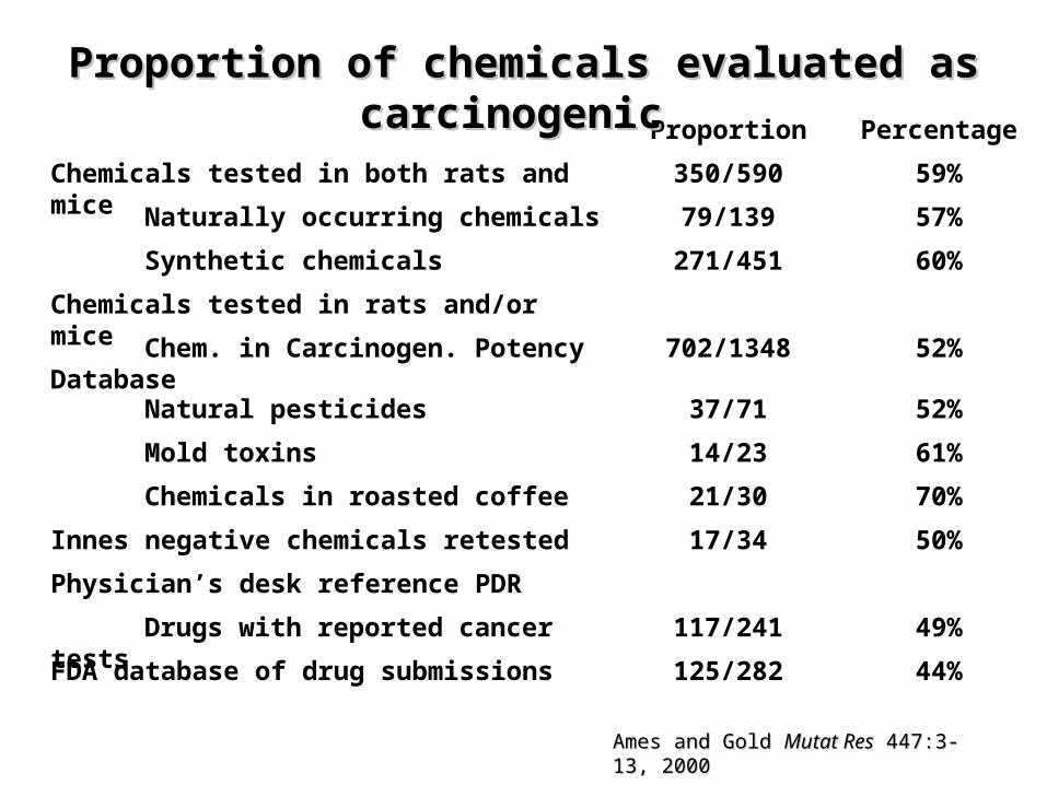
Proportion Percentage
Chemicals tested in both rats and mice 350/590 59%
Naturally occurring chemicals 79/139 57%
Synthetic chemicals 271/451 60%
Chemicals tested in rats and/or mice
Chem. in Carcinogen. Potency Database 702/1348 52%
Natural pesticides 37/71 52%
Mold toxins 14/23 61%
Chemicals in roasted coffee 21/30 70%
Innes negative chemicals retested 17/34 50%
Physician’s desk reference PDR
Drugs with reported cancer tests 117/241 49%
FDA database of drug submissions 125/282 44%
Proportion of chemicals evaluated as Proportion of chemicals evaluated as carcinogeniccarcinogenic
Ames and Gold Ames and Gold Mutat ResMutat Res 447:3-13, 2000 447:3-13, 2000
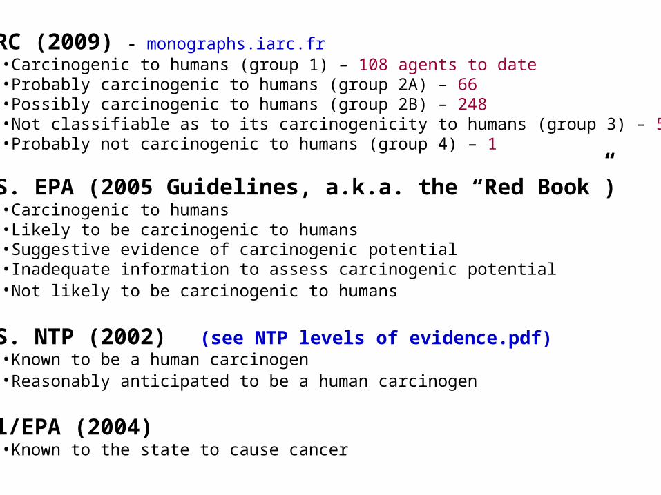
IARC (2009) - monographs.iarc.fr•Carcinogenic to humans (group 1) – 108 agents to date•Probably carcinogenic to humans (group 2A) – 66•Possibly carcinogenic to humans (group 2B) – 248•Not classifiable as to its carcinogenicity to humans (group 3) – 515 •Probably not carcinogenic to humans (group 4) – 1
U.S. EPA (2005 Guidelines, a.k.a. the “Red Book”) •Carcinogenic to humans •Likely to be carcinogenic to humans •Suggestive evidence of carcinogenic potential •Inadequate information to assess carcinogenic potential •Not likely to be carcinogenic to humans
U.S. NTP (2002) (see NTP levels of evidence.pdf)•Known to be a human carcinogen •Reasonably anticipated to be a human carcinogen
Cal/EPA (2004) •Known to the state to cause cancer
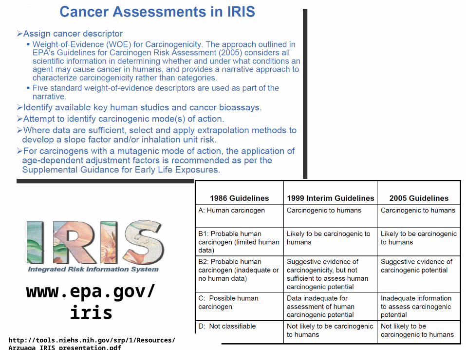
http://tools.niehs.nih.gov/srp/1/Resources/Arzuaga_IRIS_presentation.pdf
www.epa.gov/iris

WORLD HEALTH ORGANIZATIONINTERNATIONAL AGENCY FOR RESEARCH ON CANCER
IARC Monograph Evaluations
LYON, FRANCE
Slide courtesy of V. Cogliano (IARC)
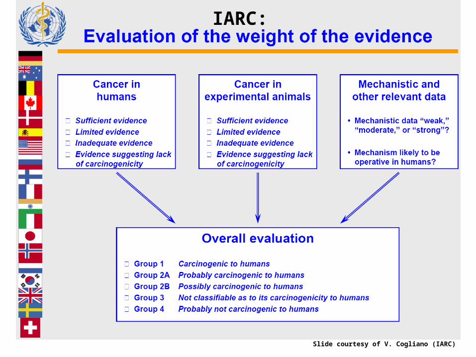
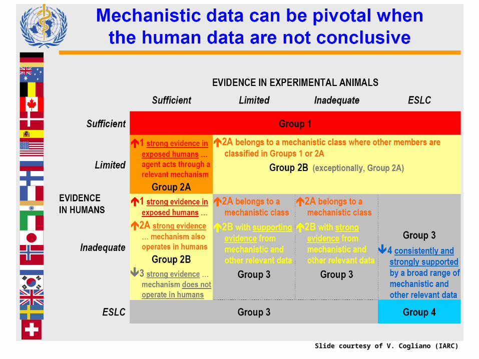
IARC:
Slide courtesy of V. Cogliano (IARC)
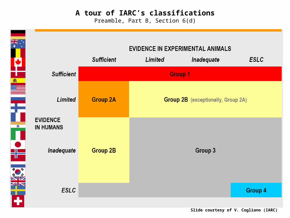
A tour of IARC’s classificationsPreamble, Part B, Section 6(d)
Slide courtesy of V. Cogliano (IARC)
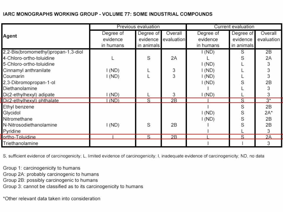
Slide courtesy of V. Cogliano (IARC)
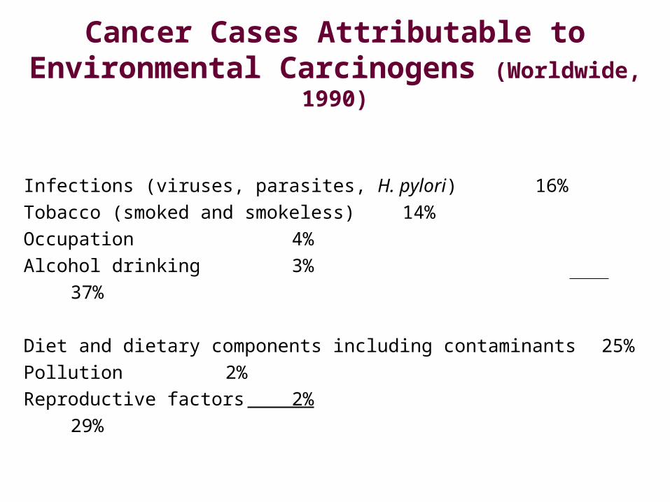
Cancer Cases Attributable to Environmental Carcinogens
(Worldwide, 1990)
Infections (viruses, parasites, H. pylori) 16%Tobacco (smoked and smokeless) 14%Occupation 4%Alcohol drinking 3%
37%
Diet and dietary components including contaminants 25%Pollution 2%Reproductive factors 2%
29%
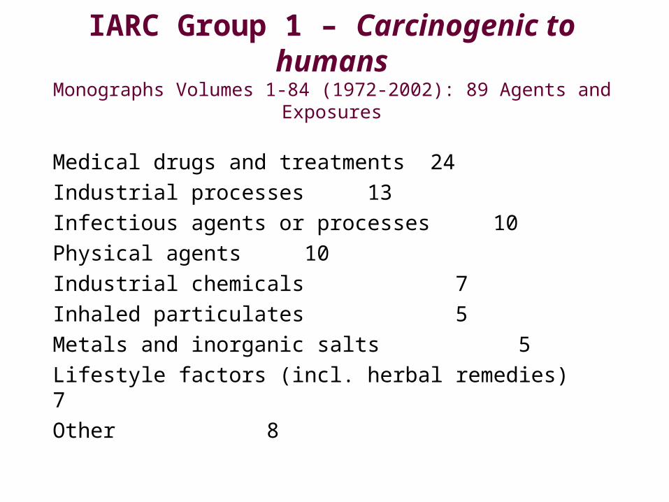
IARC Group 1 – Carcinogenic to humans
Monographs Volumes 1-84 (1972-2002): 89 Agents and Exposures
Medical drugs and treatments 24Industrial processes 13Infectious agents or processes 10Physical agents 10Industrial chemicals 7Inhaled particulates 5Metals and inorganic salts 5Lifestyle factors (incl. herbal remedies) 7Other 8
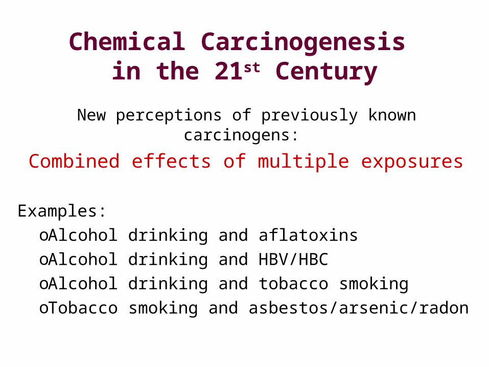
Chemical Carcinogenesis in the 21st Century
New perceptions of previously known carcinogens:
Combined effects of multiple exposures
Examples:oAlcohol drinking and aflatoxinsoAlcohol drinking and HBV/HBCoAlcohol drinking and tobacco smokingoTobacco smoking and asbestos/arsenic/radon
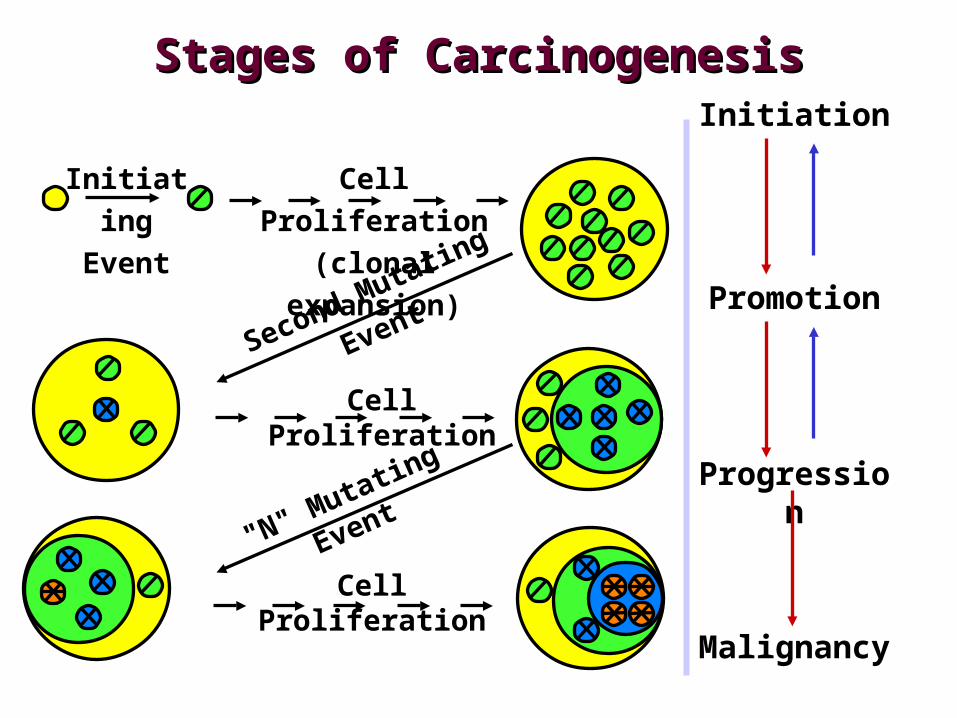
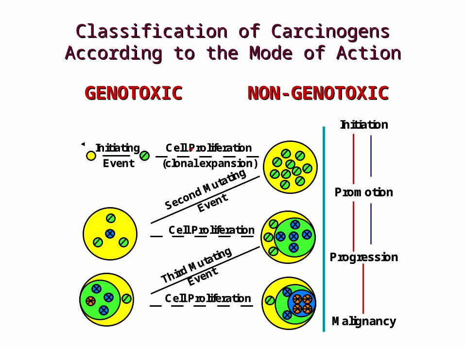
Initiating
Event
Cell Proliferation
(clonal expansion)
Progression
Cell Proliferation
Cell Proliferation
Malignancy
Second Mutating
Event
"N" Mutating Event
Initiation
Promotion
Stages of CarcinogenesisStages of Carcinogenesis
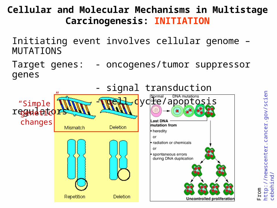
Cellular and Molecular Mechanisms in Multistage Carcinogenesis: INITIATION
Initiating event involves cellular genome – MUTATIONS
Target genes: - oncogenes/tumor suppressor genes
- signal transduction
- cell cycle/apoptosis regulators
Fro
m h
ttp:
//ne
wsc
ente
r.ca
ncer
.gov
/sci
ence
behi
nd/
“Simple” genetic changes
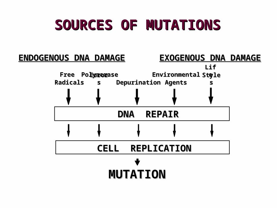
SOURCES OF SOURCES OF MUTATIONSMUTATIONS
ENDOGENOUS DNA DAMAGEENDOGENOUS DNA DAMAGE EXOGENOUS DNA DAMAGEEXOGENOUS DNA DAMAGE
DepurinatioDepurinationn
DNA REPAIRDNA REPAIR
MUTATIONMUTATION
LifLifeeStyleStyless
EnvironmentaEnvironmentallAgentAgentss
FreFreee
RadicalsRadicals
PolymerasPolymerasee
ErrorsErrors
CELL REPLICATIONCELL REPLICATION
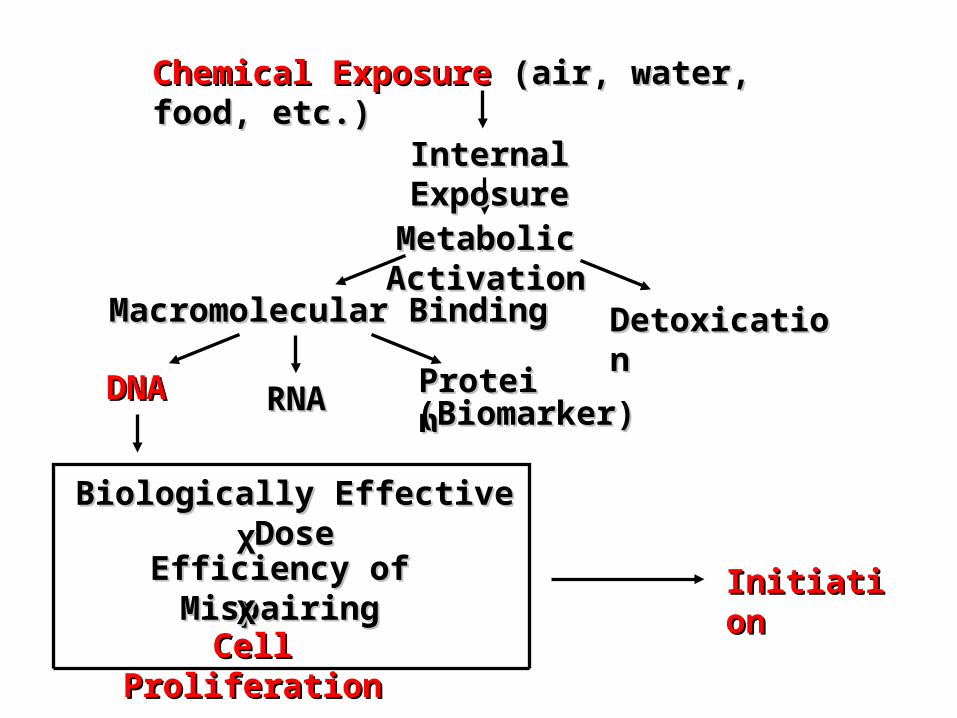
Chemical ExposureChemical Exposure (air, water, food, etc.) (air, water, food, etc.)
Internal ExposureInternal Exposure
Metabolic ActivationMetabolic Activation
Macromolecular BindingMacromolecular Binding DetoxicationDetoxication
DNADNA RNARNA ProteinProtein
Biologically Effective DoseBiologically Effective Dose
Efficiency of MispairingEfficiency of Mispairing
Cell ProliferationCell Proliferation
XX
XXInitiationInitiation
(Biomarker)(Biomarker)
Epigenetic alterations – changes induced in cells that alter the expression of the information on transcriptional, translational, or post-translational levels without changes in DNA sequence
EPIGENETICS
SAM SAH
DNMT1DNMT3aDNMT3b
Methylation of DNA
Modifications of histones
RNA-mediated modifications
• RNA-directed DNA methylation
• RNA-mediated chromatin remodeling
• RNAi, siRNA, miRNA …
A
Me
P
U
- acetylation
- methylation
- phosphorylation
- ubiquitination
P UMe
A
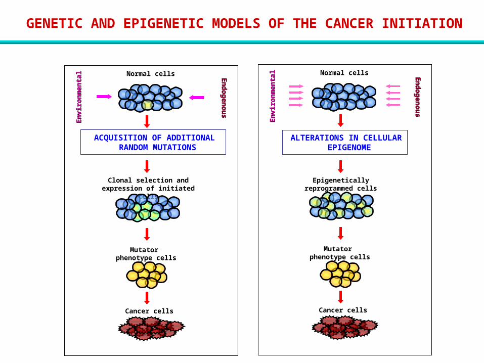
GENETIC AND EPIGENETIC MODELS OF THE CANCER INITIATION
Epigenetically reprogrammed cells
Mutator phenotype cells
En
dog
en
ou
sEn
dog
en
ou
s
En
vir
on
men
tal
En
vir
on
men
tal
ALTERATIONS IN CELLULAR EPIGENOME
Normal cells
Cancer cells
Clonal selection and expression of initiated cells
Mutator phenotype cells
En
dog
en
ou
sEn
dog
en
ou
s
En
vir
on
men
tal
En
vir
on
men
tal
ACQUISITION OF ADDITIONAL RANDOM MUTATIONS
Normal cells
Cancer cells
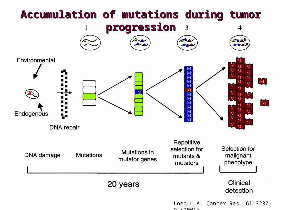
Accumulation of mutations during tumor Accumulation of mutations during tumor progressionprogression
Loeb L.A. Cancer Res. 61:3230-9 (2001)
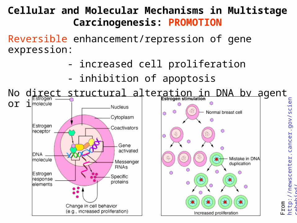
Cellular and Molecular Mechanisms in Multistage Carcinogenesis: PROMOTION
Reversible enhancement/repression of gene expression:
- increased cell proliferation
- inhibition of apoptosis
No direct structural alteration in DNA by agent or its metabolites
Fro
m h
ttp:
//ne
wsc
ente
r.ca
ncer
.gov
/sci
ence
behi
nd/
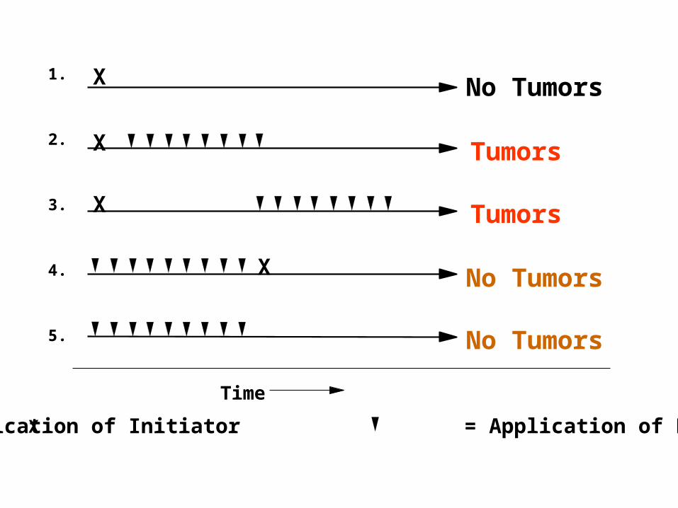
No Tumors
Tumors
No Tumors
No Tumors
Tumors
1.
2.
3.
4.
5.
X
X
X
X
Time
X = Application of Initiator = Application of Promoter
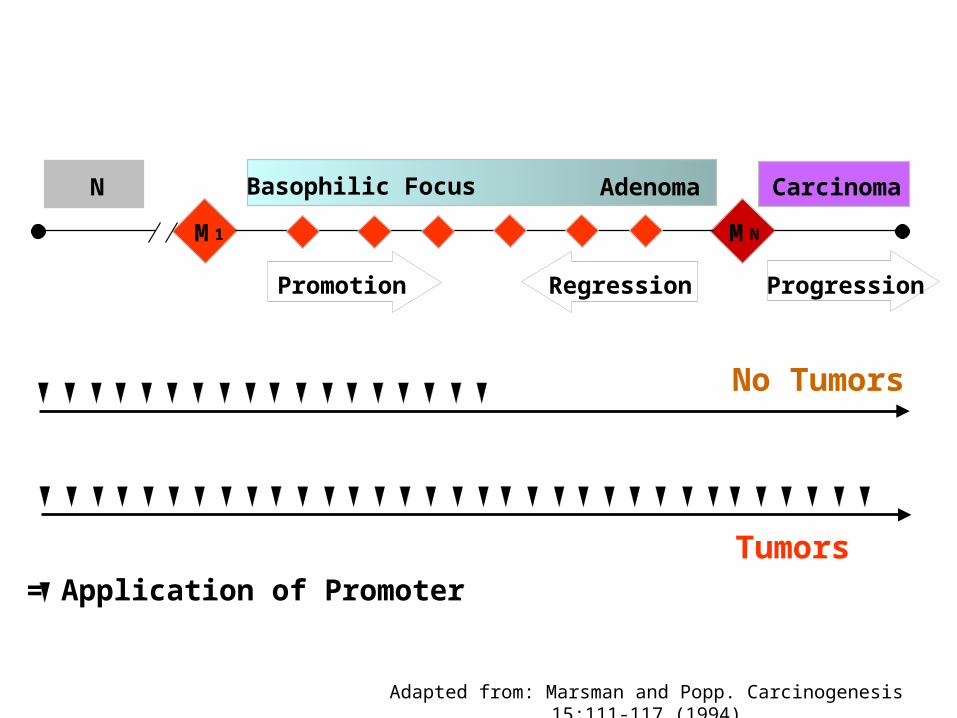
N Basophilic Focus Adenoma Carcinoma
M 1 MN
Promotion Regression Progression
Adapted from: Marsman and Popp. Carcinogenesis 15:111-117 (1994)
No Tumors
Tumors= Application of Promoter
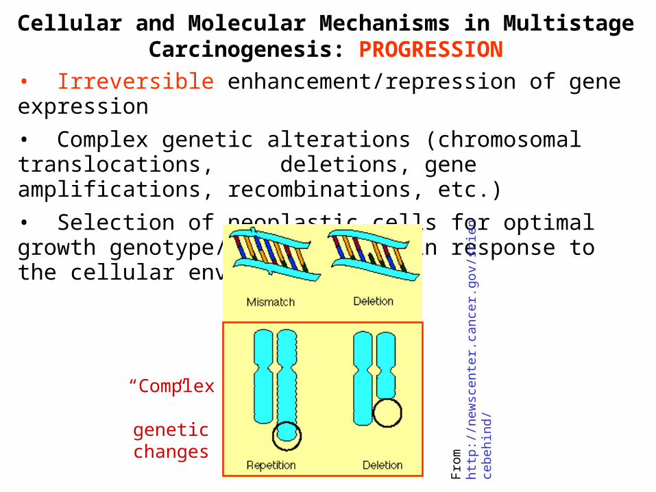
Cellular and Molecular Mechanisms in Multistage Carcinogenesis: PROGRESSION
• Irreversible enhancement/repression of gene expression
• Complex genetic alterations (chromosomal translocations, deletions, gene amplifications, recombinations, etc.)
• Selection of neoplastic cells for optimal growth genotype/ phenotype in response to the cellular environment
Fro
m h
ttp:
//ne
wsc
ente
r.ca
ncer
.gov
/sci
ence
behi
nd/
“Complex” genetic changes
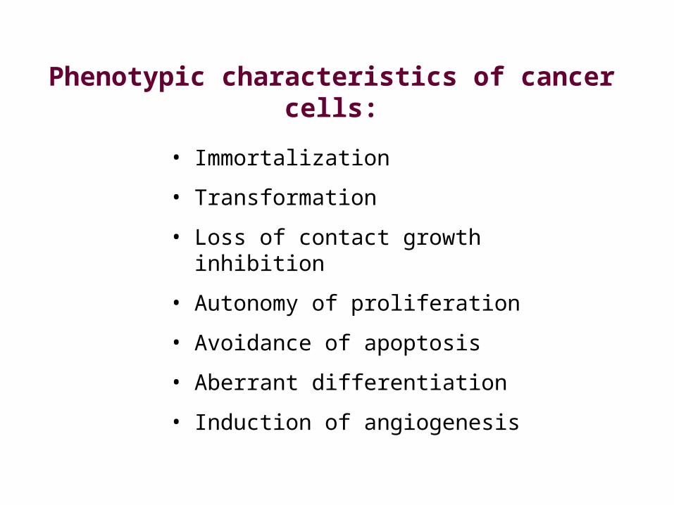
• Immortalization
• Transformation
• Loss of contact growth inhibition
• Autonomy of proliferation
• Avoidance of apoptosis
• Aberrant differentiation
• Induction of angiogenesis
Phenotypic characteristics of cancer cells:
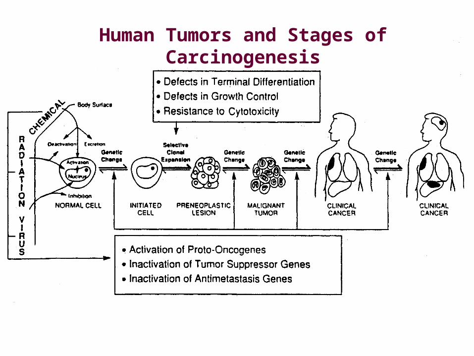
Human Tumors and Stages of Carcinogenesis
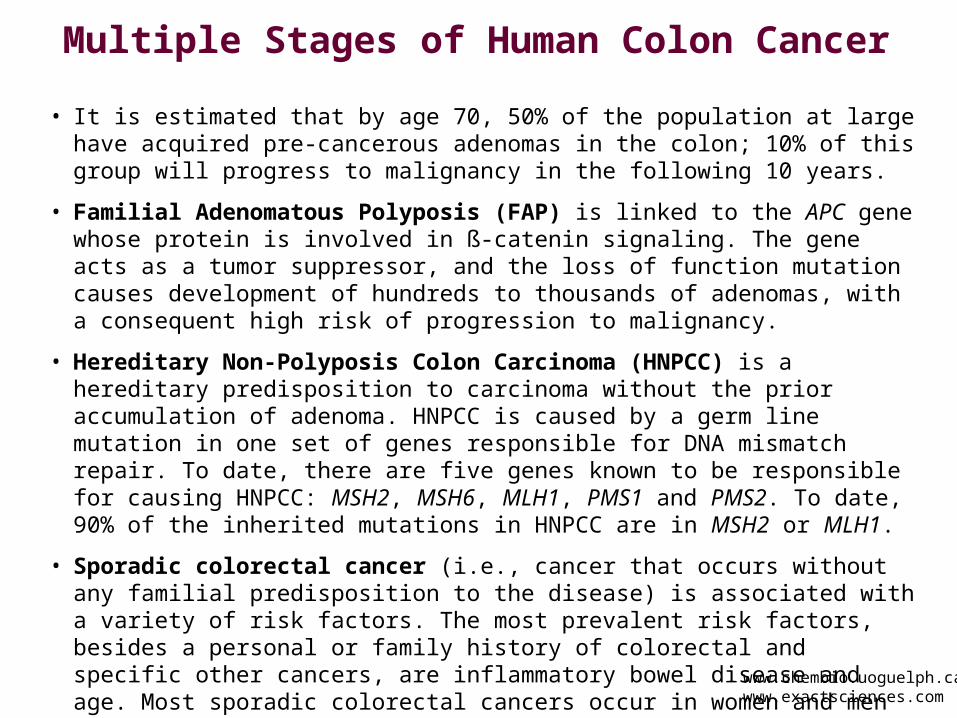
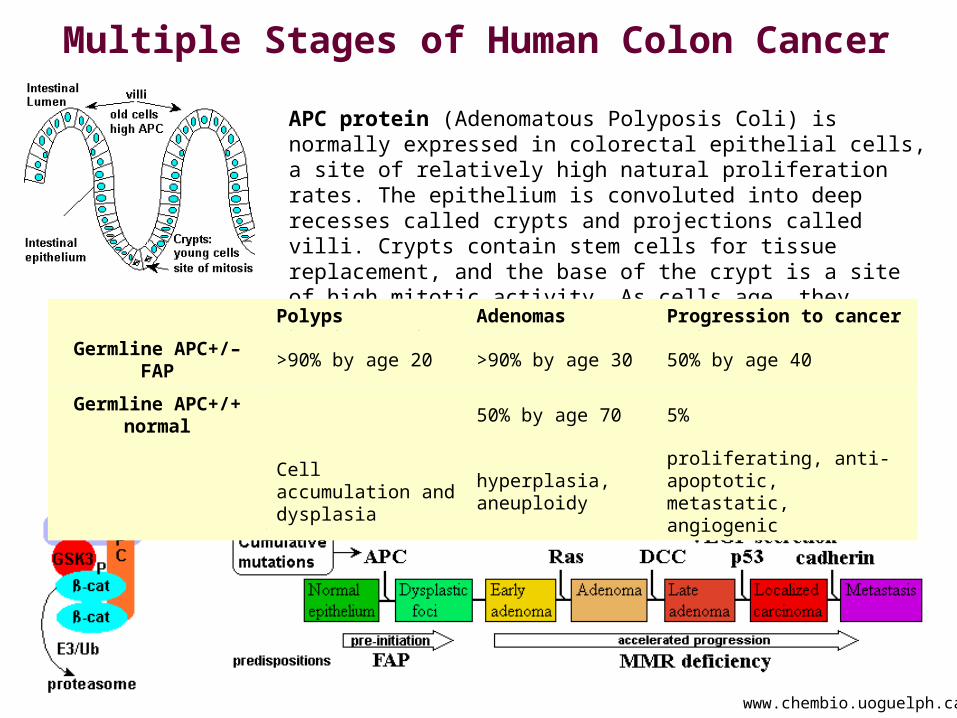
Multiple Stages of Human Colon Cancer
• It is estimated that by age 70, 50% of the population at large have acquired pre-cancerous adenomas in the colon; 10% of this group will progress to malignancy in the following 10 years.
• Familial Adenomatous Polyposis (FAP) is linked to the APC gene whose protein is involved in ß-catenin signaling. The gene acts as a tumor suppressor, and the loss of function mutation causes development of hundreds to thousands of adenomas, with a consequent high risk of progression to malignancy.
• Hereditary Non-Polyposis Colon Carcinoma (HNPCC) is a hereditary predisposition to carcinoma without the prior accumulation of adenoma. HNPCC is caused by a germ line mutation in one set of genes responsible for DNA mismatch repair. To date, there are five genes known to be responsible for causing HNPCC: MSH2, MSH6, MLH1, PMS1 and PMS2. To date, 90% of the inherited mutations in HNPCC are in MSH2 or MLH1.
• Sporadic colorectal cancer (i.e., cancer that occurs without any familial predisposition to the disease) is associated with a variety of risk factors. The most prevalent risk factors, besides a personal or family history of colorectal and specific other cancers, are inflammatory bowel disease and age. Most sporadic colorectal cancers occur in women and men over the age of 50. Additional risk factors include diet, less than moderate exercise, and obesity
www.chembio.uoguelph.cawww.exactsciences.com
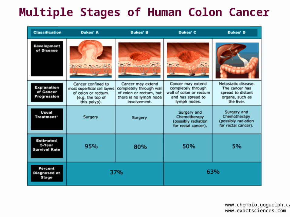
Multiple Stages of Human Colon Cancer
www.chembio.uoguelph.cawww.exactsciences.com
Multiple Stages of Human Colon Cancer
www.chembio.uoguelph.ca
APC protein (Adenomatous Polyposis Coli) is normally expressed in colorectal epithelial cells, a site of relatively high natural proliferation rates. The epithelium is convoluted into deep recesses called crypts and projections called villi. Crypts contain stem cells for tissue replacement, and the base of the crypt is a site of high mitotic activity. As cells age, they progress up the villus to the tip.
Polyps Adenomas Progression to cancer
Germline APC+/–FAP
>90% by age 20 >90% by age 30 50% by age 40
Germline APC+/+normal
50% by age 70 5%
Cell accumulation and dysplasia
hyperplasia, aneuploidy
proliferating, anti-apoptotic, metastatic, angiogenic
Initiating
Event
Cell Proliferation
(clonal expansion)
Progression
Cell Proliferation
Cell Proliferation
Malignancy
Second Mutating
Event
Third Mutating
Event
Initiation
Promotion
Stages of CarcinogenesisStages of Carcinogenesis
Initiating
Event
Cell Proliferation
(clonal expansion)
Progression
Cell Proliferation
Cell Proliferation
Malignancy
Second Mutating
Event
Third Mutating
Event
Initiation
Promotion
Stages of CarcinogenesisStages of Carcinogenesis
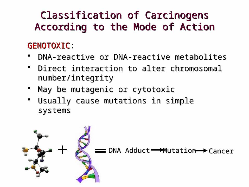
Classification of Carcinogens According to Classification of Carcinogens According to the Mode of Actionthe Mode of Action
GENOTOXIC NON-GENOTOXIC NON-GENOTOXICGENOTOXIC
Classification of Carcinogens According to Classification of Carcinogens According to the Mode of Actionthe Mode of Action
GENOTOXICGENOTOXIC:: DNA-reactive or DNA-reactive metabolitesDNA-reactive or DNA-reactive metabolites Direct interaction to alter chromosomal Direct interaction to alter chromosomal
number/integritynumber/integrity May be mutagenic or cytotoxicMay be mutagenic or cytotoxic Usually cause mutations in simple systemsUsually cause mutations in simple systems
DNA AdductDNA Adduct MutationMutation CancerCancer
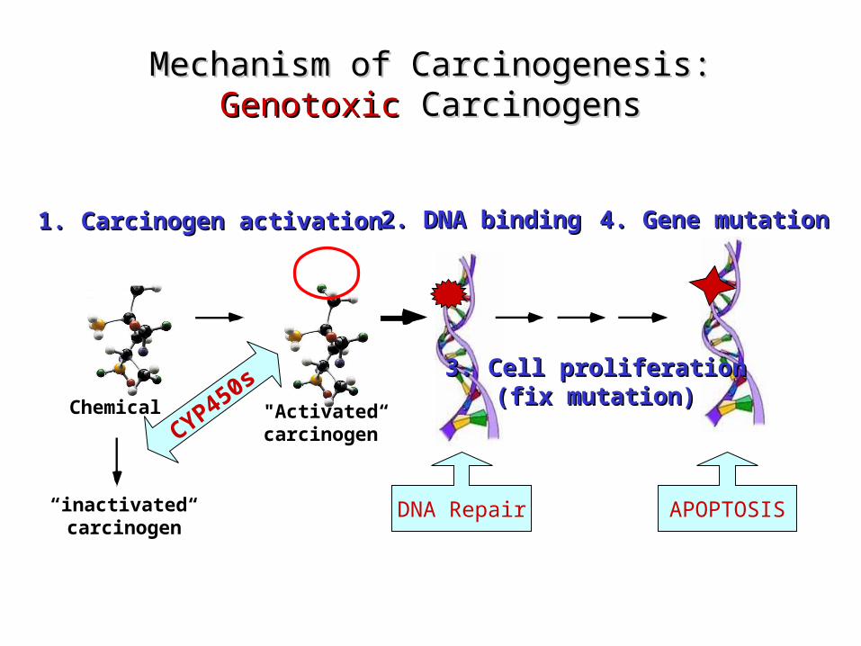
Mechanism of Carcinogenesis:Mechanism of Carcinogenesis:GenotoxicGenotoxic Carcinogens Carcinogens
1. Carcinogen activation1. Carcinogen activation 2. DNA binding2. DNA binding 4. Gene4. Gene mutationmutation
Chemical "Activated“carcinogen
3. Cell proliferation3. Cell proliferation(fix mutation)(fix mutation)
“inactivated“carcinogen
CYP450s
DNA Repair APOPTOSIS
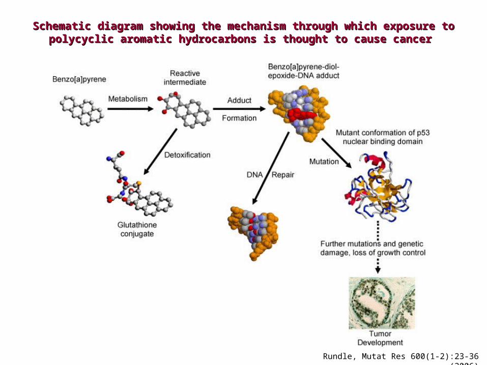
Schematic diagram showing the mechanism through which exposure to Schematic diagram showing the mechanism through which exposure to polycyclic aromatic hydrocarbons is thought to cause cancerpolycyclic aromatic hydrocarbons is thought to cause cancer
Rundle, Mutat Res 600(1-2):23-36 (2006)
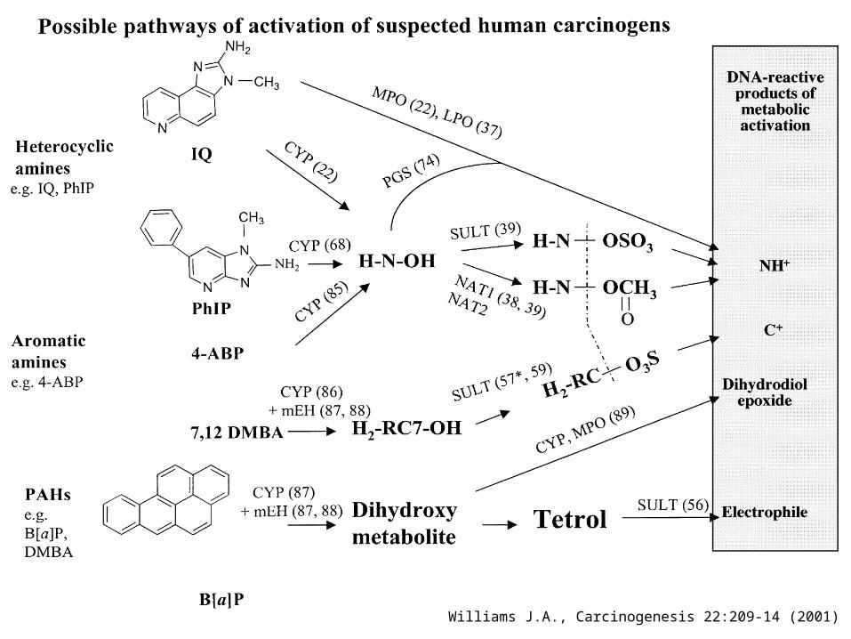
Williams J.A., Carcinogenesis 22:209-14 (2001)
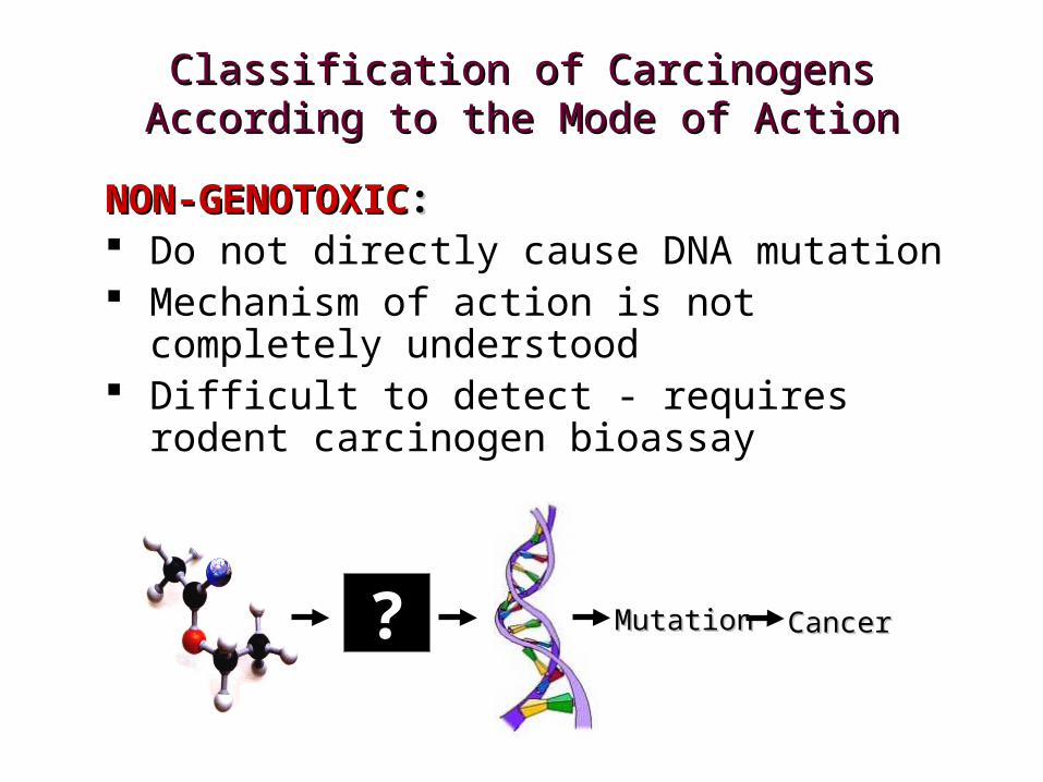
Classification of Carcinogens According to Classification of Carcinogens According to the Mode of Actionthe Mode of Action
NON-GENOTOXICNON-GENOTOXIC:: Do not directly cause DNA mutation Mechanism of action is not completely
understood Difficult to detect - requires rodent carcinogen
bioassay
?? MutationMutation CancerCancer
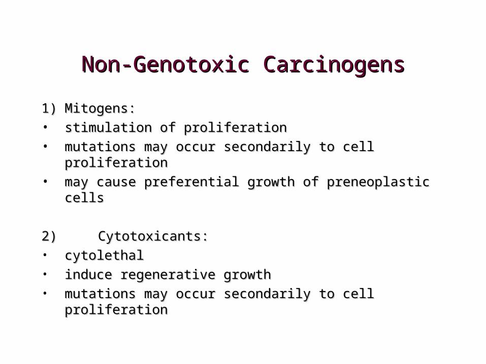
Non-Genotoxic CarcinogensNon-Genotoxic Carcinogens
1)1) Mitogens: Mitogens: • stimulation of proliferationstimulation of proliferation• mutations may occur secondarily to cell proliferationmutations may occur secondarily to cell proliferation• may cause preferential growth of preneoplastic cellsmay cause preferential growth of preneoplastic cells
2) 2) Cytotoxicants: Cytotoxicants: • cytolethalcytolethal• induce regenerative growthinduce regenerative growth• mutations may occur secondarily to cell proliferationmutations may occur secondarily to cell proliferation
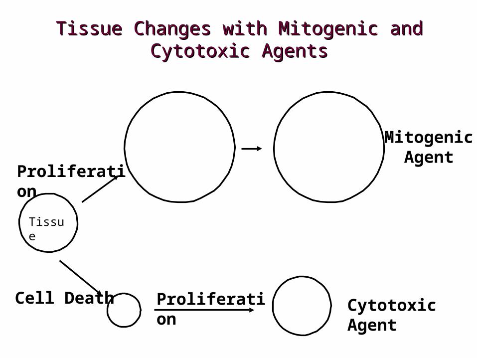
Tissue Changes with Mitogenic and Tissue Changes with Mitogenic and Cytotoxic AgentsCytotoxic Agents
Proliferation
Cell Death Proliferation
Cytotoxic Agent
MitogenicAgent
Tissue
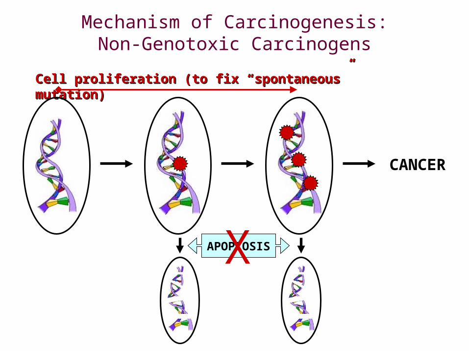
Mechanism of Carcinogenesis:Non-Genotoxic Carcinogens
Cell proliferation (to fix “spontaneous” mutation)Cell proliferation (to fix “spontaneous” mutation)
APOPTOSIS
CANCER
X
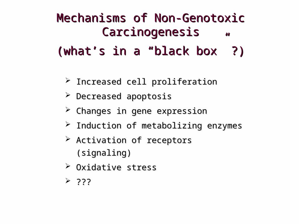
Mechanisms of Non-Genotoxic Mechanisms of Non-Genotoxic CarcinogenesisCarcinogenesis
(what’s in a “black box” ?)(what’s in a “black box” ?)
Increased cell proliferationIncreased cell proliferation
Decreased apoptosisDecreased apoptosis
Changes in gene expression Changes in gene expression
Induction of metabolizing enzymesInduction of metabolizing enzymes
Activation of receptors (signaling)Activation of receptors (signaling)
Oxidative stressOxidative stress
??????
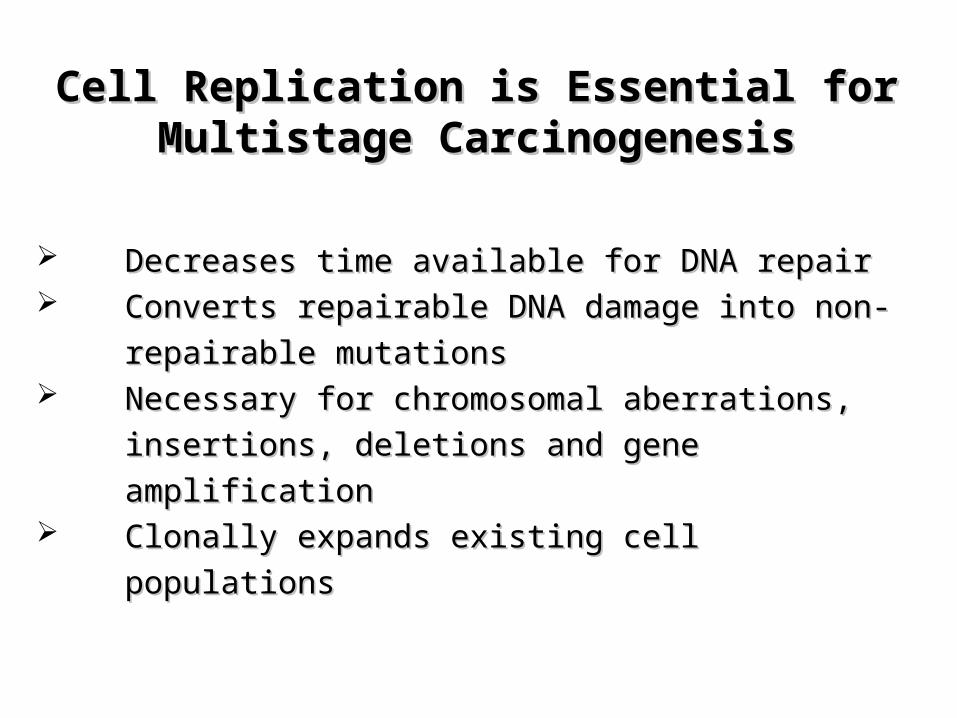
Decreases time available for DNA repairDecreases time available for DNA repair Converts repairable DNA damage into non-repairable Converts repairable DNA damage into non-repairable
mutationsmutations Necessary for chromosomal aberrations, insertions, Necessary for chromosomal aberrations, insertions,
deletions and gene amplificationdeletions and gene amplification Clonally expands existing cell populationsClonally expands existing cell populations
Cell Replication is Essential for Cell Replication is Essential for Multistage CarcinogenesisMultistage Carcinogenesis
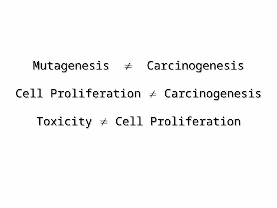
Mutagenesis Mutagenesis Carcinogenesis Carcinogenesis
Cell Proliferation Cell Proliferation Carcinogenesis Carcinogenesis
Toxicity Toxicity Cell Proliferation Cell Proliferation
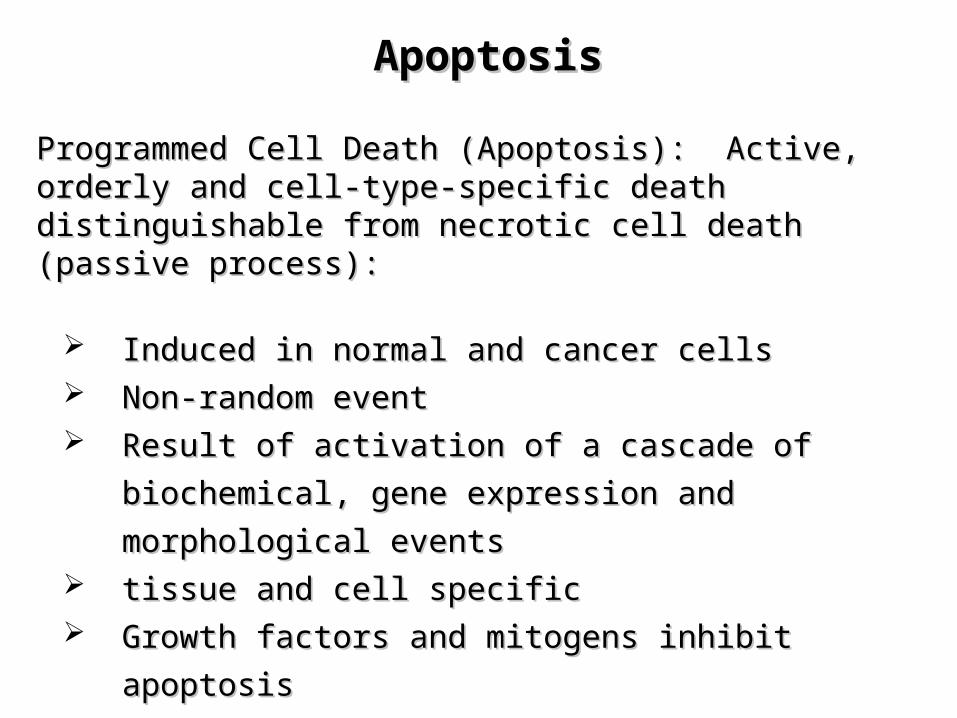
ApoptosisApoptosis
Programmed Cell Death (Apoptosis): Active, orderly and cell-Programmed Cell Death (Apoptosis): Active, orderly and cell-type-specific death distinguishable from necrotic cell death type-specific death distinguishable from necrotic cell death (passive process):(passive process):
Induced in normal and cancer cells Induced in normal and cancer cells Non-random eventNon-random event Result of activation of a cascade of biochemical, gene Result of activation of a cascade of biochemical, gene
expression and morphological eventsexpression and morphological events tissue and cell specifictissue and cell specific Growth factors and mitogens inhibit apoptosisGrowth factors and mitogens inhibit apoptosis
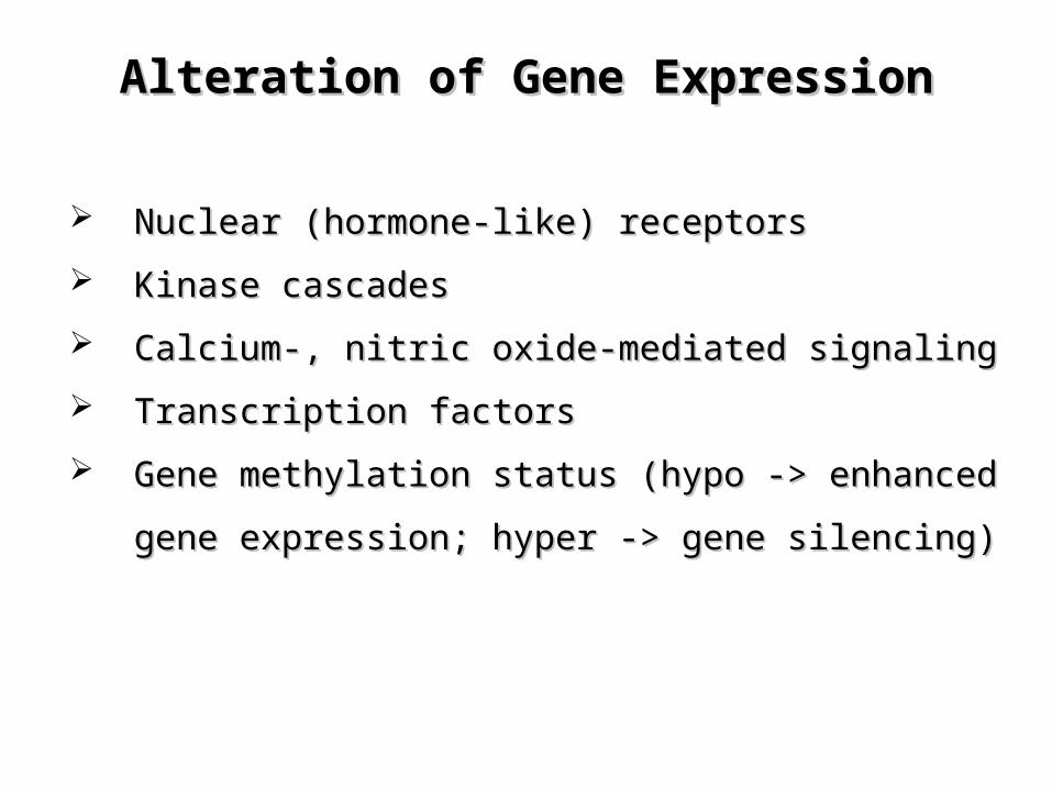
Alteration of Gene ExpressionAlteration of Gene Expression
Nuclear (hormone-like) receptors Nuclear (hormone-like) receptors
Kinase cascadesKinase cascades
Calcium-, nitric oxide-mediated signalingCalcium-, nitric oxide-mediated signaling
Transcription factorsTranscription factors
Gene methylation status (hypo -> enhanced gene Gene methylation status (hypo -> enhanced gene
expression; hyper -> gene silencing)expression; hyper -> gene silencing)
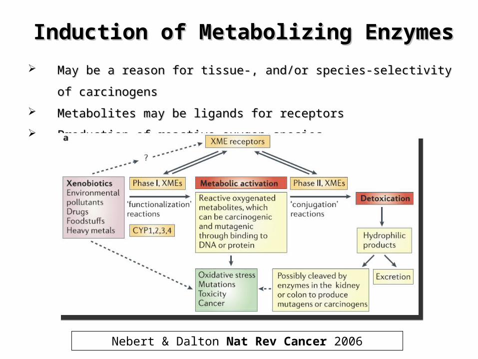
Induction of Metabolizing EnzymesInduction of Metabolizing Enzymes
May be a reason for tissue-, and/or species-selectivity of carcinogensMay be a reason for tissue-, and/or species-selectivity of carcinogens
Metabolites may be ligands for receptorsMetabolites may be ligands for receptors
Production of reactive oxygen speciesProduction of reactive oxygen species
Nebert & Dalton Nat Rev Cancer 2006
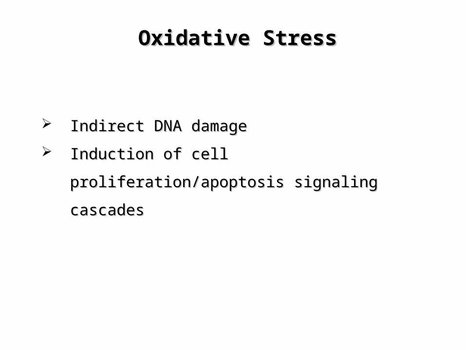
Oxidative StressOxidative Stress
Indirect DNA damageIndirect DNA damage
Induction of cell proliferation/apoptosis signaling Induction of cell proliferation/apoptosis signaling
cascadescascades
htt
p:/
/ntp
.nie
hs.
nih
.gov/fi
les/
Ag
en
da_P
rese
nta
tion
s.p
df
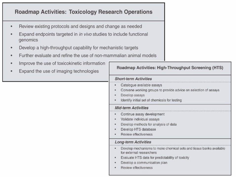
http://ntp.niehs.nih.gov/files/Agenda_Presentations.pdf
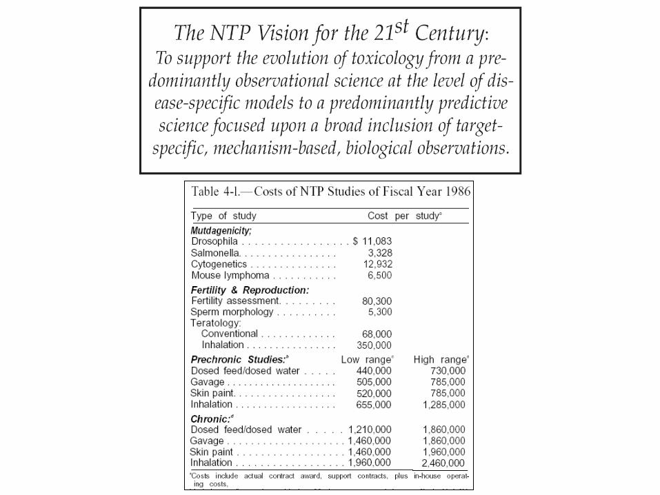
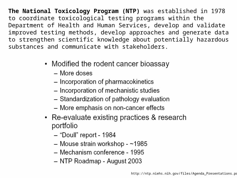
The National Toxicology Program (NTP) was established in 1978 to coordinate toxicological testing programs within the Department of Health and Human Services, develop and validate improved testing methods, develop approaches and generate data to strengthen scientific knowledge about potentially hazardous substances and communicate with stakeholders.
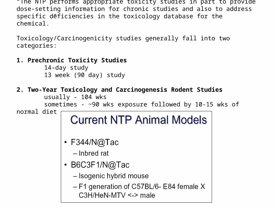
“The NTP performs appropriate toxicity studies in part to provide dose-setting information for chronic studies and also to address specific deficiencies in the toxicology database for the chemical.”
Toxicology/Carcinogenicity studies generally fall into two categories:
1. Prechronic Toxicity Studies14-day study13 week (90 day) study
2. Two-Year Toxicology and Carcinogenesis Rodent Studiesusually - 104 wkssometimes - ~90 wks exposure followed by 10-15 wks of normal diet
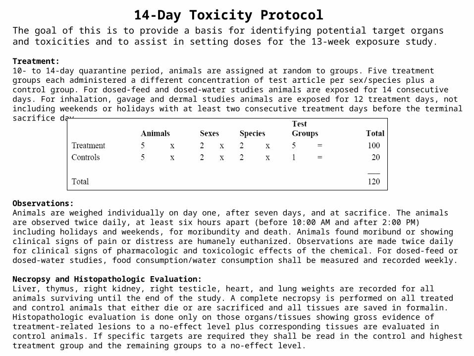
14-Day Toxicity Protocol The goal of this is to provide a basis for identifying potential target organs and toxicities and to assist in setting doses for the 13-week exposure study.
Treatment: 10- to 14-day quarantine period, animals are assigned at random to groups. Five treatment groups each administered a different concentration of test article per sex/species plus a control group. For dosed-feed and dosed-water studies animals are exposed for 14 consecutive days. For inhalation, gavage and dermal studies animals are exposed for 12 treatment days, not including weekends or holidays with at least two consecutive treatment days before the terminal sacrifice day.
Observations: Animals are weighed individually on day one, after seven days, and at sacrifice. The animals are observed twice daily, at least six hours apart (before 10:00 AM and after 2:00 PM) including holidays and weekends, for moribundity and death. Animals found moribund or showing clinical signs of pain or distress are humanely euthanized. Observations are made twice daily for clinical signs of pharmacologic and toxicologic effects of the chemical. For dosed-feed or dosed-water studies, food consumption/water consumption shall be measured and recorded weekly.
Necropsy and Histopathologic Evaluation: Liver, thymus, right kidney, right testicle, heart, and lung weights are recorded for all animals surviving until the end of the study. A complete necropsy is performed on all treated and control animals that either die or are sacrificed and all tissues are saved in formalin. Histopathologic evaluation is done only on those organs/tissues showing gross evidence of treatment-related lesions to a no-effect level plus corresponding tissues are evaluated in control animals. If specific targets are required they shall be read in the control and highest treatment group and the remaining groups to a no-effect level.
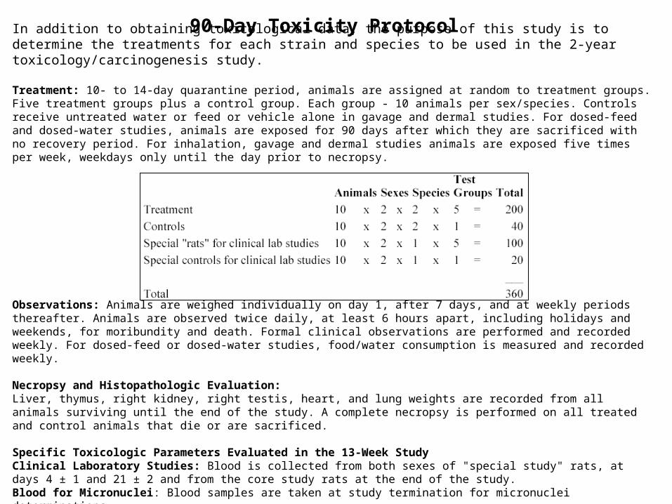
In addition to obtaining toxicological data, the purpose of this study is to determine the treatments for each strain and species to be used in the 2-year toxicology/carcinogenesis study.
Treatment: 10- to 14-day quarantine period, animals are assigned at random to treatment groups. Five treatment groups plus a control group. Each group - 10 animals per sex/species. Controls receive untreated water or feed or vehicle alone in gavage and dermal studies. For dosed-feed and dosed-water studies, animals are exposed for 90 days after which they are sacrificed with no recovery period. For inhalation, gavage and dermal studies animals are exposed five times per week, weekdays only until the day prior to necropsy.
Observations: Animals are weighed individually on day 1, after 7 days, and at weekly periods thereafter. Animals are observed twice daily, at least 6 hours apart, including holidays and weekends, for moribundity and death. Formal clinical observations are performed and recorded weekly. For dosed-feed or dosed-water studies, food/water consumption is measured and recorded weekly.
Necropsy and Histopathologic Evaluation: Liver, thymus, right kidney, right testis, heart, and lung weights are recorded from all animals surviving until the end of the study. A complete necropsy is performed on all treated and control animals that die or are sacrificed.
Specific Toxicologic Parameters Evaluated in the 13-Week Study Clinical Laboratory Studies: Blood is collected from both sexes of "special study" rats, at days 4 ± 1 and 21 ± 2 and from the core study rats at the end of the study. Blood for Micronuclei: Blood samples are taken at study termination for micronuclei determinations. Sperm Morphology and Vaginal Cytology Evaluations (SMVCE)
90-Day Toxicity Protocol
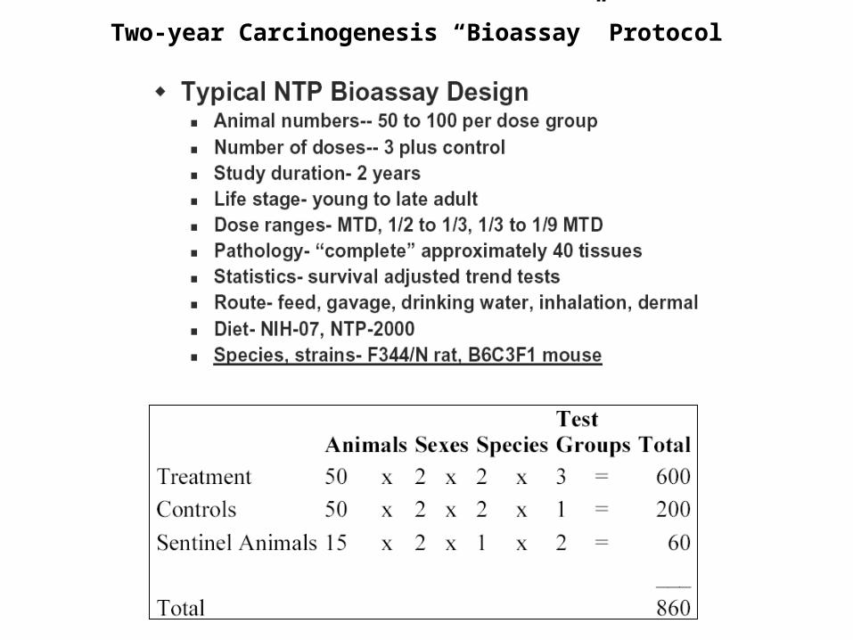
Two-year Carcinogenesis “Bioassay” Protocol