Embed Size (px)
Citation preview
#_*,t*irs+.;tift $s1.,Wi;SS.ti.ry"SSe,&iW
Chemical Control ofthe Brain and Behavior
INTRODUCTION
THE SECRETORY HYPOTHALAMUSAN OVERVIEW OFTHE HYPOTHALAMUS
Homeostosis
Structure ond Connections of the Hypotholomus
PATHWAYS TO THE PITUITARY
Hypotholomic Control of the Posterior Pituitory
Hypotholamic Control of the Anterior Pituitory
r Box 15.l OJ'Special Inlerest: Stress and the Brain
THE AUTONOMIC NERVOUS SYSTEMANS CIRCUITS
Sympothetic ond Parosympothetic Divisions
The Enteric Diyision
Centrol Control of the ANS
NEUROTRANSMITTERS AND THE PHARMACOLOGY OF AUTONOMIC FUNCTION
P rego ngli onic Neurotronsmitters
P ostga ngli o nic Neurotronsmitters
THE DIFFUSE MODULATORY SYSTEMS OF THE BRAINANATOMYAND FUNCTIONS OFTHE DIFFUSE MODULATORY SYSTEMS
r Box 15.2 O.f Special Interest:You Eat What You Are
The Norodrenergic Locus Coeruleus
The Serotonergic Rophe Nuclei
The Dopaminergic Substontio N(ro and VentrolTegmentol Areo
r Box 15.) Path of Discovery:Awakening to Dopamine, by Arvid Carlsson
The Cholinergic Bosol Forebroin ond Broin Stem Complexes
DRUGS AND THE DIFFUSE MODULATORY SYSTEMS
Hollucinogens
Stimulonts
CONCLUDING REMARKS

482 C HAPTE R I 5 . CHEMICALCONTROLOFTHE BMINAND BEHAVIOR
V INTRODUCTION
It should be obvious by now that knowing the organization of synapticconnections is essential to understanding how the brain works. It's notfrom a love of Greek and Latin that we belabor the neuroanatomyl Mostof the connections we have described are precise and specific. For exam-ple, in order for you to read these words, there must be a very fine-grainedneural mapping of the light falling on your retina-how else could you seethe dot on this question mark? The information must be carried centrallyand precisely dispersed to many parts of the brain for processing, coordi-nated with control of the motor neurons that closely regulate the six mus-cles of each eye as it scans the page.
In addition to anatomical precision, point-to-point communication inthe sensory and motor systems requires mechanisms that restrict synapticcommunication to the cleft between the axon terminal and its target. It justwouldn't do for glutamate released in somatosensory cortex to activateneurons in motor cortexl Furthermore, transmission must be brief enoughto allow rapid responses to new sensory inputs. Thus, at these synapses,only minute quantities of neurotransmitter are released with each impulse,and these molecules are then quickly destroyed enzymatically or taken upby neighboring cells. The postsynaptic actions at transmitter-gated ionchannels last only as long as the transmitter is in the cleft, a few millisecondsat most. Many axon terminals also possess presynaptic "autoreceptors" thatdetect the transmitter concentrations in the cleft and inhibit release if theyget too high. These mechanisms ensure that this type of synaptic transmis-sion is tightly constrained, in both space and time.
The elaborate mechanisms that constrain point-to-point synaptic trans-mission bring to mind a telecommunications analogy. Telephone systemsmake possible very specific connections between one place and another;your mother in Tacoma can talk just to you in Providence, reminding youthat her birthday was last week. The telephone lines can act like precisesynaptic connections. The influence of one neuron (your mother) is targetedto a small number of other neurons (in this case, only you). The embar-rassing message is limited to your ears only. For a real neuron in one of thesensory or motor systems discussed so far, its influence usually extends tothe few dozen or few hundred cells it synapses on-a conference call, to besure, but still relatively specific.
Now imagine your mother being interviewed on a television talk show,which is broadcast via a cable network. In this case, the widespread cableconnections may allow her to tell millions of people that you forgot herbirthday, and the loudspeaker in each television set will announce themessage to anyone within earshot. Likewise, certain neurons communi-cate with hundreds of thousands of other cells. These widespread systemsalso tend to act relatively slowly, over seconds to minutes. Because oftheir broad, protracted actions, such systems in the brain can orchestrateentire behaviors, ranging from falling asleep to falling in love. Indeed,many of the behavioral dysfunctions collectively known as mental disor-ders are believed to result specifically from imbalances of certain of thesechemicals.
In this chapter, we take a look at three components of the nervous systemthat operate in expanded space and time (Figure l5.I). One component isL}re secretory hypothalamus. By secreting chemicals directly into the blood-stream, the secretory hypothalamus can influence functions throughoutboth the brain and the body. A second component, controlled neurally bythe hypothalamus, is the autonomic nervous system (,4NS), introduced inChapter 7. Through extensive interconnections within the body, the ANS
V INTRODUCTION 483
-
o : F(a)
t
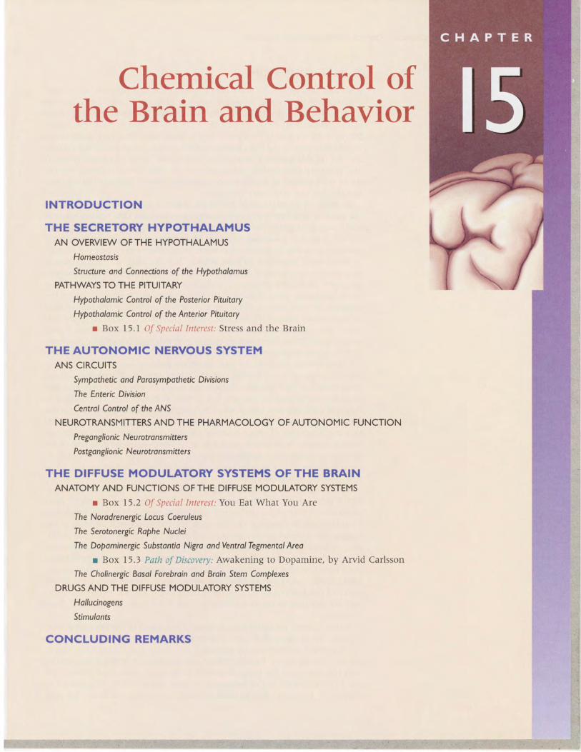
FIGURE I5.IPatterns of communication in the nervous system. (a) Most of the systems wehave discussed in this book may be described as point-to-point.The proper functioning ofthese systems requires restricted synaptic activation of target cells and signals of brief dura-tion. In contrast, three other components of the nervous system act over great distancesand for long periods of time, (b) Neurons of the secretory hypothalamus affect theirmany targets by releasing hormones directly into the bloodstream. (c) Networla of inter-connected neurons of the ANS can work together to activate tissues all over the body.(d) Diffuse modulatory systems extend their reach with widely divergent axonalproJectrons.
simultaneously controls the responses of many internal organs, blood ves-sels, and glands. The third component exists entirely within the centralnervous system (CNS) and consists of several related cell groups that differwith respect to the neurotransmitter they use. All of these cell groups ex-tend their spatial reach with highly divergent axonal projections and pro-long their actions by using metabotropic postsynaptic receptors. Membersof this component of the nervous system are called t}:'e diffuse modulatorysystems of the brain. The diffuse systems are believed to regulate, amongother things, the level of arousal and mood.
This chapter serves as a general introduction to these systems. In laterchapters, we will see how they contribute to specific behaviors and brainstates: motivation (Chapter l6), sexual behavior (Chapter l7), emotion(Chapter l8), sleep (Chapter t9), and psychiatric disorders (Chapter 22).

484 CHAPTER I 5 . CHEMICAL CONTROLOFTHE BMINAND BEHAVIOR
Optic chiasm Pituitary Hypothalamus
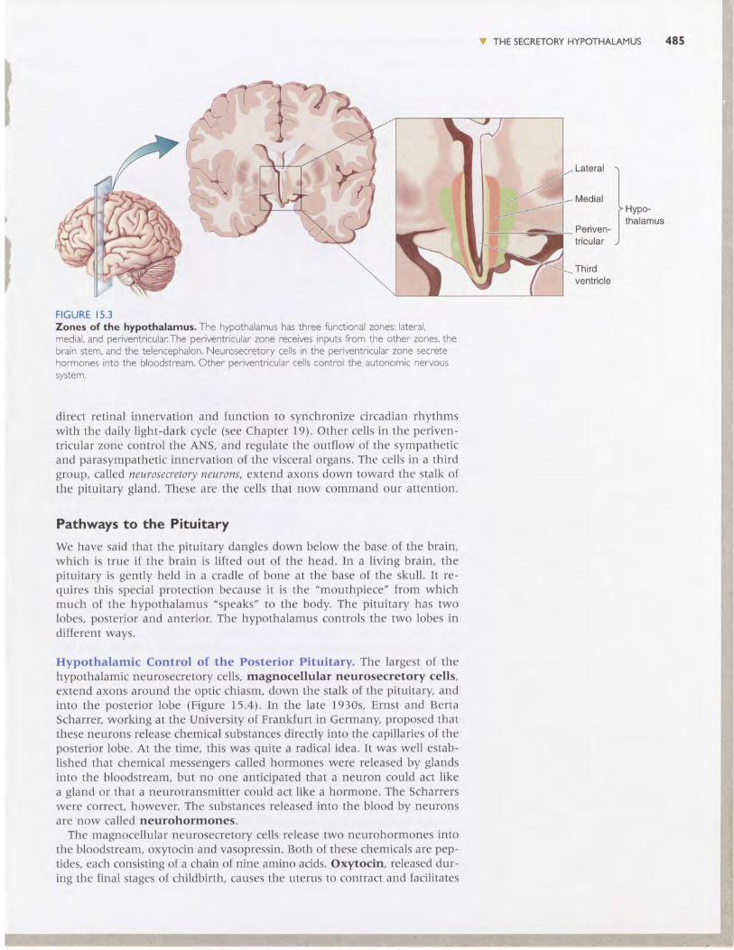
FIGURE I5.2Locations of the hypothalamus andpituitary.This is a midsagittal section. Noticethat the hypothalamus, whose borders areindicated with a dashed line, forms the wallof the third ventricle and sits below the dorsalthalamus.
V THE SECRETORY HYPOTHALAMUS
Recall from Chapter 7 that the hypothalamus sits below the thalamus, alongthe walls of the third ventricle. Ir is connecred by a stalk to the piruitarygland, which dangles below the base of the brain, just above the roof ofyour mouth (Figure 15.2). Although this tiny cluster of nuclei makes upless than Io/o of. the brain's mass, the influence of the hypothalamus on bodyphysiology is enormous. Let's take a brief tour of the hypothalamus andthen focus on some of the ways in which it exerts its powerful influence.
An Overview of the Hypothalamus
The hypothalamus and dorsal thalamus are adjacent to one another, buttheir functions are very different. As we saw in the previous seven chap-ters, the dorsal thalamus lies in the path of all the point-to-point pathwayswhose destination is the neocortex. Accordingly, the destruction of a smallpart of the dorsal thalamus can produce a discrete sensory or motor deficit:a little blind spot, or a lack of feeling on a portion of skin. In contrast, thehypothalamus integrates somatic and visceral resplnses in accordance with the needsof the brain. A tiny lesion in the hypothalamus can produce dramatic andoften fatal disruptions of widely dispersed bodily functions.
Homeostasis. In mammals, the requirements for life include a narrowrange of body temperatures and blood compositions. The hypothalamusregulates these levels in response to a changing external environment. Thisregulatory process is called homeostasis, the maintenance of the body'sinternal environment within a narrow physiological range.
Consider temperature regulation. Biochemical reactions in many cells ofthe body are fine-tuned to occur at about )7.C. A variation of more thana few degrees in either direction can be catastrophic. Temperature-sensitivecells in the hypothalamus detect variations in brain temperature and or-chestrate the appropriate responses. For example, when you stroll nakedthrough the snow, the hypothalamus issues commands that cause you toshiver (generating heat in the muscles), develop goosebumps (a futile at-tempt to fluff up your nonexistent fur-a reflexive remnant from ourhairier ancestors), and turn blue (shunting blood away from the cold surfacetissues to keep the sensitive core of the body warmer). In contrast, whenyou go for a jog in the tropics, the hypothalamus activates heat-loss mech-anisms that make you turn red (shunting blood /o the surface tissues whereheat can be radiated away) and sweat (cooling the skin by evaporation).
Other examples of homeostasis are the tight regulation of blood volume,pressure, salinity, acidity, and blood oxygen and glucose concentrations.The means by which the hypothalamus achieves these different types ofregulation are remarkably diverse.
Structure and Connections of the Hypothalamus. Each side of thehypothalamus has three functional zones: lateral, medial, and periventric-ular (Figure 15.3). The lateral and medial zones have extensive connectionswith the brain stem and the telencephalon and regulate certain types ofbehavior, as we will see in Chapter 16. Here we are concerned only withthe third zone, which actually receives much of its input from the other two.
The periventricular zone is so named because, with the exception ofa thin finger of neurons that are displaced laterally by the optic tract(called the supraoptic nucleus), the cells of this region lie right next to thewall of the third ventricle. Within this zone exists a complex mix of neuronswith different functions. one group of cells constitutes the suprachiasmaticnucleus (SCN), which lies just above the optic chiasm. These cells receive
)
V THE SECRETORY HYPOTHALAMUS 485
Thirdventricle
Periven-tricular
1,,,"-,l
thalamus
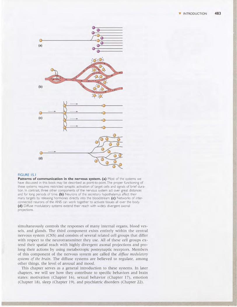
FIGURE I5.3Zones of the hypothalamus. The hypothalamus has three functional zones: lateral,medial, and periventricular:The periventricular zone receives inputs from the other zones, thebrain stem, and the telencephalon. Neurosecretory cells in the periventricular zone secretehormones into the bloodstream. Other oeriventricular cells control the autonomic nervoussystem.
direct retinal innervation and function to synchronize circadian rhythmswith the daily light-dark cycle (see Chapter 19). Other cells in the periven-tricular zone control the ANS, and regulate the outflow of the sympatheticand parasympathetic innervation of the visceral organs. The cells in a thirdgroup, called neurosecretory neurons, extend axons down toward the stalk ofthe pituitary gland. These are the cells that now command our attention.
Pathways to the Pituitary
We have said that the pituitary dangles down below the base of the brain,which is true if the brain is lifted out of the head. In a living brain, thepituitary is gently held in a cradle of bone at the base of the skull. It re-quires this special protection because it is the "mouthpiece" from whichmuch of the hypothalamus "speaks" to the body. The pituitary has twolobes, posterior and anterior. The hypothalamus controls the two lobes indifferent ways.
Hypothalamlc Control of the Posterior Pituitary. The largest of thehlpothalamic neurosecretory cells, magnocellular neurosecretory cells,extend axons around the optic chiasm, down the stalk of the pituitary andinto the posterior lobe (Figure 15.4). In the late 1930s, Ernst and BertaScharrer, working at the University of Frankfurt in Germany, proposed thatthese neurons release chemical substances directly into the capillaries of theposterior lobe. At the time, this was quite a radical idea. It was well estab-lished that chemical messengers called hormones were released by glandsinto the bloodstream, but no one anticipated that a neuron could act likea gland or that a neurotransmitter could act like a hormone. The Scharrerswere correct, however. The substances released into the blood by neuronsare now called neurohormones.
The magnocellular neurosecretory cells release two neurohormones intothe bloodstream, oxytocin and vasopressin. Both of these chemicals are pep-tides, each consisting of a chain of nine amino acids. Oxytocin, released dur-ing the final stages of childbirth, causes the uterus to contract and facilitates

485 CHAPTER I 5 . CHEMICALCONTROLOFTHE BRAINAND BEHAVIOR
FIGURE I5.4lrlagnocellular ncurprecl€tory cellr of thc lrypothelamur. This is a midsagittal viewof the hypothalamus and pituitary Magnocellular neurosecretory cells secrete oxytocin andvasopressin directly into capillaries in the posterior lobe of the pituitary
the delivery of the newbom. It also stimulates the ejection of milk from themammary glands. All lactating mothers know about the complex "let-down" reflex that involves the oxytocin neurons of the hypothalamus.Oxytocin release may be stimulated by the somatic sensations generated bya suckling baby. But the sight or cry of a baby (even someone else,s) canalso trigger the release of milk beyond the mother's conscious control. Ineach case, information about a sensory stimulus-somatic, visual, or audi-tory-reaches the cerebral cortex via the usual route, the thalamus, and thecortex ultimately stimulates the hypothalamus to trigger oxytocin release.The cortex can also suppress hypothalamic functions, as when anxiety in-hibits the letdown of milk.
Vasopressin, also called antidiuretic hormone (ADH), regulatesblood volume and salt concentration. When the body is deprived of water,the blood volume decreases and blood salt concentration increases. Thesechanges are detected by pressure receptors in the cardiovascular systemand salt concentration-sensitive cells in the hypothalamus, respectively.vasopressin-containing neurons receive information about these changesand respond by releasing vasopressin, which acts directly on the kidneysand leads to water retention and reduced urine production.
Under conditions of lowered blood volume and pressure, communicationbetween the brain and the kidneys actually occurs in both directions (Fig-ure 15.5). The kidneys secrete an enzyme into the blood called rmin.Blevatedrenin sets off a sequence of biochemical reactions in the blood. Angiotensino-gen, alarge protein released from the liver, is converted by renin to angiotensin
THE SECRETORY HYPOTHALAMUS 487
Lowered bloodpressure
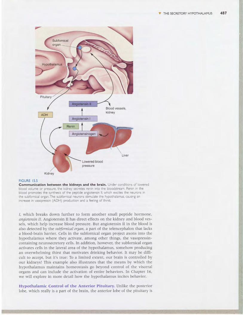
FIGURE I5.5Communication between the kidneys and the brain. Under conditions of loweredblood volume or pressure, the kidney secretes renin into the bloodstream, Renin in theblood promotes the synthesis of the peptide angiotensin ll, which excites the neurons inthe subfornical organ.The subfornical neurons stimulate the hypothalamus, causing anincrease in vasooressin (ADH) oroduction and a feelins of thirst,
1, which breaks down further to form another small peptide hormone,angiotensin I1. Angiotensin II has direct effects on the kidney and blood ves-sels, which help increase blood pressure. But angiotensin II in the blood isalso detected by the subfornical organ, a part of the telencephalon that lacksa blood-brain barrier. Cells in the subfornical organ project axons into thehypothalamus where they activate, among other things, the vasopressin-containing neurosecretory cells. In addition, however, the subfornical organactivates cells in the lateral area of the hypothalamus, somehow producingan overwhelming thirst that motivates drinking behavior. It may be diffi-cult to accept, but it's true: To a limited extent, our brain is controlled byour kidneyst This example also illustrates that the means by which thehypothalamus maintains homeostasis go beyond control of the visceralorgans and can include the activation of entire behaviors. In Chapter 16,we will explore in more detail how the hypothalamus incites behavior.
Hypothalamic Control of the Anterior Pituitary. Unlike the posteriorIobe, which really is a part of the brain, the anterior lobe of the pituitary is
488 C HAPTER I 5 . CHEMICAL CONTROL OFTHE BRAINAND BEHAVIOR
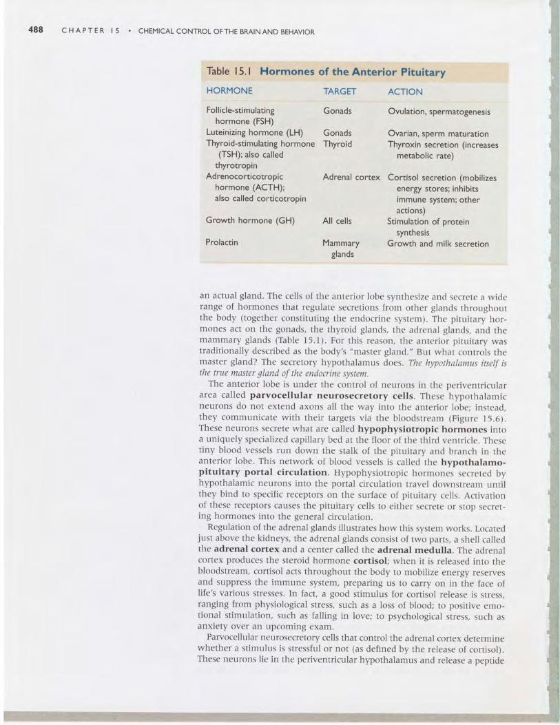
Table 15. I Hormones of the Anterior Pituitary
HORMONE
Follicle-stimulatinghormone (FSH)
Luteinizing hormone (LH)Thyroid-stimulating hormone
(TSH); also calledthyrotropin
Adrenocorticotropichormone (ACTH);also called corticotropin
Growth hormone (GH)
Prolactin
TARGET
Gonads
GonadsThyroid
Adrenal cortex
ACTION
Ovu lation, spermatogenesis
Ovarian, sperm maturationThyroxin secretion (increases
metabolic rate)
Cortisol secretion (mobilizesenergy stores; inhibitsimmune system; otheractions)
Stimulation of proteinsynthesis
Growth and milk secretion
All cells
Mammaryglands
an actual gland. The cells of the anterior lobe synthesize and secrete a widerange of hormones that regulate secretions from other glands throughoutthe body (together constituting the endocrine system). The pituitary hor-mones act on the gonads, the thyroid glands, the adrenal glands, and themammary glands (Table 15.1). For this reason, the anterior pituitary wastraditionally described as the body's "master gland." But what controls themaster gland? The secretory hypothalamus does. The hypothalamus itself isthe true master gland of the endocrine system.
The anterior lobe is under the control of neurons in the periventriculararea called parvocellular neurosecretory cells. These hypothalamicneurons do not extend axons all the way into the anterior lobe; instead,they communicate with their targers via the bloodstream (Figure 15.6).These neurons secrete what are called hypophysiotropic hormones intoa uniquely specialized capillary bed at the floor of the third ventricle. Thesetiny blood vessels run down the stalk of the pituitary and branch in theanterior lobe. This network of blood vessels is called the hypothalamo-pituitary portal circulation. Hypophysiotropic hormones secreted byhypothalamic neurons into the portal circulation travel downstream untilthey bind to specific receptors on the surface of pituitary cells. Activationof these receptors causes the pituitary cells to either secrete or stop secret-ing hormones into the general circulation.
Regulation of the adrenal glands illustrates how this system works. Locatedjust above the kidneys, the adrenal glands consist of two parts, a shell calledthe adrenal cortex and a center called the adrenal medulla. The adrenalcortex produces the steroid hormone cortisol; when it is released into thebloodstream, cortisol acts throughout the body to mobilize energy reservesand suppress the immune system, preparing us to carry on in the face oflife's various stresses. In fact, a good stimulus for cortisol release is stress,ranging from physiological stress, such as a loss of blood; to positive emo-tional stimulation, such as falling in love; to psychological stress, such asanxiety over an upcoming exam.
Parvocellular neurosecretory cells that control the adrenal cortex determinewhether a stimulus is stressful or not (as defined by the release of cortisol).These neurons lie in the periventricular hypothalamus and release a peptide

V THE SECRETORY HYPOTHALAMUS 489
FIGURE I5.6Parvocellular neunosecretory cells of the hypothalamus. Parvocellular neurosecre-tory cells secrete hypophysiotropic hormones into specialized capillary beds of the hypothal-amo-pituitary portal circulation.These hormones travel to the anterior lobe of the pituitary,where they trigger or inhibit the release of pituitary hormones from secretory cells.
called corticotropin-releasing hormone (CRII) into the blood of the portal cir-culation. CRH travels the short distance to the anterior pituitary, where,within about l5 seconds, it stimulates the release of corticotropin, or adreno-corticotropic hormone \ACIH\. ACTH enters the general circulation and travelsto the adrenal cortex where, within a few minutes, it stimulates cortisolrelease (Figure 15.7).
Blood levels of cortisol are, to some extent, self-regulated. Cortisol is asteroid, which is a class of biochemicals related to cholesterol. Thus, cortisolis a lipophilic ("fat-loving") molecule, which will dissolve easily in lipidmembranes and readily cross the blood-brain barrier. In the brain, cortisolinteracts with specific receptors that lead to the inhibition of CRH release,thus ensuring that circulating cortisol levels do not get too high. Surpris-ingly, however, neurons with cortisol receptors are found widely distributedin the brain, not just in the hypothalamus. In these other CNS locations, cor-tisol has been shown to have significant effects on neuronal activity. Thus,we see that the release of hypophysiotropic hormones by cells in the secre-tory hypothalamus can produce widespread alterations in the physiology ofboth the bodv and the brain (Box l5.l).

490 C HAPTE R I 5 . CHEMICALCONTROL OFTHE BMINAND BEHAVIOR
FIGURE I5.7The stress resPonse. Under conditions of physiological, emotional, or psychological stimu-lalion or stress, the periventricular hypothalamus secretes corticotropin-releasing hormone(CRH) into the hypothalamo-pituitary portal circulation.This triggers the release of adreno-corticotropic hormone (ACTH) into the general circulation. ACTH stimulates the release ofcortisol from the adrenal cortex. Cortisol can act directly on hypothalamic neurons, as wellas on other neurons elsewhere in the brain.
V THE AUTONOMIC NERVOUS SYSTEMBesides controlling the ingredients of the hormonal soup that flows inour veins, the periventricular zone of the hypothalamus also controls theautonomic nervous system (ANs). The ANS is an extensive network ofinterconnected neurons that are widely distributed inside the body cavity.From the Greek autonomia, autonomic roughly means ,,independence,,; au-tonomic functions are usually carried out automatically, without conscious,voluntary control. They are also highly coordinated functions. Imagine asudden crisis. In a morning class, as you are engrossed in a crosswordpuzzle, the instructor unexpectedly calls you to the blackboard to solve animpossible-looking equation. You are faced with a classic fight-or-flight sit-uation, and your body reacts accordingly, even as your conscious mindfrantically considers whether to blunder through it or beg off in humiliation.Your ANS triggers a host of physiological responses, including increasedheart rate and blood pressure, depressed digestive functions, and mobilizedglucose reserves. These responses are all produced by the sympatheticdivision of the ANS. Now imagine your relief as the class-ending bellsuddenly rings, saving you from acute embarrassment and the instructor'sanger. You settle back into your chair, breath deeply, and read the clue for24 DOWN. Within a few minutes, your sympathetic responses decrease tolow levels, and the functions of your parasympathetic division crank up

Stress and the Brain
Biological stress is created by the brain, in resPonse to
real or imagined stimuli.The many physiological responses
associated with stress help protect the body, and the
brain, from the dangers that triggered the stress in thefirst place. But stress in chronic doses can have insidiousharmful effects as well. Neuroscientists have only begunto understand the relationship between stress, the brain,
and brain damage.Stress leads to the release of the steroid hormone cor-
tisol from the adrenal cortex. Cortisol travels to the brain
through the bloodstream and binds to recePtors in the
cytoplasm of many neurons.The activated receptors travel
to the cell nucleus, where they stimulate gene transcrip-tion and ultimately protein synthesis. One consequence of
cortisol's action is that neurons admit more Ca2+ throughvoltage-gated ion channels. This may be due to a direct
change in the channels, or it may be indirectly caused by
changes in the cell 's energy metabolism. Whatever the
mechanism, presumably in the short term, cortisol makes
the brain better able to cope with the stress-perhaps byhelping it figure out a way to avoid it!
But what about the effects of chronic, unavoidablestress? In Chapter 6, we learned that too much calciumcan be a bad thing. lf neurons become overloaded with
calcium, they die (excitotoxicity). The question naturally
arises: Can cortisol kil l? Bruce McEwen and his colleagues
at Rockefeller University, and Robert Sapolsky and his
colleagues at Stanford University, have studied this ques-
tion in the rat brain. They found that daily iniections of
corticosterone (rat cortisol) for several weeks caused
Ji i THEAUTONOMIC NERVOUS SYSTEM 491
dendrites to wither on many neurons with corticosterone
receotors.A few weeks later, these cells started to die.A
similar result was found when, instead of daily hormone
injections, the rats were stressed every day.Sapolsky's studies of baboons in Kenya further reveal
the scourges of chronic stress. Baboons in the wild main-
tain a complex social heirarchy, and subordinate males
steer clear of dominant males when they can. During one
year when the baboon population boomed, local vil lagers
caged many of the animals to prevent them from de-
stroying their crops. Unable to escape the "toP baboons"
in the cages, many of the subordinate males subsequentlydied-not from wounds or malnutrit ion, but apparently
from severe and sustained stress-induced effects. They
had gastr ic u lcers, co l i t is , enlarged adrenal g lands, and
extensive degeneration of neurons in their hippocampus.
Subsequent studies suggest that it is the direct effect of
cortisol that damages the hippocampus. These effects of
cortisol and stress resemble the effects of aging on the
brain. Indeed, research has c lear ly shown that chronic
stress causes premature aging of the brain.In humans, exDosure to the horrors of combat, sexual
abuse, and other types of extreme violence can lead toposttraumatic stress disorder, with symptoms of heightened
anxiety, memory d is turbances, and int rus ive thoughts.
lmaging studies have consistently found degenerative
changes in the brains of victims, particularly in the hip-
pocampus. In Chapter 22, we wil l see that stress, and the
brain's response to it, plays a central role in several psy-
chiatric disorders.
again: Your heart rate slows and blood pressure drops, digestive functions
work harder on breakfast, and you stop sweating.Notice that you may not have moved out of your chair throughout this
unpleasant event . Maybe you d idn ' t even move your penci l . But your
body's internal workings reacted dramatically. Unlike the somaticmotlr system,
whose alpha motor neurons can rapidly excite skeletal muscles with pin-
point accuracy, the actions of the ANS are typically multiple, widespread,
and relatively slow. Therefore, the ANS operates in expanded space and
time. In addition, unlike the somatic motor system, which can only excite
its peripheral targets, the ANS balances synaptic excitation and inhibit ion
to achieve widely coordinated and graded control.
ANS Circuits
Together, the somatic motor system and the ANS constitute the total neu-
ral output of the CNS. The somatic motor system has a single function: It
innervates and commands skeletal muscle fibers. The ANS has the complex
492 cHAprER | 5 . cHEMtcALcoNTRoLoFTHEBRAINANDBEHAVIoR
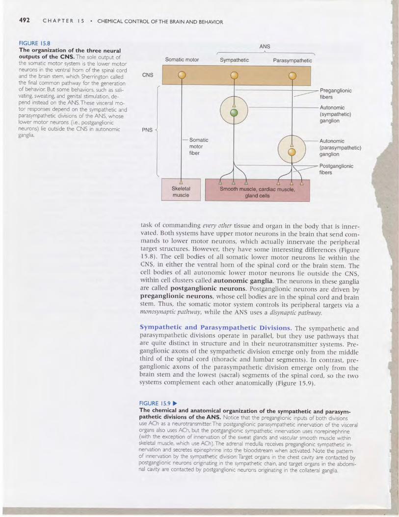
FIGURE I5.8The organization ofthe three neuraloutputs of the CNS. The sole output ofthe somatic motor system is the lower motorneurons in the ventral horn ofthe spinal cordand the brain stem, which Sherrineton calledthe final common pathway for thJgenerationof behavior: Br.rt some behaviors, such as sali-vating, sweating, and genital stimulation, de-pend instead on the ANS.These visceral mo-tor responses depend on the sympathetic andparasympathetic divisions of the ANS, whoselower motor neurons (i.e., postganglionicneurons) lie outside the CNS in autonomicganglra.
CNS
Somatic motor Sympathetic Parasympathetic
PNS
Preganglionicfibers
Autonomic(sympathetic)ganglion
Autonomic(parasympathetic)ganglion
Postganglionicfibers
task of commandingevery othertissu,e and organ in the body that is inner-vated. Both systems have upper motor neurons in the brain that send com-mands to lower motor neurons, which actually innervate the peripheraltarget structures. However, they have some interesting differences (Figure15.8). The cell bodies of all somatic lower motor neurons lie within thecNS, in either the ventral horn of the spinal cord or the brain stem. Thecell bodies of all autonomic lower motor neurons lie outside the cNS,within cell clusters called autonomic ganglia. The neurons in these gangliaare called postganglionic neurons. postganglionic neurons are driven bypreganglionic neurons, whose cell bodies are in the spinal cord and brainstem. Thus, the somatic motor system controls its peripheral targets via amonosynaptic pathway, while the ANS uses a disynaptic pathway.
sympathetic and Parasympathetic Divisions. The sympathetic andparasympathetic divisions operate in parallel, but they use pathways thatare quite distinct in structure and in their neurotransmitter systems. pre-ganglionic axons of the sympathetic division emerge only from the middlethird of the spinal cord (thoracic and lumbar segments). In conrrasr, pre-ganglionic axons of the parasympathetic division emerge only from thebrain stem and the lowest (sacral) segments of the spinal cord, so the twosystems complement each other anatomically (Figure I5.9).
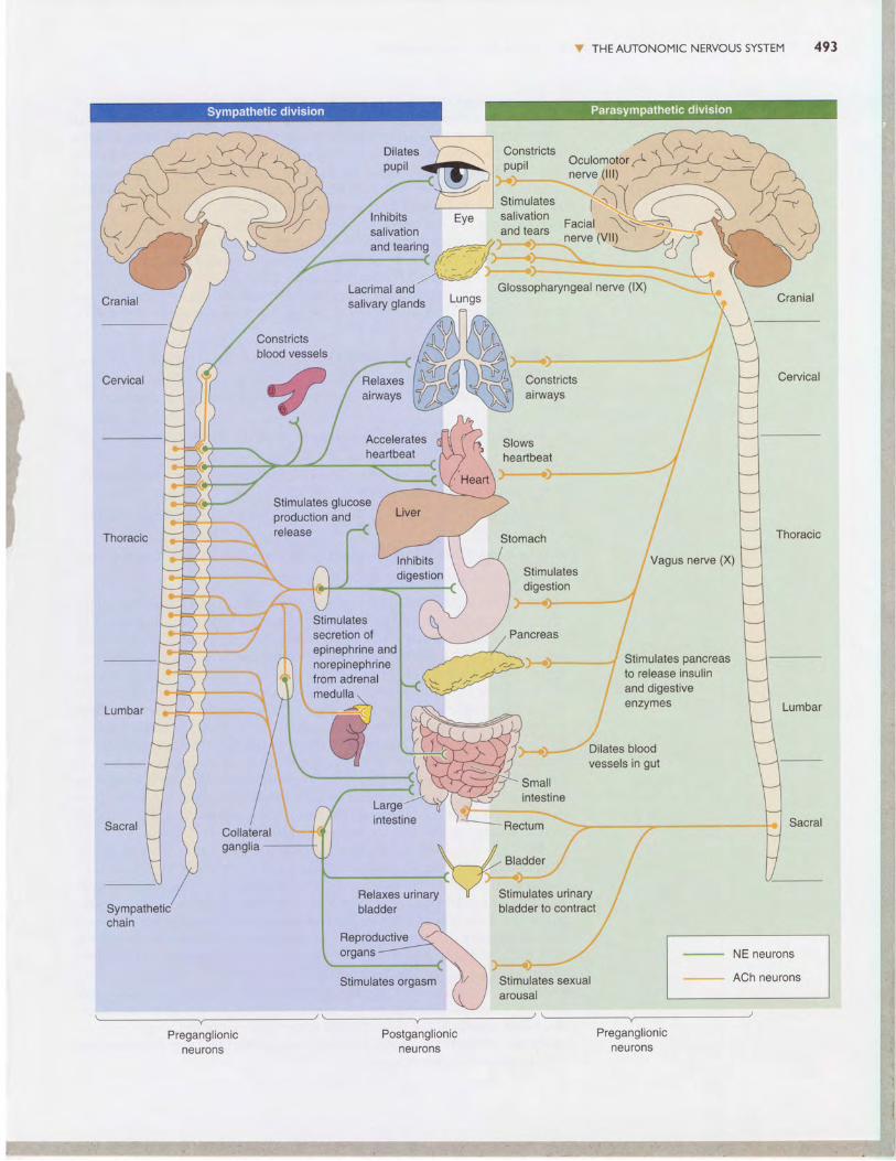
FIGURE I5.9 >The chemical and anatomical organization of the sympathetic and parasym-pathetic divisions of the ANS. Notice that the preganglionic inputs of both divisionsuse ACh as a neurotransmitter:The postganglionic parasympathetic innervation of the visceralorgans also uses ACh, but the postganglionic sympathetic innervation uses norepinephrine(with the exception of innervation of the sweat glands and vascular smooth muscle withinskeletal muscle, which use ACh).The adrenal medulla receives preganglionic sympathetic in-nervation and secretes epinephrine into the bloodstream when activated. Note the patternof innervation by the sympathetic division:Target organs in the chest cavity are contacted bypostganglionic neurons originating in the sympathetic chain, and target organs in the abdomi-nal cavity are contacted by postganglionic neurons originating in the collateral ganglia.
ANS
W THEAUTONOMIC NERVOUS SYSTEM
Dilatespupil
Constrictspupil oculomolor^
,l/
/l
, Vaous nerve (X), l '
:l',T:11:' ''\lInhibits Eye ."1".1,:-i Faciat N->salivation ,,,,"n0 1:"tt n"r" 1Vtt1'
' -,7and tearing ,4==J/',:'-:"'"ii'
*' -nerve \vu\*}f )----< ('- ,r/,"::i-": -..:ll.:. -l:f::: .. -- : I
Lacrimal ^nd"to=it Glossopharyngeat nerve (lii " " "". \
Smallintestine
Stimulates orgasm /t/ I Stimulates sexual
Postganglionicneurons
,,
\ /-r,^00",,Y'---X.'
Relaxes urinary Y Stimulates urinarybladder bladder to contract i
: * : . . . . . - - nerve ( l l l )
Stimulatesdigestion
,x -"*"'q:r '" - '" "-* "-"1/
'lcreas ,l/
;!."".*)- -- -- -^" / Stimulates pancreas
./ to release insulin,i and digestive
; enzvmes
.i
.r*,*, - -'""tilates bloodvessels in gut
Constrictsblood vessels
Stimulates glucose"".\ Production and
\ release (
Lacrimal and' Glossopharyngeal nerve (lX)
salivary glands LungsY
Acceleratesheartbeat
Relaxesairways
Stimulatessecretion ofepinephrine andnorepinephrinefrom adrenalmedulla.
AA I
t/-r--l
Sympatheticchain
NE neurons
ACh neurons
Preganglionicneurons
Preganglionicneurons
l' i
r {
l
arousal
494 CHAPTER I 5 . CHEMICALCONTROLOFTHEBRAINAND BEHAVIOR
The preganglionic neurons of the sympathetic division lie within the in-termediolateral gray matter of the spinal cord. They send their axons throughthe ventral roots to synapse on neurons in the ganglia of the sympatheticchain, which lies next to the spinal column, or within collateral gangliafound within the abdominal cavity. The preganglionic parasympatheticneurons, on the other hand, sit within a variety of brain stem nuclei andthe lower (sacral) spinal cord, and their axons travel within several cranialnerves as well as the nerves of the sacral spinal cord. The parasympatheticpreganglionic axons travel much farther than the sympathetic axons, be-cause the parasympathetic ganglia are typically located next to, on, or intheir target organs (see Figures 15.8 and I5.9).
The ANS innervates three types of tissue: glands, smooth muscle, andcardiac muscle. Thus, almost every part of the body is a target of the ANS,as shown in Figure 15.9. The ANS:
I Innervates the secretory glands (salivary sweat, tear, and various mucus-producing glands).
r Innervates the heart and blood vessels to control blood pressure andflow
t Innervates the bronchi of the lungs to meet the oxygen demands of thebody.
I Regulates the digestive and metabolic functions of the liver, gastroin-testinal tract, and pancreas.
t Regulates the functions of the kidney, urinary bladder, large intestine,and rectum.
r Is essential to the sexual responses of the genitals and reproductive organs.t Interacts with the body's immune system.
The physiological influences of the sympathetic and parasympatheticdivisions generally oppose each other. The sympathetic division tends to bemost active during a crisis, real or perceived. The behaviors related to it aresummarized in the puerile (but effective) mnemonic used by medical stu-dents, called the four Fs: fight, flight, fright, and sex. The parasympatheticdivision facilitates various non-four-F processes, such as digestion, growth,immune responses, and energy storage. In most cases, the activity levels ofthe two ANS divisions are reciprocal; when one is high, the other tends tobe low and vice versa. The sympathetic division frenetically mobilizes thebody for a short-term emergency at the expense of processes that keep ithealthy over the long term. The parasympathetic division works calmly forthe long-term good. Both cannot be stimulated strongly at the same time;their general goals are incompatible. Fortunately, neural circuits in the cNSinhibit activity in one division when the other is active.
some examples will help illustrate how the balance of activity in thesympathetic and parasympathetic divisions controls organ functions. Thepacemaker region of the heart triggers each heartbeat without the help ofneurons, but both divisions of the ANS innervate it and modulate it; sym-pathetic activity results in an increase in the rate of beating, while parasym-pathetic activity slows it down. The smooth muscles of the gastrointestinaltract are also dually innervated, but the effect of each division is the oppo-site of its effect on the heart. Intestinal motility, and thus digestion, is stim-ulated by parasympathetic axons and inhibited by sympathetic axons. Notall tissues receive innervation from both divisions of the ANS. For exam-ple, blood vessels of the skin, and the sweat glands, are innervated only byexcitatory sympathetic axons. Lacrimal (tear-producing) glands are excitedonly by parasympathetic input.
1T THE AUTONOMIC NERVOUS SYSTEM 495
Another example of the balance of parasympathetic-sympathetic activity
is the curious neural control of the male sexual response. Erection of thehuman penis is a hydraul ic process. I t occurs when the penis becomes
engorged with blood, which is triggered and sustained by parasympathetic
activity. The curious part is that orgasm and ejaculation are triggered by
sympathetic activity. You can imagine how complicated it must be for the
nervous system to orchestrate the entire sexual acu parasympathetic activ-
ity gets it going (and keeps it going), but a shift to sympathetic activity is
necessary to terminate it. Anxiety and worry, and their attendant sympa-
thetic activity, tend to inhibit erection and promote ejaculation. Not sur-prisingly, impotence and premature ejaculation are common complaints
of the overst ressed male. (We wi l l d iscuss sexual behavior fur ther in
Chap te r 17 . )
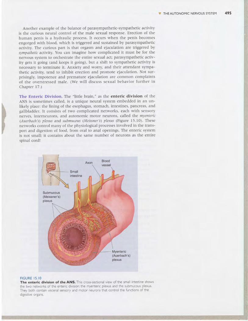
The Enteric Division. The "l itt le brain," as the enteric division of the
ANS is sometimes called, is a unique neural system embedded in an un-
likely place: the Iining of the esophagus, stomach, intestines, pancreas, andgallbladder. It consists clf two complicated networks, each with sensory
nerves, interneurons, and autonomic motor neurons, called the myenteric(Auerbach's) plexus and submucous (Meissner's) plexus (Figure 15.10). These
networks control many of the physiological processes involved in the trans-port and digestion of food, from oral to anal openings. The enteric system
is not small; i t contains about the same number of neurons as the entire
spinal cord !
F I G U R E I 5 . I OThe enteric division of the ANS. This cross-sectional view of the sma i ntestrne showsrhe two networ\s o{ tr^e ente' ic divis'o^: tne rr lente' ic plexus and t"e subnucous plex-s.They both contain vsceral sensory and motor neurons that contro the functons ofthed gestrve organs.
Axon
i t \
Bloodvessel
Smallintestine
495 C HAPTE R I 5 . CHEMICALCONTROL OFTHE BRATNAND BEHAVIOR
If the enteric division of the ANS qualifies as ,,brain,, (which may be over-stating the case), it is because it can operate with a great deal of inde-pendence. Entedc sensory neurons monitor tension and stretch of the gas-trointestinal walls, the chemical status of stomach and intestinal contents,and hormone levels in the blood. This information is used by the entericinterneuronal circuits to control the activity levels of enteric output motorneurons, which govern smooth muscle motility, the production of mucousand digestive secretions, and the diameter of the local blood vessels. Forexample, consider a partially digested pizza making its way through thesmall intestine. The myenteric plexus ensures that lubricating mucus anddigestive enzymes are delivered, that rhythmic (peristaltic) muscle actionworks to mix the pizza and enzymes thoroughly, and that intestinal bloodflow increases to provide a sufficient fluid source and transport newly ac-quired nutrients to the rest of the body.
The enteric division is not entirely autonomous. It receives input indi-rectly from the "real" brain via axons of the sympathetic and parasympa-thetic divisions. These provide supplementary control and can supersedethe functions of the enteric division in some circumstances. For example,the enteric nervous system and digestive functions are inhibited by thestrong activation of the sympathetic nervous system that occurs duringacute stress.
central control of the ANS. As we have said, the hypothalamus is themain regulator of the autonomic preganglionic neurons. Somehow thisdiminutive structure integrates the diverse information it receives about thebody's status, anticipates some of its needs, and provides a coordinated setof both neural and hormonal outputs. Essential to autonomic control arethe connections of the periventricular zone to the brain stem and spinalcord nuclei that contain the preganglionic neurons of the sympathetic andparasympathetic divisions. The nucleus of the solitary tract, located inthe medulla and connected with the hypothalamus, is another importantcenter for autonomic control. In fact, some autonomic functions operatewell even when the brain stem is disconnected from all structures above it,including the hypothalamus. The solitary nucleus integrates sensory infor-mation from the internal organs and coordinates output to the autonomicbrain stem nuclei.
Neurotransmitters and thePharmacology of Autonomic FunctionEven people who have never heard the word neurotransmitter know whatit means to "get your adrenaline flowing." (In the United Kingdom thecompound is called adrenaline, while in the united states we call it epi-nephrine.) Historically, the ANS has probably taught us more than anyother part of the body about how neurotransmitters work. Because theANS is relatively simple compared to the cNS, we understand the ANSmuch better. In addition, neurons of the peripheral parts of the ANS areoutside the blood-brain barrier, so all drugs that enter the bloodstream havedirect access to them. The relative simplicity and accessibility of the ANShave led to a deeper understanding of the mechanisms of drugs that influ-ence synaptic transmission.
Preganglionic Neurotransmitters. The primary transmitter of the pe-ripheral autonomic neurons is acetylcholine (AChl, the same transmitter usedat skeletal neuromuscular junctions. The preganglionic neurons of both sympa-

V THEAUTONOMIC NERVOUS SYSTEM 497
thetic and parasympathetic divisions release ACh.The immediate effect is that theACh binds to nicotinic ACh receptors (nAChR), which are ACh-gated chan-nels, and evokes a fast excitatory postsynaptic potential (EPSP) that usuallytriggers an action potential in the postganglionic cell. This is very similar tothe mechanisms of the skeletal neuromuscular junction, and drugs thatblock nAChRs in muscle, such as curare, also block autonomic output.
Ganglionic ACh does more than neuromuscular ACh, however. It alsoactivates muscarinic ACh receptors (mAChR), which are metabotropic(G-protein-coupled) receptors that can cause both the opening and theclosing of ion channels that lead to very slow EPSPs and inhibitory postsy-naptic potentials (IPSPs). These slow mAChR events are usually not evi-dent unless the preganglionic nerve is activated repetitively. In addition toACh, some preganglionic terminals release a variety of small, neuroactivepeptides such as NPY (neuropeptide Yl and WP (vasoactive intestinal polypep'tide\.These also interact with G-protein-coupled receptors and can triggersmall EPSPs that last for several minutes. The effects of peptides are mod-ulatory; they do not usually bring the postsynaptic neurons to firing thresh-old, but they make them more responsive to the fast nicotinic effects whenthey do come along. Because more than one action potential is required tostimulate the release of these modulatory neurotransmitters, the pattern offiring in preganglionic neurons is an important variable in determining thetype of postganglionic activity that is evoked.
Postganglionic Neurotransmitters. Postganglionic cells-the autonomicmotor neurons that actually trigger glands to secrete, sphincters to contractor relax, and so on-use different neurotransmitters in the sympatheticand parasympathetic divisions of the ANS. Postganglionic parasympatheticneurons release ACh, but those of most parts of the sympathetic divisionuse norepinephrine (NEl. Parasympathetic ACh has a very local effect on itstargets and acts entirely through mAChRs. In contrast, sympathetic NEoften spreads far, even into the blood where it can circulate widely.
The autonomic effects of a variety of drugs that interact with cholinergicand noradrenergic systems can be confidently predicted, if you under-stand some of the autonomic circuitry and chemistry (see Figure 15.9)' Ingeneral, drugs that promote the actions of norepinephrine or inhibit themuscarinic actions of acetylcholine are sympathomimetic; they cause effectsthat mimic activation of the sympathetic division of the ANS. For example,atropine, an antagonist of mAChRs, produces signs of sympathetic activa-tion, such as dilation of the pupils. This response occurs because the bal-ance of ANS activity is shifted toward the sympathetic division whenparasympathetic actions are blocked. On the other hand, drugs that pro-mote the muscarinic actions of ACh or inhibit the actions of NE are parasym-pathomimetic; they cause effects that mimic activation of the parasympa-thetic division of the ANS. For example, propranolol, an antagonist of thep receptor for NE, slows the heart rate and lowers blood pressure. For thisreason, propranolol is sometimes used to prevent the physiological conse-quences of stage fright.
But what about the familiar flow of adrenaline? Adrenaline (epinephrine)is the compound released into the blood from the adrenal medulla when ac-tivated by preganglionic sympathetic innervation. Epinephrine is actuallymade from norepinephrine (noradrenaline in the United Kingdom), and ithas effects on target tissues almost identical to those caused by sympatheticactivation. Thus, the adrenal medulla is really nothing more than a modi-fied sympathetic ganglion. You can imagine that as the epinephrine (adren-aline) flows, a coordinated, bodywide set of sympathetic effects kicks in.
498 C HAPTE R I 5 . CHEMICALCONTROLOFTHE BRAINAND BEHAVIOR
V THE DIFFUSE MODULATORY SYSTEMSOF THE BRAIN
Consider what happens when you fall asleep. The internal commands ,,youare becoming drowsy" and "You are falling asleep" are messages that mustbe received by broad regions of the brain. Dispensing this information re-quires neurons with a particularly widespread pattern of axons. The brainhas several such collections of neurons, each using a particular neuro-transmitter and making widely dispersed, diffuse, almost meandering con-nections. Rather than carrying detailed sensory information, these cells of-ten perform regulatory functions, modulating vast assemblies of postsynapticneurons (such as the cerebral cortex, the thalamus, and the spinal cord) sothat they become more or less excitable, more or less synchronously active,and so on. collectively, they are a bit like the volume, treble, and bass con-trols on a radio, which do not change the lyrics or melody of a song butdramatically regulate the impact of both. In addition, different systems ap-pear to be essential for aspects of motor control, memory, mood, motiva-tion, and metabolic state. Many psychoactive drugs affect these modulatorysystems, and the systems figure prominently in current theories about thebiological basis of certain psychiatric disorders.
Anatomy and Functions of the Difruse Modulatory SystemsThe diffuse modulatory systems differ in structure and function, yetthey have certain principles in common:
r Typically, the core of each system has a small set of neurons (severalthousand).
r Neurons of the diffuse systems arise from the central core of the brain,most of them from the brain stem.
r Each neuron can influence many others, because each one has an axonthat may contact more than 100,000 postsynaptic neurons spread widelyacross the brain.
r The synapses made by many of these systems release transmitter mole-cules into the extracellular fluid, so they can diffuse to many neuronsrather than be confined to the vicinity of the synaptic cleft.
We focus on the modulatory systems of the brain that use either NE,serotonin (5-HT), dopamine (DA), or ACh as a neurotransmitter. Recallfrom chapter 6 that all of these transmitters activate specific metabotropic(G-protein-coupled) receptors and that these receptors mediate most oftheir effects; for example, the brain has 10-100 times more metabotropicACh receptors than ionotropic nicotinic ACh receptors.
Because neuroscientists are still working hard to determine the exact func-tions of these systems in behavior, our explanations here will necessarilybe vague. It is clear, however, that the functions of the diffuse modulatorysystems depend on how electrically active they are, individually and incombination, and on how much neurotransmitter is available for release(Box 15 .2) .
The Noradrenergic Locus coeruleus. Besides being a neurotransmitterin the peripheral ANS, NE is also used by neurons of the tiny locuscoeruleus in the pons (from the Latin for "blue spot,, because of the pig-ment in its cells). Each human locus coeruleus has about 12,000 neurons.We have two of them, one on each side.
A major breakthrough occurred in the mid-1960s, when Kjell Fuxe andhis colleagues at the Karolinska Institute in sweden developed a technique
$ THE DIFFUSE MODULATORY SYSTEMS OFTHE BRAIN 499
You Eat What You AreAmericans, it seems, are always trying to lose weight.Thelow-fat, high-carbohydrate diets (think bagels) that wereall the rage in the | 990s have now been replaced by the"low-carb" craze (think omelets). Changing your diet canalter caloric intake and the body's metabolism; it can alsoalter how your brain functions.
The influence of diet on the brain is most clear in thecase of the diffuse modulatory systems. Consider serotonin.Serotonin is synthesized in two steps from the dietaryamino acid tryptophan (see Figure 6. 14).The first step iscatalyzed by the enzyme tryptophan hydroxylase.The lowaffinity of the enzyme for tryptophan makes this step rote-limiting for serotonin synthesis-that is, serotonin can beproduced only as fast as this enzyme can hydroxylatetry/ptophan. And a lot of tryptophan is required to pushthe synthetic reaction as fast as it can go. However, braintryptophan levels are well below the level required to sat-urate the enzyme.Thus, the rate of serotonin synthesis isdetermined, in part, by the availabil ity of tryptophan in thebrain-more tryptophan, more serotonin; less trypto-phan, less serotonin.
Brain tryptophan levels are controlled by how muchtryptophan there is in the blood, and by how efficiently itis transported across the blood-brain barrier.Tryptophanin the blood is derived from the proteins we digest in ourdiet, so a high-protein diet wil l lead to sharply increasedblood levels of tryptophan. Surprisingly, however, there isa decline in brain tryptophan (and serotonin) for severalhours after a hearty, high-protein meal.The paradox wasresolved by Richard \ y'urtman and his colleagues at MIT
that enabled the catecholaminergic (noradrenergic and dopaminergic)neurons to be visualized selectively in histological sections prepared fromthe bra in (F igure 15.11) . This analys is revealed that axons leave the krcuscoeruleus in several tracts, but then fan out to innervate just about everypart of the brain: all of the cerebral cortex, the thalamus and the hypo-thalan-rus, the olfactory bulb, the cerebellum, the midbrain, and the spinalcord (F igure 15.12) . The locus coeruleus must make some of the most d i f -fuse connections in the brain, considering that just one of its neurons canmake more than 250,000 synapses, and i t can have one axon branch in thecerebral cortex and another in the cerebellar cortex!
Locus ccleruleus cells seem to be involved in the regulation of attention,arousal, and sleep-wake cycles, as well as learning and memory, anxietyand pain, mood, and brain metabolism. This makes it sound as if the locuscoeruleus may run the whole show. But the key word is "involved," whichcan mean almost anything. For example, our heart, l iver, lungs, and kidneysare also involved in every brain function, for without them, all behaviorwould fail utterly. Because of its widespread connections, the locus coeruleus
who observed that several other amino acids (tyrosine,phenylalanine, leucine, isoleucine, and valine) compete withtryptophan for transport across the blood-brain barrier.These other amino acids are rich in a high-protein diet,and they suppress the entry of the tryptophan into thebrain.The situation is reversed with a high-carbohydratemeal (that also contains some protein). Insulin, released bythe pancreas in response to carbohydrates, decreases theblood levels of the compet ing amino acids re lat ive totryptophan. So the tryptophan in the blood is efficientlytransported into the brain, and serotonin levels rise.
Increased brain tryptophan correlates with elevatedmood, decreased anxiety, and increased sleepiness, l ikelydue to changes in serotonin levels. Inadequate tryptophanmay explain the phenomenon of carbohydrate cravingthat has been reported in humans with seasonal affectivedisorder-the depression of mood brought on by reduceddaylight during winter. lt may also explain why clinical trialsfor treating obesity with extreme carbohydrate depriva-tion had to be stopped because of complaints of mooddisturbances (depression, irritabil i ty) and insomnia.
Based on these and other observations,Wurtman andhis wife Judith made the intriguing suggestion that ourdietary choices may reflect our brain's need for serotonin.Consistent with this notion, drugs that elevate extracel-lular serotonin can be effective for weight loss (as well asdepression), possibly by reducing the bodys demand forcarbohydrates. We will discuss the involvement of sero-tonin in appetite regulation further in Chapter l6 and inthe regulation of mood in Chapter 22.

500 C H A P T E R I 5 CHEMICAL CONTROL OF THE BRAIN AND BEHAVIOR
F I G U R E I 5 . I INorepinephrine.containing neurons of the locus coeruleus. The react on ofnoradrenergic neurons with formadehyde gas causes them to lJuoresce green, enablinganatom cal invest gation of the r w despread prolect ons. (Source: Courtesy of Dr: Kjell Fuxe.)
can i t t f l L l ence v i r t ua l l y a l l pa r l s o l l he b ra in . B r r t t o un t l e r s l anc l i t s ac tua li t tnct ions, we s1a11 by determin ing what act ivatcs i ts ncuror . rs . Recorc l ingsl rom awake, l re l tav i r - tg rats and rnonkcys show thal locus c<lenr le us ncuronsare best act ivated by new, unexpccted, nonpainfu l sensory s l i r lu l i in thean ima l ' s env i ronn ren t . They a re l eas t ac t i ve whcn thc an iu ta l s a re no l v i g -i l a l t t , . j us t s i t t i ng a round qL r i e t l y , d i gcs t i ng a n rca l . Thc l< l cus cocn r l cus n taypa r t i c i pa te i n a gene ra l a rousa l o [ t he b ra in c l u r i ng i n te rcs l i n l ] evcn ts i n t hct l r , r ts ide wor ld. Because NE can lnake neurons of lhe ccrct r ra l cor lex ntorercs l t o t t s i ve t t l sa l i en l senso ry s t i r l r . r l i , t hc l < l cus coe ru leus rnay l unc t i ongeneral ly to increase i r ra in rcsponsive ncss, speecl ing in lorrnat i< ln proccssingby the p<l in l - to-poin1 sensory ancl r rotor systenrs and uraking lhcrn rnorce l l i c i en t .
Norepinephrine system
Hypothalamus
l e rnpora t t (J tJe a /
Locus coeruleus-
To spinal cord
F I G U R E I 5 , I 2The noradrenergic diffuse modulatory system arising from the locuscoeruleus. The small cluster of ocus coeruleus neu[ons project axons that nnervate vastareas of the CNS, nc ud ng the spinal cord, cerebe um, thalamus, and cerebra cortex,
(/,
" THE DIFFUSE MODULATORY SYSTEMS OFTHE BRAIN 50 I
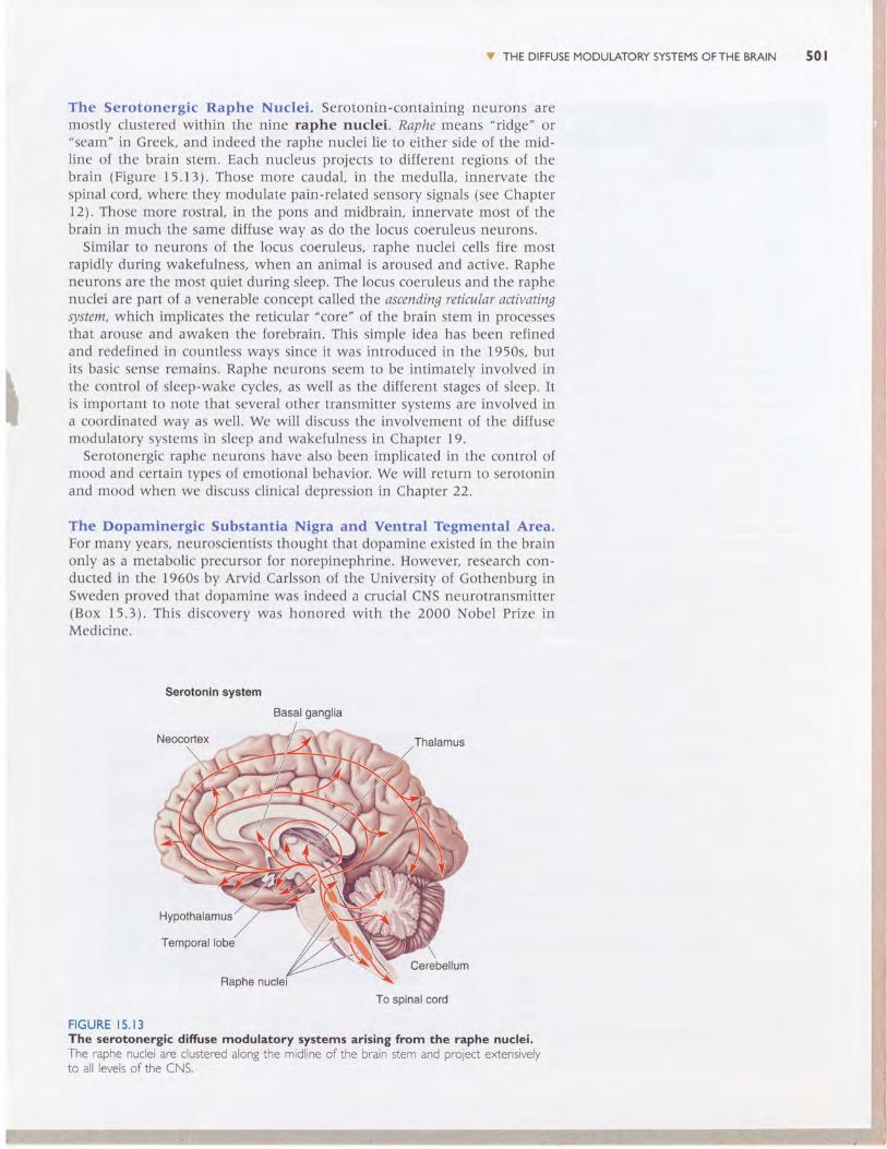
The Serotonergic Raphe Nuclei. Serotonin-containing neurons aremostly clustered within the nine raphe nuclei. Raphe means "ridge" or"seam" in Greek, and indeed the raphe nuclei lie to either side of the mid-line of the brain stem. Each nucleus projects to different regions of thebrain (Figure 15.13). Those more caudal, in the medulla, innervate thespinal cord, where they modulate pain-related sensory signals (see Chaptert2). Those more rostral, in the pons and midbrain, innervate most of thebrain in much the same diffuse way as do the locus coeruleus neurons.
Similar to neurons of the locus coeruleus, raphe nuclei cells fire mostrapidly during wakefulness, when an animal is aroused and active. Rapheneurons are the most quiet during sleep. The locus coeruleus and the raphenuclei are part of a venerable concept called the ascending reticular activatingsystem, which implicates the reticular "core" of the brain stem in processesthat arouse and awaken the forebrain. This simple idea has been refinedand redefined in countless ways since it was introduced in the 1950s, butits basic sense remains. Raphe neurons seem to be intimately involved inthe control of sleep-wake cycles, as well as the different stages of sleep. Itis important to note that several other transmitter systems are involved ina coordinated way as well. We will discuss the involvement of the diffusemodulatory systems in sleep and wakefulness in Chapter 19.
Serotonergic raphe neurons have also been implicated in the control ofmood and certain types of emotional behavior. We will return to serotoninand mood when we discuss clinical depression in Chapter 22.
The Dopaminergic Substantia Nigra and Ventral Tegmental Area.For many years, neuroscientists thought that dopamine existed in the brainonly as a metabolic precursor for norepinephrine. However, research con-ducted in the 1960s by Arvid Carlsson of the University of Gothenburg inSweden proved that dopamine was indeed a crucial CNS neurotransmitter(Box 15.3). This discoverv was honored with the 2000 Nobel Prize inMedicine.
Serotonln system
FIGURE I5 . I 3The serotonergic difruse modulatory systems arising from the raphe nuclei.The raphe nuclei are clustered alongthe midline of the brain stem and pro1ect extensivelyto all levels of the CNS.
Basal ganglia
502 C H A P T E R I 5 . C H E M I C A L C O N T R O L O F T H E B R A I N A N D B E H A V I O R
Awakeningto Dopamine
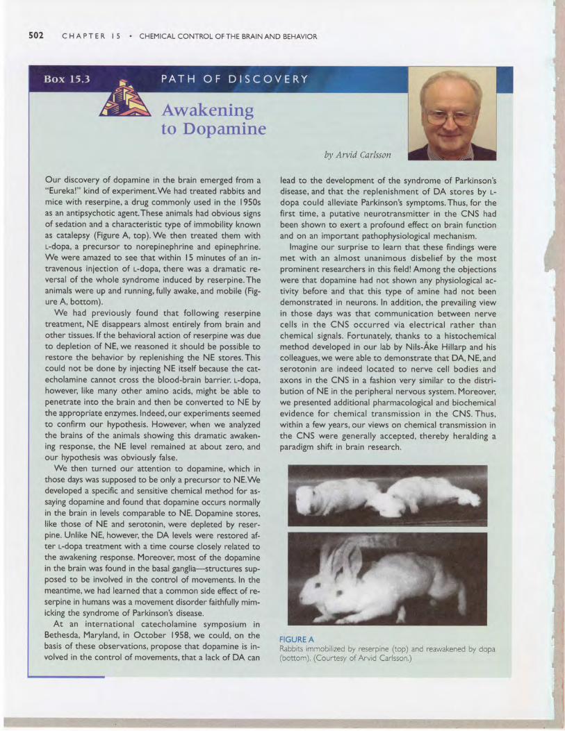
Our discovery of dopamine in the brain emerged from a"Eureka!" kind of experiment.We had treated rabbits andmice with reserpine, a drug commonly used in the 1950sas an antipsychotic agent.These animals had obvious signsof sedation and a characteristic type of immobility knownas catalepsy (Figure A, top). We then treated them withl-dopa, a precursor to norepinephrine and epinephrine.We were amazed to see that within l5 minutes of an in-travenous injection of r--dopa, there was a dramatic re-versal of the whole syndrome induced by reserpine.Theanimals were up and running, fully awake, and mobile (Fig-ure A, bottom).
We had previously found that following reserpinetreatment, NE disappears almost entirely from brain andother tissues. lf the behavioral action of reserpine was dueto depletion of NE, we reasoned it should be possible torestore the behavior by replenishing the NE stores.Thiscould not be done by injecting NE itself because the cat-echolamine cannot cross the blood-brain barrier. r--dopa,however, l ike many other amino acids, might be able topenetrate into the brain and then be converted to NE bythe appropriate enzymes. lndeed, our experiments seemedto confirm our hypothesis. However, when we analyzedthe brains of the animals showing this dramatic awaken-ing response, the NE level remained at about zero, andour hypothesis was obviously false.
We then turned our attention to dopamine, which inthose days was supposed to be only a precursor to NE.Wedeveloped a specific and sensitive chemical method for as-saying dopamine and found that dopamine occurs normallyin the brain in levels comparable to NE. Dopamine stores,like those of NE and serotonin, were depleted by reser-pine. Unlike NE, however, the DA levels were restored af-ter l-dopa treatment with a time course closely related tothe awakening response. Moreover, most of the dopaminein the brain was found in the basal ganglia-structures sup-oosed to be involved in the control of movements. ln themeantime, we had learned that a common side effect of re-serpine in humans was a movement disorder faithfully mim-icking the syndrome of Parkinson's disease.
At an in ternat ional catecholamine symposium inBethesda, Maryland, in October 1958, we could, on thebasis of these observations, propose that dopamine is in-volved in the control of movements. that a lack of DA can
by Arvid Carlsson
lead to the development of the syndrome of Parkinson'sdisease, and that the replenishment of DA stores by r--dopa could alleviate Parkinson's symptoms.Thus, for thefirst t ime, a putative neurotransmitter in the CNS hadbeen shown to exert a profound effect on brain functionand on an important pathophysiological mechanism.
lmagine our surprise to learn that these findings weremet wi th an a lmost unanimous d isbel ie f by the mostprominent researchers in this field! Among the objectionswere that dopamine had not shown any physiological ac-tivity before and that this type of amine had not beendemonstrated in neurons. In addition, the prevail ing viewin those days was that communication between nervecel ls in the CNS occurred v ia e lect r ica l rather thanchemical signals. Fortunately, thanks to a histochemicalmethod developed in our lab by Nils-Ake Hil larp and hiscolleagues, we were able to demonstrate that DA, NE, andserotonin are indeed located to nerve cell bodies andaxons in the CNS in a fashion very similar to the distri-bution of NE in the peripheral nervous system. Moreover,we presented additional pharmacological and biochemicalev idence for chemical t ransmiss ion in the CNS. Thus,within a few years, our views on chemical transmission inthe CNS were Senerally accepted, thereby heralding aparadigm shift in brain research.
FIGURE ARabbits rmmobilized by reserptne (top) and reawakened by dopa(botton ). (Courtesy of Arvid Carlsson.)

V THE DIFFUSE MODULATORY SYSTEMS OF THE BMIN 503
I
Dopamlne system
FIGURE I5 . I4The dopaminergic difruse modulatory systems arising from the substantianigra and the ventral tegmental area. The substantia nigra and ventral tegmental arealie close together in the midbrain,They prolect to the striatum (caudate nucleus andputamen) and limbic and frontal cortical regions, respectively,
Although there are dopamine-containing neurons scattered throughoutthe CNS, including some in the retina, the olfactory bulb, and the periven-tricular hypothalamus, two closely related groups of dopaminergic cellshave the characteristics of the diffuse modulatory systems (Figure 15.14).One of these arises in the substantia nigra in the midbrain. Recall fromChapter 14 that these cells project axons to the striatum (the caudate nu-cleus and the putamen), where they somehow facilitate the initiation ofvoluntary movements. Degeneration of the dopamine-containing cells inthe substantia nigra is all that is necessary to produce the progressive,dreadful motor disorders of Parkinson's disease. Although we do not en-tirely understand the function of DA in motor control, in general it facili-tates the initiation of motor responses by environmental stimuli.
The midbrain is also the origin of the other dopaminergic modulatorysystem, a group of cells that lie very close to the substantia nigra, in theventral tegmental area. Axons from these neurons innervate a circumscribedregion of the telencephalon that includes the frontal cortex and parts of thelimbic system. (The limbic system will be discussed in Chapter lS.) Thisdopaminergic projection from the midbrain is sometimes called Ihe meso-corticolimbic dopamine system. A number of different functions have been as-cribed to this complicated projection. For example, evidence indicates thatit is involved in a "reward" system that somehow assigns value to, or rein-forces, certain behaviors that are adaptive (see Chapter 16). We will see inChapter 18 that if rats (or humans) are given a chance to do so, they willwork to electrically stimulate this pathway. In addition, this projection hasbeen implicated in psychiatric disorders, as we will discuss in Chapter 22.
The Cholinergic Basal Forebrain and Brain Stem Complexes. Acetyl-choline is the familiar transmitter at the neuromuscular junction, at synapsesin autonomic ganglia, and at postganglionic parasympathetic synapses.Cholinergic interneurons also exist within the brain-in the striatum andthe cortex, for example. In addition, there are two major diffuse modulatorycholinergic systems in the brain, one of which is called the basal forebrain

504 C H A P T E R I 5 . C H E M I C A L C O N T R O L O F T H E B M I N A N D B E H A V I O R
FIGURE I5 . I5The cholinergic difruse modulatory systems arising from the basal forebrainand brain stem. The medial septal nuclei and basal nucleus of Meynert project widelyupon the cerebral cortex, including the hippocampus.The pontomesencephalotegmentalcomplex projects to the thalamus and parts of the forebrain.
complex. It is a "complex" because the cholinergic neurons lie scatteredamong several related nuclei at the core of the telencephalon, medial andventral to the basal ganglia. The best known of these are the medial septalnuclei, which provide the cholinergic innervation of the hippocampus, andthe basal nucleus of Meynert, which provides most of the cholinergic inner-vation of the neocortex.
The function of the cells in the basal forebrain complex remains mostlyunknown. But interest in this region has been fueled by the discovery thatthese are among the first cells to die during the course of Alzheimer's dis-ease, which is characterized by a progressive and profound loss of cognitivefunctions. (However, there is widespread neuronal death in Alzheimer'sdisease, and no specific link between the disease and cholinergic neuronshas been established.) Like the noradrenergic and serotonergic systems, thecholinergic system has been implicated in regulating general brain ex-citability during arousal and sleep-wake cycles. The basal forebrain complexmay also play a special role in learning and memory formation.
The second diffuse cholinergic system is called t}re pontomesencephalotegmen-tal complex. These are ACh-utilizing cells in the pons and midbrain tegmen-tum. This system acts mainly on the dorsal thalamus, where, together withthe noradrenergic and serotonergic systems, it regulates the excitability ofthe sensory relay nuclei. These cells also project up to the telencephalon,providing a cholinergic link between the brain stem and basal forebraincomplexes. Figure I5.I5 shows the cholinergic systems.
Drugs and the Difruse Modulatory Systems
Psychoactive drugs, compounds with "mind-altering" effects, all act on theCNS, and most do so by interfering with chemical synaptic transmission.Many abused drugs act directly on the modulatory systems, particularly thenoradrenergic, dopaminergic, and serotonergic systems.
V THE DIFFUSE MODUTATORY SYSTEMS OFTHE BRAIN
Hallucinogens. The use of hallucinogens, drugs that produce hallucinations,goes back thousands of years. Hallucinogenic compounds are contained ina number oI plants consumed as part of religious ritual, for example, thePsiloqtbe mushroom by the Maya and the peyote cactus by the Aztec. Themodern era of hallucinogenic drug use was unwittingly ushered in at thelaboratory of Swiss chemist Albert Hofmann. In 1938, Hofmann chemicallysynthesized a new compound, lysergic acid diethylamide (LSD). For 5 years,the LSD sat on the shelf. Then one day in 1943, Hofmann accidentallyingested some of the powder. His report on the effects attracted the imme-diate interest of the medical community. Psychiatrists began to use LSD inattempts to unlock the subconscious of mentally disturbed patients. Later,the drug was discovered by intellectuals, artists, students, and the U.S. De-fense Department, which investigated its "mind-expanding" effects. (Achief advocate of LSD use was former Harvard psychologist Timothy Leary.)In the I960s, LSD made its way to the street and was widely abused. To-day, the possession of LSD is illegal.
LSD is extremely potent. A dose sufficient to produce a full-blown hal-lucinogenic effect is only 25 micrograms (compared to a normal dose of as-pirin at 650 milligrams, which is 25,000 times larger). Among the reportedbehavioral effects of LSD is a dreamlike state with heightened awareness ofsensory stimuli, often with a mixing of perceptions such that sounds canevoke images, images can evoke smells, and so on.
The chemical structure of LSD (and the active ingredients of. Psilocybemushrooms and peyote) is very close to that of serotonin, suggesting thatit acts on the serotonergic system. Indeed, LSD is a potent agonist at theserotonin receptors on the presynaptic terminals of neurons in the raphenuclei. Activation of these receptors markedly inhibits the firing of rapheneurons. Thus, one known CNS effect of LSD is a reduction in the outflowof the brain's serotonergic diffuse modulatory system. It is interesting tonote in this regard that decreased activity of the raphe nuclei is also char-acteristic of dream-sleep (see Chapter I9).
Can we conclude that LSD produces hallucinations by silencing the brain'sserotonin systems? If only drug effects on the brain were that simple. Un-fortunately, there are problems with this hypothesis. For one, silencingneurons in the raphe nuclei by other means-by destroying them, for ex-ample-does not mimic the effects of LSD in experimental animals. Fur-thermore, animals still respond as expected to LSD after their raphe nucleihave been destroyed.
In recent years, researchers have focused on direct LSD actions at serotoninreceptors in the cerebral cortex. Current research suggests that LSD causeshallucinations by superseding the naturally modulated release of serotoninin cortical areas where perceptions normally are formed and interpreted.
Stimulants. In contrast to the uncertainties about hallucinogens andserotonin, it is clear that the powerful CNS stimulants cocaine and amphet-amine both exert their effects at synapses made by dopaminergic and nora-drenergic systems. Both drugs give users a feeling of increased alertnessand self-confidence, a sense of exhilaration and euphoria, and a decreasedappetite. Both are also sympathomimetic-they cause peripheral effectsthat mimic activation of the sympathetic division of the ANS: increasedheart rate and blood pressure, dilation of the pupils, and so on.
Cocaine is extracted from the leaves of the coca plant and has been usedby Andean Indians for hundreds of years. In the mid-nineteenth century,cocaine turned up in Europe and North America as the magic ingredientin a wide range of concoctions touted by their salesmen as having medici-nal value. (An example is Coca-Cola, originally marketed in 1886 as a
505
I
s06 CHAPTER I 5 . CHEMICALCONTROLOFTHE BRAINAND BEHAVIOR
therapeutic agent, which contained both cocaine and caffeine.) Cocaine usefell out of favor early in the twentieth century only to reemerge with avengeance in the late 1960s as a recreational drug. Ironically, one of themain reasons for the rise in cocaine use during this period was the tight-ening of regulations against amphetamines. First chemically synthesized in1887, amphetamines did not come into wide use until World War II, whenthey were taken by soldiers of both sides (particularly aviators) to sustainthem in combat. Following the war, amphetamines became available asnonprescription diet aids, as nasal decongestants, and as "pep-pills." Regu-lations were finally tightened after recognition that amphetamines are, likecocaine, highly addictive and dangerous in large doses.
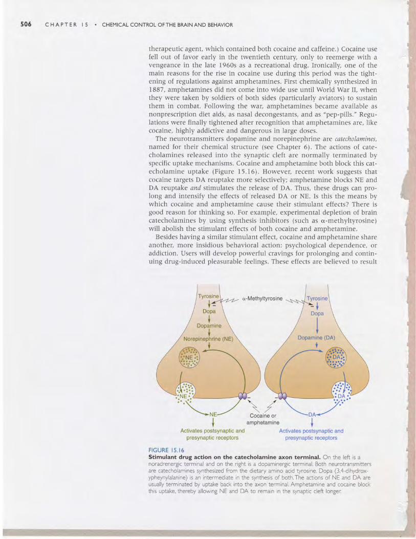
The neurotransmitters dopamine and norepinephrine are catecholamines,named for their chemical structure (see Chapter 6). The actions of cate-cholamines released into the synaptic cleft are normally terminated byspecific uptake mechanisms. Cocaine and amphetamine both block this cat-echolamine uptake (Figure 15.16). However, recent work suggests thatcocaine targets DA reuptake more selectively; amphetamine blocks NE andDA reuptake and stimulates the release of DA. Thus, these drugs can pro-long and intensify the effects of released DA or NE. Is this the means bywhich cocaine and amphetamine cause their stimulant effects? There isgood reason for thinking so. For example, experimental depletion of braincatecholamines by using synthesis inhibitors (such as o-methyltyrosine)will abolish the stimulant effects of both cocaine and amphetamine.
Besides having a similar stimulant effect, cocaine and amphetamine shareanother, more insidious behavioral action: psychological dependence, oraddiction. Users will develop powerful cravings for prolonging and contin-uing drug-induced pleasurable feelings. These effects are believed to result
a-Methyltyrosine
\ ,t/
\ ZCocaine or
amphetamin€
FIGURE I5.16Stlmulant drug actlon on the catecholamlne axon termlnal. On the left is anoradrenergic terminal and on the right is a dopaminergic terminal. Both neurotransmittersare catecholamines synthesized from the dietary amino acid tyrosine. Dopa (3,4-dihydrox-ypheynylalanine) is an intermediate in the synthesis of both.The actions of NE and DA areusually terminated by uptake back into the axon terminal. Amphetamine and cocaine blockthis uptake, thereby allowing NE and DA to remain in the synaptic cleft longer:
Activates postsynaptic andpresynaptic r€ceptors
Activates postsynaptic andpresynaptic receptors

speci f ica l ly f rom the enhanced t ransmiss ion in the mesocort ico l imbicdopamine system during drug use. Remember, this system may normallyfunction to reinforce adaptive behaviors. By short-circuiting the system,these drugs instead reinforce drug-seeking behavior. Indeed, just as rats wii lwork to electrically stimulate the mesocorticolimbic projection, they wil lalso work to receive an injection of cocaine. We'l l discuss the involvementof dopamine pathways in motivation and addiction further in Chapter 16.
,,,f ' CONCLUDING REMARKS
In th is chapter , we have examined three components of the nervous sys-tem that are characterized by the great reach of their influences. The se-cretory hypothalamus and autonomic nervous system communicate wi thcel ls a l l over the body, and the d i f fuse modulatory systems communicatewi th neurons in many d i f ferent par ts of the bra in. They are a lso character-ized by the duration of their direct effects, which can range fmm mir.rutesto hours. F inal ly , they are character ized by thei r chemical neurotransmit -ters. In many instances, the transmitter defines the system. For example, inthe per iphery, we can use the words "noradrenergic" and "sympathet ic" in-terchangeably. The same th ing goes for " raphe" and "serotonin" in the fore-bra in, and "substant ia n igra" and "dopamine" in the basal gangl ia . Thesechemical idiosyncrasies have allowed interpretations of drug effects on be-havior that are not possib le wi th most other neural systems. Thus, we havea good idea where in the bra in amphetamine and cocaine exer t thei r s t in t -ulant effects, and where in the periphery they act to raise blood pressureand heart rate.
At a deta i led level , each of the systems discussed in th is chapter per furntsdifferent functions. But at a general level, they all maintain brain homeosta-sri: They regulate different processes within a certain physiological range.For example, the ANS regulates b lood pressure wi th in a range that is ap-propr iate. Blood pressure var iat ions opt imize an animal 's per formance un-der different conditions. In a similar way, the noradrenergic krcus coeruleusand serotonergic raphe nucle i regulate levels of consciousness and mood.These levels a lso vary wi th in a range that is adapt ive to the organism. Inthe next several chapters, we wi l l encounter these systems again in thecontext of speci f ic funct ions.
The Secretory Hypothalamushomeostasis (p. a8a)periventricular zone (p. a8a)magnocellular neurosecretory cell
(p. a8s)neurohormone (p.485)oxytocin (p.485)vasopressin $.486)antidiuretic hormone (ADH)
(p.486)parvocellular neurosecretory cell
(p.488)hypophysiotropic hormone
(p. 488)
. . l , , t , i l
> v )U J :
Y ellJ
F
hypothalamo-pitu itary portalcirculation (p. 488)
adrenal cortex (p.488)adrenal medulla (p. 488)coftisol (p.488)
The Autonomic NervousSystemautonomic nervous system (ANS)
(p. aeo)sympathetic division (p. 490)parasympathetic division (P. 490)autonomic ganglia $. 492)postganglionic neuron @. 492)
CONCLUDING REMARKS 507
preganglionic neuron (p. 492)sympathetic chain (p. 494)enteric division (p. 495)nucleus of the solitary tract
$. ae6)
The Diffuse ModulatorySystems of the Braindiffuse modulatory system
(p. ae8)locus coeruleus (p. 498)raphe nuclei (p.501)basal forebrain complex (p.503)
,r/| il'1
-|-!v f
l 2- oU J Fd, rtl
urf,o
508 CHAPTER I5 . CHEMICALCONTROLOFTHE BMINAND BEHAVIOR
l. Battlefield trauma victims who hare lost large volumes of blood often exprcss a craving to drink water.Why?
2. You've sayed up all night tryrnt to meet a term paper deadline.You now are typing frantically, keeping oneeye on the paper and dre other on ttre clock How has the periventricular zone of the hypothalamus or-chestrated your bod's physiological response to this stressful situationl Describe in deail.
3.Wly is the adrenal medulla often referred to as a modified sympathetic ganglion? Why isnt the adrenalcort€r( included in this descriptionl
4. A number of famous athletes and enterainers have accidenally killed themselves by aking large quantitiesof cocaine. Usually, tlre cause of death is heart failure. How would you explain the peripheral actions ofcocainel
5. How do the diftrse modulatory and point-to-point synaptic communication systems in the brain difierl Listfour ways.
6. Under what behavioral conditions are the noradrenergic neunons of the locus coeruleus activel The nora-drenergic neurons of the ANSI