Embed Size (px)
Citation preview
Chemical Biology 03BLOOD
Biomolecular StructureMyoglobin and Hemoglobin
9/28-30/09
www.optics.rochester.edu/.../image007.gif
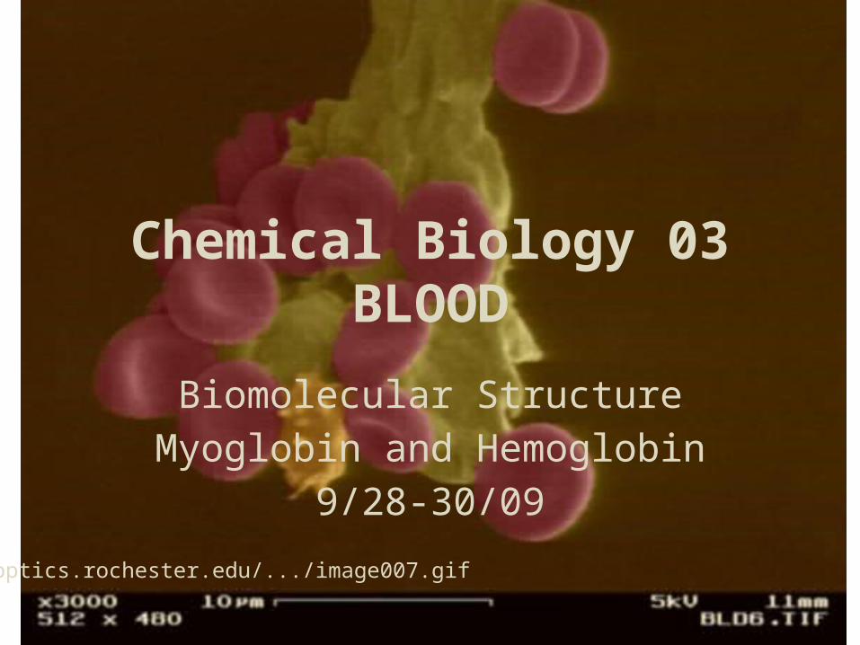
Chemical Biology 03BLOOD
Biomolecular Structure Myoglobin and Hemoglobin
Lecture 9 and 10: 9/28-30/09
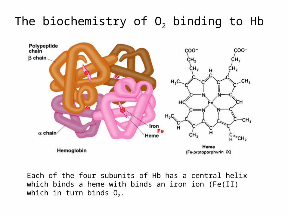
The biochemistry of O2 binding to Hb
Each of the four subunits of Hb has a central helix which binds a heme with binds an iron ion (Fe(II) which in turn binds O2.
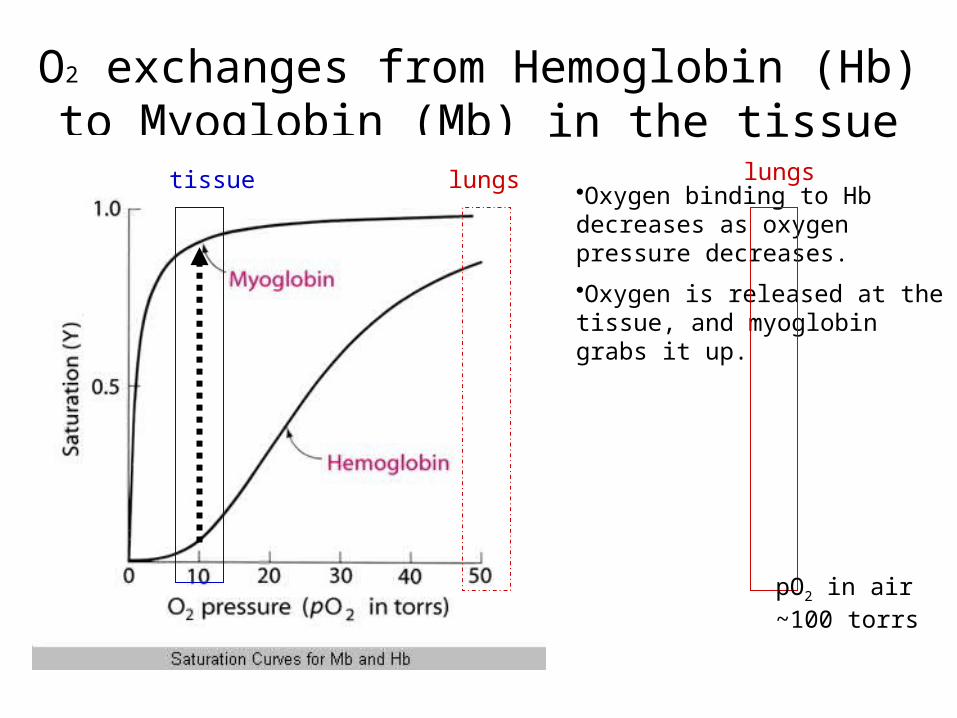
O2 exchanges from Hemoglobin (Hb) to Myoglobin (Mb) in the tissue
•Oxygen binding to Hb decreases as oxygen pressure decreases.
•Oxygen is released at the tissue, and myoglobin grabs it up.
tissue lungs lungs
pO2 in air ~100 torrs
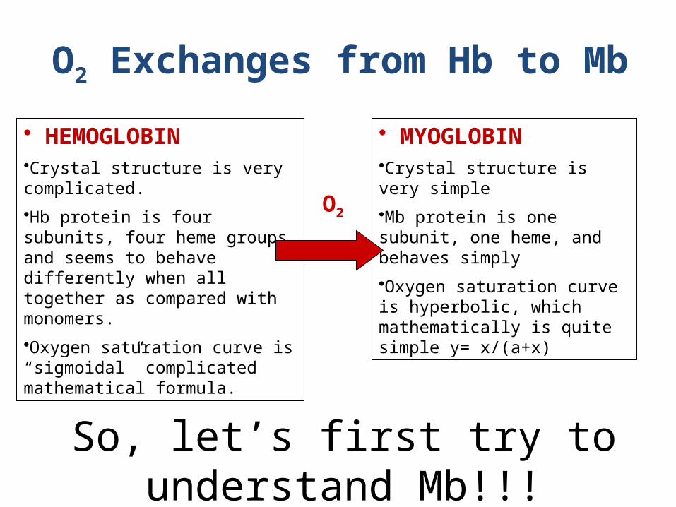
So, let’s first try to understand Mb!!!
• HEMOGLOBIN•Crystal structure is very complicated.
•Hb protein is four subunits, four heme groups, and seems to behave differently when all together as compared with monomers.
•Oxygen saturation curve is “sigmoidal” complicated mathematical formula.
• MYOGLOBIN•Crystal structure is very simple
•Mb protein is one subunit, one heme, and behaves simply
•Oxygen saturation curve is hyperbolic, which mathematically is quite simple y= x/(a+x)
O2 Exchanges from Hb to Mb
O2
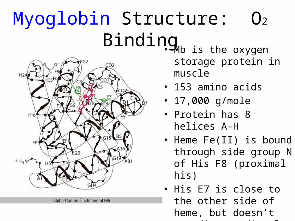
Myoglobin Structure: O2 Binding• Mb is the oxygen storage
protein in muscle• 153 amino acids • 17,000 g/mole • Protein has 8 helices A-H• Heme Fe(II) is bound through
side group N of His F8 (proximal his)
• His E7 is close to the other side of heme, but doesn’t coordinate (distal his)
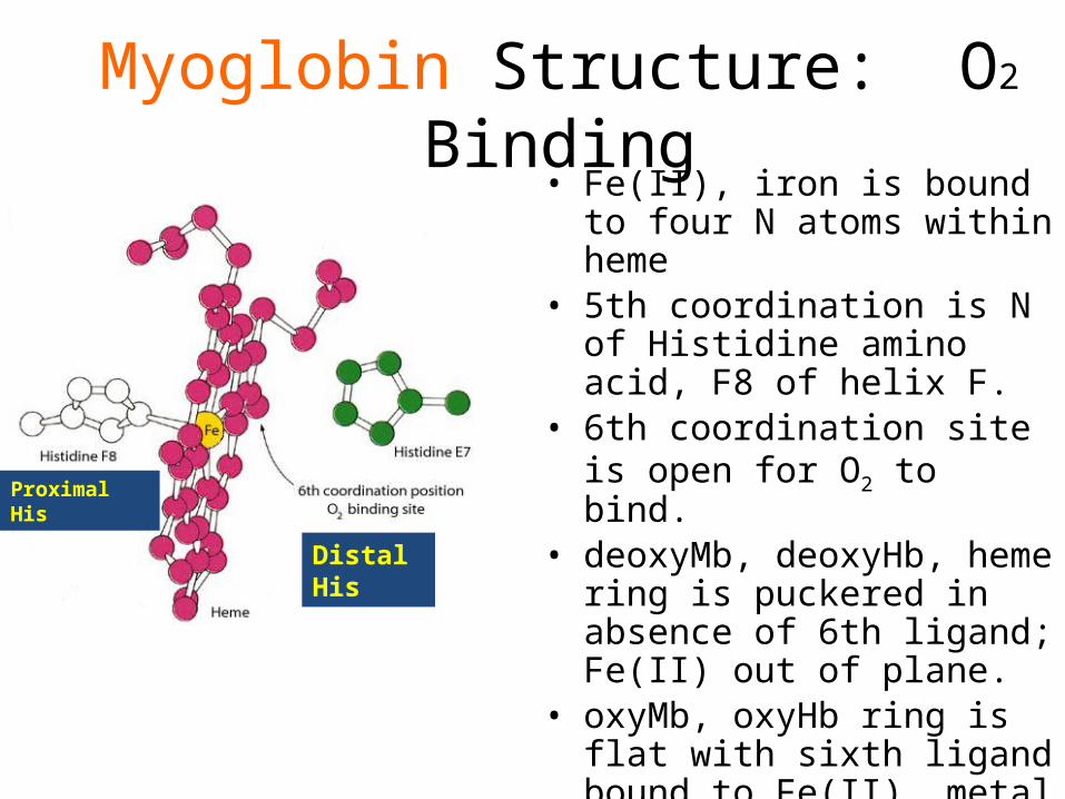
Myoglobin Structure: O2 Binding• Fe(II), iron is bound to four N
atoms within heme• 5th coordination is N of
Histidine amino acid, F8 of helix F.
• 6th coordination site is open for O2 to bind.
• deoxyMb, deoxyHb, heme ring is puckered in absence of 6th ligand; Fe(II) out of plane.
• oxyMb, oxyHb ring is flat with sixth ligand bound to Fe(II), metal is in heme plane.
Proximal His
Distal His
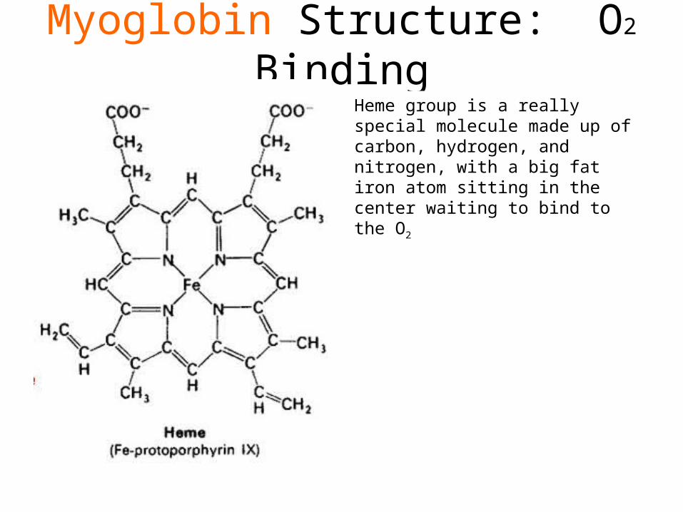
Myoglobin Structure: O2 BindingHeme group is a really special molecule made up of carbon, hydrogen, and nitrogen, with a big fat iron atom sitting in the center waiting to bind to the O2
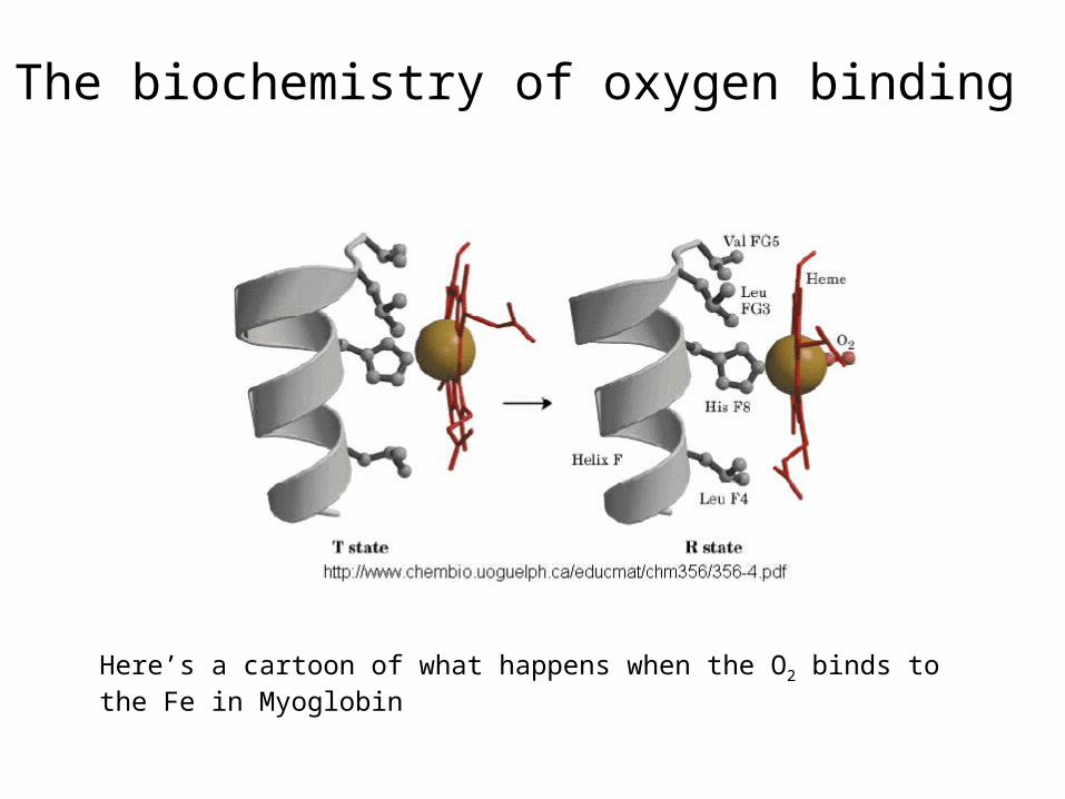
The biochemistry of oxygen binding
Here’s a cartoon of what happens when the O2 binds to the Fe in Myoglobin
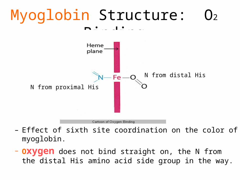
Myoglobin Structure: O2 Binding
– Effect of sixth site coordination on the color of myoglobin.
– Oxygen does not bind straight on, the N from the distal His amino acid side group in the way.
N from proximal His
N from distal His
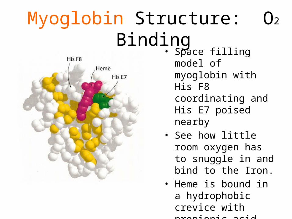
Myoglobin Structure: O2 Binding• Space filling model of
myoglobin with His F8 coordinating and His E7 poised nearby
• See how little room oxygen has to snuggle in and bind to the Iron.
• Heme is bound in a hydrophobic crevice with propionic acid groups projecting into solution orienting the heme.
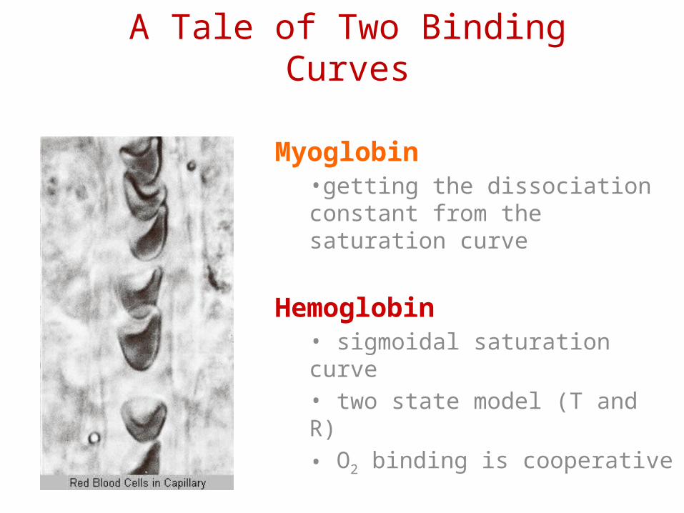
A Tale of Two Binding Curves
Myoglobin •getting the dissociation constant from the saturation curve
Hemoglobin• sigmoidal saturation curve• two state model (T and R)• O2 binding is cooperative
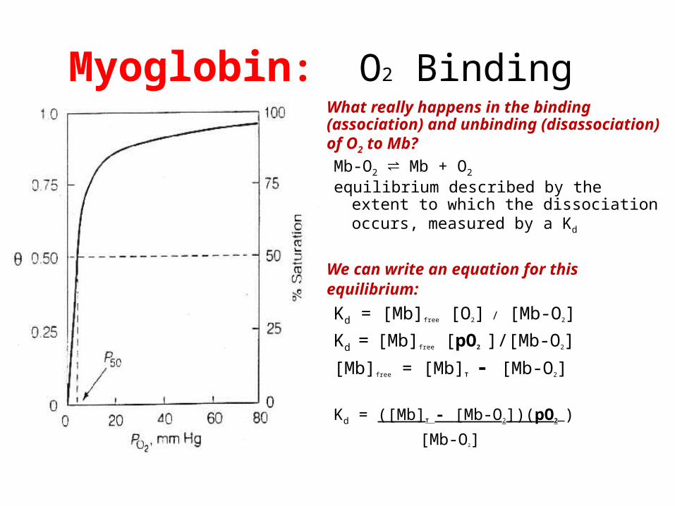
Myoglobin: O2 Binding• What really happens in the binding
(association) and unbinding (disassociation) of O2 to Mb?
Mb-O2 ⇌ Mb + O2
equilibrium described by the extent to which the dissociation occurs, measured by a Kd
• We can write an equation for this equilibrium: Kd = [Mb]free [O2] / [Mb-O2]
Kd = [Mb]free [pO2 ]/[Mb-O2]
[Mb]free = [Mb]T - [Mb-O2]
Kd = ([Mb]T - [Mb-O2])(pO2 )
[Mb-O2]
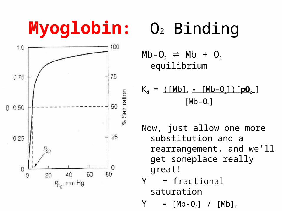
Myoglobin: O2 Binding
Mb-O2 ⇌ Mb + O2 equilibrium
Kd = ([Mb]T - [Mb-O2])[pO2 ]
[Mb-O2]
Now, just allow one more substitution and a rearrangement, and we’ll get someplace really great!
Y = fractional saturation Y = [Mb-O2] / [Mb]T
Y = pO2 / (Kd + pO2)
WOW, that’s a lot simpler!

Let’s EXCEL together

Conclusions from excel about O2 binding to Mb
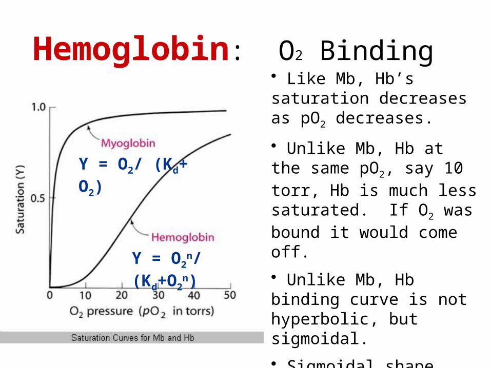
Hemoglobin: O2 Binding• Like Mb, Hb’s saturation decreases as pO2 decreases.
• Unlike Mb, Hb at the same pO2, say 10 torr, Hb is much less saturated. If O2 was bound it would come off.
• Unlike Mb, Hb binding curve is not hyperbolic, but sigmoidal.
• Sigmoidal shape suggest two states for Hb (more on this in a minute)
Y = O2n/ (Kd+O2
n)
Y = O2/ (Kd+ O2)

Let’s EXCEL together
Conclusions from excel about O2 binding to Hb
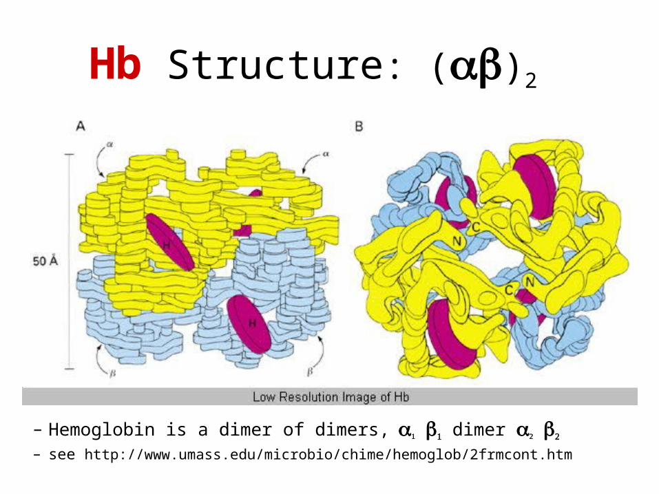
Hb Structure: (ab)2
– Hemoglobin is a dimer of dimers, a1 b1 dimer a2 b2
– see http://www.umass.edu/microbio/chime/hemoglob/2frmcont.htm
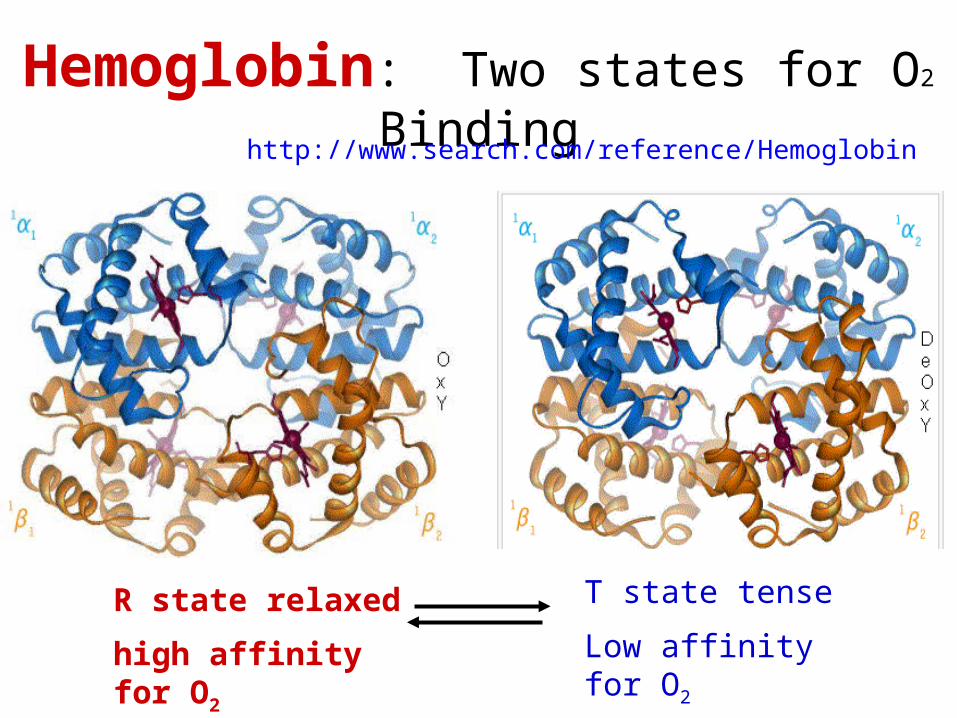
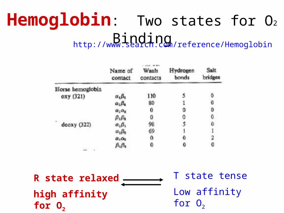
Hemoglobin: Two states for O2 Binding
R state relaxed
high affinity for O2
T state tense
Low affinity for O2
http://www.search.com/reference/Hemoglobin
Hemoglobin: Two states for O2 Binding
R state relaxed
high affinity for O2
T state tense
Low affinity for O2
http://www.search.com/reference/Hemoglobin
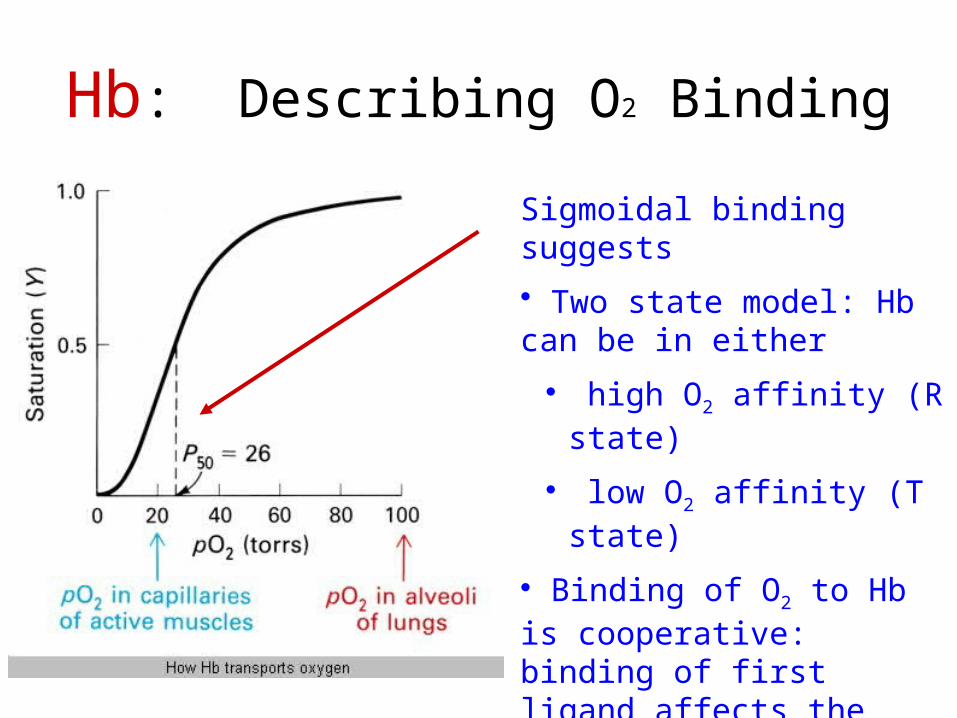
Hb: Describing O2 Binding
Sigmoidal binding suggests
• Two state model: Hb can be in either
• high O2 affinity (R state)
• low O2 affinity (T state)
• Binding of O2 to Hb is cooperative: binding of first ligand affects the affinity of the remaining sites for ligand.
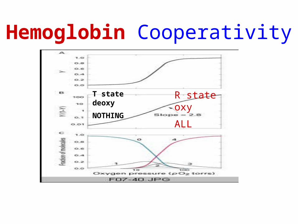
Hemoglobin Cooperativity
R state oxy
ALL
T state deoxy
NOTHING
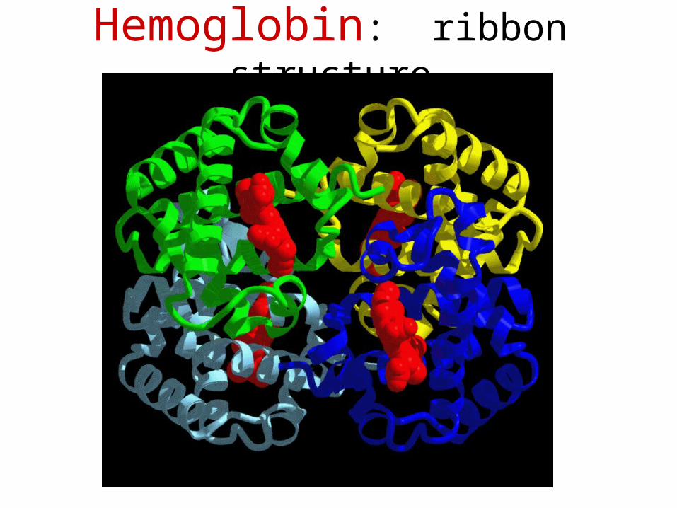
Hemoglobin: ribbon structure