Embed Size (px)
Citation preview

© 2015 Pearson Education, Inc.
PowerPoint Lectures Campbell Biology: Concepts & Connections, Eighth Edition REECE • TAYLOR • SIMON • DICKEY • HOGAN
Chapter 10
Lecture by Edward J. Zalisko
Molecular Biology of the Gene

© 2015 Pearson Education, Inc.
Introduction
• The 2009 H1N1 influenza virus • spread so quickly that it was declared a pandemic, • reached 207 countries, • infected more than 600,000 people, and • killed an estimated 20,000 people.
• Viruses share some of the characteristics of living organisms, but are generally not considered alive because they are not cellular and cannot reproduce on their own.

© 2015 Pearson Education, Inc.
Introduction
• Combating any virus requires a detailed understanding of
• molecular biology, • the study of DNA, and • how DNA serves as the basis of heredity.

© 2015 Pearson Education, Inc.
Figure 10.0-1

© 2015 Pearson Education, Inc.
Figure 10.0-2 Chapter 10: Big Ideas
The Structure of the Genetic Material
DNA Replication
The Genetics of Viruses and Bacteria
The Flow of Genetic Information from DNA
to RNA to Protein

© 2015 Pearson Education, Inc.
THE STRUCTURE OF THE GENETIC MATERIAL

© 2015 Pearson Education, Inc.
10.1 SCIENTIFIC THINKING: Experiments showed that DNA is the genetic material
• Early in the 20th century, the molecular basis for inheritance was a mystery.
• Biologists did know that genes were located on chromosomes. But it was unknown if the genetic material was
• proteins or • DNA.

© 2015 Pearson Education, Inc.
10.1 SCIENTIFIC THINKING: Experiments showed that DNA is the genetic material
• Biologists finally established the role of DNA in heredity through experiments with bacteria and the viruses that infect them.
• This breakthrough ushered in the field of molecular biology, the study of heredity at the molecular level.

© 2015 Pearson Education, Inc.
10.1 SCIENTIFIC THINKING: Experiments showed that DNA is the genetic material
• In 1928, Frederick Griffith was surprised to find that when he killed pathogenic bacteria, then mixed the bacterial remains with living harmless bacteria, some living bacterial cells became pathogenic.
• All of the descendants of the transformed bacteria inherited the newly acquired ability to cause disease.

© 2015 Pearson Education, Inc.
10.1 SCIENTIFIC THINKING: Experiments showed that DNA is the genetic material
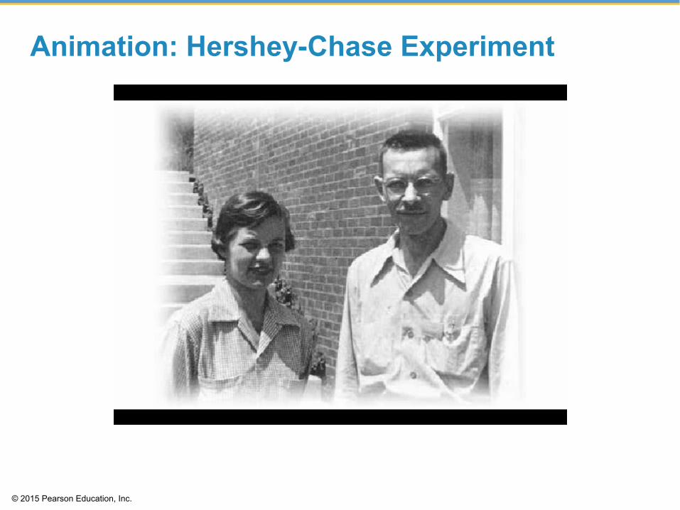
• In 1952, Alfred Hershey and Martha Chase used bacteriophages to show that DNA is the genetic material of T2, a virus that infects the bacterium Escherichia coli (E. coli).
• Bacteriophages (or phages for short) are viruses that infect bacterial cells.
• Phages were labeled with radioactive sulfur to detect proteins or radioactive phosphorus to detect DNA.
• Bacteria were infected with either type of labeled phage to determine which substance was injected into cells and which remained outside the infected cell.

© 2015 Pearson Education, Inc.
10.1 SCIENTIFIC THINKING: Experiments showed that DNA is the genetic material
• The sulfur-labeled protein stayed with the phages outside the bacterial cell, while the phosphorus-labeled DNA was detected inside cells.
• Cells with phosphorus-labeled DNA produced new bacteriophages with radioactivity in DNA but not in protein.
© 2015 Pearson Education, Inc.
Animation: Hershey-Chase Experiment

© 2015 Pearson Education, Inc.
Animation: Phage T2 Reproductive Cycle

© 2015 Pearson Education, Inc.
Figure 10.1a-0
Head DNA
Tail Tail fiber

© 2015 Pearson Education, Inc.
Figure 10.1a-1
Head
Tail Tail fiber

© 2015 Pearson Education, Inc.
Figure 10.1b-0
Phage Bacterium
Radioactive protein
DNA
Empty protein shell Phage DNA
Centrifuge
Pellet Batch 1: Radioactive protein labeled in yellow
Radioactive DNA
Centrifuge
Pellet The radioactivity is in the pellet.
The radioactivity is in the liquid.
Batch 2: Radioactive DNA labeled in green

© 2015 Pearson Education, Inc.
Figure 10.1b-1 Phage
Bacterium Radioactive protein
DNA
Empty protein shell Phage DNA
Batch 1: Radioactive protein labeled in yellow
Radioactive DNA
Batch 2: Radioactive DNA labeled in green

© 2015 Pearson Education, Inc.
10.1 SCIENTIFIC THINKING: Experiments showed that DNA is the genetic material
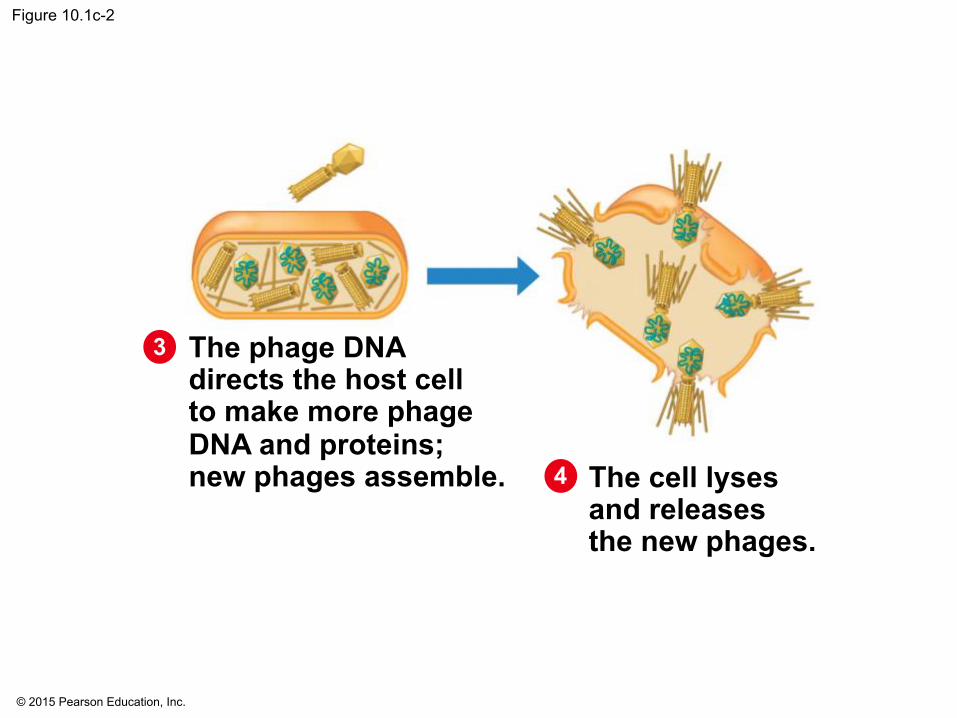
• Figure 10.1C outlines our current understanding—as originally formulated by Hershey and Chase—of the replication cycle of phage T2.

© 2015 Pearson Education, Inc.
Figure 10.1c-0
A phage attaches itself to a bacterial cell.
The phage injects its DNA into the bacterium.
The phage DNA directs the host cell to make more phage DNA and proteins; new phages assemble. The cell lyses
and releases the new phages.
1 2 3

© 2015 Pearson Education, Inc.
Figure 10.1c-1
A phage attaches itself to a bacterial cell.
The phage injects its DNA into the bacterium.
1 2
© 2015 Pearson Education, Inc.
Figure 10.1c-2
The phage DNA directs the host cell to make more phage DNA and proteins; new phages assemble. The cell lyses
and releases the new phages.
3
4

© 2015 Pearson Education, Inc.
10.2 DNA and RNA are polymers of nucleotides
• DNA and RNA are nucleic acids consisting of long chains (polymers) of chemical units (monomers) called nucleotides.
• One of the two strands of DNA is a DNA polynucleotide, a nucleotide polymer (chain).
• A nucleotide is composed of a • nitrogenous base, • five-carbon sugar, and • phosphate group.
• The nucleotides are joined to one another by a sugar-phosphate backbone.

© 2015 Pearson Education, Inc.
Figure 10.2a-0
Sugar-phosphate backbone
A T
A T
A T
T A
T A
T A
T A
C G
C G
C G
G C
G C
G A
C
T A DNA double helix
Covalent bond joining nucleotides
G
A
T
G
G G
C
Phosphate group Nitrogenous base Sugar
DNA nucleotide Phosphate
group
Sugar (deoxyribose)
DNA nucleotide
Thymine (T)
Nitrogenous base (can be A, G, C, or T)
Two representations of a DNA polynucleotide

© 2015 Pearson Education, Inc.
Figure 10.2a-1
A T
A T
A T
T A
T A
T A
T A
C G
C G
C G
G C
G C
G
A DNA double helix

© 2015 Pearson Education, Inc.
Figure 10.2a-2 Sugar-phosphate
backbone
Covalent bond joining nucleotides
A
T
G
G
C
Phosphate group Nitrogenous base Sugar
DNA nucleotide
Two representations of a DNA polynucleotide
A
T
G
G
C

© 2015 Pearson Education, Inc.
Figure 10.2a-3
Phosphate group
Sugar (deoxyribose)
DNA nucleotide
Thymine (T)
Nitrogenous base (can be A, G, C, or T)

© 2015 Pearson Education, Inc.
10.2 DNA and RNA are polymers of nucleotides
• Each type of DNA nucleotide has a different nitrogen-containing base:
• adenine (A), • cytosine (C), • thymine (T), and • guanine (G).

© 2015 Pearson Education, Inc.
Animation: DNA and RNA Structure

© 2015 Pearson Education, Inc.
Figure 10.2b-0
Thymine (T) Cytosine (C) Adenine (A) Guanine (G)
Pyrimidines Purines

© 2015 Pearson Education, Inc.
Figure 10.2b-1
Thymine (T) Cytosine (C)
Pyrimidines

© 2015 Pearson Education, Inc.
Figure 10.2b-2
Adenine (A) Guanine (G)
Purines

© 2015 Pearson Education, Inc.
10.2 DNA and RNA are polymers of nucleotides
• The full name for DNA is deoxyribonucleic acid, with nucleic referring to DNA’s location in the nuclei of eukaryotic cells.
• RNA (ribonucleic acid) is unlike DNA in that it • uses the sugar ribose (instead of deoxyribose in
DNA) and • has a nitrogenous base uracil (U) instead of
thymine.

© 2015 Pearson Education, Inc.
Figure 10.2c
Phosphate group
Sugar (ribose)
Uracil (U)
Nitrogenous base (can be A, G, C, or U)

© 2015 Pearson Education, Inc.
Figure 10.2d
Adenine Guanine
Ribose Phosphate
Uracil Cytosine

© 2015 Pearson Education, Inc.
10.3 DNA is a double-stranded helix
• After the 1952 Hershey-Chase experiment convinced most biologists that DNA was the material that stored genetic information, a race was on to determine how the structure of this molecule could account for its role in heredity.
• Researchers focused on discovering the three-dimensional shape of DNA.

© 2015 Pearson Education, Inc.
10.3 DNA is a double-stranded helix
• American James D. Watson journeyed to Cambridge University in England, where the more senior Francis Crick was studying protein structure with a technique called X-ray crystallography.
• While visiting the laboratory of Maurice Wilkins at King’s College in London, Watson saw an X-ray image of DNA produced by Wilkins’s colleague, Rosalind Franklin.

© 2015 Pearson Education, Inc.
Figure 10.3a-0

© 2015 Pearson Education, Inc.
Figure 10.3a-1

© 2015 Pearson Education, Inc.
Figure 10.3a-2
© 2015 Pearson Education, Inc.
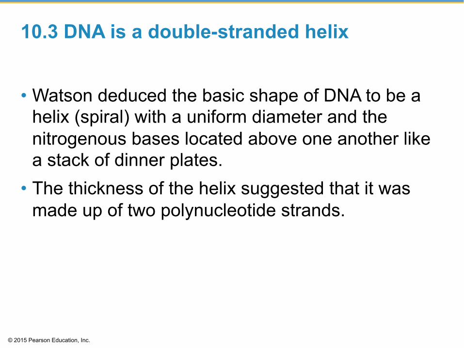
10.3 DNA is a double-stranded helix
• Watson deduced the basic shape of DNA to be a helix (spiral) with a uniform diameter and the nitrogenous bases located above one another like a stack of dinner plates.
• The thickness of the helix suggested that it was made up of two polynucleotide strands.

© 2015 Pearson Education, Inc.
10.3 DNA is a double-stranded helix
• Watson and Crick realized that DNA consisted of two polynucleotide strands wrapped into a double helix.
• The sugar-phosphate backbone is on the outside. • The nitrogenous bases are perpendicular to the
backbone in the interior. • Specific pairs of bases give the helix a uniform
shape. • A pairs with T, forming two hydrogen bonds, and • G pairs with C, forming three hydrogen bonds.

© 2015 Pearson Education, Inc.
Animation: DNA Double Helix

© 2015 Pearson Education, Inc.
Figure 10.3b

© 2015 Pearson Education, Inc.
Figure 10.3c
Twist

© 2015 Pearson Education, Inc.
Figure 10.3d-0
Ribbon model Partial chemical structure Computer model
Base pair
Hydrogen bond C G
C G G C
G C
C G
C G
C G G C
T A
T A
A T
A T
A T
C
A
T A T T A
G C
T
A
G

© 2015 Pearson Education, Inc.
Figure 10.3d-1
Ribbon model
Base pair
C G C G
G C G C
C G
C G
C G G C
T A
T A
A T
A T
A T
A
T A
T

© 2015 Pearson Education, Inc.
Figure 10.3d-2
Partial chemical structure
Hydrogen bond
C
A
T
G C
T
A
G

© 2015 Pearson Education, Inc.
Figure 10.3d-3
Computer model

© 2015 Pearson Education, Inc.
10.3 DNA is a double-stranded helix
• In 1962, the Nobel Prize was awarded to James D. Watson, Francis Crick, and Maurice Wilkins.
• Rosalind Franklin probably would have received the prize as well but for her death from cancer in 1958.
• Nobel Prizes are never awarded posthumously. • The Watson-Crick model gave new meaning to the
words genes and chromosomes. The genetic information in a chromosome is encoded in the nucleotide sequence of DNA.

© 2015 Pearson Education, Inc.
DNA REPLICATION

© 2015 Pearson Education, Inc.
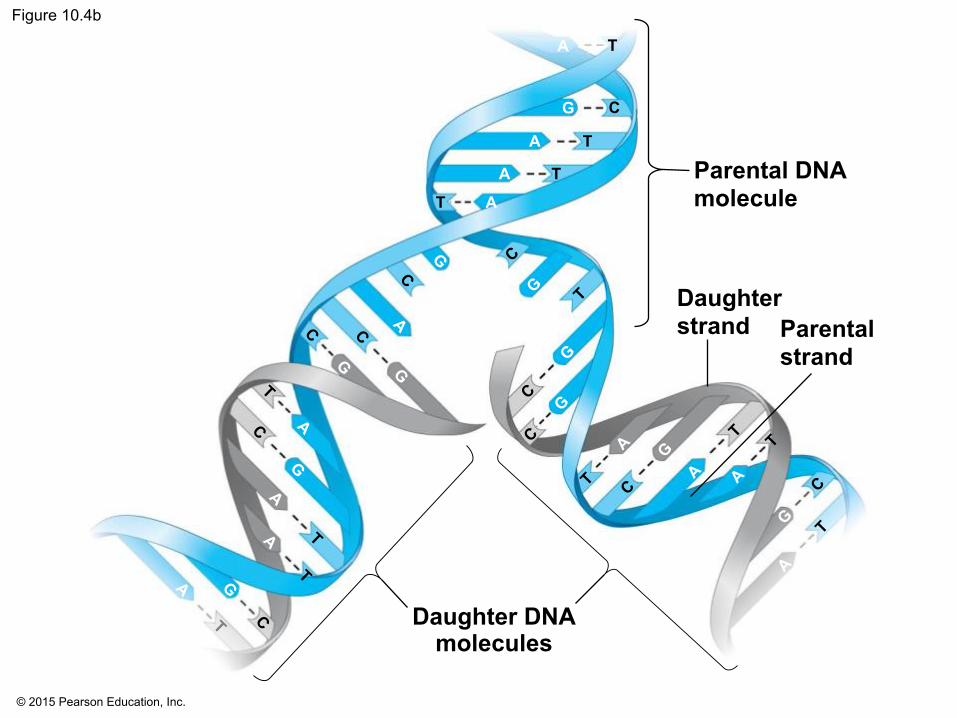
10.4 DNA replication depends on specific base pairing
• DNA replication follows a semiconservative model.
• The two DNA strands separate. • Each strand then becomes a template for the
assembly of a complementary strand from a supply of free nucleotides.
• Each new DNA helix has one old strand with one new strand.

© 2015 Pearson Education, Inc.
Animation: DNA Replication Overview

© 2015 Pearson Education, Inc.
Figure 10.4a-1
A parental molecule of DNA
A T
C G
A T
T A
G C

© 2015 Pearson Education, Inc.
Figure 10.4a-2
A parental molecule of DNA
A T
C G
A T
T A
G C
A
C
A
T
G
T
G
C
A
C
T
G
C
T
A Free nucleotides
The parental strands separate and serve as templates

© 2015 Pearson Education, Inc.
Figure 10.4a-3
A parental molecule of DNA
A T
C G
A T
T A
G C
A
C
A
T
G
T
G
C
A
C
T
G
C
T
A Free nucleotides
The parental strands separate and serve as templates
Two identical daughter molecules of DNA are formed
A T
C G
A T
T A
G C
A T
C G
A T
T A
G C
© 2015 Pearson Education, Inc.
Figure 10.4b
Parental DNA molecule
Daughter strand Parental
strand
Daughter DNA molecules
A T
G C
A
A
A
T
T
T

© 2015 Pearson Education, Inc.
10.5 DNA replication proceeds in two directions at many sites simultaneously
• Replication of a DNA molecule begins at particular sites called origins of replication, short stretches of DNA having a specific sequence of nucleotides.
• Proteins that initiate DNA replication • attach to the DNA at the origin of replication and • separate the two strands of the double helix.
• Replication then proceeds in both directions, creating replication “bubbles.”

© 2015 Pearson Education, Inc.
Figure 10.5a
Parental DNA molecule Origin of
replication Parental strand Daughter strand
“Bubble”
Two daughter DNA molecules

© 2015 Pearson Education, Inc.
10.5 DNA replication proceeds in two directions at many sites simultaneously
• DNA replication occurs in the 5ʹ to 3ʹ direction. • Replication is continuous on the 3ʹ to 5ʹ template. • DNA polymerases add nucleotides only to the 3ʹ
end of the strand, never to the 5ʹ end. • Replication is discontinuous on the 5ʹ to 3ʹ
template, forming short Okazaki fragments. • An enzyme, called DNA ligase, links (or ligates)
the pieces together into a single DNA strand.

© 2015 Pearson Education, Inc.
Figure 10.5b
5′
5′ 1′
2′
2′
4′ 3′
4′ 3′
P
P P
P P
A T
HO
C G
G C
P P
P
T A
OH
5′ end 3′ end
3′ end 5′ end
1′

© 2015 Pearson Education, Inc.
Figure 10.5c
Parental DNA
Replication fork
DNA ligase
DNA polymerase molecule This daughter
strand is synthesized continuously
This daughter strand is synthesized in pieces
5′ 3′
5′ 3′
3′
3′
5′
5′
Overall direction of replication

© 2015 Pearson Education, Inc.
10.5 DNA replication proceeds in two directions at many sites simultaneously
• DNA polymerases and DNA ligase also repair DNA damaged by harmful radiation and toxic chemicals.
• DNA replication ensures that all the somatic cells in a multicellular organism carry the same genetic information.

© 2015 Pearson Education, Inc.
Animation: Origins of Replication

© 2015 Pearson Education, Inc.
Animation: Leading Strand

© 2015 Pearson Education, Inc.
Animation: Lagging Strand

© 2015 Pearson Education, Inc.
Animation: DNA Replication Review

© 2015 Pearson Education, Inc.
THE FLOW OF GENETIC INFORMATION FROM DNA TO RNA TO PROTEIN

© 2015 Pearson Education, Inc.
10.6 Genes control phenotypic traits through the expression of proteins
• DNA specifies traits by dictating protein synthesis. • Proteins are the links between genotype and
phenotype. • The molecular chain of command is from DNA in
the nucleus to RNA and RNA in the cytoplasm to protein.

© 2015 Pearson Education, Inc.
10.6 Genes control phenotypic traits through the expression of proteins
• Transcription is the synthesis of RNA under the direction of DNA.
• Translation is the synthesis of proteins under the direction of RNA.

© 2015 Pearson Education, Inc.
Figure 10.6a-1
DNA
NUCLEUS
CYTOPLASM

© 2015 Pearson Education, Inc.
Figure 10.6a-2
DNA
NUCLEUS
CYTOPLASM
RNA
Transcription

© 2015 Pearson Education, Inc.
Figure 10.6a-3
DNA
NUCLEUS
CYTOPLASM
RNA
Transcription
Translation
Protein

© 2015 Pearson Education, Inc.
10.6 Genes control phenotypic traits through the expression of proteins
• Genes provide the instructions for making specific proteins.
• The initial one gene–one enzyme hypothesis was based on studies of inherited metabolic diseases.
• The one gene–one enzyme hypothesis was expanded to include all proteins.

© 2015 Pearson Education, Inc.
Figure 10.6b

© 2015 Pearson Education, Inc.
10.6 Genes control phenotypic traits through the expression of proteins
• Most recently, the one gene–one polypeptide hypothesis recognizes that some proteins are composed of multiple polypeptides.
• Even this description is not entirely accurate, in that the RNA transcribed from some genes is not translated but nonetheless has important functions.
• In addition, many eukaryotic genes code for a set of polypeptides (rather than just one) by a process called alternative splicing.

© 2015 Pearson Education, Inc.
10.7 Genetic information written in codons is translated into amino acid sequences
• The sequence of nucleotides in DNA provides a code for constructing a protein.
• Protein construction requires a conversion of a nucleotide sequence to an amino acid sequence.
• Transcription rewrites the DNA code into RNA, using the same nucleotide “language.”

© 2015 Pearson Education, Inc.
10.7 Genetic information written in codons is translated into amino acid sequences
• The flow of information from gene to protein is based on a triplet code.
• The genetic instructions for the amino acid sequence of a polypeptide chain are written in DNA and RNA as a series of nonoverlapping three-base “words” called codons.
• Translation involves switching from the nucleotide “language” to the amino acid “language.”
• Each amino acid is specified by a codon. • 64 codons are possible. • Some amino acids have more than one possible codon.

© 2015 Pearson Education, Inc.
Figure 10.7-0
DNA molecule
Gene 2
Gene 1
Gene 3
DNA
RNA
Amino acid
Codon
Transcription
Translation
Polypeptide
A A A C C G G C A A A A
U U U G G C C G U U U U

© 2015 Pearson Education, Inc.
Figure 10.7-1
DNA
RNA
Amino acid
Codon
Transcription
Translation
Polypeptide
A A A C C G G C A A A A
U U U C C GG G U U U U

© 2015 Pearson Education, Inc.
10.8 The genetic code dictates how codons are translated into amino acids
• The genetic code is the amino acid translations of each of the nucleotide triplets.
• Three nucleotides specify one amino acid. • Sixty-one codons correspond to amino acids. • AUG codes for methionine and signals the start of
transcription. • Three “stop” codons signal the end of translation.

© 2015 Pearson Education, Inc.
10.8 The genetic code dictates how codons are translated into amino acids
• The genetic code is • redundant, with more than one codon for some
amino acids, • unambiguous, in that any codon for one amino acid
does not code for any other amino acid, and • nearly universal, in that the genetic code is shared
by organisms from the simplest bacteria to the most complex plants and animals.

© 2015 Pearson Education, Inc.
Figure 10.8a Second base of RNA codon
Third
bas
e of
RN
A co
don
Firs
t bas
e of
RN
A co
don
U C A G
U
C
A
G
U C A G
U C A G
U C A G
UUU
UUC UUA
UUG
UCU
UCC UCA
UCG
UAU
UAC
UGU
UGC
UGG
Phe
Leu
Leu
lle
Val Ala
Thr
Pro
Ser
Tyr Cys
Trp UGA Stop UAA Stop
UAG Stop
U C A G
CUU
CUC
CUA
CUG
CCU
CCC
CCA
CCG
His
Gln
Arg
Ser
Gly
CAU
CAC CAA
CAG
CGU CGC CGA
CGG
AUU
AUC
AUA AUG
ACU
ACC
ACA ACG
AAU
AAC AAA AAG
AGU
AGC AGA
AGG
GUU
GUC
GUA GUG
GCU
GCC
GCA GCG
GAU
GAC GAA GAG
GGU
GGC GGA
GGG
Asn
Lys
Asp
Glu
Met or start
Arg

© 2015 Pearson Education, Inc.
Figure 10.8b-1 Strand to be transcribed
DNA T A C T T C A A A A T C
A T G A A G T T T T A G

© 2015 Pearson Education, Inc.
Figure 10.8b-2 Strand to be transcribed
DNA T A C T T C A A A A T C
A T G A A G T T T T A G
RNA
Transcription
A U G A A G U U U U A G

© 2015 Pearson Education, Inc.
Figure 10.8b-3 Strand to be transcribed
DNA T A C T T C A A A A T C
A T G A A G T T T T A G
RNA
Transcription
A U G A A G U U U U A G
Translation
Polypeptide Met Lys Phe
Stop codon
Start codon

© 2015 Pearson Education, Inc.
Figure 10.8c

© 2015 Pearson Education, Inc.
10.9 VISUALIZING THE CONCEPT: Transcription produces genetic messages in the form of RNA • Transcription of a gene occurs in three main steps:
1. initiation, involving the attachment of RNA polymerase to the promoter and the start of RNA synthesis,
2. elongation, as the newly formed RNA strand grows, and
3. termination, when RNA polymerase reaches the terminator DNA and the polymerase molecule detaches from the newly made RNA strand and the gene.

© 2015 Pearson Education, Inc.
Animation: Transcription

© 2015 Pearson Education, Inc.
Figure 10.9-1 Initiation RNA synthesis begins after RNA polymerase attaches to the promoter.
RNA polymerase
DNA of gene
Promoter
Terminator DNA
Newly formed RNA
Template strand of DNA
Unused strand of DNA
Direction of transcription

© 2015 Pearson Education, Inc.
Figure 10.9-2 Initiation RNA synthesis begins after RNA polymerase attaches to the promoter.
RNA polymerase
DNA of gene
Promoter
Terminator DNA
Newly formed RNA
Template strand of DNA
Unused strand of DNA
Direction of transcription
Elongation Using the DNA as a template, RNA polymerase adds free RNA nucleotides one at a time.
Newly made RNA
DNA strands reunite
Direction of transcription
Free RNA nucleotide
DNA strands separate
T C C A A
A G G T T T G A U C C A A U
A
T

© 2015 Pearson Education, Inc.
Figure 10.9-3 Initiation RNA synthesis begins after RNA polymerase attaches to the promoter.
RNA polymerase
DNA of gene
Promoter
Terminator DNA
Newly formed RNA
Template strand of DNA
Unused strand of DNA
Direction of transcription
Elongation Using the DNA as a template, RNA polymerase adds free RNA nucleotides one at a time.
Newly made RNA
DNA strands reunite
Direction of transcription
Free RNA nucleotide
DNA strands separate
Termination RNA synthesis ends when RNA polymerase reaches the terminator DNA sequence.
Terminator DNA
RNA polymerase detaches
Completed RNA
T C C A A
A G G T T T G A U C C A A U
A
T

© 2015 Pearson Education, Inc.
10.10 Eukaryotic RNA is processed before leaving the nucleus as mRNA
• Messenger RNA (mRNA) • encodes amino acid sequences and • conveys genetic messages from DNA to the
translation machinery of the cell. • In prokaryotes, this occurs in the same place that
mRNA is made. • But in eukaryotes, mRNA must exit the nucleus via
nuclear pores to enter the cytoplasm.
• Eukaryotic mRNA has introns, interrupting sequences that separate exons, the coding regions.

© 2015 Pearson Education, Inc.
10.10 Eukaryotic RNA is processed before leaving the nucleus as mRNA
• Eukaryotic mRNA undergoes processing before leaving the nucleus.
• RNA splicing removes introns (intervening sequences) and joins exons (expressed sequences) to produce a continuous coding sequence.

© 2015 Pearson Education, Inc.
10.10 Eukaryotic RNA is processed before leaving the nucleus as mRNA
• A cap and tail of extra nucleotides are added to the ends of the mRNA to
• facilitate the export of the mRNA from the nucleus, • protect the mRNA from degradation by cellular
enzymes, and • help ribosomes bind to the mRNA.
• The cap and tail themselves are not translated into protein.

© 2015 Pearson Education, Inc.
Figure 10.10
Exon Exon Exon
DNA Intron
Intron Transcription Addition of cap and tail
Tail Introns removed
Exons spliced together
NUCLEUS
CYTOPLASM
Cap
Coding sequence
RNA transcript with cap and tail
mRNA

© 2015 Pearson Education, Inc.
10.11 Transfer RNA molecules serve as interpreters during translation
• Transfer RNA (tRNA) molecules function as an interpreter, converting the genetic message of mRNA into the language of proteins.
• Transfer RNA molecules perform this interpreter task by
• picking up the appropriate amino acid and • using a special triplet of bases, called an
anticodon, to recognize the appropriate codons in the mRNA.

© 2015 Pearson Education, Inc.
Figure 10.11a
Amino acid attachment site
Hydrogen bond
RNA polynucleotide chain
A simplified representation of a tRNA
A tRNA molecule, showing its polynucleotide strand and hydrogen bonding
Anticodon

© 2015 Pearson Education, Inc.
Figure 10.11b tRNA
Enzyme
ATP

© 2015 Pearson Education, Inc.
10.12 Ribosomes build polypeptides
• Translation occurs on the surface of the ribosome. • Ribosomes coordinate the functioning of mRNA and
tRNA and, ultimately, the synthesis of polypeptides. • Ribosomes have two subunits: small and large. • Each subunit is composed of ribosomal RNAs and
proteins. • Ribosomal subunits come together during
translation. • Ribosomes have binding sites for mRNA and
tRNAs.

© 2015 Pearson Education, Inc.
Figure 10.12-0
tRNA molecules
Growing polypeptide
Ribosome Large subunit
tRNA binding sites
Small subunit
mRNA binding site
P site
A site
Growing polypeptide
mRNA tRNA
The next amino acid to be added to the polypeptide
Codons

© 2015 Pearson Education, Inc.
Figure 10.12-1
tRNA molecules
Growing polypeptide
Large subunit
Small subunit
mRNA

© 2015 Pearson Education, Inc.
Figure 10.12-2
Large subunit
tRNA binding sites
Small subunit
mRNA binding site
P site
A site

© 2015 Pearson Education, Inc.
Figure 10.12-3
Growing polypeptide
mRNA tRNA
The next amino acid to be added to the polypeptide
Codons

© 2015 Pearson Education, Inc.
10.12 Ribosomes build polypeptides
• The ribosomes of bacteria and eukaryotes are very similar in function.
• Those of eukaryotes are slightly larger and different in composition.
• The differences are medically significant. • Certain antibiotic drugs can inactivate bacterial
ribosomes while leaving eukaryotic ribosomes unaffected.
• These drugs, such as tetracycline and streptomycin, are used to combat bacterial infections.

© 2015 Pearson Education, Inc.
10.13 An initiation codon marks the start of an mRNA message
• Translation can be divided into the same three phases as transcription:
1. initiation, 2. elongation, and 3. termination.
• Initiation brings together • mRNA, • a tRNA bearing the first amino acid, and • the two subunits of a ribosome.
© 2015 Pearson Education, Inc.

© 2015 Pearson Education, Inc.
10.13 An initiation codon marks the start of an mRNA message
• Initiation establishes where translation will begin. • Initiation occurs in two steps.
1. An mRNA molecule binds to a small ribosomal subunit, and a special initiator tRNA binds to mRNA at the start codon. • The start codon reads AUG and codes for
methionine. • The first tRNA has the anticodon UAC.
© 2015 Pearson Education, Inc.

© 2015 Pearson Education, Inc.
10.13 An initiation codon marks the start of an mRNA message
• Initiation establishes where translation will begin. • Initiation occurs in two steps.
2. A large ribosomal subunit joins the small subunit, allowing the ribosome to function. • The first tRNA occupies the P site, which will hold
the growing polypeptide. • The A site is available to receive the next amino-
acid-bearing tRNA.
© 2015 Pearson Education, Inc.

© 2015 Pearson Education, Inc.
Figure 10.13a
Cap
Start of genetic message
End
Tail

© 2015 Pearson Education, Inc.
Figure 10.13b-1
Small ribosomal subunit
U
Start codon
A C mRNA
Initiator tRNA
A U G
Met
1

© 2015 Pearson Education, Inc.
Figure 10.13b-2
Small ribosomal subunit
U
Start codon
A C mRNA
Initiator tRNA
A U G
Met
1 2
Met
Large ribosomal subunit
P site
A site
U A C
A U G

© 2015 Pearson Education, Inc.
10.14 Elongation adds amino acids to the polypeptide chain until a stop codon terminates translation • Once initiation is complete, amino acids are added
one by one to the first amino acid. • Each addition occurs in a three-step elongation
process.

© 2015 Pearson Education, Inc.
10.14 Elongation adds amino acids to the polypeptide chain until a stop codon terminates translation • Each cycle of elongation has three steps.
1. The anticodon of an incoming tRNA molecule, carrying its amino acid, pairs with the mRNA codon in the A site of the ribosome.
2. The polypeptide separates from the tRNA in the P site and attaches by a new peptide bond to the amino acid carried by the tRNA in the A site.
3. The P site tRNA (now lacking an amino acid) leaves the ribosome, and the ribosome translocates (moves) the remaining tRNA (which has the growing polypeptide) from the A site to the P site.

© 2015 Pearson Education, Inc.
Figure 10.14-1
mRNA
Polypeptide Amino acid
Anticodon A site
P site
Codons Codon recognition
1

© 2015 Pearson Education, Inc.
Figure 10.14-2
mRNA
Polypeptide Amino acid
Anticodon A site
P site
Codons Codon recognition
1
Peptide bond formation
2

© 2015 Pearson Education, Inc.
Figure 10.14-3
mRNA
Polypeptide Amino acid
Anticodon A site
P site
Codons Codon recognition
1
Peptide bond formation
2 New peptide bond
Translocation 3

© 2015 Pearson Education, Inc.
Figure 10.14-4
mRNA
Polypeptide Amino acid
Anticodon A site
P site
Codons Codon recognition
1
Peptide bond formation
2 New peptide bond
Translocation 3
mRNA movement
Stop codon

© 2015 Pearson Education, Inc.
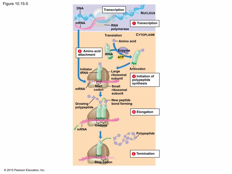
10.14 Elongation adds amino acids to the polypeptide chain until a stop codon terminates translation • Elongation continues until the termination stage of
translation, when • the ribosome reaches a stop codon, • the completed polypeptide is freed from the last
tRNA, and • the ribosome splits back into its separate subunits.

© 2015 Pearson Education, Inc.
Animation: Translation

© 2015 Pearson Education, Inc.
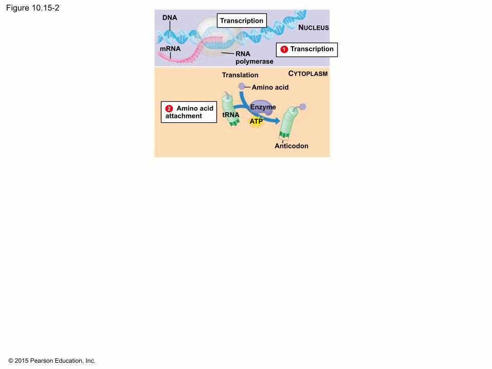
10.15 Review: The flow of genetic information in the cell is DNA → RNA → protein
• The flow of genetic information is from DNA to RNA to protein.
• In transcription (DNA → RNA), the mRNA is synthesized on a DNA template.
• In eukaryotic cells, transcription occurs in the nucleus, and the messenger RNA is processed before it travels to the cytoplasm.
• In prokaryotes, transcription occurs in the cytoplasm.

© 2015 Pearson Education, Inc.
Figure 10.15-1 DNA
mRNA RNA polymerase
NUCLEUS Transcription
Transcription 1
© 2015 Pearson Education, Inc.
2 Amino acid attachment
Figure 10.15-2 DNA
mRNA RNA polymerase
NUCLEUS
Transcription 1
Translation
Amino acid
Enzyme
ATP tRNA
CYTOPLASM
Anticodon
Transcription

© 2015 Pearson Education, Inc.
10.15 Review: The flow of genetic information in the cell is DNA → RNA → protein
• Translation can be divided into four steps, all of which occur in the cytoplasm:
1. amino acid attachment, 2. initiation of polypeptide synthesis, 3. elongation, and 4. termination.

© 2015 Pearson Education, Inc.
2 Amino acid attachment
Figure 10.15-3 DNA
mRNA RNA polymerase
Transcription 1
Translation
Amino acid
Enzyme
ATP tRNA
Anticodon
Initiation of polypeptide synthesis
3
Large ribosomal subunit
Small ribosomal subunit
Start codon mRNA
Initiator tRNA
A U G U C A
CYTOPLASM
Transcription NUCLEUS

© 2015 Pearson Education, Inc.
Amino acid attachment 2
Figure 10.15-4 DNA
mRNA RNA polymerase
Transcription 1
Translation
Amino acid
Enzyme
ATP tRNA
Anticodon
3
Large ribosomal subunit
Small ribosomal subunit
Start codon mRNA
Initiator tRNA
A U G U C A
Initiation of polypeptide synthesis
mRNA Codons
New peptide bond forming Growing
polypeptide Elongation 4
CYTOPLASM
Transcription NUCLEUS
© 2015 Pearson Education, Inc.
Figure 10.15-5
Amino acid attachment 2
DNA
mRNA RNA polymerase
Transcription 1
Translation
Amino acid
Enzyme
ATP tRNA
Anticodon
3
Large ribosomal subunit
Small ribosomal subunit
Start codon mRNA
Initiator tRNA
A U G U C A
Initiation of polypeptide synthesis
mRNA Codons
New peptide bond forming Growing
polypeptide Elongation 4
Polypeptide
Stop codon
Termination 5
CYTOPLASM
Transcription NUCLEUS

© 2015 Pearson Education, Inc.
10.16 Mutations can affect genes
• A mutation is any change in the nucleotide sequence of DNA.
• Mutations can involve • large chromosomal regions or • just a single nucleotide pair.

© 2015 Pearson Education, Inc.
10.16 Mutations can affect genes
• Mutations within a gene can be divided into two general categories.
1. Nucleotide substitutions involve the replacement of one nucleotide and its base-pairing partner with another pair of nucleotides. Base substitutions may • have no effect at all, producing a silent mutation, • change the amino acid coding, producing a missense
mutation, which produces a different amino acid, • lead to a base substitution that produces an improved
protein that enhances the success of the mutant organism and its descendants, or
• change an amino acid into a stop codon, producing a nonsense mutation.

© 2015 Pearson Education, Inc.
10.16 Mutations can affect genes
2. Nucleotide insertions or deletions of one or more nucleotides in a gene may • cause a frameshift mutation, which alters the
reading frame (triplet grouping) of the genetic message,
• lead to significant changes in amino acid sequence, and
• produce a nonfunctional polypeptide.

© 2015 Pearson Education, Inc.
10.16 Mutations can affect genes
• Mutagenesis is the production of mutations. • Mutations can be caused
• by spontaneous errors that occur during DNA replication or recombination or
• by mutagens, which include • high-energy radiation such as X-rays and ultraviolet
light and • chemicals.

© 2015 Pearson Education, Inc.
Figure 10.16a
Normal hemoglobin DNA Mutant hemoglobin DNA
Sickle-cell hemoglobin Normal hemoglobin
mRNA mRNA
C T T C A T
G U A G A A
Glu Val

© 2015 Pearson Education, Inc.
Figure 10.16b-0 Normal gene
Nucleotide substitution
mRNA Protein
Nucleotide deletion
Nucleotide insertion
Inserted
Deleted
Met
Met
Met
Met
Lys
Lys
Lys
Lys
Phe
Phe
Leu
Leu
Gly
Trp
Ala
Ser
Ala
Ala
Arg
A U G A A G U U U G G C G C A
A U G A A G U U U A G C G C A
A U G A A G U U G G C G C A
A U G A A G U U U G G C G C

© 2015 Pearson Education, Inc.
Figure 10.16b-1
Normal gene
Nucleotide substitution
mRNA Protein Met
Met
Lys
Lys
Phe
Phe
Gly
Ser
Ala
Ala
A U G A A G U U U G G C G C A
A U G A A G U U U A G C G C A

© 2015 Pearson Education, Inc.
Figure 10.16b-2
Normal gene
mRNA Protein Met Lys Phe Gly Ala
A U G A A G U U U G G C G C A
Nucleotide deletion
A U G A A G U U G G C G C A
Met Lys Leu Ala
Deleted

© 2015 Pearson Education, Inc.
Figure 10.16b-3
Normal gene
mRNA Protein Met Lys Phe Gly Ala
A U G A A G U U U G G C G C A
Nucleotide insertion
Inserted
Met Lys Leu Trp Arg
A U G A A G U U U G G C G C

© 2015 Pearson Education, Inc.
THE GENETICS OF VIRUSES AND BACTERIA

© 2015 Pearson Education, Inc.
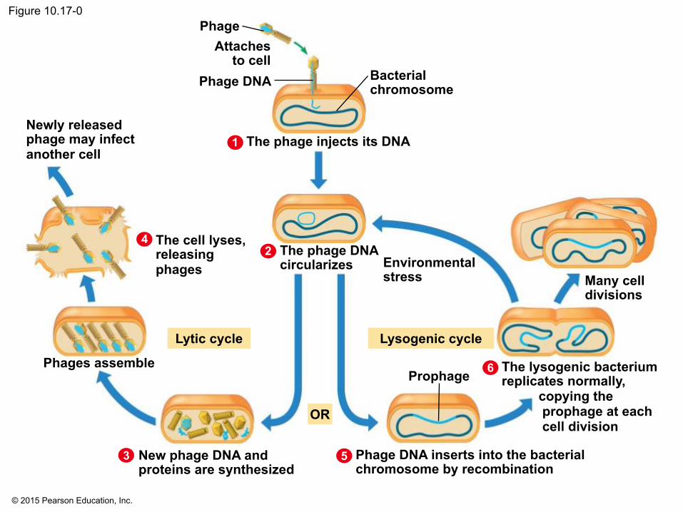
10.17 Viral DNA may become part of the host chromosome
• A virus is an infectious particle consisting of little more than “genes in a box,” a bit of nucleic acid, wrapped in a protein coat called a capsid, and in some cases, a membrane envelope.
• Viruses have two types of reproductive cycles. 1. In the lytic cycle:
• Viral particles are produced using host cell components.
• The host cell lyses. • Viruses are released.
© 2015 Pearson Education, Inc.
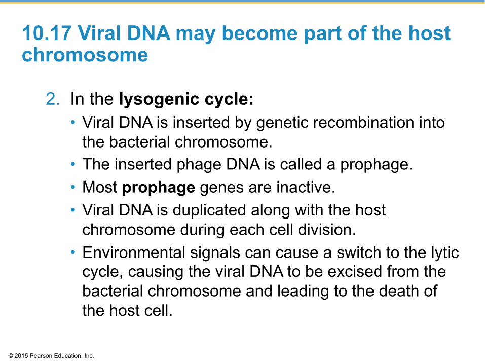
10.17 Viral DNA may become part of the host chromosome
2. In the lysogenic cycle: • Viral DNA is inserted by genetic recombination into
the bacterial chromosome. • The inserted phage DNA is called a prophage. • Most prophage genes are inactive. • Viral DNA is duplicated along with the host
chromosome during each cell division. • Environmental signals can cause a switch to the lytic
cycle, causing the viral DNA to be excised from the bacterial chromosome and leading to the death of the host cell.

© 2015 Pearson Education, Inc.
Animation: Phage Lambda Lysogenic and Lytic Cycles

© 2015 Pearson Education, Inc.
Animation: Phage T4 Lytic Cycle
© 2015 Pearson Education, Inc.
Figure 10.17-0 Phage
Attaches to cell
Phage DNA
Newly released phage may infect another cell
The cell lyses, releasing phages
The phage injects its DNA 1
2 4
3 5
6
Bacterial chromosome
Many cell divisions
Environmental stress
Prophage
Lysogenic cycle
OR
The phage DNA circularizes
Lytic cycle
Phage DNA inserts into the bacterial chromosome by recombination
New phage DNA and proteins are synthesized
Phages assemble The lysogenic bacterium replicates normally, copying the prophage at each cell division
4

© 2015 Pearson Education, Inc.
Figure 10.17-1 Phage
Attaches to cell
Phage DNA
The phage injects its DNA
Bacterial chromosome
The phage DNA circularizes
New phage DNA and proteins are synthesized
Phages assemble
The cell lyses, releasing phages
Newly released phage may infect another cell
Lytic cycle

© 2015 Pearson Education, Inc.
Figure 10.17-2 Phage
Attaches to cell
Phage DNA
The phage injects its DNA
Bacterial chromosome
The phage DNA circularizes
Many cell divisions
Environmental stress
Prophage
Lysogenic cycle
The lysogenic bacterium replicates normally, copying the prophage at each cell division
Phage DNA inserts into the bacterial chromosome by recombination

© 2015 Pearson Education, Inc.
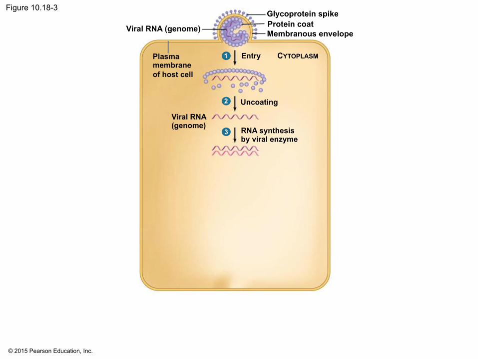
10.18 CONNECTION: Many viruses cause disease in animals and plants
• Viruses can cause disease in animals and plants. • DNA viruses and RNA viruses cause disease in
animals. • A typical animal virus has a membranous outer
envelope and projecting spikes of glycoprotein. • The envelope helps the virus enter and leave the host
cell. • Many animal viruses have RNA rather than DNA as
their genetic material. These include viruses that cause the common cold, measles, mumps, polio, and AIDS.

© 2015 Pearson Education, Inc.
10.18 CONNECTION: Many viruses cause disease in animals and plants
• The reproductive cycle of the mumps virus, a typical enveloped RNA virus, has six major steps:
1. Entry of the protein-coated RNA into the cytoplasm
2. Uncoating—the removal of the protein coat 3. RNA synthesis—mRNA synthesis using a viral
enzyme

© 2015 Pearson Education, Inc.
10.18 CONNECTION: Many viruses cause disease in animals and plants
• The reproductive cycle of the mumps virus, a typical enveloped RNA virus, has six major steps:
4. Protein and RNA synthesis—mRNA is used to make viral proteins and for synthesizing new viral genome RNA.
5. Assembly—the new coat proteins assemble around the new viral RNA.
6. Exit—the viruses leave the cell by cloaking themselves in the host cell’s plasma membrane.

© 2015 Pearson Education, Inc.
10.18 CONNECTION: Many viruses cause disease in animals and plants
• Some animal viruses, such as herpesviruses, replicate in the cell nucleus.
• Most plant viruses are RNA viruses. • To infect a plant, they must get past the outer
protective layer of the plant. • Viruses spread from cell to cell through
plasmodesmata. • Infection can spread to other plants by insects,
herbivores, humans, or farming tools.
• There are no cures for most viral diseases of plants or animals.

© 2015 Pearson Education, Inc.
Animation: Simplified Viral Reproductive Cycle

© 2015 Pearson Education, Inc.
Figure 10.18-1
Entry Plasma membrane of host cell
CYTOPLASM
Glycoprotein spike Protein coat Membranous envelope Viral RNA (genome)
1

© 2015 Pearson Education, Inc.
Figure 10.18-2
Entry Plasma membrane of host cell
Glycoprotein spike Protein coat Membranous envelope Viral RNA (genome)
1
Uncoating
Viral RNA (genome)
2
CYTOPLASM
© 2015 Pearson Education, Inc.
Figure 10.18-3
Entry Plasma membrane of host cell
Glycoprotein spike Protein coat Membranous envelope Viral RNA (genome)
1
Uncoating
Viral RNA (genome)
2
3 RNA synthesis by viral enzyme
CYTOPLASM

© 2015 Pearson Education, Inc.
Figure 10.18-4
Entry Plasma membrane of host cell
Glycoprotein spike Protein coat Membranous envelope Viral RNA (genome)
1
Uncoating
Viral RNA (genome)
2
3 RNA synthesis by viral enzyme
4 Template
New viral genome
mRNA
New viral proteins
Protein and RNA synthesis (other strand)
CYTOPLASM

© 2015 Pearson Education, Inc.
Figure 10.18-5
Entry Plasma membrane of host cell
Glycoprotein spike Protein coat Membranous envelope Viral RNA (genome)
1
Uncoating
Viral RNA (genome)
2
3 RNA synthesis by viral enzyme
4 Template
New viral genome
mRNA
New viral proteins
Protein and RNA synthesis (other strand)
5 Assembly
CYTOPLASM

© 2015 Pearson Education, Inc.
Figure 10.18-6
Entry Plasma membrane of host cell
Glycoprotein spike Protein coat Membranous envelope Viral RNA (genome)
1
Uncoating
Viral RNA (genome)
2
3 RNA synthesis by viral enzyme
4 Template
New viral genome
mRNA
New viral proteins
Protein and RNA synthesis (other strand)
5
6
Assembly
Exit
CYTOPLASM

© 2015 Pearson Education, Inc.
10.19 EVOLUTION CONNECTION: Emerging viruses threaten human health
• Viruses that appear suddenly or are new to medical scientists are called emerging viruses. These include the
• 2009 H1N1 influenza virus, • HIV (human immunodeficiency virus), which causes
AIDS, • Ebola virus, which causes hemorrhagic fever, • West Nile virus, spread primarily by mosquitoes,
and • SARS (severe acute respiratory syndrome) virus.

© 2015 Pearson Education, Inc.
10.19 EVOLUTION CONNECTION: Emerging viruses threaten human health
• Three processes contribute to the emergence of viral diseases:
1. Mutation—RNA viruses mutate rapidly. 2. Contact between species—viruses from other
animals spread to humans. 3. Spread—viruses spread from isolated human
populations to larger human populations, often over great distances.

© 2015 Pearson Education, Inc.
Figure 10.19-0

© 2015 Pearson Education, Inc.
Figure 10.19-1

© 2015 Pearson Education, Inc.
Figure 10.19-2

© 2015 Pearson Education, Inc.
10.20 The AIDS virus makes DNA on an RNA template
• AIDS (acquired immunodeficiency syndrome) is caused by HIV (human immunodeficiency virus).
• HIV • has two identical copies of its RNA, • is a retrovirus, an RNA virus that reproduces by
means of a DNA molecule, and • carries molecules of reverse transcriptase, which
catalyzes reverse transcription: the synthesis of DNA on an RNA template.

© 2015 Pearson Education, Inc.
Figure 10.20a
Envelope
Glycoprotein Protein coat
RNA (two identical strands)
Reverse transcriptase

© 2015 Pearson Education, Inc.
10.20 The AIDS virus makes DNA on an RNA template
• After HIV RNA is uncoated in the cytoplasm of the host cell,
1. reverse transcriptase makes one DNA strand from RNA,
2. reverse transcriptase adds a complementary DNA strand,
3. double-stranded viral DNA enters the nucleus and integrates into the chromosome, becoming a provirus,

© 2015 Pearson Education, Inc.
10.20 The AIDS virus makes DNA on an RNA template
• After HIV RNA is uncoated in the cytoplasm of the host cell,
4. the provirus DNA is used to produce mRNA, 5. the viral mRNA is translated to produce viral
proteins, and 6. new viral particles are assembled, leave the host
cell, and can then infect other cells.

© 2015 Pearson Education, Inc.
Animation: HIV Reproductive Cycle

© 2015 Pearson Education, Inc.
Figure 10.20b
Viral RNA Reverse transcriptase
CYTOPLASM
NUCLEUS
Chromosomal DNA
Provirus DNA
RNA
DNA strand Double-stranded DNA
Viral RNA and proteins
6
5
3
4
2
1

© 2015 Pearson Education, Inc.
10.21 Viroids and prions are formidable pathogens in plants and animals
• Some infectious agents are made only of RNA or protein.
• Viroids are small, circular RNA molecules that infect plants.
• Viroids replicate within host cells without producing proteins and interfere with plant growth.
• Prions are infectious proteins that cause degenerative brain diseases in animals.
• Prions appear to be misfolded forms of normal brain proteins, which convert normal protein to misfolded prion versions.

© 2015 Pearson Education, Inc.
10.22 Bacteria can transfer DNA in three ways
• Viral replication allows researchers to learn more about the mechanisms that regulate DNA replication and gene expression in living cells.
• Bacteria are equally valuable as microbial models in genetics research.
• Most of a bacterium’s DNA is found in a single, closed-loop chromosome.
• Bacterial cells divide by replication of the bacterial chromosome and then by binary fission.
• Because binary fission is an asexual process, bacteria in a colony are genetically identical to the parent cell.

© 2015 Pearson Education, Inc.
10.22 Bacteria can transfer DNA in three ways
• Bacteria use three mechanisms to move genes from cell to cell.
1. Transformation is the uptake of DNA from the surrounding environment.
2. Transduction is gene transfer by phages. 3. Conjugation is the transfer of DNA from a donor
to a recipient bacterial cell.

© 2015 Pearson Education, Inc.
Figure 10.22a
DNA enters cell
A fragment of DNA from another bacterial cell
Bacterial chromosome (DNA)

© 2015 Pearson Education, Inc.
Figure 10.22b
Phage
A fragment of DNA from another bacterial cell (former phage host)

© 2015 Pearson Education, Inc.
Figure 10.22c
Mating bridge
Donor cell
Sex pili
Recipient cell

© 2015 Pearson Education, Inc.
Figure 10.22d
Donated DNA Crossovers Degraded DNA
Recipient cell’s chromosome
Recombinant chromosome

© 2015 Pearson Education, Inc.
10.22 Bacteria can transfer DNA in three ways
• Once new DNA gets into a bacterial cell by any mechanism, part of it may then integrate into the recipient's chromosome.
© 2015 Pearson Education, Inc.
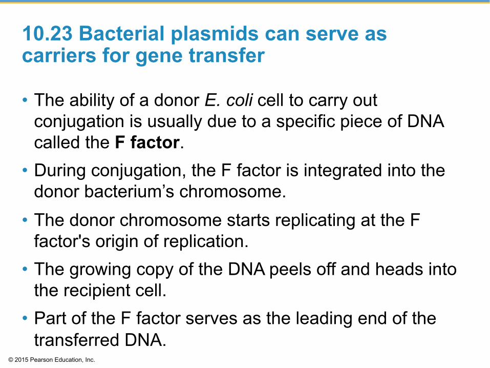
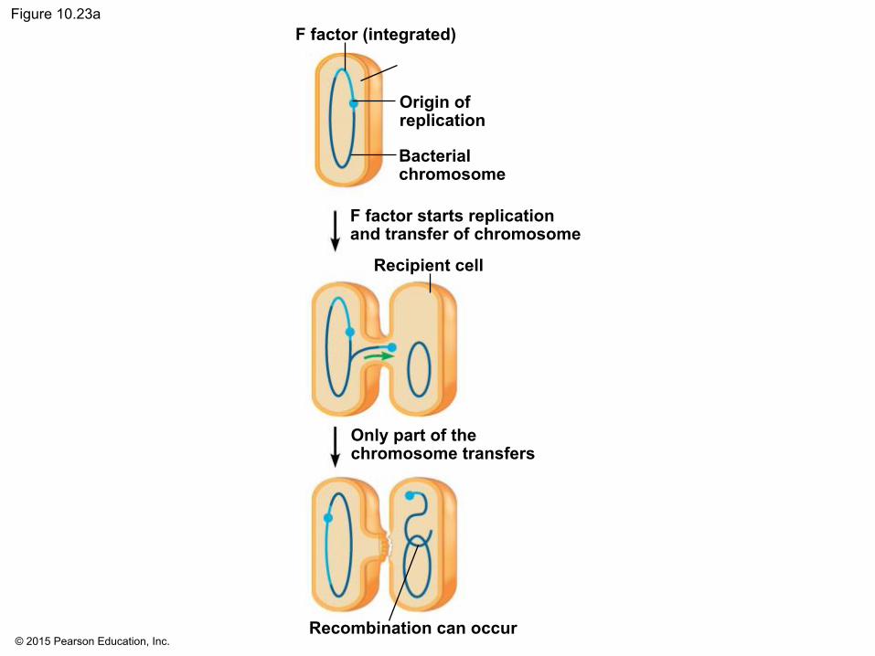
10.23 Bacterial plasmids can serve as carriers for gene transfer
• The ability of a donor E. coli cell to carry out conjugation is usually due to a specific piece of DNA called the F factor.
• During conjugation, the F factor is integrated into the donor bacterium’s chromosome.
• The donor chromosome starts replicating at the F factor's origin of replication.
• The growing copy of the DNA peels off and heads into the recipient cell.
• Part of the F factor serves as the leading end of the transferred DNA.

© 2015 Pearson Education, Inc.
Figure 10.23a-b F factor (integrated) F factor (plasmid)
Donor
Bacterial chromosome
Donor
Origin of replication
Bacterial chromosome
F factor starts replication and transfer
The plasmid completes its transfer and circularizes
The cell is now a donor Recombination can occur
Only part of the chromosome transfers
Recipient cell
F factor starts replication and transfer of chromosome
© 2015 Pearson Education, Inc.
Figure 10.23a F factor (integrated)
Origin of replication
Bacterial chromosome
Recombination can occur
Only part of the chromosome transfers
Recipient cell
F factor starts replication and transfer of chromosome

© 2015 Pearson Education, Inc.
10.23 Bacterial plasmids can serve as carriers for gene transfer
• An F factor can also exist as a plasmid, a small, circular DNA molecule separate from the bacterial chromosome.
• Some plasmids, including the F factor, can bring about conjugation and move to another cell in linear form.
• The transferred plasmid re-forms a circle in the recipient cell, and the cell becomes a donor.
• R plasmids pose serious problems for human medicine by carrying genes for enzymes that destroy antibiotics.

© 2015 Pearson Education, Inc.
Figure 10.23b F factor (plasmid)
Donor
Bacterial chromosome
F factor starts replication and transfer
The plasmid completes its transfer and circularizes
The cell is now a donor

© 2015 Pearson Education, Inc.
Figure 10.23c
Plasmids

© 2015 Pearson Education, Inc.
You should now be able to
1. Describe the experiments of Griffith, Hershey, and Chase, which supported the idea that DNA was life’s genetic material.
2. Compare the structures of DNA and RNA. 3. Explain how the structure of DNA facilitates its
replication. 4. Describe the process of DNA replication. 5. Describe the locations, reactants, and products
of transcription and translation.

© 2015 Pearson Education, Inc.
You should now be able to
6. Explain how the “languages” of DNA and RNA are used to produce polypeptides.
7. Explain how mRNA is produced using DNA. 8. Explain how eukaryotic RNA is processed
before leaving the nucleus. 9. Relate the structure of tRNA to its functions in
the process of translation. 10. Describe the structure and function of
ribosomes.

© 2015 Pearson Education, Inc.
You should now be able to
11. Describe the step-by-step process by which amino acids are added to a growing polypeptide chain.
12. Diagram the overall process of transcription and translation.
13. Describe the major types of mutations, causes of mutations, and potential consequences.
14. Compare the lytic and lysogenic reproductive cycles of a phage.
15. Compare the structures and reproductive cycles of the mumps virus and a herpes virus.

© 2015 Pearson Education, Inc.
You should now be able to
16. Describe three processes that contribute to the emergence of viral disease.
17. Explain how the AIDS virus enters a host cell and reproduces.
18. Describe the structure of viroids and prions and explain how they cause disease.
19. Define and compare the processes of transformation, transduction, and conjugation.
20. Define a plasmid and explain why R plasmids pose serious human health problems.
© 2015 Pearson Education, Inc.
Figure 10.UN01

© 2015 Pearson Education, Inc.
Figure 10.UN02

© 2015 Pearson Education, Inc.
Figure 10.UN03
Sugar- phosphate backbone
A
C
Nitrogenous base
Phosphate group
Sugar Nucleotide
DNA RNA
DNA Polynucleotide Sugar
Nitrogenous bases
Deoxy- ribose Ribose
T
G
G
C G A T
C G A U
A T
C G
T A
C G
T A
G C
G
A T
G C
T A T A
T A C G

© 2015 Pearson Education, Inc.
Figure 10.UN04
Growing polypeptide Amino acid
tRNA
Anticodon
Small ribosomal subunit
Codons
mRNA
Large ribosomal subunit

© 2015 Pearson Education, Inc.
Figure 10.UN05
DNA
RNA
(b)
(d)
(e)
(a)
(c)
(f)
(g)
(h)
(i) Protein
is a polymer made from
monomers called
is performed by an enzyme called
comes in three
kinds called
is performed by structures
called
use amino-acid-bearing molecules called
one or more polymers made from
monomers called
molecules are components of