Embed Size (px)
Citation preview
TIBS 23 – OCTOBER 1998
399Copyright © 1998, Elsevier Science Ltd. All rights reserved. 0968 – 0004/98/$19.00 PII: S0968-0004(98)01291-2
THE AIM OF cell division is to distributecomplete and accurate replicas of thegenome to daughter cells. However,there is no physical requirement forchromosomes to be replicated or re-paired prior to nuclear division. Check-point controls therefore monitor DNAreplication and repair, and thereby cou-ple the completion of these events tothe onset of mitosis. In relatively simpleorganisms, such as haploid yeasts, check-point controls are vital for survival in situ-ations where DNA replication is inhib-ited or DNA is damaged. In multicellulardiploid organisms, such as humans,checkpoint controls are important formaintenance of genomic integrity. In hu-mans, loss of genomic integrity can causecancer and genetic diseases.
Studies of yeasts have played a pio-neering role in the discovery and analy-sis of cell cycle and checkpoint controls.Important contributions have comefrom studies of both the budding yeastSaccharomyces cerevisiae and the fis-sion yeast Schizosaccharomyces pombe.In many respects, the checkpoints in thetwo yeasts appear to be similar, employ-ing homologous proteins to sense DNAdamage or replication defects. However,there also appear to be important differ-ences. Several fission yeast proteinsthat are required for checkpoints do notappear to have homologs in buddingyeast. Moreover, the mechanisms thatare used to restrain mitosis are quite dif-ferent in the two yeasts, because of thedifferent modes of division (i.e. buddingversus fission). The mitotic-controlmechanisms of fission yeast and meta-zoans are highly conserved. Indeed, the
discovery of several of the central el-ements of mitotic control in mammaliancells was a direct result of their previousdiscovery in fission yeast.
Recent studies of fission yeast haveled to major new insights into themolecular features of checkpoint con-trols. These investigations have broughtto light key similarities and differencesbetween the checkpoints activated byDNA damage or agents that inhibit DNAreplication. In this review, I highlightthese recent findings andpoint out some questionsthat remain to be answered.
The importance of phosphateThe cyclin-dependent ki-
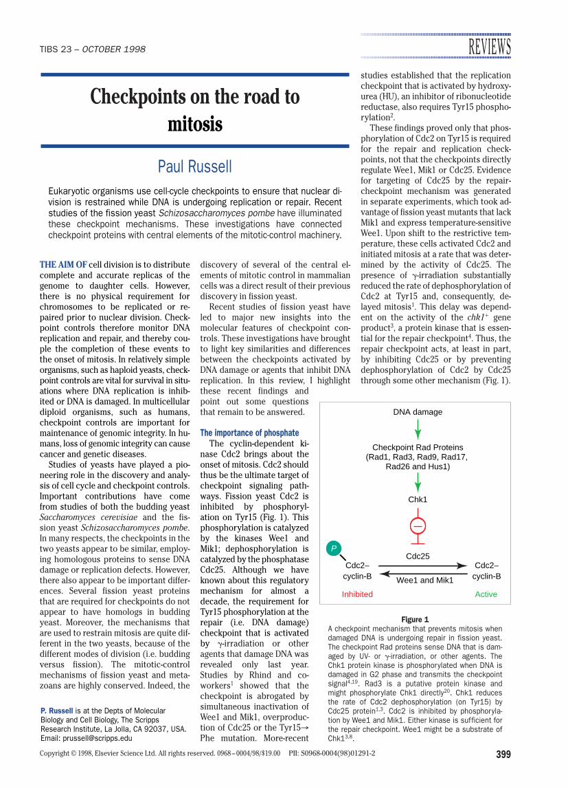
nase Cdc2 brings about theonset of mitosis. Cdc2 shouldthus be the ultimate target ofcheckpoint signaling path-ways. Fission yeast Cdc2 isinhibited by phosphoryl-ation on Tyr15 (Fig. 1). Thisphosphorylation is catalyzedby the kinases Wee1 andMik1; dephosphorylation iscatalyzed by the phosphataseCdc25. Although we haveknown about this regulatorymechanism for almost adecade, the requirement forTyr15 phosphorylation at therepair (i.e. DNA damage)checkpoint that is activatedby g-irradiation or otheragents that damage DNA wasrevealed only last year.Studies by Rhind and co-workers1 showed that thecheckpoint is abrogated bysimultaneous inactivation ofWee1 and Mik1, overproduc-tion of Cdc25 or the Tyr15®Phe mutation. More-recent
studies established that the replicationcheckpoint that is activated by hydroxy-urea (HU), an inhibitor of ribonucleotidereductase, also requires Tyr15 phospho-rylation2.
These findings proved only that phos-phorylation of Cdc2 on Tyr15 is requiredfor the repair and replication check-points, not that the checkpoints directlyregulate Wee1, Mik1 or Cdc25. Evidencefor targeting of Cdc25 by the repair-checkpoint mechanism was generatedin separate experiments, which took ad-vantage of fission yeast mutants that lackMik1 and express temperature-sensitiveWee1. Upon shift to the restrictive tem-perature, these cells activated Cdc2 andinitiated mitosis at a rate that was deter-mined by the activity of Cdc25. Thepresence of g-irradiation substantiallyreduced the rate of dephosphorylation ofCdc2 at Tyr15 and, consequently, de-layed mitosis1. This delay was depend-ent on the activity of the chk11 geneproduct3, a protein kinase that is essen-tial for the repair checkpoint4. Thus, therepair checkpoint acts, at least in part,by inhibiting Cdc25 or by preventing dephosphorylation of Cdc2 by Cdc25through some other mechanism (Fig. 1).
REVIEWS
Checkpoints on the road tomitosis
Paul RussellEukaryotic organisms use cell-cycle checkpoints to ensure that nuclear di-vision is restrained while DNA is undergoing replication or repair. Recentstudies of the fission yeast Schizosaccharomyces pombe have illuminatedthese checkpoint mechanisms. These investigations have connectedcheckpoint proteins with central elements of the mitotic-control machinery.
P. Russell is at the Depts of MolecularBiology and Cell Biology, The ScrippsResearch Institute, La Jolla, CA 92037, USA.Email: [email protected]
Chk1
Checkpoint Rad Proteins(Rad1, Rad3, Rad9, Rad17,
Rad26 and Hus1)
DNA damage
Cdc25Cdc2–
Inhibited
cyclin-BCdc2–
Active
cyclin-B
P
Wee1 and Mik1
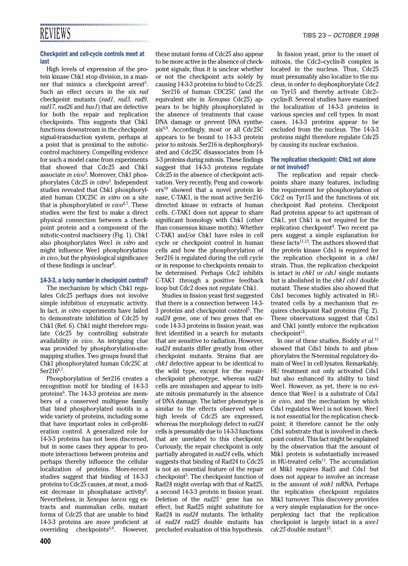
Figure 1A checkpoint mechanism that prevents mitosis whendamaged DNA is undergoing repair in fission yeast.The checkpoint Rad proteins sense DNA that is dam-aged by UV- or g-irradiation, or other agents. TheChk1 protein kinase is phosphorylated when DNA isdamaged in G2 phase and transmits the checkpointsignal4,19. Rad3 is a putative protein kinase andmight phosphorylate Chk1 directly20. Chk1 reducesthe rate of Cdc2 dephosphorylation (on Tyr15) byCdc25 protein1,3. Cdc2 is inhibited by phosphoryla-tion by Wee1 and Mik1. Either kinase is sufficient forthe repair checkpoint. Wee1 might be a substrate ofChk13,8.
REVIEWS TIBS 23 – OCTOBER 1998
400
Checkpoint and cell-cycle controls meet atlast
High levels of expression of the pro-tein kinase Chk1 stop division, in a man-ner that mimics a checkpoint arrest5.Such an effect occurs in the six radcheckpoint mutants (rad1, rad3, rad9,rad17, rad26 and hus1) that are defectivefor both the repair and replicationcheckpoints. This suggests that Chk1functions downstream in the checkpointsignal-transduction system, perhaps at a point that is proximal to the mitotic-control machinery. Compelling evidencefor such a model came from experimentsthat showed that Cdc25 and Chk1associate in vivo3. Moreover, Chk1 phos-phorylates Cdc25 in vitro3. Independentstudies revealed that Chk1 phosphoryl-ated human CDC25C in vitro on a sitethat is phosphorylated in vivo6,7. Thesestudies were the first to make a directphysical connection between a check-point protein and a component of themitotic-control machinery (Fig. 1). Chk1also phosphorylates Wee1 in vitro andmight influence Wee1 phosphorylationin vivo, but the physiological significanceof these findings is unclear8.
14-3-3, a lucky number in checkpoint control?The mechanism by which Chk1 regu-
lates Cdc25 perhaps does not involvesimple inhibition of enzymatic activity.In fact, in vitro experiments have failedto demonstrate inhibition of Cdc25 byChk1 (Ref. 6). Chk1 might therefore regu-late Cdc25 by controlling substrateavailability in vivo. An intriguing cluewas provided by phosphorylation-site-mapping studies. Two groups found thatChk1 phosphorylated human Cdc25C atSer2166,7.
Phosphorylation of Ser216 creates arecognition motif for binding of 14-3-3proteins6. The 14-3-3 proteins are mem-bers of a conserved multigene familythat bind phosphorylated motifs in awide variety of proteins, including somethat have important roles in cell-prolif-eration control. A generalized role for14-3-3 proteins has not been discerned,but in some cases they appear to pro-mote interactions between proteins andperhaps thereby influence the cellularlocalization of proteins. More-recentstudies suggest that binding of 14-3-3proteins to Cdc25 causes, at most, a mod-est decrease in phosphatase activity9.Nevertheless, in Xenopus laevis egg ex-tracts and mammalian cells, mutantforms of Cdc25 that are unable to bind14-3-3 proteins are more proficient atoverriding checkpoints6,9. However,
these mutant forms of Cdc25 also appearto be more active in the absence of check-point signals; thus it is unclear whetheror not the checkpoint acts solely bycausing 14-3-3 proteins to bind to Cdc25.
Ser216 of human CDC25C (and theequivalent site in Xenopus Cdc25) ap-pears to be highly phosphorylated inthe absence of treatments that causeDNA damage or prevent DNA synthe-sis6,9. Accordingly, most or all Cdc25Cappears to be bound to 14-3-3 proteinprior to mitosis. Ser216 is dephosphoryl-ated and Cdc25C disassociates from 14-3-3 proteins during mitosis. These findingssuggest that 14-3-3 proteins regulateCdc25 in the absence of checkpoint acti-vation. Very recently, Peng and co-work-ers10 showed that a novel protein ki-nase, C-TAK1, is the most active Ser216-directed kinase in extracts of humancells. C-TAK1 does not appear to sharesignificant homology with Chk1 (otherthan consensus kinase motifs). WhetherC-TAK1 and/or Chk1 have roles in cellcycle or checkpoint control in humancells and how the phosphorylation ofSer216 is regulated during the cell cycleor in response to checkpoints remain tobe determined. Perhaps Cdc2 inhibits C-TAK1 through a positive feedbackloop but Cdc2 does not regulate Chk1.
Studies in fission yeast first suggestedthat there is a connection between 14-3-3 proteins and checkpoint control5. Therad24 gene, one of two genes that en-code 14-3-3 proteins in fission yeast, wasfirst identified in a search for mutantsthat are sensitive to radiation. However,rad24 mutants differ greatly from othercheckpoint mutants. Strains that arechk1 defective appear to be identical tothe wild type, except for the repair-checkpoint phenotype, whereas rad24cells are misshapen and appear to initi-ate mitosis prematurely in the absenceof DNA damage. The latter phenotype issimilar to the effects observed whenhigh levels of Cdc25 are expressed,whereas the morphology defect in rad24cells is presumably due to 14-3-3 functionsthat are unrelated to this checkpoint.Curiously, the repair checkpoint is onlypartially abrogated in rad24 cells, whichsuggests that binding of Rad24 to Cdc25is not an essential feature of the repaircheckpoint5. The checkpoint function ofRad24 might overlap with that of Rad25,a second 14-3-3 protein in fission yeast.Deletion of the rad251 gene has no effect, but Rad25 might substitute forRad24 in rad24 mutants. The lethality of rad24 rad25 double mutants has precluded evaluation of this hypothesis.
In fission yeast, prior to the onset ofmitosis, the Cdc2–cyclin-B complex islocated in the nucleus. Thus, Cdc25must presumably also localize to the nu-cleus, in order to dephosphorylate Cdc2on Tyr15 and thereby activate Cdc2– cyclin-B. Several studies have examinedthe localization of 14-3-3 proteins in various species and cell types. In mostcases, 14-3-3 proteins appear to be excluded from the nucleus. The 14-3-3proteins might therefore regulate Cdc25by causing its nuclear exclusion.
The replication checkpoint: Chk1 not aloneor not involved?
The replication and repair check-points share many features, includingthe requirement for phosphorylation ofCdc2 on Tyr15 and the functions of sixcheckpoint Rad proteins. CheckpointRad proteins appear to act upstream ofChk1, yet Chk1 is not required for thereplication checkpoint4. Two recent pa-pers suggest a simple explanation forthese facts11,12. The authors showed thatthe protein kinase Cds1 is required forthe replication checkpoint in a chk1strain. Thus, the replication checkpointis intact in chk1 or cds1 single mutantsbut is abolished in the chk1 cds1 doublemutant. These studies also showed thatCds1 becomes highly activated in HU-treated cells by a mechanism that re-quires checkpoint Rad proteins (Fig. 2).These observations suggest that Cds1and Chk1 jointly enforce the replicationcheckpoint11.
In one of these studies, Boddy et al.11
showed that Cds1 binds to and phos-phorylates the N-terminal regulatory do-main of Wee1 in cell lysates. Remarkably,HU treatment not only activated Cds1but also enhanced its ability to bindWee1. However, as yet, there is no evi-dence that Wee1 is a substrate of Cds1in vivo, and the mechanism by whichCds1 regulates Wee1 is not known. Wee1is not essential for the replication check-point; it therefore cannot be the onlyCds1 substrate that is involved in check-point control. This fact might be explainedby the observation that the amount ofMik1 protein is substantially increasedin HU-treated cells11. The accumulationof Mik1 requires Rad3 and Cds1 butdoes not appear to involve an increasein the amount of mik1 mRNA. Perhapsthe replication checkpoint regulatesMik1 turnover. This discovery providesa very simple explanation for the once-perplexing fact that the replicationcheckpoint is largely intact in a wee1cdc25 double mutant13.
TIBS 23 – OCTOBER 1998
401
A mechanism in which Cds1 and Chk1jointly enforce the HU arrest explainsmost genetic studies of the replicationcheckpoint in fission yeast, but the datado not exclude other models. An alter-native model proposes that Cds1 andcheckpoint Rad proteins stabilize pro-tein complexes at stalled replicationforks (Fig. 3)12. These protein complexescould be part of an intrinsic checkpointmechanism that couples mitosis to thecompletion of DNA replication, perhapsby sequestration of Cdc2. This idea issupported by studies that show that mitosis is delayed or prevented by pro-duction of large amounts of certainreplication proteins, such as Cdc18 orOrp2 in fission yeast14,15. Cdc18 appearsto play a rate-limiting role in the acti-vation of replication origins, whereasOrp2 is a component of the origin-recog-nition complex (ORC). Cdc18 appears tohave a high affinity for Cdc2 in vivo andin vitro16,17, unlike most Cdc2 substrates.Cdc18 is normally degraded after com-pletion of the DNA synthesis (S) phaseof the cell cycle, apparently by a mecha-nism that is regulated by Cdc2 pro-tein14,17,18. Thus, Cdc18, and perhaps otherreplication proteins, might sequesterCdc2 during S phase and prevent the ini-tiation of mitosis. Degradation of Cdc18upon exit from S phase would disengagethe replication checkpoint.
Another reason for proposing thismodel involves Chk1 phosphorylation.DNA damage induces Chk1 phosphoryl-ation (which can be detected as achange in electrophoretic mobility) by amechanism that requires checkpointRad proteins19. A catalytically inactiveform of Chk1 is not phosphorylated,which suggests that autophosphoryl-ation occurs during Chk1 activation19.HU does not stimulate phosphorylationof Chk1, except in a cds1 mutant12. A failure to stabilize protein complexes atstalled replication forks in cds1 cells mightcause DNA damage and phosphorylationof Chk1 (Fig. 3). Thus, Lindsay et al.12
proposed that Chk1 does not normallyparticipate in the replication checkpointand only becomes essential in cds1 cells,which lack the intrinsic checkpoint.
The possibility that an intrinsiccheckpoint mechanism that involvestitration of Cdc2 exists runs counter tothe evidence that phosphorylation ofCdc2 on Tyr15 is required for the repli-cation checkpoint. Likewise, a checkpoint-dependent increase in the amount of Mik1provides very straightforward evidencethat the replication checkpoint operatesby controlling the phosphorylation of
Cdc2 on Tyr15. However, the possibilitythat regulation of Mik1 or Wee1 works in conjunction with an intrinsic check-point cannot be excluded. Studies onCdc25 might help to distinguish betweenthese models. Rhind and Russell2 recentlyshowed that the rate of dephosphoryl-ation of Cdc2 Tyr15 is diminished in cellsarrested by treatment with HU. Thesefindings suggest that HU treatment in-hibits Cdc25. Recent studies haveshown that this effect is due to an au-thentic checkpoint that requires check-point Rad proteins and is abolished in achk1 cds1 double mutant (B. Furnari andP. Russell, unpublished). These findingsindicate that both Chk1 and Cds1 con-tribute to the inhibition of Cdc25.
New members of the checkpoint teamThe molecular functions of the six
checkpoint Rad proteins are poorly un-derstood. The most studied is Rad3, amember of a family of proteins that
share structural homology with lipid kinases but appear to function as proteinkinases20. One member of this family is ATM, a protein that is implicated in g-irradiation checkpoint control in hu-mans. Another member of this family is DNA-PK, a protein kinase that is acti-vated through association with otherproteins that interact with the ends ofDNA. It is tempting to hypothesize thatRad3 is activated by specific DNA struc-tures (such as broken or unpaired DNA)and, because both proteins are phos-phorylated when replication or repaircheckpoints are activated, that Rad3phosphorylates Cds1 and Chk112,19.
Analysis of cut genes in fission yeastrevealed another member of the check-point team. The cut mutants undergo agrisly death, bisecting their nuclei withtheir septum (division plate). One of thecut mutants, cut5 (also known as rad4),suffers this fate because it cannot repli-cate its DNA or restrain mitosis21,22.
REVIEWS
Cdc25
Wee1 and Mik1
Inhibited Active
P
Stalled replication forks
Hydroxyurea
DNA damage
Checkpoint Rad proteins
Cds1
Checkpoint Rad proteins
Chk1
Cdc2–cyclin-B
Cdc2–cyclin-B
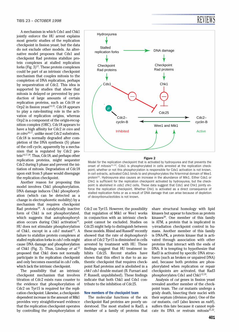
Figure 2Model for the replication checkpoint that is activated by hydroxyurea and that prevents theonset of mitosis11,12. Cds1 is phosphorylated in cells arrested at the replication check-point; whether or not this phosphorylation is responsible for Cds1 activation is not known.In cell extracts, activated Cds1 binds to and phosphorylates the N-terminal domain of Wee1protein11. Hydroxyurea also causes an increase in the abundance of Mik1. Either Cds1 orChk1 is sufficient for the replication checkpoint activated by hydroxyurea, but the check-point is abolished in cds1 chk1 cells. These data suggest that Cds1 and Chk1 jointly en-force the replication checkpoint. Whether Chk1 is activated as a direct consequence ofstalled replication forks or as a result of DNA damage that can arise because of depletionof deoxyribonucleotides is not known.
REVIEWS TIBS 23 – OCTOBER 1998
402
Unlike the checkpoint rad genes, cut5 isessential; this makes its analysis moredifficult. A careful series of experimentsshowed that Cut5 restrains mitosiswhen cells are incubated with HU21–23.Cut5 is required for the damage check-point that is activated by g-irradiationbut, surprisingly, Cut5 is not needed for the checkpoint activated by UV irra-diation or UV mimetic drugs. These obser-vations suggest that Cut5 processes cer-tain types of DNA damage or structuresin order to activate the checkpoint sig-nal-transduction system. This idea gainsstrong support from experiments thatused a temperature-sensitive cut5 mu-tant. These studies showed that Cut5 isneeded to establish the g-irradiationcheckpoint signal, but that once the signalis established Cut5 is no longer neededto maintain the checkpoint arrest23.
Crb2 (also known as Rhp9) is the lat-est addition to the checkpoint team. Thecrb2 gene was identified by two ap-proaches: (1) examination of mutantsthat are sensitive to methylmethanesulphonate (MMS); and (2) detection ofproteins that interact with Cut5 in ayeast two-hybrid screen24,25. A convinc-ing feature of the two-hybrid interaction
was the demonstration that a missensemutation of Cut5 that abolishes thecheckpoint also greatly diminishes theinteraction between Crb2 and Cut5.Mutants that are crb2 defective cannotarrest division in response to DNA dam-age, but the replication checkpoint is in-tact in these cells. This phenotype isreminiscent of chk1 mutants, which sug-gests that Chk1 and Crb2 have closelyinvolved functions. Yeast two-hybrid interactions (between Chk1 and Crb2)support this idea24. Moreover, damage-induced phosphorylation of Chk1 isabolished in a crb2 mutant. PerhapsCrb2 provides a link between Cut5 andChk1, and thereby connects a proteinthat might be directly involved in pro-cessing damaged DNA to a kinase thatlinks the checkpoint to cell-cycle control.
Outlook: yeast, flies and mammalsExcellent evidence supports the idea
that inhibitory phosphorylation of Cdc2is important for the replication and re-pair checkpoints that regulate the onsetof mitosis in mammalian cells26,27. More-recent studies have indicated that Chk1is structurally and functionally con-served in mammals and Drosophila
melanogaster. The D. melanogaster grapesmutant undergoes defective chromo-some segregation during embryogen-esis28,29. This defect is caused by a fail-ure to couple the onset of mitosis to thecompletion of DNA replication. The grapesgene product is a protein kinase that isapproximately 59% identical to fissionyeast Chk1. The discoveries of thesehomologies led to the discovery of Chk1sequence homologs from humans andmice7,30. Remarkably, both fission yeastChk1 and human CHK1 phosphorylatehuman CDC25C on Ser216. These findings,coupled with the similarities between fis-sion yeast Rad3 and human ATM, con-firm the notion that fission yeast willcontinue to serve as a valuable modelsystem for uncovering key elements ofcell-cycle and checkpoint control.
References1 Rhind, N., Furnari, B. and Russell, P. (1997)
Genes Dev. 11, 504–5112 Rhind, N. and Russell, P. (1998) Mol. Cell. Biol.
18, 3782–37873 Furnari, B., Rhind, N. and Russell, P. (1997)
Science 277, 1495–14974 Walworth, N., Davey, S. and Beach, D. (1993)
Nature 363, 368–3715 Ford, J. C. et al. (1994) Science 265, 533–5356 Peng, C. Y. et al. (1997) Science 277,
1501–15057 Sanchez, Y. et al. (1997) Science 277, 1497–15018 O’Connell, M. J., Raleigh, J. M., Verkade, H. M.
and Nurse, P. (1997) EMBO J. 16, 545–5549 Kumagai, A., Yakowec, P. S. and Dunphy, W. G.
(1998) Mol. Biol. Cell 9, 345–35410 Peng, C. Y. et al. (1998) Cell Growth Differ. 9,
197–20811 Boddy, M. N., Furnari, B., Mondesert, O. and
Russell, P. (1998) Science 280, 909–91212 Lindsay, H. et al. (1998) Genes Dev. 12, 382–39513 Enoch, T., Carr, A. M. and Nurse, P. (1993)
Genes Dev. 6, 2035–204614 Nishitani, H. and Nurse, P. (1995) Cell 83,
397–40515 Leatherwood, J., Lopez-Girona, A. and Russell,
P. (1996) Nature 379, 360–36416 Lopez-Girona, A., Mondesert, O., Leatherwood, J.
and Russell, P. (1998) Mol. Biol. Cell 9, 63–7317 Jallepalli, P. V. et al. (1997) Genes Dev. 11,
2767–277918 Kominami, K. and Toda, T. (1997) Genes Dev. 11,
1548–156019 Walworth, N. C. and Bernards, R. (1996)
Science 271, 353–35620 Bentley, N. J. et al. (1996) EMBO J. 15,
6641–665121 Saka, Y. et al. (1994) EMBO J. 13, 5319–532922 Saka, Y. and Yanagida, M. (1993) Cell 74,
383–39323 McFarlane, R. J., Carr, A. M. and Price, C.
(1997) Mol. Gen. Genet. 255, 332–34024 Saka, Y. et al. (1997) Genes Dev. 11, 3387–340025 Willson, J., Wilson, S., Warr, N. and Watts, F. Z.
(1997) Nucleic Acids Res. 25, 2138–214526 Jin, P., Gu, Y. and Morgan, D. O. (1996) J. Cell
Biol. 134, 963–97027 Blasina, A., Paegle, S. and McGowan, C. H.
(1997) Mol. Biol. Cell 8, 1013–102328 Sibon, O. C. M., Stevenson, V. A. and Theurkauf,
W. E. (1997) Nature 388, 93–9729 Fogarty, P. et al. (1997) Curr. Biol. 7, 418–42630 Flaggs, G. et al. (1997) Curr. Biol. 7, 977–986
Unstable stalled replication forks
Hydroxyurea
Checkpoint Rad proteins
Stable stalled replication forks
Cds1
DNA damage
Inhibited Active
Checkpoint Rad proteins
Chk1
Sequestrationof Cdc2?
Cdc25
Wee1 and Mik1
Cdc2–cyclin-B
Cdc2–cyclin-B
P
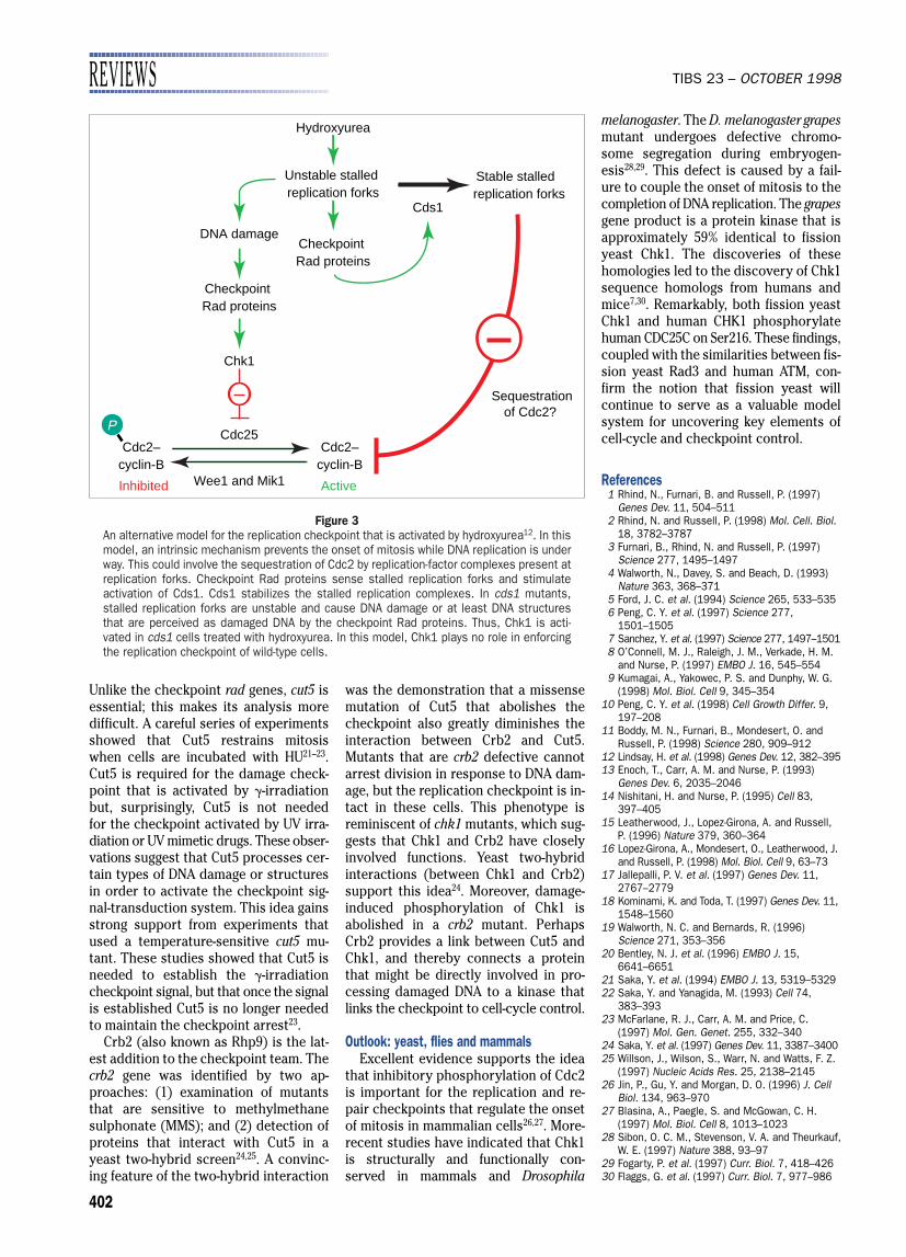
Figure 3An alternative model for the replication checkpoint that is activated by hydroxyurea12. In thismodel, an intrinsic mechanism prevents the onset of mitosis while DNA replication is underway. This could involve the sequestration of Cdc2 by replication-factor complexes present atreplication forks. Checkpoint Rad proteins sense stalled replication forks and stimulate activation of Cds1. Cds1 stabilizes the stalled replication complexes. In cds1 mutants,stalled replication forks are unstable and cause DNA damage or at least DNA structuresthat are perceived as damaged DNA by the checkpoint Rad proteins. Thus, Chk1 is acti-vated in cds1 cells treated with hydroxyurea. In this model, Chk1 plays no role in enforcingthe replication checkpoint of wild-type cells.