Embed Size (px)
Citation preview
U. P. B. Sci. Bull., Series D, Vol. 78, Iss. 1, 2016 ISSN 1454-2358
CHECKING THE CORRECTIVE LENSES FOR THE SPHERICAL DIOPTERS HYPOTHESIS AND PREPARING
THE APPROACH FOR THE ASPHERICAL DIOPTERS HYPOTHESIS
Adrian Titi PASCU1, Daniel BACESCU1, Constantin Anton MICU3
This paper is designed to simulate the reception of the corrective lenses, with spherical diopters and prepare the approach of the aspherical ones, for a patient. In the paper, the authors have completed the Lotmar mathematical eye model, for spherical diopters, with a law for variation of the refractive index of the crystalline lens, throughout its body. From there, the necessity to verify in practice the previously proposed approach has resulted. The work also proposes a method for the evaluation of the cornea shape and a method for the determination of the surfaces of the contact lens, in the case of the anterior surface of the eye as a spheroid.
Keywords: Spherical diopters, contact lens, cornea, ellipsis, eccentricity, modulation transfer function
1. Introduction
In the ophthalmologic practice, the first step for the correction of an anomaly is its determination by specific methods. After the investigation, which determines the anomaly, a recipe is issued, which is taken to the optician workshop, and the eyeglasses and manufacture or contact lenses are ordered. The acceptance of the corrective lens is made through a subjective test conducted by the patient, which may be satisfied or not. If not, then the procedure is repeated. This paper is designed to simulate the reception of the corrective lens, with spherical diopters and prepare the approach of the aspherical ones, for a patient. In [1], the authors have completed the Lotmar mathematical eye model, with the law for variation of the refractive index of the crystalline lens, throughout its body. From there, it has resulted the necessity to verify in practice the previously proposed approach. The work also proposes a method for the evaluation of the cornea shape and a method for the determination of the surfaces of the contact
1Assist , Department of Mechatronics and Precision Mechanics, University POLITEHNICA Bucharest, Romania, e-mail:adi5pascu@yahoo. com 2 Prof., Department of Mechatronics and Precision Mechanics, University POLITEHNICA Bucharest, Romania, e-mail:dbacescu@yahoo. com 3 Prof., Department of Mechatronics and Precision Mechanics, University POLITEHNICA Bucharest, Romania, e-mail: e-mail: [email protected]
120 Adrian Titi Pascu, Daniel Bacescu, Constantin Anton Micu
lenses, in the case when the anterior surface of the eye is considered as an ellipsoid of revolution. In order to process the data and achieve their graphical representation, the authors designed an optical calculation software, based on the optical calculation formulae, in an extension of the Pascal software, named Delphi 2006. 2. Simulation of the correction with contact lenses with spherical
diopters [1][2][3]
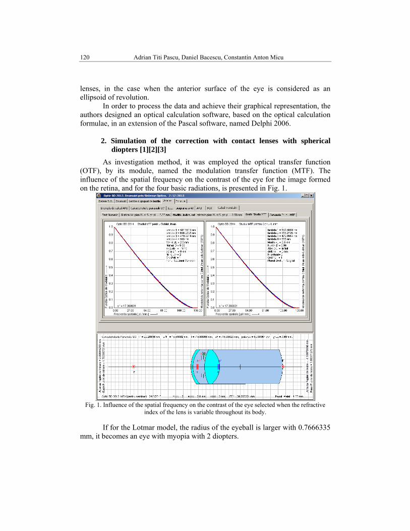
As investigation method, it was employed the optical transfer function (OTF), by its module, named the modulation transfer function (MTF). The influence of the spatial frequency on the contrast of the eye for the image formed on the retina, and for the four basic radiations, is presented in Fig. 1.
Fig. 1. Influence of the spatial frequency on the contrast of the eye selected when the refractive
index of the lens is variable throughout its body.
If for the Lotmar model, the radius of the eyeball is larger with 0.7666335 mm, it becomes an eye with myopia with 2 diopters.
Checking the corrective lenses for the spherical diopters […] the aspherical diopters hypothesis121
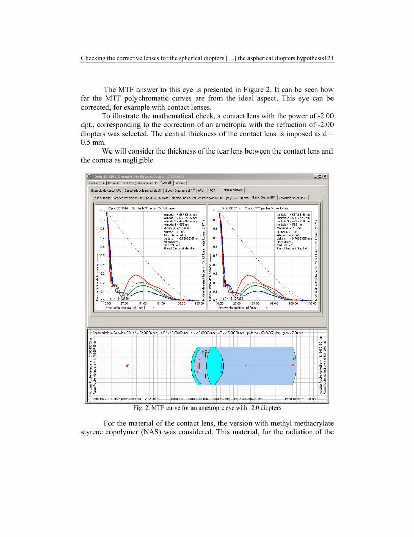
The MTF answer to this eye is presented in Figure 2. It can be seen how far the MTF polychromatic curves are from the ideal aspect. This eye can be corrected, for example with contact lenses. To illustrate the mathematical check, a contact lens with the power of -2.00 dpt., corresponding to the correction of an ametropia with the refraction of -2.00 diopters was selected. The central thickness of the contact lens is imposed as d = 0.5 mm. We will consider the thickness of the tear lens between the contact lens and the cornea as negligible.
Fig. 2. MTF curve for an ametropic eye with -2.0 diopters
For the material of the contact lens, the version with methyl methacrylate styrene copolymer (NAS) was considered. This material, for the radiation of the
122 Adrian Titi Pascu, Daniel Bacescu, Constantin Anton Micu
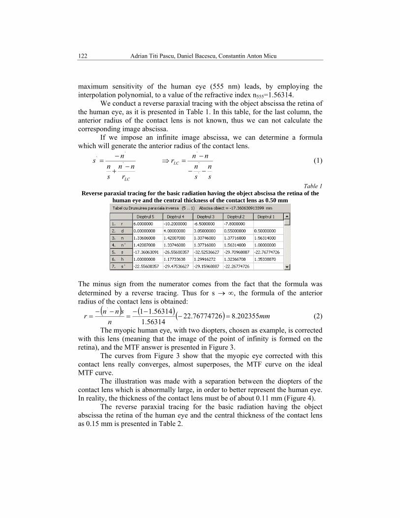
maximum sensitivity of the human eye (555 nm) leads, by employing the interpolation polynomial, to a value of the refractive index n555=1.56314. We conduct a reverse paraxial tracing with the object abscissa the retina of the human eye, as it is presented in Table 1. In this table, for the last column, the anterior radius of the contact lens is not known, thus we can not calculate the corresponding image abscissa. If we impose an infinite image abscissa, we can determine a formula which will generate the anterior radius of the contact lens.
LCrnn
sn
ns−
+
−= `
``
sn
sn
nnrLC
−−
−=⇒
`
`
`
(1)
Table 1 Reverse paraxial tracing for the basic radiation having the object abscissa the retina of the
human eye and the central thickness of the contact lens as 0.50 mm
The minus sign from the numerator comes from the fact that the formula was determined by a reverse tracing. Thus for s → ∞, the formula of the anterior radius of the contact lens is obtained:
( ) ( ) ( ) mmn
snnr 202355.876774726.2256314.1
56314.11`
=−−−
=−−
= (2)
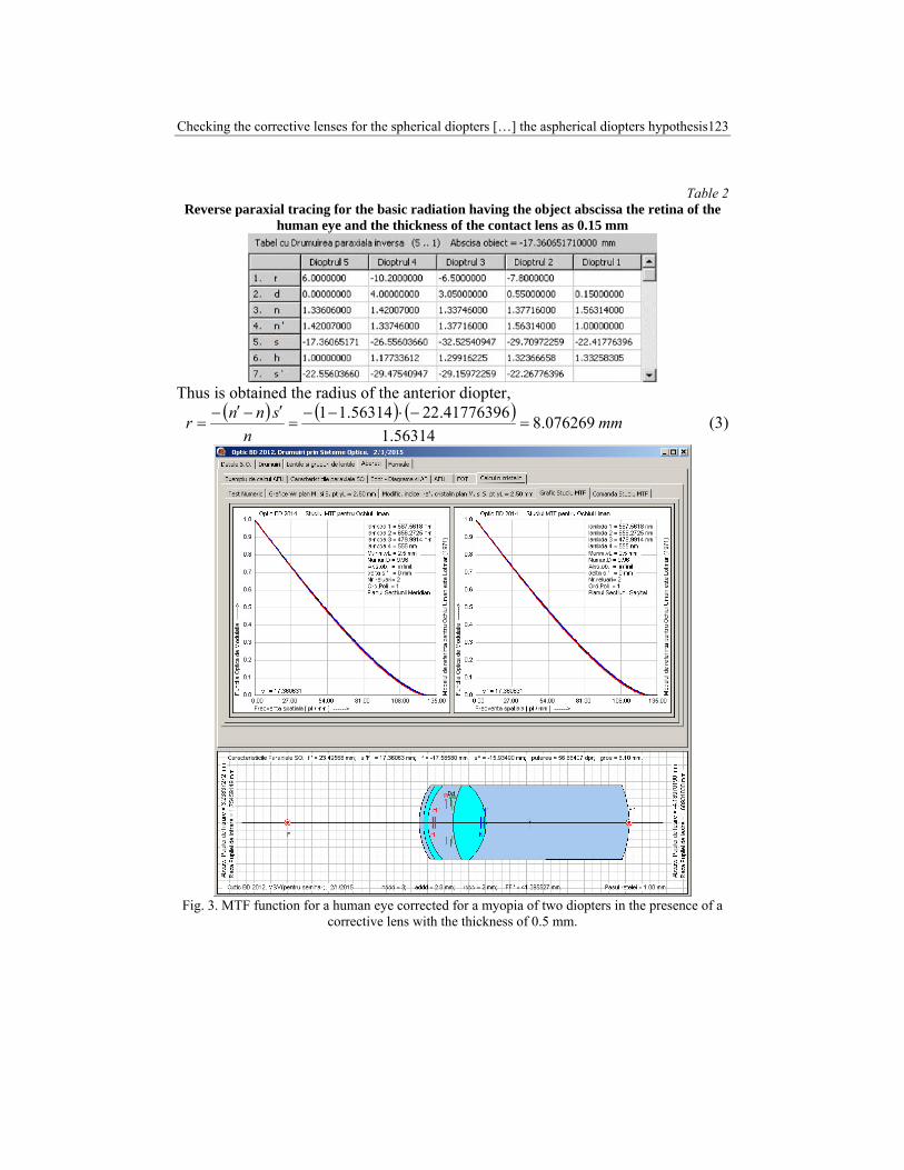
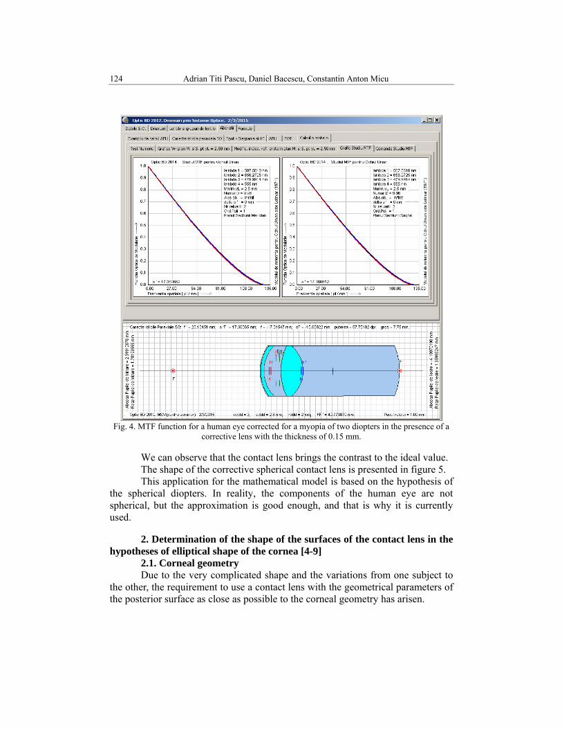
The myopic human eye, with two diopters, chosen as example, is corrected with this lens (meaning that the image of the point of infinity is formed on the retina), and the MTF answer is presented in Figure 3. The curves from Figure 3 show that the myopic eye corrected with this contact lens really converges, almost superposes, the MTF curve on the ideal MTF curve. The illustration was made with a separation between the diopters of the contact lens which is abnormally large, in order to better represent the human eye. In reality, the thickness of the contact lens must be of about 0.11 mm (Figure 4). The reverse paraxial tracing for the basic radiation having the object abscissa the retina of the human eye and the central thickness of the contact lens as 0.15 mm is presented in Table 2.
Checking the corrective lenses for the spherical diopters […] the aspherical diopters hypothesis123
Table 2 Reverse paraxial tracing for the basic radiation having the object abscissa the retina of the
human eye and the thickness of the contact lens as 0.15 mm
Thus is obtained the radius of the anterior diopter,
( ) ( ) ( ) mmn
snnr 076269.856314.1
41776396.2256314.11=
−⋅−−=
′−′−= (3)
Fig. 3. MTF function for a human eye corrected for a myopia of two diopters in the presence of a
corrective lens with the thickness of 0.5 mm.
124 Adrian Titi Pascu, Daniel Bacescu, Constantin Anton Micu
Fig. 4. MTF function for a human eye corrected for a myopia of two diopters in the presence of a
corrective lens with the thickness of 0.15 mm.
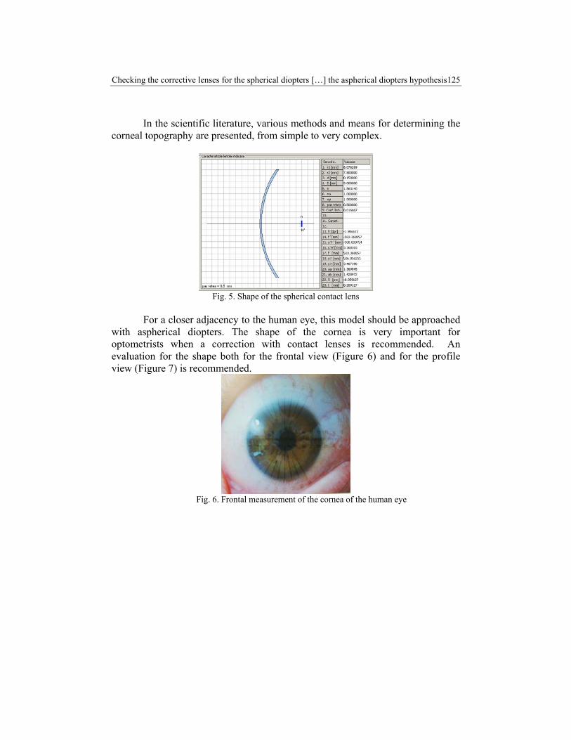
We can observe that the contact lens brings the contrast to the ideal value. The shape of the corrective spherical contact lens is presented in figure 5. This application for the mathematical model is based on the hypothesis of the spherical diopters. In reality, the components of the human eye are not spherical, but the approximation is good enough, and that is why it is currently used.
2. Determination of the shape of the surfaces of the contact lens in the hypotheses of elliptical shape of the cornea [4-9]
2.1. Corneal geometry Due to the very complicated shape and the variations from one subject to the other, the requirement to use a contact lens with the geometrical parameters of the posterior surface as close as possible to the corneal geometry has arisen.
Checking the corrective lenses for the spherical diopters […] the aspherical diopters hypothesis125
In the scientific literature, various methods and means for determining the corneal topography are presented, from simple to very complex.
Fig. 5. Shape of the spherical contact lens
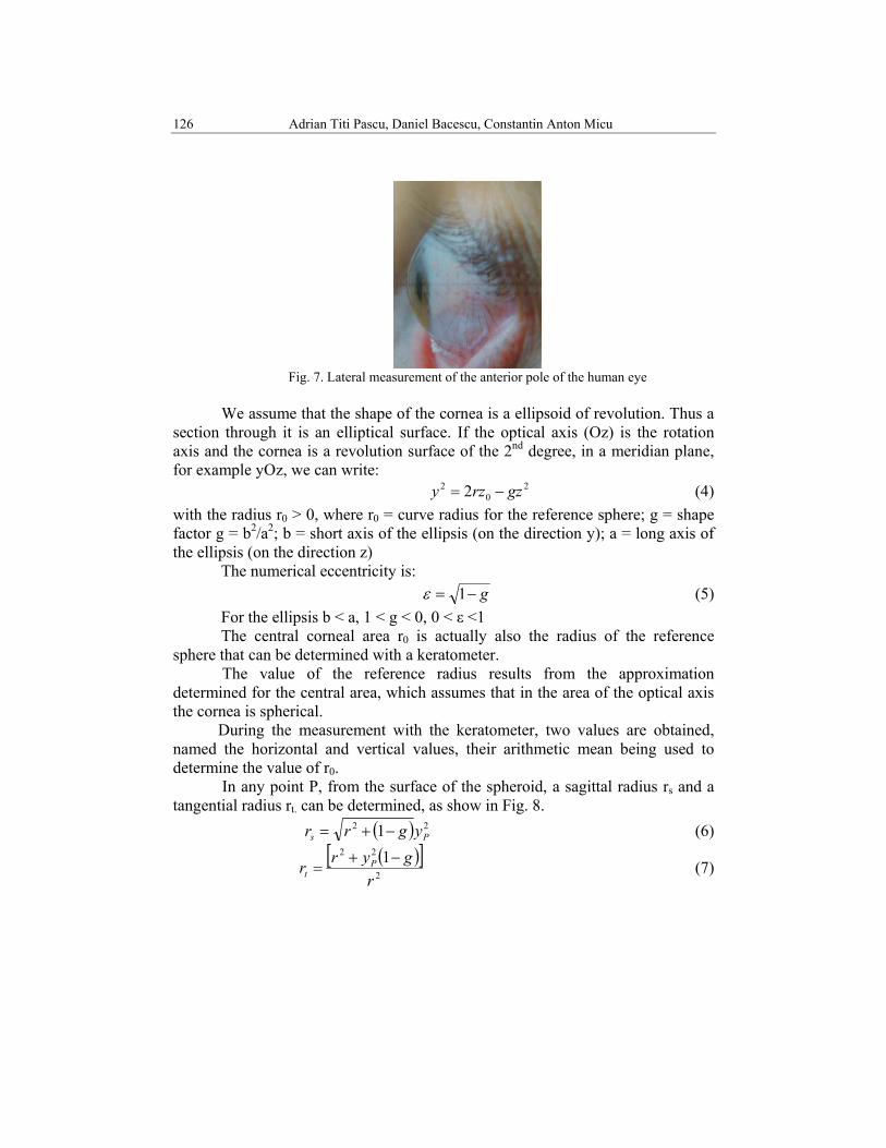
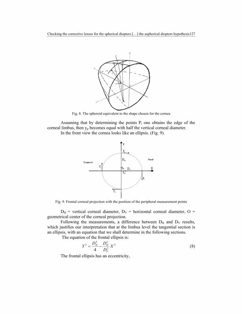
For a closer adjacency to the human eye, this model should be approached
with aspherical diopters. The shape of the cornea is very important for optometrists when a correction with contact lenses is recommended. An evaluation for the shape both for the frontal view (Figure 6) and for the profile view (Figure 7) is recommended.
Fig. 6. Frontal measurement of the cornea of the human eye
126 Adrian Titi Pascu, Daniel Bacescu, Constantin Anton Micu
Fig. 7. Lateral measurement of the anterior pole of the human eye
We assume that the shape of the cornea is a ellipsoid of revolution. Thus a
section through it is an elliptical surface. If the optical axis (Oz) is the rotation axis and the cornea is a revolution surface of the 2nd degree, in a meridian plane, for example yOz, we can write:
20
2 2 gzrzy −= (4) with the radius r0 > 0, where r0 = curve radius for the reference sphere; g = shape factor g = b2/a2; b = short axis of the ellipsis (on the direction y); a = long axis of the ellipsis (on the direction z)
The numerical eccentricity is: g−= 1ε (5)
For the ellipsis b < a, 1 < g < 0, 0 < ε <1 The central corneal area r0 is actually also the radius of the reference
sphere that can be determined with a keratometer. The value of the reference radius results from the approximation determined for the central area, which assumes that in the area of the optical axis the cornea is spherical. During the measurement with the keratometer, two values are obtained, named the horizontal and vertical values, their arithmetic mean being used to determine the value of r0. In any point P, from the surface of the spheroid, a sagittal radius rs and a tangential radius rt. can be determined, as show in Fig. 8.
( ) 22 1 Ps ygrr −+= (6)
( )[ ]2
22 1r
gyrr Pt
−+= (7)
Checking the corrective lenses for the spherical diopters […] the aspherical diopters hypothesis127
Fig. 8. The spheroid equivalent to the shape chosen for the cornea
Assuming that by determining the points Pi one obtains the edge of the corneal limbus, then yp becomes equal with half the vertical corneal diameter. In the front view the cornea looks like an ellipsis. (Fig. 9).
Fig. 9. Frontal corneal projection with the position of the peripheral measurement points
DH = vertical corneal diameter, DV = horizontal corneal diameter, O =
geometrical center of the corneal projection. Following the measurements, a difference between DH and DV results, which justifies our interpretation that at the limbus level the tangential section is an ellipsis, with an equation that we shall determine in the following sections. The equation of the frontal ellipsis is:
22
222
4X
DDDY
V
HH −= (8)
The frontal ellipsis has an eccentricity,
128 Adrian Titi Pascu, Daniel Bacescu, Constantin Anton Micu
2
2
1V
HN D
DE −= (9)
From a study conducted on a number of 56 subjects, it resulted that the numerical eccentricity of the frontal ellipsis is between 0.18 and 0.43. The evaluation and frontal measurement were conducted with the microscope model RS 7001 Kansum, provided with a high performance optical system that determines a very good quality of the images obtained for each individual environment. One of the eye glasses of the microscope is provided with a measuring crosshair with the value of the divisions of 0.1 mm, which can be rotated at 360° from its own axis. The zoom of the microscope is adjusted 10x. The microscope can be positioned frontally or laterally to the human eye.
Table 3 Illustration of the study regarding the eccentricity of the frontal corneal ellipsis for the right eye
No DH(mm) DV(mm) DV/DH (DV/DH)2 EN z(mm) 1 9.2 8.5 0.923913 0.853615 0.38 2.00 2 11.3 10.3 0.907489 0.823536 0.42 3.6 3 9 8.1 0.905556 0.820031 0.43 2.60 4 10.8 9.6 0.889401 0.791034 0.46 3.00 5 9.3 8.7 0.930481 0.865795 0.37 2.70
The measurement diagram for obtaining the coordinates of the points Pi is the one presented in Figure 8. Thus, four peripheral radiuses are obtained, that will determine an average sagittal radius. The keratometer employed for the measuring is of Javal type. We assume that the measuring angle, from the center of the entry pupil of the microscope of the ophthalmometer (keratometer) is of 30° and that in the peripheral corneal points will be reached the level z for known coordinates.
4
4
1∑== i
i
s
rr
, 2
000
yx rrr
+=
(10) Thus, it results an average numerical eccentricity of the spheroid
⎟⎟⎠
⎞⎜⎜⎝
⎛−= 2
2014s
m rrE (11)
2.2. Contact lens with the posterior surface an ellipsoid
The ellipsis equation from formula 1 results as ( ) 22
02 12 zEzry m−−= (12)
From the same study, it results the peripheral keratometric measurements for the right eye according to table 4.

Checking the corrective lenses for the spherical diopters […] the aspherical diopters hypothesis129
Table 4 Illustration of the study regarding the measuring of the central and peripheral radiuses No. Peripheral keratometry for the right eye
r0x(mm) r1(mm) r2(mm) r0y(mm) r3(mm) r4(mm) Em 1 7.50 7.90 7.80 7.50 7.60 7.70 0.505 2 7.50 7.80 7.70 7.60 7.70 7.80 0.451 3 7.60 7.80 7.70 7.50 7.60 7.70 0.392 4 7.40 7.70 7.60 7.70 7.80 7.90 0.451 5 8.00 8.10 8.20 8.20 8.60 8.50 0.485
For the example, we consider that the radius of the cornea in the center of the optical area is r0 = 7.80 mm and the eccentricity is Em = 0.50. The equation of the posterior surface of the contact lens, according to Equation (1), in order to result a parallel adaptation will be:
y2 – 15 z +0.75z 2 = 0 (13) The determination of the level z (corneal arrow) was made with the same
bio-microscope RS7001 Kansum, as in Figure 7 and the data is provided in Table 3. For illustration the level z = 2 mm, and y is corresponding to line no. 1 from Table 3, meaning y = DV /2 = 4.275 mm. The graphical representation of the ellipsis of the posterior surface of the corrective lens is in Figure 10.
Fig. 10. Graphical representation of the ellipsis of the posterior surface of the corrective lens
In a subsequent paper, the check of the corrective lenses starting from the aspherical diopters hypothesis will be presented.
2. Conclusions Analyzing the performances of this eye corrected with the contact lens, for the same aberrations, we can reach the conclusion if the contact lens is good or
130 Adrian Titi Pascu, Daniel Bacescu, Constantin Anton Micu
not. When this analysis gives an unfavorable conclusion, some of the constructive parameters of the contact lens can be modified, meaning we can conduct a fine correction, or, in other words, an optimization of the constructive parameters of the contact lens up to an acceptable value. With regard to the situation of the spherical diopters hypothesis, applied on a mathematical model of the human eye, the checking of the visual comfort with the corrective lens was conducted with the application of the modulation transfer function. This result is close to reality, but not precise. To approach a real situation was drafted the approach with aspherical diopters, applied to the anterior diopter of the cornea. For this, measurements for a number of representative subjects were conducted. There is usually a gradual increase of the radius with a higher rate of variation towards the periphery, determining practically an elliptical shape for the cornea. The central section is approximated with a spherical shape even if not all patients have this area. The peak of the central area is usually irregular, usually an ellipsis or another geometric shape. This area is usually of center from the line of sight, and tilted up temporally or up and nasally. A large enough centering deviation can affect considerably the centering of the contact lenses. There were no significant variation depending on age, sex, fixing direction or accommodation when maturing and almost absent with any modifications during the day. There are considerable variations from person to person, some even present negative asphericity. So far there are no satisfactory clinical methods for routine and follow up measurements for the changes of the topography. The continuous improvement of the mathematical model of the eye, with aspherical diopters, involves a huge mathematical effort, because one must determine the formulae for the 3D tracing, for aspherical surfaces.
R E F E R E N C E S [1]. D. Băcescu , A. T.Pascu ,Completing the Lotmar model for the human eye with the crystalline
lens refraction index variation function,JOAM, iss.5-6, 2015 [2]. D.Băcescu , Visual system modeling, (in romanian) ,PUB- Course notes, 2012 [3]. D.Băcescu , Applied Optical. Analysis and synthesis components ,(in romanian), Ed. Medro
2004. [4]. A.T. Pascu, The role, design and manufacture of contact lenses,(in romanian) Ed. Atkins,
Bucuresti, 2000 [5]. N.Dumitrescu , Contact lens. Course notes, (in romanian ),Bucharest 2001 [6]. W.Grimm, vorlesungsskript, 12. durchgesehene Auflage [7]. T Olsen, On the calculation of power from curvature of the cornea,Br.Journ.Ophthalmol.1986. [8]. M. Gersten, R.J .Mammore, N.J. Brunswick. And Larchmont N.Y.,System for topographical
modeling of anatomical surfaces,US Patent nr.4,863,260,1989 [9]. J.D. Doss, R..L. Hutson, J.J.,Rowswy and R. Brown ,Method for calculation of corneal profile
and power distribution.Arch.Ophthalmol.1981;99,1261-1265.