Embed Size (px)
Citation preview
8/3/2019 Charlotte G. Jørgensen et al- Synthesis and pharmacology of glutamate receptor ligands: new isothiazole analogue…
http://slidepdf.com/reader/full/charlotte-g-jorgensen-et-al-synthesis-and-pharmacology-of-glutamate-receptor 1/9
PAPER www.rsc.org/obc | Organic & Biomolecular Chemistry
Synthesis and pharmacology of glutamate receptor ligands: new isothiazoleanalogues of ibotenic acid†
Charlotte G. Jørgensen,a Rasmus P. Clausen,a Kasper B. Hansen,b Hans Brauner-Osborne,a Birgitte Nielsen,a
Bjørn Metzler,a Jan Kehler,b Povl Krogsgaard-Larsena and Ulf Madsen*a
Received 18th October 2006, Accepted 5th December 2006 First published as an Advance Article on the web 20th December 2006
DOI: 10.1039/b615162k
The naturally occurring heterocyclic amino acid ibotenic acid (Ibo) and the synthetic analogue
thioibotenic acid (Thio-Ibo) possess interesting but dissimilar pharmacological activity at ionotropic
and metabotropic glutamate receptors (iGluRs and mGluRs). Therefore, a series of Thio-Ibo analogues
was synthesized. The synthesis included introduction of substituents by Suzuki and Grignard reactions
on 4-halogenated 3-benzyloxyisothiazolols, reduction of the obtained alcohols, followed by
introduction of the amino acid moiety by use of 2-( N -tert-butoxycarbonylimino)malonic acid diethyl
ester. The obtained Thio-Ibo analogues (1, 2a–g) were characterized in functional assays on
recombinant mGluRs and in receptor binding assays on native iGluRs. At mGluRs, the activity at
Group II was retained for compounds with small substituents (2a–2d), whereas the Group I and Group
III receptor activities for all new compounds were lost. Detection of NMDA receptor affinity prompted
further characterization, and two-electrode voltage-clamp recordings at recombinant NMDA receptor
subtypes NR1/NR2A-D expressed in Xenopus oocytes were carried out for compounds with small
substituents (chloro, bromo, methyl or ethyl, compounds 2a–d). This series of Thio-Ibo analogues
defines a structural threshold for NMDA receptor activation and reveals that the individual subtypes
have different steric requirements for receptor activation. The compounds 2a and 2c are the first
examples of agonists discriminating individual NMDA subtypes.
Introduction
(S )-Glutamic acid (Glu) is the main excitatory neurotransmitter
in the central nervous system and activates ionotropic glutamate
receptors (iGluRs) as well as metabotropic glutamate receptors(mGluRs). The mGluRs and iGluRs are both implicated in
fundamental physiological processes and are involved in the devel-
opment of several neurological diseases.1–4 Within the last decade
many selective compounds have emerged providing important
pharmacological tools, but the pharmacological coverage of the
Glu receptors is still incomplete.2
According to activation by the selective agonists, the iGluRs
are divided into N -methyl-D-aspartic acid (NMDA), (S )-2-amino-
3-(3-hydroxy-5-methyl-4-isoxazolyl)propionic acid (AMPA) and
kainic acid (KA) receptors. Seven NMDA (NR1, NR2A-D,
NR3A-B), four AMPA (GluR1-4) and five KA (GluR5-7 and
KA1-2) receptor subunits have been identified.1 The native
iGluRs are tetrameric combinations of the respective subunits,of which NMDA and AMPA receptors are likely heteromeric
and KA receptors may be either homomeric or heteromeric in
composition.1,5,6 Functional NMDA receptors are heteromeric
assemblies typically of two NR1 subunits and two NR2 subunits,
aDepartment of Medicinal Chemistry, The Danish University of Pharma-ceutical Sciences, 2100, Copenhagen, Denmark. E-mail: [email protected];Fax: +45 35306040bH. Lundbeck A/S, 2500, Valby, Copenhagen, Denmark
† Electronic supplementary information (ESI) available: Detailed exper-imental procedures and compound characterization data of compounds1, 2b–g, 3, 5, 6, 7b–c, f–g, 9c,f–g, 10, 11, 12, 13b–g, 14b–g. See DOI:10.1039/b615162k
that are activated by simultaneous binding of glycine and Glu.
The NR1 subunits provide the glycine binding sites and the NR2
subunits form the Glu binding sites.5
Eight subtypes of mGluRs have been characterized and these
are divided into three groups according to protein sequenceidentity and signal transduction pathways: Group I (mGluR1
and mGluR5), Group II (mGluR2 and mGluR3) and Group III
(mGluR4, mGluR6, mGluR7 and mGluR8).2,4
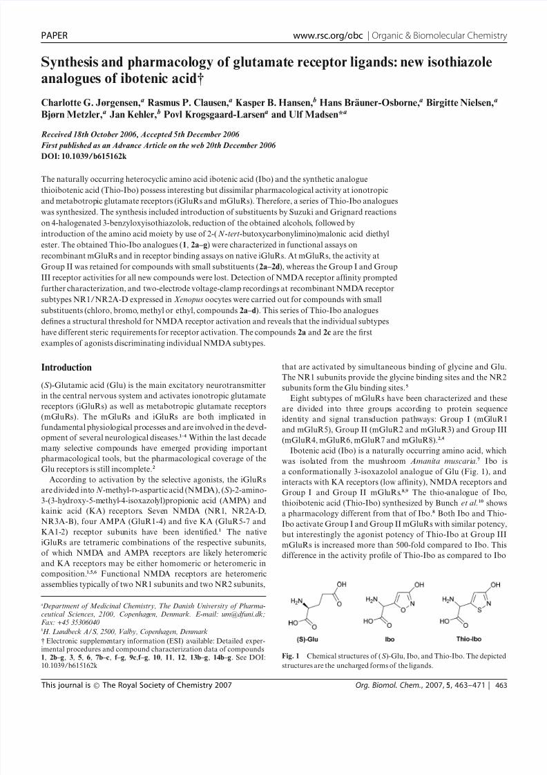
Ibotenic acid (Ibo) is a naturally occurring amino acid, which
was isolated from the mushroom Amanita muscaria.7 Ibo is
a conformationally 3-isoxazolol analogue of Glu (Fig. 1), and
interacts with KA receptors (low affinity), NMDA receptors and
Group I and Group II mGluRs.8,9 The thio-analogue of Ibo,
thioibotenic acid (Thio-Ibo) synthesized by Bunch et al.10 shows
a pharmacology different from that of Ibo.8 Both Ibo and Thio-
Ibo activate Group I and Group II mGluRs with similar potency,
but interestingly the agonist potency of Thio-Ibo at Group III
mGluRs is increased more than 500-fold compared to Ibo. Thisdifference in the activity profile of Thio-Ibo as compared to Ibo
Fig. 1 Chemical structures of (S )-Glu, Ibo, and Thio-Ibo. The depicted
structures are the uncharged forms of the ligands.
This journal is © The Royal Society of Chemistry 2007 Org. Biomol. Chem., 2007, 5, 463–471 | 463

8/3/2019 Charlotte G. Jørgensen et al- Synthesis and pharmacology of glutamate receptor ligands: new isothiazole analogue…
http://slidepdf.com/reader/full/charlotte-g-jorgensen-et-al-synthesis-and-pharmacology-of-glutamate-receptor 2/9
was ascribed to a lowering of the relative energy for the preferred
mGluR4 active conformation.8
Based on the unique pharmacology of Thio-Ibo, a selection of
analogues was chosen as model compounds in order to further
investigate the structure–activity relationship at both iGluRs and
mGluRs.
Eight different substituents were chosen from three categories:
(1) a-substituents (compound 1). Some mGluR agonists have
successfully been converted into antagonists by introducing sub-
stituents in the a-position of the amino acid.4,11 (2) Halogen
and alkyl substituents (compounds 2a–d), in analogy with other
series of subtype selective Glu receptor active compounds.12,13 (3)
Aromatic substituents (compounds 2e–g) (Fig. 2).
Fig. 2 Chemical structures of the new Thio-Ibo analogues 1 and 2a–g.
Results
Chemistry
Direct functionalization of a protected Thio-Ibo derivative was
not feasible and instead the amino acid analogues were prepared
via 3-benzyloxyisothiazoles.
An amino acid synthon, ketimine 3, for the preparation of
a-methyl amino acids, was prepared by an aza-Wittig reaction
(Scheme 1).14 Regioselective lithiation of the 5-position of 4 was
accomplished with LDA,10 and further reaction with ketimine 3
resulted in the protected amino acid 5. Ketimine 3 exists as a
tautomeric mixture (Scheme 1) as observed by NMR, and this
may explain the moderate yield (36%) of the protected amino acid
5. The ester 5 was hydrolyzed under basic conditions yielding 6.
Subsequent treatment with hydrogen bromide in glacial acetic acid
removed the Boc and benzyl protecting groups to give compound
1. This two-step deprotection procedure was found to be more
efficient than acidic deprotection of 5 in one step.
Scheme 1 Reagents and conditions: a) THF, reflux, 30%; b) LDA, Et2O,
−78 ◦C, then imine 3, −78 ◦C, 36%; c) LiOH (aq), THF, rt, 75%; d) HBr,
AcOH, rt, 21%.
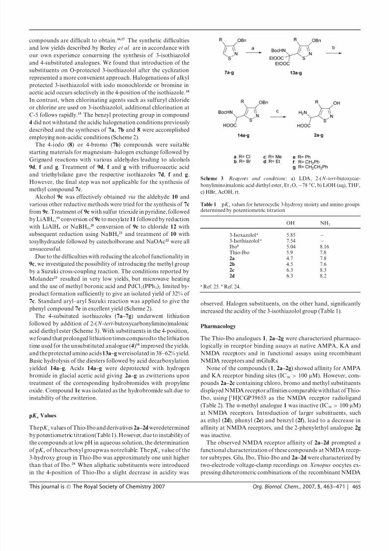
The 4-substituted Thio-Ibo derivatives (2a–g) were prepared
from the corresponding 4-substituted 3-benzyloxyisothiazoles
(7a–7g) (Scheme 2).
Earlier reports by Lewis et al. on the synthesis of 3-isothiazolol
and 4-methylisothiazolol have involved cyclization reactions of
3,3-dithiodipropionamides.15 In 1994, Beeley et al. reported very
low yields using this method to synthesize 3-isothiazolol.16 More-
over, the acyclic starting materials for the synthesis of 4-substituted
Scheme 2 Reagents and conditions: a) 7a: NCS, MeCN, 58%; 7b: NBS, MeCN, 88%; 8: ICl, K2CO3, CHCl3, 82%; b) i PrMgCl, RCHO, THF; 9d: 84%
(from 8); 9f : 86% (from 8); 9g: 26% (from 8) or 43% (from 7b); c) TFA, Et3SiH, CH2Cl2; 7d: 70%; 7f : 54%; 7g: 65%; d) i PrMgCl, DMF, THF, 82% (from
8); e) NaBH4, MeOH, 98%; f) 11: MsCl, Et3N, THF; 12: SOCl2; h) 7c: MeB(OH)2, PdCl2(PPh3)2, K2CO3, DMF, MW 160 ◦C, 5 min, 32% (from 7b); 7e:
PhB(OH)2, PdCl2(PPh3)2, K2CO3 (aq), DMF, 94% (from 8).
464 | Org. Biomol. Chem., 2007, 5, 463–471 This journal is © The Royal Society of Chemistry 2007
8/3/2019 Charlotte G. Jørgensen et al- Synthesis and pharmacology of glutamate receptor ligands: new isothiazole analogue…
http://slidepdf.com/reader/full/charlotte-g-jorgensen-et-al-synthesis-and-pharmacology-of-glutamate-receptor 3/9
compounds are difficult to obtain.16,17 The synthetic difficulties
and low yields described by Beeley et al. are in accordance with
our own experience concerning the synthesis of 3-isothiazolol
and 4-substituted analogues. We found that introduction of the
substituents on O-protected 3-isothiazolol after the cyclization
represented a more convenient approach. Halogenations of alkyl
protected 3-isothiazolol with iodo monochloride or bromine in
acetic acid occurs selectively in the 4-position of the isothiazole.18
In contrast, when chlorinating agents such as sulfuryl chloride
or chlorine are used on 3-isothiazolol, additional chlorination at
C-5 follows rapidly.15 The benzyl protecting group in compound
4 did not withstand the acidic halogenation conditions previously
described and the syntheses of 7a, 7b and 8 were accomplished
employing non-acidic conditions (Scheme 2).
The 4-iodo (8) or 4-bromo (7b) compounds were suitable
starting materials for magnesium–halogen exchange followed by
Grignard reactions with various aldehydes leading to alcohols
9d, f and g. Treatment of 9d, f and g with trifluoroacetic acid
and triethylsilane gave the respective isothiazoles 7d, f and g.
However, the final step was not applicable for the synthesis of
methyl compound 7c.
Alcohol 9c was effectively obtained via the aldehyde 10 and
various other reductive methods were tried for the synthesis of 7c
from 9c. Treatment of 9c with sulfur trioxide in pyridine, followed
by LiAlH4,19 conversion of 9c to mesylate 11 followed by reduction
with LiAlH4 or NaBH4,20 conversion of 9c to chloride 12 with
subsequent reduction using NaBH421 and treatment of 10 with
tosylhydrazide followed by catecholborane and NaOAc22 were all
unsuccessful.
Due to the difficulties with reducing the alcohol functionality in
9c, we investigated the possibility of introducing the methyl group
by a Suzuki cross-coupling reaction. The conditions reported by
Molander23 resulted in very low yields, but microwave heating
and the use of methyl boronic acid and PdCl2(PPh3)2 limited by-
product formation sufficiently to give an isolated yield of 32% of
7c. Standard aryl–aryl Suzuki reaction was applied to give the
phenyl compound 7e in excellent yield (Scheme 2).
The 4-subsituted isothiazoles (7a–7g) underwent lithiation
followed by addition of 2-(N -tert-butoxycarbonylimino)malonic
acid diethyl ester (Scheme 3). With substituents in the 4-position,
we found that prolonged lithiation times comparedto the lithiation
time used for the unsubstituted analogue (4)10 improved the yields,
and the protected amino acids13a–g wereisolated in 38–62% yield.
Basic hydrolysis of the diesters followed by acid decarboxylation
yielded 14a–g. Acids 14a–g were deprotected with hydrogen
bromide in glacial acetic acid giving 2a–g as zwitterions upon
treatment of the corresponding hydrobromides with propylene
oxide. Compound 1c was isolated as the hydrobromide salt due to
instability of the zwitterion.
pK a Values
ThepK a values of Thio-Ibo and derivatives 2a–2d weredetermined
by potentiometric titration(Table 1). However, due to instability of
the compounds at low pH in aqueous solution, the determination
of pK a of thecarboxyl groupwas notreliable. The pK a value of the
3-hydroxy group in Thio-Ibo was approximately one unit higher
than that of Ibo.24 When aliphatic substituents were introduced
in the 4-position of Thio-Ibo a slight decrease in acidity was
Scheme 3 Reagents and conditions: a) LDA, 2-(N -tert-butoxycar-
bonylimino)malonic acid diethyl ester, Et2O,−78 ◦C, b) LiOH (aq), THF,
c) HBr, AcOH, rt.
Table 1 pK a values for heterocyclic 3-hydroxy moiety and amino groupsdetermined by potentiometric titration
OH NH2
3-Isoxazolola 5.85 −
3-Isothiazolol a 7.54 −
Ibob 5.04 8.16Thio-Ibo 5.9 7.82a 4.7 7.82b 4.5 7.62c 6.3 8.32d 6.3 8.2
a Ref. 25. b Ref. 24.
observed. Halogen substituents, on the other hand, significantly
increased the acidity of the 3-isothiazolol group (Table 1).
Pharmacology
The Thio-Ibo analogues 1, 2a–2g were characterized pharmaco-
logically in receptor binding assays at native AMPA, KA and
NMDA receptors and in functional assays using recombinant
NMDA receptors and mGluRs.
None of the compounds (1, 2a–2g) showed affinity for AMPA
and KA receptor binding sites (IC50 > 100 lM). However, com-
pounds 2a–2c containing chloro, bromo and methyl substituents
displayed NMDA receptor affinities comparable withthat of Thio-
Ibo, using [3H]CGP39653 as the NMDA receptor radioligand
(Table 2). The a-methyl analogue 1 was inactive (IC50 > 100 lM)
at NMDA receptors. Introduction of larger substituents, such
as ethyl (2d), phenyl (2e) and benzyl (2f ), lead to a decrease in
affinity at NMDA receptors, and the 2-phenylethyl analogue 2g
was inactive.
The observed NMDA receptor affinity of 2a–2d prompted a
functional characterization of these compounds at NMDA recep-
tor subtypes. Glu, Ibo, Thio-Ibo and 2a–2d were characterized by
two-electrode voltage-clamp recordings on Xenopus oocytes ex-
pressing diheteromeric combinations of the recombinant NMDA
This journal is © The Royal Society of Chemistry 2007 Org. Biomol. Chem., 2007, 5, 463–471 | 465
8/3/2019 Charlotte G. Jørgensen et al- Synthesis and pharmacology of glutamate receptor ligands: new isothiazole analogue…
http://slidepdf.com/reader/full/charlotte-g-jorgensen-et-al-synthesis-and-pharmacology-of-glutamate-receptor 4/9
Table 2 Receptor binding affinities at iGluRs in rat cortical membranesa
[3H]AMPAIC50/lM
[3H]KAIC50/lM
[3H]CGP39653K i/lM
Glu 0.34b 0.38b 0.20c
Ibo >100d 22d 5.3d
Thio-Ibo >100d >100d 13d
1 >100 >100 >100
2a>
100>
100 19 [18;21]2b >100 >100 21 [19;24]2c >100 >100 14 [13;15]2d >100 >100 41 [37;46]2e >100 >100 86 [74;99]2f >100 >100 80 [75;86]2g >100 >100 >100
a Values are expressed as the antilog to the log mean of three individualexperiments. The numbers in brackets [min;max] indicate ± S.E.M.according to a logarithmic distribution of K i.
b Ref. 26. c Ref. 27. d Ref.8.
receptor subunits NR1 and NR2A-D (Fig. 3 and Table 3).
Ibo and Thio-Ibo were found to be partial agonists at all four
subunit combinations, with maximal activities ranging from 57–
79% relative to Glu. The chloro-substituted compound 2a was a
partial agonist at the NR1/NR2A, NR1/NR2B and NR1/NR2D
subtypes, as illustrated by the concentration–response curves in
Fig. 3, whereas 2a was an antagonist at the NR1/NR2C subtype
combination as illustrated by the electrophysiological traces in
Fig. 4. Compounds 2b and 2d were antagonists at all four
subunit combinations (data not shown), and compound 2c was
a partial agonist at NR2B and NR2D containing receptors and
an antagonist at NR2A and NR2C containing receptors (Fig. 3
and 4).
The compounds were also tested for activity at mGluRs
using mGluR1, mGluR2 and mGluR4 as representatives of
metabotropic Group I, II and III receptors, respectively. In
contrast to Ibo and Thio-Ibo, none of the compounds 1 and 2a–
2g showed any activity at mGluR1 at 1 mM. Only compound
2a showed activity as a partial agonist at mGluR4 (Table 4). At
mGluR2, activity was lost for compounds 1 and 2e–g, whereas
compounds containing smaller substituents (2a–2d) retained
mGluR2 agonist activity comparable to that of Ibo and Thio-
Ibo. None of the compounds had antagonist activity at any of the
mGluRs (at 1 mM).
Table 4 Pharmacological activities at cloned mGluRs expressed in CHOcellsa
EC50/lM [pEC50 ± S.E.M.]
mGluR1 mGluR2 mGluR4
(S )-Glu 13b 4.4b 13b
Ibo 43 [4.37 ± 0.01]b 110 [3.97± 0.04]b >1000b
Thio-Ibo 12 [4.96 ± 0.11]c 52 [4.30 ± 0.06]c 2.6 [5.63 ± 0.14]c
1 >1000 >1000 >10002a >1000 150 [3.89 ± 0.16] 295 [3.53± 0.18]d
2b >1000 89 [4.05 ± 0.02] >10002c >1000 140 [3.94 ± 0.20] >10002d >1000 140 [3.92 ± 0.13] >10002e >1000 >1000 >10002f >1000 >1000 >10002g >1000 >1000 >1000
a Functional data were obtained from CHO cell lines stably expressingthe mGluR1a, 2 or 4a receptor subtype. Results on mGluR1a receptorswere obtained by measurement of intracellular Ca2+ levels and resultsfor mGluR2 and 4a were determined by measurement of intracellularcAMP. Data are given as means of at least three independent experiments.b Ref. 9. c Ref. 8. d Partial agonism (relative efficacy = 54%).
Molecular modeling
Ligand binding domains from all three classes of iGluRs have
been crystallized and homology models have been constructed.1,28
The X-ray crystal structures of soluble AMPA-selective GluR2
constructs of the extracellular Glu binding domain (S1S2) cut off
from the membrane spanning part have revealed a clamshell-like
binding domain which closes around the ligand.29 This activation
of the GluR2 receptor leads to a positive correlation between the
agonist efficacy of the ligand and the degree of domain closure it
can induce. We have recently performed a mutation study using a
homology model of the Glu binding domain of NR2B based on
the crystal structure of the glycine binding domain of NR1.30 The
published crystal structure of the Glu binding domain of NR2A31
prompted us to rebuildthe modelof NR2B–S1S2 using Prime 3.532
now including two water molecules in the binding site. Although
there are small changes from the previous structure, the models
of NR2B produce similar results in the ligand–protein docking
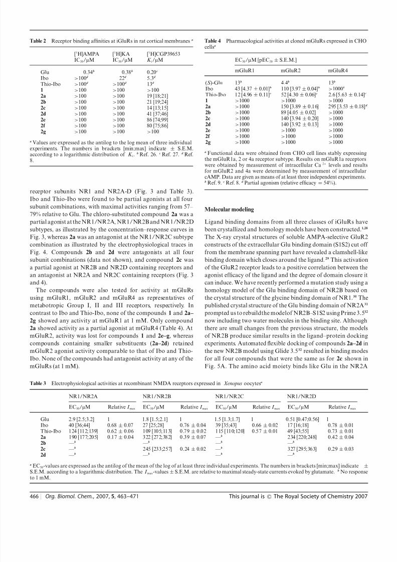
experiments. Automated flexible docking of compounds 2a–2d in
the new NR2B model using Glide 3.532 resulted in binding modes
for all four compounds that were the same as for 2c shown in
Fig. 5A. The amino acid moiety binds like Glu in the NR2A
Table 3 Electrophysiological activities at recombinant NMDA receptors expressed in Xenopus oocytesa
NR1/NR2A NR1/NR2B NR1/NR2C NR1/NR2D
EC50/lM Relative I max EC50/lM Relative I max EC50/lM Relative I max EC50/lM Relative I max
Glu 2.9 [2.5;3.2] 1 1.8 [1.5;2.1] 1 1.5 [1.3;1.7] 1 0.51 [0.47;0.56] 1Ibo 40 [36;44] 0.68 ± 0.07 27 [25;28] 0.76 ± 0.04 39 [35;43] 0.66 ± 0.02 17 [16;18] 0.78 ± 0.01Thio-Ibo 124 [112;139] 0.62± 0.06 109 [105;113] 0.79± 0.02 115 [110;120] 0.57± 0.01 49 [43;55] 0.73 ± 0.012a 190 [177;205] 0.17± 0.04 322 [272;382] 0.39± 0.07 —b 234 [220;248] 0.42± 0.042b —b —b —b —b
2c —b 245 [233;257] 0.24 ± 0.02 —b 327 [295;363] 0.29± 0.032d —b —b —b —b
a EC50-values are expressed as the antilog of the mean of the log of at least three individual experiments. The numbers in brackets [min;max] indicate ±S.E.M. according to a logarithmic distribution. The I max-values± S.E.M. are relative to maximal steady-state currents evoked by glutamate. b No responseto 1 mM.
466 | Org. Biomol. Chem., 2007, 5, 463–471 This journal is © The Royal Society of Chemistry 2007

8/3/2019 Charlotte G. Jørgensen et al- Synthesis and pharmacology of glutamate receptor ligands: new isothiazole analogue…
http://slidepdf.com/reader/full/charlotte-g-jorgensen-et-al-synthesis-and-pharmacology-of-glutamate-receptor 5/9
Fig. 3 A–D: Mean concentration–response curves obtained by two-electrode voltage-clamp recordings at NR1/NR2A-D receptorsexpressed in Xenopus
oocytes.
Fig. 4 Two-electrode voltage-clamp recordings showing antagonist ac-
tivities of compound 2c at NR1/NR2A and of compounds 2a and 2c at
NR1/NR2C.
structure and the heterocycle is positioned by hydrogen bonds to
the hydroxyl group and amide N-H of T691. In this binding mode
the substituents in the 4-position of the heterocycle points like an
interdomain wedge between H486 and V686 that are part of the
upper andlower domainin theclamshell structure(Fig.5B). Thus,
it seems likely that the decreasing efficacy with increasing size of
the substituent arises from steric clash with these two residues,
resulting in steric hindrance of clamshell closure. This finding is
in agreement with our mutation study on NR2B, where His486
and Val686 are mutated to residues of increased size resulting in
decreased efficacy of ligands containing substituents protruding
towards this region.30
Discussion
The striking difference in the pharmacological profiles of Ibo
and Thio-Ibo8 prompted the synthesis of a series of a- and 4-
substituted analogues of Thio-Ibo. Direct functionalization of a
protected Thio-Ibo derivative was not feasible and instead the
analogues were prepared via 4-substituted 3-benzyloxyisothiazole
derivatives. Incorporation of substituents into the molecule of
Thio-Ibo significantly narrowed the pharmacological profile com-
pared to Thio-Ibo. Most of the activity at mGluRs was lost
although four compounds were equipotent with Thio-Ibo as
weak agonists at mGluR2. However, the NMDA receptor binding
This journal is © The Royal Society of Chemistry 2007 Org. Biomol. Chem., 2007, 5, 463–471 | 467
8/3/2019 Charlotte G. Jørgensen et al- Synthesis and pharmacology of glutamate receptor ligands: new isothiazole analogue…
http://slidepdf.com/reader/full/charlotte-g-jorgensen-et-al-synthesis-and-pharmacology-of-glutamate-receptor 6/9
Fig. 5 A: (S )-2c (green carbon atoms) docked in a homology model of
NR2B binding domain showing residues within 5 A (grey carbon atoms).
B: (S )-2c docked in a homology model of NR2B showing the protein
backboneas well as residueH486(upper residue) andV686 (lower residue).
H486 from S1 (dark blue) and V686 from S2 (light blue) may be important
determinants for the agonist efficacy of the Thio-Ibo analogues.
site seemed to accommodate most of the substituents in the 4-
position. In particular, analogues containing smaller substituents
retained affinity similar to that of Thio-Ibo. Therefore, the 4-
substituted chloro, bromo, methyl and ethyl Thio-Ibo analogues,
2a–2d, were characterized at recombinant NMDA receptors. In-
terestingly, thisseries of compounds displayed quite heterogeneous
pharmacological profiles on these subtypes. Thio-Ibo analogues
containing the more bulky bromo and ethyl substituents, 2b and
2d, did not activate any of the NMDA receptor subtypes. In
contrast, the smaller chloro and methyl substituents in 2a and
2c mediate agonist activity at several NMDA receptor subtypes,
and Thio-Ibo activates all subtypes. It is notable that the change
in efficacy, when substituent bulk is increased, is different between
the individual subtypes. Thus, the relative agonist efficacy of Thio-
Ibo at NR1/NR2A is approximately 4-fold higher than that of the
chloro derivative (2a), whereas methyl substitution (2c) leads to
antagonism at this subtype. In contrast, only a minor decrease in
relative agonist efficacy is observed for 2a and 2c at NR1/NR2B
and NR1/NR2D. At NR1/NR2C agonist activity is lost for all
four Thio-Ibo analogues (2a–2d).
The determination of the pK a values has substantiated that the
compounds may function as Glu bioisosteres. The pK a values
of the 3-isothiazolol, representing the distal carboxylic acid in
Glu, are in the range of 4.5–6.3 as compared with Ibo which has
a pK a value of 5.04. Thus, at physiological pH the compounds
are expected to be virtually fully ionized. No correlation between
pharmacological activity and the range of pK a values observed
can be concluded.
In this series of Thio-Ibo derivatives it seems plausible, that
the increased bulk of the substituents could limit domain closure
due to steric clashes. The observed differences in efficacy across
subtypes may reflect that the Thio-Ibo analogues are recognized
by theindividualsubtypesin a dissimilar manner or that activation
of the receptor subtypes are differently coupled to binding of the
Thio-Ibo derivatives. A difference in relative efficacy at various
subtypes based on variations in the recognition of the ligands
does not seem likely, given that the residues in the ligand binding
domainof theNMDA receptor arefully conservedthroughoutthe
different NR2 subtypes. Consequently, it maybe hypothesizedthat
the ligands induce similar conformational changes in the ligand
binding domain in all subtypes, but that the energy barrier to acti-
vate theion channel part of thereceptor variesamong thesubtypes.
In conclusion, the substituted Thio-Ibo analogues 2a and 2c are
the first examples of NMDA receptor agonists that differentiate
among the individual receptor subtypes.
Experimental
Chemistry
Detailed experimental procedures and compound characteriza-
tion data of compounds 1, 2b–g, 3, 5, 6, 7b–c, f–g, 9c,f–g, 10, 11,
12, 13b–g, 14b–g are described in supplementary information.†
(RS )-2-Amino-(4-chloro-3-hydroxy-5-isothiazolyl)acetic acid
(2a). (RS )-(3-Benzyloxy-4-chloro-5-isothiazolyl)-(N -tert-butoxy-
carbonylamino)acetic acid (14a) (236 mg,0.6 mmol)was dissolved
in AcOH (3 mL). 10% HBr in AcOH (3 mL) was added. After
stirring for 8 h the reaction was concentrated in vacuo. The
solid was triturated with Et2O, redissolved in 70% EtOH (aq),
treated with propylene oxide whereupon the zwitterion of 2a
was isolated as colorless crystals, which turned light brown
overnight (104 mg, 84%). Mp. >140 ◦C (Found: C, 28.6; H, 2.5;
N, 13.1. Calc. for C5H5ClN2O3S: C, 28.8; H, 2.4; N, 13.4%); TLC(nBuOH–H2O–AcOH–EtOAc 1 : 1 : 1 : 1) Rf 0.4; dH(DMSO-d 6)
4.60 (s, 1H).
3-Benzyloxy-4-chloroisothiazole (7a). 3-Benzyloxyisothiazole
(4) (3.0 g, 16 mmol) was dissolved in MeCN (50 mL) and NCS
(2.3 g, 17 mmol) was added. The mixture was left stirring for 3 d
at rt. The mixture was concentrated to approximately 2 mL and
EtOAc was added. The organic solution was washed with H2O
(2×), followed by brine and dried using MgSO4. Concentration
in vacuo followed by FC (petroleum ether–toluene 1 : 0–1 : 1) gave
pure 7a (2.0 g, 58%) as a clear oiland 0.2g of3-benzyloxy-4-chloro-
5-chloroisothiazole as a yellow oil (Found: 226.0082 [M + H +].
468 | Org. Biomol. Chem., 2007, 5, 463–471 This journal is © The Royal Society of Chemistry 2007
8/3/2019 Charlotte G. Jørgensen et al- Synthesis and pharmacology of glutamate receptor ligands: new isothiazole analogue…
http://slidepdf.com/reader/full/charlotte-g-jorgensen-et-al-synthesis-and-pharmacology-of-glutamate-receptor 7/9
Calc. for C10H9ClNOS: 226.0093 [M + H+]); TLC (petroleum
ether–toluene 1 : 1) Rf 0.6; dH(CDCl3) 5.41 (s, 2H), 7.30–7.44 (m,
5H), 8.15 (s, 1H); dC(CDCl3) 71.0 (CH2), 111.1 (C), 127.9 (CH),
128.1 (CH), 128.4 (CH), 135.9 (C), 143.2 (CH), 163.6 (C); m/z
(EI) 225 (9%, M+), 190, 106, 91.
3-Benzyloxy-4-ethylisothiazole (7d). 1-(3-Benzyloxy-4-iso-
thiazolyl)ethanol (9d) (1.00 g, 4 mmol) was dissolved in dry
CH2Cl2 (15 mL) under N2 and cooled to 0 ◦C. Triethylsilane(1.15 mL, 7 mmol) was added followed by the addition of
trifluoroacetic acid (10 mL). The mixture was left at ∼5 ◦C
overnight with no stirring. H2O (20 mL) was added to the cooled
reaction mixture followed by extraction with Et2O (3×). The
combined organic phases were washed with satd. NaHCO3 (aq)
until a pH of 7 was reached. The organic phase was washed with
brine and dried with MgSO4. A silane impurity was removed
under high vacuum overnight. FC (petroleum ether–toluene 2 :
1–1 : 1) afforded 7d as a clear oil (0.66 g, 70%) (Found: 220.0789
[ M + H+]. Calc. for C12H14NOS: 220.0796 [M + H+]); TLC
(petroleum ether–toluene 1 : 1) Rf 0.3; dH(CDCl3) 1.22 (t, J 7,
3H), 2.52 (dq, J 1 and 7, 2H), 5.42 (s, 2H), 7.23–7.45 (m, 5H),
8.03 (t, J 1, 1H); dC(CDCl3) 13.6 (CH3), 19.8 (CH2), 70.3 (CH2),127.8 (CH), 128.0 (CH), 128.3 (C), 128.5 (CH), 137.0 (C), 142.5
(CH), 167.1 (C); m/z (EI) 219 (6%, M+), 202, 186, 106, 91.
3-Benzyloxy-4-phenylisothiazole (7e). 3-Benzyloxy-4-iodoiso-
thiazole (8) (700 mg, 2.2 mmol) was dissolvedin dry DMF (14 mL)
under N2. PhB(OH)2 (540 mg,4.4 mmol)and PdCl2(PPh3)2 (77mg,
0.1 mmol) was added under N2. 3M K2CO3 (aq) (1.5 mL) was
added and the mixture was left refluxing at 75 ◦C overnight under
N2. The reaction mixture was cooled to rt and H 2O (20 mL) was
added. The reaction mixture was extracted with Et2O (3×). The
combined organic phases were washed with 2 M NaOH (2 ×
20 mL). Theorganicphasewas further washedwith H2O and brine
and dried using MgSO4. The mixture was concentrated in vacuoand purified using FC (petroleum ether–toluene 1 : 1) giving 7e as
a clear oil (552 mg, 94%) (Found: 268.0791 [M + H+]. Calc. for
C16H14NOS: 268.0796 [M + H+]); TLC (petroleum ether–toluene
1 : 1 ) Rf 0.27; dH(CDCl3) 5.54 (s, 2H), 7.31–7.42 (m, 6H), 7.45–7.49
(m, 2H), 7.67–7.70 (m, 2H), 8.53 (s, 1H); dC(CDCl3) 70.8 (CH2),
126.1 (C), 127.6 (CH), 127.7 (CH), 127.7 (CH), 128.0 (CH), 128.5
(CH), 128.6 (CH), 132.2 (C), 136.7 (C), 144.9 (CH), 165.8 (C);
m/z (EI) 267 (11%, M+), 162, 134, 106, 91.
3-Benzyloxy-4-iodoisothiazole (8). 3-Benzyloxyisothiazole (4)
(3.0 g, 16 mmol) was dissolved in CHCl3 (20 mL). Dry K2CO3
(3.2 g, 22 mmol) was added followed by ICl (3.8 g, 24 mmol) in
CHCl3 (10 mL). The reaction mixture was refluxed for 2 d underN2. The reaction was quenched with 10 mL of 1 M Na2SO3 (aq),
followed by extraction with CH2Cl2. Thecombined organic phases
were dried with MgSO4, concentrated in vacuo and purified by FC
(petroleum ether–toluene 1 : 1) giving 8 as a clear oil (4.1 g, 82%)
(Found: 317.9455 [M + H+]. Calc. for C10H9INOS: 317.9450 [M +
H+]); TLC (petroleum ether–toluene 1 : 1) Rf 0.4; dH(CDCl3) 5.44
(s, 2H), 7.30–7.47 (m, 5H), 8.46 (s, 1H); dC(CDCl3) 65.0 (C), 71.3
(CH2), 127.8 (CH), 128.2 (CH), 128.6 (CH), 136.3 (C), 151.6 (CH),
167.9 (C); m/z (EI) 317 (3%, M+), 190, 106, 91.
1-(3-Benzyloxy-4-isothiazolyl)ethanol (9d). 8 (3.00 g, 9.5 mmol)
was dissolved in dry THF (30 mL) under N2. The mixture was
cooled to −30 ◦C and i PrMgCl in Et2O (5.7 mL, 2 M, 11 mmol)
was added to the mixture. The mixture was slowly warmed to 0 ◦C
andleftat0 ◦C for 20 min beforeacetaldehyde (0.53 mL, 9.5mmol)
in THF (2 mL) was added. The mixture was left at rt overnight.
The reaction was quenched with satd. NH4Cl (aq) (10 mL) and
additional H2O (40 mL) was added. The pH was adjusted to ∼7
using 1 M HCl (aq). The aqueous mixture was extracted with
EtOAc (3×) and the combined organic phases were washed with
brine and dried with MgSO4. FC (toluene–EtOAc 9 : 1) gave 9d
as a brown oil (1.88 g, 84%) (Found: 236.0759 [M + H+]. Calc.
for C12H14NO2S: 236.0745 [M + H+]); TLC (toluene–EtOAc 9 : 1)
Rf 0.22; dH(CDCl3) 1.48 (d, J 7, 3H), 2.63 (br s, 1H), 4.87 (dq, J
7, 1H), 5.43 (s, 2H), 7.32–7.44 (m, 5H), 8.27 (s, 1H); dC(CDCl3)
22.9 (CH3), 63.8 (CH), 70.6 (CH2), 127.9 (CH), 128.2 (CH), 128.5
(CH), 130.2 (C), 136.4 (C), 143.6 (CH), 165.6 (C).
2-(3-Benzyloxy-4-chloro-5-isothiazolyl)-2-( N -tert -butoxycar-
bonylamino)malonicacid diethyl ester (13a). 7a (1.00 g, 4.4mmol)
in Et2O (10 mL) was added over 5 min to freshly prepared
LDA (5.8 mmol) in Et2O (40 mL) at −78 ◦C under N2. The
mixture was left stirring at −78 ◦C for 2 h. 2-(N -tert-Butoxy-
carbonylimino)malonic acid diethyl ester (1.30 g, 4.8 mmol) in
Et2O (5 mL) was added slowly. The mixture was warmed over
1 h to −30 ◦C. Satd. NH4Cl (aq) was added and the mixture was
warmedto rt.The mixture was extractedwith EtOAc (3×), washed
with brine, dried with MgSO4. and concentrated in vacuo. FC
(petroleum ether–toluene–EtOAc 4 : 4 : 1) yielded 13a as a yellow
oil (0.93 g, 42%). Starting material 7a was recovered (0.25 g, 25%).
TLC (petroleum ether–toluene–EtOAc 4 : 4 : 1) Rf 0.3; dH(CDCl3)
1.24 (t, J 7, 2 × CH3, 6H), 1.39 (s, tBu, 9H), 4.24–4.34 (m, 2 ×
CH2, 4H), 5.42 (s, CH2, 2H), 6.64 (br s, NH, 1H), 7.30–7.44 (m,
5H); dC(CDCl3) 13.9 (CH3), 28.1 (CH3), 63.9 (CH2), 65.8 (C), 70.5
(CH2), 81.2 (C) 109.4 (C), 128.0 (CH), 128.2 (CH), 128.5 (CH),
136.2 (C), 153.3 (C), 154.5 (C), 162.3 (C), 165.2 (C).
(RS )-(3-Benzyloxy-4- chloro-5- isothiazolyl)-(N -tert -butoxycar-
bonylamino)acetic acid (14a). The title compound was prepared
according to the procedure described for 6 starting with 13a
(900 mg, 1.80 mmol) and a reaction time of 3 1
2h. Purification
using FC (CH2Cl2 –MeOH–AcOH 100 : 5 : 2) followed by
recrystallization (EtOAc–petroleum ether) yielded 14a as colorless
crystals (337 mg, 47%). Mp. 128.9–129.2 ◦C (Found, C, 51.13;
H, 4.93; N, 6.84. Calc. for C17H19ClN2O5S: C, 51.19; H, 4.80;
N, 7.03%); TLC (CH2Cl2 –MeOH–AcOH 100 : 5 : 2) Rf 0.19;
dH(CDCl3) 1.30 and 1.45 (2 × br s, tBu, 9H), 5.43–5.66 (m, CH2,
a-H, NH (0.4H), 3.4H), 7.33–7.47 (m, phenyl, 5H), 8.10 (br s,
COOH, 1H); dC(CDCl3) 28.2 (CH3), 52.8 (CH), 70.8 (CH2), 83.4
(C), 110.9 (C), 128.1 (CH), 128.4 (CH), 128.7 (CH), 136.1 (C),
156.8 (C), 158.5 (C), 163.4 (C), 170.3 (C).
Pharmacology
iGluR receptor binding. Rat brain membrane preparations
used in the receptor binding experiments for iGluRs were
prepared according to the method described by Ransom and
Stec.33 Affinities for native AMPA, KA and NMDA receptors
were determined using 5 nM [3H]AMPA,34 5 nM [3H]KA35 and
2 nM [3H]CGP39653,36 respectively, with some modifications (see
Hermit et al.8 for further details).
This journal is © The Royal Society of Chemistry 2007 Org. Biomol. Chem., 2007, 5, 463–471 | 469

8/3/2019 Charlotte G. Jørgensen et al- Synthesis and pharmacology of glutamate receptor ligands: new isothiazole analogue…
http://slidepdf.com/reader/full/charlotte-g-jorgensen-et-al-synthesis-and-pharmacology-of-glutamate-receptor 8/9
Two-electrode voltage-clamp electrophysiology. For expression
in Xenopus oocytes, rat NR1-1a (GenBank U11418) cDNA was
subcloned into a pCI-IRES-neo vector and rat NR2A (GenBank
D13211), NR2B (GenBank M91562), NR2C (GenBank D13212),
and NR2D (GenBank D13214) cDNAs were subcloned into
a pCI-IRES-bla vector containing a T7 site upstream from
the 5 untranslated region. Constructs used for expression in
Xenopus oocytes were linearized by restriction enzymes in order
to produce cRNAs, using mMessage mMachine kits.37 Oocytes
were surgically removed from mature female Xenopus laevis as
previously described27 and co-injected with cRNA encoding NR1-
1a and NR2B at a 1 : 1 ratio and maintained at 18 ◦C in Barth’s
solution (in mM: 88 NaCl, 1.0 KCl, 2.4 NaHCO3, 0.41 CaCl2,
0.82 MgSO4, 0.3 Ca(NO3)2, and 15 HEPES pH 7.6) supplemented
with 100 IU mL−1 penicillin and 100 lg mL−1 streptomycin
(Invitrogen, Carlsbad, CA). The electrophysiological recordings
and data analysis were performed as previously described.30
mGluR activity. Chinese hamster ovary (CHO) cell lines stably
expressing ratmGluR1a, mGluR2 and mGluR4a wereprepared as
previously described38 (see also Clausen et al.27 for further details).
Measurement of intracellular Ca2+ levels and cyclic AMPformation: pharmacological activity at mGluR1a was assessed by
measurement of intracellular Ca2+ levels as previously described.13
Pharmacological activity at mGluR2 and mGluR4a was assessed
by measuring intracellular cAMP levels as previously described39
(see also Clausen et al.27 for further details).
Molecular modeling
A homology model of the agonized ligand binding domain of
NR2B was constructed using the crystal structure of the soluble
NR2A–S1S2 construct in complex with Glu (Protein Data Bank
code 2A5S) as a template. Thesequenceof NR2B wasaligned with
NR2A and truncated so that a sequence consisting of residuesD404–R540 from S1, followed by the GT linker and residues
Q662–H826 from S2 forms a virtual NR2B–S1S2 construct.
Residues are numbered according to the sequence of total NR2B
protein. From this sequence, a model structure of NR2B was
generated using Prime 3.5.32 Glu and water molecules 6 and 48
from 2A5S were added to the resulting structure and submitted
to the standard preparation and refinement procedure in Glide
3.532 to assign charges, add hydrogens, and perform a series of
constrained minimizations (OPLS-AA force field). On this final
model, Van der Waals and electrostatic grids within a 20 A3
box around the ligand were calculated using Glide 3.5, and these
grids were then used for ligand docking. The ligands 2a–2d were
submitted to a conformational search (Monte Carlo) in tri-ionizedforms using the MMFFs force field and water as solvent in
Macromodel 9.032 The lowest energy conformations were docked
with Glide 3.5 to the agonist binding site of the NR2B–S1S2
model using the automated flexible procedure. Default parameters
were used. All figures of the models were prepared using PyMol
software.32
Acknowledgements
The authors wish to acknowledge Preben Friis Hansen, H. Lund-
beck A/S, for assistance in up-scaling of the synthesis of 3-
benzyloxyisothiazole and Erling B. Jørgensen, H. Lundbeck A/S,
for determining the pK a values. This work was supported by
the Danish Medical Research Council and the Novo Nordisk
Foundation.
References
1 M. L. Mayer, Curr. Opin. Neurobiol., 2005, 15, 282–288.2 J. N. C. Kew and J. A. Kemp, Psychopharmacology, 2005, 179, 4–29.3 J. P. Pin,T.Galvez andL. Prezeau,Pharmacol. Ther., 2003, 98, 325–354.4 H. Brauner-Osborne, J. Egebjerg, E. Ø. Nielsen, U. Madsen and P.
Krogsgaard-Larsen, J. Med. Chem., 2000, 43, 2609–2645.5 S. Schorge and D. Colquhoun, J. Neurosci., 2003, 23, 1151–1158; S.
Cull-Candy, S. Brickley and M. Farrant, Curr. Opin. Neurobiol., 2001,11, 327–335.
6 R. Dingledine, K. Borges, K. Bowie and S. F. Traynelis, Pharmacol.Rev., 1999, 51, 7–61.
7 C. H. Eugster, G. F. R. Muller and R. Good, Tetrahedron Lett., 1965,23, 1813–1815; D. Michelot and L. M. Melendez-Howell, Mycol. Res.,2003, 107, 131–146; C. Li and N. H. Oberlies, Life Sci., 2005, 78, 532–538.
8 M. B. Hermit, J. R. Greenwood,B. Nielsen, L. Bunch, C.G. Jørgensen,H. T. Vestergaard, T. B. Stensbøl, C. Sanchez, P. Krogsgaard-Larsen,U. Madsen and H. Brauner-Osborne, Eur. J. Pharmacol., 2004, 486,241–250.
9 H. Brauner-Osborne, B. Nielsen and P. Krogsgaard-Larsen,Eur. J. Pharmacol., 1998, 350, 311–316.
10 L. Bunch, P. Krogsgaard-Larsen and U. Madsen, J. Org. Chem., 2002,67, 2375–2377.
11 P. L. Ornstein, T. J. Bleisch, M. B. Arnold, R. A. Wright, B. G. Johnsonand D. D. Schoepp, J. Med. Chem., 1998, 41, 346–357.
12 H. T. Vestergaard, S.B. Vogensen, U. Madsenand B.Ebert,Eur. J. Phar-macol., 2004, 488, 101–109.
13 E. J. Bjerrum, A. S. Kristensen, D. S. Pickering, J. R. Greenwood,B. Nielsen, T. Liljefors, A. Schousboe, H. Brauner-Osborne and U.Madsen, J. Med. Chem., 2003, 46, 2246–2249.
14 P. Calı and M. Begtrup, Synthesis, 2002, 63–66.15 S. N. Lewis, G. A. Miller, M. Hausman and E. C. Szamborski,
J. Heterocycl. Chem., 1971, 8, 571–580.16 N. R. A. Beeley, L. M. Harwood and P. C. Hedger, J. Chem. Soc.,
Perkin Trans. 1, 1994, 1, 2245–2251.17 K. Taubert, S. Kraus and B. Schulze, Sulfur Rep., 2002, 23, 79–121.18 E. D. Weiler, M. Hausman and G. A. Miller, J. Heterocycl. Chem.,
1977, 14, 725–728; E. D. Weiler, M. Hausman and G. A. Miller, US Pat., 3 957 808, 1976.
19 E. J. Corey and K. Achiwa, J. Org. Chem., 1969, 34, 3667–3668.20 Y. Torisawa, T. Nishi and J. Minamikawa, Bioorg. Med. Chem., 2002,
10, 2583–2587.21 H. M. Bell, C. W. Vanderslice and A. Spehar, J. Org. Chem., 1969, 34,
3923–3926.22 G. W. Kabalka and J. D. Baker, J. Org. Chem., 1975, 40, 1834–1835.23 G. A. Molander and C. S. Yun, Tetrahedron, 2002, 58, 1465–1470;
G. A. Molander, C. S. Yun, M. Ribagorda and B. Biolatto, J. Org.Chem., 2003, 68, 5534–5539.
24 C. H. Eugster, Prog. Chem. Org. Nat. Prod., 1969, 27, 261–321.25 K. Frydenvang, L. Matzen, P. Norrby, F. A. Sløk, T. Liljefors, P.
Krogsgaard-Larsen and J. W. Jaroszewski, J. Chem. Soc., Perkin Trans.2, 1997, 2, 1783–1791.
26 T. N. Johansen, Y. L. Janin, B. Nielsen, K. Frydenvang, H. Br auner-Osborne, T. B. Stensbøl, S.B. Vogensen, U. Madsen and P. Krogsgaard-Larsen, Bioorg. Med. Chem., 2002, 10, 2259–2266.
27 R. P. Clausen, K. B. Hansen, P. Calı, B. Nielsen, J. R. Greenwood,M. Begtrup, J. Egebjerg and H. Brauner-Osborne, Eur. J. Pharmacol.,2004, 499, 35–44.
28 B.Laube, R. Schemm and H. Betz, Neuropharmacology, 2004, 47, 994–1007; P. E. Chen, M. T. Geballe, P. J. Stansfeld, A. R. Johnston, H.Yuan, A. L. Jacob, J. P. Snyder, S. F. Traynelis and D. J. A. Wyllie,Mol. Pharmacol., 2005, 67, 1470–1484; L. Kinarsky, B. Feng, D. A.Skifter, R. M. Morley, S. Sherman, D. E. Jane and D. T. Monaghan,J. Pharmacol. Exp. Ther., 2005, 313, 1066–1074.
29 N. Armstrong and E. Gouaux, Neuron, 2000, 28, 165–181.30 K. B. Hansen, R. P. Clausen, E. J. Bjerrum, C. Bechmann, J. R.
Greenwood, C. Christensen, J. L. Kristensen, J. Egebjerg and H.Brauner-Osborne, Mol. Pharmacol., 2005, 68, 1510–1523.
470 | Org. Biomol. Chem., 2007, 5, 463–471 This journal is © The Royal Society of Chemistry 2007
8/3/2019 Charlotte G. Jørgensen et al- Synthesis and pharmacology of glutamate receptor ligands: new isothiazole analogue…
http://slidepdf.com/reader/full/charlotte-g-jorgensen-et-al-synthesis-and-pharmacology-of-glutamate-receptor 9/9
31 H. Furukawa, S. K. Singh, R. Mancussoand E. Gouaux, Nature, 2005,438, 185–192.
32 Prime 3.5, Schrodinger, Portland, OR. Glide, 3.5, Schrodinger, Port-land, OR. Macromodel, 9.0, Schrodinger, Portland, OR. PyMol,software, DeLano Scientific, San Francisco, CA.
33 R. W. Ransom and N. L. Stec, J. Neurochem., 1988, 51, 830–836.34 T. Honore and M. Nielsen, Neurosci. Lett., 1985, 54, 27–32.35 D. J. Braitman and J. T. Coyle, Neuropharmacology, 1987, 26, 1247–
1251.36 M. A. Sills, G. Fagg, M. Pozza, C. Angst, D. E. Brundish, S. D. Hurt,
E. J. Wilusz and M. Williams, Eur. J. Pharmacol., 1991, 192, 19–24.37 Message mMachine kits, Ambion, Huntingdon, UK. Oocyte, clamp
amplifier, 0C–725C, Warner Instruments, Hampden, CT. Digidata,
1322 interface, Axon Instruments, Union City, CA. pClamp7 suite of programs, Axon Instruments. Borosilicate glass capillaries, GC150TF-10, Harvard Apparatus, Holliston, MA. PC-,10 puller, NarishigeInstruments, Tokyo, Japan. Valvebank 8, Automate Scientific, SanFrancisco, CA. GraphPad, Prism 4.0, GraphPad Software, San Diego,CA.
38 I. Aramori and S. Nakanishi, Neuron, 1992, 8, 757–765; Y. Tan-abe, M. Masu, T. Ishii, R. Shigemoto and S. Nakanishi, Neuron,1992, 8, 169–179; Y. Tanabe, A. Nomura, M. Masu, R. Shige-moto, N. Mizuno and S. Nakanishi, J. Neurosci., 1993, 13, 1372–1378.
39 H. Brauner-Osborne and P. Krogsgaard-Larsen, Br. J. Pharmacol.,1998, 123, 269–274.
This journal is © The Royal Society of Chemistry 2007 Org. Biomol. Chem., 2007, 5, 463–471 | 471