Embed Size (px)
Citation preview
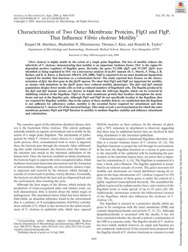
JOURNAL OF BACTERIOLOGY, Sept. 2009, p. 5669–5679 Vol. 191, No. 180021-9193/09/$08.00�0 doi:10.1128/JB.00632-09Copyright © 2009, American Society for Microbiology. All Rights Reserved.
Characterization of Two Outer Membrane Proteins, FlgO and FlgP,That Influence Vibrio cholerae Motility�
Raquel M. Martinez, Madushini N. Dharmasena, Thomas J. Kirn, and Ronald K. Taylor*Department of Microbiology and Immunology, Dartmouth Medical School, Hanover, New Hampshire 03755
Received 13 May 2009/Accepted 6 July 2009
Vibrio cholerae is highly motile by the action of a single polar flagellum. The loss of motility reduces theinfectivity of V. cholerae, demonstrating that motility is an important virulence factor. FlrC is the sigma-54-dependent positive regulator of flagellar genes. Recently, the genes VC2206 (flgP) and VC2207 (flgO) wereidentified as being regulated by FlrC via a microarray analysis of an flrC mutant (D. C. Morris, F. Peng, J. R.Barker, and K. E. Klose, J. Bacteriol. 190:231–239, 2008). FlgP is reported to be an outer membrane lipoproteinrequired for motility that functions as a colonization factor. The study reported here focuses on the charac-terization of flgO, the first gene in the flgOP operon. We show that FlgO and FlgP are important for motility,as strains with mutations in the flgOP genes have reduced motility phenotypes. The flgO and flgP mutantpopulations display fewer motile cells as well as reduced numbers of flagellated cells. The flagella produced bythe flgO and flgP mutant strains are shorter in length than the wild-type flagella, which can be restored byinhibiting rotation of the flagellum. FlgO is an outer membrane protein that localizes throughout the mem-brane and not at the flagellar pole. Although FlgO and FlgP do not specifically localize to the flagellum, theyare required for flagellar stability. Due to the nature of these motility defects, we established that the flagellumis not sufficient for adherence; rather, motility is the essential factor required for attachment and thuscolonization by V. cholerae O1 of the classical biotype. This study reveals a novel mechanism for which the outermembrane proteins FlgO and FlgP function in motility to mediate flagellar stability and influence attachmentand colonization.
The causative agent of the infectious diarrheal disease chol-era is the bacterium Vibrio cholerae. This enteric pathogennaturally inhabits an aquatic environment and is motile by theaction of a single polar flagellum. The mechanism of patho-genesis by which V. cholerae causes disease is a stepwise andcyclic process. First, contaminated food or water is ingested;then, the bacteria pass through the stomach. After withstand-ing that acidic environment, the bacteria enter the lumen ofthe intestine and attach to the intestinal epithelium of thehuman host. Once the bacteria establish an initial attachment,the bacteria begin to express the toxin-coregulated pilus, whichfacilitates bacterium-bacterium interactions and the formationof microcolonies. Subsequently, an enterotoxin, cholera toxin,is expressed and secreted by the bacteria, which through acascade of events leads to profuse, watery diarrhea. Eventually,the bacteria are shed from the host and are dispersed back intothe environment, thus renewing the cycle.
Although the later stages of the disease, which include theproduction of toxin-coregulated pilus and cholera toxin, arewell characterized, little is known about the initial stages ofpathogenesis. The attachment factor GbpA has been shown tobind chitin, an abundant substance found in the environmentthat is a polymer of N-acetylglucosamine (GlcNAc) carbohy-drate subunits (17). GbpA is also involved in the initial attach-ment of V. cholerae to epithelial cells, which have exposed
GlcNAc moieties on their surfaces. In the absence of gbpA,only a 50% reduction in attachment is observed, suggestingthat there may be additional factors that are involved in initi-ating attachment to the intestinal epithelium.
Colonization requires a functional flagellum; therefore, mo-tility is a crucial virulence factor of V. cholerae (9, 12). Theflagellum functions to propel the cell through its environment.In the host, the flagellum functions as a means to gain accessto the microvilli of the epithelial cells by facilitating the pen-etration of the intestinal mucus layer, an action that is impor-tant for colonization (1, 4, 14). The flagellum is composed of abase, a hook, and a filament. The flagellar apparatus is assem-bled in an ordered cascade of events. The genes necessary formotility and chemotaxis are found distributed among six re-gions on the large chromosome of V. cholerae (regions I to VI)(28). The expression of these genes is under the control of atightly regulated transcriptional hierarchy. The assembled fla-gellum is powered by sodium motive force, and rotation of theflagellum leads to swim speeds of up to 65 �m/s (20, 30).Additionally, chemotaxis controls the bidirectional rotationof the flagellum, which also influences the virulence of V.cholerae (4).
The flagellum is encased in a protective sheath, which ap-pears to be contiguous with the outer membrane (OM) andcontains lipopolysaccharide and protein (7, 13, 31). Althoughlipopolysaccharide is associated with the sheath, it has notbeen concluded whether the sheath is indeed a continuation ofthe OM or a separate entity. The flagellar sheath of V. choleraehas been widely studied; however, its origin and function arenot completely understood. It has recently been proposed thatthe flagellar sheath of V. cholerae functions as a means to evade
* Corresponding author. Mailing address: Dartmouth MedicalSchool, Department of Microbiology and Immunology, HB7550, Han-over, NH 03755. Phone: (603) 650-1632. Fax: (603) 650-1318. E-mail:[email protected].
� Published ahead of print on 10 July 2009.
5669
Dow
nloa
ded
from
http
s://j
ourn
als.
asm
.org
/jour
nal/j
b on
17
Oct
ober
202
1 by
164
.163
.92.
233.
host immune responses (36). The sheath was also shown toprotect the filament from dissociation when exposed to hightemperatures and low pH, conditions that easily dissociate thenonsheathed flagellum of Salmonella enterica serovar Typhi-murium (36). Furthermore, the flagellum is also thought tohave adhesive properties, which would allow it to function asan attachment factor (1, 9, 14, 29).
Pilus-mediated-attachment, motility, nitrogen metabolism,alginate production, glutamine synthesis, and type III secretiongenes are regulated by RpoN (�54), an alternate sigma factor,in various pathogenic gram-negative bacteria (16). In V. chol-erae, RpoN plays an important role in positively regulatingflagellar gene expression. A V. cholerae rpoN mutant is aflagel-lated and displays a 10- to 20-fold reduction in its ability tocolonize the infant mouse intestine compared to the wild-type(WT) strain (19). However, the nonflagellated flaA mutantshows only a twofold reduction in the same infant mouse com-petitive assay, suggesting that RpoN regulates the expressionof additional factors other than flagella that are important forattachment and colonization. Given that RpoN appears toplay a major role in the ability of V. cholerae to adhere to theintestinal epithelium, we chose to investigate other genes reg-ulated by RpoN that might contribute to intestinal coloniza-tion.
A recent study of flrC, which encodes the RpoN-dependenttranscriptional activator of the class III flagellar genes, identi-fied the genes VC2207 (flgO) and VC2206 (flgP) via a microar-ray analysis of an flrC mutant of the classical biotype strainO395 (25). The authors report the effects of �flgP on motilityand colonization. The deletion of flgP resulted in the produc-tion of abnormal flagella that were jagged and lacked thecharacteristic sinusoidal curvature of the flagellar structure,resulting in the loss of motility. Intriguingly, FlgP is an OMlipoprotein, in which lipidation at residue C18 is essential forlocalization to the OM, as well as improved colonization, butnot motility. A new and contradicting report by Cameron et al.states that an flgP mutant of the El Tor biotype strain C6706displays normal flagella (5).
Whereas FlgP was the primary focus of the study by Morriset al. (25), the work reported here encompasses an initialcharacterization of FlgO as well as additional findings withrespect to FlgP in the classical biotype strain O395. We havedetermined that both FlgO and FlgP play a role in motility andintestinal colonization. The flgO and flgP mutant strains exhibitlower numbers of motile cells and have fewer flagella. We alsodetermined that the flagella produced by the flgO and flgPmutant strains were normal in appearance except for theirlength. Transmission electron microscopy studies revealedtruncated flagella that averaged half the length of the WTflagella. Interestingly, inhibition of the flagellar motor restoredthe length of the flagellum to the length of the WT flagella,suggesting a stability defect, which is further supported byincreased sensitivity of the flagella elaborated by the flgO andflgP mutant strains to high temperature and low pH. Addition-ally, we demonstrated that FlgO localizes to the OM and thatneither FlgO nor FlgP requires the other for proper localiza-tion. Further localization studies indicate that FlgO and FlgPare present throughout the membrane and not only at theflagellar pole. Also, unlike other known OM proteins (OMPs),FlgO and FlgP associate primarily with the cell and not the
flagellar fraction. The results presented here provide a novelmechanism by which the OMPs FlgO and FlgP function inmotility to influence the stability of the flagellum of V. cholerae.
MATERIALS AND METHODS
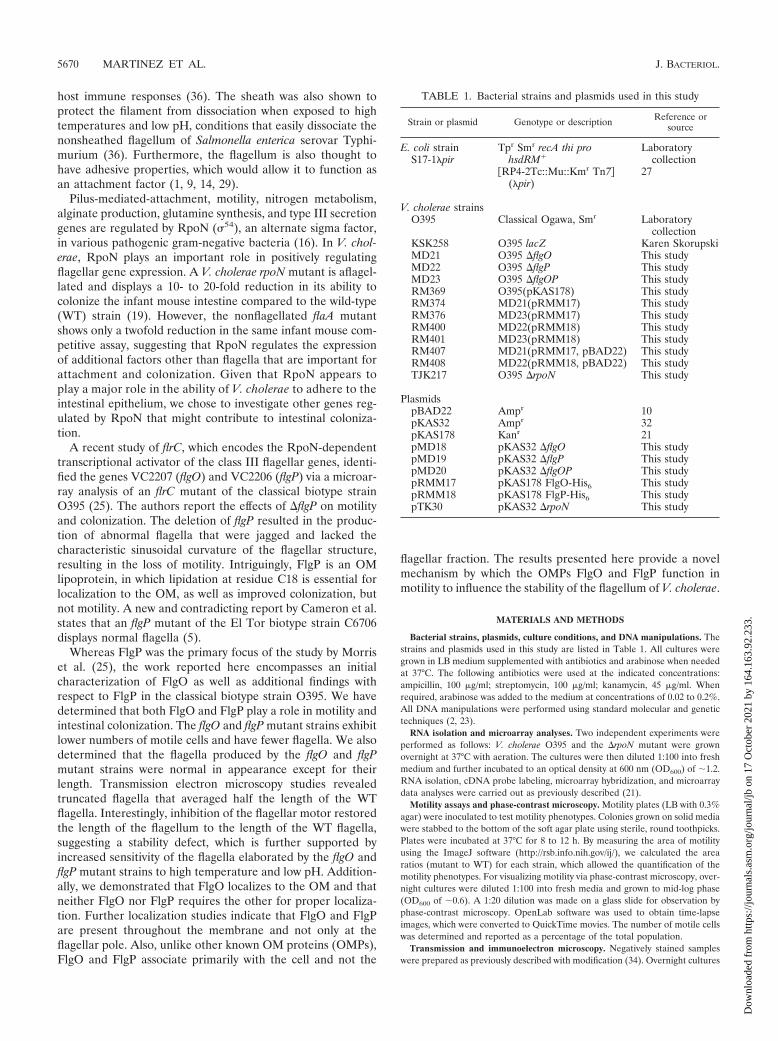
Bacterial strains, plasmids, culture conditions, and DNA manipulations. Thestrains and plasmids used in this study are listed in Table 1. All cultures weregrown in LB medium supplemented with antibiotics and arabinose when neededat 37°C. The following antibiotics were used at the indicated concentrations:ampicillin, 100 �g/ml; streptomycin, 100 �g/ml; kanamycin, 45 �g/ml. Whenrequired, arabinose was added to the medium at concentrations of 0.02 to 0.2%.All DNA manipulations were performed using standard molecular and genetictechniques (2, 23).
RNA isolation and microarray analyses. Two independent experiments wereperformed as follows: V. cholerae O395 and the �rpoN mutant were grownovernight at 37°C with aeration. The cultures were then diluted 1:100 into freshmedium and further incubated to an optical density at 600 nm (OD600) of �1.2.RNA isolation, cDNA probe labeling, microarray hybridization, and microarraydata analyses were carried out as previously described (21).
Motility assays and phase-contrast microscopy. Motility plates (LB with 0.3%agar) were inoculated to test motility phenotypes. Colonies grown on solid mediawere stabbed to the bottom of the soft agar plate using sterile, round toothpicks.Plates were incubated at 37°C for 8 to 12 h. By measuring the area of motilityusing the ImageJ software (http://rsb.info.nih.gov/ij/), we calculated the arearatios (mutant to WT) for each strain, which allowed the quantification of themotility phenotypes. For visualizing motility via phase-contrast microscopy, over-night cultures were diluted 1:100 into fresh media and grown to mid-log phase(OD600 of �0.6). A 1:20 dilution was made on a glass slide for observation byphase-contrast microscopy. OpenLab software was used to obtain time-lapseimages, which were converted to QuickTime movies. The number of motile cellswas determined and reported as a percentage of the total population.
Transmission and immunoelectron microscopy. Negatively stained sampleswere prepared as previously described with modification (34). Overnight cultures
TABLE 1. Bacterial strains and plasmids used in this study
Strain or plasmid Genotype or description Reference orsource
E. coli strainS17-1�pir
Tpr Smr recA thi prohsdRM�
Laboratorycollection
�RP4-2Tc::Mu::Kmr Tn7�(�pir)
27
V. cholerae strainsO395 Classical Ogawa, Smr Laboratory
collectionKSK258 O395 lacZ Karen SkorupskiMD21 O395 �flgO This studyMD22 O395 �flgP This studyMD23 O395 �flgOP This studyRM369 O395(pKAS178) This studyRM374 MD21(pRMM17) This studyRM376 MD23(pRMM17) This studyRM400 MD22(pRMM18) This studyRM401 MD23(pRMM18) This studyRM407 MD21(pRMM17, pBAD22) This studyRM408 MD22(pRMM18, pBAD22) This studyTJK217 O395 �rpoN This study
PlasmidspBAD22 Ampr 10pKAS32 Ampr 32pKAS178 Kanr 21pMD18 pKAS32 �flgO This studypMD19 pKAS32 �flgP This studypMD20 pKAS32 �flgOP This studypRMM17 pKAS178 FlgO-His6 This studypRMM18 pKAS178 FlgP-His6 This studypTK30 pKAS32 �rpoN This study
5670 MARTINEZ ET AL. J. BACTERIOL.
Dow
nloa
ded
from
http
s://j
ourn
als.
asm
.org
/jour
nal/j
b on
17
Oct
ober
202
1 by
164
.163
.92.
233.

were diluted 1:100 into fresh media and grown to mid-log phase (OD600 of �0.6).Formvar-coated grids were incubated with cultures for 2 min. The grids werethen fixed in a 4% paraformaldehyde solution and washed three times for 1 mineach in 0.1 M sodium cacodylate. Whole-cell preparations were negativelystained with 0.5% phosphotungstic (PTA) acid (pH 6.5) for 2 min. Thin sectionswere immunolabeled by blocking in 3% (wt/vol) casein in Tris-buffered saline(15). A 1:10 dilution of the primary antibody was incubated for 1 h and thenwashed with Tris-buffered saline containing Tween 20. The gold-conjugatedsecondary antibody was diluted 1:25 and incubated for 1 h in the dark.
Subcellular fractionation, SDS-PAGE, and immunoblot analysis. Whole-cellextracts were prepared by suspending overnight cultures in 2 sodium dodecylsulfate-polyacrylamide gel electrophoresis (SDS-PAGE) buffer and were boiledfor 10 min. 2 SDS-PAGE without 2-mercaptoethanol and bromophenol bluewas used for protein estimation. Subcellular fractionation was performed aspreviously described (35). Protein concentrations of whole-cell extracts and sub-cellular fractions were determined with a BCA protein assay kit (Pierce). Sam-ples containing equal amounts of total protein were resolved by SDS-PAGE.Proteins were transferred to nitrocellulose membranes at 4°C. All primary anti-bodies (antiflagellin, anti-His, anti-OmpA, anti-OmpS, anti-OmpU, anti-OmpW,and anti-ToxR) were used at a dilution of 1:1,000. The horseradish peroxidase-conjugated secondary antibodies (anti-rabbit or anti-mouse antibody) were usedat a dilution of 1:10,000. Enhanced chemiluminescence detection reagents wereutilized to visualize reactive bands (Amersham Pharmacia).
Tissue culture and bacterial attachment assay. HT-29 cells were grown inDulbecco’s modified Eagle medium supplemented with 10% fetal bovine serumand 1 Pen-Strep (Gibco) under 5% CO2, as previously described (3, 18).Overnight bacterial cultures were diluted 1:100 in phosphate-buffered saline(PBS) before incubation with confluent HT-29 monolayers. The bacterial sus-pension was added to the epithelial cells and centrifuged for 10 min at 1,000 g toensure bacterium-epithelial cell interaction. The bacterial cells were allowed toattach to the HT-29 cells for 1 h at 37°C. The HT-29 monolayers and attachedbacteria were washed three times with PBS to remove unattached bacterial cells.The HT-29 cells were then lysed to enumerate the attached bacteria by incubat-ing the coculture for 30 min in a 1% Triton X-100 solution. The resultingbacterial suspension was serially diluted and plated for enumeration of bacteriaon LB medium plates supplemented with streptomycin and 5-bromo-4-chloro-3-indolyl--D-galactopyranoside (X-Gal) (40 �g/ml) when required for compet-itive attachment assays.
Infant mouse cholera model. As previously described, test strains were mixedwith equal numbers (1:1) of a �lacZ O395 reference strain (18). Four- to6-day-old CD-1 mice from mixed litters were orally inoculated with 50 �l of a 1 10�2 dilution of the bacterial mixture and incubated at 30°C for 24 h. Thebacteria were then recovered by homogenizing harvested intestines with a TissueTearor in 4 ml of LB broth containing 10% glycerol. The homogenate wasserially diluted and plated on LB agar plates containing streptomycin and X-Gal.The competitive index was calculated using the equation [test (output/input)]/[reference (output/input)]. Animal experiments were done in compliance withinstitutional animal care and use guidelines.
Isolation of crude flagellar fractions and flagellar treatments. Broth cultureswere diluted 1:100 in fresh medium and grown overnight. The cells were pelletedby centrifugation at 10,000 g, and the supernatant was discarded. The cellswere then suspended in PBS, vortexed for 2 min to shear the flagella from thecells, and centrifuged. The supernatant was removed and subjected to ultracen-trifugation at 208,000 g for 60 min to pellet the flagella. The resulting crudeflagellar pellet was suspended in PBS. Flagella were kept at room temperature(no treatment) or treated at 50°C or 80°C for 10 min, followed by incubation at4°C until needed. Equal volumes of flagella and buffered 50 mM sodium citratesolutions (pH 7, 5, and 3) were incubated at room temperature for a treatmenttime of 10 min as previously described (36). Following treatment, flagellar sam-ples were loaded on a 4 to 20% polyacrylamide gel and electrophoresed undernative conditions.
Microarray data accession number. The microarray data discussed hereinhave been deposited in the NCBI Gene Expression Omnibus (GEO; http://www.ncbi.nlm.nih.gov/geo/) and are accessible through GEO series accession numberGSE17144.
RESULTS
RpoN regulates the expression of the genes VC2206 andVC2207. Because the rpoN mutant shows a greater coloniza-tion defect than a flaA mutant in an in vivo competitive index,we were interested in identifying the gene(s) regulated by
RpoN that might contribute to attachment and colonization ofV. cholerae (19). RpoN is known to regulate the expression ofgenes involved in flagellar motility, glutamate synthesis, and apossibly unknown colonization factor(s) in V. cholerae (19). Inan attempt to identify such novel factors, a �rpoN microarrayanalysis was carried out. In two separate microarray studies, atotal of 139 genes showed changes in expression in the �rpoNmutant of at least threefold compared to that of the WT. Ofthe 139 genes, 86 genes were upregulated, whereas 53 geneswere downregulated compared to the expression in the WT.We were interested in gene products that contained signalsequences, which is a common characteristic of attachment andcolonization factors. The genes VC2206 and VC2207 appearedto be positively regulated by RpoN. The microarray datashowed an average change of 18.79 � 0.93-fold in gene expres-sion for VC2207 and an average change of 5.35 � 1.32-fold forVC2206. Independent of this study, Morris et al. recently re-ported that the genes VC2206 and VC2207 compose anoperon positively regulated by FlrC, an RpoN-dependentflagellar regulatory protein (25). The genes VC2207 andVC2206 were named flgO and flgP. Our findings corroboratethose reported by Morris et al. that RpoN regulates the geneexpression of flgOP (25).
flgO and flgP are important for proper motility function inV. cholerae. The flgOP operon can be considered to lie withinan extension of the original flagellar gene cluster, a regionconsisting of flagellar structural genes, chemotaxis genes, andvarious hypothetical genes (25, 28). Given that FlgP plays animportant role in motility in V. cholerae, as well as in Campy-lobacter jejuni, we next chose to investigate the role of FlgO inV. cholerae motility (25, 33). According to the data resultingfrom motility agar assays, the deletion of flgO resulted in ap-proximately a 60% reduction in motility compared to that ofthe WT (Fig. 1A and B). The �flgP and �flgOP strains dis-played even greater reductions in motility (�90%). Nonethe-less, the flgP and flgOP mutants were significantly more motilethan the flaA mutant. The motility defect of the flgO mutantcan be partially complemented by plasmid-encoded FlgO-His6
in soft agar (Fig. 1C). Likewise, expressing FlgP-His6 in theflgP mutant increased the zone of motility (Fig. 1D). In addi-tion, the flgO and flgP mutants appear to be epistatic, since thedouble mutant has the same phenotype as the �flgP mutant,suggesting that they operate in the same pathway. Thus, flgOPseem to be required for optimal motility of V. cholerae.
The flgO and flgP mutants exhibit low numbers of motilecells. Given that the �flgP motility agar phenotype suggestedthat motility is not completely lost, we observed the motility ofthe �flgO and �flgP broth cultures under phase-contrast lightmicroscopy. Motile cells were enumerated, and the proportionof motile cells for each strain was quantified (Fig. 2A). Thirty-four percent of the WT cells were observed to be motile,whereas only 17% of the �flgO population was motile, which isa 50% reduction (17% versus 34%) in motility compared to theWT strain. An even greater reduction was discovered forthe �flgP strain, where only 6% of the population was motilecompared to 34% for the WT strain (an 82% reduction). Thedecreased percentage of motile cells in the flgO and flgP mu-tant populations observed under the light microscope is con-sistent with the motility agar data (Fig. 1).
VOL. 191, 2009 TWO PROTEINS ASSOCIATED WITH V. CHOLERAE MOTILITY 5671
Dow
nloa
ded
from
http
s://j
ourn
als.
asm
.org
/jour
nal/j
b on
17
Oct
ober
202
1 by
164
.163
.92.
233.

The flgO and flgP mutants produce fewer flagella than theWT. Because the flgO mutant and flgP mutant strains displayeda decline in the percentage of motile cells, we hypothesizedthat reduced rotation of the flagellum, a damaged filament, orperhaps the lack of a flagellum could explain why a populationwould show decreased numbers of motile cells compared to theWT. To test this, transmission electron microscopy was used tovisualize the flagella of the flgO, flgP, and flgOP mutant strains.There were no visible abnormalities in the sheathing of theflagella; the flagella appeared to be smooth and not rough aspreviously reported (25) (Fig. 2B). One hundred cells werethen analyzed for the presence of flagella. As expected, almostall of the WT cells had intact flagella (90%), whereas none ofthe nonmotile flaA mutant cells had flagella (Fig. 2C). Morethan half of the �flgO cells were flagellated (59%), with sig-nificantly fewer �flgP and �flgOP cells maintaining their fla-gella (41% and 42%, respectively). These results indicate thatthese mutant strains are still flagellated, but at a lower levelthan the WT. In the case of the flgP mutant, a large portion ofthese flagellated cells are not motile. This suggests a defect ineither the rotation or the integrity of the flagellum.
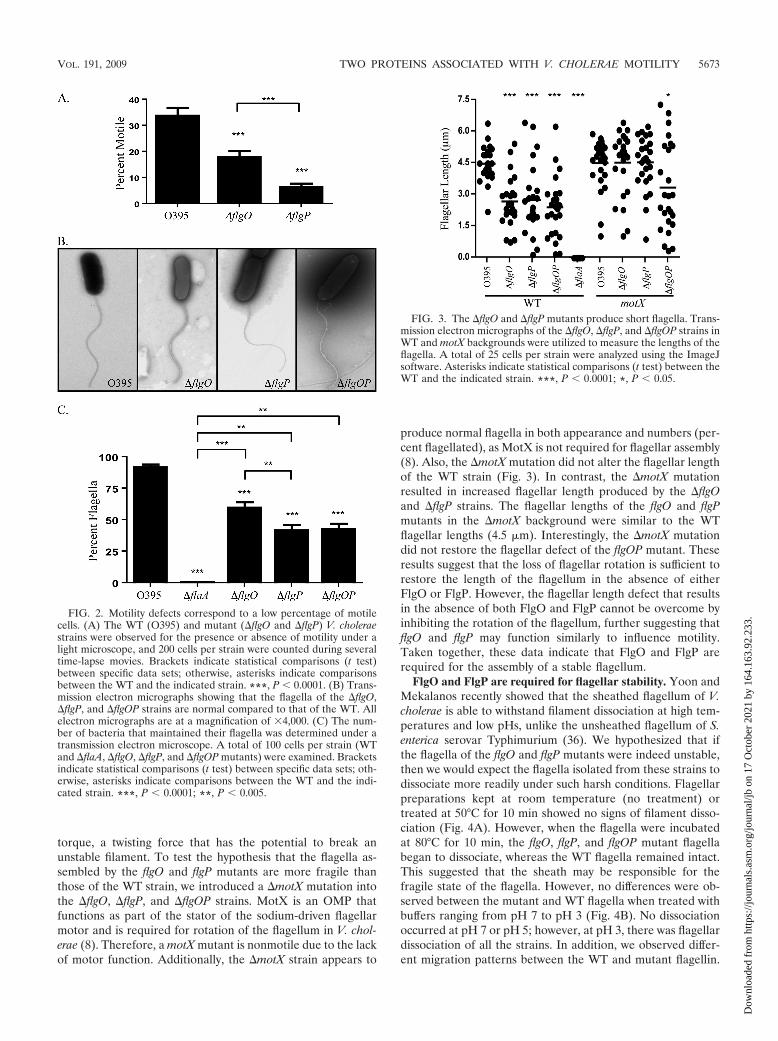
The �flgO and �flgP mutants have truncated flagella. Al-though the flgO and flgP mutant flagella appeared smooth andsinusoidal like the WT flagella, we did observe variations inflagellar length. In order to study the potential differences inthe flagellar structures produced by the flgO and flgP mutants,the ImageJ software was utilized to directly measure the lengthof flagella from electron micrographs. The average length of aWT flagellum was found to be 4.5 �m, with a range of lengthsbetween 2.0 and 6.5 �m (Fig. 3). The average lengths of theflgO, flgP, and flgOP mutants were 2.5 �m. In other words, themajority of the flagella produced by the mutant strains wereshorter than those of the WT. However, a small proportion ofthe mutant cells had flagella of lengths comparable to those ofWT flagella (�4.5 �m), indicating that these strains are capa-ble of making full-length flagella. These data show that the flgOand flgP mutant strains are not able to properly modulate thelength of the flagellum, which would imply a role for flgO andflgP in the stabilization of the flagellum.
Because we have observed full-length flagella among theflgO and flgP mutants, we hypothesize that the shorter flagellaare the result of breakage. Rotation of the flagellum requires
FIG. 1. �flgO and �flgP mutants are defective in motility. (A) Area of motility of the WT (O395) and �flaA (nonmotile), �flgO, �flgP, and�flgOP V. cholerae strains in motility agar. (B) Graphical representation of the area of motility shown in panel A. The NIH ImageJ software wasused to measure the area of the zone of motility of each strain in quadruplicate. Brackets indicate statistical comparisons (t tests) between specificdata sets; otherwise, asterisks indicate comparisons between the WT and the indicated strain. ***, P 0.0001; **, P 0.005. (C) The �flgO mutantcan be partially complemented by a C-terminal His-tagged version of FlgO but not by the vector alone (0.02% arabinose). (D) Similarly, the �flgPmutant can be partially complemented by a C-terminal His-tagged version of FlgP but not by the vector alone (0.2% arabinose).
5672 MARTINEZ ET AL. J. BACTERIOL.
Dow
nloa
ded
from
http
s://j
ourn
als.
asm
.org
/jour
nal/j
b on
17
Oct
ober
202
1 by
164
.163
.92.
233.
torque, a twisting force that has the potential to break anunstable filament. To test the hypothesis that the flagella as-sembled by the flgO and flgP mutants are more fragile thanthose of the WT strain, we introduced a �motX mutation intothe �flgO, �flgP, and �flgOP strains. MotX is an OMP thatfunctions as part of the stator of the sodium-driven flagellarmotor and is required for rotation of the flagellum in V. chol-erae (8). Therefore, a motX mutant is nonmotile due to the lackof motor function. Additionally, the �motX strain appears to
produce normal flagella in both appearance and numbers (per-cent flagellated), as MotX is not required for flagellar assembly(8). Also, the �motX mutation did not alter the flagellar lengthof the WT strain (Fig. 3). In contrast, the �motX mutationresulted in increased flagellar length produced by the �flgOand �flgP strains. The flagellar lengths of the flgO and flgPmutants in the �motX background were similar to the WTflagellar lengths (4.5 �m). Interestingly, the �motX mutationdid not restore the flagellar defect of the flgOP mutant. Theseresults suggest that the loss of flagellar rotation is sufficient torestore the length of the flagellum in the absence of eitherFlgO or FlgP. However, the flagellar length defect that resultsin the absence of both FlgO and FlgP cannot be overcome byinhibiting the rotation of the flagellum, further suggesting thatflgO and flgP may function similarly to influence motility.Taken together, these data indicate that FlgO and FlgP arerequired for the assembly of a stable flagellum.
FlgO and FlgP are required for flagellar stability. Yoon andMekalanos recently showed that the sheathed flagellum of V.cholerae is able to withstand filament dissociation at high tem-peratures and low pHs, unlike the unsheathed flagellum of S.enterica serovar Typhimurium (36). We hypothesized that ifthe flagella of the flgO and flgP mutants were indeed unstable,then we would expect the flagella isolated from these strains todissociate more readily under such harsh conditions. Flagellarpreparations kept at room temperature (no treatment) ortreated at 50°C for 10 min showed no signs of filament disso-ciation (Fig. 4A). However, when the flagella were incubatedat 80°C for 10 min, the flgO, flgP, and flgOP mutant flagellabegan to dissociate, whereas the WT flagella remained intact.This suggested that the sheath may be responsible for thefragile state of the flagella. However, no differences were ob-served between the mutant and WT flagella when treated withbuffers ranging from pH 7 to pH 3 (Fig. 4B). No dissociationoccurred at pH 7 or pH 5; however, at pH 3, there was flagellardissociation of all the strains. In addition, we observed differ-ent migration patterns between the WT and mutant flagellin.
FIG. 2. Motility defects correspond to a low percentage of motilecells. (A) The WT (O395) and mutant (�flgO and �flgP) V. choleraestrains were observed for the presence or absence of motility under alight microscope, and 200 cells per strain were counted during severaltime-lapse movies. Brackets indicate statistical comparisons (t test)between specific data sets; otherwise, asterisks indicate comparisonsbetween the WT and the indicated strain. ***, P 0.0001. (B) Trans-mission electron micrographs showing that the flagella of the �flgO,�flgP, and �flgOP strains are normal compared to that of the WT. Allelectron micrographs are at a magnification of 4,000. (C) The num-ber of bacteria that maintained their flagella was determined under atransmission electron microscope. A total of 100 cells per strain (WTand �flaA, �flgO, �flgP, and �flgOP mutants) were examined. Bracketsindicate statistical comparisons (t test) between specific data sets; oth-erwise, asterisks indicate comparisons between the WT and the indi-cated strain. ***, P 0.0001; **, P 0.005.
FIG. 3. The �flgO and �flgP mutants produce short flagella. Trans-mission electron micrographs of the �flgO, �flgP, and �flgOP strains inWT and motX backgrounds were utilized to measure the lengths of theflagella. A total of 25 cells per strain were analyzed using the ImageJsoftware. Asterisks indicate statistical comparisons (t test) between theWT and the indicated strain. ***, P 0.0001; *, P 0.05.
VOL. 191, 2009 TWO PROTEINS ASSOCIATED WITH V. CHOLERAE MOTILITY 5673
Dow
nloa
ded
from
http
s://j
ourn
als.
asm
.org
/jour
nal/j
b on
17
Oct
ober
202
1 by
164
.163
.92.
233.

These results indicate that the flagella produced by the flgOand flgP mutants are less stable than the WT but probably notstrictly due to any sheath defect.
FlgO localizes to the OM and does not require FlgP forproper localization. Because FlgP was identified to be anOMP in V. cholerae and C. jejuni, we were interested indetermining the localization of FlgO (25, 33). Subcellularfractionation was used to identify the OM localization of theFlgO-His6 protein (Fig. 5A). An anti-His immunoblot re-vealed that the majority of the FlgO protein resides in theOM when FlgO is expressed under an inducible PBAD pro-moter. To ensure the purity of the inner membrane (IM)and OM fractions, the fractionated samples were probedwith anti-OmpA, a known OMP, and anti-ToxR, an estab-lished IM protein (IMP) (6, 24). As expected, OmpA wasdetected only in the OM fraction and the majority of ToxRwas located in the IM, indicating that the cellular fractionsobtained were not contaminated. As expected, the anti-Hisantibody was specific, as no protein was detected when anempty vector was expressed in the same �flgO strain (Fig.5B). Additionally, when either FlgO-His6 or FlgP-His6 wasexpressed in the flgOP mutant, we found that OM localiza-tion was not dependent on the other protein (Fig. 5C and
D). These data demonstrate that FlgO and FlgP do notrequire each other for proper OM localization.
FlgO and FlgP do not localize to either pole. Because of therole of FlgO and FlgP in motility and their effect on theflagellum, we hypothesized that these proteins might localizeto the flagellar pole or to the flagellar sheath. Thin-sectionedcells expressing either FlgO-His6 or FlgP-His6 were immuno-gold labeled in order to localize each protein (Fig. 6). BothFlgO-His6 and FlgP-His6 were observed to localize throughoutthe membrane and were not enriched at either pole (Fig. 6A,panels i and ii). As expected, the vector controls did not resultin protein detection via immunogold labeling (Fig. 6A, panelsiii and iv). These results suggest that FlgO and FlgP localizethroughout the OM and do not preferentially localize to theflagellar pole.
One concern about using the PBAD promoter in localiza-tion experiments was the possibility of improper localizationin the OM due to overexpression of the protein. To addressthe influence that expression levels have on localization, astrain that expresses FlgO-His6 from its native putative pro-moter was constructed (the �flgO � flgOpFlgO-His6 strain).Although FlgO-His6 expressed from its native promoter wasnot detectable via Western blot analysis, it fully comple-mented the motility phenotype, suggesting that the endog-enous level of FlgO protein in the cell is low (data notshown). Therefore, due to the low level of protein present,the detection of immunogold particles identifying the flgOp-FlgO-His6 protein in thin-section preparations required theobservation of a large number of specimens. The majority ofthe flgOpFlgO-His6 protein detected in the complementedstrain (the �flgO � flgOpFlgO-His6 strain) was not localizedto either pole (Fig. 6B, panels i and ii; Fig. 6C). A goldparticle was observed at the pole opposite the flagellum
FIG. 4. The �flgO and the �flgP strains produce fragile flagella.(A) O395, �flgO, �flgP, and �flgOP crude flagellar fractions were keptfor 10 min at room temperature (no treatment) or incubated at 50°Cor 80°C. Treated and control samples were run on 4 to 20% Tris-glycine precast gels under native conditions. Filament dissociation wasdetermined by immunoblot analysis with antiflagellin antibody.(B) O395, �flgO, �flgP, and �flgOP flagellar fractions were treated for10 min at room temperature with equal volumes of flagella and 50 mMcitrate buffers at pH 7, 5, and 3. Treated samples were run on 4 to 20%Tris-glycine precast gels under native conditions. Filament dissociationwas determined by immunoblot analysis with antiflagellin antibody.Arrows indicate migration differences between WT and mutantflagellin.
FIG. 5. FlgO localizes to the OM. Subcellular fractionation wascarried out on the �flgO � pFlgO-His6 (A), �flgO � vector (B),�flgOP � pFlgO-His6 (C), and �flgOP � pFlgP-His6 (D) strains. Thecytoplasmic (lane C), periplasmic (lane P), IM (lane I), and OM (laneO) samples were separated by SDS-PAGE, followed by either anti-His,anti-OmpA, or anti-ToxR immunoblotting.
5674 MARTINEZ ET AL. J. BACTERIOL.
Dow
nloa
ded
from
http
s://j
ourn
als.
asm
.org
/jour
nal/j
b on
17
Oct
ober
202
1 by
164
.163
.92.
233.

(Fig. 6B, panel ii), suggesting that the polar particles de-tected may not necessarily indicate localization at the flagel-lar pole. As expected, the FlgO protein was not detected atthe pole or in the nonpolar membrane area in the vector-
only control (Fig. 6B, panels iii and iv; Fig. 6C). Takentogether, these results suggest that FlgO localizes through-out the OM and does not specifically localize to the flagellarpole.
FIG. 6. FlgO and FlgP do not localize to either pole. (A) Thin-section immunoelectron microscopy was utilized to determine where FlgO and FlgP localizedin the OM of the �flgO � pFlgO-His6 (panel i) and �flgP � pFlgP-His6 (panel ii) complemented strains compared to the vector-only controls. Panel iii, �flgO �vector control strain; panel iv, �flgP � vector control strain. An arrow indicates the polar flagellum. (B) Thin-section immunoelectron micrographs of the �flgOmutant expressing FlgO-His6 under the control of its native putative flgO promoter (panels i and ii), compared to the vector control strains (panels iii and iv).Black arrows indicate gold particles, and the white arrow signifies the flagellar pole. All electron micrographs are at a magnification of 20,000. (C) Graphicalrepresentation of polar versus nonpolar (lateral) localization of flgOpFlgO-His6 protein. A total of 50 cells were counted per strain. Asterisks indicate statisticalcomparisons (t test) between the nonpolar (lateral) and indicated regions (pole or nonspecific). ***, P 0.0001; **, P 0.005. NSB, nonspecific binding.
VOL. 191, 2009 TWO PROTEINS ASSOCIATED WITH V. CHOLERAE MOTILITY 5675
Dow
nloa
ded
from
http
s://j
ourn
als.
asm
.org
/jour
nal/j
b on
17
Oct
ober
202
1 by
164
.163
.92.
233.

FlgO and FlgP are not associated with the flagellar sheath.As a result of the thin-sectioning process, specimens yield veryfew cells with intact flagella. Consequently, we could not de-termine whether FlgO and FlgP localize to the sheath. Sincethe fragile state of the �flgO and �flgP flagella could be due todefects in the sheath or the filament, we next investigatedwhether FlgO and FlgP associated with the flagellum as ameans to understand the role of FlgO and FlgP in filamentstability. Crude flagellar fractions were isolated and analyzedby antiflagellin, anti-OMPs (OmpS, OmpU, and OmpW), andanti-His immunoblotting (Fig. 7). As expected, more flagellinwas detected in the flagellar fraction than in the cellular frac-tion for each of the WT and mutant strains (Fig. 7A). As acontrol, no flagellin was detected in either fraction of theaflagellated �flaA strain. While the OMPs were equally dis-tributed in the cell and flagellar fractions (Fig. 7B to D), theanti-His immunoblot revealed that the majority of both theFlgO and FlgP proteins were associated with the cell, with onlya slight association present in the flagellar fraction (Fig. 7E).All in all, these data suggest that although FlgO and FlgP arenot part of the flagellum (Fig. 7), they do play a role in main-taining the integrity of the flagellum (Fig. 3 and 4).
FlgO plays a role in colonization. Motility plays an impor-tant role in colonization of the intestinal epithelium by V.cholerae (9, 12, 22). Recently, FlgP was shown to play a signif-icant role in the intestinal colonization of the infant mouse(25). Therefore, we hypothesized that an flgO mutant wouldalso play a similar role in virulence. To determine the potentialrole of FlgO in colonization, the �flgO mutant and the �flgOPdouble mutant were assayed using the infant mouse model ofcholera in which an in vivo competitive index was determinedand compared to those of the flgP and flaA deletion mutants.We observed that the flaA mutant displayed a sixfold decreasein colonization compared to the WT strain (Fig. 8A). The flgOmutant also displayed a sixfold reduction in colonization,whereas the flgOP mutant revealed a slightly greater decreasein its ability to colonize the infant mouse intestine. The flgO,flgP, and flgOP mutants showed reduced colonization pheno-types similar to that of the flaA mutant. These data suggest thatFlgO also plays a role in colonization, although not as great arole as FlgP.
FlgO and FlgP are involved in epithelial cell attachment.Because of the roles played by FlgO and FlgP in colonization,we next were interested in determining whether FlgO and FlgPare important for attachment. Also, due to the fact that mo-tility is important for colonization and attachment, as it pro-motes association between the bacterial and epithelial cells, ithas yet to be conclusively determined whether the flagellumcontains adhesive properties (1, 9, 14, 29). To address whetherthe flagellum functions in adherence (contains adhesive prop-erties) or is solely the propeller by which the cell comes incontact with the intestinal epithelium, we carried out a com-petitive attachment assay. The attachment assay evaluates theability of a strain to adhere to epithelial cells in vitro. Com-pared to the WT strain, the nonmotile, aflagellate flaA mutantdisplayed a threefold decrease in attachment, whereas the flag-ellated, nonmotile motX mutant displayed a sixfold decrease inattachment (Fig. 8B). These data demonstrate that it is in factmotility, and not the adhesive ability of a flagellum, that isimportant for adherence. The flgO mutant showed a threefoldreduction in attachment compared to the WT strain, and theflgP mutant displayed a similar decrease (fourfold) in attach-ment. The flgOP mutant attachment defect was the same as
FIG. 7. FlgO and FlgP associate predominately with the bacterialcell and not the flagellum. Crude flagellar fractions were isolated fromthe O395, �flgO � vector, �flgO � pFlgO-His6, �flgP � vector,�flgP � pFlgP-His6, and �flaA (control) strains. Samples were sepa-rated by SDS-PAGE analysis followed by immunoblot analyses. Rows:A, antiflagellin; B, anti-OmpS; C, anti-OmpU; D, anti-OmpW; E,anti-His. F, flagellar fraction; C, cellular fraction.
FIG. 8. The �flgO and �flgP mutants display a defect in colonization and epithelial cell adherence. (A) V. cholerae strain O395 and the �flaA,�flgO, �flgP, and �flgOP mutants were competed against an O395 �lacZ strain (KSK258), using four to seven mice per group. (B) V. cholerae strainO395 and the �motX (nonmotile, flagellate), �flaA (nonmotile, aflagellate), �flgO, �flgP, and �flgOP mutants were competed against an O395�lacZ strain (KSK258) for in vitro attachment to HT-29 epithelial cells. Brackets indicate statistical comparisons (t tests) between specific data sets;otherwise asterisks indicate comparisons between the WT and the indicated strain. ***, P 0.0001; **, P 0.005; *, P 0.05.
5676 MARTINEZ ET AL. J. BACTERIOL.
Dow
nloa
ded
from
http
s://j
ourn
als.
asm
.org
/jour
nal/j
b on
17
Oct
ober
202
1 by
164
.163
.92.
233.

that of an flgP mutant, suggesting that FlgO and FlgP functionin a common pathway. These data suggest that proper motilityis crucial for attachment. Furthermore, FlgO and FlgP do notfunction as attachment factors per se but rather function topromote proper motility.
DISCUSSION
The purpose of this study was to identify novel factors thatmediate colonization in V. cholerae. We have shown here thattwo proteins, FlgO and FlgP, appear to influence colonizationby playing an important role in the stability of the flagellum.Analysis of microarray data from this study as well as data fromthe study presented by Morris et al. has determined that thealternate sigma factor RpoN (�54) and the class III flagellargene activator FlrC positively regulate the flgOP operon (25).Morris et al. established that flgP is necessary for colonizationby V. cholerae. Our studies confirm that flgP is involved inintestinal colonization as well as epithelial cell attachment. Inaddition, we have demonstrated that the adjacent gene, flgO,also plays an important role in motility, attachment, and col-onization.
Here we report that the role of FlgO and FlgP in motility isto maintain the integrity of the flagellum. The report by Morriset al. determined that the flgP mutant is nonmotile; however,we have found flgP to be slightly motile. Despite the “occa-sional motile cell” that could be detected within the flgP mu-tant population by Morris et al., which is consistent with ourfindings, the authors did not further investigate this matter(25). We, too, noticed a small population of motile �flgP cellsand therefore quantified the zones of motility as well as thepercentage of motile cells within each of the flgO, flgP, and WTpopulations to determine the full extent of the motility defectsdisplayed by these mutants. Based on our data, it appears thatboth the flgO and flgP mutants are motile; however, the mo-tility is significantly reduced.
Morris et al. additionally report rough and perturbed flagellaproduced by the flgP mutant (25). On the contrary, we ob-served flagella that were smooth and displayed normal sinewave appearances for the mutant strains, similar to the WTstrain. One possible cause of these conflicting observationscould be the use of different negative stains. In this study weused 0.5% PTA, whereas Morris et al. used 7% uranyl acetate(UA) (25). UA and PTA are two widely used reagents fornegatively staining samples for the purpose of transmissionelectron microscopy. One concern with using UA to negativelystain samples is the inherently low pH of the solution (11). A1 to 2% aqueous solution of UA has a pH of 4.5 and cannot bebuffered due to rapid precipitation of UA upon the addition ofalkali (above pH 5.0). On the other hand, PTA can be adjustedto a more neutral pH of 6.5 for staining purposes. It wasrecently demonstrated that the flagella of V. cholerae begin todissociate at pH 3.6, with some dissociation at pH 4.0 (36).Therefore, it is possible that the observations made by Morriset al. are due to the effect that a high concentration of UAwould have on the flagellum or sheath (25). Supporting data bySommerlad and Hendrixson showed that in the case of C.jejuni, the �flgP flagella appeared normal when stained withPTA (33). Likewise, Cameron et al. recently reported a normalflagellar appearance for the flgP mutant of the El Tor biotype
strain C6706 (5). Therefore, we conclude that the �flgO and�flgP flagella are smooth and maintain a sinusoidal appear-ance.
Although the appearance of the filaments appeared normal,we did observe truncated flagella in the case of the flgO, flgP,and flgOP mutant strains. Measurements of the flgO, flgP, andflgOP mutant flagella resulted in a wide distribution of lengths,ranging from no visible flagella to WT-length flagella. Theseobservations suggest either that filament assembly is randomlyinterrupted or that the filament is more susceptible to break-age. Since the mutants are capable of producing normal fla-gella, we are in favor of the latter hypothesis. We tested theidea that the flagella of the flgO and flgP mutants were moresusceptible to breakage by inhibiting the rotation of the flagel-lum. Flagellar rotation generates and consequently exertsforces that act upon the flagellum, which could theoreticallycause a fragile flagellum to break more readily. Therefore, theuse of a motX mutant, which is flagellated yet nonmotile,allowed us to determine whether the flgO and flgP flagella aremore susceptible to breakage. Indeed, by introducing a motXmutation into the �flgO and �flgP background strains, we wereable to restore the mutant flagella to WT lengths. This exper-iment implies that the flagella produced by the flgO and �flgPmutants are fragile and more susceptible to breakage thanthose of the WT. This also led us to believe that the percentageof nonflagellated cells observed in the �flgO and �flgP popu-lations is probably the result of breakage at the base of thefilament rather than lack of production of a flagellum. Wepropose that the shorter of the truncated flagella are not ca-pable of further propelling the cell, thus resulting in the re-duction of overall motility.
Yoon and Mekalanos recently demonstrated that thesheathed flagellum of V. cholerae is less susceptible to filamentdissociation in the presence of high temperature or low pHthan is the unsheathed flagellum of Salmonella serovar Typhi-murium (36). To provide more evidence that the flagella of theflgO and flgP mutants are unstable, we subjected crude flagellarfractions to heat and pH treatments. We hypothesized that ifthe flagella of the �flgO and �flgP strains were more suscep-tible to breakage, then perhaps they would dissociate morereadily than the WT flagella under the same harsh conditions.We found that high temperature caused the mutant flagella todissociate more than the WT flagella, which suggests one oftwo things. First, the sheath of the mutant flagellum may bedefective, thus allowing dissociation. Second, perhaps thesheath is normal but the rate of dissociation is higher for ashorter flagellum (�flgO, �flgP, and �flgOP mutants) than foran intact flagellum (WT) at high temperatures. However, inthis case it is difficult to determine whether the flagellar insta-bility is due to the sheath or filament.
The flagella treated with low pH did not show any differ-ences in dissociation. All flagella dissociated at pH 3, which isconsistent with the findings of Yoon et al. (36). An intriguingfeature that we reproducibly observed was a distinct differencebetween the migration of the mutant flagellin and that of theWT flagellin under native conditions. In an attempt to identifythese differences in the flagella/sheath, subsequent SDS-PAGEanalysis of the WT, flgO, flgP, and flgOP flagellar and OMfractions was carried out and revealed a few differences in theresulting protein profiles, suggesting that the filament makeup
VOL. 191, 2009 TWO PROTEINS ASSOCIATED WITH V. CHOLERAE MOTILITY 5677
Dow
nloa
ded
from
http
s://j
ourn
als.
asm
.org
/jour
nal/j
b on
17
Oct
ober
202
1 by
164
.163
.92.
233.

is somehow altered in these mutants; however, the profileswere not robust enough to make any precise conclusions (datanot shown). Taken together, these results strongly suggest afilament defect. It is possible that the WT flagella are brokendown into flagellin monomers while the mutant flagella (i) arenot completely broken down, (ii) have some other protein orlipid moiety associated with the flagellin, or (iii) perhaps aremade up of altered flagellin. If indeed flagellin is changed insome way in the mutants, then the flagellin would migratedifferently under native conditions and the altered flagellinwould explain why the mutant flagella are unstable. In otherwords, if the flagellin monomers are unable to properly poly-merize, then the filament would be easily broken. Although wehypothesize that FlgO and FlgP function in proper filamentassembly, we cannot ignore the possibility that these OMPs areinvolved in sheath stability. It is unclear whether the sheath isan extension of the OM. If this is true, then we are left with thepossibility that in the absence of FlgO and/or FlgP, the sheathmay be fragile, thus rendering the filament unstable.
Because of the effects that FlgO and FlgP had on flagellarstability, we were certain that we would find FlgO and FlgP tobe associated with the flagellum. However, to our surprise,FlgO localization studies revealed that not only is FlgO anOMP but FlgO and FlgP can be found throughout the mem-brane and not enriched at either pole. In addition, we deter-mined that FlgO and FlgP associate more readily with thecellular fraction than the flagellar fraction, suggesting thattheir role in flagellar stability may be indirect.
The colonization defects of the flgO and flgP mutants cor-relate with their motility defects. For example, the �flgO strainis more motile than the �flgP strain, and as such, it is betterable to colonize. Additionally, for decades many have ques-tioned whether colonization required motility or if it was themere presence of a flagellum that was necessary (1, 9, 14, 22,29). Due to the lack of molecular tools, it was not until recentlythat Lee et al. conclusively determined that a functional fla-gellum is absolutely required for colonization (22). Knowingthis, we argue that because the flgO and flgP mutants aremotile, they should colonize better than the �flaA strain. Thisholds true for the flgO but not the flgP mutant. In fact, the�flgP and �flgOP strains are significantly more defective thanthe flaA mutants in colonization. This suggests that the defectin colonization of the flgP mutant is separate from its motilitydefect. Therefore, it is possible that FlgP may also function asa colonization factor, which is consistent with the findings ofMorris et al. (25). Morris et al. further hypothesize that FlgPmay function as an adhesin due to the decreased competitiveindex of the flgP mutant, relative to those of other nonmotilestrains (25). Based on our attachment assay results, we foundthe adherence index of the �flgP strain to be relatively similarto those of the motX and flaA mutants. Since the flgP mutantis largely defective in motility, it makes sense that its level ofadherence is comparable to those of nonmotile mutants. IfFlgP were functioning as an attachment factor, then we wouldexpect to observe a significant decrease in attachment com-pared to the nonmotile mutants, as is seen for colonization.However, this is not the case. These data indicate that the littlemotility retained by the �flgO and �flgP strains is not capableof increasing their chances of coming into contact with theepithelial cells compared to those of the nonmotile strains. In
addition, since the flaA and motX mutants behaved similarly inattachment, we conclude that the V. cholerae flagellum doesnot contain adhesive properties and that a functional flagellumis absolutely required for attachment.
It remains unknown how these two proteins function withinthe OM to contribute to the production of a stable flagellum.We hypothesize that FlgO and FlgP affect motility in an indi-rect manner. Morris et al. showed that a strain expressing FlgPwith a C18G mutation, which localizes to the IM rather thanthe OM, remained motile but was defective for colonization(25). It is important to note that OM lipoproteins are firstanchored to the periplasmic side of the IM before being trans-ported to the OM (26). Since localization of FlgP to eithermembrane is sufficient for proper motility, it is likely thatFlgP’s role in motility is carried out in the periplasm. Webelieve that FlgO and FlgP interact with another protein(s)that in turn interacts with a known flagellar protein(s) as ameans to influence flagellar stability. Further studies are underway to identify FlgO and FlgP interacting proteins to betterunderstand their role in flagellar stability.
ACKNOWLEDGMENTS
We thank Karen Skorupski and Emily Stonehouse for helpful dis-cussions and critical reading of the manuscript. We thank LouisaHoward for assistance with transmission electron microscopy andCarol Ringelberg for microarray analysis. We thank Gabriela Ko-vacikova for technical assistance. We also thank Debra Milton ofUmea University for the generous gift of the Vibrio anguillarum anti-flagellin antibody.
This work was supported by NIH grants AI025096 and AI039654and NSF grant OCN-0120677 to R.K.T. R.M.M. was also supported byNIH training grant T32GM008704.
REFERENCES
1. Attridge, S. R., and D. Rowley. 1983. The role of the flagellum in theadherence of Vibrio cholerae. J. Infect. Dis. 147:864–872.
2. Ausubel, F. M., R. Brent, R. E. Kingston, D. D. Moore, J. G. Seidman, J. A.Smith, and K. Struhl (ed.). 1987. Current protocols in molecular biology.Greene Publishing Associates and John Wiley & Sons, New York, NY.
3. Benítez, J. A., R. G. Spelbrink, A. Silva, T. E. Phillips, C. M. Stanley, M.Boesman-Finkelstein, and R. A. Finkelstein. 1997. Adherence of Vibrio chol-erae to cultured differentiated human intestinal cells: an in vitro colonizationmodel. Infect. Immun. 65:3474–3477.
4. Butler, S. M., and A. Camilli. 2004. Both chemotaxis and net motility greatlyinfluence the infectivity of Vibrio cholerae. Proc. Natl. Acad. Sci. USA 101:5018–5023.
5. Cameron, D. E., J. M. Urbach, and J. J. Mekalanos. 2008. A defined trans-poson mutant library and its use in identifying motility genes in Vibriocholerae. Proc. Natl. Acad. Sci. USA 105:8736–8741.
6. Chai, T. J., and J. Foulds. 1977. Purification of protein A, an outer mem-brane component missing in Escherichia coli K-12 ompA mutants. Biochim.Biophys. Acta 493:210–215.
7. Fuerst, J. A., and J. W. Perry. 1988. Demonstration of lipopolysaccharide onsheathed flagella of Vibrio cholerae O:1 by protein A-gold immunoelectronmicroscopy. J. Bacteriol. 170:1488–1494.
8. Gosink, K. K., and C. C. Hase. 2000. Requirements for conversion of theNa�-driven flagellar motor of Vibrio cholerae to the H�-driven motor ofEscherichia coli. J. Bacteriol. 182:4234–4240.
9. Guentzel, M. N., and L. J. Berry. 1975. Motility as a virulence factor forVibrio cholerae. Infect. Immun. 11:890–897.
10. Guzman, L.-M., D. Belin, M. J. Carson, and J. Beckwith. 1995. Tight regu-lation, modulation, and high-level expression by vectors containing the ar-abinose PBAD promoter. J. Bacteriol. 177:4121–4130.
11. Harris, J. R. 1997. Negative staining and cryoelectron microscopy. p. 21–25.RMS microbiology handbook. BIOS Scientific Publishers Limited, Oxford,United Kingdom.
12. Hase, C. C. 2001. Analysis of the role of flagellar activity in virulence geneexpression in Vibrio cholerae. Microbiology 147:831–837.
13. Hranitzky, K. W., A. Mulholland, A. D. Larson, E. R. Eubanks, and L. T.Hart. 1980. Characterization of a flagellar sheath protein of Vibrio cholerae.Infect. Immun. 27:597–603.
14. Jones, G. W., G. D. Abrams, and R. Freter. 1976. Adhesive properties of
5678 MARTINEZ ET AL. J. BACTERIOL.
Dow
nloa
ded
from
http
s://j
ourn
als.
asm
.org
/jour
nal/j
b on
17
Oct
ober
202
1 by
164
.163
.92.
233.

Vibrio cholerae: adhesion to isolated rabbit brush border membranes andhemagglutinating activity. Infect. Immun. 14:232–239.
15. Kaur, R., K. L. Dikshit, and M. Raje. 2002. Optimization of immunogoldlabeling TEM: an ELISA-based method for evaluation of blocking agents forquantitative detection of antigen. J. Histochem. Cytochem. 50:863–873.
16. Kazmierczak, M. J., M. Wiedmann, and K. J. Boor. 2005. Alternative sigmafactors and their roles in bacterial virulence. Microbiol. Mol. Biol. Rev.69:527–543.
17. Kirn, T. J., B. A. Jude, and R. K. Taylor. 2005. A colonization factor linksVibrio cholerae environmental survival and human infection. Nature 438:863–866.
18. Kirn, T. J., and R. K. Taylor. 2005. TcpF is a soluble colonization factor andprotective antigen secreted by El Tor and classical O1 and O139 Vibriocholerae serogroups. Infect. Immun. 73:4461–4470.
19. Klose, K. E., and J. J. Mekalanos. 1998. Distinct roles of an alternative sigmafactor during both free-swimming and colonizing phases of the Vibrio chol-erae pathogenic cycle. Mol. Microbiol. 28:501–520.
20. Kojima, S., K. Yamamoto, I. Kawagishi, and M. Homma. 1999. The polarflagellar motor of Vibrio cholerae is driven by an Na� motive force. J.Bacteriol. 181:1927–1930.
21. Kovacikova, G., W. Lin, and K. Skorupski. 2005. Dual regulation of genesinvolved in acetoin biosynthesis and motility/biofilm formation by the viru-lence activator AphA and the acetate-responsive LysR-type regulator AlsRin Vibrio cholerae. Mol. Microbiol. 57:420–433.
22. Lee, S. H., S. M. Butler, and A. Camilli. 2001. Selection for in vivo regulatorsof bacterial virulence. Proc. Natl. Acad. Sci. USA 98:6889–6894.
23. Maniatis, T., E. F. Fritsch, and J. Sambrook (ed.). 1982. Molecular cloning:a laboratory manual. Cold Spring Harbor Laboratory Press, Cold SpringHarbor, NY.
24. Miller, V. L., R. K. Taylor, and J. J. Mekalanos. 1987. Cholera toxin tran-scriptional activator toxR is a transmembrane DNA binding protein. Cell48:271–279.
25. Morris, D. C., F. Peng, J. R. Barker, and K. E. Klose. 2008. Lipidation of an
FlrC-dependent protein is required for enhanced intestinal colonization byVibrio cholerae. J. Bacteriol. 190:231–239.
26. Narita, S., S. Matsuyama, and H. Tokuda. 2004. Lipoprotein trafficking inEscherichia coli. Arch. Microbiol. 182:1–6.
27. Priefer, U. B., R. Simon, and A. Puhler. 1985. Extension of the host range ofEscherichia coli vectors by incorporation of RSF1010 replication and mobi-lization functions. J. Bacteriol. 163:324–330.
28. Prouty, M. G., N. E. Correa, and K. E. Klose. 2001. The novel sigma54- andsigma28-dependent flagellar gene transcription hierarchy of Vibrio cholerae.Mol. Microbiol. 39:1595–1609.
29. Richardson, K. 1991. Roles of motility and flagellar structure in pathoge-nicity of Vibrio cholerae: analysis of motility mutants in three animal models.Infect. Immun. 59:2727–2736.
30. Sen, A., R. K. Nandi, and A. N. Ghosh. 2005. Ion-swimming speed variationof Vibrio cholerae cells. J. Biosci. 30:465–467.
31. Sjoblad, R. D., C. W. Emala, and R. N. Doetsch. 1983. Bacterial flagellarsheaths: structures in search of a function. Cell Motil. 3:93–103.
32. Skorupski, K., and R. K. Taylor. 1996. Positive selection vectors for allelicexchange. Gene 169:47–52.
33. Sommerlad, S. M., and D. R. Hendrixson. 2007. Analysis of the roles of FlgPand FlgQ in flagellar motility of Campylobacter jejuni. J. Bacteriol. 189:179–186.
34. Taylor, R. K., T. J. Kirn, M. D. Meeks, T. K. Wade, and W. F. Wade. 2004.A Vibrio cholerae classical TcpA amino acid sequence induces protectiveantibody that binds an area hypothesized to be important for toxin-coregu-lated pilus structure. Infect. Immun. 72:6050–6060.
35. Tripathi, S. A., and R. K. Taylor. 2007. Membrane association and multim-erization of TcpT, the cognate ATPase ortholog of the Vibrio cholerae toxin-coregulated-pilus biogenesis apparatus. J. Bacteriol. 189:4401–4409.
36. Yoon, S. S., and J. J. Mekalanos. 2008. Decreased potency of the Vibriocholerae sheathed flagellum to trigger host innate immunity. Infect. Immun.76:1282–1288.
VOL. 191, 2009 TWO PROTEINS ASSOCIATED WITH V. CHOLERAE MOTILITY 5679
Dow
nloa
ded
from
http
s://j
ourn
als.
asm
.org
/jour
nal/j
b on
17
Oct
ober
202
1 by
164
.163
.92.
233.