Embed Size (px)
Citation preview
This file is part of the following reference:
Ukolova, Svetlana (2012) Characterization of the Wnt
signalling system in the coral Acropora millepora. PhD
thesis, James Cook University.
Access to this file is available from:
http://eprints.jcu.edu.au/27998/
The author has certified to JCU that they have made a reasonable effort to gain
permission and acknowledge the owner of any third party copyright material
included in this document. If you believe that this is not the case, please contact
[email protected] and quote http://eprints.jcu.edu.au/27998/
ResearchOnline@JCU
Characterization of the Wnt
signalling system in the coral
Acropora millepora
Thesis submitted by
Svetlana Ukolova
James Cook University, Townsville,
Australia
April 2012
Thesis submitted in fulfillment of the requirements of the degree of
Doctor of Philosophy in the School of Pharmacy and Molecular
Sciences at James Cook University
i
Statement of Sources
I declare that this thesis is my own work and has not been submitted in any form
for another degree or diploma at any university or other institution of tertiary
education. Information derived from the published or unpublished work of others
has been acknowledged in the text and a list of references is given.
Svetlana Ukolova
Signature Date
ii
Statement of Access
I, the undersigned, author of this work, understand that James Cook University
will make this thesis available for use within the University Library and, via the
Australian Digital Theses network, for use elsewhere.
I understand that, as an unpublished work, a thesis has significant protection
under the Copyright Act and I do not wish to place any further restriction on
access to this work.
Svetlana Ukolova
Signature Date
iii
Acknowledgements
Firstly, I would like to thank my supervisor Prof. David J. Miller for giving me the
opportunity to do my PhD in his laboratory. My project was one of the most interesting and I
thoroughly enjoyed my PhD studies.
I also thank Dr. Eldon Ball and Dr. David Hayward for their advices and material sent from
ANU, Canberra.
Also, I would like to thank all past and present members of the David Miller’s lab for
constant support and help with any troubles on my way to completing my PhD studies: Brent
Knack, Lauretta Grasso, Lubna Ukani, Marcelo Kitahara, Sylvain Forret, Aurelie Moya,
Yvonne Weiss, Susanne Sprungala, Huibin Zoe, Zoe Richards and others.
I thank people who have supported my PhD study in Australia, especially my family and
friends.
Я также хочу сказать как сильно я люблю свою маму, без её поддержки моя
аспирантура никогда бы не была завершена. Также, большое спасибо всей моей семье,
близким и друзьям.
I also wish to acknowledge three and a half years of financial support from the James Cook
University (JCUPRS). I would not have been able to complete my PhD without this
scholarship.
Lastly, my PhD studies at James Cook University were a great experience and a great
change in my life and I am sincerely grateful to my friends and colleagues who supported me
on my journey to becoming a Doctor.
iv
Table of Contents
Statement of Sources…………………………………………………………………………………………....i
Statement of Access……………………………………………………………………………………………..ii
Acknowledgements…………………………………………………………………………..………………...iii
Abstract……………………………………………………………………………………………………..….…..viii
List of Figures…..……………………………………………………………………………………………..…..x
List of Tables…………………………………………………………………………………………….....…xviii
Chapter 1 – General Introduction…………………......................................................................1
1.1 Importance of the coral Acropora millepora in the research
of the developmental pathways responsible for axial patterning 1
1.1.1 Acropora millepora – representative of the basal class in the phylum
Cnidaria............................................................................................................................3
1.1.2 The unexpected genetic complexity of cnidarians.............................................6
1.2 The Wnt system is responsible for axis patterning in lower animals 7
1.2.1 Absence of a true Hox code system in cnidarians..............................................8
1.2.2 Introduction to the Wnt signalling network......................................................10
1.2.3 “Wnt” code in Nematostella vectensis................................................................15
1.2.4 Wnt system in other cnidarians – diversity of functions 17
1.3 Regulation of the Wnt signalling network...................................................................19
1.3.1 Possible molecular mechanisms of Wnt signalling 19
1.3.2 Cnidarian Wnt system regulation and Wnt inhibitors...................................22
v
1.4 Project objectives.................................................................................................................23
Chapter 2 – Characterization of the Wnt signalling system in Acropora
millepora 25
2.1 The genetic complexity of the Wnt signalling system in basal
Metazoans 25
2.1.1 Complex Wnt signalling system in Acropora millepora 29
2.2 Materials and Methods......................................................................................................30
2.2.1 Animal sampling 30
2.2.2 Isolation of Wnt, Fzd genes and other components of Wnt system 30
2.2.3 Phylogenetic analyses................................................................................................32
2.2.4 Fixation and whole mount in situ hybridization of coral
embryos and larvae 33
2.3 Results......................................................................................................................................36
2.3.1 Analysis of Wnt proteins in Acropora millepora 36
2.3.2 Expression patterns of Wnt genes during the development of
Acropora millepora 40
2.3.3 Analysis of Fzd receptors in Acropora millepora..........................................44
2.3.4 Expression patterns of Fzd receptors in A. millepora
development 47
2.3.5 Analysis of other Wnt system components.......................................................51
2.4 Discussion...............................................................................................................................56
2.4.1 The complexity of the Wnt signalling repertoire in
Acropora millepora 56
2.4.2 Comparative analysis of Wnt proteins and their expression patterns in
Acropora millepora and other cnidarians 57
vi
Chapter 3 – The significance of non-canonical Wnt signalling in the oral region
during larval development of Acropora 64
3.1 Functional analysis of Wnt signalling system in cnidarians................................64
3.1.1 Role of -catenin Wnt signalling in cnidarians...............................................66
3.1.2 Possible molecular mechanisms of Wnt system in cnidarians 68
3.1.3 Molecular mechanisms of Wnt system in Acropora millepora –
functional studies 69
3.2 Materials and methods.......................................................................................................71
3.2.1 Animal sampling 72
3.2.2 Isolation and in situ hybridization of components involved in the
regulation of Wnt network 72
3.2.3 Alsterpaullone inhibition experiments on coral larvae................................73
3.2.4 In situ hybridization experiments on alsterpaullone-treated
Acropora larvae........................................................................................................75
3.2.5 Light microscopy and phalloidin staining of alsterpaullone-treated
and non-treated Acropora millepora larvae 75
3.3 Results.....................................................................................................................................76
3.3.1 Expression of Wnt16 and Bra (Brachyury) after alsterpaullone
treatment/ in response to nuclear -catenin 76
3.3.2 Alsterpaullone treatment during larval development causes
morphological abnormalities in Acropora......................................................79
3.3.3 Expression of other Wnt system components during Acropora
development...............................................................................................................82
3.3.4 Characterization of Axin in Acropora millepora..........................................88
3.4 Discussion..............................................................................................................................91
3.4.1 The mechanisms of Wnt signalling in Acropora millepora are
different from hydrozoan and other anthozoan representatives 91
vii
3.4.2 Oral region – centre for non-canonical signalling centre during larval
life of Acropora.....................................................................................................95
3.4.3 No simple relationship between Wnt ligands and inhibitors in
Acropora..................................................................................................................98
Chapter 4 – General Conclusions........................................................................................100
4.1 No simple relationship between Wnt molecular mechanisms of different
cnidarian representatives..............................................................................................100
4.2 Future directions.................................................................................................................101
References....................................................................................................................................104
Appendix A – Abbreviations.................................................................................................121
Appendix B – Supplementary data.....................................................................................122
A. millepora Wnt protein alignments................................................................123-133
ML phylogenetic analysis of A.digitifera Wnt proteins........................................134
A. millepora Fzd protein alignments.................................................................135-139
A. millepora LRP 5/6 protein alignment...................................................................140
A. millepora β-catenin protein alignment..................................................................141
viii
Abstract
The Wnt signalling system plays essential roles in many animal developmental processes.
While in higher animals it is involved in morphogenesis and organogenesis, in lower metazoans
Wnt signalling is proposed to be primarily responsible for axis formation and patterning along
the body axis. The numbers of Wnt genes in triploblasts range from 4-7 among
protostomes and 11-19 among deuterostomes, which cluster into 12 different subfamilies.
Recent studies have shown that members of the Cnidaria have Wnt repertoires that are as
complex as those of vertebrates - the genome of Nematostella vectensis (Anthozoa) encodes
fourteen Wnts, with representatives of 11 vertebrate Wnt subfamilies, and that of Hydra
magnipapillata (Hydrozoa) encodes eleven. My studies have shown that the complexity of the
Wnt system in the coral Acropora millepora is comparable to that of Nematostella with the
presence of most of its components including the antagonists of Wnt signalling system. We
have identified most of the Wnt ligands known from Nematostella, as well as a range of Fzd
receptors, other downstream players and antagonists. My research of Wnt expression patterns in
Acropora millepora revealed that expression of all of the known Wnt genes starts after
gastrulation and that the expression patterns of some Wnt genes are quite different from their
sea anemone homologues. The late onset of Wnt gene expression in Acropora suggests t h a t
they play only minor roles in gastrulation, but a r e more significantly involved in axial
patterning during larval life. This also implies that the processes of gastrulation and axis
specification are less coupled in Acropora millepora than in Nematostella. Acropora therefore,
potentially permits insights into the molecular basis of axial patterning without the
complication of additional roles in germ-layer specification and differentiation – unlike in
Nematostella vectensis, where Wnt genes participate in both processes. On the other hand, the
difference in expression patterns of Wnt homologues between these two anthozoans are
intriguing and may be a result of the differences in the development of coral and sea anemone.
For example, Wnt16, which is involved in planar cell polarity pathway in vertebrates (Muy-
Teck et al., 2007) is expressed in a different cell layer in these two species, endoderm in
Nematostella and ectoderm in Acropora suggesting that different molecular mechanisms may
underlie oral pore specification in the two animals.
Another major difference between coral and sea anemone is that, while Wnts are continuously
expressed from the embryo or larva to the primary polyp in Nematostella, the expression of
their homologues in Acropora is completely abolished during settlement and metamorphosis.
The interruption of Wnt signalling seen during Acropora development may be essential to
ix
permit the much more complex metamorphosis seen in the coral.
In Hydra, the hypostome is considered to be the centre for canonical Wnt signalling with the
vast majority of Hydra Wnts being expressed in this region, but this is not the case for
Acropora millepora. Although the main transcription factor responding to canonical Wnt
signalling, -catenin, is expressed in the oral region of coral larvae, a growing body of data,
including analysis of Wnt ligands, inhibitors and other downstream components, implies that
the oral pole in Acropora is the centre for non-canonical, rather than canonical signalling. In
addition to the oral region being a centre for Wnt non-canonical signalling, there is no simple
relationship between Wnt genes and their antagonists in Acropora. For example, one of the
Acropora Wnt antagonists, WIF (Wnt Inhibitory Factor), is co-expressed with two non-
canonical Wnt genes in the oral region of Acropora larvae. Further research and functional
analysis is required to better understand the regulation of Wnt signalling at both the molecular
and cellular levels in Acropora.
x
List of Figures
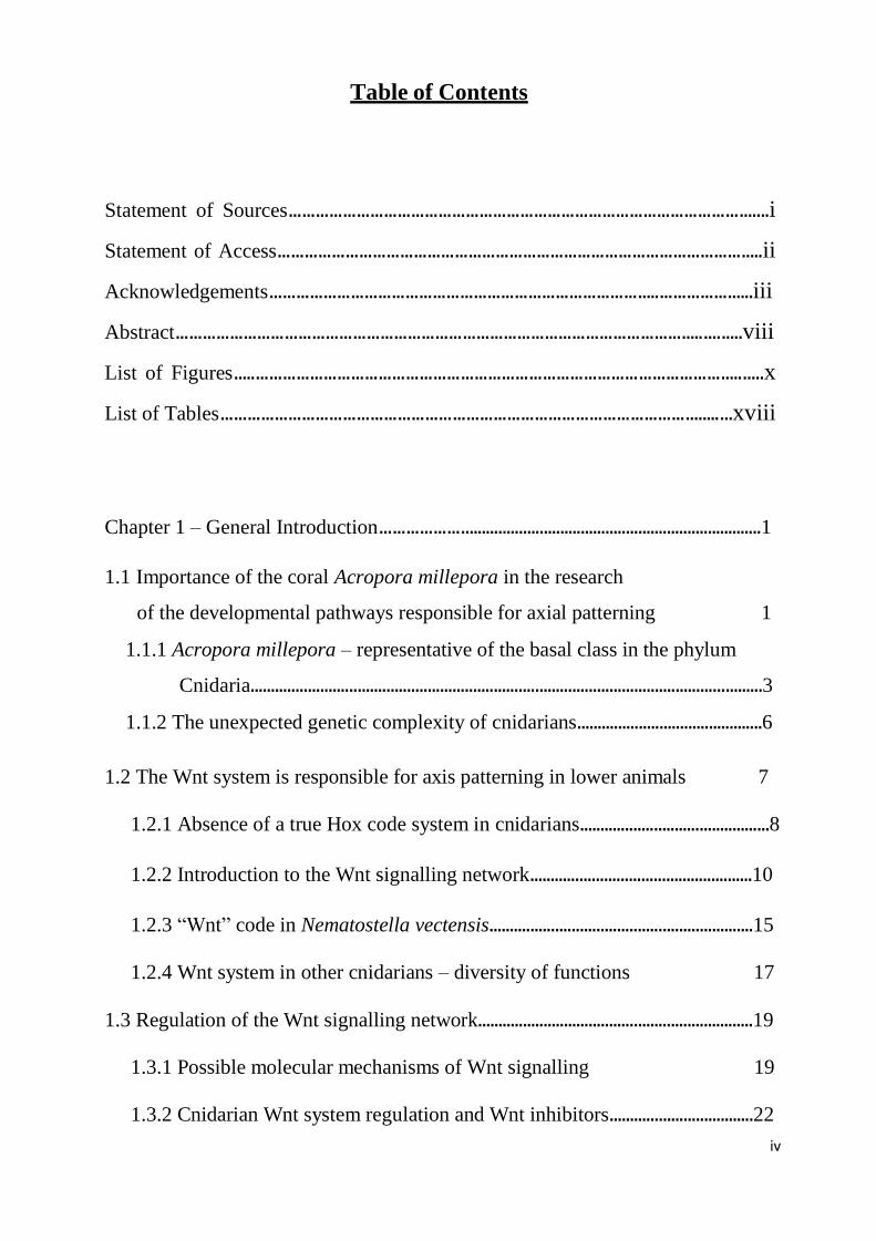
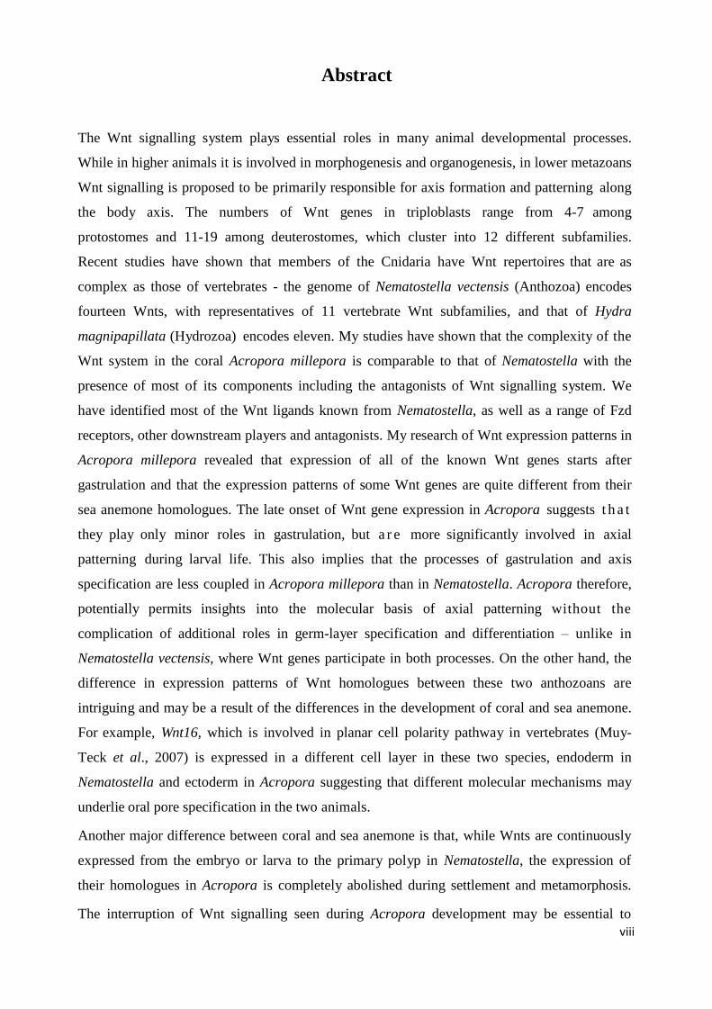
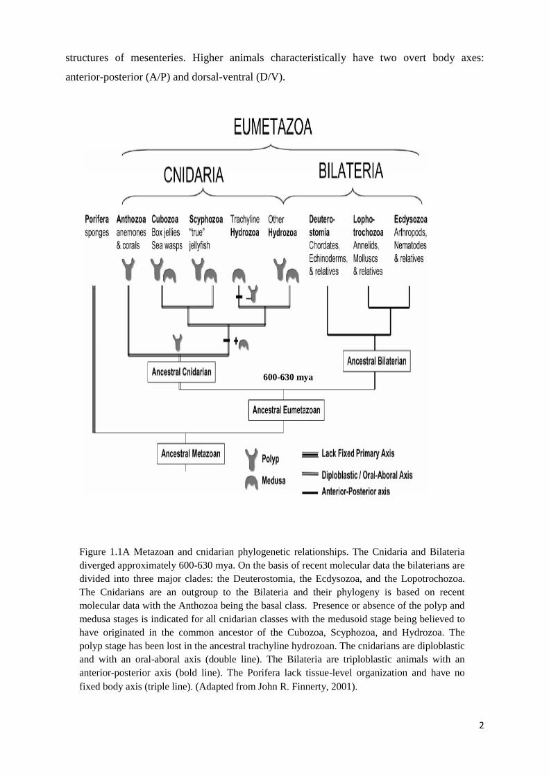
Chapter 1 Figure 1.1A Metazoan and cnidarian phylogenetic relationships. The Cnidaria and Bilateria
diverged approximately 600-630 mya. On the basis of recent molecular data the bilaterians
are divided into three major clades: the Deuterostomia, the Ecdysozoa, and the
Lopotrochozoa. The Cnidarians are an outgroup to the Bilateria and their phylogeny is based
on recent molecular data with the Anthozoa being the basal class. Presence or absence of the
polyp and medusa stages is indicated for all cnidarian classes with the medusoid stage being
believed to have originated in the common ancestor of the Cubozoa, Scyphozoa, and
Hydrozoa. The polyp stage has been lost in the ancestral trachyline hydrozoan. The cnidarians
are diploblastic and with an oral-aboral axis (double line). The Bilateria are triploblastic
animals with an anterior-posterior axis (bold line). The Porifera lack tissue-level organization
and have no fixed body axis (triple line). (Adapted from John R. Finnerty,
2001)…………………………………………………………………………………………….
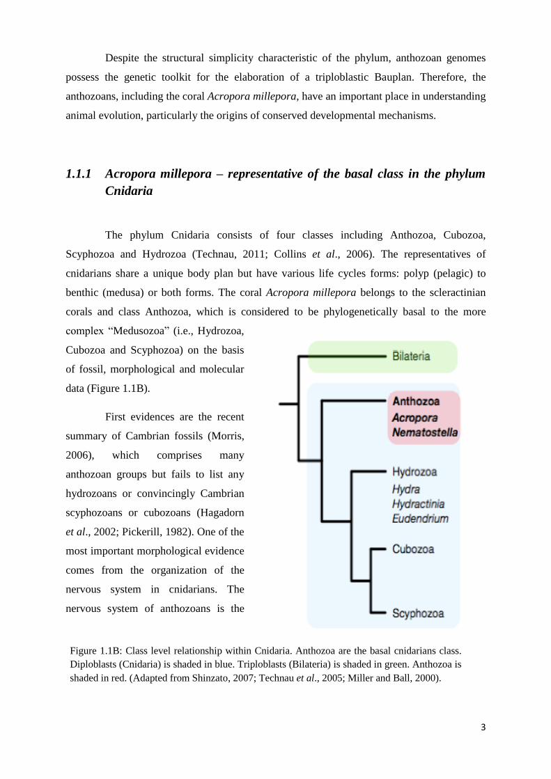
Figure 1.1B: Class level relationship within Cnidaria. Anthozoa are the basal cnidarians class.
Diploblasts (Cnidaria) is shaded in blue. Triploblasts (Bilateria) is shaded in green. Anthozoa
is shaded in red. (Adapted from Shinzato, 2007; Technau et al., 2005; Miller and Ball,
2000)…………………………………………………………………………………………….
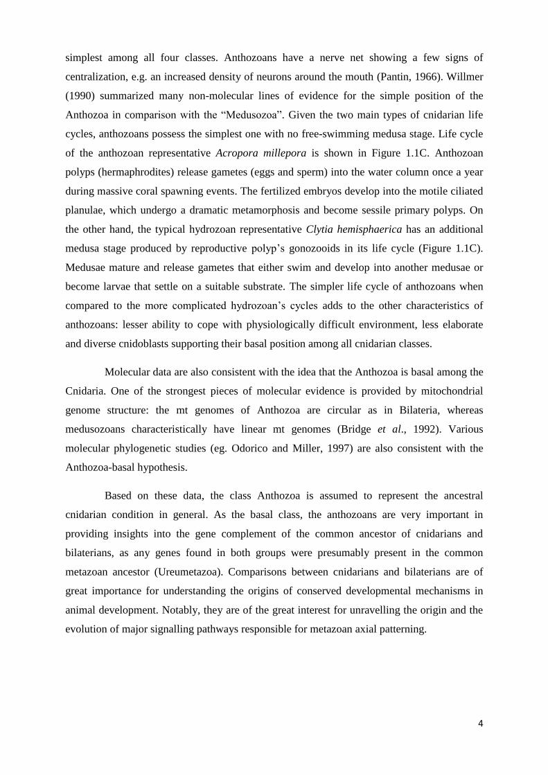
Figure 1.1C: Life cycles of the anthozoan Acropora millepora (first picture) and the
hydrozoan Clytia hemispaerica (below). Clytia has a more complex life cycle with polyp
and medusa generations compared to the simpler cycle of Acropora which lacks a
medusa stage (Adapted from Ulrich Technau, 2011)…………………………………………...
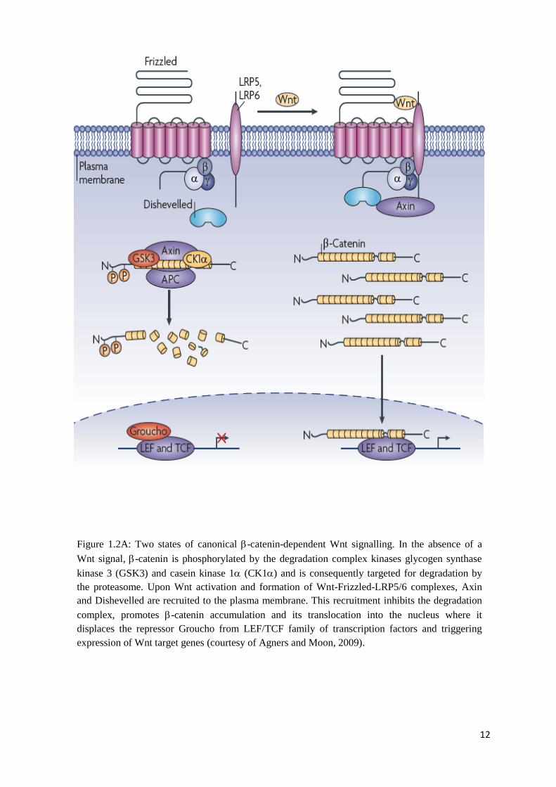
Figure 1.2A: Two states of canonical -catenin-dependent Wnt signalling. In the absence of a
Wnt signal, -catenin is phosphorylated by the degradation complex kinases glycogen
synthase kinase 3 (GSK3) and casein kinase 1 (CK1) and is consequently targeted for
degradation by the proteasome. Upon Wnt activation and formation of Wnt-Frizzled-LRP5/6
complexes, Axin and Dishevelled are recruited to the plasma membrane. This recruitment
inhibits the degradation complex, promotes -catenin accumulation and its translocation into
the nucleus where it displaces the repressor Groucho from LEF/TCF family of transcription
factors and triggering expression of Wnt target genes (courtesy of Agners and Moon,
2009)…………………………………………………………………………………………….
xi
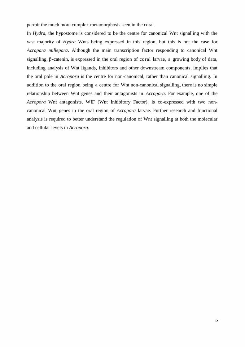
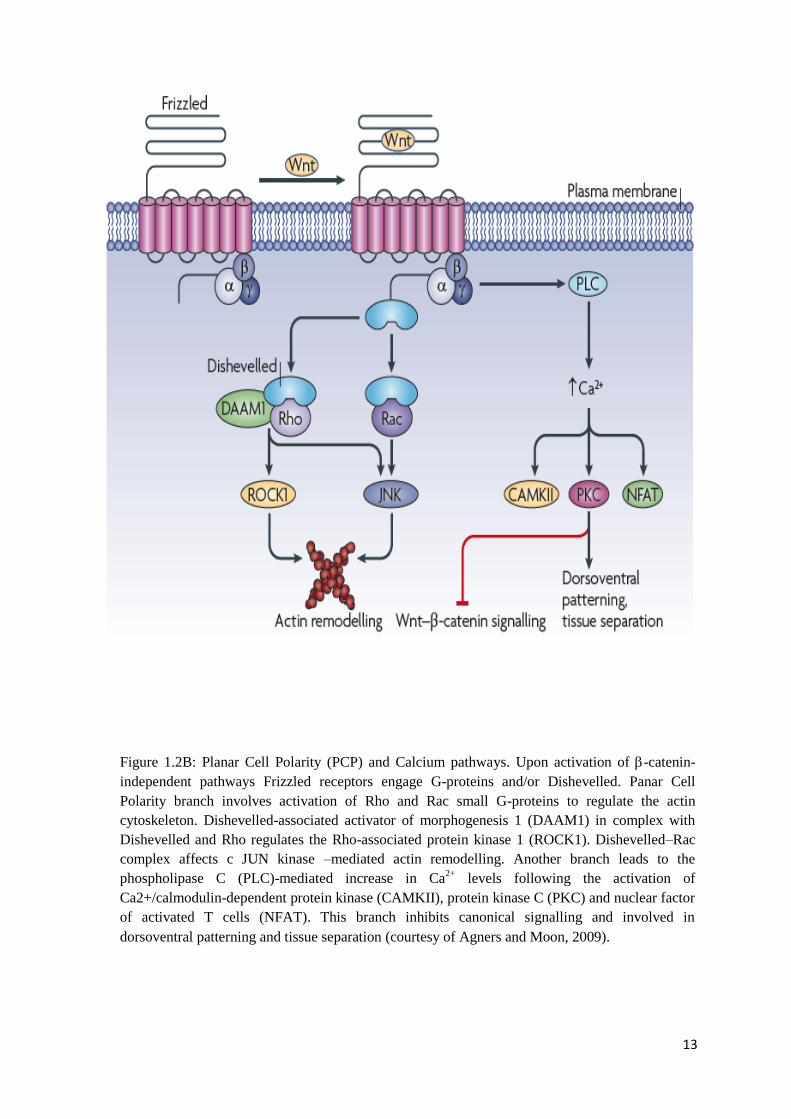
Figure 1.2B: Planar Cell Polarity (PCP) and Calcium pathways. Upon activation of -catenin-
independent pathways Frizzled receptors engage G-proteins and/or Dishevelled. Panar Cell
Polarity branch involves activation of Rho and Rac small G-proteins to regulate the actin
cytoskeleton. Dishevelled-associated activator of morphogenesis 1 (DAAM1) in complex
with Dishevelled and Rho regulates the Rho-associated protein kinase 1 (ROCK1).
Dishevelled–Rac complex affects c JUN kinase –mediated actin remodelling. Another branch
leads to the phospholipase C (PLC)-mediated increase in Ca2+
levels following the activation
of Ca2+/calmodulin-dependent protein kinase (CAMKII), protein kinase C (PKC) and nuclear
factor of activated T cells (NFAT). This branch inhibits canonical signalling and involved in
dorsoventral patterning and tissue separation (courtesy of Agners and Moon, 2009)…………
Figure 1.2C: Expression patterns of Wnt genes in Nematostella vectensis (Kusserow et al.,
2005). Majority of Wnts in Nematostella are expressed in the staggered domains along oral-
aboral axis, which is reminiscent of the Hox code in higher animals. Based on their
expression patterns in sea anemone they are proposed to be mainly involved in axis patterning
of lower animals…………………………………........................................................................
Picture 1.3: Key Wnt regulators in the canonical Wnt pathway. In the absence of Wnt ligands,
-catenin binds to the destruction complex containing APC, Axin, and the CK1 and GSK-
3Β kinases and is marked for proteolytic degradation. Wnt signals promote
hyperphosphorylation of LRP5/6 and Dsh, which recruit Axin to the receptor complex, where
it undergoes proteolytic degradation. Unphosphorylated -catenin is no longer rapidly
degraded and enters the nucleus. Also, part of the -catenin pool is bound to E-cadherins
participating in adherens junctions (AJ). (Courtesy of Karl Willert and Katherine A. Jones,
Genes & Development, 2006)……………………………………………………………………
Chapter 2 Figure 2.3A: Maximum likelihood (ML) phylogenetic analysis of Acropora millepora Wnt
proteins. Bootstrap support values (100 replicates) are given on all branches. Species
abbreviations: Am, Acropora millepora; Nv, Nematostella vectensis; Hv, Hydra vulgaris; Hs,
Homo sapiens; Xl, Xenopus laevis; Bf, Branchiostoma floridae (amphioxus); A, Apis
mellifera; Ag, Anopheles gambiae; Pd, Plathynereis dumerlii; Sp, Strongylocentrotus
purpuratus; Sk, Saccoglossus kowalevskii……………………………………………………...
Figure 2.3B: Alignment of Wnt11 proteins from Am, Acropora millepora, Nv, Nematostella
vectensis, Hv, Hydra vulgaris, Bf, Branchiostoma floridae, Xl, Xenopus laevis and Hs, Homo
sapiens and the comparison of their cysteine patterns show a high degree of similarity within
Wnt11 family. Small domain is positioned between 200 and 240 aa, and large one is at the
end of the protein sequence (320-360 aa).……………………………………………………..
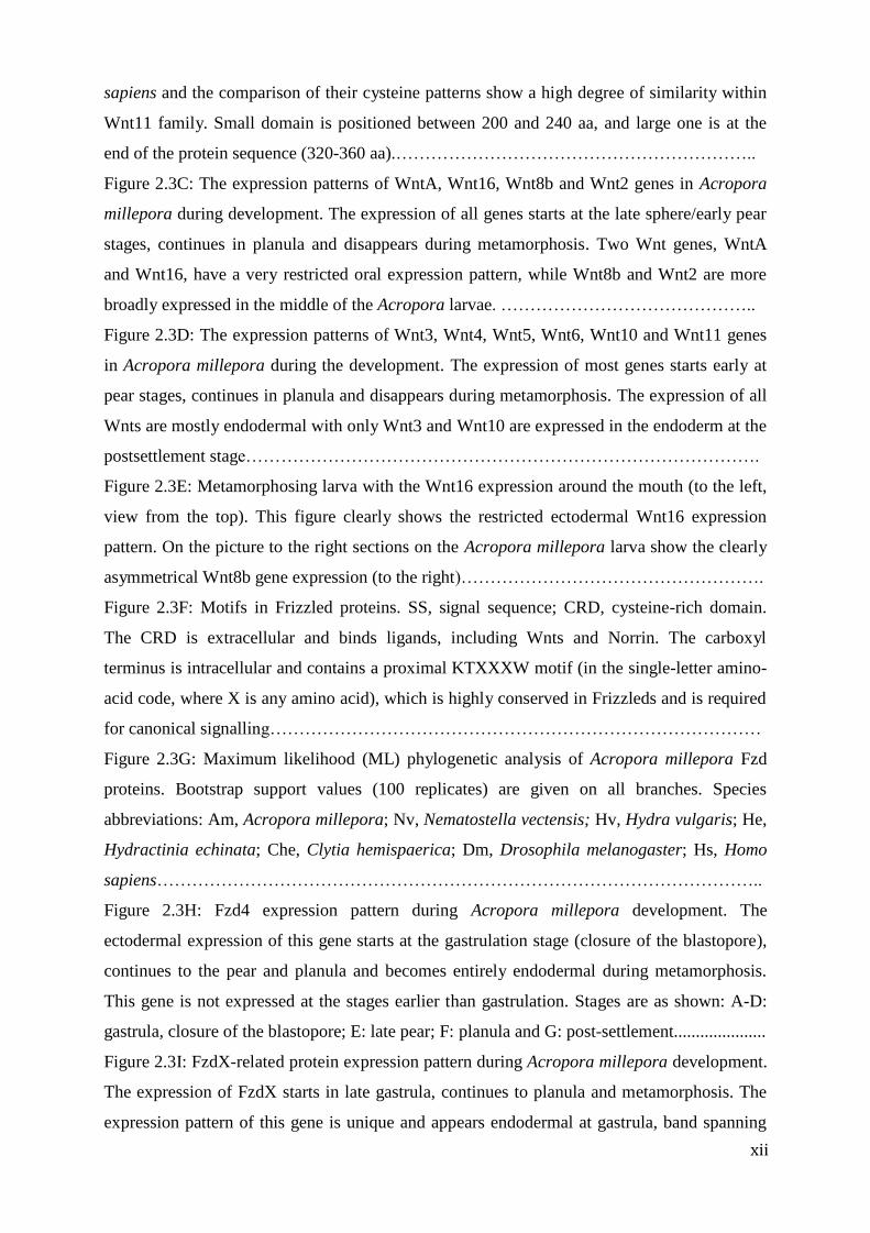
Figure 2.3C: The expression patterns of WntA, Wnt16, Wnt8b and Wnt2 genes in Acropora
millepora during development. The expression of all genes starts at the late sphere/early pear
stages, continues in planula and disappears during metamorphosis. Two Wnt genes, WntA
and Wnt16, have a very restricted oral expression pattern, while Wnt8b and Wnt2 are more
broadly expressed in the middle of the Acropora larvae. ……………………………………..
Figure 2.3D: The expression patterns of Wnt3, Wnt4, Wnt5, Wnt6, Wnt10 and Wnt11 genes
in Acropora millepora during the development. The expression of most genes starts early at
pear stages, continues in planula and disappears during metamorphosis. The expression of all
Wnts are mostly endodermal with only Wnt3 and Wnt10 are expressed in the endoderm at the
postsettlement stage…………………………………………………………………………….
Figure 2.3E: Metamorphosing larva with the Wnt16 expression around the mouth (to the left,
view from the top). This figure clearly shows the restricted ectodermal Wnt16 expression
pattern. On the picture to the right sections on the Acropora millepora larva show the clearly
asymmetrical Wnt8b gene expression (to the right)…………………………………………….
Figure 2.3F: Motifs in Frizzled proteins. SS, signal sequence; CRD, cysteine-rich domain.
The CRD is extracellular and binds ligands, including Wnts and Norrin. The carboxyl
terminus is intracellular and contains a proximal KTXXXW motif (in the single-letter amino-
acid code, where X is any amino acid), which is highly conserved in Frizzleds and is required
for canonical signalling…………………………………………………………………………
Figure 2.3G: Maximum likelihood (ML) phylogenetic analysis of Acropora millepora Fzd
proteins. Bootstrap support values (100 replicates) are given on all branches. Species
abbreviations: Am, Acropora millepora; Nv, Nematostella vectensis; Hv, Hydra vulgaris; He,
Hydractinia echinata; Che, Clytia hemispaerica; Dm, Drosophila melanogaster; Hs, Homo
sapiens…………………………………………………………………………………………..
Figure 2.3H: Fzd4 expression pattern during Acropora millepora development. The
ectodermal expression of this gene starts at the gastrulation stage (closure of the blastopore),
continues to the pear and planula and becomes entirely endodermal during metamorphosis.
This gene is not expressed at the stages earlier than gastrulation. Stages are as shown: A-D:
gastrula, closure of the blastopore; E: late pear; F: planula and G: post-settlement.....................
Figure 2.3I: FzdX-related protein expression pattern during Acropora millepora development.
The expression of FzdX starts in late gastrula, continues to planula and metamorphosis. The
expression pattern of this gene is unique and appears endodermal at gastrula, band spanning
xii
xiii
ectoderm and endoderm in the middle of the embryo in planula, and this gene is also
expressed in the post-settlement stage showing an intriguing expression pattern in the
endoderm, surrounding the mouth. Stages depicted in figure are as follows: left up: late
gastrula; right up: planula; left down: post-settlement and right down: sections of post-
settlement. ……………....................... Figure 2.3J: Fzd8 expression pattern during Acropora
millepora development. The expression of Fzd8 starts at the pear stage (A, B), continues to
planula (C, D) and postsettlement (E). This gene shows a very restricted expression pattern in
the aboral ectoderm during early larval development entirely switches to the endoderm at
the postsettlement stage. Oral pore is shown to the
left………………………………………………………………………………… Figure 2.3K:
Domain structure of LRP5/6 low-density lipoprotein from Xenopus laevis, Nematostella
vectensis and Acropora millepora species. All proteins contain Ly-domains type “B”, EGF
domains, LDLa domains and one transmembrane domain (TD) at the end of the
sequence……………………………………………………….....................................................
Figure 2.3L: Upon Wnt activation Frizzled receptors recruit the Dishevelled proteins to the
plasma membrane. Dishevelled recruits the Axin-glycogen synthase kinase 3 (GSK-3Β)
complex, which phosphorylates PPS(T)P motifs on the cytoplasmic tail of LRP5/6. LRP5/6 is
further phosphorylated by CK1 and GSK-3Β. Axin sequestration by LRP5/6 inhibits the
formation of the GSK-3Β degradation complex and leads to -catenin stabilization and
pathway activation………………………………………………………………………………
Figure 2.3M: Domain structure of Dsh3 proteins from Homo sapiens, Nematostella vectensis
and Acropora millepora. This figure shows that all three proteins contain all domains of Dsh,
including DIX, PDZ and DEP…………………………………………………………………..
Figure 2.3N: Expression pattern of Acropora millepora homologue of Dishevelled is entirely
endodermal and it expression starts at the late sphere stage. No expression detected at earlier
stages in development. At the postsettlement stage the Dsh expression weakens but remains
endodermal. Shown stages of Acropora millepora development are as follows: A: prawnchip;
B: early sphere; C: early planula and D: post-settlement……………………………………….
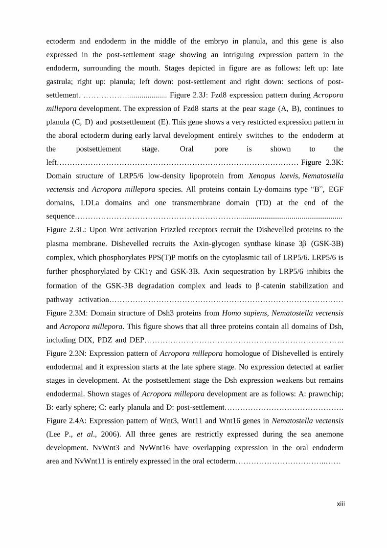
Figure 2.4A: Expression pattern of Wnt3, Wnt11 and Wnt16 genes in Nematostella vectensis
(Lee P., et al., 2006). All three genes are restrictly expressed during the sea anemone
development. NvWnt3 and NvWnt16 have overlapping expression in the oral endoderm
area and NvWnt11 is entirely expressed in the oral ectoderm……………………………..……
xiv
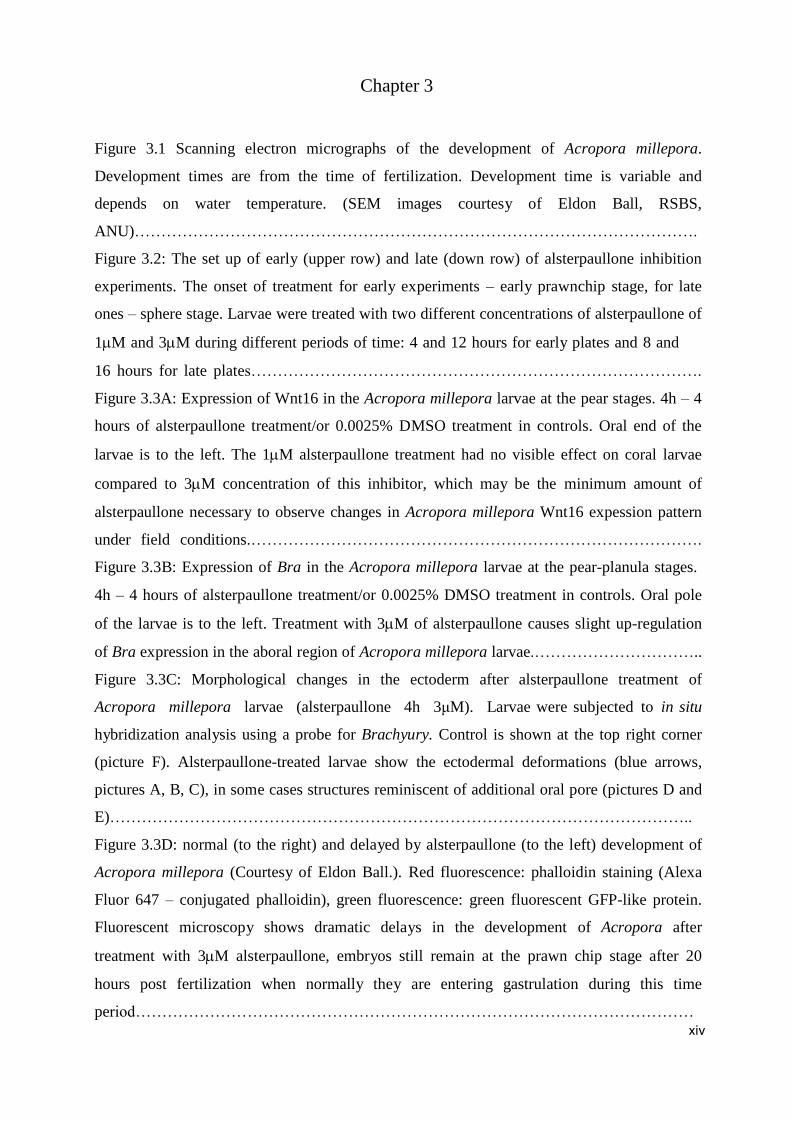
Chapter 3 Figure 3.1 Scanning electron micrographs of the development of Acropora millepora.
Development times are from the time of fertilization. Development time is variable and
depends on water temperature. (SEM images courtesy of Eldon Ball, RSBS,
ANU)…………………………………………………………………………………………….
Figure 3.2: The set up of early (upper row) and late (down row) of alsterpaullone inhibition
experiments. The onset of treatment for early experiments – early prawnchip stage, for late
ones – sphere stage. Larvae were treated with two different concentrations of alsterpaullone of
1M and 3M during different periods of time: 4 and 12 hours for early plates and 8 and
16 hours for late plates………………………………………………………………………….
Figure 3.3A: Expression of Wnt16 in the Acropora millepora larvae at the pear stages. 4h – 4
hours of alsterpaullone treatment/or 0.0025% DMSO treatment in controls. Oral end of the
larvae is to the left. The 1M alsterpaullone treatment had no visible effect on coral larvae
compared to 3M concentration of this inhibitor, which may be the minimum amount of
alsterpaullone necessary to observe changes in Acropora millepora Wnt16 expession pattern
under field conditions.………………………………………………………………………….
Figure 3.3B: Expression of Bra in the Acropora millepora larvae at the pear-planula stages.
4h – 4 hours of alsterpaullone treatment/or 0.0025% DMSO treatment in controls. Oral pole
of the larvae is to the left. Treatment with 3M of alsterpaullone causes slight up-regulation
of Bra expression in the aboral region of Acropora millepora larvae.…………………………..
Figure 3.3C: Morphological changes in the ectoderm after alsterpaullone treatment of
Acropora millepora larvae (alsterpaullone 4h 3μM). Larvae were subjected to in situ
hybridization analysis using a probe for Brachyury. Control is shown at the top right corner
(picture F). Alsterpaullone-treated larvae show the ectodermal deformations (blue arrows,
pictures A, B, C), in some cases structures reminiscent of additional oral pore (pictures D and
E)………………………………………………………………………………………………..
Figure 3.3D: normal (to the right) and delayed by alsterpaullone (to the left) development of
Acropora millepora (Courtesy of Eldon Ball.). Red fluorescence: phalloidin staining (Alexa
Fluor 647 – conjugated phalloidin), green fluorescence: green fluorescent GFP-like protein.
Fluorescent microscopy shows dramatic delays in the development of Acropora after
treatment with 3M alsterpaullone, embryos still remain at the prawn chip stage after 20
hours post fertilization when normally they are entering gastrulation during this time
period……………………………………………………………………………………………
xv
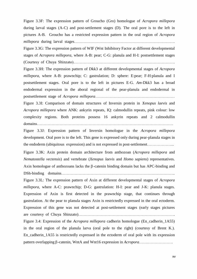
Figure 3.3F: The expression pattern of Groucho (Gro) homologue of Acropora millepora
during larval stages (A-C) and post-settlement stages (D). The oral pore is to the left in
pictures A-B. Groucho has a restricted expression pattern in the oral region of Acropora
millepora during larval stages………………………………………………………………….
Figure 3.3G: The expression pattern of WIF (Wnt Inhibitory Factor at different developmental
stages of Acropora millepora, where A-B: pear; C-G: planula and H-I: postsettlement stages
(Courtesy of Chuya Shinzato).………………………………………………………………….
Figure 3.3H: The expression pattern of Dkk3 at different developmental stages of Acropora
millepora, where A-B: prawnchip; C: gastrulation; D: sphere: E:pear; F-H:planula and I
postsettlement stages. Oral pore is to the left in pictures E-G. Am-Dkk3 has a broad
endodermal expression in the aboral regional of the pear-planula and endodermal in
postsettlement stage of Acropora millepora……………………………………………………
Figure 3.3I: Comparison of domain structures of Inversin protein in Xenopus laevis and
Acropora millepora where ANK: ankyrin repeats, IQ: calmodullin repeats, pink colour: low
complexity regions. Both proteins possess 16 ankyrin repeats and 2 calmodullin
domains………………………………………………………………………………………….
Figure 3.3J: Expression pattern of Inversin homologue in the Acropora millepora
development. Oral pore is to the left. This gene is expressed only during pear-planula stages in
the endoderm (ubiquitous expression) and is not expressed in post-settlement………………...
Figure 3.3K: Axin protein domain architecture from anthozoan (Acropora millepora and
Nematostella vectensis) and vertebrate (Xenopus laevis and Homo sapiens) representatives.
Axin homologue of anthozoans lacks the -catenin binding domain but has APC-binding and
DSh-binding domains…………………………………………………………………………..
Figure 3.3L: The expression pattern of Axin at different developmental stages of Acropora
millepora, where A-C: prawnchip; D-G: gastrulation: H-I: pear and J-K: planula stages.
Expression of Axin is first detected in the prawnchip stage, that continues through
gastrulation. At the pear to planula stages Axin is restrictedly expressed in the oral ectoderm.
Expression of this gene was not detected at post-settlement stages (early stages pictures
are courtesy of Chuya Shinzato)……………………………………………………………….
Figure 3.4: Expression of the Acropora millepora cadherin homologue (En_cadherin_1A55)
in the oral region of the planula larva (oral pole to the right) (courtesy of Brent K.).
En_cadherin_1A55 is restrictedly expressed in the ectoderm of oral pole with its expression
pattern overlapping -catenin, WntA and Wnt16 expression in Acropora…….……………….
xvi
Supplementary material
Figure 2.2A: Alignment of Wnt16 proteins from Am, Acropora millepora and
Nv, Nematostella vectensis. The comparison of their cysteine patterns patterns show a high
degree of similarity within Wnt16 family. Small Wnt domain is positioned between 200
and 240 aa, and large one is at the end of the protein sequence (280-360 aa)………………...
Figure 2.2B: Alignment of Wnt1 proteins from Am, Acropora millepora and Nv,
Nematostella vectensis. The comparison of their cysteine patterns shows high degree
of similarity in the small Wnt domain of Acropora and Nematostella, positioned between
200 and 240 aa………………………………………………………………...........................
Figure 2.2C: Alignment of Wnt6 proteins from Am, Acropora millepora and
Nv, Nematostella vectensis. The comparison of their cysteine patterns shows high
degree of similarity between these homologues, though complete Acropora Wnt6
sequence needed for the small and large Wnt domains comparisons………………….……..
Figure 2.2D: Alignment of Wnt4 proteins from Am, Acropora millepora and
Nv, Nematostella vectensis. The comparison of their cysteine patterns shows a high
degree of similarity between small and large Wnt4 domains of Acropora and
Nematostella, positioned at 200-240 aa and 280-360 aa, respectively…………………..……
Figure 2.2E: Alignment of Wnt2 proteins from Am, Acropora millepora and Nv,
Nematostella vectensis. The comparison of their cysteine patterns shows a high degree
of similarity between small and large Wnt2 domains of Acropora and Nematostella,
positioned at 200-240 aa and at 280-360 aa, respectively………………………………...…..
Figure 2.2F: Alignment of Wnt5 proteins from Am, Acropora millepora and Nv,
Nematostella vectensis. The comparison of their cysteine patterns shows a high degree
of similarity between small and large Wnt domains of Acropora and Nematostella,
positioned 200-240 aa and 280-360 aa, respectively……………….........................................
Figure 2.2G: Alignment of Wnt10 proteins from Am, Acropora millepora and
Nv, Nematostella vectensis. The comparison of their cysteine patterns shows a high
degree of similarity between small and large Wnt domains of Acropora and Nematostella,
positoned at 200-240 aa and at 280-360 aa, respectively………………………………..……
Figure 2.2H: Alignment of WntA proteins from Am, Acropora millepora and Nv,
Nematostella vectensis. The comparison of their cysteine patterns shows a high degree
of similarity between small and large Wnt domains of Acropora and Nematostella,
positioned at 200-240 aa and at 280-360 aa, respectively………………………….…………
Figure 2.2I: Alignment of Wnt8b proteins from Am, Acropora millepora and Nv,
xvii
Nematostella vectensis. The comparison of their cysteine patterns shows a high degree of
similarity between small and large Wnt domains of Acropora and Nematostella, positioned
at 160-200 aa and at 240-340 aa, respectively……………………………………….…………
Figure 2.2J: Alignment of Wnt3 proteins from Am, Acropora millepora and Nv,
Nematostella vectensis. The comparison of their cysteine patterns shows a high degree
of similarity between small and large Wnt3 domains from Acropora and Nematostella,
positioned at 200-240 aa and at 280-360 aa, respectively…………………………...…………
Figure 2.2K: Alignment of Wnt7b proteins from Am, Acropora millepora and Nv,
Nematostella vectensis. The comparison of their cysteine patterns shows a high degree
of similarity between small and large Wnt domains of Acropora and Nematostella,
positioned at 200-240 aa and at 280-360 aa, respectively……………………………………...
Figure 2.2L: Alignment of Fzd8 proteins from Am, Acropora millepora and Nv,
Nematostella vectensis. The figure shows the similarities in the N-terminal region of
Acropora and Nematostella terminal regions………………………………………………..…
Figure 2.3O: Alignment of Fzd4 proteins from Am, Acropora millepora and Nv,
Nematostella vectensis shows a high degree of similarity of these anthozoan Fzd4
protein sequences……………………………………….............................................................
Figure 2.3P: Alignment of Fzd3 proteins from Am, Acropora millepora and Nv,
Nematostella vectensis shows areas degree of similarity of these anthozoan Fzd3
protein sequences excluding region of insertion for Acropora homologue, positioned
between 200 and 250 aa…………............................................................................................
Figure 2.3Q: Alignment of Fzd2 proteins from Am, Acropora millepora and Nv,
Nematostella vectensis shows a high degree of similarity of these anthozoan Fzd2
protein sequences.……………………………………………………………………...……….
Figure 2.3R: Alignment of Fzd1 proteins from Am, Acropora millepora and Nv,
Nematostella vectensis vectensis shows a high degree of similarity of these anthozoan
Fzd1 protein sequences………………………………………………………………………………..………………
Figure 2.3S: Alignment of LRP5/6 proteins from Am, Acropora millepora and Nv,
Nematostella vectensis shows a high degree of similarity between theanthozoan homologues
Figure 2.4: Maximum likelihood (ML) phylogenetic analysis of Acropora millepora,
Acropora digitifera and Nematostella vectensis Wnt proteins. Bootstrap support values
(100 replicates) are given on all branches. Species abbreviations: Am, Acropora
millepora; Nv, Nematostella vectensis; Ad, Acropora digitifera………………………………
Figure 3.3E: The alignment of Nematostella vectensis and Acropora millepora
xviii
armadillo repeats in -catenin shows very high level of sequence similarity between the
two..……………………………………………………………………………………………
List of Tables
Table 2.2A: Name of the gene, primer sequences, Tm, length of the sequenced fragments
and used plasmids are given……………………………………………………………...……
Table 2.2B: Name of the gene fragment, restriction enzyme, RNA polymeraze and the
size of the probe in bp (base pairs) are given………………………………………………….
Table 2.4.2: Distribution of Wnt genes among metazoans. Cnidaria -Am: Acropora
millepora; Nv: Nematostella vectensis; Hm: Hydra magnipapillata; Che: Clytia
hemispaerica. Vertebrata – Hs: Homo sapiens; Xt: Xenopus tropicalis. Ecdusozoa – Apm:
Apis mellifera; Dm: Drosophila melanogaster; Tc: Tribolium castaneum. Lophotrochozoa
– Pd: Platynereis dumerilli; Pv: Patella vulgata; Gt: Girardia tigrina……………….………
Table 3.2.2: Name of the gene, primer sequences, Tm, cycles required for PCR reaction
and the length of the fragment………………………………………………………...………
1
Chapter 1
General Introduction
1.1 Importance of the coral Acropora millepora in the research of the
developmental pathways responsible for axial patterning.
Our model, the coral Acropora millepora belongs to the phylum Cnidaria, class
Anthozoa. This phylum is a sister group to Bilateria and positioned among the four early-
branching phyla of the Metazoa (Porifera, Ctenophora, Placozoa and Cnidaria). While there is
a long-standing debate on whether the Porifera or Ctenophora represents the earliest divergent
metazoans with conserved ancestral state of animal multicellularity (Schierwater et al., 2010,
2009; Dunn et al., 2008; Dellaporta et al., 2006), cnidarians (corals, sea anemones, jellyfish)
are the most complex among early-diverged phyla. At the same time, cnidarian anthozoan
representatives have been found in the Cambrian fossil record (Morris, 2006) showing that
cnidarian diversification could have been occurred as early as after the Precambrian period
over 500 million years ago (Technau, 2011).
Moreover, as it was shown that all body plan characteristics of the 35 phyla that exist
today were already present in the Cambrian period, it has been suggested that cnidarians
might have been the cause (or ‘‘explosives’’) of the Cambrian explosion (Boero et al., 2007).
As one of the most complex of early diverging metazoan phyla, cnidarians also represent a
sister group to the Bilateria. The Cnidaria and Bilateria are thought to have diverged from the
common Eumetazoan ancestor approximately 600-630 million years ago according to the
recent studies of components of Toll-like receptor signalling in Bilateria and Cnidaria
(Irazoqui et al., 2010) (Figure 1.1A). Several morphological and structural characteristics
distinguish bilaterians and cnidarians as being higher and lower animals respectively. The
representatives of the Bilateria are triploblastic animals with three germ layers, the ectoderm,
endoderm and mesoderm. In contrast, cnidarians have only two germ layers, the ectoderm and
endoderm. Although, another character distinguishing Cnidaria from bilaterians would be the
possession of only one overt body axis, known as the oral-aboral axis in cnidarians, many
corals including A. millepora, have the dorsal-ventral axis recognized by apparent asymmetric
2
structures of mesenteries. Higher animals characteristically have two overt body axes:
anterior-posterior (A/P) and dorsal-ventral (D/V).
600-630 mya
Figure 1.1A Metazoan and cnidarian phylogenetic relationships. The Cnidaria and Bilateria
diverged approximately 600-630 mya. On the basis of recent molecular data the bilaterians are
divided into three major clades: the Deuterostomia, the Ecdysozoa, and the Lopotrochozoa.
The Cnidarians are an outgroup to the Bilateria and their phylogeny is based on recent
molecular data with the Anthozoa being the basal class. Presence or absence of the polyp and
medusa stages is indicated for all cnidarian classes with the medusoid stage being believed to
have originated in the common ancestor of the Cubozoa, Scyphozoa, and Hydrozoa. The
polyp stage has been lost in the ancestral trachyline hydrozoan. The cnidarians are diploblastic
and with an oral-aboral axis (double line). The Bilateria are triploblastic animals with an
anterior-posterior axis (bold line). The Porifera lack tissue-level organization and have no
fixed body axis (triple line). (Adapted from John R. Finnerty, 2001).
3
Despite the structural simplicity characteristic of the phylum, anthozoan genomes
possess the genetic toolkit for the elaboration of a triploblastic Bauplan. Therefore, the
anthozoans, including the coral Acropora millepora, have an important place in understanding
animal evolution, particularly the origins of conserved developmental mechanisms.
1.1.1 Acropora millepora – representative of the basal class in the phylum
Cnidaria
The phylum Cnidaria consists of four classes including Anthozoa, Cubozoa,
Scyphozoa and Hydrozoa (Technau, 2011; Collins et al., 2006). The representatives of
cnidarians share a unique body plan but have various life cycles forms: polyp (pelagic) to
benthic (medusa) or both forms. The coral Acropora millepora belongs to the scleractinian
corals and class Anthozoa, which is considered to be phylogenetically basal to the more
complex “Medusozoa” (i.e., Hydrozoa,
Cubozoa and Scyphozoa) on the basis
of fossil, morphological and molecular
data (Figure 1.1B).
First evidences are the recent
summary of Cambrian fossils (Morris,
2006), which comprises many
anthozoan groups but fails to list any
hydrozoans or convincingly Cambrian
scyphozoans or cubozoans (Hagadorn
et al., 2002; Pickerill, 1982). One of the
most important morphological evidence
comes from the organization of the
nervous system in cnidarians. The
nervous system of anthozoans is the
Figure 1.1B: Class level relationship within Cnidaria. Anthozoa are the basal cnidarians class.
Diploblasts (Cnidaria) is shaded in blue. Triploblasts (Bilateria) is shaded in green. Anthozoa is
shaded in red. (Adapted from Shinzato, 2007; Technau et al., 2005; Miller and Ball, 2000).
4
simplest among all four classes. Anthozoans have a nerve net showing a few signs of
centralization, e.g. an increased density of neurons around the mouth (Pantin, 1966). Willmer
(1990) summarized many non-molecular lines of evidence for the simple position of the
Anthozoa in comparison with the “Medusozoa”. Given the two main types of cnidarian life
cycles, anthozoans possess the simplest one with no free-swimming medusa stage. Life cycle
of the anthozoan representative Acropora millepora is shown in Figure 1.1C. Anthozoan
polyps (hermaphrodites) release gametes (eggs and sperm) into the water column once a year
during massive coral spawning events. The fertilized embryos develop into the motile ciliated
planulae, which undergo a dramatic metamorphosis and become sessile primary polyps. On
the other hand, the typical hydrozoan representative Clytia hemisphaerica has an additional
medusa stage produced by reproductive polyp’s gonozooids in its life cycle (Figure 1.1C).
Medusae mature and release gametes that either swim and develop into another medusae or
become larvae that settle on a suitable substrate. The simpler life cycle of anthozoans when
compared to the more complicated hydrozoan’s cycles adds to the other characteristics of
anthozoans: lesser ability to cope with physiologically difficult environment, less elaborate
and diverse cnidoblasts supporting their basal position among all cnidarian classes.
Molecular data are also consistent with the idea that the Anthozoa is basal among the
Cnidaria. One of the strongest pieces of molecular evidence is provided by mitochondrial
genome structure: the mt genomes of Anthozoa are circular as in Bilateria, whereas
medusozoans characteristically have linear mt genomes (Bridge et al., 1992). Various
molecular phylogenetic studies (eg. Odorico and Miller, 1997) are also consistent with the
Anthozoa-basal hypothesis.
Based on these data, the class Anthozoa is assumed to represent the ancestral
cnidarian condition in general. As the basal class, the anthozoans are very important in
providing insights into the gene complement of the common ancestor of cnidarians and
bilaterians, as any genes found in both groups were presumably present in the common
metazoan ancestor (Ureumetazoa). Comparisons between cnidarians and bilaterians are of
great importance for understanding the origins of conserved developmental mechanisms in
animal development. Notably, they are of the great interest for unravelling the origin and the
evolution of major signalling pathways responsible for metazoan axial patterning.
5
Figure 1.1C: Life cycles of the anthozoan Acropora millepora (first picture) and the hydrozoan Clytia
hemisphaerica (second picture). Clytia has a more complex life cycle with polyp and medusa
generations compared to the simpler cycle of Acropora which lacks a medusa stage (Adapted from
Ulrich Technau, 2011).
Acropora millepora
Clytia hemisphaerica
6
1.1.2 The unexpected genetic complexity of cnidarians
In spite of cnidarians being amongst the simplest metazoans in terms of morphology,
they possess unexpected genetic complexity (Guder et al., 2006a; Technau et al., 2005).
Analyses of ESTs from anthozoan representatives, Acropora millepora and Nematostella
vectensis, have led to the discovery that anthozoan genomes are likely to contain around
25,000 genes which is more than Drosophila melanogaster (14,000 protein coding genes) and
Caenorhabditis elegance (19,000 protein coding genes) and in the same range as vertebrates.
Although the class Anthozoa is basal and may therefore best represent ancestral cnidarian
character states, the accumulating data have shown that this class possesses a much more
complex genome than was expected, including also genes that are not previously known from
the animal kingdom (Technau et al., 2005).
Importantly, searches of Cnidaria have revealed all of the major families of
developmentally regulated signalling pathways represented in higher animals and many of
their downstream components. For example, both Acropora millepora and Nematostella
vectensis have at least five different transforming growth factor β (TGFβ) ligands,
representing four of the six known subfamilies and the corresponding subclasses of receptors
(TGFβ, Activin and bone morphogenetic proteins (BMP)) (Technau et al., 2005). They also
have representatives of all of the Smad subfamilies (Smad1/5/8; Smad2/3; Smad4, Smad6/7;
downstream members of the TGFβ pathway) known from vertebrates (Technau et al., 2005).
Many components of the Wnt, Notch and Hedgehog signalling pathways have also been
identified in cnidarians, whereas hedgehog is missing from Caenorhabditis.
It is also becoming clear that many antagonists of these developmentally-regulated
signalling systems are present in cnidarians, although these are not present in Caenorhabditis
elegans or Drosophila melanogaster. Wnt Inhibitory Factor (WIF) and members of the Dkk
family are known from both Nematostella and Acropora (Guder et al., 2006a; Guder et al.,
2006b; Lee et al., 2006; Technau et al., 2005). The Wnt antagonists of Acropora millepora
are discussed in Chapter 3.
Surprisingly, many genes involved in mesoderm formation are present in cnidarians
despite the fact that they lack true mesoderm. In Acropora (Hayward et al., 2004), one of the
snail genes is expressed during the early stages of gastrulation in a pattern that strikingly
resembles that of its Drosophila counterpart (Ball et al., 2004). The Technau group has
identified many “mesodermal” genes in Nematostella including the transcription factors
7
Brachyury (Scholz and Technau, 2003), Snail (Fritzenwanker et al., 2004), Twist (Martindale
et al., 2004), Forkhead (Fritzenwanker et al., 2004) and gata factors (Martindale et al., 2004).
Studying the functions of these genes in cnidarians can hopefully shed some light on the
evolutionary origins of the bilaterian mesoderm.
Another feature of anthozoan genomes is the presence of many independently
duplicated genes; more than 5% of proteins in both Acropora millepora and Nematostella
vectensis, are likely to represent paralogues that arose independently in the cnidarian lineage
(Technau et al., 2005). Wnt8a and Wnt8b are the products of a Nematostella-specific
duplication event, but a substantial number of key transcription factors have also been
independently duplicated in the anthozoan lineage. The significance of these duplications is as
yet unclear.
The picture that is emerging is that, in terms of gene content, the complexity of the
anthozoan genome is comparable to that of vertebrates. Most of the genes that are important
for bilaterian development, including genes of all major signalling pathways (Technau et al,
2005) are present including the key mesodermal inducers. Therefore, despite their structural
simplicity, cnidarians are of crucial importance for studying the evolution of developmental
signalling pathways throughout the animal kingdom.
1.2 The Wnt signalling system is responsible for axis patterning in lower
animals.
Understanding how signalling systems interact and regulate animal embryogenesis
has always been a central concern of developmental biology. One of the most important
processes in embryogenesis – axial patterning – is controlled by several signalling systems,
which crosstalk with each other. Higher animals have two major body axes – oral/aboral and
dorsal/ventral, and the Hox genes and TGFβ system that play key roles in patterning these
(Ball et al., 2004). Apart from Hox genes, other genes are known to have conserved and
crucial roles in A/P patterning of higher animals. Among them are head-patterning genes
Otd/Otx and Ems/Emx, and genes of the Wnt signalling pathway (Kusserow et al., 2005; Ball
et al., 2004). As lower animals have only one overt body axis – oral/aboral, the majority of
studies on cnidarians were focused on systems that are responsible for A/P patterning. Recent
investigations on various cnidarian species show that whereas the diversification of
8
Hox/ParaHox genes unambiguously predates the origin of bilaterians, the true Hox code with
its role in anterior-posterior patterning is entirely bilaterian feature (Chiori et al., 2009). It has
been suggested that, in the absence of a Hox system, a Wnt code plays the same role in axial
patterning in cnidarians (Kusserow et al., 2005).
1.2.1 Absence of a true Hox system in cnidarians
According to recent studies cnidarians are the most primitive animals that possess
Hox genes. No Hox, ParaHox or extended Hox genes are present in sponges (studies on A.
queenslandica, Larroux et al., 2006, 2007; Seimiya et al., 1994, and other species, Manuel
and Le Parco, 2000). As sponge larvae are clearly axially organized (Adamska et al., 2007;
Degnan et al., 2005) axis patterning in this animal is most likely controlled by Wnt and TGF
systems (Adamska et al., 2007). Moreover, no Hox-related genes have been found in
Placozoa (Montiero et al., 2006) or the ctenophore Mnemiopsis (Pang and Martindale, 2008).
As the Hox system is ubiquitous throughout the Bilateria, these recent studies suggest that
Cnidaria may be unique amongst non-bilaterian Phyla in that some Hox genes are present but,
unlike the Bilateria, the key step of evolving the Hox code had not been made.
The genomic organization and expression patterns of Hox genes in bilaterians are
responsible for their central roles in the A/P (anterior/posterior) axis specification. Although
Hox and ParaHox genes have been identified in a range of cnidarians (Kamm et al., 2006;
Ryan et al., 2006; Gauchat et al., 2000), their expression data do not confirm the conservation
of the Hox code in these lower animals (Chiori et al., 2009).
According to the previous phylogenetic analyses cnidarians possess the “anterior”
Hox1 and Hox2 and posterior Hox genes (Hox9-14 like) but lack the “median” genes (Hox3-
8) (Chiori et al., 2009; Ryan and Baxevanis, 2007; Chourrout et al., 2006; Finnerty et al.,
2004; Gauchat et al., 2000). Also, many independent duplications have occurred during
cnidarian evolution and the vast majority of the Hox genes that have been identified do not fit
into the classes known from bilaterians. For example, Hox9-14 genes were independently
duplicated in the Scyphozoa and the Hydrozoa, and several duplications of Hox2 gene
occurred in the Anthozoa (Chiori et al., 2009). Thus, many of the Hox/Hox-like genes of
cnidarians are paralogs and the only report of the genomic linkage between several Hox genes
was based on the Nematostella vectensis genomic data (Chourrout et al., 2006). In
9
Nematostella, three paralogous Hox-like genes are linked to the eve/anthox6 pair, but this
“cluster” arose since the bilaterian divergence, and is therefore unrelated to the Hox clusters
of bilaterians. These facts are further supported by the studies of the corresponding Hydra
genes, which were shown not to be linked within a range of 150 kb (Gauchat et al., 2000) and
Clytia hemisphaerica Hox genes, also found not to be linked (Chiori et al., 2009).
Apart from the absence of clustered organization, the cnidarian Hox genes do not
have the kind of colinear expression patterns that would be required if they fulfilled roles in
axis patterning. The expression patterns of the cnidarian Hox-like genes are highly divergent
and significantly different not only from the relatively conserved expression patterns in
bilaterians but also within the phylum. Total absence of colinearity was previously reported
for the hydromedusae Eleutheria (Kamm et al., 2006) and Podocoryne as their Hox genes are
not expressed at the same stage of life cycle (Kamm et al., 2006; Yanze et al., 2001). Also,
when orthologous genes are expressed at the same stage, their transcripts are localized at
opposite poles along the oral/aboral axis. For example, CheHox9-14B in Clytia is expressed at
the oral end of the planula and its Nematostella counterpart NveAnthox1 has an aboral
expression in Nematostella (Ryan and Baxevanis, 2007; Finnerty et al., 2004). And finally,
orthologous genes that have similar expression domains are often expressed in different
tissues. For instance, while Podocoryne gene PcaCnox1 is expressed in both ectoderm and
endoderm at the aboral pole in larva its orthologue in Eleutheria EdiCnox5 is expressed
although similarly at the aboral end but only in the ectoderm (Kamm et al., 2006; Yanze et
al., 2001).
Thus, the expression and genomic data from cnidarians suggest that the Hox system
is not essential for axial patterning in lower animals and the role of Hox-like genes in corals is
still unclear. As cnidarians lack the canonical Hox clusters of higher Metazoans, it was
assumed that the true Hox system with clustered organization developed after the
Cnidaria/Bilateria split and was not mandatory for the axial patterning and elaboration of
different morphological variations of a basic body plan in cnidarians.
In the absence of a canonical Hox cluster, it was proposed that the genes of the Wnt
signalling system have significant roles in the establishing of axial polarity during cnidarian
development (Lee et al., 2006; Kusserow et al., 2005).
10
1.2.2 Introduction to the Wnt signalling network
The Wnt signalling system is one of the systems that regulates anterior-posterior
(A/P) patterning in the cnidarian development (studies on cnidarian species, Kusserow et al.,
2005). This pathway is evolutionarily conserved and crosstalks with many other signalling
systems, including the TGF/BMP pathway, which is responsible for establishing the
dorsal/ventral axis (Ball et al., 2004).
The Wnt system is traditionally divided into one canonical and two non-canonical
pathways, planar cell polarity and calcium dependent pathway. Mechanisms by which these
pathways operate are diverse and, apart from the conserved canonical pathway, are still poorly
understood. A major function of the canonical pathway in cnidarians is thought to be axial
patterning (Kusserow et al., 2005), whereas in higher animals the two non-canonical
pathways are responsible for cell adhesion, polarity and proliferation (Croce and McClay,
2008). The role of non-canonical pathways in lower animals is yet to be understood. The
central player of the canonical pathway is -catenin (Huelsken and Behrens, 2002). Other
pathways are considered to signal through -catenin independent mechanisms (Huelsken and
Behrens, 2002). The ligands of all Wnt system pathways are secreted cysteine-rich Wnt
glycoproteins. Different classes of secreted ligands signal through different Wnt pathways.
Wnt proteins of the canonical pathway (Figure 1.2A) bind to Frizzled receptors (Fzd)
and the single-pass transmembrane receptor protein LRP5/6 (Huelsken and Behrens, 2002).
The formation of a Frizzled-LRP-Wnt complex triggers the recruitment of Axin to the
LRP5/6 co-receptor with the involvement of G-proteins. The cytoplasmic phosphoprotein
Dishevelled is recruited to the G proteins and Axin leading to a series of events that ultimately
disrupt the adenomatous polyposis coli (APC)/Axin/glycogen synthase kinase GSK-3Β
complex (Huelsken and Behrens, 2002). The Wnt signal in the canonical pathway prevents
the phosphorylation of -catenin in the GSK-3Β complex, which otherwise is targeted for
degradation in the proteasome. Then, the accumulated -catenin, escaping degradation, is
translocated into the nucleus, where it complexes with other transcription factors, including
TCF (transcription T cell factor) and LEF (lymphoid enhancer factor) families, stimulating
the expression of the Wnt/-catenin pathway responsive genes (Kestler et al., 2008; Lee et al.,
2006).
11
Compared to the -catenin dependent signalling, non-canonical Wnt pathways are
very diverse and less characterized than the canonical one (Huelsken and Behrens, 2002). The
ones that are slightly better researched are Planar Cell Polarity and Calcium pathways
(Huelsken and Behrens, 2002) (Figure 1.2B).
The Planar Cell Polarity (PCP) pathway is complex and involves many components.
Upstream components like Dachsous and Fat (Matakatsu and Blair, 2004) provide a long-
range signal for the global direction of polarity across the whole tissue. This directional signal
is then transduced by a set of core PCP components to establish planar polarity within
individual cells along the polarity axis. The core planar cell polarity transmembrane proteins
include Fzd, Strabismus (stbm), Diego (dgo), Flamingo (Celsr), Van Gogh (Vangl), and
Prickle (Pk) that can also bind Dishevelled (Jenny et al., 2005; Bastock et al., 2003). The
asymmetric distribution of these proteins is involved in establishing planar cell polarity.
Finally, Rho and Rac kinases, cJNK cascade and other effectors conduct tissue-specific
downstream effects of Wnt signals (Wang, 2009). The main function of this pathway is
establishing cell polarity by the regulation of the cytoskeletal organization (Huelsken and
Behrens, 2002).
The Wnt/Ca2+
pathway also uses non-canonical secreted Wnts and membrane Fzd
proteins for signal transduction. The activation of this pathway leads to the increase of the
intracellular Ca2+
concentration through the activation of trimeric GTP-binding proteins and
activation of calcium/calmodulin-dependent protein kinase II and protein kinase C (PKC).
This pathway is responsible mainly for cell migration (adhesion), proliferation, dorsal-ventral
patterning and tissue separation (Westfall et al., 2003b; Saneyoshi et al., 2002; Kuhl et al.,
2000).
The regulation of the Wnt signalling pathway includes multiple secreted antagonists
(Kawano and Kypta, 2003). Using two different mechanisms of inhibition they prevent
ligand-receptor interactions. The sFRP (secreted Frizzled-related family of proteins), WIF-1
(Wnt Inhibitory Factor-1) and Cerberus comprise the first class of antagonists. They
antagonize the Wnt system by direct binding to the Wnt proteins. The second class includes
the Dickkopf family of proteins (Dkk), which bind to the LRP5/6 co-receptor and are
considered to act as antagonists only for canonical Wnts.
12
Figure 1.2A: Two states of canonical -catenin-dependent Wnt signalling. In the absence of a
Wnt signal, -catenin is phosphorylated by the degradation complex kinases glycogen synthase
kinase 3 (GSK3) and casein kinase 1 (CK1) and is consequently targeted for degradation by
the proteasome. Upon Wnt activation and formation of Wnt-Frizzled-LRP5/6 complexes, Axin
and Dishevelled are recruited to the plasma membrane. This recruitment inhibits the degradation
complex, promotes -catenin accumulation and its translocation into the nucleus where it
displaces the repressor Groucho from LEF/TCF family of transcription factors and triggering
expression of Wnt target genes (courtesy of Agners and Moon, 2009).
13
Figure 1.2B: Planar Cell Polarity (PCP) and Calcium pathways. Upon activation of -catenin-
independent pathways Frizzled receptors engage G-proteins and/or Dishevelled. Panar Cell
Polarity branch involves activation of Rho and Rac small G-proteins to regulate the actin
cytoskeleton. Dishevelled-associated activator of morphogenesis 1 (DAAM1) in complex with
Dishevelled and Rho regulates the Rho-associated protein kinase 1 (ROCK1). Dishevelled–Rac
complex affects c JUN kinase –mediated actin remodelling. Another branch leads to the
phospholipase C (PLC)-mediated increase in Ca2+
levels following the activation of
Ca2+/calmodulin-dependent protein kinase (CAMKII), protein kinase C (PKC) and nuclear factor
of activated T cells (NFAT). This branch inhibits canonical signalling and involved in
dorsoventral patterning and tissue separation (courtesy of Agners and Moon, 2009).
14
Apart from the antagonists, the components of the Wnt network participate in its
regulation, as lots of the effectors of the Wnt pathways are likely to overlap/intersect with
each other. For example, the canonical and planar cell polarity pathways share Dishevelled
and Axin proteins. Further, it has been shown that non-canonical Wnt proteins can inhibit
canonical signalling. In Xenopus it was shown that Wnt5A and Wnt11 antagonize Xwnt8-
mediated axis induction, which is mediated by the canonical Wnt/-catenin pathway (Torres
et al., 1996). Also the Wnt/calcium pathway has been linked to the activation of nemo-like
kinase (NLK) in human that is able to phosphorylate TCF transcription factors and thereby
inhibiting canonical Wnt signalling (Ishitani et al., 2003, 1999; Meneghini et al., 1999).
Intriguingly, recent findings have shown that Wnt signals can also be transduced by
non-Frizzled receptors (Agners and Moon, 2009). In vertebrates, Wnt can signal through the
receptor Tyr kinase ROR2, which leads to the inhibition of Wnt--catenin-dependent
signalling and to the activation of planar cell polarity non-canonical signalling pathway
(cJUN-mediated). Derailed (DRL) in Drosophila melanogaster, the receptor Tyr kinase RYK
in vertebrates and LIN-18 in C. elegance possess Wnt inhibitory factor domains that allow
them to bind Wnt proteins. Collectively, all these studies suggest the complexity of the Wnt
signal transduction and point to the possibility of finding more branches of the Wnt signalling
system.
Although these three Wnt signalling pathways, one canonical and two non-canonical,
were investigated as distinct and linear, the more recent view of Wnt signalling is based on
the latest data of multiple interactions of its components (Amerongen and Nusse, 2009;
Kestler and Kuhl, 2008). According to the latest research, the Wnt signalling system is
considered to be a network rather than several linear pathways. Firstly, it was shown that
intracellular components are often shared between different pathways. For example,
phosphoprotein Dishevelled together with G proteins is involved in the activation of all three
pathways (Katanaev et al., 2005; Sheldahl et al., 2003; Boutros et al., 1998). Secondly,
different Wnt pathways act in vivo in one cell type to regulate each other at the same time.
One of the most interesting example is the JNK kinase being involved not only in regulating
cell movements through the Wnt/JNK/planar cell polarity pathway, but also inhibiting the
action of the Wnt/-catenin pathway (Liao et al., 2006). Therefore, the Wnt signalling
cascade is a complex network of protein interactions, with multiple crosstalks and often
simultaneous regulatory inputs at every level of both Wnt-receptor binding and the
downstream, intracellular response.
15
1.2.3 “Wnt code” in Nematostella vectensis
Wnt signalling system in cnidarians has multiple functions during embryogenesis
and adult stages. Apart from acting as a main system in axis patterning, this signalling
network participates in gastrulation, germ layer specification, regeneration, cell differentiation
and other processes in cnidarians, particularly Nematostella vectensis.
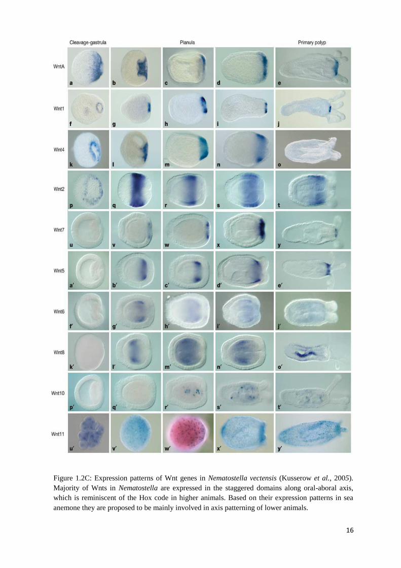
The overlapping expression patterns of Wnt genes in the sea anemone Nematostella
vectensis (Figure 1.2C) resemble the patterning of the true Hox genes in bilaterians indicating
that these genes may be able to participate in axis patterning in Cnidaria (Kusserow et al.,
2005). Expression patterns of ten Wnt genes were analyzed on five stages of the Nematostella
vectensis (Nv) development: from the early blastula through to newly settled polyps forming
their first tentacles. Most of the sea anemone Wnt genes are expressed along the primary body
axis, where they are restricted to the blastopore during gastrulation and to the oral region in
planulae or polyps (NvWntA, -1, -4, and -5, -7). Each subfamily of Wnt genes is restricted to
one of the two body layers, the ectoderm (staggered domains of expression of NvWntA, -1, -4
and -7, spanning the entire oral-aboral axis except for the aboral pole itself) or the endoderm
(NvWnt5, -6 and -8). Although the boundaries between gene expression domains are not sharp
and overlap one another, there are distinct regional differences in their expression along the
oral-aboral axis reminiscent of Hox gene expression in bilaterian animals. Therefore, Arne
Kusserow and his group propose that the Wnt system in basal cnidarians is responsible for the
axis patterning during the development and predates the appearance of the “Hox code” in the
animal evolution.
Moreover, as most of the Wnt genes are expressed in the oral pole of the embryo and
are also co-expressed together with many transcription factors, including Brachyury,
Forkhead, Snail, Notch and Caudal, the authors propose that these Nv-Wnts can represent a
conserved cassette of genes defining the blastoporal signalling centre (Kusserow et al., 2005;
Holland, 2002; Lengyel and Iwaki, 2002). This hypothesis leads them to suggest that Wnt
genes probably had an ancient and primary function in gastrulation and axial patterning.
Particularly, there is a hypothesis that the Wnt genes of this ancient blastoporal
signalling centre gave rise to various mesodermal and neuro-ectodermal derivatives in the
lophotrochozoan, ecdysozoan and deuterostome lineages (Kusserow et al., 2005). The Wnt
16
Figure 1.2C: Expression patterns of Wnt genes in Nematostella vectensis (Kusserow et al., 2005).
Majority of Wnts in Nematostella are expressed in the staggered domains along oral-aboral axis,
which is reminiscent of the Hox code in higher animals. Based on their expression patterns in sea
anemone they are proposed to be mainly involved in axis patterning of lower animals.
17
genes with endodermal expression in Nematostella vectensis in deuterostomes can be found in
the developing mesoderm with overlapping expression domains along a ventral-posterior
direction. These similarities show a close link between endoderm and mesoderm during
gastrulation and a function of this ancient cluster of Nematostella vectensis Wnt genes in
mesoderm evolution.
Therefore, on the basis of the recent data, the Wnt genes in Nematostella vectensis
have primary function in the axis patterning. Moreover, they define the blastoporal signalling
centre in the developing embryo during the development and can play a major role in the
mesoderm evolution (Kusserow et al., 2005).
1.2.4 Wnt system in other cnidarians – diversity of functions
Clytia hemisphaerica – axis determinants
Several components of the Wnt signalling system have been shown to be strong
molecular candidates for the Wnt-activating determinants. Two maternally localized Fzd
proteins (CheFzd1 and CheFzd3) and CheWnt3 are required for the activation of the canonical
Wnt pathway in the hydrozoan Clytia hemisphaerica (Momose et al., 2008, 2007).
The maternal CheFzd1 RNA is localized at the future oral half of the embryo
(gradient with the highest levels coinciding with the gastrulation initiation site) while the
distribution of the maternal CheFzd3 RNA is opposite (restricted domain at the future aboral
side of the embryo) (Momose et al., 2008, 2007). Morpholino-mediated experiments showed
that CheFzd1 and CheFzd3 are the positive and negative regulators of the canonical Wnt
pathway, respectively. Moreover, CheFzd1 and CheFzd3 are mutually downregulated as the
CheFzd3 morpholino-injected embryos showed a massive expansion of the territory of
CheFzd1 and vice versa. However, the co-injection of two morpholinos together resulted in
the up regulation of both RNAs. The main conclusion based on these experiments is that the
mechanism of mutual down-regulation between CheFzd1 and CheFzd3 is not simple and
involves -catenin independent mechanisms.
The RNA mis-expression experiments showed that ectopic injections of CheFzd1 or
CheFzd3 (rescue experiments) could redirect embryonic polarity in Clytia hemisphaerica
18
(Momose et al., 2007, 2008). Therefore, the receptors of the Wnt systems, namely Fzd1 and
Fzd3 in Clytia act as axis determinants and represent a complex system for the regulation of
the canonical Wnt pathway.
Unlike in the Nematostella vectensis, where the Wnt genes repertoire is not
detectable in unfertilized eggs (as maternal transcripts), in Clytia a maternally localized
CheWnt3 is essential for axis patterning. Experiments with local injection of CheWnt3 RNA
in CheWnt3 morpholino-treated embryos have shown that this transcript can induce ectopic
oral poles. These findings demonstrate that the ligand CheWnt3 is required not only for the
establishment of axis polarity but also for the activation of the canonical system in Clytia.
Hydra magnipapillata and Hydractinia echinata – organizer formation and
regeneration capacity
All main components of the Wnt signalling system including eleven Wnt genes from
Hydra magnipapillata (Lengfeld et al., 2009), HeWnt3, HeTcf from Hydractinia echinata
(Duffy et al., 2010) and Dishevelled, GSK-3β, β-catenin from both hydrozoan species were
cloned (Hobmayer et al., 2000; Hobmayer, 1996).
Loss-of-function experiments using pharmacological inhibitors clearly demonstrated
the role of the canonical Wnt/β-catenin pathway in the axial patterning of Hydra
magnipapillata, particularly for the maintenance of the active head organizer and establishing
axis formation during asexual budding and regeneration (Bode, 2011). The alsterpaullone,
LiCl and DAG (diacyl glycerol) treatments, specifically inhibiting GSK-3β activity, resulted
in the formation of multiple ectopic tentacles and, in some cases, heads along the whole
Hydra body column. Also these experiments showed an increased level of β-catenin in all
cells of the adult polyp following either chemical treatment. These treatments also affect the
expression patterns of HyWnts, HyTcf and HyBra1 (the Hydra Brachyury ortholog) genes in
the hypostome. In non-treated species the expression of HyWnts is restricted to the apical tip
of the hypostome whereas in chemical-treated the expression of HyWnts expanded to the
entire region and some cells in the upper part of the body. The expression of other genes
(HyTcf and HyBra1) is also expanded beyond of their normal expression domains. Also, when
bisected, HyWnt3 is the earliest gene expressed during head regeneration suggestive of its
main role in stimulating axis patterning (Lengfeld et al., 2009). Moreover, after appearance of
19
HyWnt3 following Hydra bisection, other six Wnt genes (HyWnt11, HyWnt1, HyWnt9/10c,
HyWnt16, HyWnt9/10a and HyWnt7) are expressed at the site of regeneration but with
different kinetics (Lengfeld et al., 2009).
Also, recent data from Hydractinia echinata studies provide evidence that Wnt
system in this model animal not only promotes head formation, but also represses the
formation of stolons and vice versa. This study was the first to provide the functional data on
the effect of Wnt pathway inhibition on aboral structures (Duffy et al., 2010). These results
suggest an additional role for the Wnt system in cnidarian axis formation beyond specification
of head development.
All this data support the evidence that Wnt signalling in hydrozoans is responsible
for the active state of the head organizer and axis formation during development and head
regeneration.
1.3 Regulation of the Wnt signalling network
1.3.1 Possible molecular mechanisms of Wnt signalling.
The molecular mechanisms of Wnt signalling network are very complex and are
regulated at different levels. The best-studied canonical Wnt pathway signals through changes
in the cytoplasmic level of one of its key players, -catenin (Huelsken and Behrens, 2002). In
the absence of Wnt signalling, the level of cytoplasmic -catenin is kept low via its targeting
to degradation in the multisubunit destruction complex, which includes Axin, the APC tumor
suppressor, protein phosphatase 2A (PP2A), and the protein kinases GSK-3Β and CK1
(Figure 1.3). Sequential phosphorylation of -catenin within this complex targets it for
ubiquitination by E3 ubiquitin ligase and subsequent proteolytic destruction by the
proteasome (Price, 2006). Activation of Wnt signalling leads to the inhibition of the GSK-3Β
destruction complex and the accumulation of cytoplasmic (signalling) -catenin, which enters
the nucleus and together with other transcription factors displaces Groucho (Gro) from
LEF/TCF factors and acts as a transcriptional co-activator to regulate the Wnt target gene
expression.
20
The mechanisms of non-canonical Wnt signalling, which comprises the Wnt/c-Jun
NH2-terminal kinase (JNK) pathways (planar cell polarity) and the Wnt/calcium branches, are
still poorly understood. Interestingly, according to one of the earliest observations, non-
canonical Wnt signalling has the ability to inhibit the canonical Wnt pathway (Amerongen
and Nusse, 2009; Kestler and Kuhl, 2008; Kuhl et al., 2000; Torres et al., 1996). In Xenopus
laevis the nuclear translocation of -catenin is observed transiently in dorsal blastomeres
during the mid-blastula stage, and the -catenin/Tcf3 complex regulates the expression of
dorsal-specific genes. Recent findings further show that in Xenopus the Wnt/calcium/NFAT
pathways promotes ventral cell fate, and one of their ligands, Wnt-5A induces the degradation
of -catenin. Further, one of the most intriguing examples of the canonical pathway inhibition
comes from the studies of the planar cell polarity pathways. One of the downstream
components of planar cell polarity pathways is JNK (JUN NH2-terminal kinase), which was
originally identified by its ability to phosphorylate c–Jun and mediate stress-induced cell
death (Karin et al., 2005; Davis, 2000). Moreover, JNK is downstream of the Dsh-mediated
non-canonical Wnt pathway in establishing the planar cell polarity in Drosophila (Boutros et
al., 1998) and is implicated in gastrulation and convergent extension movements in Xenopus
(Yamanaka et al., 2002; Wallingford et al., 2000). Recently it was also discovered that JNK
antagonizes canonical signalling by reducing nuclear -catenin in both Xenopus and
mammalian cells. These interesting findings show that -catenin is involved in both canonical
and non-canonical signalling and that the molecular mechanisms of canonical and planar cell
polarity (PCP) pathways are opposite at level of -catenin accumulation in the nucleus.
Another study has shown that ciliary proteins such as Inversin are implicated in the
regulation of Wnt and PCP signalling (Simons et al., 2005). It was shown that Inversin, which
is required for Xenopus gastrulation governed by Wnt/PCP signalling, binds to and promotes
the degradation of cytosolic, but not membrane-associated, Dishevelled (Dsh), a critical
component of both Wnt/-catenin and PCP pathways. This led to the hypothesis that Inversin
switches Dsh function from -catenin to PCP signalling. In mammals Diversin (homolog of
the Drosophila Diego and Xenopus Inversin) acts as a molecular switch that blocks Wnt/-
catenin pathway and promotes -catenin–independent signalling (Haribaskar et al., 2009).
More studies needed to decipher this mechanism of regulation between canonical and
planar cell polarity Wnt signalling.
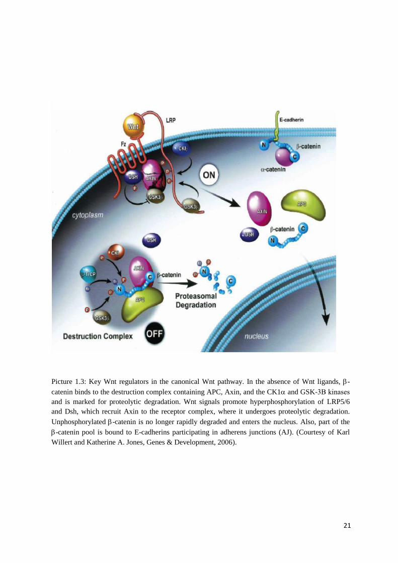
21
Picture 1.3: Key Wnt regulators in the canonical Wnt pathway. In the absence of Wnt ligands, -
catenin binds to the destruction complex containing APC, Axin, and the CK1 and GSK-3Β kinases
and is marked for proteolytic degradation. Wnt signals promote hyperphosphorylation of LRP5/6
and Dsh, which recruit Axin to the receptor complex, where it undergoes proteolytic degradation.
Unphosphorylated -catenin is no longer rapidly degraded and enters the nucleus. Also, part of the
-catenin pool is bound to E-cadherins participating in adherens junctions (AJ). (Courtesy of Karl
Willert and Katherine A. Jones, Genes & Development, 2006).
22
1.3.2 Cnidarian Wnt system regulation and Wnt inhibitors
Although, recent data on Wnt signalling network in cnidarians, has shown that two of
the most popular cnidarian models, Nematostella vectensis (anthozoan) and Hydra
magnipapillata (hydrozoan) possess nearly all Wnt gene complement known from higher
animals (Lengfeld et al., 2009; Guder et al., 2006a; Kusserow et al., 2005), little is known
about the molecular mechanisms of Wnt signalling system within this phylum. Studies on
Hydra have demonstrated that nearly all Hydra Wnt genes are expressed in the hypostome,
where -catenin is concentrated in the nucleus. Therefore, hypostome in Hydra is the centre
for canonical Wnt signalling, where this system is responsible for the maintenance of the head
organizer activity (Gee et al., 2010). More detailed description of Hydra Wnt signalling
system is given in the Chapter 3. In Nematostella vectensis nuclear -catenin is differently
stabilized along the oral/aboral axis and is translocated into the nuclei of cells at the site of
gastrulation for endoderm specification (Wikramanayake et al., 2003). Also, Wnt genes are
the main candidates responsible for axis patterning of this model as they are expressed in a
similar pattern to that of the Hox genes in bilaterians (Kusserow et al., 2005).
In Hydractinia and Clytia embryos, Wnt3, Tcf and frizzled are maternally deposited
at the pole of the oocyte corresponding to the future posterior pole of the larvae and the future
head/oral pole of the adult polyp (Momose and Houliston, 2007; Plickert et al., 2006) and
additionally may act as axis determinants. It has also been shown that misexpression of Wnt3,
frizzled (Momose et al., 2008; Momose and Houliston, 2007) and -catenin (Gee et al.,
2010), as well as blocking GSK-3 activity (Muller et al., 2004b), affects axis formation in
cnidarians. Moreover, apart from the Wnts role in the head/oral pole specification, recent
studies on Hydractinia echinata provided additional piece of functional evidence that Wnt
signalling promotes oral but suppresses aboral structures in this animal (Duffy et al., 2010).
Further studies of Wnt system components on these cnidarian models needed for
unravelling their roles and possible regulation mechanisms in many crucial cnidarian
developmental processes.
While Wnt ligands and cytoplasmic components of Wnt signalling pathways affect
the stability and localization of -catenin, secreted Wnt antagonists/inhibitors represent a
totally different level of regulation. There are two known classes of extracellular Wnt
antagonists. The first class comprises sFRP (secreted Frizzled-related proteins), WIF (Wnt
23
Inhibitory Factor) and Cerberus. Primary binding of these molecules to secreted Wnt ligands
prevents the ligand-receptor interaction. The second class includes the members of the
Dickkopf (Dkk) family, which bind to the LRP5/6 (low-density lipoprotein receptor-related
protein 5/6) subunit of the Wnt receptor complex also preventing signal transduction.
Dickkopf (Dkk) protein family members comprise of Dkk1/2/4 and Dkk3 proteins.
So far three unique sequences with homology to the Dkk gene family have been cloned from
Nematostella vectensis; two of these sequences group within Dkk-3 subfamily and the third is
a dkk1/2/4 homologue (Guder et al., 2006b). Also, a Dkk-3 related protein was identified in
Hydra magnipapillata (Fedders et al., 2004) together with a dkk1/2/4 homologue. A putative
Dkk-3 sequence has been recovered from an EST screen of the coral Acropora millepora
(Guder et al., 2006b).
The expression pattern of Hydra dkk1/2/4 gene was inverted in relation to the head
specific genes (HyWnt3, Hytcf and Brachyury) in the adult polyp and its expression was
restricted to endodermal gland cells (Guder et al., 2006b). The main role proposed for this
protein in Hydra is to antagonize canonical Wnt/β-catenin signalling. Also, as stress
experiments showed the significant up-regulation of the Hydkk1/2/4 at the site of the injury it
can be one of the early injury responsive genes. Less is known about the Wnt antagonists in
Nematostella vectensis and Acropora millepora. In situ RNA hybridization experiments
showed that NvDkk1/2/4 is expressed in the aboral ectoderm from gastrula to the early polyp
and later in the endoderm of the mesenteries and that is consistent with the proposed
antagonizing function of this subfamily in Cnidarians.
Based on recent functional studies of the Wnt system and its antagonists in Cnidaria,
mechanisms of Wnt regulation are very complex and differ even within this phylum. More
functional analyses and comparative studies between different representatives are needed to
provide insights into attempts of deciphering the complex Wnt signalling network in
cnidarians.
1.4 Project objectives
The Wnt signalling pathway is one of the key developmentally regulated cell
signalling systems that are unique to the animal kingdom. Although molecules related to some
components of the Wnt pathway have been identified in members of other kingdoms (Prabhu
24
et al., 2006; Droucheau et al., 2004; Grimson et al., 2000), the complete system is restricted
to animals, where it appears to be involved in axial patterning and gastrulation from sponges
to mammals (Petersen and Reddien, 2009). Deciphering the mechanisms of Wnt signalling is
one of the most important tasks in understanding the crucial developmental processes, such as
axis patterning and gastrulation. Cnidarians are likely to provide insights on the evolution and
diversification of patterning mechanisms operating in higher animals because here most of the
genes are present but they operate in a much less complex context.
There have been a number of recent studies of the Wnt system in the anthozoan
Nematostella vectensis and the hydrozoans Hydra magnipapillata and Clytia hemisphaerica,
that revealed possible roles of the cnidarian Wnt signalling, but studies of other cnidarians
species with different life history traits will improve our understanding of the roles and the
evolution of the Wnt signalling pathways. The aim of my project is to investigate and
characterize Wnt signalling components in the coral Acropora millepora, an anthozoan with
very different characteristics to Nematostella.
Chapter 2 of my thesis describes the retrieval and analysis of the Wnt system
components from recent ESTs collections, transcriptome data and genome sequencing data
and analysis of their expression patterns together with comparisons to other cnidarian species.
The aim here was to obtain full coding sequences of the Acropora repertoire of Wnts and
Fzds and to subject these to phylogenetic analysis. As Wnt expression data are available for
Nematostella vectensis, the comparison with Acropora millepora could highlight similarities
and differences in Wnt use during the very different developmental processes in these
animals. In Hydra, the oral pore is thought be a centre of Wnt canonical signalling, the
hypostome acting as a head organizer, and studies described in chapter 2 revealed oral-
specific expression of several Wnt ligands in coral.
Chapter 3 explores this further, focusing on the significance of Wnt signalling in the
oral region of Acropora pear-planula stages. In this chapter we propose possible molecular
mechanisms for Wnt signalling in the oral region of Acropora millepora planulae and
highlight the importance of the oral pore in Acropora as the main centre for Wnt non-
canonical signalling.
Chapter 4 provides main conclusions of my studies and gives further directions in the
research of the Wnt signalling system in Acropora millepora.
25
Chapter 2
Characterization of the Wnt signalling system in Acropora
millepora
2.1 The genetic complexity of the Wnt signalling system in basal
metazoans
Wnt glycoproteins are the secreted ligands of the Wnt signalling network. Typically
the individual Wnt proteins are classified as canonical or non-canonical based on the ability of
the former, but not the latter, type to induce an ectopic axis in Xenopus embryos (McMahon
and Moon, 1989) and to cause the morphological transformation of mouse C57MG mammary
cells (Wong et al., 1994). Both of these properties correlate with an increase in the levels of -
catenin and a concomitant increase in canonical signalling. On the contrary, specific non-
canonical Wnts, for example Wnt5a and Wnt11, have been shown to be involved in
convergent extension movements during vertebrate development, regulating cell adhesion and
triggering an increase in the intracellular calcium levels (Westfall et al., 2003; Eisenberg and
Eisenberg, 2002; Wallingford et al., 2001; Heisenberg et al., 2000; Tada and Smith, 2000).
Although the ability of Wnt proteins to trigger canonical or non-canonical pathways has
recently been shown to be highly context dependent (Amerongen and Nusse, 2009),
particularly on the availability of specific Fzd receptors, Wnts are often still classified as
canonical or non-canonical. In vertebrates, Wnt1, Wnt3a and Wnt8 trigger the canonical
pathway, whereas Wnt4, Wnt5a and Wnt11 may participate in non-canonical type of Wnt
signalling (Grumolato et al., 2010). In total, the Wnt family includes nineteen members in
metazoans, representing twelve Wnt subfamilies with additional WntA subfamily being
absent from majority of animals except of sea urchin Strongylocentrotus purpuratus
(Amerongen and Nusse, 2009; Croce and McClay, 2008; Croce et al., 2006).
Wnt genes are specific to animals (metazoans) and, so far, no Wnts genes have been
found in plants, fungi or protists (Croce and McClay, 2008). Although, it is important to note
that a transducing pathway related to the canonical Wnt pathway has been reported for
Dictyostelium (Coates and Harwood, 2001). Wnt system components including Wnt proteins
have been discovered in the Porifera phyla, which is the most ancient phyletic lineage
26
according to latest views on animal evolution (Philippe et al., 2009). The three Wnt ligands
present in the genome of the demosponge Amphimedon queenslandica are quite divergent,
forming a poorly supported clade with Wnt8/9/10 representatives in phylogenetic analyses
(Adamska et al., 2010). Two divergent Wnt11-related genes have also been recovered from
the homoscleromorph sponge Oscarella lobularis together with well-conserved genes for
intracellular pathway components (-catenin, GSK3; Lapebie et al., 2009). Interestingly, the
expression patterns of the Oscarella Wnt genes are reminiscent of Wnt expression during skin
appendage formation in vertebrates (Lapebie et al., 2009; Liu et al., 2008; Chang et al., 2004;
Reddy et al., 2001). This suggests that, participation in epithelial patterning and
morphogenesis may be an ancient feature of canonical Wnt signalling (Lapebie et al., 2009).
By contrast to a very poor representation of Wnt genes in Porifera, recent studies
have revealed that almost all of the twelve bilaterian Wnt gene subfamilies are present in
representatives of the phylum Cnidaria (Lengfeld et al., 2009; Holstein et al., 2008; Guder et
al., 2006a; Lee et al., 2006b; Kusserow et al., 2005). As well as having members of the other
Wnt subfamilies, cnidarians possess WntA homologue, which is absent from most
deuterostomes (Croce et al., 2006). The genetic complexity of the Wnt system in cnidarians
indicates that this gene family was already diversified in the common ancestor of cnidarians
and bilaterians (but most likely not earlier in evolution) and suggests that some Wnt roles in
patterning of the body axis may be ancient. Canonical Wnts participate in pattern formation
and regeneration of Hydra (Lengfeld et al., 2009) and in regeneration, gastrulation and early
embryogenesis in the anthozoan Nematostella vectensis (Trevino et al., 2011; Holstein, 2008;
Kusserow et al., 2005). Non-canonical Wnt proteins are involved in tissue evagination of
Hydra (Philipp et al., 2009) and in the initial archenteron formation during gastrulation in
Nematostella (Kumburegama et al., 2011).
The Holstein group showed that the anthozoan sea anemone Nematostella vectensis
has members of eleven of the twelve known Wnt gene subfamilies. In total, fourteen different
Wnt genes were isolated from Nematostella (Holstein et al., 2008; Guder et al., 2006a; Lee et
al., 2006b; Kusserow et al., 2005). Of the bilaterian Wnt subfamilies, the sea anemone lacks
only a Wnt9 member. Two representatives of both the Wnt7 and Wnt8 subfamilies have been
described in N. vectensis; NvWnt8a and NvWnt8b are paralogues that probably arose from a
cnidarian-specific duplication event (Kusserow et al., 2005), whereas Wnt7a and Wnt7b are
now known to be splice variants from a single locus as was suggested by their structural
identity in the C-terminal region (Guder et al., 2006).
27
Like Nematostella, Hydra magnipapillata also has a complex Wnt repertoire (eleven
Wnt genes), but it is more derived than in the former case; eight of the Hydra Wnt genes are
members of the Wnt1, -2, -3, -5, -7, -8, -11, and -16 subfamilies, but the remaining three
(HyWnt9/10a, -9/10b, and -9/10c) are related to the Wnt9 and Wnt10 types but are not clearly
members of either subfamily (Lengfeld et al., 2009).
The data available from cnidarians indicates that in general members of this phylum
have more complex Wnt repertoires than do members of the bilaterian super-phylum
Ecdysozoa. For example, the insect Drosophila melanogaster and the nematode
Caenorhabditis elegans have only seven and five Wnt genes respectively. These data support
the hypothesis that early multicellular animals at more than 550 million years ago had already
evolved a surprisingly complex Wnt signalling system that included all or most of the ligands
known from higher animals. Thus, the last common ancestor of bilaterians and cnidarians, the
Ur-Eumetazoa, already had many Wnt ligands and members of the ecdysozoan lineage have
subsequently lost a large number of these.
Analysis of the Nematostella genome revealed close linkage of the NvWnt6 and
NvWnt10 loci, these being separated by less than 4.5kb (Sullivan et al., 2007). In the
crustacean Daphnia pulex two clusters consisting of Wnt9-Wnt1-Wnt6-Wnt10 and Wnt5-
Wnt7 were also discovered (Janssen et al., 2010). Moreover, a linkage of Wnt5-Wnt7 genes
was also observed in Nematostella, where the similarity of expression patterns of these genes
suggests the possibility that common regulation mechanisms may apply to the Wnt5 and
Wnt7 genes in the sea anemone (Guder et al., 2006). As Wnt1, -3, -6, -9, -10 members are
linked in both human and fly, the Wnt6-Wnt10 and Wnt5-Wnt7 linkages seen in Nematostella
are possible remnants of an ancient Wnt cluster that originated in the common ancestor of
cnidarians and bilaterians (Guder et al., 2006). Analysis of the organisation and expression of
Wnts in Acropora may add substantially to our understanding of the evolution of the genes
more generally. Interestingly, analysis of the Hydra and human genomes indicate that the
different Wnt gene subfamilies share a surprisingly high degree of conservation within four
main exons (Lengfeld et al., 2009).
The majority of the downstream components of the canonical and non-canonical Wnt
signalling systems have been identified in both Nematostella and Hydra. In addition to Wnt
genes, at least seven Frizzled (Fzd) receptor genes have been identified in Nematostella
(Guder et al., 2006a; Lee et al., 2006; Minobe et al., 2000). The Fzd homologues CheFzd1
and CheFzd3 are known from Clytia hemisphaerica (Momose and Houliston, 2007). The first
28
cnidarian homologue of -catenin was cloned from the freshwater polyp Hydra
magnipapillata (Hobmayer, 1996), and subsequently other components including Dishevelled
(Dsh) and Fzd homologues have been identified and characterised from Hydra vulgaris
(Minobe et al., 2000). Intracellular Wnt signalling components identified in both
Nematostella and Hydra include homologues of APC (adenomatous polyposis coli), Axin,
CamKII, -catenin, casein kinase 1, 1, 12, 13, and 1, Dsh, Flamingo, GSK3, JNK,
PKC, Tcf/Lef, Van Gogh, and Wntless (Lee et al., 2006; Technau et al., 2005; Philipp et al.,
2005; Rentzsch et al., 2005; Hobmayer et al., 2000; Hobmayer et al., 1996). The HeTcf1
(transcription factor) is also known from Hydractinia echinata (Plickert et al., 2006) as well
as Hydra.
Surveys of Hydra and other cnidarians also indicate that not only are most of the Wnt
system components present in these basal animals, but so too are many of the Wnt antagonists
known from bilaterians. The work of Gunter Plickert led to the identification of Hydra
proteins clearly resembling dkk-3 and dkk1/2/4 (Guder et al., 2005). Whereas the Hydra
Dkk3 homologue participates in neuronal differentiation, the dkk1/2/4 homologue is likely to
be involved in inhibition of canonical Wnt signalling as this gene is expressed at the opposite
end of the Hydra body axis to the domain of Wnt expression (Lengfeld et al., 2009; Lee et al.,
2006; Guder et al., 2006b). Other Wnt antagonists, namely secreted Frizzled-related proteins
(sFRP) and Wnt inhibitory factor (WIF) have been identified from both Hydra and
Nematostella (Guder et al., 2006a; Guder et al., 2006b; Lee et al., 2006; Technau et al.,
2005). In total, there are eight sFRPs in Nematostella and several in Hydra have been found
(Holstein, 2008).
Although cnidarians possess a highly complex and diversified Wnt signalling
repertoire, functional data is lacking in many cases. Nevertheless, analysis of Nematostella
and Hydra Wnt signalling system implies several aspects of Wnt functions in cnidarians
(Guder et al., 2006; Lee et al., 2006). The major functions of Wnts in Nematostella appear to
be in axis formation, gastrulation and regeneration (Trevino et al., 2011; Kusserow et al.,
2005). Cnidarians lack the true Hox clusters (Kamm et al., 2006) that play central roles in
axial patterning throughout the Bilateria. However, the expression patterns of Nematostella
Wnt genes are strikingly reminiscent of bilaterian Hox genes patterns, suggesting that Wnts
may have an ancient function in specifying position along the main body axis instead of Hox
patterning. In Hydra, Wnt/-catenin signalling is also involved in head regeneration
(Hobmayer et al., 2000).
29
In summary, we can conclude that Wnt signalling system is very complex in basal
metazoans, particularly in cnidarians. The Wnt network is active during early embryogenesis,
gastrulation, in larval life of Nematostella and adult life of both Nematostella and Hydra. We
are far from a complete understanding of the Wnt system interactions and roles in cnidarians
but the studies of other representatives, particularly the coral Acropora millepora (class
Anthozoa), may help to further enrich our knowledge of one of the most important signalling
systems in cnidarian development.
2.1.1 Complex Wnt signalling system in Acropora millepora
During the course of this study, we discovered that the Wnt signalling system in
Acropora millepora is complex, with many secreted ligands, receptors and intracellular
components. This coral has most or all of the Wnt genes known from the other anthozoan,
Nematostella vectensis. In addition to the secreted Wnt ligands, six Acropora Frizzled
receptors were retrieved, belonging to different Fzd subfamilies (Fzd1, Fzd2, Fzd3, Fzd4,
Fzd8 (partial) and FzdX-related protein) together with the LRP5/6 transmembrane protein.
One of the dishevelled homologues (Dsh3), Axin, β-catenin, GSK3 and CKI kinases,
transcription factors (TCF/LEF and repressor Groucho) and inhibitors of the Wnt pathways –
WIF (Wnt Inhibitory Factor) and one of the Dickkopf homologues (Dkk3) were also
discovered. The presence of planar cell polarity (PCP) components has not previously been
reported in corals, however analysis of the on-going Acropora transcriptome project has
revealed many of the core PCP pathway proteins to be present including (in addition to
Frizzleds) Flamingo/CELSR, Van Gogh, prickled and Dachsous (Knack, unpublished data).
More components may yet be discovered, as this is an ongoing study.
Based on the results obtained, twelve Wnt genes were identified in Acropora
millepora, belonging to different Wnt subfamilies. Although each of the Acropora Wnt genes
has a clear homologue in Nematostella vectensis, some intriguing differences in expression
patterns were observed, which will be discussed later.
In Acropora, most of the Wnt signalling system components are expressed only after
the closure of the blastopore (i.e. at or after the end of gastrulation), indicating that activation
of coral Wnt signalling begins in early larval developmental stages, with expression
disappearing again around metamorphosis. Overall, these results suggest that, unlike several
30
other cnidarians, including Nematostella, in Acropora Wnt signalling is unlikely to play a
major role in gastrulation, and is likely to function primarily in larval patterning.
2.2 Materials and methods
2.2.1 Animal sampling
Eggs and embryos of Acropora millepora were collected at Heron Island, QLD, Australia
(Latitude 23°26’ South; Longitude 151°55 East), Magnetic Island (Latitude 19°09’ South;
Longitude 146°49’ East) and Orpheus Island (Latitude 18°28’ South; Longitude 146°25’
East) during the coral mass-spawning events in 2008, 2009 and 2010.
2.2.2 Isolation of Wnt, Fzd genes and other components of Wnt system
Homologues of Wnt2, three Fzd receptors (Fzd4, FzdX and Fzd8), GSK3, Axin and dkk3
were identified in Acropora millepora as ESTs (Kortschak et. al., 2003; Technau et al., 2005).
pGEMT plasmids with Fzd4 (C007-H4), Fzd8 (D016-E8), FzdX-related protein (D019-E7),
Wnt2 (D018-E10), Dsh, Axin (D027-D10) and dkk3 (B027-D10) gene fragments were kindly
provided by Dr. David Hayward (Australian National University, Canberra).
In order to search for more Wnt genes, we performed cDNA library screening with primers
designed for two relatively short and fairly well conserved domains of Wnt genes from
Acropora. The λ-UniZap XR A.millepora cDNA library was constructed by Dr. David
Hayward (Australian National University, Canberra) with the messenger RNA from
prawnchip, presettlement, postsettlement and adult tips stages. Degenerative primer sequences
for two highly conserved Wnt regions were: Wnt_forward (5’–TGGSANTGGGGNGGNTG-
3’) and Wnt_reverse_outer (5’–TYNCCRTGRCAYTTRCA–3’), Wnt_reverse_inner (5’–
CCNGCNYBRTTBTG–3’). PCR conditions were as follows: 95°C for 5 min as hot start,
95°C for 30 second, gradient 38°C-55°C with one degree lower each cycle, 72°C for 1
minute, 35 cycles in total. The fragments of expected size (750 bp) were cloned into pGEMT
vector (Promega) and sequenced by Macrogen company (South Korea). Following the
31
screening two novel Acropora millepora Wnt genes were identified, Wnt5 and Wnt16,
respectively.
RACE method was used to obtain the complete 5’-end of the Wnt5 homologue in Acropora
millepora. We used SMARTTM
RACE cDNA Amplification Kit (Clontech). 5’- First-Strand
cDNA synthesis was performed on total RNA (extracted from larva stages). Primers used:
NGSP1 5’-TTCACAGTACCCTACAGCCCTAGC-3’, and GSP1 5’-
CTCTCTTAACAGCGACAAGCAGCC-3’. PCR conditions were as follows: 94°C for 30
seconds, 72°C for 3 mins for 5 cycles; 94°C for 30 seconds, 70°C for 30 seconds, 72°C for 3
minutes for 5 cycles; 94°C for 30 seconds, 68°C for 30 seconds, 72°C for 3 minutes for 25
cycles. The expected sizes of DNA fragments were cloned into pGEMT-T vector (Promega)
and sequenced by Macrogen company (South Korea).
Most of the components of Wnt signalling system in Acropora millepora were retrieved using
several recent extensive EST projects, 454 data and genomic Illumina sequencing data (on
going). In total, we identified twelve Wnt genes, six Fzd receptors, LRP5/6 protein and
multiple intracellular downstream effectors of the Wnt signalling network in our model coral
Acropora millepora.
The primers for all retrieved Acropora millepora Wnt signalling system genes were designed
with Primer3 program (Table 2.2.2) based on the latest transcriptome data available and PCR
reactions on mixed cDNA libraries were performed using prawnchip, presettlement,
metamorphosis, postsettlement and adult stages. Primer sequences, length of the fragment,
temperature of annealing (Tm), vector, and additional comments are given in Table 2.2A.
Name
of the
gene
Primers Tm/Cycl
es
Length
of the
fragment
Plasmid used
for cloning
Wnt16 F - 5’GTACAGTGGTTGGGGAATGG3’
R - 5’CAGTGCAAGAGCCAGAAACA3’ 60C/35 860bp pGEMT
WntA F - 5’GGCTTGTGAATGCGATCAAGCACT3’
R - 5’TTCCTTCCCGCTTCATTGTTGTGC3’ 68C/30 867bp pGEMT
Wnt8b F - 5'TTGGTGCTTGCTTCATTCGCTCTG3'
R -5'ACGCCTACCTAGTGCATGTTGTGA3' 57C/35 984bp pGEMT
Wnt6 F – 5’AAGCAATTAATGGCAAACCAA3’
R - 5’CCATACTGGTGTCGTTGCAT3’ 58C/35 381bp pGEMT
Wnt4 F - 5’ACAACGGAAGGTCTGAGAAAGGCT3’
R - 5’CGCAGCACATCAATTCACACCCAT3’ 72C/30 513bp pGEMT
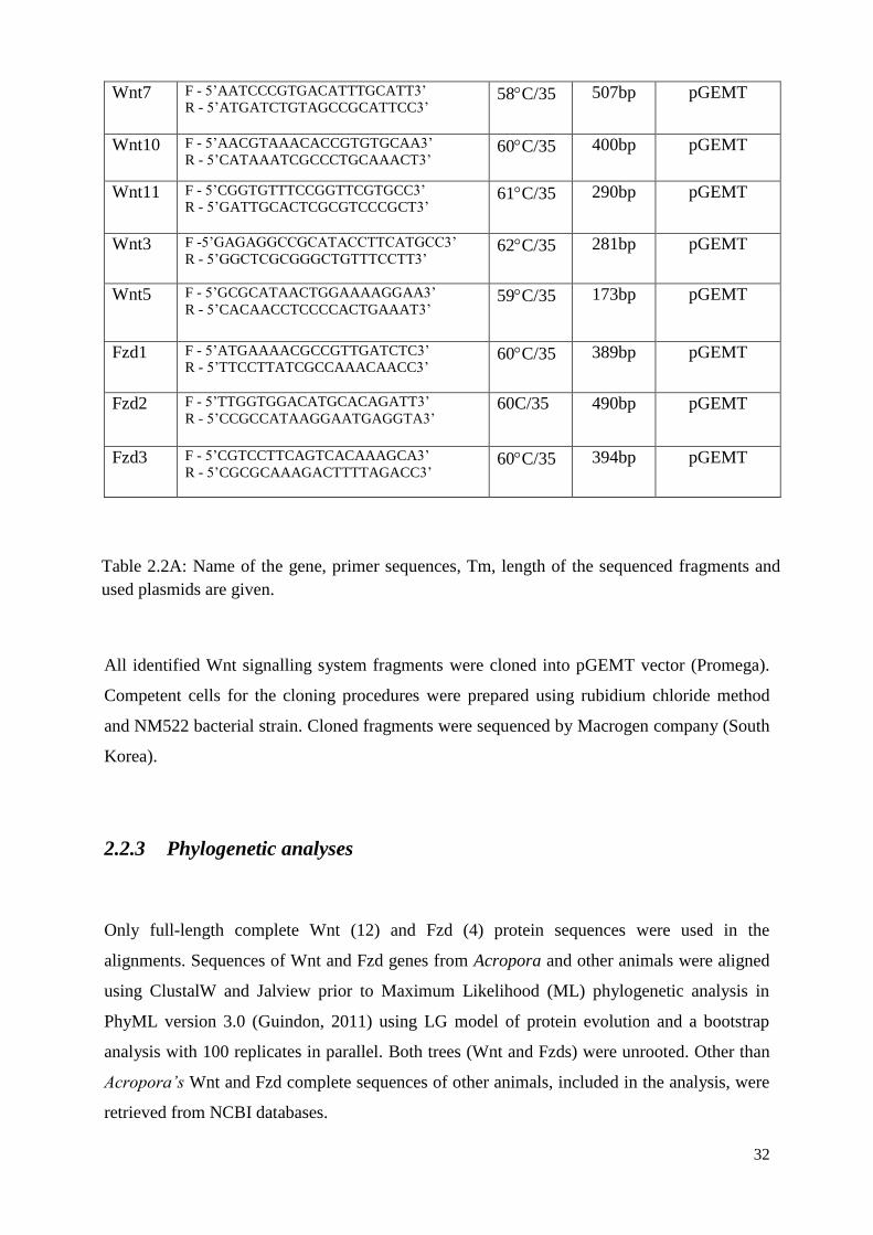
32
Wnt7 F - 5’AATCCCGTGACATTTGCATT3’
R - 5’ATGATCTGTAGCCGCATTCC3’ 58C/35 507bp pGEMT
Wnt10 F - 5’AACGTAAACACCGTGTGCAA3’
R - 5’CATAAATCGCCCTGCAAACT3’ 60C/35 400bp pGEMT
Wnt11 F - 5’CGGTGTTTCCGGTTCGTGCC3’
R - 5’GATTGCACTCGCGTCCCGCT3’ 61C/35 290bp pGEMT
Wnt3 F -5’GAGAGGCCGCATACCTTCATGCC3’
R - 5’GGCTCGCGGGCTGTTTCCTT3’ 62C/35 281bp pGEMT
Wnt5 F - 5’GCGCATAACTGGAAAAGGAA3’
R - 5’CACAACCTCCCCACTGAAAT3’ 59C/35 173bp pGEMT
Fzd1 F - 5’ATGAAAACGCCGTTGATCTC3’
R - 5’TTCCTTATCGCCAAACAACC3’ 60C/35 389bp pGEMT
Fzd2 F - 5’TTGGTGGACATGCACAGATT3’
R - 5’CCGCCATAAGGAATGAGGTA3’ 60C/35 490bp pGEMT
Fzd3 F - 5’CGTCCTTCAGTCACAAAGCA3’
R - 5’CGCGCAAAGACTTTTAGACC3’ 60C/35 394bp pGEMT
All identified Wnt signalling system fragments were cloned into pGEMT vector (Promega).
Competent cells for the cloning procedures were prepared using rubidium chloride method
and NM522 bacterial strain. Cloned fragments were sequenced by Macrogen company (South
Korea).
2.2.3 Phylogenetic analyses
Only full-length complete Wnt (12) and Fzd (4) protein sequences were used in the
alignments. Sequences of Wnt and Fzd genes from Acropora and other animals were aligned
using ClustalW and Jalview prior to Maximum Likelihood (ML) phylogenetic analysis in
PhyML version 3.0 (Guindon, 2011) using LG model of protein evolution and a bootstrap
analysis with 100 replicates in parallel. Both trees (Wnt and Fzds) were unrooted. Other than
Acropora’s Wnt and Fzd complete sequences of other animals, included in the analysis, were
retrieved from NCBI databases.
Table 2.2A: Name of the gene, primer sequences, Tm, length of the sequenced fragments and
used plasmids are given.
33
Alignments of incomplete Acropora millepora Wnt genes with possible Nematostella
vectensis homologues for Wnts were made using ClustalW and Jalview. The cysteine patterns
of complete and incomplete Wnt glycoproteins were analysed and comparisons with other
organisms made (Figure 2.3B, discussed later in chapter 2 and supplementary material,
Figures 2.2A, 2.2B, 2.2C, 2.2D, 2.2E, 2.2F, 2.2G, 2.2H, 2.2I, 2.2J, 2.2K).
The incomplete protein sequence of Fzd8 identified in Acropora millepora was aligned with
the predicted Fzd8 protein sequence of Nematostella vectensis using ClustalW and Jalview
(Supplementary material, Figure 2.2L).
Protein structures for all identified components were identified from SMART
(http://smart.embl-heidelberg.de/).
2.2.4 Fixation and whole mount in situ hybridization of coral embryos
Acropora millepora embryos were fixed for 12 min in 3.7% (v/v) formaldehyde in Millipore-
filtered seawater (MPFSW) buffered to pH 8.0 with HEPES. We fixed embryos at different
stages of development from unfertilized eggs to the postsettlement. The embryos were then
washed repeatedly in MPFSW, dehydrated through a gradient methanol/dd H2O series, and
stored in absolute methanol at -20C until needed.
In-vitro transcription and probe preparation
After isolation of Wnt signalling system genes fragments, antisence probes for in situ
hybridizations were synthesized according to the standard in situ protocol (Kucharsky et al.,
2000).
In situ hybridizations were performed following standard protocols (Kucharsky et al., 2000)
using digoxigenin-labelled antisense RNA probes (Roche). The digoxigenin-labelled
antisense RNA probes for Acropora millepora Wnt system genes fragments were synthesized
in an in vitro transcription with the T7 or SP6 RNA polymerase (Fermentas) after the
following digestions (Table 2.2B):
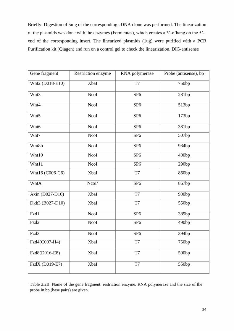
34
Briefly: Digestion of 5mg of the corresponding cDNA clone was performed. The linearization
of the plasmids was done with the enzymes (Fermentas), which creates a 5’-o’hang on the 5’-
end of the corresponding insert. The linearized plasmids (1ug) were purified with a PCR
Purification kit (Qiagen) and run on a control gel to check the linearization. DIG-antisense
Gene fragment Restriction enzyme RNA polymerase Probe (antisense), bp
Wnt2 (D018-E10) XbaI T7 750bp
Wnt3 NcoI SP6 281bp
Wnt4 NcoI SP6 513bp
Wnt5 NcoI SP6 173bp
Wnt6 NcoI SP6 381bp
Wnt7 NcoI SP6 507bp
Wnt8b NcoI SP6 984bp
Wnt10 NcoI SP6 400bp
Wnt11 NcoI SP6 290bp
Wnt16 (C006-C6) XbaI T7 860bp
WntA NcoI/ SP6 867bp
Axin (D027-D10) XbaI T7 900bp
Dkk3 (B027-D10) XbaI T7 550bp
Fzd1 NcoI SP6 389bp
Fzd2 NcoI SP6 490bp
Fzd3 NcoI SP6 394bp
Fzd4(C007-H4) XbaI T7 750bp
Fzd8(D016-E8) XbaI T7 500bp
FzdX (D019-E7) XbaI T7 550bp
Table 2.2B: Name of the gene fragment, restriction enzyme, RNA polymeraze and the size of the
probe in bp (base pairs) are given.
35
RNA probes were synthesized with DIG-RNA labelling mix (Roche). A mixture of template
plasmid, labelling mix and the appropriate RNA polymerase were incubated for 4 hours at
37C. EDTA (2l, 0.2M) was used to stop the probe synthesis. The aliquot of the reaction
(2l) was run on a formaldehyde gel to check the labelling efficiency. For probes longer that
750bp, the probe alkaline hydrolysis was performed. The carbonate buffer (5.5l, 0.2M) was
added to the reaction, which was then incubated @ 60C as per length of time denoted by the
following equation:
t = (Lo - Lf) / (K)(Lo)(Lf)
Lo = starting length (kb), including all parts of the vector that are transcribed
Lf = final desired length (kb) = .075-.1 kb,
K = 0.11 (constant, kb/min), calculated time is in minutes.
After hydrolysis the precipitated probes were resuspended in a volume of 80l of formamide
resuspension buffer (50% formamide, 50%TE, 0.1% Tween) and 2l of the probe was used in
the 250l hybridization reaction.
Hybridization and probe detection
In situ hybridizations were carried out as reported previously (Hayward et al., 2001) with
some modifications on all fixed stages. Fixed animals were rehydrated through a methanol
series from 70% to 25% and washed in PBT. In order to remove large amount of lipids, the
embryos were treated in RIPA at 4C overnight. After RIPA treatment, embryos were
dehydrated through ethanol series (50% to 100% and treated with Xylene for 5 hours. Xylene
was removed by several absolute ethanol washes and the embryos were rehydrated and
washed by PBT. The embryos were transferred into the embryo hybridization solution (50%
(v/v) formamide, 4SSC, 1Denhardts solutions, 5g/ml heparin, 5% (w/v) dextran sulphate,
500g/ml denatured sonicated salmon sperm DNA, 0.1% (v/v) Tween 20). Hybridization was
carried out at 50-58C for 48-72 hours with a final probe concentration of 0.25-1 ng/l.
Excess probes were removed by washing at 55-58 C for 24 hours with hybridization wash
solution (4SSC, 50% (v/v) formamide, 0.1% (v/v) Tween 20). Probe detection was carried
out by 1500-fold diluted anti-digoxigenin antibody conjugated to alkaline phosphatase
(Roche). After several washes in PBT, animals were rinsed with 100mM Tris pH 9.5, and the
presence of the alkaline phosphatase was detected by the substrate BCIP-NBT (Vector
36
laboratories). Colour development was stopped by several PBT washes, and the background
was cleared by ethanol.
2.3 Results
2.3.1 Analysis of Wnt proteins in Acropora millepora
Twelve novel Wnt genes were identified in Acropora millepora based on the
extensive available transcriptome data. Among Wnts presumably participating in the -
catenin dependent pathways we have identified five, these being Wnt1, Wnt8b, Wnt2, Wnt7,
and Wnt3 homologues. Amongst other Wnts identified are homologues of Wnt4, Wnt5,
Wnt6, Wnt10, Wnt11, Wnt16 and WntA. Sequence analysis indicated this complement to be
comparable to that of another anthozoan, Nematostella vectensis, for which a total of fourteen
Wnt proteins have been reported (Lee et al., 2006a, 2006b; Kusserow et al., 2005). Acropora
millepora therefore has more Wnt protein subfamilies than either of the hydrozoan cnidarians
for which extensive data are available, Hydra magnipapillata and Clytia hemisphaerica
(Table 2.4.2, discussion section). Clear orthology between Acropora and Nematostella Wnts
is observed for all but four families (Wnt1, Wnt3, Wnt6 and Wnt7b). Single transcripts only
were identified for each of the Wnt7 and Wnt8 families in Acropora, whereas in Nematostella
Wnt7 is alternatively spliced to two transcripts and there are two Wnt8 paralogues. The one
Wnt8 gene identified in Acropora millepora is a clear homologue of Wnt8b in Nematostella
vectensis. As in the case of both Nematostella vectensis and Hydra magnipapillata (Lengfeld
et al., 2009; Kusserow et al., 2005), Wnt9 is also absent from Acropora millepora. Some of
the sequences (Wnt1, Wnt3, Wnt6 and Wnt7b) are as yet incomplete. Alignments of the
incomplete protein sequences with their likely counterparts from Nematostella are presented
in the Supplementary material (Figures 2.2B, 2.2J, 2.2C, 2.2K).
Searching the raw transcriptome (454 and Illumina) data, in conjunction with 5’-
RACE, allowed the identification of complete coding sequences for 8 of the 12 Acropora Wnt
genes identified (Wnt2, Wnt4, Wnt5, WntA, Wnt8b, Wnt10, Wnt11, Wnt16). Phylogenetic
analyses were conducted on the complete Acropora millepora Wnt proteins and Wnt16. This
latter was included as a sequence comparison implying that the sequence lacks only
approximately 27 amino acid residues at the N-terminus, and the two Wnt domains being
37
complete (alignment in Supplementary material, Figure 2.2A). These protein sequences were
aligned with data for other cnidarians and representatives of the deuterostomes (including
human sequences), ecdysozoans and lopotrochozoans. Phylogenetic analysis was conducted
using maximum likelihood methods (ML, 100 bootstrap support) and the ML tree (with 100
bootstraps) is shown as Figure 2.3A.
With the exception of WntA, each of the Acropora Wnt sequences fell into a strongly
supported clade together with bilaterian members of a specific subfamily. The association
between anthozoan sequences is close to 100% for each of the eight Wnt protein sequences
(100% for WntA and Wnt4; 99% for Wnt10; 98% for Wnt5 and Wnt16; 97% for Wnt2 and
Wnt11; and 77% for Wnt8b), whereas the Hydra sequences were more divergent, clustering
with their anthozoan counterparts with significantly lower support.
Wnt proteins characteristically have two domains, the spacing between them is
highly conserved, and domains typically contain at least 20 cysteine residues (Miller, 2002).
The Wnt domains are positioned close to and at the C-terminus of the protein and contain four
and twelve cysteine residues, respectively, in a characteristic pattern. Comparison of the
Acropora Wnt proteins to their deuterostome counterparts based on Jalview alignment
revealed similar or identical patterns of cysteine residues. For example, as can be seen in
Figure 2.3B, each Wnt11 protein contains the same number of cysteine residues organised in
a conserved pattern, these being clustered towards the C-terminal ends of the two domains;
Branchiostoma floridae is exceptional in that one cysteine residue is missing at the C-end of
the protein. The Acropora and Nematostella Wnt11 proteins have identical patterns of
cysteine residues, whereas the positions in Hydra are similar but not identical. In the case of
Acropora millepora the C-terminal cysteine pattern closely resembles that of deuterostomes.
Similar conclusions can be drawn for the other complete Acropora Wnts (Supplementary
material, Figures 2.2A - 2.2K). Hence, each of the Acropora millepora Wnts is considered to
be a bona fide member of specific bilaterian Wnt subfamilies.
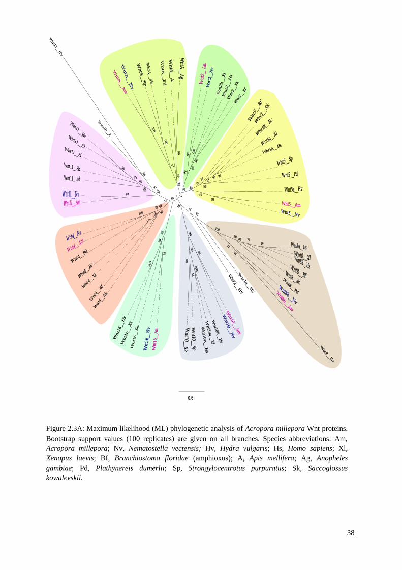
38
Figure 2.3A: Maximum likelihood (ML) phylogenetic analysis of Acropora millepora Wnt proteins.
Bootstrap support values (100 replicates) are given on all branches. Species abbreviations: Am,
Acropora millepora; Nv, Nematostella vectensis; Hv, Hydra vulgaris; Hs, Homo sapiens; Xl,
Xenopus laevis; Bf, Branchiostoma floridae (amphioxus); A, Apis mellifera; Ag, Anopheles
gambiae; Pd, Plathynereis dumerlii; Sp, Strongylocentrotus purpuratus; Sk, Saccoglossus
kowalevskii.
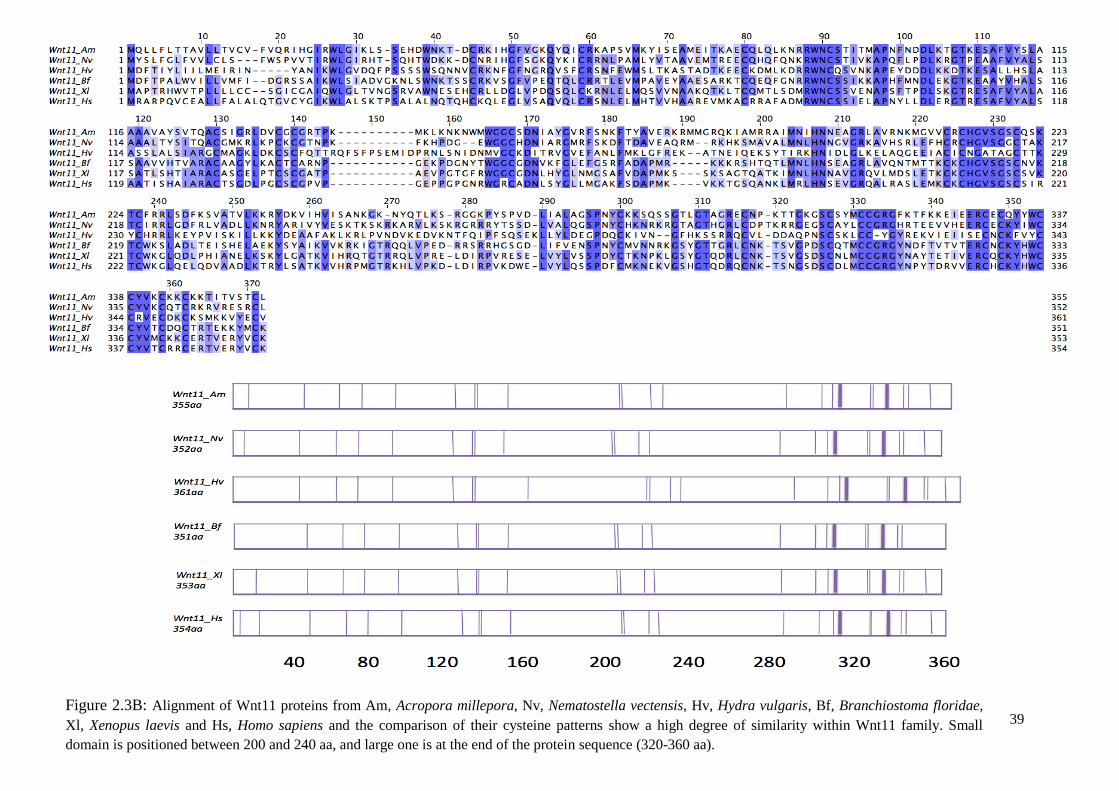
39
Figure 2.3B: Alignment of Wnt11 proteins from Am, Acropora millepora, Nv, Nematostella vectensis, Hv, Hydra vulgaris, Bf, Branchiostoma floridae,
Xl, Xenopus laevis and Hs, Homo sapiens and the comparison of their cysteine patterns show a high degree of similarity within Wnt11 family. Small
domain is positioned between 200 and 240 aa, and large one is at the end of the protein sequence (320-360 aa).
40
2.3.2 Expression patterns of Wnt genes during the development of
Acropora millepora
In situ hybridization demonstrates that each of the Acropora millepora Wnt genes
analysed are expressed only after gastrulation, with stable patterns of expression identifiable
throughout the motile larval stages (early pear to late planula). In no case was expression
detected in unfertilized eggs, and in all cases expression diminished or disappeared after
metamorphosis, at the postsettlement stage.
The Wnt genes in Acropora millepora can be classified into three groups based on
their spatial expression patterns during the larval stages of development. The first group
consists of WntA and Wnt16, genes that are expressed at the oral end of the planula in the
ectodermal tissue layer (Figure 2.3C). Whilst WntA is expressed in a gradient, which is most
concentrated at the oral pole, Wnt16 expression is restricted to a ring of ectodermal cells
surrounding the mouth of the larva. A metamorphosing coral larva with a clear ring of Wnt16
expression is shown in Figure 2.3E.
The second group consists of the Acropora millepora Wnt8b and Wnt2 homologues,
which are both expressed in the central region of the larva, between the oral and aboral poles.
Whereas Wnt2 expression is restricted to a band of ectodermal and endodermal cells
spanning the middle of the larva, Wnt8b is expressed in the endoderm and, intriguingly, its
expression is asymmetrical along the directive axis of the embryo, supporting the notion of a
second axis in cnidarians (Ball et al., 2007) (Figure 2.3C). This asymmetric distribution of
Wnt8b mRNA can be seen in sections of the Acropora millepora larvae, shown in Figure
2.3E.
The remaining Wnt genes are expressed throughout the endoderm of the pear-
planula stages (Figure 2.3D). In only two cases, Wnt3 and Wnt10, staining was detected in
the endoderm at the post-settlement stages. Wnt3, Wnt5, Wnt6 and Wnt11 are all expressed
in the endoderm, staining being more intense toward the aboral end of the larva. Wnt4 is
highly expressed in the entire endoderm and oral ectoderm at the planula stage and Wnt10 is
expressed in the endoderm stronger at the oral end.
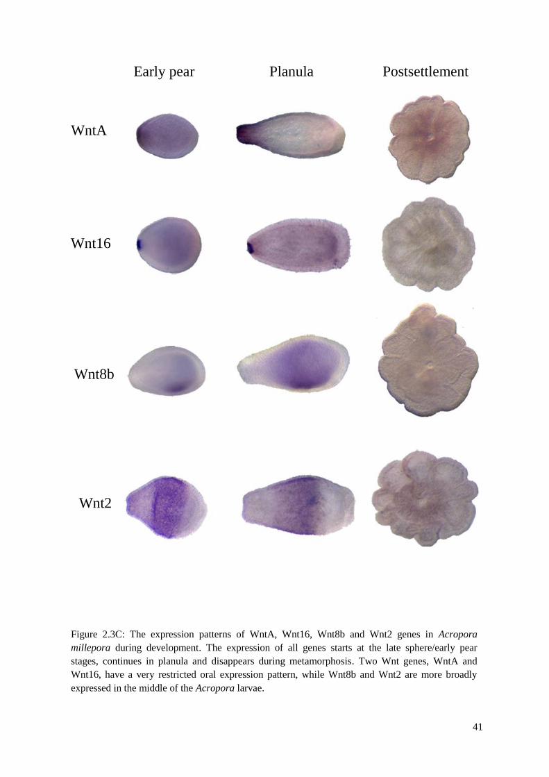
41
Figure 2.3C: The expression patterns of WntA, Wnt16, Wnt8b and Wnt2 genes in Acropora
millepora during development. The expression of all genes starts at the late sphere/early pear
stages, continues in planula and disappears during metamorphosis. Two Wnt genes, WntA and
Wnt16, have a very restricted oral expression pattern, while Wnt8b and Wnt2 are more broadly
expressed in the middle of the Acropora larvae.
WntA
Wnt16
Wnt8b
Wnt2
Early pear Planula Postsettlement
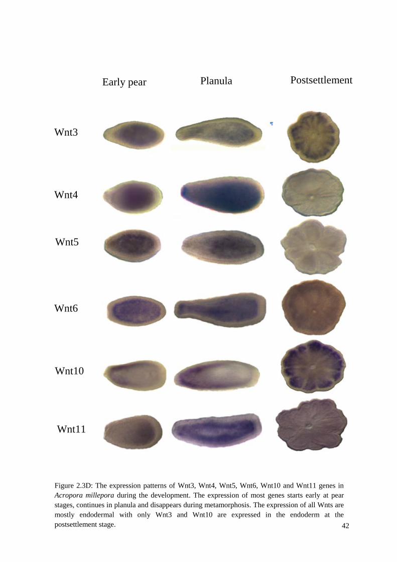
42
Early pear Planula Postsettlement
Wnt3
Wnt4
Wnt5
Wnt6
Figure 2.3D: The expression patterns of Wnt3, Wnt4, Wnt5, Wnt6, Wnt10 and Wnt11 genes in
Acropora millepora during the development. The expression of most genes starts early at pear
stages, continues in planula and disappears during metamorphosis. The expression of all Wnts are
mostly endodermal with only Wnt3 and Wnt10 are expressed in the endoderm at the
postsettlement stage.
Wnt10
Wnt11
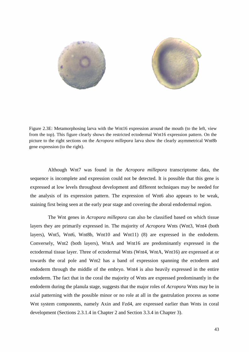
43
Although Wnt7 was found in the Acropora millepora transcriptome data, the
sequence is incomplete and expression could not be detected. It is possible that this gene is
expressed at low levels throughout development and different techniques may be needed for
the analysis of its expression pattern. The expression of Wnt6 also appears to be weak,
staining first being seen at the early pear stage and covering the aboral endodermal region.
The Wnt genes in Acropora millepora can also be classified based on which tissue
layers they are primarily expressed in. The majority of Acropora Wnts (Wnt3, Wnt4 (both
layers), Wnt5, Wnt6, Wnt8b, Wnt10 and Wnt11) (8) are expressed in the endoderm.
Conversely, Wnt2 (both layers), WntA and Wnt16 are predominantly expressed in the
ectodermal tissue layer. Three of ectodermal Wnts (Wnt4, WntA, Wnt16) are expressed at or
towards the oral pole and Wnt2 has a band of expression spanning the ectoderm and
endoderm through the middle of the embryo. Wnt4 is also heavily expressed in the entire
endoderm. The fact that in the coral the majority of Wnts are expressed predominantly in the
endoderm during the planula stage, suggests that the major roles of Acropora Wnts may be in
axial patterning with the possible minor or no role at all in the gastrulation process as some
Wnt system components, namely Axin and Fzd4, are expressed earlier than Wnts in coral
development (Sections 2.3.1.4 in Chapter 2 and Section 3.3.4 in Chapter 3).
Figure 2.3E: Metamorphosing larva with the Wnt16 expression around the mouth (to the left, view
from the top). This figure clearly shows the restricted ectodermal Wnt16 expression pattern. On the
picture to the right sections on the Acropora millepora larva show the clearly asymmetrical Wnt8b
gene expression (to the right).
44
2.3.3 Analysis of Fzd receptors in Acropora millepora
Fzd receptors are seven transmembrane proteins that belong to the large family of G
protein-coupled receptors. These proteins usually contain 500-700 amino acids, and bind Wnt
ligands from the extracellular space as well as components (e.g. Axin in the canonical
pathway) from the intracellular compartment. Fzd receptors have an extracellular N-terminal
cysteine-rich domain (CRD) responsible for Wnt binding and a C-terminal cytoplasmic tail
for signal transduction. Most vertebrates have ten Frizzled family proteins. Searching the
Acropora millepora transcriptome assembly led to the identification of four complete Fzd
proteins (Fzd1, Fzd2, Fzd3 and Fzd4), one partial Fzd8-like sequence and one Fzd-like
protein (named FzdX_Am) with several extracellular cysteine-rich domains. The domain
structures of all the Fzd proteins identified are shown in Figure 2.3F along with the
generalised structure of Frizzled proteins. All of the four complete Fzd proteins are
approximately 550 amino acids long and in each case contain the KTXXXW motif that is
responsible for activation of the Wnt/-catenin pathway (Umbhauer et al., 2000) at the C-
terminal of the hydrophobic domains. Except in the case of FzdX_Am, all of the Acropora
Fzd proteins have signal sequences (Figure 2.3F). All discovered Frizzleds contain the
cysteine-rich domains that are responsible for binding of Wnt secreted glycoproteins (note
that the FzdX_Am partial sequence has two CRD domains and its function is unknown).
To better understand the relationships of Frizzled proteins sequences, maximum
likelihood (ML) phylogenetic analysis was performed with Fzds from Acropora millepora
(Am), Nematostella vectesis (Nv), Clytia hemisphaerica (Ch), Hydractinia echinata (He),
Hydra vulgaris (Hv), Drosophila melanogaster (Dm) and Homo sapiens (Hs) (Figure 2.3G).
Only those Frizzled proteins for which full-length sequences were available (Fzd1, Fzd2,
Fzd3 and Fzd4) were included in the phylogenetic analysis. The Acropora millepora Fzd
sequences are named based on their likely homologues in Nematostella vectenisis.
Alignments of the Acropora millepora Fzd protein sequences with their Nematostella
vectensis homologues (except of FzdX-related protein) are shown in the Supplementary
material Figures 2.2L, 2.3O, 2.3P, 2.3Q, 2.3R).
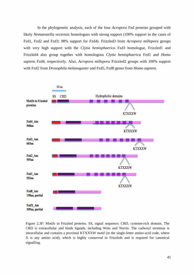
45
In the phylogenetic analysis, each of the four Acropora Fzd proteins grouped with
likely Nematostella vectensis homologues with strong support (100% support in the cases of
Fzd1, Fzd2 and Fzd3; 98% support for Fzd4). Frizzled3 from Acropora millepora groups
with very high support with the Clytia hemisphaerica Fzd3 homologue, Frizzled1 and
Frizzled4 also group together with homologous Clytia hemisphaerica Fzd1 and Homo
sapiens Fzd4, respectively. Also, Acropora millepora Frizzled2 groups with 100% support
with Fzd2 from Drosophila melanogaster and Fzd5, Fzd8 genes from Homo sapiens.
Figure 2.3F: Motifs in Frizzled proteins. SS, signal sequence; CRD, cysteine-rich domain. The
CRD is extracellular and binds ligands, including Wnts and Norrin. The carboxyl terminus is
intracellular and contains a proximal KTXXXW motif (in the single-letter amino-acid code, where
X is any amino acid), which is highly conserved in Frizzleds and is required for canonical
signalling.
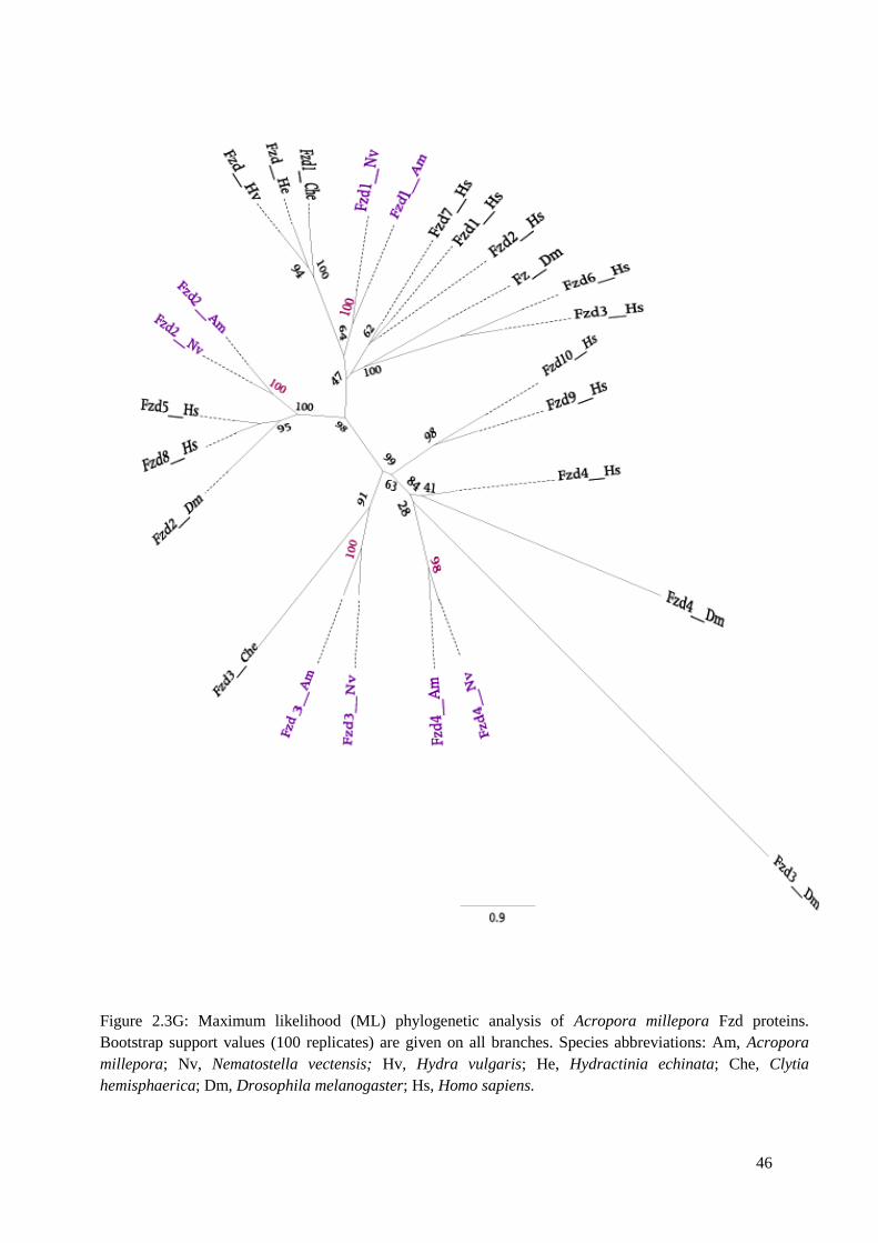
46
Figure 2.3G: Maximum likelihood (ML) phylogenetic analysis of Acropora millepora Fzd proteins.
Bootstrap support values (100 replicates) are given on all branches. Species abbreviations: Am, Acropora
millepora; Nv, Nematostella vectensis; Hv, Hydra vulgaris; He, Hydractinia echinata; Che, Clytia
hemisphaerica; Dm, Drosophila melanogaster; Hs, Homo sapiens.
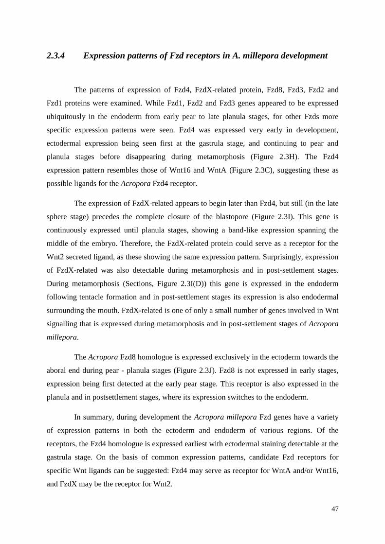
47
2.3.4 Expression patterns of Fzd receptors in A. millepora development
The patterns of expression of Fzd4, FzdX-related protein, Fzd8, Fzd3, Fzd2 and
Fzd1 proteins were examined. While Fzd1, Fzd2 and Fzd3 genes appeared to be expressed
ubiquitously in the endoderm from early pear to late planula stages, for other Fzds more
specific expression patterns were seen. Fzd4 was expressed very early in development,
ectodermal expression being seen first at the gastrula stage, and continuing to pear and
planula stages before disappearing during metamorphosis (Figure 2.3H). The Fzd4
expression pattern resembles those of Wnt16 and WntA (Figure 2.3C), suggesting these as
possible ligands for the Acropora Fzd4 receptor.
The expression of FzdX-related appears to begin later than Fzd4, but still (in the late
sphere stage) precedes the complete closure of the blastopore (Figure 2.3I). This gene is
continuously expressed until planula stages, showing a band-like expression spanning the
middle of the embryo. Therefore, the FzdX-related protein could serve as a receptor for the
Wnt2 secreted ligand, as these showing the same expression pattern. Surprisingly, expression
of FzdX-related was also detectable during metamorphosis and in post-settlement stages.
During metamorphosis (Sections, Figure 2.3I(D)) this gene is expressed in the endoderm
following tentacle formation and in post-settlement stages its expression is also endodermal
surrounding the mouth. FzdX-related is one of only a small number of genes involved in Wnt
signalling that is expressed during metamorphosis and in post-settlement stages of Acropora
millepora.
The Acropora Fzd8 homologue is expressed exclusively in the ectoderm towards the
aboral end during pear - planula stages (Figure 2.3J). Fzd8 is not expressed in early stages,
expression being first detected at the early pear stage. This receptor is also expressed in the
planula and in postsettlement stages, where its expression switches to the endoderm.
In summary, during development the Acropora millepora Fzd genes have a variety
of expression patterns in both the ectoderm and endoderm of various regions. Of the
receptors, the Fzd4 homologue is expressed earliest with ectodermal staining detectable at the
gastrula stage. On the basis of common expression patterns, candidate Fzd receptors for
specific Wnt ligands can be suggested: Fzd4 may serve as receptor for WntA and/or Wnt16,
and FzdX may be the receptor for Wnt2.
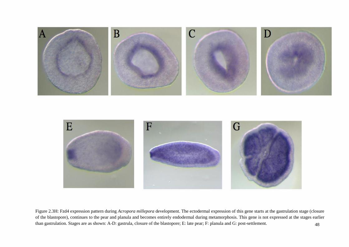
48
Figure 2.3H: Fzd4 expression pattern during Acropora millepora development. The ectodermal expression of this gene starts at the gastrulation stage (closure
of the blastopore), continues to the pear and planula and becomes entirely endodermal during metamorphosis. This gene is not expressed at the stages earlier
than gastrulation. Stages are as shown: A-D: gastrula, closure of the blastopore; E: late pear; F: planula and G: post-settlement.
49
Figure 2.3I: FzdX-related protein expression pattern during Acropora millepora development. The expression
of FzdX starts in late gastrula, continues to planula and metamorphosis. The expression pattern of this gene is
unique and appears endodermal at gastrula, band spanning ectoderm and endoderm in the middle of the
embryo in planula, and this gene is also expressed in the post-settlement stage showing an intriguing
expression pattern in the endoderm, surrounding the mouth. Stages depicted in figure are as follows: left up:
late gastrula; right up: planula; right down: post-settlement and left down
: sections of post-settlement.
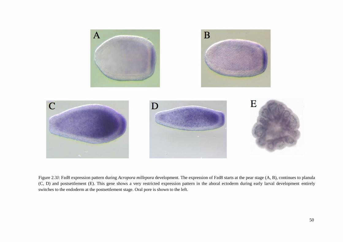
50
Figure 2.3J: Fzd8 expression pattern during Acropora millepora development. The expression of Fzd8 starts at the pear stage (A, B), continues to planula
(C, D) and postsettlement (E). This gene shows a very restricted expression pattern in the aboral ectoderm during early larval development entirely
switches to the endoderm at the postsettlement stage. Oral pore is shown to the left.
51
2.3.5 Analysis of other Wnt system components
In addition to the Wnt ligands and Fzd receptors, the majority of the intracellular
components of the Wnt signalling network were also identified in Acropora millepora,
including the LRP5/6 co-receptor of Frizzled proteins that is essential for -catenin
dependent canonical signalling, Dishevelled, Axin, APC, β-catenin, the GSK3 and CKI
kinases, the transcription factor TCF/LEF and the repressor Groucho, and two types of Wnt
inhibitors – WIF (Wnt Inhibitory Factor) and a homologue of one of the Dickkopf types
(Dkk3). The domain structures of key Wnt system components were analysed and compared
with their counterparts from Nematostella vectensis and other animals.
LRP5/6 transmembrane protein in Acropora millepora
The low-density lipoprotein receptor (LDLR5/6) is the major cholesterol-carrying
lipoprotein of plasma, acting to regulate cholesterol homeostasis in mammalian cells. This
protein is also the Wnt co-receptor, which acts together with Frizzled proteins in the
recruitment of Axin molecules to the Frizzled-LRP complexes. This event leads to the
activation of Dishevelled cytoplasmic phosphoproteins and canonical (-catenin dependent)
Wnt signalling. According to recent reviews on Wnt signalling (Angers and Moon, 2009),
Axin binds to the carboxyl-terminal domains of the LRP5/6 co-receptor. In particular, the
recruitment of Axin to the LRP co-receptor is mediated by five Pro-Pro-(Ser/Thr)-Pro
repeats that are present in the cytoplasmic domains of Xenopus laevis LRP5 and the
LRP6/Arrow homologue in Drosophila melanogaster (Tamai et al., 2000).
The near full-length coding sequence (1684 amino acid residues) of a clear
homologue of LRP5/6 was retrieved from the Acropora millepora transcriptome assembly.
Alignments of the Acropora and Nematostella LRP5/6 sequences are given in
Supplementary material (Figure 2.3S) and the domain structures of the Acropora,
Nematostella and Xenopus LRP5/6 homologues are shown in Figure 2.3K.
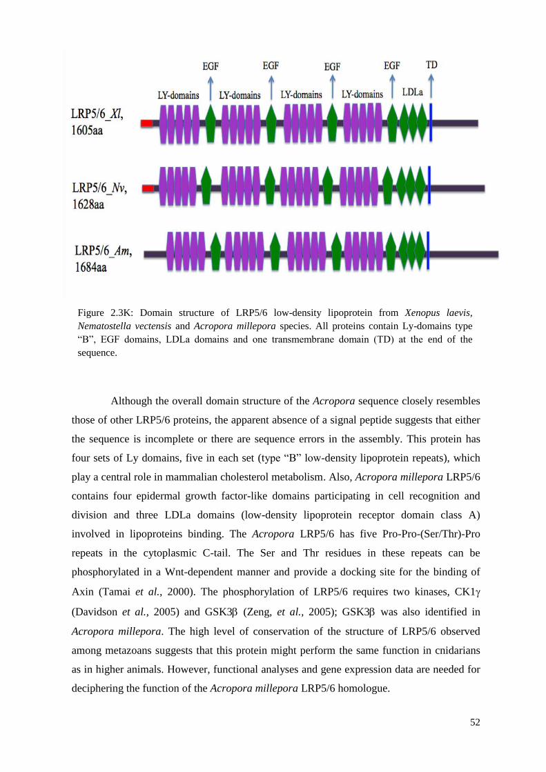
52
Although the overall domain structure of the Acropora sequence closely resembles
those of other LRP5/6 proteins, the apparent absence of a signal peptide suggests that either
the sequence is incomplete or there are sequence errors in the assembly. This protein has
four sets of Ly domains, five in each set (type “B” low-density lipoprotein repeats), which
play a central role in mammalian cholesterol metabolism. Also, Acropora millepora LRP5/6
contains four epidermal growth factor-like domains participating in cell recognition and
division and three LDLa domains (low-density lipoprotein receptor domain class A)
involved in lipoproteins binding. The Acropora LRP5/6 has five Pro-Pro-(Ser/Thr)-Pro
repeats in the cytoplasmic C-tail. The Ser and Thr residues in these repeats can be
phosphorylated in a Wnt-dependent manner and provide a docking site for the binding of
Axin (Tamai et al., 2000). The phosphorylation of LRP5/6 requires two kinases, CK1
(Davidson et al., 2005) and GSK3 (Zeng, et al., 2005); GSK3 was also identified in
Acropora millepora. The high level of conservation of the structure of LRP5/6 observed
among metazoans suggests that this protein might perform the same function in cnidarians
as in higher animals. However, functional analyses and gene expression data are needed for
deciphering the function of the Acropora millepora LRP5/6 homologue.
Figure 2.3K: Domain structure of LRP5/6 low-density lipoprotein from Xenopus laevis,
Nematostella vectensis and Acropora millepora species. All proteins contain Ly-domains type
“B”, EGF domains, LDLa domains and one transmembrane domain (TD) at the end of the
sequence.
53
Dishevelled protein in Acropora millepora
The Dishevelled (Dsh) phosphoproteins are located in the cytoplasm and function
in both -catenin dependent canonical and non-canonical Wnt signalling (Chan and Ye-
Guang, 2010). These proteins function downstream of Frizzled receptors. In the Wnt--
catenin pathway Dishevelled inactivates the GSK3 degradation complex through its
association with Axin (Smalley et al., 1999; Kishida et al., 1999) (Figure 2.3L). Dsh also
participates in the Wnt-mediated phosphorylation of LRP5/6 receptors (Zeng et al., 2008).
When the non-canonical Wnt pathway is activated, Frizzled proteins engage heterotrimeric
G proteins and Dishevelled to initiate signalling.
In man and the mouse, three genes encode dishevelled proteins (Dsh1, 2 and 3)
(Semenov and Snyder, 1997; Klingensmith et al., 1996; Pizzuti et al., 1996; Tsang et al.,
1996; Sokol et al., 1995; Sussman et al., 1994). Dsh proteins typically consist of about 750
amino acid residues and have three conserved domains: the amino-terminal DIX (binding to
Dishevelled/polymerization and Axin) of 80 amino acids, a central PDZ domain of about 90
amino acids, and a carboxyl-terminal DEP domain of 80 amino acids. Dishevelled proteins
exploit the PDZ domain to transduce signals from the membrane receptor Fzd to
downstream components by direct interaction with Fzd. The DEP domain is crucial for the
interaction between Dsh and other proteins possessing further two conserved regions, the
basic region and the proline-rich region, both involved in protein-protein interaction and/or
phosphorylation (Chan et al., 2010).
Only one Dsh protein was identified in Acropora millepora, which is most similar
to Dishevelled 3 in Homo sapiens and Mus musculus. Acropora Dsh consists of 689 amino
acids and possesses all three domains identifiable in human Dsh3 (Figure 2.3M). The
analysis of expression pattern of Acropora millepora Dsh showed that this protein is not
expressed prior to the early sphere stage and its expression disappears during
metamorphosis. In pear-planula stages expression spans the endodermal aboral region. After
metamorphosis, Dsh expression weakens and at the post-settlement stage only weak
endodermal staining is observed (Figure 2.3N).
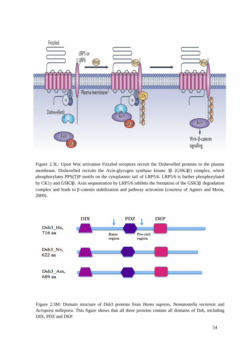
54
Figure 2.3L: Upon Wnt activation Frizzled receptors recruit the Dishevelled proteins to the plasma
membrane. Dishevelled recruits the Axin-glycogen synthase kinase 3 (GSK3) complex, which
phosphorylates PPS(T)P motifs on the cytoplasmic tail of LRP5/6. LRP5/6 is further phosphorylated
by CK1 and GSK3. Axin sequestration by LRP5/6 inhibits the formation of the GSK3 degradation
complex and leads to -catenin stabilization and pathway activation (courtesy of Agners and Moon,
2009).
Figure 2.3M: Domain structure of Dsh3 proteins from Homo sapiens, Nematostella vectensis and
Acropora millepora. This figure shows that all three proteins contain all domains of Dsh, including
DIX, PDZ and DEP.
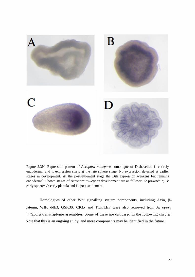
55
Homologues of other Wnt signalling system components, including Axin, -
catenin, WIF, ddk3, GSK3, CKI and TCF/LEF were also retrieved from Acropora
millepora transcriptome assemblies. Some of these are discussed in the following chapter.
Note that this is an ongoing study, and more components may be identified in the future.
Figure 2.3N: Expression pattern of Acropora millepora homologue of Dishevelled is entirely
endodermal and it expression starts at the late sphere stage. No expression detected at earlier
stages in development. At the postsettlement stage the Dsh expression weakens but remains
endodermal. Shown stages of Acropora millepora development are as follows: A: prawnchip; B:
early sphere; C: early planula and D: post-settlement.
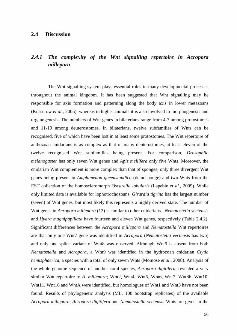
56
2.4 Discussion
2.4.1 The complexity of the Wnt signalling repertoire in Acropora
millepora
The Wnt signalling system plays essential roles in many developmental processes
throughout the animal kingdom. It has been suggested that Wnt signalling may be
responsible for axis formation and patterning along the body axis in lower metazoans
(Kusserow et al., 2005), whereas in higher animals it is also involved in morphogenesis and
organogenesis. The numbers of Wnt genes in bilaterians range from 4-7 among protostomes
and 11-19 among deuterostomes. In bilaterians, twelve subfamilies of Wnts can be
recognised, five of which have been lost in at least some protostomes. The Wnt repertoire of
anthozoan cnidarians is as complex as that of many deuterostomes, at least eleven of the
twelve recognised Wnt subfamilies being present. For comparison, Drosophila
melanogaster has only seven Wnt genes and Apis mellifera only five Wnts. Moreover, the
cnidarian Wnt complement is more complex than that of sponges, only three divergent Wnt
genes being present in Amphimedon queenslandica (demosponge) and two Wnts from the
EST collection of the homoscleromorph Oscarella lobularis (Lapebie et al., 2009). While
only limited data is available for lophotrochozoans, Girardia tigrina has the largest number
(seven) of Wnt genes, but most likely this represents a highly derived state. The number of
Wnt genes in Acropora millepora (12) is similar to other cnidarians - Nematostella vectensis
and Hydra magnipapillata have fourteen and eleven Wnt genes, respectively (Table 2.4.2).
Significant differences between the Acropora millepora and Nematostella Wnt repertoires
are that only one Wnt7 gene was identified in Acropora (Nematostella vectensis has two)
and only one splice variant of Wnt8 was observed. Although Wnt9 is absent from both
Nematostella and Acropora, a Wnt9 was identified in the hydrozoan cnidarian Clytia
hemisphaerica, a species with a total of only seven Wnts (Momose et al., 2008). Analysis of
the whole genome sequence of another coral species, Acropora digitifera, revealed a very
similar Wnt repertoire to A. millepora; Wnt2, Wnt4, Wnt5, Wnt6, Wnt7, Wnt8b, Wnt10,
Wnt11, Wnt16 and WntA were identified, but homologues of Wnt1 and Wnt3 have not been
found. Results of phylogenetic analysis (ML, 100 bootstrap replicates) of the available
Acropora millepora, Acropora digitifera and Nematostella vectensis Wnts are given in the
57
Supplementary material, Figure 2.4. Following the complete genome sequencing of the
Acropora millepora we hope to gain more data on the Wnt gene complement of this coral.
Six Fzd receptors have so far been identified in Acropora millepora (Fzd1, Fzd2,
Fzd3, Fzd4, FzdX, Fzd8), which is more than the numbers in Nematostella vectensis (
Guder et al., 2006) or Hydra magnipapillata (Minobe et al., 2000). In addition to Wnt
ligands and Fzd receptors most of the downstream players and antagonists of the Wnt
system could be identified in our model. Further, the presence of planar cell polarity
components has not previously been reported in coral, however analysis of the Acropora
millepora transcriptome has revealed the presence of many of the core planar cell polarity
proteins including Frizzled, Flamingo/CELSR, Van Gogh, prickled, Dishevelled, and
Dachsous (Brent Knack, unpublished data).
In summary, we can conclude that the Wnt signalling repertoire of anthozoan
cnidarians, including Acropora millepora, is complex. The complexity seen in cnidarian
representatives suggests that a genetically complex Wnt signalling network was already in
place in Ureumetazoa, the common ancestor of cnidarians and bilaterians, whose
appearance has been dated at more than 700 Mya (Dawkins, 2005). Moreover, the early
appearance of a complex Wnt system is consistent with the idea that Wnts were part of the
basic molecular toolkit that enabled the diversification of all of the eumetazoan body plans
(Boero et al., 2007).
2.4.2 Comparative analysis of Wnt proteins and their expression patterns
in Acropora millepora and other cnidarians.
The twelve Wnt genes we have identified from Acropora millepora exhibit a range
of spatial expression patterns, some of which differ significantly to the patterns reported for
their nominal homologues genes in the sea anemone Nematostella vectensis). Whereas roles
have been suggested for WntA, Wnt1, Wnt2 and Wnt4 in axial patterning during
gastrulation (as suggested by their oral expression in and around the blastopore) in
Nematostella (Kusserow et al., 2005), in Acropora such roles are unlikely because
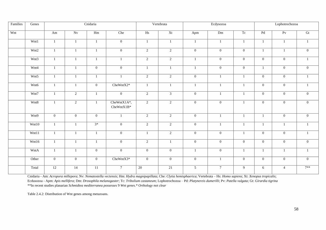
58
Families Genes Cnidaria Vertebrata Ecdysozoa Lophotrochozoa
Wnt Am Nv Hm Che Hs Xt Apm Dm Tc Pd Pv Gt
Wnt1 1 1 1 0 1 1 1 1 1 1 1 1
Wnt2 1 1 1 0 2 2 0 0 0 1 1 0
Wnt3 1 1 1 1 2 2 1 0 0 0 0 1
Wnt4 1 1 0 0 1 1 1 0 0 1 0 0
Wnt5 1 1 1 1 2 2 0 1 1 0 0 1
Wnt6 1 1 0 CheWntX2* 1 1 1 1 1 0 0 1
Wnt7 1 2 1 0 2 3 0 1 1 0 0 0
Wnt8 1 2 1 CheWntX1A*,
CheWntX1B*
2 2 0 0 1 0 0 0
Wnt9 0 0 0 1 2 2 0 1 1 1 0 0
Wnt10 1 1 3* 0 2 2 0 1 1 1 1 1
Wnt11 1 1 1 0 1 2 0 0 1 0 0 1
Wnt16 1 1 1 0 2 1 0 0 0 0 0 0
WntA 1 1 0 0 0 0 1 0 1 1 1 1
Other 0 0 0 CheWntX3* 0 0 0 1 0 0 0 0
Total 12 14 11 7 20 21 5 7 9 6 4 7**
Cnidaria - Am: Acropora millepora; Nv: Nematostella vectensis; Hm: Hydra magnipapillata; Che: Clytia hemisphaerica; Vertebrata – Hs: Homo sapiens; Xt: Xenopus tropicalis;
Ecdusozoa - Apm: Apis mellifera; Dm: Drosophila melanogaster; Tc: Tribolium castaneum; Lophotrochozoa – Pd: Platynereis dumerilli; Pv: Patella vulgata; Gt: Girardia tigrina
**In recent studies planarian Schmidtea mediterranea possesses 9 Wnt genes.* Orthology not clear
Table 2.4.2: Distribution of Wnt genes among metazoans.
59
expression could not be detected prior to blastopore closure. In the case of Acropora, only
two genes of the Wnt signalling network are expressed during the early stages of
development: Fzd4, for which expression starts at the blastula stage (closure of the
blastopore) and Axin, the only gene for which maternal transcripts were detected (Shinzato,
unpublished). Based on these data, it appears unlikely that canonical Wnt signalling plays
significant roles during gastrulation in Acropora millepora. Rather, the complexity of Wnt
expression patterns seen during larval developmental is suggestive of primary roles in larval
patterning processes. The apparent down-regulation of Wnt expression at metamorphosis
coincides with the strong up-regulation of nemo-like kinase (NLK) in response to
neuropeptides of the LWamide family or crustose coralline algae (CCA) extract, both of
which trigger metamorphosis in coral larvae (Grasso et al., 2010). The expression of NLK
has been linked to activation of the Wnt calcium pathway (Ishitani et al., 1999, 2003;
Meneghini et al., 1999), which may be significant in the context of calcification initiation in
the coral. Moreover, in other animals up-regulation of NLK results in the inhibition of
canonical Wnt signalling (Ishitani et al., 1999, 2003; Meneghini et al., 1999;), implying that
non-canonical (calcium) Wnt signalling inhibits the canonical Wnt pathway in coral during
metamorphosis and the initiation of calcification.
Despite differences in the time at which expression of Wnts is initiated, the
expression patterns of WntA and Wnt2 along the oral aboral axis (Figure 2.3C and 2.4A) are
strikingly similar in Acropora millepora and Nematostella vectensis. Wnt2 is classically
thought of as a canonical Wnt and in both animals is expressed as a broad band spanning
both cell layers (ectoderm and endoderm) in the central region of the embryo. However,
Wnt2 expression is detectable much earlier (cleavage-gastrula stage) in the case of
Nematostella vectensis than in Acropora millepora (early pear). Based on the expression
data, Wnt2 may function primary as a canonical Wnt in the anthozoans, presumably
participating in axial patterning during larval development. In the hydrozoan, Hydra
magnipapillata, the corresponding gene (HyWnt2) is expressed only during the early stages
of bud formation, suggestive of a role in the early bud development. A second gene with
similar expression patterns in the two anthozoans is WntA, which typically functions in
non-canonical Wnt signalling in ecdysozoans (Janssen et al., 2010). In both Acropora and
Nematostella, the WntA genes are expressed in the oral region of the embryo, the
expression domain being somewhat broader in the case of the coral. Again, the expression
of WntA starts much earlier (at the cleavage-gastrula stage) in Nematostella vectensis, and
60
the sea anemone WntA may be involved in the blastopore organizer/gastrulation processes.
Interestingly, this gene has apparently been lost from the majority of bilaterian animals and
hydrozoans, and no functional data are yet available for cnidarians. In summary, the
similarity of both Wnt2 and WntA expression patterns between Acropora and Nematostella
suggest that these genes may play important roles in specification and/or maintenance of the
oral/aboral axis in anthozoans.
In contrast to the situation with WntA and Wnt2, the expression patterns of many
of the other Wnt genes in Acropora millepora are very different from those of their
homologues in Nematostella vectensis. The differences include not only the size and
position of the expression domain within one cell layer along the larvae axis (WntA, Wnt8b,
Wnt5 and others), but also the cell layers in which the genes are expressed differing
between the two species (Wnt16, Wnt11). Moreover, in Acropora millepora Wnt8b is
differentially expressed along the directive axis, whereas this is not the case in Nematostella
vectensis. Most of the Wnt genes in Acropora millepora are expressed only after
gastrulation, whereas in Nematostella many of the Wnt genes are expressed much earlier, at
the cleavage-gastrula stages. While in Nematostella some Wnt genes (Wnt10 and Wnt11)
are expressed in individual cells nothing like this was seen for any of the Acropora
millepora Wnt genes – indeed, these genes have highly divergent expression patterns in the
two anthozoans. Whereas in Nematostella vectensis Wnt11 is expressed (in individual cells)
throughout the planula and Wnt10 closer to the oral end, both Wnt10 and Wnt11
homologues of Acropora millepora are more generally expressed throughout the endoderm
(Figure 2.3D and Figure 2.4A). Interestingly in hydrozoans (Hydra magnipapillata) the
expression of Wnt11 is restricted to the ectoderm in the hypostome.
One of the most interesting expression patterns was observed in the case of
Acropora Wnt8b; the transcript appears to be asymmetrically distributed along directive
axis with the onset of expression at early pear stage. Few cases of asymmetrical expression
in the directive axis have been documented to date. For example, homeobox gene goosecoid
in Nematostella (NvGsc) is not expressed during gastrulation, but in the planula is expressed
asymmetrically with respect to the directive axis – two bands of expression are seen with
stronger one is on the side of anthox1a/7/8 (Hox-like), gdf5-like (D/V patterning gene) and
gremlin (inhibitor of TGF-beta signalling pathway) expression and less extensive stripe on
the side of dpp (morphogene), chordin (involved in D/V patterning) and noggin (inhibitor of
61
TGF- signalling) expression (Ball et al., 2007). In contrast to the Acropora Wnt8b gene, in
Nematostella vectensis, the homologous gene is symmetrically expressed in the endoderm in
band spanning the middle of the embryo. Hydra Wnt8 is expressed at the base of and in the
tentacles (Lengfeld et al., 2009; Philipp et al., 2009) but not in the hypostome and may be
involved in non-canonical Wnt pathways. The group of Isabelle Philipp have shown that
HyWnt8 together with HyWnt5 act in mass tissue movements during bud and tentacle
evagination and/or regeneration (Philipp et al., 2009). Whereas in anthozoans Wnt8 is most
likely to perform canonical function, as this Wnt is only expressed during larval life of
Acropora asymmetrically along the directive axis (Figure 2.3C).
Only two of the Acropora millepora Wnt genes studied have very restricted
expression patterns, WntA and Wnt16. While the WntA (discussed earlier) expression seen
in the coral closely resembles that of its Nematostella homologue, in Acropora millepora
Wnt16 is expressed in a different cell layer than are its counterparts in both Nematostella
and Hydra. In Acropora, Wnt16 is expressed in the ring of ectodermal cells surrounding the
mouth of the swimming planula (Figure 2.3E), whilst in Nematostella vectensis the
homologous gene is expressed specifically in the oral endoderm (Figure 1.2C, Chapter 1). In
adult Hydra magnipapillata, Wnt16 is expressed exclusively in the hypostome in both cell
layers but predominantly (60%) in the endoderm (Lengfeld et al., 2009). Based on data
presented in Chapter 3 of this thesis, Wnt16 seems to function primarily in the planar cell
polarity pathway and convergent extension movements during Acropora development.
With the exception of those cases discussed above, the expression patterns
observed for Wnt genes differ substantially between Acropora millepora and Nematostella;
for example, the Acropora Wnt3 gene, although expressed in the same cell layer as in
Nematostella, differs markedly between these two cnidarian species (Figure 2.3D and
Figure 2.4A). In Acropora the Wnt3 gene is uniformly expressed throughout the endoderm
during larval stages and whereas in the sea anemone Wnt3 expression is restricted to the
oral endoderm. While Acropora Wnt4 is also expressed throughout the endoderm and in the
oral ectoderm (Figure 2.3D), the Nematostella gene is expressed in a broad (ectoderm and
endoderm) band toward the oral pore (Figure 1.2C, Chapter 1). Whereas both Wnt5 and
Wnt6 are expressed only weakly in the endoderm in Acropora (Figure 2.3D); expression of
their Nematostella homologues is both stronger and more restricted (Figure 1.2C, Chapter
1).
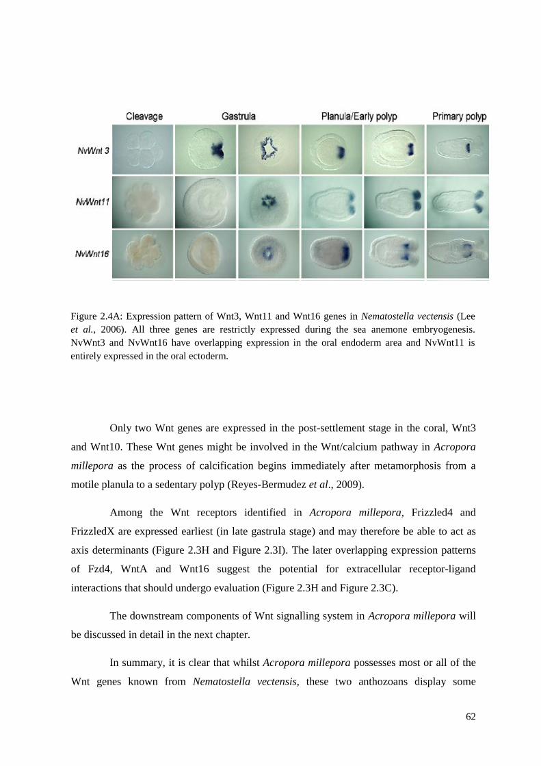
62
Only two Wnt genes are expressed in the post-settlement stage in the coral, Wnt3
and Wnt10. These Wnt genes might be involved in the Wnt/calcium pathway in Acropora
millepora as the process of calcification begins immediately after metamorphosis from a
motile planula to a sedentary polyp (Reyes-Bermudez et al., 2009).
Among the Wnt receptors identified in Acropora millepora, Frizzled4 and
FrizzledX are expressed earliest (in late gastrula stage) and may therefore be able to act as
axis determinants (Figure 2.3H and Figure 2.3I). The later overlapping expression patterns
of Fzd4, WntA and Wnt16 suggest the potential for extracellular receptor-ligand
interactions that should undergo evaluation (Figure 2.3H and Figure 2.3C).
The downstream components of Wnt signalling system in Acropora millepora will
be discussed in detail in the next chapter.
In summary, it is clear that whilst Acropora millepora possesses most or all of the
Wnt genes known from Nematostella vectensis, these two anthozoans display some
Figure 2.4A: Expression pattern of Wnt3, Wnt11 and Wnt16 genes in Nematostella vectensis (Lee
et al., 2006). All three genes are restrictly expressed during the sea anemone embryogenesis.
NvWnt3 and NvWnt16 have overlapping expression in the oral endoderm area and NvWnt11 is
entirely expressed in the oral ectoderm.
63
significant differences in the patterns of expression of homologous genes. Similarities in
Wnt expression patterns can be attributed to the fact that Acropora and Nematostella belong
to the same cnidarian class (Anthozoa). The divergent patterns of expression of Wnt
homologues may be a consequence of major differences in the development of these two
species – for example, the sophisticated metamorphosis that occurs in Acropora millepora
has no counterpart in Nematostella.
As in Acropora millepora the majority of Wnt genes are expressed only after
gastrulation, during pear-planula stages (Figures 2.3C and 2.3D), the main function of Wnts
is likely to be in axial patterning, whereas in Nematostella vectensis, Wnts are thought to
participate in both gastrulation and the axial patterning (Figure 1.2C and 2.4A). The
processes of gastrulation and axial patterning may be less tightly coupled in Acropora
millepora than in Nematostella vectensis, hence Acropora may permit insights into the
molecular bases of axial patterning of lower animals without the complication of additional
roles for genes in germ-layer specification and differentiation.
Finally, the absence of detectable gene expression in the primary polyp of
Acropora millepora is in stark contrast to the maintenance of Wnt signalling in the primary
polyps of both Nematostella vectensis and Hydra magnipapillata. The dramatic differences
between these animals in how a conserved signalling system is used are most likely due to
developmental differences. Whereas Nematostella polyps develop from larvae as the
product of a continuous and uninterrupted program, Acropora millepora larvae undergo a
dramatic metamorphosis from free-living planulae to sessile polyps in response to
environmental cues (Grasso et al., 2010). Together this suggests that Wnt signals in coral
are important in oral patterning of free-living stages, and in maintaining oral specification
throughout the metamorphosis. Temporarily relaxing or abolishing this system of head
specification after metamorphosis may be necessary to enable the specification of the
colonial adult form, a process which remains completely enigmatic in corals.
64
Chapter 3
The significance of non-canonical Wnt signalling in the oral
region during larval development of Acropora
3.1 Functional analysis of Wnt signalling system in cnidarians
The canonical Wnt signalling system has been studied extensively and is well
conserved from humans to cnidarians (Eisenmann, 2005). Canonical Wnt genes play roles in
patterning of the anterior-posterior axis (A/P) in various triploblastic animals during
embryogenesis (Huelsken et al., 2000) and regeneration (Gurley et al., 2008). Non-canonical
Wnt signalling pathways are less well understood and characterized. Despite being
understudied, a growing body of data provides strong evidence for the importance of -
catenin independent Wnt signalling in the regulation of convergent extension movements
during vertebrate gastrulation (Peifer and Polakis, 2000), establishing the polarity of hairs,
bristles and ommatidia in Drosophila (Veeman et al., 2003), control of intracellular calcium
release (Kuhl et al., 2000) and also in negative regulation of canonical Wnt signaling
(Amerongen and Nusse, 2009; Kestler and Kuhl, 2008). Additionally, while homologues of
some non-canonical components can be identified in the Amphimedon queenslandica genome
(Adamska et al., 2010), several key elements including Diversin, Strabismus/van Gogh,
Flamingo/Starry night and Fuzzy are missing (Adamaska et al., 2010). Therefore, as the
simplest animals in which most of the core PCP components are present (see below),
cnidarians are critically important in terms of understanding the evolution of non-canonical
Wnt signalling pathways.
In cnidarians, it has been suggested that canonical Wnt signalling is of primary
importance in axial patterning, as well as functioning in gastrulation and regeneration
(Trevino et al., 2011; Lengfeld et al., 2009; Kusserow et al., 2005) and its role is supported
by studies of both main cnidarian branches, the Anthozoa and the Medusozoa (Duffy et al.,
2010; Gee et al., 2010; Momose et al., 2008; Broun et al., 2005; Hobmayer et al., 2000). In
Hydra, seven Wnt genes are expressed in the hypostome, participating in head organizer
formation and regeneration (Lengfeld et al., 2009). Additionally, main downstream
65
component of the canonical Wnt pathway, -catenin, is involved in oral-aboral (O/A)
patterning during embryogenesis and/or regeneration of Hydra (Gee et al., 2010; Broun et al.,
2005; Hobmayer et al., 2000), Hydractinia (Duffy et al., 2010) and Clytia (Momose et al.,
2008) (detailed description is given below). In Nematostella, Wnt signalling is responsible for
germ layer specification and axial patterning (Trevino et al., 2011; Kumburegama et al.,
2011; Kusserow et al., 2005). Specifically, overlapping expression of NvWnt genes is
reminiscent of Hox genes patterning in vertebrates, suggesting that the cnidarian Wnt code
plays major role in the cnidarian axis patterning (O/A) (Kusserow et al., 2005).
However, data presented in Chapter 2 of this thesis demonstrates that in Acropora
millepora the Wnt genes are not expressed in the staggered and overlapping domains that
have been reported for Nematostella vectensis (Kusserow et al., 2005). Rather, the expression
patterns of the Acropora Wnts are diverse and mostly quite different from those of the
corresponding sea anemone genes. The substantial differences in expression patterns of
homologous Wnts between these species are presumably largely consequences of
Nematostella and Acropora developmental differences; note that similar conclusions have
been drawn in the case of the Sox genes (Shinzato et al., 2008). Nematostella vectensis
planulae directly develop into the juvenile polyps, whereas in the case of Acropora millepora
larvae undergo dramatic metamorphosis to polyps. These developmental and molecular
differences led us to the hypothesis that whilst in Nematostella vectensis Wnts proteins are
thought to be responsible for axial patterning and have roles in gastrulation (Trevino et al.,
2011; Kusserow et al., 2005), in Acropora millepora Wnts function primarily during the
larval stages but do not play Hox-like roles. Based on data presented in this chapter, we
propose that in Acropora millepora the oral pore is a centre for PCP (planar cell polarity)-type
Wnt signalling, and that this non-canonical signalling pathway plays critical roles during
Acropora millepora development. As discussed in Chapter 2, the majority of core PCP
proteins have already been identified in Acropora, including Flamingo/CELSR, Van Gogh,
prickled and Dachsous. The hypothesis of a coral larvae oral region being the centre of non-
canonical Wnt signalling is based on analysis of the expression patterns of Wnt inhibitors and
downstream components of the Wnt signalling pathways, which indicate that there is no
simple relationship between the expression patterns of Wnt genes and their antagonists in our
model system. The importance of this novel developmental centre is also discussed.
Non-canonical Wnt pathways are also present and function in other cnidarians, such
as Nematostella vectensis and Hydra (Kumburegama et al. 2011; Philipp et al., 2009). The
group of Wikramanayake showed that the Wnt/PCP-mediated process of archenteron
66
initiation is uncoupled from the Wnt/-catenin-dependent endoderm cell fate specification in
Nematostella, and that the initial gut invagination requires NvStrabismus (NvStbm), a
maternally expressed core component of the PCP pathway (Kumburegama et al., 2011).
Probable homologues of non-canonical Wnt pathway components, including Wnt5, Wnt8,
frizzled2 (Fz2), Strabismus (Stbm)/Van Gogh, Rho-kinase, dishevelled (Dsh) and JNK have
been reported in Hydra (Philipp et al., 2009, Philipp et al., 2005; Hobmayer et al., 2000). The
Hydra homologues of Wnt5, Wnt8, Frizzled2, and JNK are involved in bud and tentacle
evagination (Philipp et al., 2009). Interestingly, Wnt5 is also expressed at the base of
developing tentacles during larval metamorphosis in N. vectensis, indicating that the function
of Wnt5 in cell polarization and movement is conserved among cnidarians (Kusserow et al.,
2005). As Wnt5 functions in non-canonical signalling in deuterostomes and insects (Gordon
and Nusse, 2002), this function may predate the cnidarian/bilaterian split (Philipp et al.,
2009). In summary, the data available for cnidarians implies that the canonical and non-
canonical Wnt branches were already distinct in the common cnidarian-bilaterian ancestor
and that non-canonical Wnt functions in cell movements and polarization evolved early in the
evolution.
3.1.1 Role of -catenin Wnt signalling in cnidarians
Studies of Hydra, Hydractinia, Clytia and Nematostella imply that -catenin plays
important roles in oral-aboral (O/A) patterning during development throughout the Cnidaria
(Gee et al., 2010; Duffy et al., 2010; Momose et al., 2008; Broun et al., 2005; Hobmayer et
al., 2000). As previously mentioned, Hydra -catenin plays central role in setting up the head
organizer (Gee et al., 2010). In Hydractinia and Clytia, -catenin is also implicated in O/A
patterning during regeneration and embryogenesis, respectively (Duffy et al., 2010; Momose
et al., 2008). In Nematostella, -catenin appears to be differently stabilized along the oral-
aboral axis, leading to selective degradation of Nv--catenin at one pole and stabilization and
nuclear localization at the other pole (Wikramanayake et al., 2003). This selective
degradation and nuclear localization of -catenin plays an essential role in gastrulation in N.
vectensis (Wikramanayake et al., 2003).
67
The studies of Chuya Shinzato (Shinzato, 2007) have led to the suggestion that -
catenin plays important roles in gastrulation and/or germ layer specification in Acropora
millepora. In Acropora planulae, -catenin expression is restricted to the oral region (Ball et
al., unpublished). By analogy with Hydra (Broun et al., 2005; Hobmayer et al., 2000), the
Acropora expression pattern was assumed to indicate canonical Wnt signalling activity in the
oral region. Surprisingly, this initial hypothes is now appearing to be incorrect; below
evidence is presented that the oral region is a centre for non-canonical, rather than canonical,
Wnt signalling. In the oral region of Acropora planulae -catenin does not participate in
triggering canonical Wnt signalling in the nucleus, but rather functions primarily in adherens
junctions complexes and participates in convergent extension movements.
It is becoming clear that -catenin is a critical regulator of Wnt signalling network;
not only is it a key component of both canonical and PCP pathways, but it also functions as a
component of the cadherin complex that controls cell-cell adhesion and cell migration
(Schambony et al., 2004).
A connection between Wnt signalling and cadherin-mediated cell-cell adhesion has
previously been suggested in vertebrates because of the dual function of -catenin in both
systems (Schambony et al., 2004). In Xenopus, binding of -catenin to cadherins antagonizes
Wnt signalling activity during axis formation (Fagotto et al., 1996). Additionally, structural
analyses indicate that cadherins, APC and LEF/TCF bind to the same region of -catenin,
implying that binding partners may compete for that site (Graham et al., 2002, 2001; Hulsken
et al., 1994).
Work presented here led us to suggest that -catenin binds to cadherin in the oral
region of planulae (Section 3.4.3), and thus functions primarily in regulating cell movements
at this stage. This is the first indication of the importance of cadherin--catenin interactions in
cnidarian development, and suggests that this regulatory mechanism appeared very early in
animal evolution and is conserved in cnidarians and bilaterians.
68
3.1.2 Possible molecular mechanisms of Wnt system in cnidarians
The hypostome of Hydra – a centre for canonical (-catenin dependent) Wnt
signalling
The canonical (-catenin dependent) Wnt signalling pathway and its regulation
mechanisms have been extensively studied in cnidarians, particularly in Hydra
magnipapillata. Seven of the eleven known Wnt genes are expressed in the Hydra hypostome
(Lengfeld et al., 2009), where they are thought to play roles in axial patterning and in the head
organizer. HyWnt3, which is the earliest expressed Hydra Wnt, is thought to be a critical
player during head regeneration, stimulating the other Wnts, regulating axial patterning and
organizer activity (Lengfeld et al., 2009). The hypostome of a Hydra polyp appears to
perform some of the same functions as do vertebrate developmental organizers such as the
Spemann-Mangold organizer of amphibians. Essentially, the hypostome is thought to transmit
signal(s) that lead to the formation of a polyp axis (Broun et al., 2002). Moreover, Wnt genes
and -catenin signalling are actively involved in the formation of the Spemann organizer in
Xenopus embryos (Du et al., 1995) and in axial patterning (Kiecker et al., 2001). By analogy,
it was proposed that the hypostome in Hydra is also a centre of canonical Wnt -catenin
signalling (Lengfeld et al., 2009; Broun et al., 2005), and assumed that canonical Wnt
signalling is the main pathway responsible for axial patterning in Hydra. Further support for
this hypothesis was provided by studies of -catenin expression. -catenin is expressed
uniformly throughout adult Hydra (Hobmayer et al., 2000) however, only in the hypostome it
is located in the nucleus (Broun et al., 2005), where many of the Wnts are also expressed
(Lengfeld et al., 2009). Expression of a modified -catenin gene driven by an actin promoter,
has been shown to result in elevated head organizer activity throughout the body column in
transgenic Hydra (Gee et al., 2010), providing functional support for the importance of -
catenin in maintaining Hydra hypostome organizer activity.
69
3.1.3 Molecular mechanisms of Wnt system in Acropora millepora –
functional studies
The development of Acropora millepora
During normal development, fertilized Acropora millepora eggs undergo series of
unilateral cleavages and after the eight-cell stage (approximately 9 hours post fertilization)
cell divisions become irregular (Figure 3.1). At the 13 hours post fertilization the embryo
becomes an irregular-shaped cellular bilayer, the “prawnchip” stage, which is unique to corals
(Ball et al., 2002; Miller and Ball, 2000). The gastrulation occurs at approximately 22 hours
post fertilization which culminate in the closure of the blastopore and the formation of the two
cell layers, ectoderm and endoderm. The closure of the blastopore signifies the transition from
embryo to larva. At 50 hours into development, newly recognizable cell types first appear, for
example putative neurons that can be stained with an antibody against the cnidarian
neurotransmitter RFamide during this developmental stage. At the 72 hours post fertilization,
the larva becomes pear-shaped and cilia develop for directed swimming. Various additional
cell types are differentiated by the late planula stage. On receipt of appropriate settlement
cues, such as chemicals released from coralline algae or neuropeptides of the LWamide
family (Grasso et al., 2011), larvae attach to the substratum with the aboral end and
metamorphosis starts. During this stage Acropora larvae undergo dramatic re-organization of
their tissues and initiate the process of calcification. Following settlement, mesenteries appear
within the flattened disk and tentacles begin to form in the area surrounding the oral pore,
which begins to rise as a growing tip (Figure 3.1).
First steps in functional analysis of Acropora Wnt signalling system
Attempts at functional analysis of developmental genes in Acropora millepora are
constrained by the fact that Acropora is not a laboratory animal – reproduction occurs once
per year during mass coral spawning events. Hence in pioneering functional analyses of
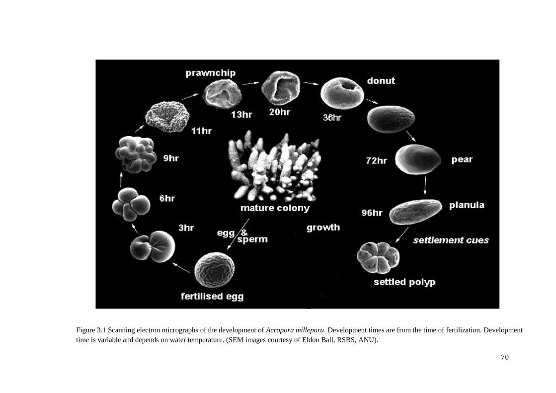
70
Figure 3.1 Scanning electron micrographs of the development of Acropora millepora. Development times are from the time of fertilization. Development
time is variable and depends on water temperature. (SEM images courtesy of Eldon Ball, RSBS, ANU).
71
developmental genes in Acropora millepora, it was necessary to conduct experiments during
the spawning period on the Great Barrier Reef, and many technical limitations had to be
applied.
To better understand the roles of Wnt signalling during coral development,
pharmacological inhibition experiments were performed. Alsterpaullone is widely used as a
specific inhibitor of GSK-3 activity, which results in ligand-independent activation of the
canonical (-catenin dependent) Wnt pathway. In Hydra, 5M alsterpaullone has been shown
to block GSK-3 activity throughout the organism, leading to elevated -catenin levels in
nuclei, which resulted in the formation of multiple ectopic tentacles (Broun et al., 2005).
Alsterpaullone-dependent increases in -catenin protein levels have also been reported in the
Hydractinia echinata (Teo et al., 2006). In the case of Acropora millepora, treatment of
larvae with 5M alsterpaullone resulted in failure to close the blastopore and termination of
development (Shinzato, 2007), suggesting that -catenin may play a critical role in
gastrulation and/or germ layer specification of Acropora millepora.
Because we now know (Chapter 2) that few Wnt system components are expressed
during embryonic development, the focus in this study was on alsterpaullone effects on later
(pear-planula) development of Acropora. Effects in the oral region during pear-planula stages
were of particular interest because the expression of a number of Wnt system components,
including non-canonical Wnts (Wnt16 and WntA) and -catenin, is restricted to this part of
the larva.
3.2 Materials and methods
All basic molecular techniques were carried out using standard protocols (Sambrook and
Russell, 2001).
72
3.2.1 Animal sampling
Inhibition treatments on Acropora millepora embryos were performed at Heron Island
(Latitude 23°26’ South; Longitude 151°55 East), Magnetic Island (Latitude 19°09’ South;
Longitude 146°49’ East) and Orpheus Island (Latitude 18°28’ South; Longitude 146°25’
East) during the coral mass-spawning events in 2008, 2009 and 2010, respectively.
3.2.2 Isolation and in situ hybridization of components involved in the
regulation of Wnt network
Ambcat, Acropora millepora -catenin homologue, was first isolated during a preliminary
project to identify expressed sequence tags (ESTs) in Acropora (Kortschak et al., 2003).
Brachyury (Bra), GSK-3, Groucho, WIF (Wnt Inhibitory Factor), Axin and Dickkopf3
(dkk3) homologues were also isolated during the Acropora millepora EST projects. pGEMT
plasmids with Brachyury, GSK-3, Groucho, WIF (DO52-B8), Axin (D027-D10), and Dkk3
(B027-D10) gene fragments were kindly provided by Dr. David Hayward (Australian
National University, Canberra). Additionally, based on the latest transcriptome assembly, I
have identified the homologue of the Inversin gene, which is considered to act as a molecular
switch between the two Wnt signalling pathways, canonical -catenin dependent and planar
cell polarity (Table 3.2.2).
Name of
the gene
Primers Tm/Cycle
s
Length of the fragment
Inversin For-
5’ ATGGAGGAGTCGGGTTCTTT 3’ Rev-
5’ GCCTTAGAAATGCCGCTGTA 3’
60C/35 521bp
Table 3.2.2: Name of the gene, primer sequences, annealing temperature (Tm), cycles required for
PCR reaction and the length of the fragment.
73
The primers for the Acropora millepore Inversin homologue were designed using Primer3
program (http://frodo.wi.mit.edu/primer3/) based on transcriptome data. PCR was performed
on mixed cDNA libraries using prawnchip, presettlement, metamorphosis, postsettlement and
adult stages. PCR conditions were as follows: 95°C for 5 min as hot start, 95°C for 30
seconds, 55°C for 30 seconds, 72°C for 1 min for 5 cycles, after 35 cycles of 95°C for 30
seconds, 52°C for 30 seconds, 72°C for 1 min. Primer sequences, length of the fragment,
temperature of annealing (Tm) and number of PCR cycles required are given in Table 3.2.2.
Identified fragment of Acropora Inversin homologue was cloned into the pGEMT vector
(Promega). Competent cells for the cloning procedures were prepared using rubidium
chloride method and NM522 bacterial strain. Cloned fragment of Inversin was sequenced by
Macrogen (South Korea) to check direction and correctness of the Inversin plasmid insert.
In situ hybridizations of coral embryos and larvae with the fragment of the Acropora
millepora Inversin homologue and other downstream components of Wnt signalling system
were performed as described in Chapter 2. The digoxigenin-labelled antisense RNA probes
were synthesized in an in vitro transcription with the T7/SP6 RNA polymerase (Fermentas)
after the digestion with NotI/NcoI restriction enzyme. The choice of the polymerase and the
restriction enzyme was dependent on the direction of the gene insert in each case. Subsequent
hybridizations were carried out as described in Chapter 2 at 52C for 48 hours with a final
probe concentration of 0.25 ng/l.
3.2.3 Alsterpaullone inhibition experiments on coral larvae
Alsterpaullone (Calbiochem, 126870) was dissolved in DMSO and diluted in Millipore-
filtered seawater (MFSW) to final concentrations of 1M and 3M in 0.025% DMSO. This
concentration of DMSO showed no effect on coral development. The levels of alsterpaullone
were chosen based on recent (Shinzato, unpublished) experiments in which 5M inhibitor
completely blocked gastrulation in Acropora (Shinzato, 2007). Therefore, two concentrations
(1M and 3M) were tested which were known to be toxic. The inhibition experiment was
divided into two parts: in the first part (early plates) I commenced alsterpaullone treatments
from the early prawnchip stage (11hpf) and in the second from the sphere stage (32hpf; late
plates) onwards. Coral material was exposed to 1M or 3M alsterpaullone for 4 or 12 hours
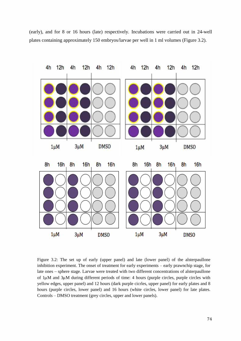
74
(early), and for 8 or 16 hours (late) respectively. Incubations were carried out in 24-well
plates containing approximately 150 embryos/larvae per well in 1 ml volumes (Figure 3.2).
Figure 3.2: The set up of early (upper panel) and late (lower panel) of the alsterpaullone
inhibition experiment. The onset of treatment for early experiments – early prawnchip stage, for
late ones – sphere stage. Larvae were treated with two different concentrations of alsterpaullone
of 1M and 3M during different periods of time: 4 hours (purple circles, purple circles with
yellow edges, upper panel) and 12 hours (dark purple cicrles, upper panel) for early plates and 8
hours (purple circles, lower panel) and 16 hours (white circles, lower panel) for late plates.
Controls – DMSO treatment (grey circles, upper and lower panels).
75
At the end of exposure, the embryos/larvae were washed several times in MFSW
and collected at each washing step following the fixation and transfer of all treated and
control embryos/larvae to the 100% methanol solution and storage at -20C prior to in situ
experiments. I collected treated embryos/larvae at the late prawnchip stage, the onset of
Brachyury expression, folding, donut, sphere, swimming sphere, pre-pear, pear, swimming
pear and spindles (late planulae) stages. Although the embryos/larvae have shown signs of
deviation from normal development, it did not stop completely until planula stage. None of
the treated animals entered metamorphosis stage and the survival rate was zero for all
alsterpaullone-treated larvae after the planula stage.
3.2.4 In situ hybridization experiments on alsterpaullone-treated Acropora
larvae.
In situ hybridization experiments were carried out as described in the chapter 2 on the latest
stages of Acropora millepora development under alsterpaullone treatment conditions (57
hours post fertilization) spanning the pear to planula stages. Control in situ hybridizations
were performed on larvae exposed to DMSO (0.0025%).
The expression patterns of Wnt ligands restrictedly expressed in the oral region of Acropora
larvae, namely WntA and Wnt16 were examined. Also, the expression pattern of a possible
downstream target of Wnt canonical -catenin dependent signalling pathway, a homologue of
Acropora transcription factor Brachyury, was analysed using alsterpaullone-treated larvae of
same treatment conditions (57 hours post fertilization).
3.2.5 Light microscopy and phalloidin staining of alsterpaullone treated and
non-treated Acropora millepora larvae
Images of Acropora millepora alsterpaullone-treated larvae were taken using SPOT advanced
software and LEICA MZFLIII microscope (light microscopy).
76
Phalloidin staining
Acropora larvae from various stages were fixed in fresh 4% paraformaldehyde for 20 mins.
Fixed larvae were rinsed 5 in PBT (PBS with 0.1% Tween 20) and stored at +4C.
Preparation for staining included dehydration of larvae in EtOH series (50%, 70%, 90% and
2100% ) for 5 mins each step. After preparation step Acropora larvae were washed in 50:50
Xylene : EtOH (10mins), 100% Xylene (for 7 hours). After Xylene treatment the larvae were
washed in EtOH (gradually replacing Xylene with 3 100% EtOH) and rehydrated through
EtOH : water series (5 mins each of 20%, 50%, 70%, 90% PBS, and 2 100% PBT). Before
adding the Alexa Fluor 647 – conjugated phalloidin solution containing methanol to the
larvae, it was dried out at room temperature for 2 hours to allow methanol evaporation.
Acropora larvae were then incubated in 10l Alexa Fluor 647 – conjugated phalloidin
(200U/ml; Invitrogen) in 1ml PBT (with Tween) for 2 hours at room temperature and then
washed in PBT for 1 hour with subsequent visualization on a Zeiss LSM 720 confocal
microscope.
3.3 Results
3.3.1 Expression of Wnt16 and Bra (Brachyury) after alsterpaullone
treatment/in response to nuclear -catenin
During larval development (pear-planula stages) of Acropora millepora, expression
of Wnt16 is restricted to the oral ectoderm (Chapter 2, Figure 2.3C). In vertebrates, the
corresponding gene function primarily in the (non-canonical) planar cell polarity (PCP)
pathway (Muy-Teck et al., 2007). To investigate the regulation of Acropora Wnt16, in situ
hybridization was conducted on larvae (paraformaldehyde-fixed material; 57h post
fertilization) that had been treated with either 1M or 3M alsterpaullone. Treatment with
1M alsterpaullone had no obvious effect on the spatial expression pattern of Wnt16,
whereas treatment with 3M alsterpaullone totally abolished the oral staining (Figure 3.3A).
Based on the assumption that alsterpaullone treatment inhibits GSK3 activity in Acropora
77
(Shinzato, 2007) and triggers nuclear localization of -catenin as it does in Hydra (Broun et
al., 2005), these results imply that activation of the canonical Wnt pathway (i.e. nuclear
localisation of -catenin) suppresses the expression of Wnt16 in Acropora larvae. By analogy
with vertebrates, Wnt16 is therefore likely to signal through the PCP pathway (i.e. function as
a non-canonical Wnt ligand) in the oral ectoderm of Acropora larvae and its expression is
inhibited by canonical Wnt signalling/activation of -catenin and TCF/LEF transcription.
Because the transcription factor Brachyury (Bra) is regulated by canonical Wnt
signalling in the mouse (Sebastian et al., 2000), the effect of alsterpaullone treatment on Bra
expression was also examined in Acropora planulae (Figure 3.3B). Bra was considered to be
a good oral marker as it is expressed around the blastopore in basal deuterostomes and
protostomes (Gross and McClay, 2002, Arendt et al., 2001, Shoguchi et al., 1999) as well as
in the sea anemone Nematostella vectensis (Nv). Nv-Brachyury is expressed around the
blastopore and its derivatives, the endodermal mesenteries (Scholz and Technau, 2003).
Expression of HyBra, the Hydra Brachyury, is associated with the mouth, which arises from
the blastopore (Technau, 2001; Technau and Bode, 1999). Therefore the blastoporal
expression of Brachyury is thought to be an ancestral characteristic for the Eumetazoa. The
Acropora Brachyury homologue (AmBra) is also expressed around the blastopore (Ball,
personal communication).
If in Acropora, as in the mouse, the expression of Bra was regulated by canonical
Wnt signalling, one might expect its expression pattern to be modified by alsterpaullone
treatment. However, in situ hybridization of Acropora larvae showed that, treatment with
1M alsterpaullone has no apparent effect on the expression of Brachyury, and 3M
alsterpaullone may have resulted in a slight increase/up-regulation of this transcription factor,
mostly in the area of aboral endoderm (Figure 3.3B). These results suggest that in Acropora,
expression of Brachyury is not regulated by canonical Wnt signalling.
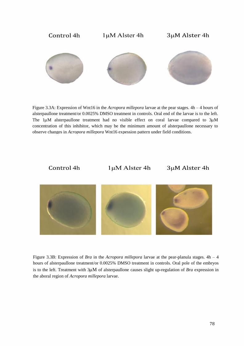
78
Figure 3.3A: Expression of Wnt16 in the Acropora millepora larvae at the pear stages. 4h – 4 hours of
alsterpaullone treatment/or 0.0025% DMSO treatment in controls. Oral end of the larvae is to the left.
The 1M alsterpaullone treatment had no visible effect on coral larvae compared to 3M
concentration of this inhibitor, which may be the minimum amount of alsterpaullone necessary to
observe changes in Acropora millepora Wnt16 expession pattern under field conditions.
Figure 3.3B: Expression of Bra in the Acropora millepora larvae at the pear-planula stages. 4h – 4
hours of alsterpaullone treatment/or 0.0025% DMSO treatment in controls. Oral pole of the embryos
is to the left. Treatment with 3M of alsterpaullone causes slight up-regulation of Bra expression in
the aboral region of Acropora millepora larvae.
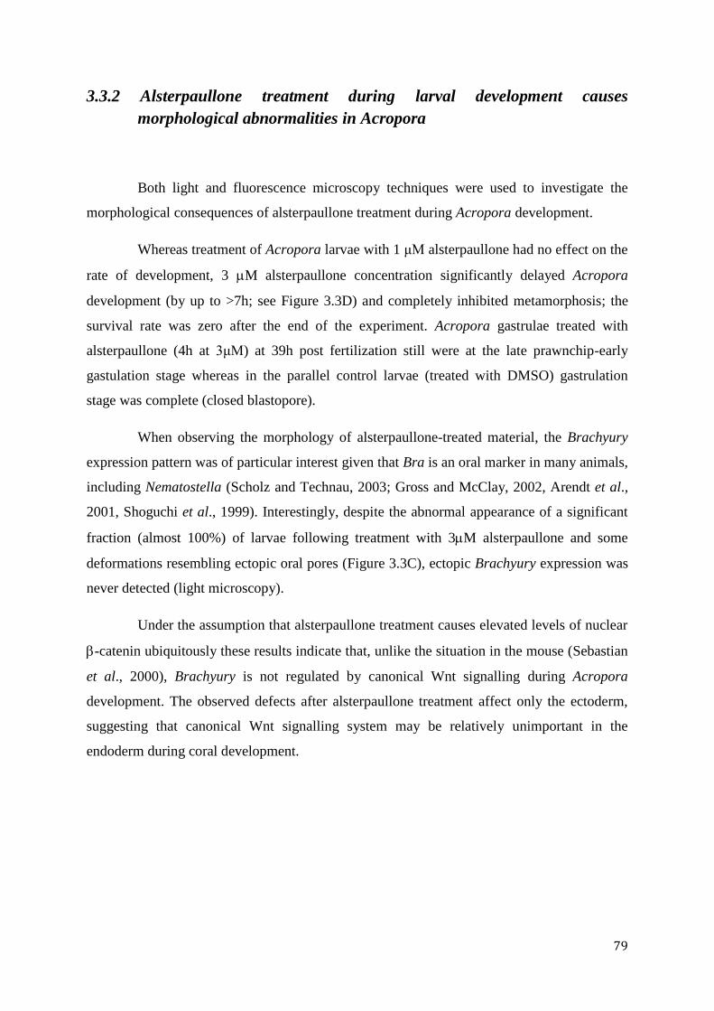
79
3.3.2 Alsterpaullone treatment during larval development causes
morphological abnormalities in Acropora
Both light and fluorescence microscopy techniques were used to investigate the
morphological consequences of alsterpaullone treatment during Acropora development.
Whereas treatment of Acropora larvae with 1 μM alsterpaullone had no effect on the
rate of development, 3 M alsterpaullone concentration significantly delayed Acropora
development (by up to >7h; see Figure 3.3D) and completely inhibited metamorphosis; the
survival rate was zero after the end of the experiment. Acropora gastrulae treated with
alsterpaullone (4h at 3μM) at 39h post fertilization still were at the late prawnchip-early
gastulation stage whereas in the parallel control larvae (treated with DMSO) gastrulation
stage was complete (closed blastopore).
When observing the morphology of alsterpaullone-treated material, the Brachyury
expression pattern was of particular interest given that Bra is an oral marker in many animals,
including Nematostella (Scholz and Technau, 2003; Gross and McClay, 2002, Arendt et al.,
2001, Shoguchi et al., 1999). Interestingly, despite the abnormal appearance of a significant
fraction (almost 100%) of larvae following treatment with 3M alsterpaullone and some
deformations resembling ectopic oral pores (Figure 3.3C), ectopic Brachyury expression was
never detected (light microscopy).
Under the assumption that alsterpaullone treatment causes elevated levels of nuclear
-catenin ubiquitously these results indicate that, unlike the situation in the mouse (Sebastian
et al., 2000), Brachyury is not regulated by canonical Wnt signalling during Acropora
development. The observed defects after alsterpaullone treatment affect only the ectoderm,
suggesting that canonical Wnt signalling system may be relatively unimportant in the
endoderm during coral development.
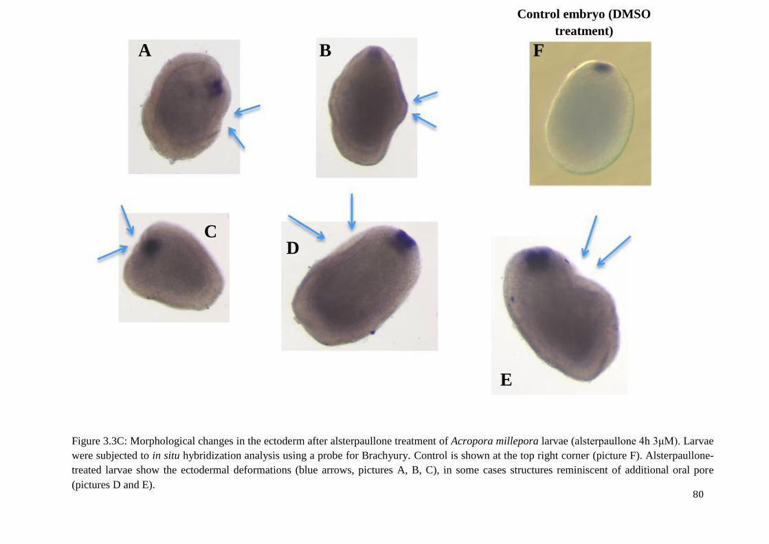
80
Control embryo (DMSO
treatment)
Figure 3.3C: Morphological changes in the ectoderm after alsterpaullone treatment of Acropora millepora larvae (alsterpaullone 4h 3μM). Larvae
were subjected to in situ hybridization analysis using a probe for Brachyury. Control is shown at the top right corner (picture F). Alsterpaullone-
treated larvae show the ectodermal deformations (blue arrows, pictures A, B, C), in some cases structures reminiscent of additional oral pore
(pictures D and E).
A B
C D
E
F
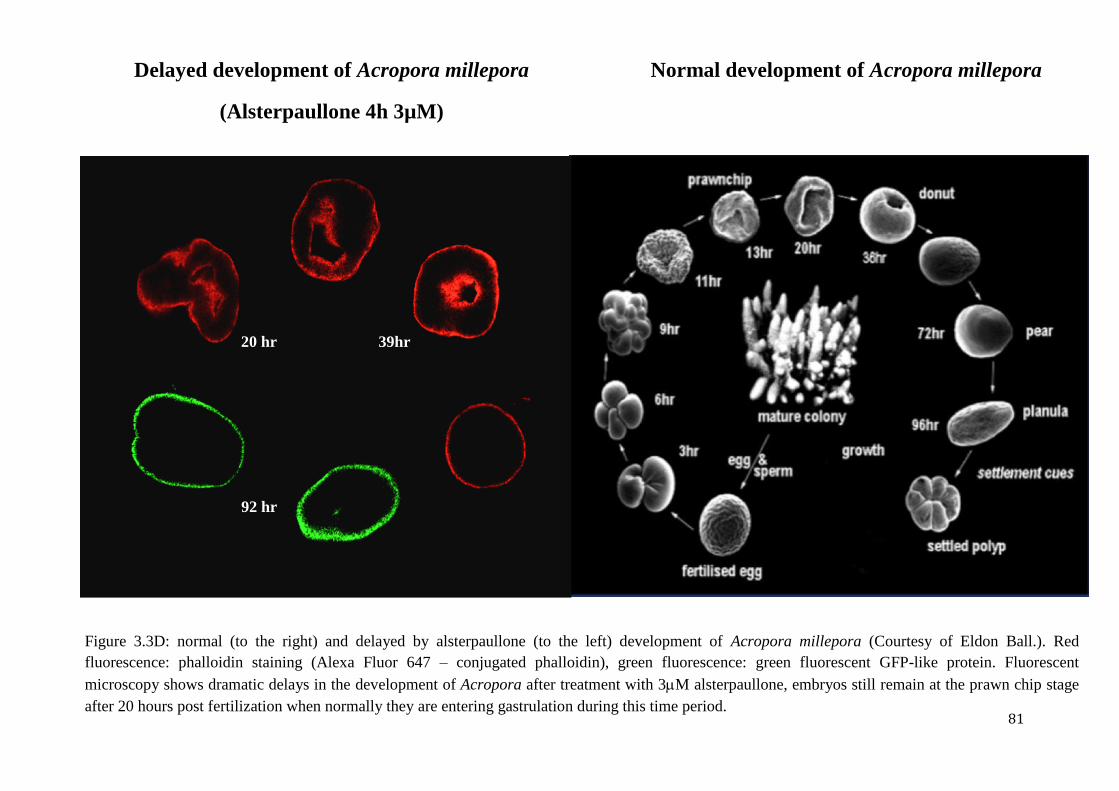
81
Delayed development of Acropora millepora
(Alsterpaullone 4h 3µM)
)
Normal development of Acropora millepora
Figure 3.3D: normal (to the right) and delayed by alsterpaullone (to the left) development of Acropora millepora (Courtesy of Eldon Ball.). Red
fluorescence: phalloidin staining (Alexa Fluor 647 – conjugated phalloidin), green fluorescence: green fluorescent GFP-like protein. Fluorescent
microscopy shows dramatic delays in the development of Acropora after treatment with 3M alsterpaullone, embryos still remain at the prawn chip stage
after 20 hours post fertilization when normally they are entering gastrulation during this time period.
20 hr 39hr
92 hr
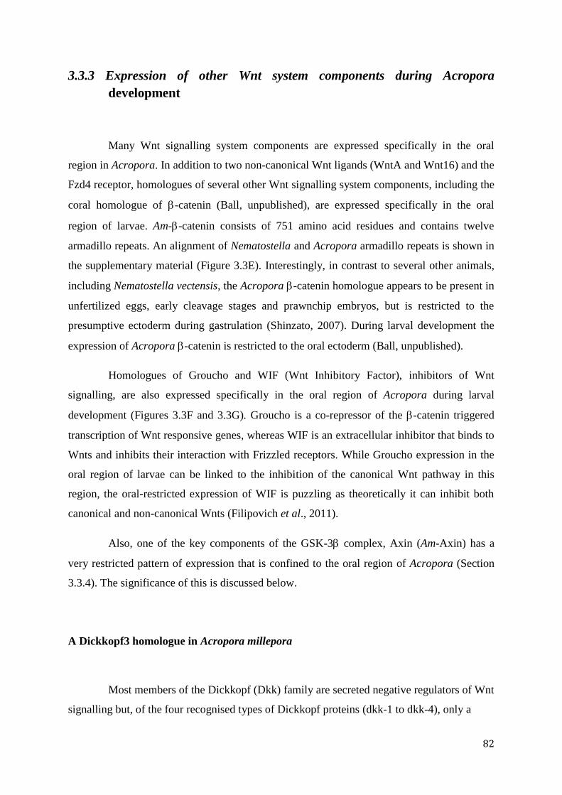
82
3.3.3 Expression of other Wnt system components during Acropora
development
Many Wnt signalling system components are expressed specifically in the oral
region in Acropora. In addition to two non-canonical Wnt ligands (WntA and Wnt16) and the
Fzd4 receptor, homologues of several other Wnt signalling system components, including the
coral homologue of -catenin (Ball, unpublished), are expressed specifically in the oral
region of larvae. Am--catenin consists of 751 amino acid residues and contains twelve
armadillo repeats. An alignment of Nematostella and Acropora armadillo repeats is shown in
the supplementary material (Figure 3.3E). Interestingly, in contrast to several other animals,
including Nematostella vectensis, the Acropora -catenin homologue appears to be present in
unfertilized eggs, early cleavage stages and prawnchip embryos, but is restricted to the
presumptive ectoderm during gastrulation (Shinzato, 2007). During larval development the
expression of Acropora -catenin is restricted to the oral ectoderm (Ball, unpublished).
Homologues of Groucho and WIF (Wnt Inhibitory Factor), inhibitors of Wnt
signalling, are also expressed specifically in the oral region of Acropora during larval
development (Figures 3.3F and 3.3G). Groucho is a co-repressor of the -catenin triggered
transcription of Wnt responsive genes, whereas WIF is an extracellular inhibitor that binds to
Wnts and inhibits their interaction with Frizzled receptors. While Groucho expression in the
oral region of larvae can be linked to the inhibition of the canonical Wnt pathway in this
region, the oral-restricted expression of WIF is puzzling as theoretically it can inhibit both
canonical and non-canonical Wnts (Filipovich et al., 2011).
Also, one of the key components of the GSK-3 complex, Axin (Am-Axin) has a
very restricted pattern of expression that is confined to the oral region of Acropora (Section
3.3.4). The significance of this is discussed below.
A Dickkopf3 homologue in Acropora millepora
Most members of the Dickkopf (Dkk) family are secreted negative regulators of Wnt
signalling but, of the four recognised types of Dickkopf proteins (dkk-1 to dkk-4), only a
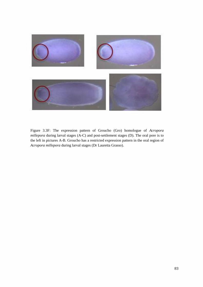
83
Figure 3.3F: The expression pattern of Groucho (Gro) homologue of Acropora
millepora during larval stages (A-C) and post-settlement stages (D). The oral pore is to
the left in pictures A-B. Groucho has a restricted expression pattern in the oral region of
Acropora millepora during larval stages (Dr Lauretta Grasso).
84
Figure 3.3G: The expression pattern of WIF (Wnt Inhibitory Factor at different developmental stages of Acropora millepora, where A-
B: pear; C-D: pear, E-G: planula and H-I: postsettlement stages. Timeline goes from picture A to picture I. Red circle point at the
position of oral pore (pictures C-G oral pore to the left). WIF has an ectodermally-restricted expression pattern in the oral region of
Acropora millepora, that continues to the postsettlement stage (Courtesy of Chuya Shinzato).
85
single representative of one type was identified in Acropora millepora. By comparison, two
Dkk subfamilies (dkk3 and dkk1/2/4) are represented in both Hydra (Guder et al., 2006b;
Fedders et al., 2004) and Nematostella (Guder et al., 2006b).
The coral protein is a clear homologue of Hydra Dickkopf3 (Dkk3) subfamily,
members of which are unique in not being Wnt inhibitors (Guder et al., 2005, Mao and
Niehrs, 2003, Krupnik et al., 1999). However, recent work suggests a novel role for Dkk3 as
a positive modulator of Wnt pathways through interactions with Kremen proteins (Kremen1
and Kremen2), which act as Dkk receptors (Nakamura et al., 2010, Mao et al., 2002). In situ
hybridization experiments (Figure 3.3H) show that, in the case of Acropora, Dkk3 is
expressed only after gastrulation, throughout the endoderm at sphere stage and in the aboral
endoderm during larval stages. Post-settlement Dkk3 expression is also entirely endodermal.
Although the significance of these expession patterns is unclear, functional studies of coral
Dkk3 may shed some light on the evolution of mechanisms of Wnt system regulation and
inhibition/mutual down-regulation of different Wnt signalling pathways.
An Inversin homologue in Acropora millepora
Recent work in Xenopus laevis has highlighted the ability of Inversin to act as a regulatory
switch between canonical and non-canonical Wnt signalling (He, 2008). Inversin can form
protein complexes with the cytoplasmic phosphoprotein Dishevelled, targeting it for
degradation. At the same time, inversin permits the accumulation of Dishevelled at the
plasma membrane of polarized cells, facilitating non-canonical Wnt signalling via the PCP
pathway. In zebrafish, the structurally related switch molecule diversin interacts with two
components of the canonical Wnt pathway, casein kinase I (CKI) and axin/Condictin; both
interactions facilitate efficient phosphorylation of -catenin, thereby inhibiting canonical Wnt
signalling. Diversin also plays a crucial role in gastrulation movements that are controlled by
the Wnt/JNK pathway. Therefore, this molecule is a central regulator of the Wnt system and
suppresses canonical Wnt signals while stimulating PCP JNK-dependent signalling
(Schwarz-Romond et al., 2002). An Inversin-like protein is also present in the sponge
Aphimedon queenslandica (spongezome.metazome.net), indicating that this protein family
has a very long evolutionary history.
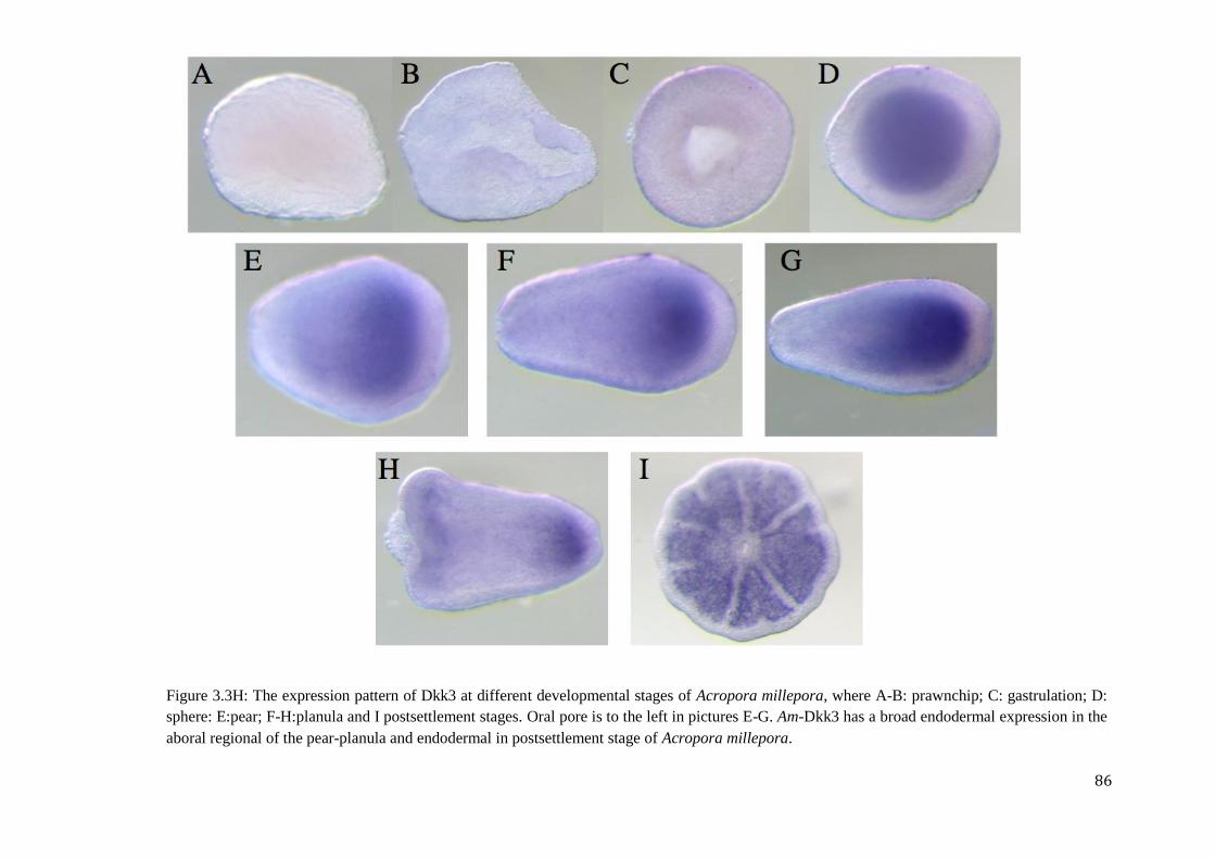
86
Figure 3.3H: The expression pattern of Dkk3 at different developmental stages of Acropora millepora, where A-B: prawnchip; C: gastrulation; D:
sphere: E:pear; F-H:planula and I postsettlement stages. Oral pore is to the left in pictures E-G. Am-Dkk3 has a broad endodermal expression in the
aboral regional of the pear-planula and endodermal in postsettlement stage of Acropora millepora.
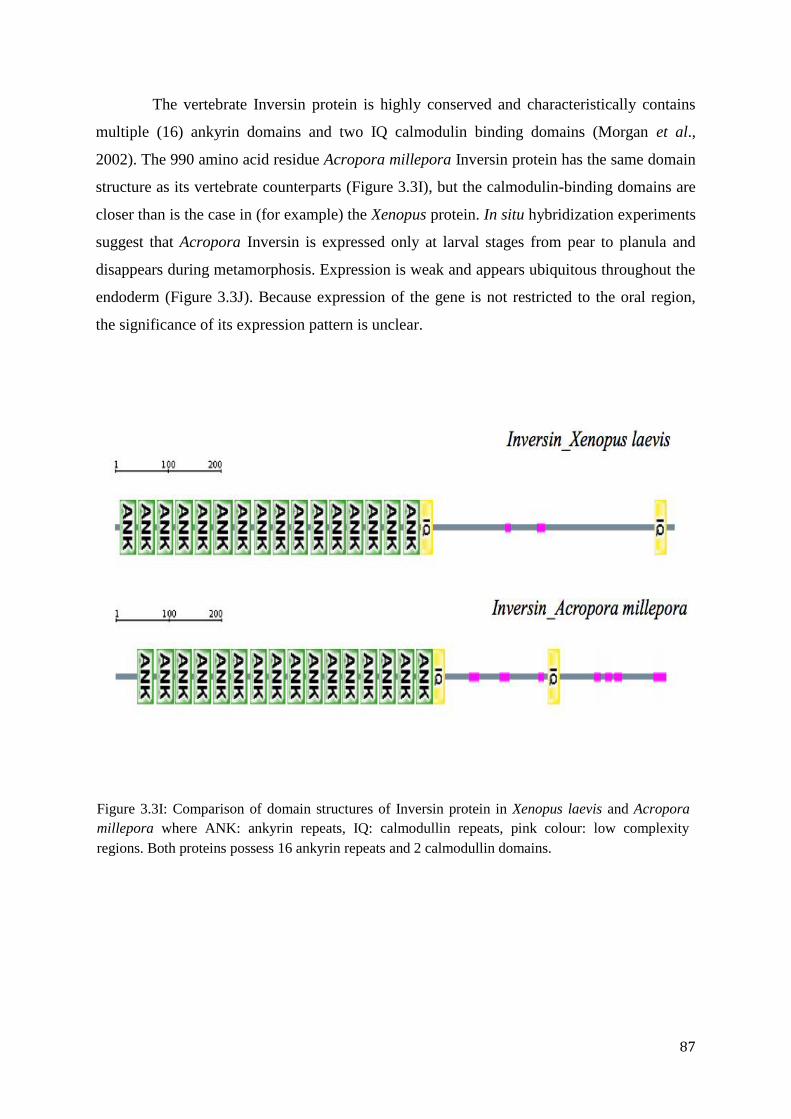
87
The vertebrate Inversin protein is highly conserved and characteristically contains
multiple (16) ankyrin domains and two IQ calmodulin binding domains (Morgan et al.,
2002). The 990 amino acid residue Acropora millepora Inversin protein has the same domain
structure as its vertebrate counterparts (Figure 3.3I), but the calmodulin-binding domains are
closer than is the case in (for example) the Xenopus protein. In situ hybridization experiments
suggest that Acropora Inversin is expressed only at larval stages from pear to planula and
disappears during metamorphosis. Expression is weak and appears ubiquitous throughout the
endoderm (Figure 3.3J). Because expression of the gene is not restricted to the oral region,
the significance of its expression pattern is unclear.
Figure 3.3I: Comparison of domain structures of Inversin protein in Xenopus laevis and Acropora
millepora where ANK: ankyrin repeats, IQ: calmodullin repeats, pink colour: low complexity
regions. Both proteins possess 16 ankyrin repeats and 2 calmodullin domains.
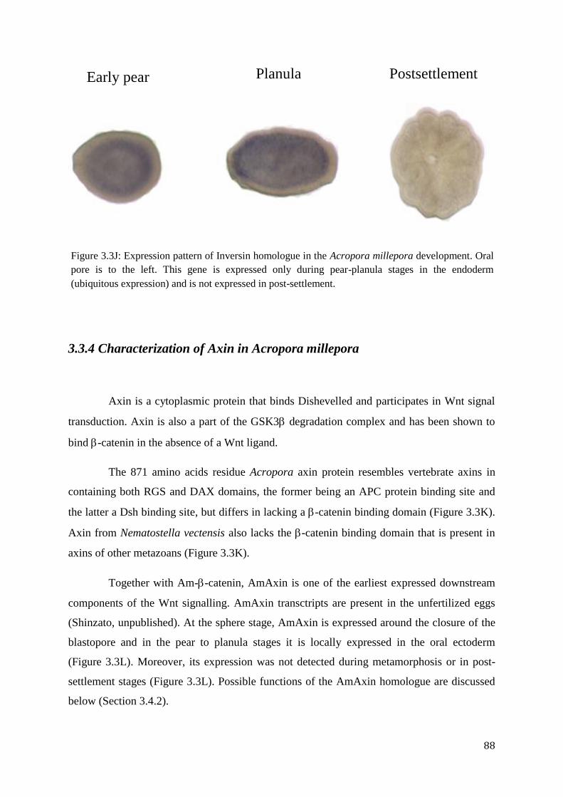
88
3.3.4 Characterization of Axin in Acropora millepora
Axin is a cytoplasmic protein that binds Dishevelled and participates in Wnt signal
transduction. Axin is also a part of the GSK3 degradation complex and has been shown to
bind -catenin in the absence of a Wnt ligand.
The 871 amino acids residue Acropora axin protein resembles vertebrate axins in
containing both RGS and DAX domains, the former being an APC protein binding site and
the latter a Dsh binding site, but differs in lacking a -catenin binding domain (Figure 3.3K).
Axin from Nematostella vectensis also lacks the -catenin binding domain that is present in
axins of other metazoans (Figure 3.3K).
Together with Am--catenin, AmAxin is one of the earliest expressed downstream
components of the Wnt signalling. AmAxin transctripts are present in the unfertilized eggs
(Shinzato, unpublished). At the sphere stage, AmAxin is expressed around the closure of the
blastopore and in the pear to planula stages it is locally expressed in the oral ectoderm
(Figure 3.3L). Moreover, its expression was not detected during metamorphosis or in post-
settlement stages (Figure 3.3L). Possible functions of the AmAxin homologue are discussed
below (Section 3.4.2).
Figure 3.3J: Expression pattern of Inversin homologue in the Acropora millepora development. Oral
pore is to the left. This gene is expressed only during pear-planula stages in the endoderm
(ubiquitous expression) and is not expressed in post-settlement.
Early pear Planula Postsettlement
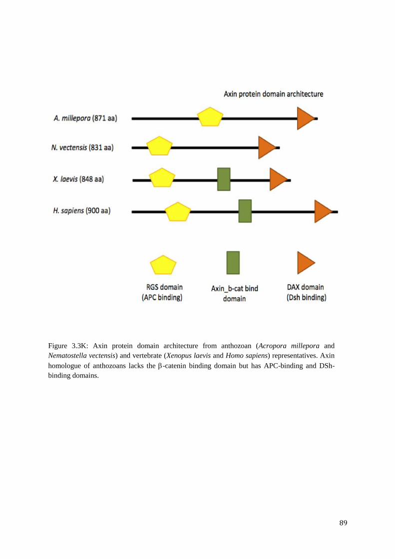
89
Figure 3.3K: Axin protein domain architecture from anthozoan (Acropora millepora and
Nematostella vectensis) and vertebrate (Xenopus laevis and Homo sapiens) representatives. Axin
homologue of anthozoans lacks the -catenin binding domain but has APC-binding and DSh-
binding domains.
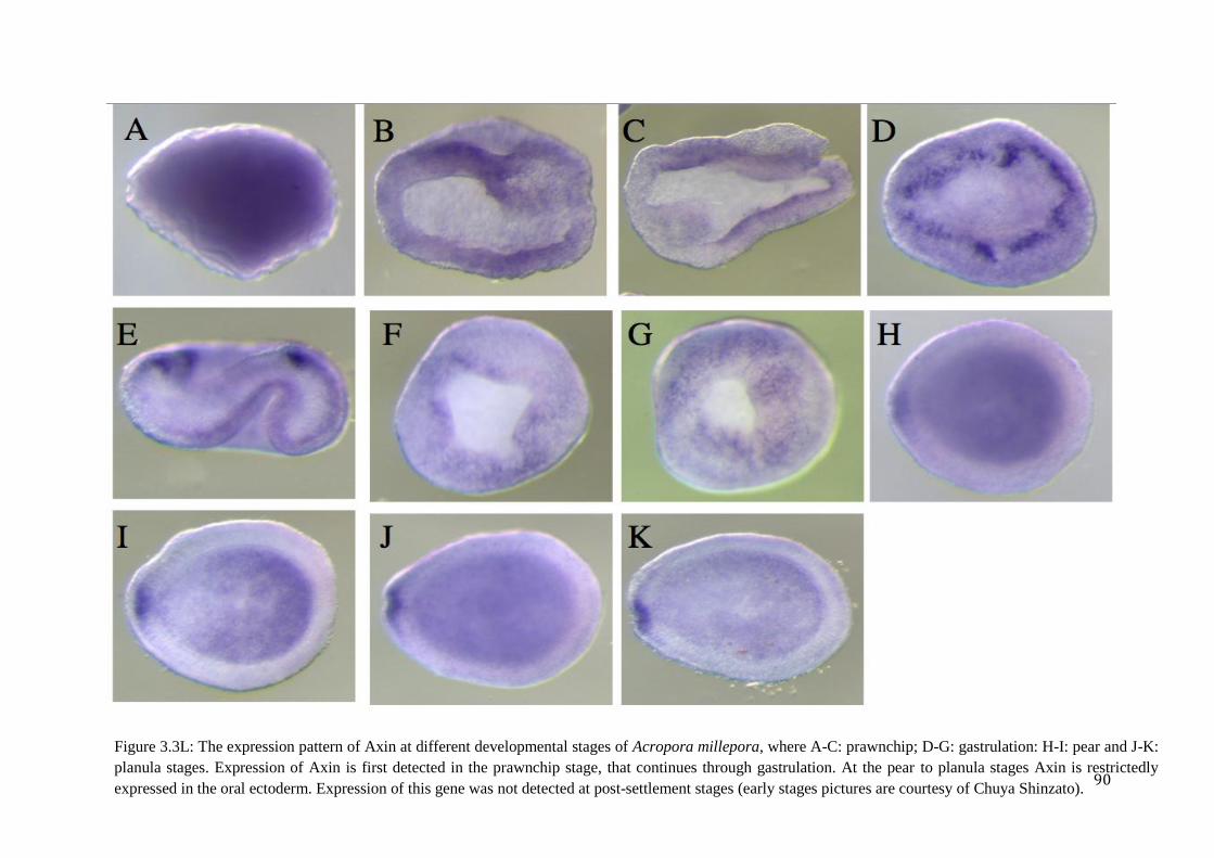
90
Figure 3.3L: The expression pattern of Axin at different developmental stages of Acropora millepora, where A-C: prawnchip; D-G: gastrulation: H-I: pear and J-K:
planula stages. Expression of Axin is first detected in the prawnchip stage, that continues through gastrulation. At the pear to planula stages Axin is restrictedly
expressed in the oral ectoderm. Expression of this gene was not detected at post-settlement stages (early stages pictures are courtesy of Chuya Shinzato).
91
3.4 Discussion
3.4.1 The mechanisms of Wnt signalling in Acropora millepora are different
from hydrozoan and other anthozoan representatives
The developmental deployment of Wnt signalling mechanisms in Acropora millepora
appears to differ substantially not only from what has been observed in hydrozoans such as
Hydra magnipapillata, but also from a representative of the same class, the sea anemone
Nematostella vectensis (Anthozoa). In Hydra, seven Wnt genes (HyWnt1, -3, -9/10a, 9/10c, -
11, and -16) are continuously expressed in the hypostome, maintaining head organizer
activity (Lengfeld et al., 2009). All of these, including HyWnt3, are expressed when a new
head organizer is formed during head regeneration or budding (Lengfeld et al., 2009)
indicating that each of the Wnt genes may play a role in the initial stages of bud formation
(Lengfeld et al., 2009). In Nematostella vectensis, ten Wnt genes (NvWntA, -1, -2, -3, -5, -7,
-8, -10, -11, -16) are expressed in the primary polyp. When compared to our model the coral
Acropora millepora, while majority of Wnts are expressed in the Nematostella and Hydra
primary polyps, expression of only a few Wnt genes is seen immediately at the stage of post-
settlement in Acropora (Chapter 2). Therefore, the Wnt signalling system in Acropora is
inhibited at the metamorphosis stage, and does not appear to be important in early post-
settlement life, although nothing is known about later stages. On the other hand, the time at
which zygotic Wnt expression begins is quite different between Nematostella and Acropora;
in the latter the majority of Wnt genes are initially expressed during larval life, whereas in
Nematostella the expression of WntA, -1, -2, -4, -11 was detected as early as the cleavage-
gastrula stages.
In another cnidarian, Clytia hemisphaerica, canonical Wnt signalling has also been
shown to play important roles during early embryogenesis. Momose and Houliston (2007)
identified two Wnt receptors, CheFzd1 and CheFzd3, that are localised at opposite poles of
the egg. Morpholino-mediated inhibition experiments showed that CheFzd1, localized at the
animal pole, activates the canonical Wnt pathway, promotes oral fates including gastrulation,
and may also mediate global polarity in the ectoderm. In the same study it was shown that the
Clytia -catenin homologue is stabilized in the oral half of the embryo, making the animal
92
pole the centre for canonical Wnt signalling (Momose and Houliston, 2007). In addition to
downstream Wnt signalling components, a maternally coded Wnt ligand, CheWnt3, have also
been identified in Clytia and is localised to the future oral side of the egg (Momose et al.,
2008). The studies indicate that the Wnt signalling is involved in early embryonic
development in some cnidarians, whilst in Acropora this does not appear to be true.
Moreover, whereas in the Nematostella larva Wnt genes are expressed in nested and
overlapping patterns that are reminiscent of the Hox genes in bilaterians (Kusserow et al.,
2005; Figure 2.4A), nothing like this is seen in Acropora. Instead, a few of the coral Wnts
exhibit orally-restricted (ectodermal) expression patterns and majority are expressed in the
endoderm (detailed discussion is given in Chapter 2).
As well as the distribution and timing of expression of Wnt ligands differing in
Acropora from other cnidarians, the expression patterns of many of the other signalling
system components vary substantially, suggesting quite different use of Wnt pathways. In
Hydra, canonical Wnt signalling is thought to be the primary influence for the maintenance
of head organizer activity and the process of regeneration (Lee et al., 2006). Hydra Wnts are
expressed at the hypostome, whereas the transcriptional components of Wnt/-catenin
signalling HyTcf and -catenin are more broadly distributed along the oral-aboral axis with
higher levels of expression in the hypostome (Lengfeld et al., 2009; Guder et al., 2006;
Broun et al., 2005; Hobmayer et al., 2000). Although -catenin is expressed uniformly
throughout adult Hydra (Hobmayer et al., 2000), only in the hypostome it is located in the
nucleus (Broun et al., 2005), where many (seven out of eleven) Hydra Wnt genes are also
expressed (Lengfeld et al., 2009). The overlapping expression of Wnts and -catenin together
with nuclear localization of -catenin in the Hydra hypostome suggest that in this region the
canonical Wnt signalling branch is active. The fact that transgenic Hydra expressing a
modified -catenin gene driven by the actin promoter form heads and secondary axes in
multiple locations along the body column (Gee et al., 2010) illustrates the importance of
canonical Wnt signalling in Hydra head organizer activity.
Studies of the colonial hydrozoan, Hydractinia echinata provide more evidence for
the importance of canonical Wnt signalling in the oral region of hydrozoans (Duffy et al.,
2010). In Hydractinia the activation of Wnt downstream events during metamorphosis
resulted in complete oralization of the animals and repression of aboral structures. Under
these conditions the expression of Wnt3, Tcf and Brachyury was upregulated. These lines of
93
evidence suggest that in Hydractinia Wnt signalling promotes head formation and represses
the formation of stolons (aboral structures) (Duffy et al., 2010).
Whilst the expression of the Wnt ligands has been studied throughout development in
Nematostella, the distribution of β-catenin has been reported only during early
embryogenesis. At the cleavage stage, stabilization of β-catenin in one hemisphere allows
nuclear localisation of the protein in cells at the site of gastrulation which marks the future
oral end of the animal, where it is able to act as a transcriptional co-activator
(Wikramanayake et al., 2003). The studies of Wikramanayake have showed that induction of
Nv--catenin nuclear localisation with lithium chloride results in elongated planulae
defective for the ability to make the ectodermal components of the pharynx and tentacles.
Therefore, planulae treated with LiCl exhibit overproliferation of endodermal tissue
(Wikramanayake et al., 2003). These studies imply that Wnt/β-catenin signalling pathway is
responsible for the endoderm cell fate specification during early embryogenesis of
Nematostella vectensis. Another study suggests that the induction of canonical Wnt signalling
is sufficient for the oral tissue fate during regeneration and embryogenesis in Nematostella
vectensis (Trevino et al., 2011). Kumburegama et al. (2011) showed that the Wnt/PCP-
mediated initial archenteron invagination is uncoupled from Wnt/-catenin dependent
endodermal cell fate specification during early embryogenesis of the sea anemone. These
studies indicate that the PCP pathway is active during embryogenesis in at least some
cnidarians and suggest that two Wnt signalling branches, (canonical and PCP) operate
independently during early embryogenesis of Nematostella vectensis.
In A. millepora, the expression patterns of WntA and Wnt2 resemble those of their sea
anemone orthologues, but the corresponding patterns for all of the other Wnts and β-catenin
are quite different. The majority of AmWnts are not expressed in staggered domains as in
Nematostella, nor are they concentrated at the oral pole as in Hydra. Rather, in Acropora two
non-canonical Wnts are expressed in the ectodermal oral region, the majority of the other
Wnts being more generally expressed (Chapter 2). The fact that β-catenin is co-expressed
with non-canonical Wnts in the oral pole (Ball, unpublished) is one of the lines of evidence
that led us to hypothesise that the oral pole in A. millepora is a centre for PCP non-canonical
Wnt signalling, with β-catenin functioning in convergent extension movements rather than
participating in the canonical β-catenin-dependent pathway in the oral region. Data from
other cnidarians with different developmental mechanisms might help clarify the relative
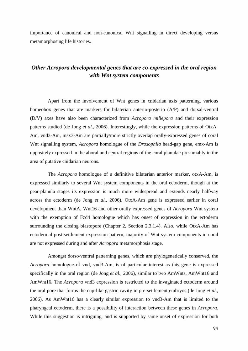
94
importance of canonical and non-canonical Wnt signalling in direct developing versus
metamorphosing life histories.
Other Acropora developmental genes that are co-expressed in the oral region
with Wnt system components
Apart from the involvement of Wnt genes in cnidarian axis patterning, various
homeobox genes that are markers for bilaterian anterio-posterio (A/P) and dorsal-ventral
(D/V) axes have also been characterized from Acropora millepora and their expression
patterns studied (de Jong et al., 2006). Interestingly, while the expression patterns of OtxA-
Am, vnd3-Am, msx3-Am are partially/more strictly overlap orally-expressed genes of coral
Wnt signalling system, Acropora homologue of the Drosophila head-gap gene, emx-Am is
oppositely expressed in the aboral and central regions of the coral planulae presumably in the
area of putative cnidarian neurons.
The Acropora homologue of a definitive bilaterian anterior marker, otxA-Am, is
expressed similarly to several Wnt system components in the oral ectoderm, though at the
pear-planula stages its expression is much more widespread and extends nearly halfway
across the ectoderm (de Jong et al., 2006). OtxA-Am gene is expressed earlier in coral
development than WntA, Wnt16 and other orally expressed genes of Acropora Wnt system
with the exemption of Fzd4 homologue which has onset of expression in the ectoderm
surrounding the closing blastopore (Chapter 2, Section 2.3.1.4). Also, while OtxA-Am has
ectodermal post-settlement expression pattern, majority of Wnt system components in coral
are not expressed during and after Acropora metamorphosis stage.
Amongst dorso/ventral patterning genes, which are phylogenetically conserved, the
Acropora homologue of vnd, vnd3-Am, is of particular interest as this gene is expressed
specifically in the oral region (de Jong et al., 2006), similar to two AmWnts, AmWnt16 and
AmWnt16. The Acropora vnd3 expression is restricted to the invaginated ectoderm around
the oral pore that forms the cup-like gastric cavity in pre-settlement embryos (de Jong et al.,
2006). As AmWnt16 has a clearly similar expression to vnd3-Am that is limited to the
pharyngeal ectoderm, there is a possibility of interaction between these genes in Acropora.
While this suggestion is intriguing, and is supported by same onset of expression for both
95
AmWnt16 and vnd3-Am genes, the expression of vnd3-Am is maintained following
metamorphosis and AmWnt16 gene is not expressed at the Acropora post-settlement stages
(Chapter 2).
Of the three identified Msx/msh genes in A. millepora (de Jong et al., 2006), msx3-
Am is ectodermally expressed during larval development. Whereas the expression of this
gene appears to be absent in the oral pore, it is broadly expressed in the ectoderm covering
approximately two-thirds of the embryo. While this gene may interact with AmWnt2 based
on overlapping expression patterns, it is more likely that it functions independently of Wnt
signalling system in coral.
In summary, the growing body of data on both Wnt and homeobox genes propose
that they function synergistically during coral axis specification. Moreover, overlapping
expression patterns for some genes like vnd3-Am and Wnt16 may imply possible interaction
and co-option of these genes under same regulation mechanism however further studies are
needed to shed light on this hypothesis. As, in most cases the spatial and temporary
expression patterns of homeobox genes are diverse and not strictly overlapping the majority
of Wnt genes patterns in coral, it may be possible that homeobox genes and Wnt signalling
genes work independently but in synergy for coral axis specification.
3.4.2 The oral region – a non-canonical signalling centre during larval life of
Acropora
AmWntA and AmWnt16, the two ligands that are exclusively expressed in the
ectoderm in the oral region of Acropora planulae, belong to subfamilies that primary function
in non-canonical Wnt signalling in metazoans. While the function of WntA remains poorly
understood and as this gene is missing from vertebrates, Wnt16 is primarily associated with
planar cell polarity signalling. By activating the JNK (Jun NH2-terminal kinase) pathway
(Muy-Teck et al., 2007), Wnt16 inhibits canonical Wnt signalling because JNK blocks the
accumulation of -catenin in the nucleus (Liao et al., 2006). Canonical and non-canonical
Wnt pathways typically show reciprocal inhibition (Liao et al., 2006), hence the suppression
of Wnt16 expression by alsterpaullone provides further support for the hypothesis that the
oral pore is a centre of Wnt-PCP signalling. A similar function has been proposed for
96
HyWnt16 in blocking the canonical Wnt signalling in the lower part of the Hydra hypostome
(Bode, 2009). Moreover, compared with seven canonical Hydra Wnts which are expressed in
the hypostome (discussed earlier, Lengfeld et al., 2009), non-canonical Wnt pathway
members of Hydra are expressed at the distal tip of the tentacles, (HyWnt5) and at the base of
the tentacles (HyWnt8, HyFzd2 and HyDsh) but not in the hypostome (Philipp et al., 2009).
These studies lead to conclusion that canonical and non-canonical Wnt signalling centres
may operate in different parts of the Hydra body. Similarly, in Acropora oral region the Wnt
PCP non-canonical pathway may be active with simultaneous inhibition of Wnt canonical
branch, so two active Wnt centres are not overlapping.
The expression of several other Wnt-pathway components is restricted to the oral
ectoderm in Acropora larvae - for example, Fzd4, which may act as a receptor for AmWntA
or AmWnt16 ligand (Chapter 2). Axin, which also has this restricted expression pattern is a
dual function protein (Zhang et al., 2001). On one hand, when complexed with APC, GSK3
and CKI kinases, Axin functions in canonical Wnt signalling (Zhang et al., 2001). On the
other hand, Axin can form complexes with MEKK1 and function in JNK activation (Zhang et
al., 2001). Because AmAxin lacks the -catenin binding domain that is present in bilaterian
Axin proteins (Figure 3.3.K), it is unable to activate canonical Wnt signalling. However,
AmAxin does contain the MEKK1 binding site that is critical for the activation of JNK
kinase in the planar cell polarity pathway (Zhang et al., 2001). Being expressed in the oral
region of Acropora larvae, the domain structure of coral Axin homologue (Figure 3.3.K)
provides additional evidence for the active PCP signalling in the Acropora oral region of
larval stages.
Groucho (Gro) is a co-repressor of -catenin-dependent gene expression (Chen et
al., 1999; Cavallo et al., 1998), and is again expressed in the oral region (Figure 3.3F),
lending further support to the hypothesis that the oral region is a centre for non-canonical
rather than canonical Wnt signalling.WIF (WNT Inhibitory Factor) is also expressed in the
same region, but in this case the function is less clear. The uncertainty stems from the fact
that vertebrate WIF can inhibit both canonical and non-canonical Wnt signalling by binding
ligands and preventing their interactions with the Fzd receptors (Kawano and Kypta, 2003).
More details of the A. millepora Wnt inhibitors are given in Section 3.4.3.
A. millepora -catenin homologue exhibits a restricted oral expression pattern and its
mRNA is co-expressed with two non-canonical Wnts (WntA and Wt16) in the oral region of
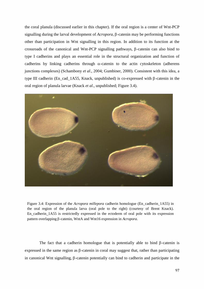
97
the coral planula (discussed earlier in this chapter). If the oral region is a center of Wnt-PCP
signalling during the larval development of Acropora, -catenin may be performing functions
other than participation in Wnt signalling in this region. In addition to its function at the
crossroads of the canonical and Wnt-PCP signalling pathways, -catenin can also bind to
type I cadherins and plays an essential role in the structural organization and function of
cadherins by linking cadherins through -catenin to the actin cytoskeleton (adherens
junctions complexes) (Schambony et al., 2004; Gumbiner, 2000). Consistent with this idea, a
type III cadherin (En_cad_1A55, Knack, unpublished) is co-expressed with -catenin in the
oral region of planula larvae (Knack et al., unpublished; Figure 3.4).
The fact that a cadherin homologue that is potentially able to bind -catenin is
expressed in the same region as -catenin in coral may suggest that, rather than participating
in canonical Wnt signalling, -catenin potentially can bind to cadherin and participate in the
Figure 3.4: Expression of the Acropora millepora cadherin homologue (En_cadherin_1A55) in
the oral region of the planula larva (oral pole to the right) (courtesy of Brent Knack).
En_cadherin_1A55 is restrictedly expressed in the ectoderm of oral pole with its expression
pattern overlapping -catenin, WntA and Wnt16 expression in Acropora.
98
formation of adherens junctions complexes in the oral region of Acropora. More functional
data and analysis of the protein interactions are needed to confirm that hypothesis.
3.4.3 No simple relationship between Wnt ligands and inhibitors in Acropora
Although several classes of Wnt antagonists known from vertebrates are missing
from the “typical” ecdysozoans Drosophila and Caenorhabditis, representatives of Dkk, the
more generally distributed sFRP (secreted Frizzled-related proteins) and WIF families have
been cloned from Hydra and Nematostella (Guder et al., 2006a; Guder et al., 2006b; Lee et
al., 2006; Technau et al., 2005). Two dickkopf homologues (belonging to the Dkk3 and
dkk1/2/4 subfamilies) have been identified in both Hydra and Nematostella (Guder et al.,
2006a; Guder et al., 2006b; Fedders et al., 2004), and in both organisms dkk1/2/4 is
expressed at the aboral end - i.e. the opposite point in the axis to that where Wnt expression is
concentrated (Lee et al., 2006; Guder et al., 2006b), which is consistent with a role as a Wnt
antagonist. To date, no Dkk1/2/4 has been identified in A. millepora, however, a Dkk3
homologue is expressed in the aboral endoderm in coral in pear-planula stages of
development (Figure 3.3H).
Although dkk proteins were identified as Wnt antagonists (Monaghan et al., 1999;
Glinka et al., 1998), Hydra Dkk3 is a divergent protein that lacks this ability, and is thought
to function primarily in neuronal differentiation as it is expressed in differentiating
nematocytes (Fedders et al., 2004). As many Wnt proteins of Acropora are also expressed in
the same region as its Dkk3 homologue, AmDkk3 protein may not be able to antagonize the
Wnt signalling system in coral. Moreover, the Acropora Dkk3 protein is very divergent from
Hydra Dkk3 homologue with less than 50% in sequence similarity and its function still
remains poorly understood and cannot be related to the Hydra Dkk3 gene.
AmDkk3, the co-repressor Groucho and the extracellular WIF protein (both
discussed earlier) are the only candidate Wnt system inhibitors so far identified in Acropora.
Although a single sFRP sequence of the sFRP 1/2/5 type has been identified in Nematostella
vectensis (Lee et al., 2006), to date no sFRP sequences have been found in coral.
99
In summary, we were unable to show a clear relationship between Wnt ligands and
their antagonists in Acropora millepora due to small number of inhibitors that we retrieved
and their ambiguous expression patterns at larval stages. Further search for Acropora Wnt
system inhibitors, analysis of their expression patterns and functional studies are needed to
provide more insights into the mechanisms of inhibition and/or regulation of Wnt signalling
pathways in Acropora millepora.
100
Chapter 4
General conclusions
4.1 No simple relationship between Wnt molecular mechanisms of
different cnidarian representatives
The Wnt signalling system has previously been studied in both hydrozoan and
anthozoan cnidarians (Chapter 1, 2 & 3). These studies have demonstrated different roles for
the Wnt system in the anthozoan Nematostella vectensis and the hydrozoans, Hydra
magnipapillata and Clytia hemisphaerica (Lengfeld et al., 2009; Momose et al., 2008, 2007;
Kusserow et al., 2005). Whilst differences between cnidarian classes are perhaps to be
expected, the differences between Acropora and Nematostella reported here were beyond our
expectations.
The main function of N. vectensis Wnt genes was suggested to be axis patterning
(Kusserow et al., 2005). The detailed description of sea anemone Wnt complement is given in
Chapter 2. Moreover, the condensation of Wnts in the oral region of the Nematostella embryo
is met by the co-expression of transcription factors, including Brachyury, Notch and Caudal,
suggesting that the Nv-Wnts cluster defines a blastoporal signalling centre (Kusserow et al.,
2005; Holland, 2002; Lengyel and Iwaki, 2002) that is reminiscent of the Hydra head
organizer region. In Hydra magnipapillata, seven Wnt genes are constitutively expressed in
the hypostome with the main function of maintaining the head organizer and regeneration
(Lengfeld et al., 2009, also see Chapter 3). It was also observed that these seven Hm-Wnt
genes are expressed in the bud indicating that each of the Wnt genes may play role in the
initial stages of the bud formation that is important for asexual reproduction (Lengfeld et al.,
2009; Lee et al., 2006a). Moreover, functional studies of the main component of the canonical
Wnt signalling pathway, -catenin, of H. magnipapillata, have shown that the hypostome is
the active centre of the Wnt canonical signalling, where -catenin interacts with the TCF/LEF
transcription factors in the nucleus and triggers the expression of Wnt responsive genes
(Chapter 3). Further, pioneering studies of Clytia hemisphaerica (Momose et al., 2008;
Momose and Houliston, 2007) have proposed a new role for Wnt components (CheFzd1,
CheFzd3 and CheWnt3) as activating determinants during the early embryogenesis of this
hydrozoan.
101
The studies of A. millepora, reported here, suggest that the oral region of the coral
planula is a centre for non-canonical Wnt signalling system, particularly of the planar cell
polarity (PCP) Wnt type. Several lines of evidence support this idea. First, expression of the
Acropora Wnt16 homologue is restricted to the oral ectoderm of coral larvae. Wnt16 is
viewed as a non-canonical Wnt – for example, Muy-Teck Teh (2007) showed that in human
epidermal keratinocytes Wnt16 is responsible for PCP signalling. Second, the canonical Wnt
pathway co-repressor Groucho shares the same orally-restricted expression pattern as Am-
Wnt16. Third, an Axin homologue is also expressed in the same region. The absence of a -
catenin binding domain in Acropora Am-Axin (Chapter 3, Section 3.3.4) suggests that the
main function of Am-Axin in the oral region is the activation of cJUN kinase, a crucial
downstream component of the PCP pathway (Wen et al., 2005; Zhang et al., 2001). Although
circumstantial, these various lines of evidence highlight the possibility that the oral region
acts as a Wnt-PCP signalling centre during early coral development.
4.2 Future directions
It is highly possible that the Wnt signalling network, particularly its planar cell
polarity branch is actively involved in the axis patterning of the coral Acropora millepora. In
order to better understand the evolution of the Wnt gene family in metazoans, it is important
to characterize the full Wnt complement of A. millepora including all downstream
components of the major branches of Wnt signalling. As the recently sequenced genome of A.
millepora (http://www.agrf.org.au/), A. digitifera (Shinzato et al., 2011) and A. palmata
(http://genome.wustl.edu/) become available for analysis, more components of the Wnt
signalling branches and their targets can be identified and compared.
After the identification of the Wnt network components in A. millepora, the next
steps will be functional analyses of all its components. The most powerful approaches for
these analyses are gain-of-function and loss-of function experiments using over-expression
constructs or morpholino antisense oligonucleotides, respectively. As morpholinos can
completely knock down expression of target sequences, it became a widely-used
developmental technique to assess the function of various genes in cnidarians. Morpholino-
mediated knockdown experiments have been successfully performed in Nematostella and
Clytia (Saina et al., 2009; Momose et al., 2008; Rentzsch et al., 2008; Momose et al., 2007).
Saina et al., 2009 studied TGF-β signalling pathway in Nematostella and using morpholino
knockdowns revealed that the function of BMP signalling is required for symmetry breaking
102
of bmp and chordin expression during gastrulation (Saina et al., 2009). Momose studies
(2008) showed that a maternally localized Wnt ligand (CheWnt3) is required for active axis
patterning in the cnidarian Clytia hemisphaerica. The establishment of morpholino
knockdown experiments in Acropora can help to investigate the regulation of complex coral
Wnt network as well as aid in deciphering the function of all Wnt system components.
Whereas for Nematostella and Clytia the delivery of morpholinos through microinjection
proved accessible, it is not yet possible for coral partly due to large amounts of yolk in A.
millepora cells (Prof. Eldon Ball, personal communication). Therefore, other methods of
delivery should be explored including the use of the cationic polymer JetPEITM
transfection
reagent (http://www.biocompare.com/). Another possible approach for gene knockdown
would be RNA interference (RNAi) introducing double-stranded (ds) RNA into coral cells via
electroporation techniques. This method was introduced in Hydra (Smith et al., 2000;
Lohmann et al., 1999) and further developed by a simultaneous knock-down of the green
fluorescent protein (GFP) as a control (Khalturin et al., 2008). The potential of this technique
on anthozoan models, particularly on A. millepora, has not been investigated yet.
A new powerful approach to functional analyses of the Acropora Wnt system
components and in particular Wnt inhibitors might be the establishment of a transgenic
system: cnidarian genes expressed in Xenopus laevis embryos. This method was successfully
used in Hydra (Wittlieb et al., 2006; Guder et al., 2006b), N. vectensis (Renfer et al., 2010)
and recently in Hydractinia (Kunzel et al., 2010). The group of Guder (2006b) demonstrated
by heterologous expression of Hydkk1/2/4 in Xenopus that Hydkk1/2/4 behaved as Xdkk1 in
inhibiting canonical Wnt signalling. Thus, heterologous expression of Acropora Wnt system
components in Xenopus may help to unravel their putative function in Cnidaria.
In addition to the successful use of pharmacological GSK3 inhibitor, alsterpaullone,
it will be of high importance to research other possible inhibitors of Wnt pathway
components. A potential, and available candidate might be SP600125 (Sigma-Aldrich), which
is an anthrapyrazolone inhibitor of Jun N-terminal kinase (part of the planar cell polarity
pathway). This particular inhibitor is of great interest as the non-canonical, PCP Wnt branch
is suggested to play one of the main roles in A. millepora axis patterning according to my
studies.
A priority for further study of Wnt system components in A. millepora is researching
the role of -catenin. My pioneering attempts to decipher the role of this protein in the oral
region of Acropora involved immunochemical investigation of the distribution of the -
103
catenin protein in the cells of Acropora larvae. However, these preliminary attempts were
inconclusive most likely as a consequence of poor reactivity of the heterologous antibody. As
in situ experiments indicate that Acropora -catenin mRNA is restricted to the oral region
during larval life, I hypothesize that in this region -catenin functions in the PCP pathway;
rather than participating in canonical Wnt signalling in nuclei of the oral region. I hypothesise
that Am--catenin is exported to the cytoplasm, and is enriched close to cell membranes
where it interacts with cadherin in adherens junctions (AJ) complexes. For my initial attempt I
used commercially available rabbit polyclonal antibodies (Abcam, ab47426) without success.
Possible reasons might be low specificity of this antibody for Am--catenin binding, despite
being designed for a very conserved region of -catenin around the phosphorylation site of
serine 33. Further immunochemistry experiments with higher antibody specificity are needed
to decode the localization of Am--catenin protein in the oral region of larvae. It will provide
one of the main evidences for my hypothesis of non-canonical pathways, particularly PCP,
playing a major role during A. millepora development.
In summary, my studies of the A. millepora Wnt signalling system revealed many
intriguing differences between the coral and other cnidarians, but there is a real need for
functional studies of key components. Moreover, the interpretation of gene expression
patterns and other molecular data are complicated by the lack of information concerning the
cell biology of coral development. Acropora is by far the best characterized coral at the
molecular level, but there is still much to learn concerning basic biological processes in these
ecologically important animals.
104
References
Adamska, M., Degnan, S.M., Green, K.M., Adamski, M., Craigie, A., Larroux, C.,
Degnan, B.M. (2007). Wnt and TGF- Expression in the Sponge Amphimedon
queenslandica and the Origin of Metazoan Embryonic Patterning. PLoS ONE
2, e1031.
Adamska, M., Larroux, C., Adamski, M., Green, K., Lovas, E., Koop, D., Richards,
G.S., Zwafink, C., and Degnan, B.M. (2010). Structure and expression of
conserved Wnt pathway components in the demosponge Amphimedon
queenslandica. Evolution & Development, 12, 494-518.
Agners, S. and Moon, R.T. (2009). Proximal events in Wnt signal transduction. Nat
Rev Mol Cell Biol. 10, 468-77
Altmann, C.R., Brivanlou, A.H., Lawrence, D.Ea K.W.J. (2001). Neural patterning in
the vertebrate embryo. International Review of Cytology 203, 447-482.
Amerongen, van R. and Nusse, R. (2009). Towards an integrated view of Wnt
signaling in development. Dev 136, 3205-3214.
Arendt, D., Technau, U., and Wittbrodt, J. (2001). Evolution the bilaterian larval
foregut. Nature 409, 81-85.
Ball, E.E., Hayward, D.C., Reece-Hoyes, J.S., Hislop, N.R., Samuel, G., Saint, R.,
Harrison, P.L., Miller, D.J. (2002). Coral development: from classical
embryology to molecular control. Intl Jour Dev Biol 46, 671-678.
Ball, E.E., Hayward, D.C., Saint, R., and Miller, D.J. (2004). A simple plan –
cnidarians and the origins of developmental mechanisms. Nature Reviews
Genetics 5, 567-577.
Ball, E.E., de Jong, D.M., Schierwater, B., Shinzato, C., Hayward, D.C., Miller, D.J.
(2007). Implications of cnidarian gene expression patterns for the origins of
bilaterality – is the glass half full or half empty? Integr Comp Biol 47, 701-
711.
Batstock, R., Strutt, H., and Strutt, D. (2003). Strabismus is asymmetrically localised
and binds to Prickle and Dishevelled during Drosophila planar patterning.
Development 130, 3007-3014.
Bode, H.R. (2009). Axial Patterning in Hydra. Cold Spring Harbor Laboratory Press.
1, a000463 Bode, H. (2011). Axis Formation in Hydra. Annual Rev Genet 45, 105-17.
Boero, F., Schierwater, B., and Piraino, S. (2007). Cnidarian milestones in metazoan
evolution. Integr Comp Biol 47, 693–700.
Bolós, V., Grego-Bessa, J., de la Pompa, J.L. (2007). Notch signaling in
105
development and cancer. Endocr. Rev. 28, 339–63. Böttcher, R.T., Niehrs, C. (2005). Fibroblast growth factor signaling during early
vertebrate development. Endocr. Rev. 26, 63–77.
Boutros, M., Paricio, N., Strutt, D.I., and Mlodzik, M. (1998). Dishevelled activates
JNK and discriminates between JNK pathways in planar polarity and wingless
signaling. Cell 94, 109-118.
Bridge, D., Cunningham, CW, Scierwater, B., Desalle, R., Buss, L.W. (1992) Class-
Level Relationships in the Phylum Cnidaria: Evidence from Mitochondrial
Genome Structure. Proceeding of the National Academy of Sciences of the
United States of America 18, 8750-8753.
Broun, M., Bode, H.R. (2002). Characterization of the head organizer in Hydra.
Development 129, 875-84.
Broun, M., Gee, L., Reinhards, B., Bode, H.R. (2005). Formation of the head
organizer in Hydra involves the canonical Wnt pathway. Development 132,
2907-2916.
Cartwright, P., and Collins, A.G. (2007). Fossils and phylogenies: integrating multiple
lines of evidence to investigate the origin of early major metazoan lineages.
Integr Comp Biol 47, 744–751.
Cavallo, R.A., Cox, R.T., Moline, M.M., Roose, J., Polevoy, G.A., Clevers, H., Peifer, M., Bejsovec, A. (1998). Drosophila Tcf and Groucho interact to repress Wingless signalling activity. Nature 395, 604-608.
Chan, G., and Ye-Guang, C. (2010). Dishevelled: The hub of Wnt signaling. Cellular
Signalling. 22, 717-727.
Chang, C.H., Jiang, T.X., Lin, C.M., Burrus, L.W., Chuong, C.M., and Widelitz, R.
(2004). Distinct Wnt members regulate the hierarchical morphogenesis of skin
regions (spinal tract) and individual feathers. Mech Dev 121, 157-171.
Chen, G., Fernandez, J., Mische, S., Courey, A.J. (1999). A functional interaction
between the histone deacetylase Rpd3 and the corepressor groucho in
Drosophila development. Genes Dev 13, 2218-30.
Chiori, R., Jager, M., Denker, E., Wincker, P., Da Silva, C., Guyader, H.L., Manuel,
M., and Queinnec, E. (2009). Are Hox Genes Ancestrally Involved in Axial
Patterning? Evidence from the Hydrozoan Clytia hemisphaerica (Cnidaria).
PLoS ONE 4, e4231.
Chodankar, R., Chang C-H., Yue, Z., Jiang, T-X., Suksaweang, S., Burrus, L.W.,
Chuong, C-M. and Widelitz, R.B. (2003). Shift of localized growth zones
contributes to skin appendage morphogenesis: role of the Wnt/-catenin
pathway. Journal of Investigative Dermatology 120, 20-26.
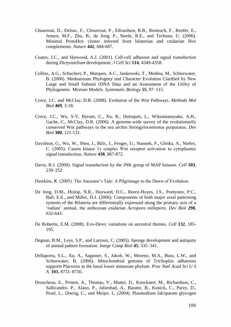
106
Chourrout, D., Delsuc, F., Chourrout, P., Edvardsen, R.B., Rentzsch, F., Renfer, E.,
Jensen, M.F., Zhu, B., de Jong, P., Steele, R.E., and Technau, U. (2006).
Minimal ProtoHox cluster inferred from bilaterian and cnidarian Hox
complements. Nature 442, 684-687.
Coates, J.C., and Harwood, A.J. (2001). Cell-cell adhesion and signal transduction
during Dictyostelium development. J Cell Sci 114, 4349-4358.
Collins, A.G., Schuchert, P., Marques, A.C., Jankowski, T., Medina, M., Schierwater,
B. (2006). Medusozoan Phylogeny and Character Evolution Clarified by New
Large and Small Subunit rDNA Data and an Assessment of the Utility of
Phylogenetic Mixture Models. Systematic Biology 55, 97–115.
Croce, J.C. and McClay, D.R. (2008). Evolution of the Wnt Pathways. Methods Mol
Biol 469, 3-18.
Croce, J.C., Wu, S-Y, Byrum, C., Xu, R., Duloquin, L., Wikramanayake, A.H.,
Gache, C., McClay, D.R. (2006). A genome-wide survey of the evolutionarily
conserved Wnt pathways in the sea urchin Strongylocentrotus purpuratus. Dev
Biol 300, 121-131.
Davidson, G., Wu, W., Shen, J., Bilic, J., Fenger, U., Stannek, P., Glinka, A., Niehrs,
C. (2005). Casein kinase 1γ couples Wnt receptor activation to cytoplasmic
signal transduction. Nature 438, 867-872.
Davis, R.J. (2000). Signal transduction by the JNK group of MAP kinases. Cell 103,
239–252
Dawkins, R. (2005). The Ancestor’s Tale: A Pilgrimage to the Dawn of Evolution.
De Jong, D.M., Hislop, N.R., Hayward, D.C., Reece-Hoyes, J.S., Pontynen, P.C.,
Ball, E.E., and Miller, D.J. (2006). Components of both major axial patterning
systems of the Bilateria are differentially expressed along the primary axis of a
‘radiate’ animal, the anthozoan cnidarian Acropora millepora. Dev Biol 298,
632-643.
De Robertis, E.M. (2008). Evo-Devo: variations on ancestral themes. Cell 132, 185-
195.
Degnan, B.M., Leys, S.P., and Larroux, C. (2005). Sponge development and antiquity
of animal pattern formation. Integr Comp Biol 45, 335–341.
Dellaporta, S.L., Xu, A., Sagasser, S., Jakob, W., Moreno, M.A., Buss, L.W., and
Schierwater, B. (2006). Mitochondrial genome of Trichoplax adhaerens
supports Placozoa as the basal lower metazoan phylum. Proc Natl Acad Sci U S
A. 103, 8751–8756.
Droucheau, E., Primot, A., Thomas, V., Mattei, D., Knockaert, M., Richardson, C.,
Sallicandro, P., Alano, P., Jafarshad, A., Baratte, B., Kunick, C., Parzy, D.,
Pearl, L., Doerig, C., and Meijer, L. (2004). Plasmodium falciparum glycogen
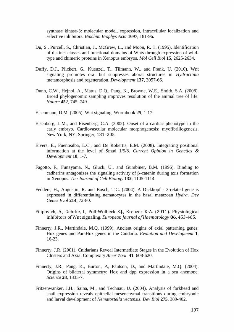
107
synthase kinase-3: molecular model, expression, intracellular localization and
selective inhibitors. Biochim Biophys Acta 1697, 181-96.
Du, S., Purcell, S., Christian, J., McGrew, L., and Moon, R. T. (1995). Identification
of distinct classes and functional domains of Wnts through expression of wild-
type and chimeric proteins in Xenopus embryos. Mol Cell Biol 15, 2625-2634.
Duffy, D.J., Plickert, G., Kuenzel, T., Tilmann, W., and Frank, U. (2010). Wnt
signaling promotes oral but suppresses aboral structures in Hydractinia
metamorphosis and regeneration. Development 137, 3057-66.
Dunn, C.W., Hejnol, A., Matus, D.Q., Pang, K., Browne, W.E., Smith, S.A. (2008).
Broad phylogenomic sampling improves resolution of the animal tree of life.
Nature 452, 745–749.
Eisenmann, D.M. (2005). Wnt signaling. Wormbook 25, 1-17.
Eisenberg, L.M., and Eisenberg, C.A. (2002). Onset of a cardiac phenotype in the
early embryo. Cardiovascular molecular morphogenesis: myofibrillogenesis.
New York, NY: Springer, 181–205.
Eivers, E., Fuentealba, L.C., and De Robertis, E.M. (2008). Integrating positional
information at the level of Smad 1/5/8. Current Opinion in Genetics &
Development 18, 1-7.
Fagotto, F., Funayama, N., Gluck, U., and Gumbiner, B.M. (1996). Binding to
cadherins antagonizes the signaling activity of -catenin during axis formation
in Xenopus. The Journal of Cell Biology 132, 1105-1114. Fedders, H., Augustin, R. and Bosch, T.C. (2004). A Dickkopf - 3-related gene is
expressed in differentiating nematocytes in the basal metazoan Hydra. Dev
Genes Evol 214, 72-80.
Filipovich, A., Gehrke, I., Poll-Wolbeck S.J., Kreuzer K-A. (2011). Physiological
inhibitors of Wnt signaling. European Journal of Haematology 86, 453-465. Finnerty, J.R., Martindale, M.Q. (1999). Ancient origins of axial patterning genes:
Hox genes and ParaHox genes in the Cnidaria. Evolution and Development 1,
16-23. Finnerty, J.R. (2001). Cnidarians Reveal Intermediate Stages in the Evolution of Hox
Clusters and Axial Complexity Amer Zool 41, 608-620.
Finnerty, J.R., Pang, K., Burton, P., Paulson, D., and Martindale, M.Q. (2004).
Origins of bilateral symmetry: Hox and dpp expression in a sea anemone.
Science 28, 1335-7.
Fritzenwanker, J.H., Saina, M., and Technau, U. (2004). Analysis of forkhead and
snail expression reveals epithelial-mesenchymal transitions during embryonic
and larval development of Nematostella vectensis. Dev Biol 275, 389-402.
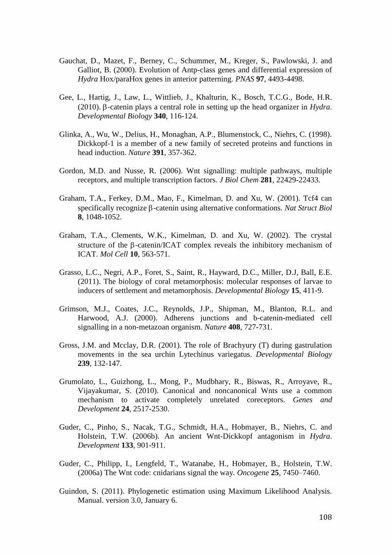
108
Gauchat, D., Mazet, F., Berney, C., Schummer, M., Kreger, S., Pawlowski, J. and
Galliot, B. (2000). Evolution of Antp-class genes and differential expression of
Hydra Hox/paraHox genes in anterior patterning. PNAS 97, 4493-4498.
Gee, L., Hartig, J., Law, L., Wittlieb, J., Khalturin, K., Bosch, T.C.G., Bode, H.R.
(2010). -catenin plays a central role in setting up the head organizer in Hydra.
Developmental Biology 340, 116-124.
Glinka, A., Wu, W., Delius, H., Monaghan, A.P., Blumenstock, C., Niehrs, C. (1998).
Dickkopf-1 is a member of a new family of secreted proteins and functions in
head induction. Nature 391, 357-362.
Gordon, M.D. and Nusse, R. (2006). Wnt signalling: multiple pathways, multiple
receptors, and multiple transcription factors. J Biol Chem 281, 22429-22433.
Graham, T.A., Ferkey, D.M., Mao, F., Kimelman, D. and Xu, W. (2001). Tcf4 can
specifically recognize -catenin using alternative conformations. Nat Struct Biol
8, 1048-1052.
Graham, T.A., Clements, W.K., Kimelman, D. and Xu, W. (2002). The crystal
structure of the -catenin/ICAT complex reveals the inhibitory mechanism of
ICAT. Mol Cell 10, 563-571.
Grasso, L.C., Negri, A.P., Foret, S., Saint, R., Hayward, D.C., Miller, D.J, Ball, E.E.
(2011). The biology of coral metamorphosis: molecular responses of larvae to
inducers of settlement and metamorphosis. Developmental Biology 15, 411-9.
Grimson, M.J., Coates, J.C., Reynolds, J.P., Shipman, M., Blanton, R.L. and
Harwood, A.J. (2000). Adherens junctions and b-catenin-mediated cell
signalling in a non-metazoan organism. Nature 408, 727-731.
Gross, J.M. and Mcclay, D.R. (2001). The role of Brachyury (T) during gastrulation
movements in the sea urchin Lytechinus variegatus. Developmental Biology
239, 132-147.
Grumolato, L., Guizhong, L., Mong, P., Mudbhary, R., Biswas, R., Arroyave, R.,
Vijayakumar, S. (2010). Canonical and noncanonical Wnts use a common
mechanism to activate completely unrelated coreceptors. Genes and
Development 24, 2517-2530.
Guder, C., Pinho, S., Nacak, T.G., Schmidt, H.A., Hobmayer, B., Niehrs, C. and
Holstein, T.W. (2006b). An ancient Wnt-Dickkopf antagonism in Hydra.
Development 133, 901-911.
Guder, C., Philipp, I., Lengfeld, T., Watanabe, H., Hobmayer, B., Holstein, T.W.
(2006a) The Wnt code: cnidarians signal the way. Oncogene 25, 7450–7460. Guindon, S. (2011). Phylogenetic estimation using Maximum Likelihood Analysis.
Manual. version 3.0, January 6.
109
Gumbiner, B.M. (2000). Regulation of cadherin adhesive activity. J Cell Biol 148,
399-404. Gurley, K.A., Rink, J.C., Alvarado, A.S. (2008). Beta-catenin defines head versus tail
identity during planarian regeneration and homeostasis. Science 319, 323-327. Janssen, R., Le Gouar, M., Pechmann, M., Poulin, F., Bolognesi, R., Scwagen, E.E.,
Hopfen, C., Colbourne, J.K., Budd, G. E., Brown, S.J., Prpic, N-M., Kosiol, C.,
Vervoort, M., Damen, W.G.M., Balavoine, G., McGregor, A.P. (2010).
Conservation, loss, and redeployment of Wnt ligands in protostomes:
implications for understanding the evolution of segment formation. BMC
Evolutionary Biology 10, 374.
Jenny, A., Reynolds-Kenneally, J., Das, G., Burnett, M., and Mlodzik, M. (2005).
Diego and Prickle regulate Frizzled planar cell polarity signaling by competing
for Dishevelled binding. Nat Cell Biol 7, 691-697. Hagadorn, J.W., Dott, Jr. R.H., and Damrow, D. (2002). Stranded on a Late Cambrian
shoreline: medusae from central Wisconsin, Geology 30, 147–150. Haribaskar, R., Putz, M., Schupp, B., Skouloudaki, K., Bietenbeck, A., Walz, G.,
Schafer, T. (2009). The planar cell polarity (PCP) protein Diversin translocates
to the nucleus to interact with the transcription factor AF9. Biochem Biophys
Res Commun 387, 212-7. Hayward, D.C., Miller, D.J., and Ball, E.E. (2004). Snail Expression during
Embryonic Development of the Coral Acropora: Blurring the
Diploblast/Triploblast Divide? Dev Genes Evol 241, 257-260. Hayward, D.C., Catmull, J., Reece-Hoyes, J.S., Berghammer, H., Dodd, H., Hann,
S.J., Miller, D.J., and Ball, E.E. (2001) Gene structure and larval expression of
cnox-2Am from the coral Acropora millepora. Dev.Genes Evol. 211, 10-19.
He, X. (2008). Cilia put a break on Wnt signalling. Nature Cell Biology 10, 11-13.
Heisenberg, C.P., Tada, M., Rauch, G.J., Saude, L., Concha, M.L., Geisler, R.,
Stemple, D.L., Smith, J.C., and Wilson, S.W. (2000). Silberblick/Wnt11
mediates convergent extension movements during zebrafish gastrulation. Nature
405, 76-81. Hobmayer, E. (1996). Identification of a Hydra homologue of the -
catenin/plakoglobin/armadillo gene family. Gene 172, 155-159. Hobmayer, B., Rentzsch, F., Kuhn, K., Happel, C.M., von Laue, C.C., Snyder, P.,
Rothbacher, U., and Holstein, T.W. (2000). WNT signalling molecules act in
axis formation in the diploblastic metazoan Hydra. Nature 407, 186-189.
110
Holland, L.Z. (2002). Heads or tails? Amphioxus and the evolution of anterior-
posterior patterning in deuterostomes. Developmental Biology 241, 209–228.
Holstein, T.W. (2008). Wnt signaling in cnidarians. Methods Mol Biol 469, 47-54.
Review.
Huelsken, J., Vogel, R., Brinkmann, V., Erdmann, B., Birchmeier, C., Birchmeier, W.
(2000). Requirement for beta-catenin in anterior-posterior axis formation in
mice. J Cell Biol 148, 567-578.
Huelsken, J., and Behrens, J. (2002). The Wnt signalling pathway. Journal of Cell
Science 115, 3977-3978.
Hulsken, J., Birchmeier, W. and Behrens, J. (1994). E-cadherin and APC compete for
the interaction with -catenin and the cytoskeleton. J Cell Biol 127, 2061-2069.
Ingham, P. W., Nakano, Y., and Seger C. (2011). Mechanism and function of
Hedgehog signaling across the metazoan. Nature Reviews Genetics 12, 393-406.
Irazoqui, J.E., Urbachm, J.M. and Ausubel, F.M. (2010). Evolution of host innate
defence: insights from Caenorhabditis elegans and primitive invertebrates, Nat
Rev Immunol 10, 47–58. Ishitani, T., Ninomiya-Tsuji, J., Nagai, S., Nishita, M., Meneghini, M., Barker, N.,
Waterman, M., Bowerman, B., Clevers, H., Shibuya, H., Matsumoto, K. (1999).
The TAK1-NLK-MAPK-related pathway antagonizes signalling between beta-
catenin and transcription factor TCF. Nature 24, 798-802. Ishitani, T., Kishida, S., Hyodo-Miura, J., Ueno, N., Yasuda, J., Waterman, M.,
Shibuya, H., Moon, R.T., Ninomiya-Tsuji, J., and Matsumoto, K. (2003). The
TAK1-NLK mitogen-activated protein kinase cascade functions in the
Wnt5a/Ca2+ pathway to antagonize Wnt/beta-catenin signaling. Mol Cell Biol
23, 131-139. Kamm, K., Schierwater, B., Wolfgang, J., Dellaporta, SL., and Miller, D.J. (2006).
Axial Patterning and Diversification in the Cnidaria Predate the Hox System.
Current Biology 16, 1-7. Karin, M., and Gallagher, E. (2005). From JNK to pay dirt: Jun kinases, their
biochemistry, physiology and clinical importance. IUBMB Life 57, 283–295. Katanaev, V.L., Ponzielli, R., Semeriva, M., and Tomlinson, A. (2005). Trimeric G
protein-dependent frizzled signaling in Drosophila. Cell 120, 111-122. Kawano, Y., and Kypta, R. (2003). Secreted antagonists of the Wnt signalling
pathway. July 1, J Cell Sci 116, 2627-2634. Kestler, H.A., and Kuhl, M. (2008). From individual Wnt pathways towards a Wnt
signaling network. Phil Trans R Soc B 363, 1333-1347.
111
Khalturin, K., Anton-Erxleben, F., Sassmann, S., Wittlieb, J., Hemmrich, G., and
Bosch, T.C. (2008). A novel gene family controls species-specific
morphological traits in Hydra. PLoS Biol 6, e278. Kiecker, C., and Niehrs, C. (2001). A morphogen gradient of Wnt/-catenin signaling
regulates anteroposterior neural patterning in Xenopus. Development 128, 4189-
4201. Kishida, S., Yamamoto, H., Hino, S., Ikeda, S., Kishida, M., Kikuchi, A. (1999). DIX
domains of Dvl and axin are necessary for protein interactions and their ability
to regulate beta-catenin stability. Mol Cell Biol 19, 4414–4422.
Klingensmith, J., Yang, Y., Axelrod, J.D., Beier, D.R., Perrimon, N., Sussman, D.J.
(1996). Conservation of dishevelled structure and function between flies and
mice: Isolation and characterization of Dvl2. Mechanisms of
Development 58, 15-26. Kortschak, R.D., Samuel, G., Saint, R., Miller, D.J. (2003). EST analysis of the
cnidarian Acropora millepora reveals extensive gene loss and rapid sequence
divergence in the model invertebrates. Current Biology, volume 13, issue 24,
December 16, 2190-2195. Krupnik, V.E., Sharp, J.D., Jiang, C., Robison, K., Chickering, T.W., Amaravadi, L.,
Brown, D.E., Guyot, D., Mays, G., Leiby, K., Chang, B., Duong, T., Goodearl,
A.D., gearing, D.P., Sokol, S.Y., McCarthy, S.A. (1999). Functional and
structural diversity of the human Dickkopf gene family. Gene 238, 301-313.
Kuhl, M., Sheldahl, L.C., Park, M., Miller, J.R., and Moon, R.T. (2000). The
Wnt/Ca2+ pathway: a new vertebrate Wnt signaling pathway takes shape.
Trends Genet 16, 279-283.
Kumburegama, S., Wijesena, N., Xu, R., and Wikramanayake, A.H. (2011).
Strabismus – mediated primary archenteron invagination is uncoupled from Wnt/-catenin-dependent endoderm cell fate specification in Nematostella vectensis (Anthozoa, Cnidaria): Implications for the evolution of gastrulation. EvoDevo 2, 2.
Kusserow, A., Pang, K., Sturm, C., Hrouda, M., Lentfer, J., Schmidt, H.A., Technau,
U., Arndt von Haeseler, Hobmayer, B., Martindale, M.Q., and Holstein, T.W.
(2005). Unexpected complexity of the Wnt gene family in a sea anemone.
Nature 433, 156-160. Kucharski, R., Ball, E.E., Hayward, D.C., Maleszka, R. (2000). Molecular cloning
and expression analysis of a cDNA encoding a glutamate transporter in the honeybee brain. Gene 244, 399-405.
Kuhl, M., Sheldahl, L.C., Malbon, C.C., Moon, R.T. (2000). Ca(2+)/calmodulin-
dependent protein kinase II is stimulated by Wnt and Frizzled homologs and
promotes ventral cell fates in Xenopus. J Biol Chem 28, 12701-11.
112
Kunzel, T., Heiermann, R., Frank, U., Muller, W., Tilmann, W., Bause, M., Nonn, A.,
Helling, M., Schwarz, R.S., and Plickert, G. (2010). Migration and
differentiation potential of stem cells in the cnidarian Hydractinia analysed in
eGFP-transgenic animals and chimeras. Dev Biol 348, 120-129. Lapebie, P., Gazave, E., Ereskovsky, A., Derelle, R., Bezac, C., Renard, E.,
Houliston, E., Borchiellini, C. (2009). WNT/-catenin signalling and epithelial
patterning in the homoscleromorph sponge Oscarella. PLoS ONE 4, e5823. Larroux, C., Fahey, B., Liubicich, D., Hinman, V.F., Gauthier, M., Gongora, M.,
Green, K., Worheide, G., Leys, S.P., Degnan, B.M. (2006). Developmental
expression of transcription factor genes in a demosponge: insights into the
origin of metazoan multicellularity. Evo Dev 8, 150-173. Larroux, C., Fahey, B., Degnan, S.M., Adamski, M., Rokhsar, D.S., Degnan, B.M.
(2007). The NK homeobox gene cluster predates the origin of Hox genes. Curr
Biol 17, 706-710. Lee, P.N., Pang, K., Matus, D.Q., and Martindale, M.Q. (2006). A WNT of things to
come: Evolution of Wnt signaling and polarity in cnidarians. Semin Cell Dev
Biol 17, 157-67. Lengfeld, T., Watanabe, H., Simakov, O., Lindgens, D., Gee, L., Law, L., Schmidt,
H.A., Ozbek, S., Bode, H., and Holstein, T.W. (2009). Multiple Wnts are
involved in Hydra organizer formation and regeneration. Developmental
Biology 330, 186-99. Lengyel, J.A., and Iwaki, D.D. (2002). It takes guts: the Drosophila hindgut as a
model system for organogenesis. Developmental Biology 243, 1-19. Liao, G., Tao, Q., Kofron, M., Chen, J.S., Schloemer, A., Davis, R.J., Hsieh, J.C.,
Wylie, C., Heasman, J., Kuan, C.Y. (2006). Jun NH2-terminal kinase (JNK)
prevents nuclear beta-catenin accumulation and regulates axis formation in
Xenopus embryos. Proc Natl Acad Sci USA 103, 16313-8. Liu, F., Chu, E.Y., Watt, B., Zhang, Y., Gallant, N.M., Andl, T., Yang, S.H., Lu,
M.M., Piccolo, S., Schmidt-Ulrich, R., Taketo, M.M., Morrisey, E.E., Atit, R.,
Dlugosz, A.A., and Millar, S.E. (2008). Wnt/beta-catenin signaling directs
multiple stages of tooth morphogenesis. Dev Biol 313, 210-224. Lohmann, J.U., Endl, I., and Bosch, T.C. (1999). Silencing of developmental genes in
Hydra. Dev Biol 214, 211-214. Mao, B., Wu, W., Davidson, G., Marhold, J., Li, M., Mechler, B.M., Delius, H.,
Hoppe, D., Stannek, P., Walter, C., Glinka, A., and Niehrs, C. (2002). Kremen
proteins are Dickkopf receptors that regulate Wnt/beta-catenin signalling.
Nature 427, 664-7. Mao, B., and Niehrs, C. (2003). Kremen2 modulates Dickkopf2 activity during
Wnt/LRP6 signaling. Gene 302, 179-183.
113
Manuel, M., and Le Parco, Y. (2000). Homeobox gene diversification in the
calcareous sponge, Sycon raphanus. Mol Phylogen Evol 17, 97-107. Martindale, M.Q., Pang, K., and Finnerty, J.R. (2004). Investigating the origins of
triploblasty: ’mesodermal’ gene expression in a diploblastic animal, the sea
anemone Nematostella vectensis (phylum, Cnidaria; Anthozoa) Development
131, 2463-2474. Matakatsu, H., and Blair, S.S. (2004). Interactions between Fat and Dachsous and the
regulation of planar cell polarity in the Drosophila wing. Development 131,
3785-3794. McMahon, A.P., Moon, R.T. (1989). Ectopic expression of the proto-oncogene int-1
in Xenopus embryos leads to duplication of the embryonic axis. Cell 58, 1975-
84. Meneghini, M.D., Ishitani, T., Carter, J.C., Hisamoto, N., Ninomiya-Tsuji, J., Thorpe.
C.J., Hamill, D.R., Matsumoto, K., and Bowerman, B. (1999). MAP kinase and
Wnt pathways converge to downregulate an HMG-domain repressor in
Caenorhabditis elegans. Nature 399, 793-7. Miller, D.J., Ball, E.E. (2000). The coral Acropora: what it can contribute to our
knowledge of metazoan evolution and the evolution of developmental
processes. Bioessays 22, 291-6. Miller, D.J., Ball, E.E., and Technau, U. (2005). Cnidarians and ancestral genetic
complexity in the animal kingdom. Trends in Genet 21, 536-9. Miller, J.R. (2001). The Wnts. Protein family review. Genome Biology 3, 300I.I-
300I.I5. Minobe, S., Fei, K., Yan, L., Michael, P., Sarras, Jr., and Werle, M.J. (2000).
Identification and characterization of the epithelial polarity receptor “Frizzled”
in Hydra vulgaris. Development Genes and Evolution 210, 258-262. Momose, T., and Houliston, E. (2007). Two oppositely localized frizzled RNAs as
axis determinants in a cnidarian embryo. PLoS Biology 5, 889-899. Momose, T., Derelle, R., and Houliston, E. (2008). A maternally localized Wnt ligand
required for axial patterning in the cnidarian Clytia hemispaerica. Development
135, 2105-2113. Monaghan, A.P., Kioschis, P., Wu, W., Zuniga, A., Bock, D., Poustka, A., Delius, H.,
Niehrs, C. (1999). Dickkopf genes are co-ordinately expressed in mesodermal
lineages. Mech Dev 87, 45-56. Monteiro, A.S., Schierwater, B., Dellaporta, S.L., and Holland, P.W.H. (2006). A low
diversity of ANTP class homeobox genes in Placozoa. Evol Dev 8, 174-182.
114
Morris, S.C. (2006). Darwin's dilemma: the realities of the Cambrian ‘explosion.
Philos Trans R Soc Lond B Biol Sci 361, 1069–1083.
Morgan, D., Goodship, J., Essner, J.J., Vogan, K.J., Turnpenny, L., Yost, H.J., Tabin,
C.J., Strachan, T. (2002). The left-right determinant inversin has highly
conserved ankyrin repeat and IQ domains and interacts with calmodulin. Hum
Genet 110, 377-384.
Muller, W.A., Teo, R., and Möhrlen, F. (2004b). Patterning a multi-headed mutant in
Hydractinia: enhancement of head formation and its phenotypic normalization.
Int J Dev Biol 48, 9-15.
Muy-Teck, T., Blaydon, D., Ghali, L., Briggs, V., Edmunds, S., Pantazi, E., Barnes,
M.R., Leigh, I.M., Kelsell, D.P., Philpott, M.P. (2007). Role for WNT16B in
human epidermal keratinocyte proliferation and differentiation. Journal of Cell
Science 120, 330-339.
Nakamura, R.E., and Hackam, A.S. (2010). Analysis of Dickkopf3 interactions with
Wnt signalling receptors. Growth Factors 28, 232-42.
Odorico, D.M., and Miller, D.J. (1997). Variation in the Ribosomal Internal
Transcribed Spacers and 5.8S rDNA Among Five Species of Acropora
(Cnidaria; Scleractinia): Patterns of Variation Consistent with Reticulate
Evolution. Mol Biol Evol 14, 465-473.
Pang, K., and Martindale, M.Q. (2008). Ctenophores. Quick Guide. Curr Biol 18,
R1119-20
Pang, K., and Martindale, M.Q. (2008). Ctenophores. In: “Emerging Model
Organisms”, Cold Spring Harbor Press.
Pantin, C.F.A. (1966). Homology, analogy and chemical identity in the Cnidaria. In:
Rees, W.J, editor. The Cnidaria and their Evolution. New York: Academic Press
1-15.
Peifer, M., and Polakis, P. (2000). Wnt signalling in oncogenesis and embryogenesis
– a look outside the nucleus. Science 287, 1606-1609.
Petersen, C.P. and Reddien, P.W. (2009). A wound-induced Wnt expression program
controls planarian regeneration polarity. PNAS 106, 17061-6
Philipp, I., Holstein, T.W., and Hobmayer, B. (2005). HvJNK, a Hydra member of the
c-Jun NH2-terminal kinase gene family, is expressed during nematocyte
differentiation. Gene Expr Patters 5, 397-402.
Philipp, I., Aufschnaiter, R., Ozbek, S., Pontasch, S., Jenewein, M., Watanabe, H.,
Rentzsch, F., Holstein, T.W., and Hobmayer, B. (2009). Wnt/-catenin and
noncanonical Wnt signalling interact in tissue evagination in the simple
eumetazoan Hydra. PNAS 106, 4290-4295.
115
Philippe, H., Derelle, R., Lopez, P., Pick, K., Borchiellini, C., Boury-Esnault, N.,
Vacelet, J., Renard, E., Houliston, E., Queinnec, E., Da Silva, C., Wincker, P.,
Le Guyader, H., Leys, S., Jackson, J.D., Schreiber, F., Erpenbeck, D.,
Morgenstern, B., Worheide, G., and Manual, M. (2009). Phylogenomics revives
traditional views on deep animal relationships. Curr. Biol 19, 706-712.
Pickerill, R.K. Cambrian medusoids from the St John Group, southern NewBrunswic
(1982) Canadian Geological Survey Paper 82-1B, 71-6.
Plickert, G., Jacoby, V., Frank, U., Muller, W., and Mokady, O. (2006). Wnt
signaling in hydroid development: formation of the primary body axis in
embryogenesis and its subsequent Patterning. Developmental Biology 298, 368–
378.
Prabhu, Y., and Eichinger, L. (2006). The Dictyostelium repertoire of seven
transmembrane domain receptors. Eur J Cell Biol 85, 937–946.
Price, M.A. (2006). CKI, there’s more than one: Casein kinase I family members in
Wnt and Hedgehog signaling. Genes & Dev 20, 399–410.
Reddy, S., Andl, T., Bagasra, A., Lu, M.M., Epstein, D.J., Morrisey, E.E., and Millar,
S.E. (2001). Characterization of Wnt gene expression in developing and
postnatal hair follicles and identification of Wnt5a as a target of Sonic hedgehog
in hair follicle morphogenesis. Mech Dev 107, 69-82.
Renfer, E., Amon-Hassenzahl, A., Steinmetz, P.R., and Technau, U. (2010). A
muscle-specific transgenic reporter line of the sea anemone, Nematostella
vectensis. Proc Natl Acad Sci USA 107, 104-108.
Rentzsch, F., Hobmayer, B., and Holstein, T.W. (2005). Glycogen synthase kinase 3
has a proapoptotic function in Hydra gametogenesis. Dev Biol 278, 1-12.
Rentzsch, F., Fritzenwanker, J.H., Scholz, C.B., and Technau, U. (2008). FGF
signalling controls formation of the apical sensory organ in the cnidarian
Nematostella vectensis. Development 135, 1761-1769.
Reyes-Bermudez, A., Lin, Z., Hayward, D.C., Miller, D.J., Ball, E.E. (2009).
Differential expression of three galaxin-related genes during settlement and
metamorphosis in the scleractinian coral Acropora millepora. BMC Evol Biol 9,
178.
Ryan, J.F., Burton, P.M., Mazza, M.E., Kwong, G.K., Mullikin, J.C., Finnerty, J.R.
(2006). The cnidarian-bilaterian ancestor possessed at least 56 homeoboxes:
evidence from the starlet sea anemone, Nematostella vectensis. Genome Biol 7,
R64.
Ryan, J.F., Mazza, M.E., Pang, K., Matus, D.Q., Baxevanis AD., Martindale MQ.,
Finnerty JR. (2007). Pre-Bilaterian Origins of the Hox Cluster and the Hox
Code: Evidence from the Sea Anemone, Nematostella vectensis. PLoS ONE 2,
e153.
116
Ryan, J.F. and Baxevanis, A.D. (2007). Hox, Wnt and the evolution of the primary
body axis: insights from the early-divergent phyla. Biology Direct 2, 37
Saina, M., Genikhovich, G., Renfer, E., and Technau, U. (2009). BMPs and chordin
regulate patterning of the directive axis in a sea anemone. Proc Natl Acad Sci
USA 106, 18592-18597.
Sambrook, J., and Russel, D.W. (2001). Molecular cloning: a laboratory manual,
volume 2.
Saneyoshi, T., Kume, S., Amasaki, Y., and Mikoshiba, K. (2002). The Wnt/calcium
pathway activates NF-AT and promotes ventral cell fate in Xenopus embryos.
Nature 417, 295-9.
Schambony, A., Kunz, M., and Gradl, D. (2004). Cross-regulation of Wnt signalling
and cell adhesion. Differentiation 72, 307-318.
Schierwater, B., Eitel M., Osigus, Hans, H.J, von der Chevallerie, K., Tjard, B.,
Hadrys, H., Cramm, M., Heck, L., Wolfgang, J., Lang, M., and DeSalle, R.
(2010). Key Transitions in Animal Evolution. pages 289 -326.
Schierwater, B., Eitel, M., Jakob, W., Osigus, H-J., Hadrys, H., Dellaporta, S.L.,
Kolokotronis, S.O., and Desalle, R. (2009). Concatenated Analysis Sheds Light
on Early Metazoan Evolution and Fuels a Modern “Urmetazoon” Hypothesis.
PLoS Biol 7, e20.
Scholz, C.B., and Technau, U. (2003). The ancestral role of Brachyury: expression of
Nembra1 in the basal cnidarian Nematostella vectensis (Anthozoa). Dev Genes
Evol 212, 563-570.
Schwarz-Romond, T. (2002). The ankyrin repeat protein Diversin recruits Casein
kinase lepsilon to the beta-catenin degradation complex and acts in both
canonical Wnt and Wnt/JNK signalling. Genes Dev 16, 2073-2084.
Sebastian, J.A., Stappert, J., Bauer, A., Kispert, A., Herrmann, B.G., Kemler, R.
(2000). Brachyury is a target gene of the Wnt/ - catenin signalling pathway.
Mechanisms of development 91, 249-258.
Seimiya, M., Ishiguro, K., Miura, K., and Watanabe, Y. (1994). Homeobox-
containing genes in the most primitive metazoa, the sponges. Eur J Biochem,
221, 219-225
Semenov, M.V., and Snyder, M. (1997). Human Dishevelled genes constitute a DHR-
containing multigene family. Genomics 42, 302-310.
Sheldahl, L.C., Slusarski, D.C., Pandur, P., Miller, J.R., Kuhl, M., and Moon, R.T.
(2003). Dishevelled activates Ca2+
flux, PKC, and CamKII in vertebrate
embryos. J Cell Biol 161, 769-777.
117
Shinzato. C. (2007). Cnidarian Sox genes and the evolution of function in the Sox
gene family. PhD thesis.
Shoguchi, E., Satoh, N., and Maruyama, Y.K. (1999). Pattern of Brachyury gene
expression in starfish embryos resembles that of hemichordate embryos but not
of sea urchin embryos. Mech Dev 82, 185-189.
Simeone, A., Acampora, D., Gulisano, M., Stornaiuolo, A., Boncinelli, E. (1992).
Nested expression domains of four homeobox genes in developing rostal brain.
Nature 358, 687-90.
Simons, M., Gloy, J., Ganner, A., Bullerkotte, A., Bashkurov, M., Kronig, C.,
Schermer, B., Benzing, T., Cabello, O.A., Jenny, A., Mlodzik, M., Polok, B.,
Driever, W., Obara, T., Walz, G. (2005). Inversin, the gene product mutated in
nephronophthisis type II, functions as a molecular switch between Wnt
signaling pathways. Nat Genet 37, 537–543. Smalley, M.J., Sara, E., Paterson, H., Naylor, S., Cook, D., Jayatilake, H., Fryer, L.G.,
Hutchinson, L., Fry, M.J., Dale, T.C. (1999). Interaction of axin and Dvl-2
proteins regulates Dvl-2-stimulated TCF-dependent transcription. EMBO J 18,
2823–2835.
Smith, K.M., Gee, L.. and Bode, H.R. (2000). HyAlx, an aristaless-related gene, is
involved in tentacle formation in Hydra. Development 127, 4743-4752. Sullivan, J.C., Joseph, F.R., Mullikin, J.C., and Finnerty, J.R. (2007). Conserved and
novel Wnt clusters in the basal eumetazoan Nematostella vectensis.
Development Genes and Evolution 217, 235-239.
Sussman, D.J., Klingensmith, J., Salinas, P., Adams, P.S., Nusse, R., Perrimon, N.
(1994). Isolation and characterization of a mouse homolog of the Drosophila
segment polarity gene dishevelled. Developmental Biology 166, 73-86.
Tada M., and Smith, J.C. (2000). Xwnt11 is a target of Xenopus Brachyury:
regulation of gastrulation movements via Dishevelled, but not through the
canonical Wnt pathway. Development 127, 2227-38.
Tamai, K., Semenov, M., Kato, Y., Spokony, R., Liu, C., Katsuyama, Y., Hess, F.,
Saint-Jeannet, J.P., and He, X. (2000). LDL-receptor-related proteins in Wnt
signal transduction. Nature 407, 530-535.
Technau, U. and Bode, H.R. (1999). HyBra1, a Brachyury homologue, acts during
head formation in Hydra. Development 126, 999-1010.
Technau, U. (2001). Brachyury, the blastopore and the evolution of the mesoderm. Bioessays 23, 788-794.
Technau, U., Scholz, C.B. (2003). Origin and evolution of endoderm and mesoderm.
Int J Dev Biol 47, 531–539.
118
Technau, U., Rudd, S., Maxwell, P., Gordon M.K.P., Saina, M., Grasso, L.C.,
Hayward, D.C., Sensen, C.W., Saint, R., Holstein, T.W., Ball, E.E, and Miller,
D.J. (2005). Maintenance of ancestral complexity and non-metazoan genes in
two basal cnidarians. Trends in Genetics 21, 633-9.
Technau, U., Steele, R.E. (2011). Evolutionary crossroads in developmental biology:
Cnidaria. Development 138, 1447-1458.
The, M.T., Blaydon, D., Ghali, L.R., Briggs, V., Edmunds, S., Pantazi, E., Barnes,
M.R., Leigh, I.M., Kelsell, D.P., Phipott, M.P. (2007). Role of WNT16B in
human epidermal keratinocyte proliferation and differentiation. J Cell Sci 120,
330-9. Teo, R., Mohrlen, F., Plickert, G., Muller, W.A., Frank, U. (2006). An evolutionary
conserved role of Wnt signalling in stem cell fate decision. Developmental
Biology 289, 91-99.
Torres, M.A., Yang-Snyder, J.A., Purcell, S.M., DeMarais, A.A., McGrew, L.L., and
Moon, R.T. (1996). Activities of the Wnt-1 Class of Secreted Signaling Factors
Are Antagonized by the Wnt-5A class and by a Dominant Negative Cadherin in
Early Xenopus Development. The Journal of Cell Biology 133, 1123-1137.
Trevino, M., Stefanik, D.J., Rodriguez, R., Harmon, S., and Burton, P.M. (2011).
Induction of canonical Wnt signalling by alsterpaullone is sufficient for oral
tissue fate during regeneration and embryogenesis in Nematostella vectensis.
Developmental Dynamics 240, 2673-2679.
Umbhauer, M., Djiane, A., Goisset, C., Penzo-Mendez, A., Riou, J.F., Boucaut, J.C.,
and Shi, D.L. (2000). The C-terminal cytoplasmic Lys-thr-X-X-X-Trp motif in
frizzled receptors mediates Wnt/beta-catenin signalling. EMBO J 19, 4944-54.
Veeman, M.T., Axelrod, J.D., and Moon, R.T. (2003). A second canon. Functions and
mechanisms of -catenin-independent Wnt signalling. Dev. Cell 5, 367-377.
Wang, Y. (2009). Wnt/Planar cell polarity signalling: A new paradigm for cancer
therapy. Mol Cancer Ther, 2103-9. Wallingford, J.B., Rowning, B.A., Vogeli, K.M., Rothbacher, U., Fraser, S.E., and
Harland, R.M. (2000). Dishevelled controls cell polarity during Xenopus
gastrulation. Nature 405, 81-5.
Wallingford, J.B., Vogeli, K.M., and Harland, R.M. (2001). Regulation of convergent
extension in Xenopus by Wnt5a and Frizzled-8 is independent of the canonical
pathway. Int J Dev Biol 45, 225-7.
119
Wallingford, J.B., and Habas, R. (2005). The developmental biology of Dishevelled:
an enigmatic protein governing cell fate and cell polarity. Development 132,
4421-4436. Weinberg, R.A. (2006). Biology of cancer. Garland Science; Hamden, CT: 119-158. Wen, L., Haiying, Z., Lihua, J., Shuyong, L., Qinxi, L., Zhiyun, Y., Hongliang, R.,
and Sheng-Cai, L. (2005). Axin contains three separable domains that confer
intramolecular, homodimeric, and heterodimeric interactions involved in
distinct functions. The Journal of Biological Chemistry 280, 5054-5060. Westfall, T.A., Brimeyer, R., Twedt, J., Gladon, J., Olbering, A., Furutani-Seiki, M.,
and Slusarski, D.C. (2003). Wnt-5/pipetail functions in vertebrate axis
formation as a negative regulator of Wnt/beta-catenin activity. Journal of Cell
Biology 162, 889-898. Westfall, T.A., Hjertos, B., and Slusarski, D.C. (2003b). Requirement for intracellular
calcium modulation in zebrafish dorsal-ventral patterning. Developmental
Biology 259, 380-391. Wikramanayake, A.H., Hong, M., Lee, P.N., Pang, K., Byrum, C.A., Bince, J.M., Xu,
R., and Martindale, M.Q. (2003). An ancient role for nuclear beta-catenin in the
evolution of axial polarity and germ layer segregation. Nature 426, 446-50. Willert, K. and Jones, K.A. (2006). Wnt signalling: is the party in the nucleus? Genes
& Dev 20, 1394-1404. Willmer, P. (1990). Invertebrate relationships: patterns in animal evolution.
Cambridge: Cambridge University Press.
Wittlieb, J., Khalturin, K., Lohmann, J.U., Anton-Erxleben, F., and Bosch, T.C.
(2006). Transgenic Hydra allow in vivo tracking of individual stem cells during
morphogenesis. Proc Natl Acad Sci USA 103, 6208-6211.
Wong, G.T., Gavin, B.J., and McMahon, A.P. (1994). Different transformation of
mammary epithelial cells by Wnt genes. Mol Cell Biol 14, 6278-6286.
Yamanaka, H., Moriguchi, T., Masuyama, N., Kusakabe, M., Hanafusa, H., Takada,
R., Takada, S., and Nishida, E. (2002). JNK functions in the non-canonical Wnt
pathway to regulate convergent extension movements in vertebrates. EMBO
Rep. 3, 69–75.
120
Yanze, N., Spring, J., Schmidli, C., and Schmid, V. (2001). Conservation of
Hox/ParaHox-related genes in the early development of a cnidarian. Dev Biol
236, 89-98.
Zeng, X., Tamai, K., Doble, B., Li, S., Huang, H., Habas, R., Okamura, H., Woodgett,
J., and He, X. (2005). A dual-kinase mechanism for Wnt co-receptor
phosphorylation and activation. Nature 438, 873-877.
Zeng, X., Huang, H., Tamai, K., Zhang, X., Harada, Y., Yokota, C., Almeida, K.,
Wang, J., Doble, B., Woodgett, J., Wynshaw-Boris, A., Hsieh, J.C., and He,
X. (2008). Initiation of Wnt signaling: Control of Wnt coreceptor Lrp6
phosphorylation/activation via frizzled, dishevelled and axin functions.
Development 135, 367-375.
Zhang, Y., Qiu, W.J., Liu, D.X., Neo, S.Y., He, X., and Lin, S.C. (2001). Differential
Molecular Assemblies Underlie the Dual Function of Axin in Modulating the
Wnt and JNK pathways. The Journal of Biological Chemistry 276, 32152-
32159.
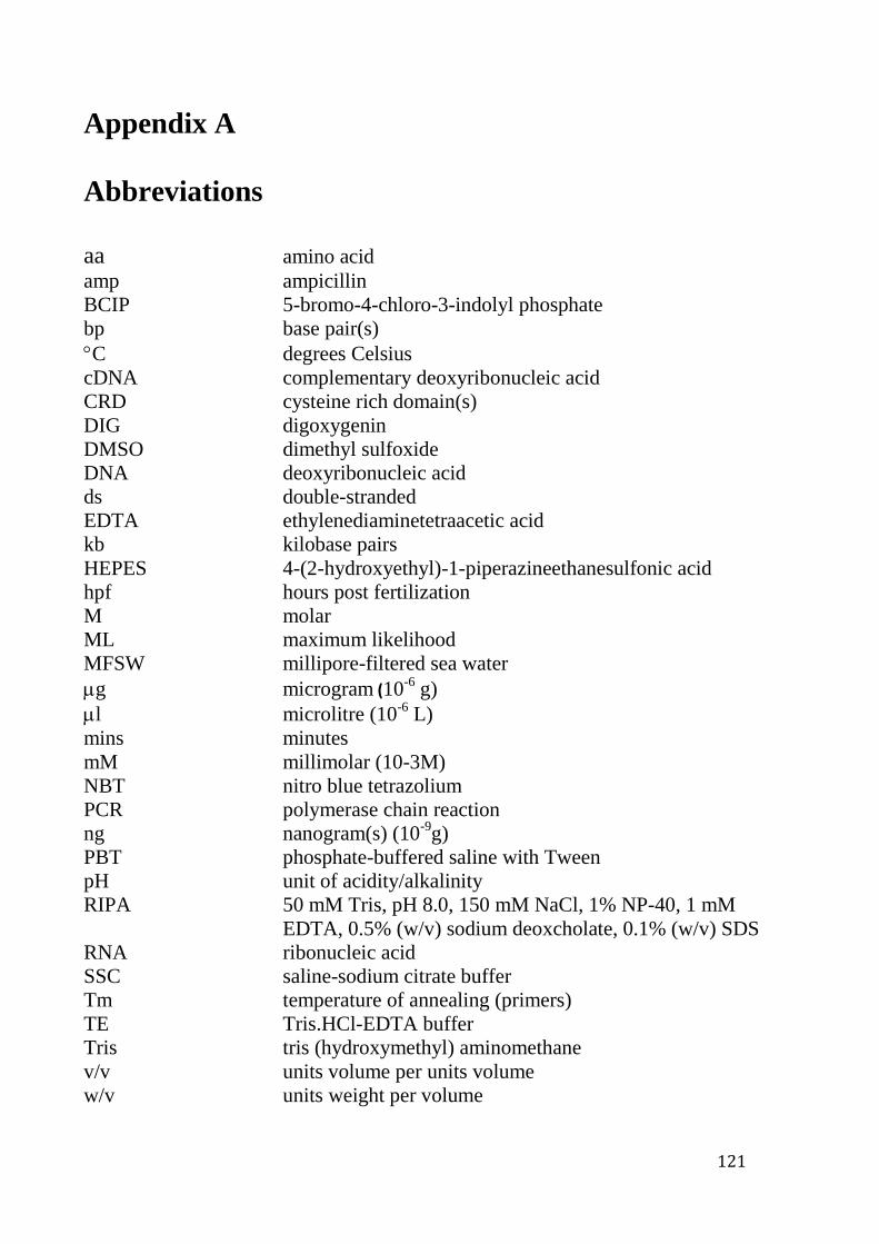
121
Appendix A
Abbreviations
aa amino acid
amp ampicillin
BCIP 5-bromo-4-chloro-3-indolyl phosphate
bp base pair(s)
C degrees Celsius
cDNA complementary deoxyribonucleic acid
CRD cysteine rich domain(s)
DIG digoxygenin
DMSO dimethyl sulfoxide
DNA deoxyribonucleic acid
ds double-stranded
EDTA ethylenediaminetetraacetic acid
kb kilobase pairs
HEPES 4-(2-hydroxyethyl)-1-piperazineethanesulfonic acid
hpf hours post fertilization
M molar
ML maximum likelihood
MFSW millipore-filtered sea water
g microgram (10-6
g)
l microlitre (10-6
L)
mins minutes
mM millimolar (10-3M)
NBT nitro blue tetrazolium
PCR polymerase chain reaction
ng nanogram(s) (10-9
g)
PBT phosphate-buffered saline with Tween
pH unit of acidity/alkalinity
RIPA 50 mM Tris, pH 8.0, 150 mM NaCl, 1% NP-40, 1 mM
EDTA, 0.5% (w/v) sodium deoxcholate, 0.1% (w/v) SDS
RNA ribonucleic acid
SSC saline-sodium citrate buffer
Tm temperature of annealing (primers)
TE Tris.HCl-EDTA buffer
Tris tris (hydroxymethyl) aminomethane
v/v units volume per units volume
w/v units weight per volume

Appendix B
- Supplementary data -
122
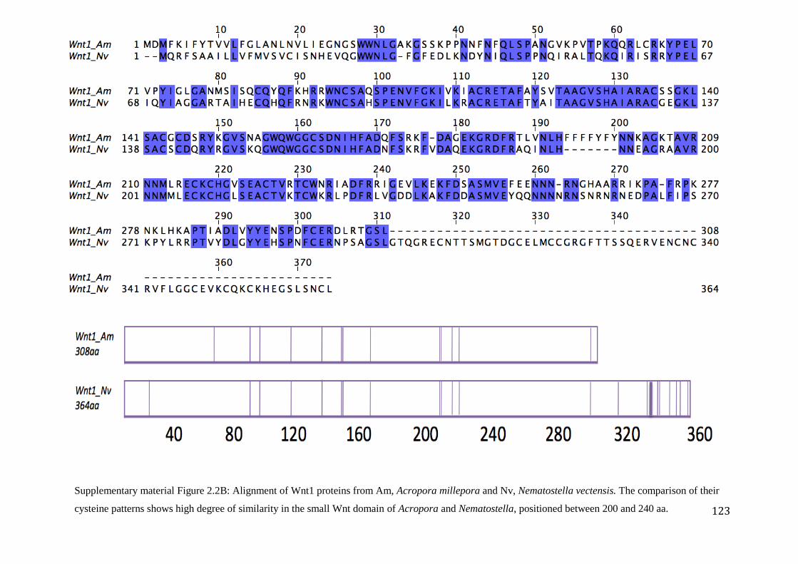
123
Supplementary material Figure 2.2B: Alignment of Wnt1 proteins from Am, Acropora millepora and Nv, Nematostella vectensis. The comparison of their
cysteine patterns shows high degree of similarity in the small Wnt domain of Acropora and Nematostella, positioned between 200 and 240 aa.
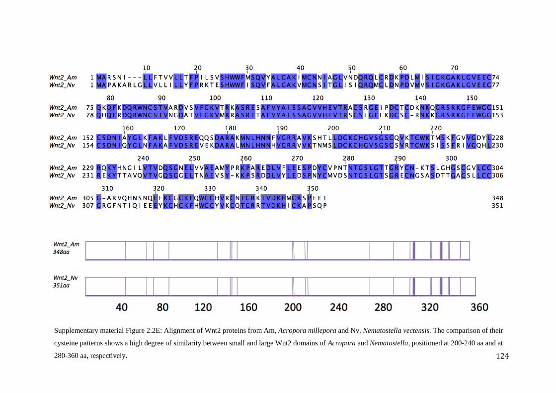
124
Supplementary material Figure 2.2E: Alignment of Wnt2 proteins from Am, Acropora millepora and Nv, Nematostella vectensis. The comparison of their
cysteine patterns shows a high degree of similarity between small and large Wnt2 domains of Acropora and Nematostella, positioned at 200-240 aa and at
280-360 aa, respectively.
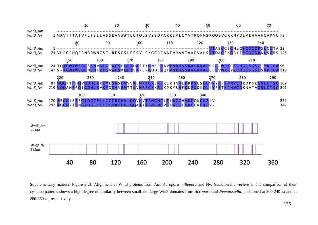
125
Supplementary material Figure 2.2J: Alignment of Wnt3 proteins from Am, Acropora millepora and Nv, Nematostella vectensis. The comparison of their
cysteine patterns shows a high degree of similarity between small and large Wnt3 domains from Acropora and Nematostella, positioned at 200-240 aa and at
280-360 aa, respectively.
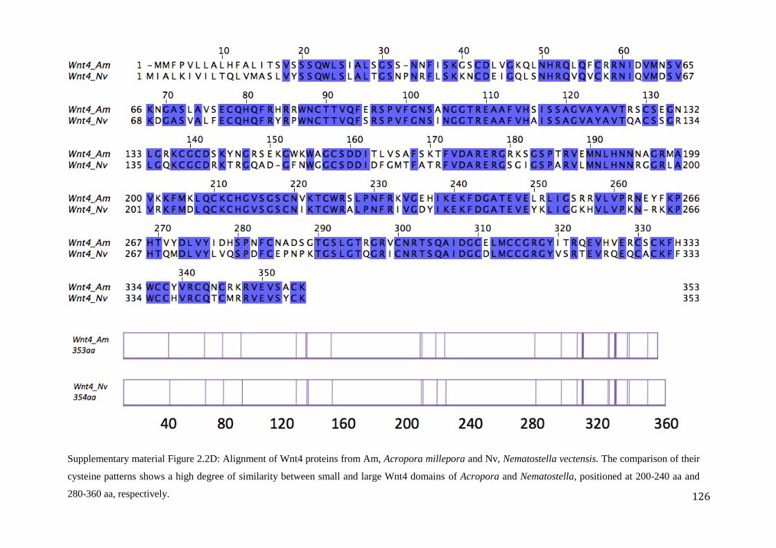
126
Supplementary material Figure 2.2D: Alignment of Wnt4 proteins from Am, Acropora millepora and Nv, Nematostella vectensis. The comparison of their
cysteine patterns shows a high degree of similarity between small and large Wnt4 domains of Acropora and Nematostella, positioned at 200-240 aa and
280-360 aa, respectively.
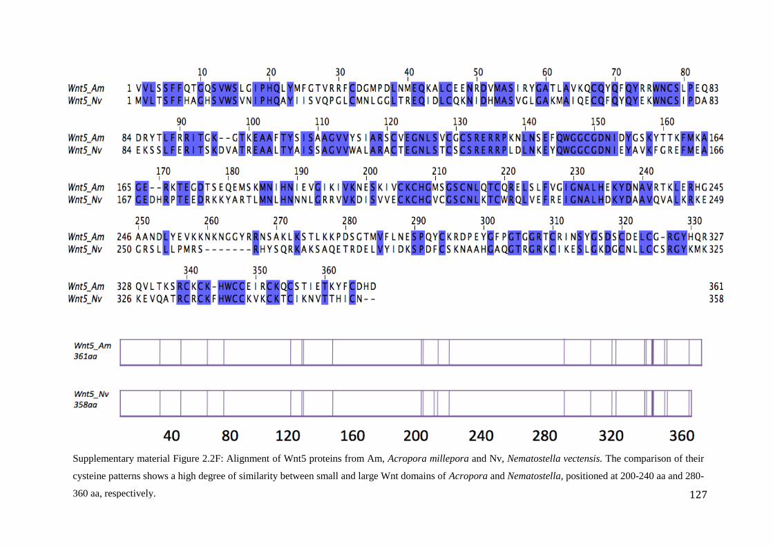
127
Supplementary material Figure 2.2F: Alignment of Wnt5 proteins from Am, Acropora millepora and Nv, Nematostella vectensis. The comparison of their
cysteine patterns shows a high degree of similarity between small and large Wnt domains of Acropora and Nematostella, positioned at 200-240 aa and 280-
360 aa, respectively.
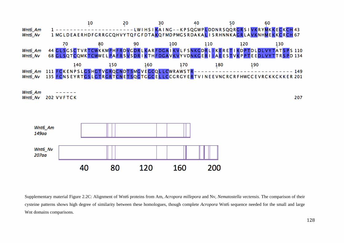
128
Supplementary material Figure 2.2C: Alignment of Wnt6 proteins from Am, Acropora millepora and Nv, Nematostella vectensis. The comparison of their
cysteine patterns shows high degree of similarity between these homologues, though complete Acropora Wnt6 sequence needed for the small and large
Wnt domains comparisons.
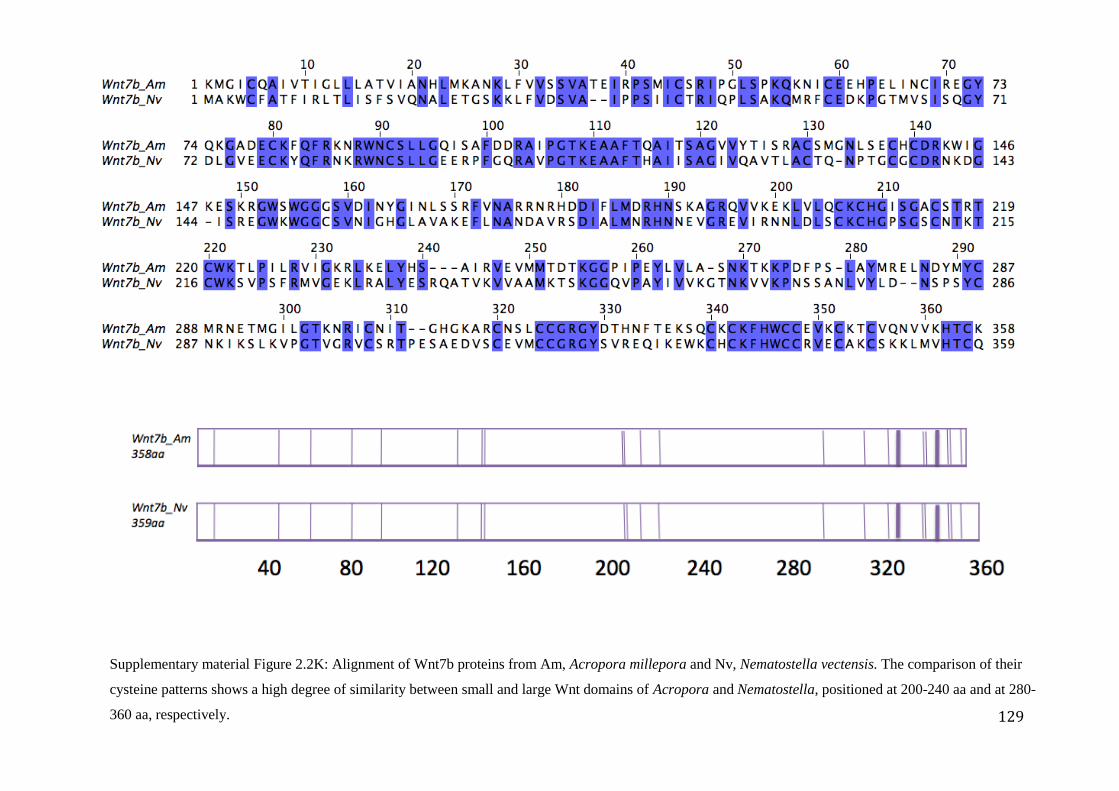
129
Supplementary material Figure 2.2K: Alignment of Wnt7b proteins from Am, Acropora millepora and Nv, Nematostella vectensis. The comparison of their
cysteine patterns shows a high degree of similarity between small and large Wnt domains of Acropora and Nematostella, positioned at 200-240 aa and at 280-
360 aa, respectively.
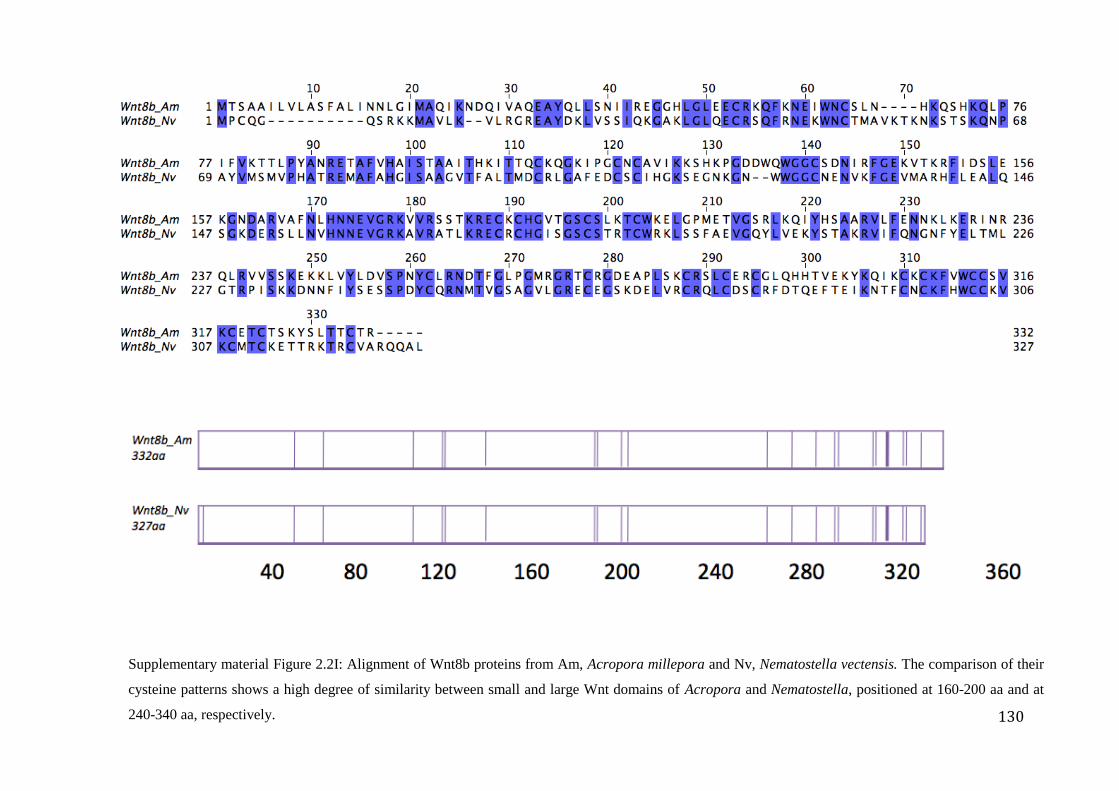
130
Supplementary material Figure 2.2I: Alignment of Wnt8b proteins from Am, Acropora millepora and Nv, Nematostella vectensis. The comparison of their
cysteine patterns shows a high degree of similarity between small and large Wnt domains of Acropora and Nematostella, positioned at 160-200 aa and at
240-340 aa, respectively.
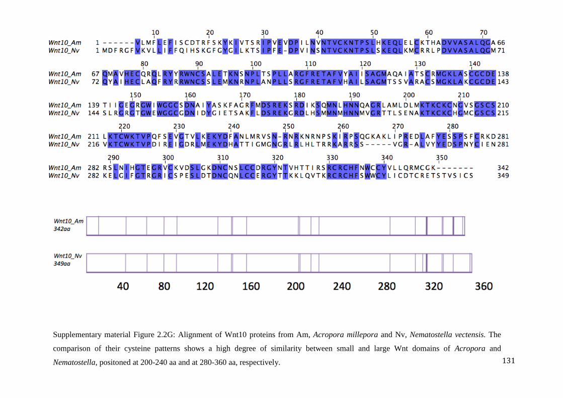
131
Supplementary material Figure 2.2G: Alignment of Wnt10 proteins from Am, Acropora millepora and Nv, Nematostella vectensis. The
comparison of their cysteine patterns shows a high degree of similarity between small and large Wnt domains of Acropora and
Nematostella, positoned at 200-240 aa and at 280-360 aa, respectively.
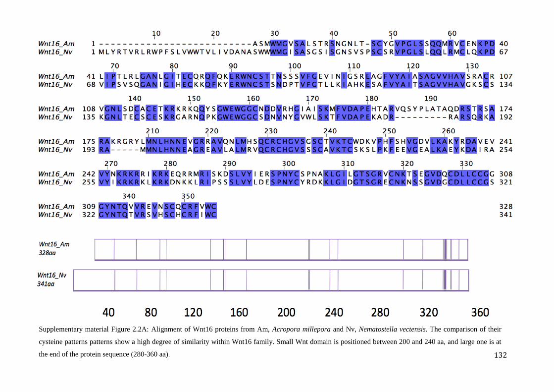
132
Supplementary material Figure 2.2A: Alignment of Wnt16 proteins from Am, Acropora millepora and Nv, Nematostella vectensis. The comparison of their
cysteine patterns patterns show a high degree of similarity within Wnt16 family. Small Wnt domain is positioned between 200 and 240 aa, and large one is at
the end of the protein sequence (280-360 aa).
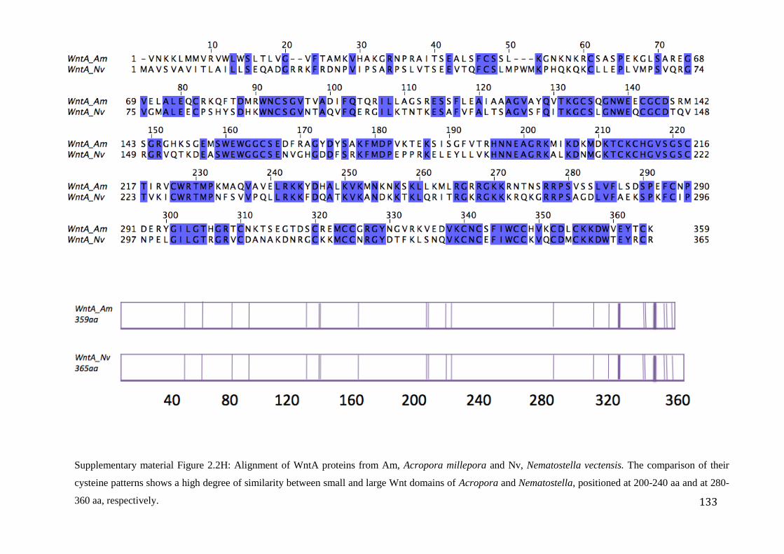
133
Supplementary material Figure 2.2H: Alignment of WntA proteins from Am, Acropora millepora and Nv, Nematostella vectensis. The comparison of their
cysteine patterns shows a high degree of similarity between small and large Wnt domains of Acropora and Nematostella, positioned at 200-240 aa and at 280-
360 aa, respectively.
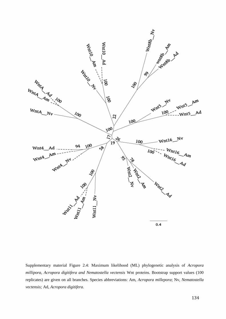
134
Supplementary material Figure 2.4: Maximum likelihood (ML) phylogenetic analysis of Acropora
millipora, Acropora digitifera and Nematostella vectensis Wnt proteins. Bootstrap support values (100
replicates) are given on all branches. Species abbreviations: Am, Acropora millepora; Nv, Nematostella
vectensis; Ad, Acropora digitifera.
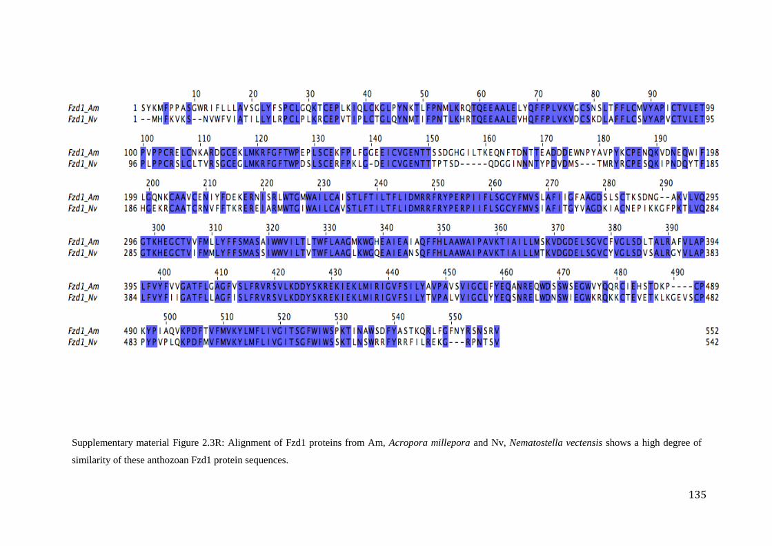
135
Supplementary material Figure 2.3R: Alignment of Fzd1 proteins from Am, Acropora millepora and Nv, Nematostella vectensis shows a high degree of
similarity of these anthozoan Fzd1 protein sequences.
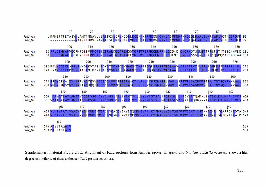
136
Supplementary material Figure 2.3Q: Alignment of Fzd2 proteins from Am, Acropora millepora and Nv, Nematostella vectensis shows a high
degree of similarity of these anthozoan Fzd2 protein sequences.
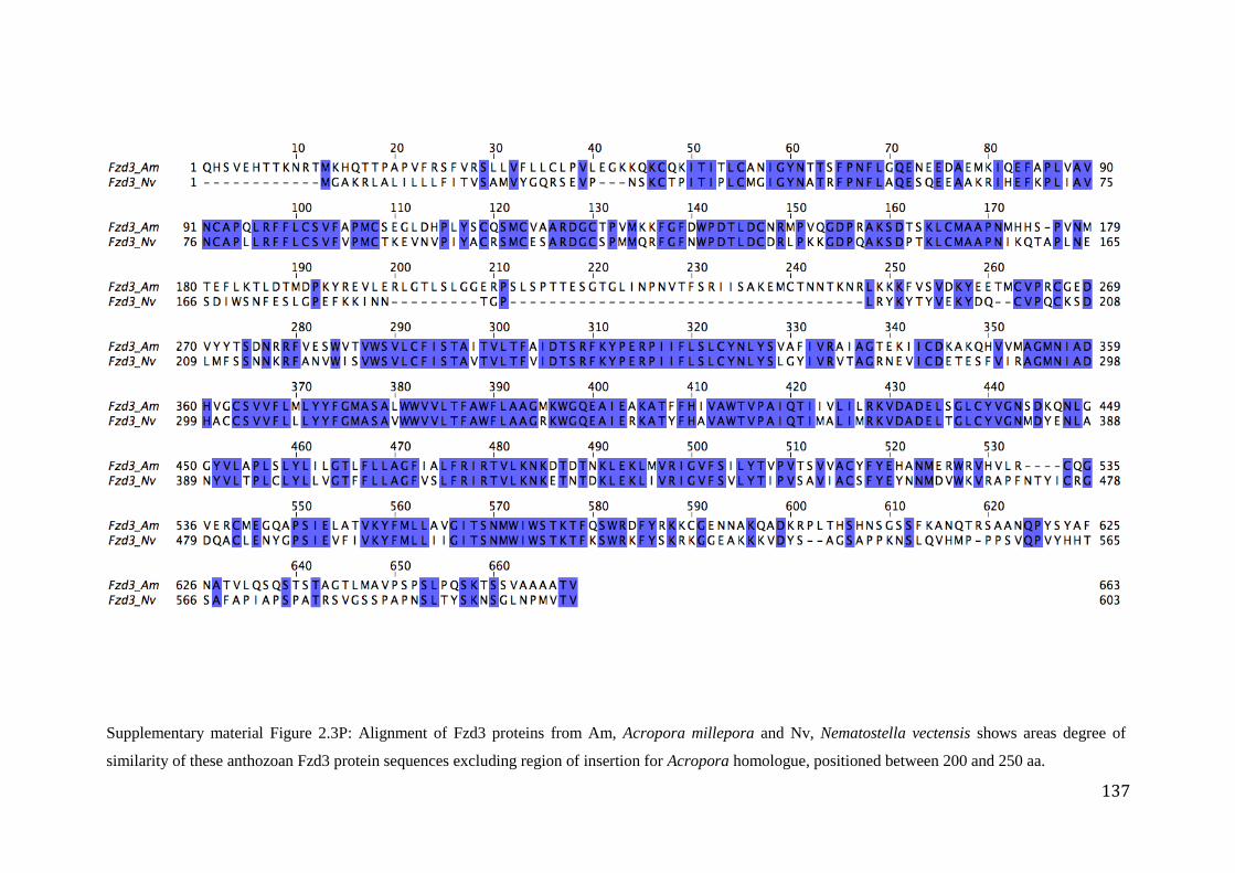
137
Supplementary material Figure 2.3P: Alignment of Fzd3 proteins from Am, Acropora millepora and Nv, Nematostella vectensis shows areas degree of
similarity of these anthozoan Fzd3 protein sequences excluding region of insertion for Acropora homologue, positioned between 200 and 250 aa.
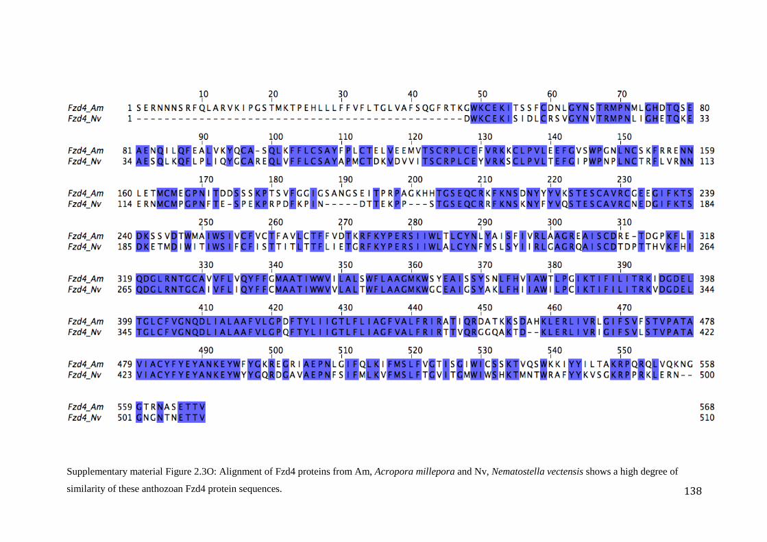
138
Supplementary material Figure 2.3O: Alignment of Fzd4 proteins from Am, Acropora millepora and Nv, Nematostella vectensis shows a high degree of
similarity of these anthozoan Fzd4 protein sequences.
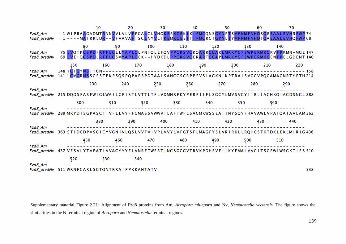
139
Supplementary material Figure 2.2L: Alignment of Fzd8 proteins from Am, Acropora millepora and Nv, Nematostella vectensis. The figure shows the
similarities in the N-terminal region of Acropora and Nematostella terminal regions.
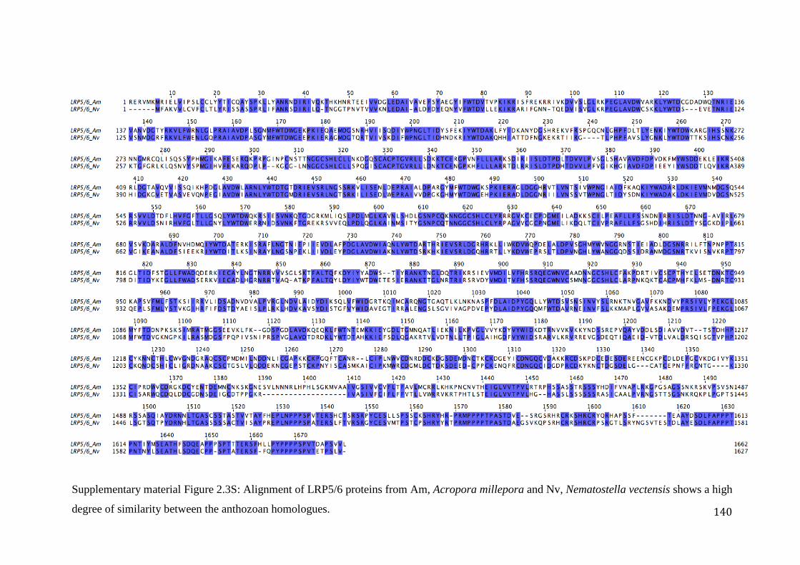
140
Supplementary material Figure 2.3S: Alignment of LRP5/6 proteins from Am, Acropora millepora and Nv, Nematostella vectensis shows a high
degree of similarity between the anthozoan homologues.
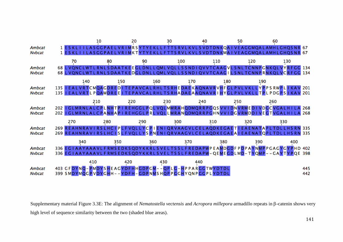
141
Supplementary material Figure 3.3E: The alignment of Nematostella vectensis and Acropora millepora armadillo repeats in -catenin shows very
high level of sequence similarity between the two (shaded blue areas).