Embed Size (px)
Citation preview
CHARACTERIZATION OF THE HUMAN CYT19 GENE PRODUCT: AN ARSENIC
METHYLTRANSFERASE
By
ALEX J. MCNALLY
A THESIS PRESENTED TO THE GRADUATE SCHOOL OF THE UNIVERSITY OF FLORIDA IN PARTIAL FULFILLMENT
OF THE REQUIREMENTS FOR THE DEGREE OF MASTER OF SCIENCE
UNIVERSITY OF FLORIDA
2006

Copyright 2006
by
Alex J. McNally
iii
ACKNOWLEDGMENTS
I would like to thank my mentor, Dr. David S. Barber, for all his help, guidance,
and knowledge he has given me. I would also like to thank my committee members, Dr.
Nancy Denslow, Dr. Lena Ma, and Dr. Steve Roberts, for their suggestions and advise. I
thank my fiancé and my parents for their support and encouragement throughout my
graduate studies.
iv
TABLE OF CONTENTS page
ACKNOWLEDGMENTS ................................................................................................. iii
LIST OF TABLES............................................................................................................. vi
LIST OF FIGURES .......................................................................................................... vii
ABSTRACT....................................................................................................................... ix
CHAPTER
1 INTRODUCTION ........................................................................................................1
Sources of Arsenic........................................................................................................1 Natural Sources .....................................................................................................1 Anthropogenic Sources .........................................................................................1
Exposure and Health Effects ........................................................................................2 Exposure ................................................................................................................2 Health Effects ........................................................................................................3 Mechanisms of Toxicity ........................................................................................4
Arsenic Biotransformation............................................................................................6 Reduction of Pentavalent Arsenicals.....................................................................6 Methylation of Trivalent Arsenicals......................................................................7
Variation in Arsenic Methylation .................................................................................8 Role of Methylation in Arsenic Toxicity....................................................................10 Specific Aims of Research..........................................................................................11
2 MOLECULAR CLONING AND CHARACTERIZATION OF HUMAN CYT19, AN S-ADENOSYL-L-METHIONINE:AS-METHYLTRANSFERASE FROM HEPG2 CELLS...........................................................................................................13
Introduction.................................................................................................................13 Materials and Methods ...............................................................................................14
Molecular Cloning...............................................................................................14 RACE PCR..........................................................................................................15 Expression of Recombinant cyt19.......................................................................15 Characterization...................................................................................................16 Confirmation of Methylated Arsenicals ..............................................................17
v
Results and Discussion ...............................................................................................18
3 IDENTIFICATION OF A SPLICE VARIANT OF HUMAN CYT19 ARSENIC METHYLTRANSFERASE........................................................................................33
Introduction.................................................................................................................33 Materials and Methods ...............................................................................................34
Molecular Cloning of cyt19 Splice Variants .......................................................34 Human Liver Samples .........................................................................................35 qPCR of cyt19 Splice Variants............................................................................35
Results.........................................................................................................................36 Discussion...................................................................................................................37
4 GENERAL CONCLUSIONS.....................................................................................45
APPENDIX
ROLE OF CYT19 IN ACUTE ARSENIC TOXICITY. IS CYT19 THE ONLY HUMAN ARSENIC METHYLTRANSFERASE?....................................................49
Materials and Methods ...............................................................................................49 Results and Discussion ...............................................................................................50
cyt19 mRNA Knockdown by siRNA..................................................................50 Antibody Specificity and Purification .................................................................51 Future Experiments .............................................................................................51
LIST OF REFERENCES...................................................................................................57
BIOGRAPHICAL SKETCH .............................................................................................67
vi
LIST OF TABLES
Table page 2-1. Primers used in the PCR amplification of cyt19 ......................................................23
2-2. Kinetic analysis of the methylation activity of cyt19-WT and cyt19S81R..............23
3-1. The individual information and Shapiro’s score of cyt19 exon 2 and exon 3. ........44
3-2. The amount of cyt19 and cyt19∆E2 in different human liver samples and HepG2 cells...........................................................................................................................44
A-1. Sequence of double stranded siRNA........................................................................53
vii
LIST OF FIGURES
Figure page 2-1. Sequence alignment of cyt19t and Genbank sequence (accession AF226730) ......24
2-2. Sequence alignment of cyt19S81R and Genbank sequence (accession AK057833)...............................................................................................................25
2-3. Sequence alignment of cyt19-WT and cyt19t ..........................................................26
2-4. Sequence alignments of the 5’ & 3’RACE-PCR products and the Genbank sequences (accession AK057833 and AF226730) ...................................................27
2-5. Purification of recombinant human cyt19................................................................28
2-6. The effects of AsIII & MMAV concentrations ..........................................................29
2-7. The effect of pH on activity .....................................................................................30
2-8. The effects of reductants on methylation activity ....................................................30
2-9. The effect of SAM concentration on activity...........................................................31
2-10. Arsenical metabolites formed after incubation with [3H]SAM and cyt19 for 30 min at 37C ................................................................................................................32
3-1. The hypothesized scheme of iAs methylation proposed by Cullen, McBride et al. 1984. ....................................................................................................................40
3-2. PCR products of cyt19 amplification .......................................................................40
3-3. Alignment of the reference cyt19 nucleotide sequence and cyt19∆E2....................41
3-4. cyt19 isoforms ..........................................................................................................42
3-5. Alignment of the reference cyt19 amino acid sequence and product of cyt19∆E2 uORF ........................................................................................................................43
A-1. Determination of the most efficient siRNA in the knockdown of cyt19 mRNA levels assayed by qPCR............................................................................................54
A-2. The siRNA pool knockdown of cyt19 mRNA relative to control............................54
viii
A-3. Determination of cell viability after siRNA knockdown by the XTT assay. ...........55
A-4. Western blot of the specificity of the crude antisera to the antigen, purified cyt19 protein.......................................................................................................................55
A-5. Western blot of the specificity of the crude antisera to cyt19 in human liver cytosolic preparations...............................................................................................55
A-6. Western blot of the specificity of the purified antibody to the antigen, purified cyt19 protein.............................................................................................................56
A-7. Western blot of the specificity of the crude antisera to cyt19 in human liver cytosolic preparations...............................................................................................56
ix
Abstract of Thesis Presented to the Graduate School
of the University of Florida in Partial Fulfillment of the Requirements for the Degree of Master of Science
CHARACTERIZATION OF THE HUMAN CYT19 GENE PRODUCT: AN ARSENIC METHLTRANSFERASE
By
Alex J. McNally
May 2006
Chair: David S. Barber Major Department: Veterinary Medicine
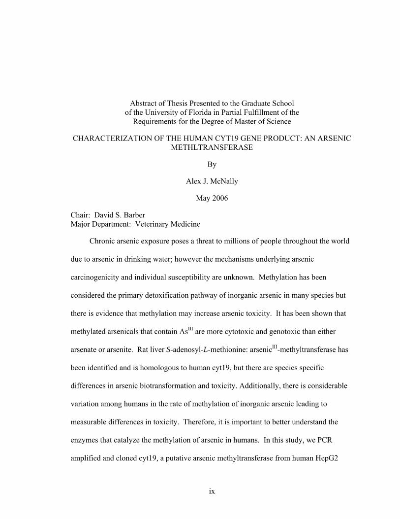
Chronic arsenic exposure poses a threat to millions of people throughout the world
due to arsenic in drinking water; however the mechanisms underlying arsenic
carcinogenicity and individual susceptibility are unknown. Methylation has been
considered the primary detoxification pathway of inorganic arsenic in many species but
there is evidence that methylation may increase arsenic toxicity. It has been shown that
methylated arsenicals that contain AsIII are more cytotoxic and genotoxic than either
arsenate or arsenite. Rat liver S-adenosyl-L-methionine: arsenicIII-methyltransferase has
been identified and is homologous to human cyt19, but there are species specific
differences in arsenic biotransformation and toxicity. Additionally, there is considerable
variation among humans in the rate of methylation of inorganic arsenic leading to
measurable differences in toxicity. Therefore, it is important to better understand the
enzymes that catalyze the methylation of arsenic in humans. In this study, we PCR
amplified and cloned cyt19, a putative arsenic methyltransferase from human HepG2
x
hepatoma cells. The PCR product was ligated into an E. coli pET expression vector with
a polyhistidine tag at the amino-terminal residue. The recombinant human cyt19 was
successfully expressed in BL21 (DE3) and purified using a nickel-nitrilotriacetic acid
metal-affinity chromatography. The recombinant protein catalyzes the methylation of
arsenite as well as monomethylarsonic acid (MMA). The specific activity of arsenite
methylation was 597 pmol/mg protein/min in a reaction mixture containing 5mM GSH, 1
mM DTT, 1 mM MgCl2, 100 µM S-adenosyl-L-methionine, 50µM sodium m-arsenite,
and 5 µg of S-adenosyl-L-methionine: arsenic methyltransferase in 100mM tris/100mM
sodium phosphate buffer pH 7.4 at 37 °C for 30 minutes. The results suggest that the
human cyt19 gene, in fact, is translated to an S-adenosyl-L-methionine: arsenic
methyltransferase which methylates both arsenite and MMA.
Studies have shown that humans exposed to arsenic excrete variable amounts of
methylated arsenicals in the urine which may be due to differences in arsenic
methyltransferase activity. While polymorphisms in the coding region of cyt19 may
account for some of the observed variation in arsenic methylation, other mechanisms are
likely to be involved. In this study we identified an alternative splice variant of the
human cyt19 (cyt19∆E2), in which exon 2 is removed creating a bicistronic transcript
that is unlikely to produce an active protein. This variant was expressed in 7 out of 7
male Caucasian human liver samples tested and in HepG2 cells. The human cyt19
appears to be alternatively spliced in many individuals and may play a role in the
observed variation in arsenic methylation seen in individuals.
1
CHAPTER 1 INTRODUCTION
Sources of Arsenic
Natural Sources
Arsenic (As) is a member of the nitrogen group in the periodic table and is
classified as a metalloid. This metalloid is a naturally occurring element and is the 20th
most abundant element in the earth’s crust [1]. Arsenic is found in the environment as
sulfides, and complex sulfides of iron, nickel, and cobalt. The natural weathering of
rocks and soils containing various forms of arsenic contribute to its levels in the
environment. Arsenic is present in the atmosphere, aquatic environments, soils &
sediments, and in organisms. This metalloid is found naturally in rocks, geothermal
wells, minerals, and metal ores such as copper and lead. Arsenic is present in the
environment in both organic and inorganic forms and exists in four valence states, -3, 0
(elemental), +3 (trivalent), and +5 (pentavalent arsenic), however it exists mainly in the
latter two valence states. Many marine plant and animal species have naturally high
levels of As, but in organic forms that appear to cause little toxicity. The main species of
arsenic in marine animals is the arsenosugar, arsenobetaine [2]. In general, organic forms
of arsenic are less toxic than inorganic forms of arsenic and the pentavalent inorganic
forms are less toxic than trivalent inorganic arsenic compounds.
Anthropogenic Sources
Anthropogenic sources of arsenic stem from its use in pesticides and wood
preservatives as well as mining and smelting wastes. In the U.S., 2,200 tons of arsenic
2
was produced in 1985 [3]. Since 1985, the domestic production of arsenic in the US has
ceased. However, arsenic is still used domestically and therefore is imported from
countries such as China, Japan and Mexico. In 2003, the United States was world’s
largest consumer of arsenic, demanding 21,600 metric tons [4] . Arsenic has also been
used in the production of dessicants and as growth stimulants for plants and animals.
Organoarsenicals have been shown to have both therapeutic and growth promoting
properties in poultry and swine. Arsanilic acid and its sodium salts, such as 4-
nitrophenylarsonic acid are added to pet feed [5]. Most of this arsenic passes through
the animal and becomes part of the waste stream resulting from animal production. In
addition, arsenic has been used for therapeutic purposes and of course as a poison.
Arsenic has been used to treat syphilis, tropical diseases such as trypanosomiasis (African
sleeping sickness), yaws, amoebic dysentery and recently as an anticancer. Paul Ehrlich
developed an organic arsenical, arsphenamine, also known as Salvarsan, which was used
to treat syphilis [6]. Arsphenamine was also believed to be effective in treating
trypanosomiasis [6]. Arsenic trioxide has been shown to be highly effective in the
treatment of various cancers especially of acute promyelocytic leukaemia [7].
Exposure and Health Effects
Exposure
Arsenic is present throughout our environment, in the air we breathe, the water we
drink, and the food we eat. Water contributes more to iAs exposure than food or air. On
average Americans are exposed to 50 µg per day of arsenic of which 10 µg is in the
inorganic form [8]. People are exposed to higher than average arsenic due to living or
working around higher exposure sources. For example living or working near a
hazardous waste site can lead to exposure via the air, ingestion, or the food chain.
3
Arsenic is present in 47% of all sites on the National Priorities List (NPL) sites or
Superfund sites making it second only to lead as the most common contaminant of
concern [9]. Occupational settings such as workers who use or produce arsenic
compounds: vineyards, ceramics, glass-making, smelting, pharmaceuticals, refining of
metallic ores, pesticide manufacturing & application, wood preservation, or
semiconductor manufacturing, have the potential to be exposed to higher than average
arsenic levels [1].
Currently, people in Taiwan, Mexico, western United States, western South
America, China, West Bengal and Bangladesh are exposed to high levels of arsenic due
to anthropogenic and/or natural contamination of potable water. It has been estimated
that 200 million people worldwide are at risk from health effects associated with this
exposure [10]. The two most affected areas in the world are Bangladesh and West
Bengal, India; it has been estimated that around 122 million people in these areas are
exposed to groundwater arsenic concentrations above the World Health Organization
maximum permissible limit of 50µg/L [11]. The study showed that in West Bengal,
26.4% (n=10,991 tube wells) of the water samples had arsenic ranging in 100-299 µg/L.
In the U.S. the arsenic maximum contaminant level (MCL) was decreased to 10µg/L by
the US Environmental Protection Agency (EPA) in January 2001. Frost et al. [12]
identified 33 counties in 11 states in the western United States with mean arsenic
concentrations of 10µg/L or greater. In addition, from 1950-1990 there were over 60
million people in the US exposed to arsenic contaminated water exceeding 10µg/L [12].
Health Effects
Ingestion of arsenic is a widespread human health problem. There are different
symptoms associated with acute and chronic arsenic exposure. Acute exposure to arsenic
4
can result in acute paralytic syndrome, acute GI syndrome, and even death. Oral
exposure above 60 ppm in food or water can result in death [3]. Chronic arsenic
exposure can result in skin disorders such as hyper and hypo pigmentation, and
hyperkeratosis [3]. It was found that even at 0.005-0.01mg/L of arsenic in water there is
an increase in the prevalence of skin lesions [13]. In addition, chronic iAs exposure can
affect the circulatory and nervous systems leading to diseases such as Blackfoot disease.
Major organs such as the liver, kidneys, lung, bladder, and heart can be affected as a
result of arsenic cytotoxicity. There is an increase of cancer, death from cancer, and
diabetes mellitus associated with chronic arsenic exposure. Epidemiological studies
show that there is dose-response relationship between exposure to iAs and skin cancer
[14]. A study in Taiwan[15] and Japan[16] demonstrated a significant association
between long-term arsenic exposure in drinking water with lung and bladder cancer. In
northern Chile an increase in mortality from bladder, lung, kidney, and skin cancer is
associated with As exposure; bladder and lung cancer showing highest increase in
mortality [17]. A follow up study in Taiwan compared the incidence of diabetes mellitus
in an arsenic exposed population to two control areas showed an association between As
exposure and diabetes mellitus [18].
Mechanisms of Toxicity
The mechanism of arsenic toxicity is dependent on oxidation state. Trivalent
arsenicals, including methylated arsenicals produce toxicity by enzyme inhibition by
interactions with sulfhydryl groups in proteins [19] and the generation of reactive oxygen
species (ROS). For example, in vitro studies have shown that MMAIII and arsenite are
capable of inhibiting pyruvate dehydrogenase (PDH) activity in hamster kidney and
purified porcine heart PDH resulting in the subsequent blockage of adeonosine
5
triphosphate (ATP) production because of the disruption of the citric acid due to the
depletion of cellular citrate [20]. Inorganic arsenicals, arsenite and arsenate, have been
shown to induce ROS and reactive nitrogen species (RNS) which result in DNA and
protein oxidative damage [21]. Another study demonstrated that MMAIII and DMAIII
induced ROS result in DNA damage [22]. Arsenate has been shown to interfere with
ATP production by substituting for phosphate leading to production of an unstable ADP-
arsenate complex which spontaneously hydrolyzes [23]. This process leads to a depletion
of cellular energy due to this futile cycle.
Although there is strong evidence of the carcinogenicity of arsenic in humans, the
mechanism by which tumors are produced is unknown. Studies of arsenic carcinogenesis
have been hampered because there are very few animal models in which arsenic induces
carcinogenesis [3]. DMA concentrations of 50 and 200 ppm have been shown to be
carcinogenic in F344 rats urinary bladder [24]. Recent work has demonstrated the
promotion effects of inorganic arsenicals and methylated arsenicals. Inorganic arsenic
(42.5 and 85 ppm)has been shown to be a transplacental carcinogen in mice [25].
Organic arsenicals, such as MMA, DMA, and TMAO have been shown to act as
promoters in carcinogenesis of several rat organs [26]. However, some of these studies
have received much criticism due to the high arsenical exposure levels used ranging from
about 50 to 400 ppm and the use of several initiators such as diethylnitrosamine, N-butyl-
N-(4-hydroxybutyl)nitrosamine, N-methyl-N-nitrosourea, dihydroxy-di-N-
propylnitrosamine and N-N’-dimethylhydrazine, prior to arsenic exposure.
Several possibilities for mechanisms of arsenic induced malignancies have been
hypothesized such as chromosomal abnormality, oxidative stress, and the promotion of
6
tumorigenesis. Zhao et al. [27], have shown that chronic low level arsenic (0-0.5µM)
exposure will result in the malignant transformation of epithelial cells associated with
DNA hypomethylation due to depletion of SAM and aberrant gene expression. An in
vivo long-term arsenic exposure study to mice demonstrated that arsenic in potable water
can induce aberrant gene expression, global DNA hypomethylation, and hypomethylation
of the gene for the estrogen receptor-α resulting in enhanced transcription, which
cumulatively could lead to arsenic hepatocarcinogenesis [28]. Arsenate was shown to
have a dose-dependent transcriptional induction of several different signal transduction
pathways, including the dose-response induction of several promoters and/or response
elements responsive to oxidative damage and DNA damage [29] which may help
understand the mechanisms of carcinogenicity for arsenic. Binet et al. [30] has shown
that arsenic induced apoptosis via reactive oxygen species (ROS) production occurs but,
the ROS is not produced from nicotinamide adenine dinucleotide phosphate
dehydrogenase activation. Trivalent methylated arsenicals have been shown to indirectly
cause DNA damage by ROS. One study showed that DMAIII promotes tumorigenesis
and gentoxicity via dimethylated arsenic peroxides [31]
Arsenic Biotransformation
Reduction of Pentavalent Arsenicals
The biotransformation of iAs alternates between the reduction of arsenate (iAsV) to
arsenite (iAsIII) followed by oxidative methylation. The hypothesized scheme of iAs
methylation involves oxidative methylation and reduction[32]:
AsvO43- + 2e → AsIIIO3
3- + CH3+ → CH3Asv O3
2- + 2e → CH3AsIIIO22- + CH3
+ →
(CH3)2AsvO2- + 2e → (CH3)2AsIIIO
7
Arsenic reduction must occur first before it can be methylated. Several enzymes
have been shown capable to reduce arsenic. Purine nucleoside phosphorylase (PNP) has
the ability to reduce iAsV to iAsIII in the presence of a dithiol and a purine nucleoside
(guanosine or inosine) in vitro [33, 34]. However, studies performed by Németi et al.
2003 showed that PNP does not play a role in iAsV reduction in vivo [35].
Glyceraldehyde 3-phosphate dehydrogenase (GAPDH) in the presence of glutathione
(GSH) and NAD has the ability to reduce iAsV to iAsIII in human red blood cells and rat
liver cytosol [36, 37]. A human arsenate reductase was discovered capable of reducing
arsenate but not methylarsonic acid (MMAV) [38]. Zakharyan et al 1999 presented an
enzyme from rabbit liver capable of reducing MMAV, arsenate, and dimethylarsinic acid
(DMAV) in the presence of GSH; this enzyme was also present in human liver [39].
MMAV reductase was sequenced and 92% of the sequences were identical to human
glutathione-S-transferase Omega class (hGSTO-1) [40]. This hGSTO-1 catalyzes the
reduction of iAsV, MMAV, and DMAV [39, 41]. There is evidence that pentavalent
arsenicals can be reduced nonenzymatically. Glutathione (GSH) has been shown to
reduce pentavalent arsenicals [42, 43].
Methylation of Trivalent Arsenicals
Following the first reduction step, arsenite is enzymatically oxidatively methylated
to MMAV. In this reaction, a high energy methyl group from S-adenosyl-L-methione
(SAM) is transferred to a trivalent arsenical in an oxidative process that produces a
pentavalent methylated arsenical. The resulting monomethylarsonic acid (MMA) can be
reduced a second time and methylated again to form dimethylarsinic acid (DMA). In
West Bengal, MMAIII and DMAIII were detected in the urine of exposed humans in 48%
and 72% respectively out of the 428 subjects [44]. In some animals, including humans, a
8
third methylation can occur resulting in formation of trimethylarsine oxide (TMAO). It
was shown that a single dose of arsenic trioxide in hamsters resulted in a very small
amount of TMAO production in the liver[45]. Yoshida et al 1997 demonstrated that rats
excrete TMAO in their urine after a single oral administration of DMA [46]. Urine
excretion of TMAO in man has been observed after ingestion of an arsenosugar and
DMA [47, 48].
Arsenic methyltransferases (AS3MT) have been isolated from many mammalian
species. An AS3MT has been purified 2000-fold from rabbit liver by DEAE
chromatography to a single band [49]. The rabbit liver AS3MT was capable of
performing both methylation steps. The Golden Syrian hamster liver was used to purify
AS3MT and was shown to have similar activities as the rabbit AS3MT [50]. Rat liver S-
adenosyl-L-methionine:arsenicIII-methyltransferase has been identified and is
homologous to human cyt19 [51]. The rat arsenic methyltransferase has been shown to
perform both mono and dimethylation of arsenic. Arsenic methyltransferase activity has
been determined in mice and primates, including humans [52-55].
Variation in Arsenic Methylation
There is significant variation in the arsenic methylation rate and arsenic metabolite
production among mammalian species. The variability of arsenic methylation is apparent
in the amounts of methylated arsenic metabolites seen in the urine of exposed mammals
such as the rat, rabbit, hamster, dog, and mouse. For example, mice quickly excrete
about 90% of the dose in two days of which 80% is DMA [56]. The rat efficiently
methylates arsenic, but it accumulates DMA in red blood cells resulting in subsequent
lower DMA excretion levels making it a poor model for human metabolic studies [57].
Healy et al. [58], purified arsenic methyltransferases from livers of rabbit, hamster, and
9
rhesus monkey and found different rates of methylation which may affect arsenic
elimination and toxicity.
However, these mammals with arsenic methylation capacity have something in
common which distinguishes them from humans. On average, humans exposed to
arsenic excrete more MMA in the urine than other mammals , specifically 10-30% iAs,
10-20% MMA, and 60-80% DMA [59]. This suggests that non-human mammals are
more efficient at catalyzing the second methylation step which produces MMA. This
may relate to their lower susceptibility to iAs carcinogenesis following exposure versus
man. Additionally, there is significant variation in human susceptibility to As induced
toxicity, which may be related to differences in arsenic biotransformation between
individuals. Epidemiological studies have shown differences in the amount of MMA and
DMA excreted in the urine of exposed populations which may be associated with
differences in arsenic methyltransferase activity. Several studies on the urinary excretion
of arsenic metabolites in native Andean people and mixed ethnicities in northeastern
Argentina exposed to arsenic in potable water revealed low excretion of MMA [60-63].
One study revealed higher than normal MMA in urine, on average 27%, in people
exposed to arsenic in drinking water on the northeast coast of Taiwan [64].
Only a few polymorphisms have been found in the coding region of cyt19 to date.
The Met287Thr mutation has been reported on three different occasions [65-67]. In two
of these studies, the methylation activity of this allozyme was determined and showed to
have a higher methylation capacity than the wild-type [65, 67]. However, the activity of
this allozyme was determined either from a cytosol preparation or by analysis of cells and
culture media of exposed human hepatocytes. This type of analysis does not take into
10
account the possible different expression levels of the arsenic methyltransferase. In fact
one study shows that the allozyme Met287Thr was expressed at higher levels than the
wild-type arsenic methyltransferase [67], making unclear as to whether the increase in
activity results from the mutation or the increased expression levels. There are
differences in arsenic methylation capacities among individuals, which cannot be
supported alone by polymorphisms within the cyt19 coding region. All of the single
nucleotide polymorphisms (SNPs) in cyt19 available as seen in the NCBI SNP database
on January 12, 2006 are within the intron or untranslated region (UTR). In addition, two
separate studies which examined the frequency of polymorphisms within cyt19 found one
nonsynonymous SNPs (nsSNPs) out of 58 SNPs [66] and the other study found 3
nsSPNPs out of 26 SNPs [67], the remainder of the SNPs occurring in introns or UTRs.
There is another mechanism which may help explain the differences seen in
arsenic methylation, alternative splicing. Alternative splicing is frequently used to
regulate gene expression and to generate tissue-specific mRNA and protein isoforms [68,
69]. Introns contain sequence elements in which splicesome assembly occurs [70].
Mutations within these sequence elements could alter the constitutive splicing of a gene
which may affect the methylation capacity within and among different population groups.
Role of Methylation in Arsenic Toxicity
Arsenic methylation has traditionally been thought to be a detoxification pathway.
Pentavalent methylated arsenic metabolites are less reactive and are readily excreted in
the urine compared to iAs [56]. Pentavalent methylated arsenicals have also been shown
to be less cytotoxic and genotoxic compared to arsenite [71]. Mure et al. [72],
demonstrated that arsenite induces delayed mutagenesis and transformation in human
osteosarcoma cells but MMAIII showed no significant increase in mutagenesis or

11
transformation. While As methylation has been viewed as a detoxification pathway,
recent studies have shown monomethylarsonous acid (MMAIII) and dimethylarsinous
acid (DMAIII) to be more toxic than inorganic arsenicals. In vitro and in vivo studies
have shown that MMAIII is more cytotoxic than AsIII. Petrick et al [20], revealed that the
order of toxicity in Chang human hepatocytes is as follows: MMAIII > arsenite > arsenate
> MMAV = DMAV. In vivo studies performed in hamsters demonstrated that MMAIII is
more lethal than arsenite [73]. In addition, Hirano et al. [74], has shown that
monomethylarsonous acid diglutathione is more acutely toxic than other arsenicals. It is
important to point out that not all mammals methylate arsenic such as the marmoset
monkey, tamarin, chimpanzee, and the guinea pig [57, 75-78]. These mammals have not
been shown to be more susceptible to acute arsenic intoxication. One study showed no
correlation between the induction of micronuclei and the ability to methylate arsenic in
the leukocytes of four mammalian species, humans, mice, rats, and guinea pigs [79].
Another factor that questions the role of methylation is the fact that arsenic is a known
human carcinogen, but very few animals exist in which arsenic initiates carcinogenesis.
The debate over whether As methylation is a detoxification or bioactivation pathway
leads to confusion over the role of methylation in toxicity.
Specific Aims of Research
The proposed study will address the role of arsenic methylation in human toxicity
by better understanding the kinetics of the human arsenic methyltransferase. The overall
hypothesis is the following: Human arsenic methyltransferase, cyt19, activity is the
determining factor in the rate of arsenic methylation and toxicity. To test this hypothesis,
I examined two specific aims: 1) clone and characterize the human arsenic
methyltransferase (cyt19) and 2) determine cyt19s role in toxicity and arsenic
12
methylation variability. Specific aim one was addressed by determining rate of arsenic
methylation by an in vitro assay. The optimum conditions required for human cyt19
activity were determined such as pH optimum, substrate specificity and concentration,
and thiol requirements. Specific aim two was addressed by examining the role of
polymorphisms and splice variants on arsenic methylation variability. The determination
of cyt19s role in toxicity was addressed by As toxicity in the presence and absence of
methylation activity. In order to determine if cyt19 is the only arsenic methyltransferase
in humans, the mRNA levels, protein concentration, and activity, in the presence and
absence of siRNA knockdown was determined.
13
CHAPTER 2 MOLECULAR CLONING AND CHARACTERIZATION OF HUMAN CYT19, AN S-ADENOSYL-L-METHIONINE:AS-METHYLTRANSFERASE FROM HEPG2 CELLS
Introduction
Chronic arsenic exposure is a threat to millions of people throughout the world.
Exposure to arsenic has been linked to various types of cancers such as skin cancer, lung
cancer, and cancer of other internal organs [71]. Methylation has been considered the
major route of biotransformation and excretion of inorganic arsenic (iAs) in many species
including humans. The hypothesized scheme of iAs methylation involves reduction
followed by oxidative methylation [80]:
AsvO43- + 2e → AsIIIO3
3- + CH3+ → CH3Asv O3
2- + 2e → CH3AsIIIO22- + CH3
+ →
(CH3)2AsvO2- + 2e → (CH3)2AsIIIO
While traditionally thought to be a detoxification pathway, recent studies have
shown monomethylarsonous acid (MMAIII) and dimethylarsonous acid (DMAIII) to be
more toxic than inorganic arsenicals [73]. In addition, Hirano et al.[74], has shown that
monomethylarsonous acid diglutathione is more acutely toxic than other arsenicals. The
debate over whether As methylation is a detoxification or bioactivation pathway leads to
confusion over the role of methylation in toxicity. Rat liver S-adenosyl-L-
methionine:arsenicIII-methyltransferase has been identified and is homologous to human
cyt19 [51]. While this enzyme can be used as a model for human arsenic
biotransformation, the rat is considered a poor model for metabolic studies due to its
accumulation in red blood cells and subsequent lower DMA excretion levels [57]. There
14
are also other species specific differences in arsenic biotransformation and toxicity.
Healy et al. (1999) [58], purified arsenic methyltransferases from livers of rabbit,
hamster, and rhesus monkey and found different rates of methylation which may affect
arsenic elimination and toxicity. Humans excrete greater amounts of monomethylarsonic
acid (MMAV) compared to most other mammals [81]. Additionally, there is considerable
variation among humans in the rate of methylation of inorganic arsenic possibly leading
to measurable differences in toxicity [82]. Therefore it is important to better understand
the arsenic methylation capacity in human. To date, human cyt19 has been expressed,
but it has not been fully characterized [52]. In this study, we cloned, expressed, and
characterized cyt19, an arsenic methyltransferase from human HepG2 hepatoma cells.
Materials and Methods
Molecular Cloning
Two separate sequences available from Genbank (accession number AK057833
and AF226730) were used to design primers to amplify the open reading frame (ORF) of
cyt19, an arsenic methyltransferase (Table 1). Total RNA was isolated from HepG2 cells
using Trizol reagent (Invitrogen, Carlsbad, USA)). Total RNA was treated with DNAase
(DNA-free kit, Ambion, Austin, USA), and reverse transcribed (RETROscript for RT-
PCR, Ambion) using 2 µg of RNA. HepG2 cDNA was polymerase chain reaction
(PCR)-amplified, the PCR product was ligated in pET100/D-TOPO (Invitrogen) and
transformed into chemically competent Escherichia coli One Shot TOP10 chemically
competent cells (Invitrogen). The PCR reaction consisted of 2.5 U of Pfu DNA
polymerase, 0.4 µM each primer, 5µl of the RT reaction, 0.2 mM dNTP mix, 5µl of 10X
PCR Buffer, and nuclease-free water to 50µl. The PCR conditions were as follows: an
initial denaturation at 94°C for 2min, followed by 35 cycles of denaturation at 94°C for
15
1min, annealing at 60°C for 1min, extension for 72°C for 2min 30s and a final extension
at 72°C for 7min. The PCR products, the complete open reading frame of cyt19 (cyt19-
WT), the mutated cyt19 (cyt19S81R), and the truncated cyt19 (cyt19t) were then ligated
and transformed. Ampicillin resistant colonies were analyzed by PCR and visualized by
agarose gel electrophoresis. Once a correct clone was identified it was sent for
sequencing to the DNA Sequencing Core Laboratory at the University of Florida. Each
clone was sequenced several times and the consensus sequence determined.
RACE PCR
Rapid amplification of cDNA ends (RACE) was performed to determine which of
the two separate sequences available in Genbank was actually expressed in HepG2 cells.
The RACE-PCR was performed using the FirstChoice® RLM-RACE kit from Ambion.
Primers were designed (Table 1) according to the instruction manual and the PCR
reaction consisted of 1.25 U of Taq DNA polymerase, 0.4 µM each primer, 1µl of the RT
reaction, 0.2 mM dNTP mix, 5µl of 10X PCR Buffer, and nuclease-free water to 50µl.
The PCR conditions were as follows: an initial denaturation at 94°C for 3min, followed
by 35 cycles of denaturation at 94°C for 30s, annealing at 60°C for 30s, extension for
72°C for 1min and a final extension at 72°C for 7min. The PCR product was ligated in
pGEM®-T Easy Vector (Promega) and transformed into chemically competent
Escherichia coli JM109 chemically competent cells (Promega).
Expression of Recombinant cyt19
The pET100/D-TOPO constructs (cyt19-WT, cyt19S81R, and cyt19t) were
transformed into BL21 Star (DE3) E. coli strain for expression (Invitrogen). First, 10ml
of Luria-Bertani (LB) broth containing ampicillin (100µg/ml) and 1% glucose were
inoculated with the transformed bacteria and the cultures were grown overnight. The
16
next day, 5ml of the overnight culture was used to inoculate 250ml of LB broth
containing ampicillin and 1% glucose and grown to an OD600 of 0.5. Expression was
induced by the addition of 1mM isopropyl-1-thio-β-D-galactoside. The culture was
allowed to grow for one hour.The pET100/D-TOPO construct was then transformed into
BL21 Star (DE3) E. coli strain for expression (Invitrogen).
The cells were harvested by centrifugation at 5,000g for 15 minutes at 4°C. The
pellet was resuspended in binding buffer (50mM NaH2PO4, 300mM NaCl, 10mM
imidazole, pH 8.0). The cells were lysed by addition of lysozyme to a final concentration
of 1mg/ml and incubated on ice for 30 minutes followed by further incubation for 10
minutes at 4°C on a rocking platform. Triton X-100 was added to a final concentration of
1% and the incubation continued for another 10 minutes at 4°C with rocking. The
cellular debris was removed by centrifugation of the lysate at 3000g for 30 minutes at
4°C The recombinant 6xHis-tagged protein was purified using a nickel-nitrilotriacetic
acid (Ni-NTA) metal-affinity chromatography according to the manufacturer’s
instructions (QIAGEN, Valencia, USA).
Characterization
Activity of the expressed proteins was determined by the rate of arsenic
methylation. All incubations were carried out at 37°C for 30 minutes in a final volume of
250 µl, unless otherwise noted. The reaction mixtures contained 5mM glutathione
(GSH), 1 mM dithiothreitol (DTT), 1 mM MgCl2, 100 µM S-adenosyl-L-methionine
(SAM), 13pM (3H-methyl)-SAM (S.A.), 50µM sodium m-arsenite, and 5 µg of S-
adenosyl-L-methionine: arsenic methyltransferase in 100mM tris/100mM sodium
phosphate buffer pH 7.4. The pH optimum was determined using the above conditions
17
but at different pHs (6.0 – 11). The substrate specificity and optimum substrate
concentrations were also determined by addition of various concentrations of sodium m-
arsenite or MMA, ranging from 1µM to 200 µM or 10 µM to 1000 µM respectively. The
requirements of SAM and reductants by cyt19 were determined by addition of various
concentrations of SAM, and the reductants GSH and tris(2-carboxyethyl)-phosphine
(TCEP). The methylation reactions were stopped by placing on ice. The standard
extraction procedure described by Zakharyan et al. [49] was used to separate radioactive
SAM from radioactive MMA and DMA. Briefly, the reaction mixture (250 µl) was
treated with 10 µl of 40% KI, 20 µl of 1.5% potassium dichromate, 750 µl of
concentrated HCl and 750 µl of chloroform. The mixture was then mixed on a vortex for
3 min followed by centrifugation at 1500g for 3 min. The upper aqueous phase contained
SAM and was discarded. The lower organic phase was washed twice with 250 µl of
water, 5 µl of 40% KI, and 750 µl of concentrated HCl. The mixture was mixed on a
vortex and centrifuged and the upper aqueous phase was discarded each time. The
methylated arsenicals contained in the organic phase were back extracted with 1 mL of
water, vortexed for 3min and centrifuged at 1500g for 5 min. Half a milliliter from the
final aqueous phase after back extraction was counted in a liquid scintillation counter.
The activity was calculated from the dpm 3H transferred from SAM to arsenic.
Confirmation of Methylated Arsenicals
Methylated arsenicals were separated from each other and contaminating species
using the ion exchange method described previously by Zakharyan et al. (1995) [49]. A
10 mL glass pipette was filled to 2 mL with Bio-Rad AG 50W-X4 cation exchange resin
(100-200 µM mesh). The column was equilibrated by addition of 0.5N HCl (30 mL),
followed by water until the pH of the effluent was 5.5, 0.5N NaOH (30 mL), water until
18
the pH of the effluent was 5.5, 0.5N HCl (30 mL), and 0.05N HCl (50 mL). After
equilibrating the column, 0.5 mL of the final aqueous phase extract from above was
applied to the column. The columns were eluted by 6 mL of 0.05M HCl to obtain MMA
and 10 mL of 0.5M NaOH for DMA elution. One milliliter of the these fractions were
counted in a liquid scintillation counter.
Results and Discussion
Both cyt19 and cyt19t transcripts were amplified by PCR from HepG2 cells and
human liver samples. The sequencing results of cyt19t showed 4 point mutations, 3
transverions and 1 transition, (Figure 2-1A) resulting in 3 missense mutations (Figure 2-
1B) compared to the Genbank sequence (accession AF226730). The wild-type cyt19 was
also amplified, cloned, and sequenced from two different populations of HepG2 cells.
One of the clones, designated cyt19-WT, was aligned to the Genbank sequence,
accession AK057833, and showed a 100% homology (data not shown). The other clone
designated cyt19S81R contained a nonsynonymous single nucleotide polymorphism
(nsSNPs) when compared to the Genbank sequence, accession AK057833 (Figure 2-2A),
which results in a change from serine to arginine at residue 81 in the peptide sequence
(Figure 2-2B). This change occurs in the SAM-binding site, however SAM-dependent
methyltransferases have poor conservation of SAM-binding residues. SAM-dependent
methyltransferases contain 3 regions of sequence similarity (motif I, II, and III) which are
thought to be important in SAM binding. The only highly conserved residues in the
SAM-binding N-terminal region appear to be the glycine-rich sequence E/DXGXGXG
found at residues 76 to 82 [83]. Therefore, the amino acid change may not have a
significant effect on the activity of the recombinant protein.
19
The cyt19t clone and the cyt19-WT clone are identical except for the deletion of a
nucleotide at position 997 resulting in a premature stop codon (Figure 2-3A). Further
analysis of the protein sequences revealed 6 missense mutations including a cysteine to
valine mutation and deletions of the final 37 amino acids from the C-terminus including 4
cysteines due to the deletion in the nucleotide sequence (Figure 2-3B). In other SAM-
dependent methyltransferases, the C-terminus is important in substrate binding [83]. The
cyt19t protein showed no arsenic methylation activity. This indicates that the cysteine
rich C-terminus is important for As binding and critical for activity.
RACE-PCR was performed on both the 5’ and 3’ ends. The sequencing results
revealed that the 5’end of the cDNA was identical to the ORF of both sequences
available in Genbank (accession AK057833 and AF226730). The 5’-untranslated region
(UTR) is different from the Genbank sequences containing 18 mutations (Figure 2-4A).
The 3’RACE-PCR revealed that HepG2 cells expressed mRNA identical to the 3’end of
the Genbank sequence, accession AK057833. In particular, the sequencing showed that
HepG2 cells cyt19 mRNA does not have a nucleotide deletion resulting in a premature
stop codon (Figure 2-4B).
The recombinant human cyt19s (cyt19-WT, cyt19t, and cyt19S81R) were
successfully expressed in BL21 (DE3) and purified to homogeneity using a nickel-
nitrilotriacetic acid metal-affinity chromatography (Figure 5). The recombinant proteins,
cyt19S81R and cyt19-WT, catalyze the transfer of a methyl group from SAM to AsIII as
well as MMAIII, which is consistent with previous studies (Figure 2-6) [51, 52].
However, the different arsenite methyltransferase activity profiles between cyt19-WT and
cyt19S81R are apparent. Arsenite concentrations above 50 µM appears to have an
20
inhibitory effect on cyt19S81R activity, which is similar to what is seen in the rabbit [49].
This inhibitory effect is not seen in the cyt19-WT arsenite methylation activity. The
apparent Km and Vmax of cyt19 AsIII methyltransferase (AS3MT) activity for cyt19-WT
and cyt19S81R are 251.6 µM, 3505 pmole/mg/min, 6.176 µM, and 804.9 pmole/mg/min
respectively (Table 2). The Km and Vmax values of cyt19-WT MMA methylation is 164.6
µM and 926.8 pmole/mg/min (Table 2). The MMA methylation profile is very similar to
that seen in the rabbit. These enzymes seem to saturate at MMA concentrations of 1000
µM [49].
The Km values can be used to interpret the affinity of an enzyme for its substrate (a
larger Km implies a weak affinity). Other kinetic analysis of the human arsenic
methyltransferase had very low Km [67, 84] values compared to cyt19-WT but, the Km
value of cyt19S81R was very similar to the other kinetic analysis. However, these other
studies did not use purified enzymes which may explain the difference, especially the
difference seen with cyt19-WT. The kinetic analysis suggests that the cyt19S81R has a
higher affinity for arsenite than cyt19-WT. However, cyt19-WT has a considerably
higher Vmax value compared to cyt19S81R. The Vmax values of both cyt19-WT and
cyt19S81R are considerably higher than that seen among other mammals such as the
hamster, rabbit, and rhesus monkey [50]. Kinetic analysis of MMA methylation
demonstrates that the Vmsx and Km values for cyt19-WT are much higher than the values
seen in the hamster, rabbit, and rhesus monkey. The higher Vmax values of arsenite
methyltransferase compared to MMA methyltransferase in cyt19-WT may explain the
higher MMA urine excretion levels seen in humans compared to other mammals. The
rabbit, which excretes higher amounts of MMA than most other mammals, has a higher
21
MMA than arsenite methyltransferase Km [50]. Possibly, arsenite is converted very
quickly to MMA, allowing it to accumulate before the dimethylation resulting in the
higher excretion of MMA seen in humans.
The optimum pH of AsIII methylation for cyt19-WT and cyt19S81R was found to
be about 8 and about 9 respectively (Figure 2-7). This is similar to previous results
which show that at basic pHs, methylation activity of rat cyt19, and AsIII
methyltransferase & MMAIII methyltransferase activity from rabbit liver increase [49,
51]. This may be due to the deprotonation of cysteines at higher pHs, which increases the
rate of binding between arsenic and cysteines in the substrate binding domain. The
reductant requirements were examined and it was determined that GSH is not required
for cyt19-WT to methylate arsenic (Figure 2-8). In addition, it was determined that
cyt19S81R does not require GSH. Previous study suggests that the substrates for cyt19
are arsenic triglutathione and monomethylarsonic glutathione [52]. Our results
demonstrate that only a strong reductant such as TCEP is necessary for methylation of
arsenic by cyt19, however, the addition of GSH appears to increase the activity above the
reductant alone. Finally, the effect that different SAM concentrations would have on
activity was determined. It was found that above 500 µM, SAM began to have an
inhibitory effect (Figure 2-9). This differs from what is seen in rat, where SAM
concentrations above 50 µM have an inhibitory effect [51].
The ion exchange method confirmed that cyt19 indeed produces MMA when
arsenite is the substrate (Figure 2-10A). When MMAV is used as the substrate, both
MMA and DMA are seen as products (Figure 2-10B). However, DMA is the major
22
metabolite. The MMAV used as a substrate is not 100% pure and likely contains some
iAs as contaminants. It is possible that the contaminating iAs is methylated to MMA.
In conclusion, we have shown that cyt19 is in fact an arsenic methyltransferase
methylating both arsenite and MMA. Examination of the cyt19t activity, indicates that
the cysteine rich C-terminus is important for As binding and critical for activity. The
data suggests that a mutation within the SAM-binding site of cyt19 can drastically change
the methylation capacity of the enzyme. The characterization and kinetic analysis may
explain the higher MMA urine excretion levels and increased susceptibility seen in
humans compared to other mammals. It appears that arsenite is converted very quickly to
MMA, allowing it to accumulate before the dimethylation resulting in the higher
excretion of MMA seen in humans. The apparently deficient dimethylation activity in
humans compared to other mammals is supported by the kinetic analysis and suggests
that methylation may actually be a detoxification pathway.
23
Table 2-1. Primers used in the PCR amplification of cyt19 Primer Sequence cyt19 Forward:
CACCATGGCTGCACTTCGTGACGCTGAGATACAG Reverse:
TTAGCAGCTTTTCTTTGTGCCACAGCAGCCTCC cyt19t Forward:
CACCATGGCTGCACTTCGTGACGCTGAGATACAG Reverse:
TTAACTCCAAAGCAGAACAGCTCCAGATGT 5’RACE Outer: TTTCAGCCACTTCCACCTGGCCTT Inner: CAGGGATCACCAGACCACAGCCAT 3’RACE Outer: AGGACCAACCAAGAGATGCCAA Inner: GCCAGAAGAAATCAGGACACACAA Figure 2-2. Kinetic analysis of the methylation activity of cyt19-WT and cyt19S81R. cyt19-WT cyt19S81R Arsenite methyltransferase activity
Km (µM) 83.0±10.9 6.2±0.9
Vmax (pmole/mg/min) 1585±142 804.9±33.4
MMA methyltransferae activity
Km (µM) 164.6±41.2 43.7±13.3
Vmax (pmole/mg/min) 926.8±75.4 365.1±25.7
24
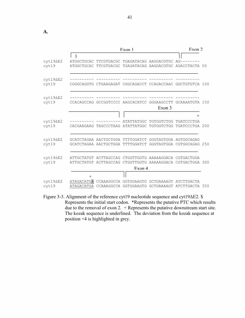
A.
301 350 AF226730 ATAGACATGA CCAAAGGCCA GGTGGAAGTG GCTGAAAAGT ATCTTGACTA cyt19t ATAGACATGA CCAAAGGCCA GGTGGAAGTG GCTGAAAAGT ATCTTGACTA 351 400 AF226730 TCACATGGAA AAATATGGCT TCCAGGCATC TAATGTGACT TTTTTCCATG cyt19t TCACATGGAA AAATATGGCT TCCAGGCATC TAATGTGACT TTTATTCATG 401 450 AF226730 GCAACATTGA GAAGTTGGCA GAGGCTGGAA TCAAGAATGA GAGCCATGAT cyt19t GCTACATTGA GAAGTTGGGA GAGGCTGGAA TCAAGAATGA GAGCCATGAT 451 500 AF226730 ATTGTTGTAT CAAACTGTGT TATTAACCTT GTGCCTGATA AACAACAAGT cyt19t ATTGTTGTAT CAAACTGTGT TATTAACCTT GTGCCTGATA AACAACAAGT B. 1 50 AF226730 MAALRDAEIQ KDVQTYYGQV LKRSADLQTN GCVTTARPVP KHIREALQNV cyt19t MAALRDAEIQ KDVQTYYGQV LKRSADLQTN GCVTTARPVP KHIREALQNV 51 100 AF226730 HEEVALRYYG CGLVIPEHLE NCWILDLGSG SGRDCYVLSQ LVGEKGHVTG cyt19t HEEVALRYYG CGLVIPEHLE NCWILDLGSG SGRDCYVLSQ LVGEKGHVTG 101 150 AF226730 IDMTKGQVEV AEKYLDYHME KYGFQASNVT FFHGNIEKLA EAGIKNESHD cyt19t IDMTKGQVEV AEKYLDYHME KYGFQASNVT FIHGYIEKLG EAGIKNESHD 151 200 AF226730 IVVSNCVINL VPDKQQVLQE AYRVLKHGGE LYFSDVYTSL ELPEEIRTHK cyt19t IVVSNCVINL VPDKQQVLQE AYRVLKHGGE LYFSDVYTSL ELPEEIRTHK Figure 2-1. Sequence alignment of cyt19t and Genbank sequence (accession AF226730).
(A) Nucleotide alignment of cyt19t and Genbank sequence. The four point mutation are in red. (B) Alignment of the deduced cyt19t amino acid sequence and Genbank sequence. The 3 resulting missense mutations are in red.
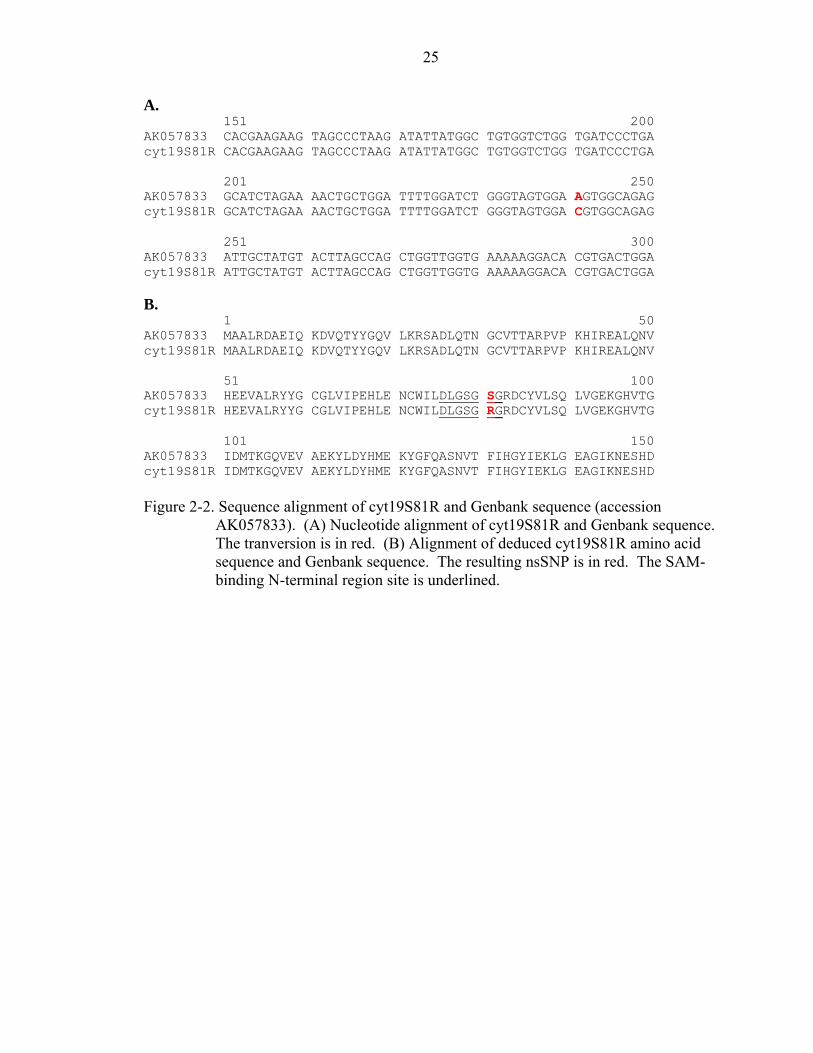
25
A. 151 200 AK057833 CACGAAGAAG TAGCCCTAAG ATATTATGGC TGTGGTCTGG TGATCCCTGA cyt19S81R CACGAAGAAG TAGCCCTAAG ATATTATGGC TGTGGTCTGG TGATCCCTGA 201 250 AK057833 GCATCTAGAA AACTGCTGGA TTTTGGATCT GGGTAGTGGA AGTGGCAGAG cyt19S81R GCATCTAGAA AACTGCTGGA TTTTGGATCT GGGTAGTGGA CGTGGCAGAG 251 300 AK057833 ATTGCTATGT ACTTAGCCAG CTGGTTGGTG AAAAAGGACA CGTGACTGGA cyt19S81R ATTGCTATGT ACTTAGCCAG CTGGTTGGTG AAAAAGGACA CGTGACTGGA B. 1 50 AK057833 MAALRDAEIQ KDVQTYYGQV LKRSADLQTN GCVTTARPVP KHIREALQNV cyt19S81R MAALRDAEIQ KDVQTYYGQV LKRSADLQTN GCVTTARPVP KHIREALQNV 51 100 AK057833 HEEVALRYYG CGLVIPEHLE NCWILDLGSG SGRDCYVLSQ LVGEKGHVTG cyt19S81R HEEVALRYYG CGLVIPEHLE NCWILDLGSG RGRDCYVLSQ LVGEKGHVTG 101 150 AK057833 IDMTKGQVEV AEKYLDYHME KYGFQASNVT FIHGYIEKLG EAGIKNESHD cyt19S81R IDMTKGQVEV AEKYLDYHME KYGFQASNVT FIHGYIEKLG EAGIKNESHD Figure 2-2. Sequence alignment of cyt19S81R and Genbank sequence (accession
AK057833). (A) Nucleotide alignment of cyt19S81R and Genbank sequence. The tranversion is in red. (B) Alignment of deduced cyt19S81R amino acid sequence and Genbank sequence. The resulting nsSNP is in red. The SAM-binding N-terminal region site is underlined.
26
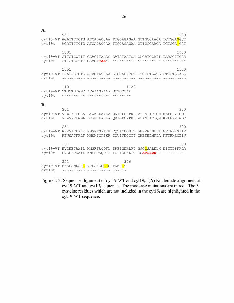
A. 951 1000 cyt19-WT AGATTTTCTG ATCAGACCAA TTGGAGAGAA GTTGCCAACA TCTGGAGGCT cyt19t AGATTTTCTG ATCAGACCAA TTGGAGAGAA GTTGCCAACA TCTGGA.GCT 1001 1050 cyt19-WT GTTCTGCTTT GGAGTTAAAG GATATAATCA CAGATCCATT TAAGCTTGCA cyt19t GTTCTGCTTT GGAGTTAA~~ ~~~~~~~~~~ ~~~~~~~~~~ ~~~~~~~~~~ 1051 1100 cyt19-WT GAAGAGTCTG ACAGTATGAA GTCCAGATGT GTCCCTGATG CTGCTGGAGG cyt19t ~~~~~~~~~~ ~~~~~~~~~~ ~~~~~~~~~~ ~~~~~~~~~~ ~~~~~~~~~~ 1101 1128 cyt19-WT CTGCTGTGGC ACAAAGAAAA GCTGCTAA cyt19t ~~~~~~~~~~ ~~~~~~~~~~ ~~~~~~~~ B. 201 250 cyt19-WT VLWGECLGGA LYWKELAVLA QKIGFCPPRL VTANLITIQN KELERVIGDC cyt19t VLWGECLGGA LYWKELAVLA QKIGFCPPRL VTANLITIQN KELERVIGDC 251 300 cyt19-WT RFVSATFRLF KHSKTGPTKR CQVIYNGGIT GHEKELMFDA NFTFKEGEIV cyt19t RFVSATFRLF KHSKTGPTKR CQVIYNGGIT GHEKELMFDA NFTFKEGEIV 301 350 cyt19-WT EVDEETAAIL KNSRFAQDFL IRPIGEKLPT SGGCSALELK DIITDPFKLA cyt19t EVDEETAAIL KNSRFAQDFL IRPIGEKLPT SGAVLLWS*~ ~~~~~~~~~~ 351 376 cyt19-WT EESDSMKSRC VPDAAGGCCG TKKSC* cyt19t ~~~~~~~~~~ ~~~~~~~~~~ ~~~~~~ Figure 2-3. Sequence alignment of cyt19-WT and cyt19t. (A) Nucleotide alignment of
cyt19-WT and cyt19t sequence. The missense mutations are in red. The 5 cysteine residues which are not included in the cyt19t are highlighted in the cyt19-WT sequence.
27
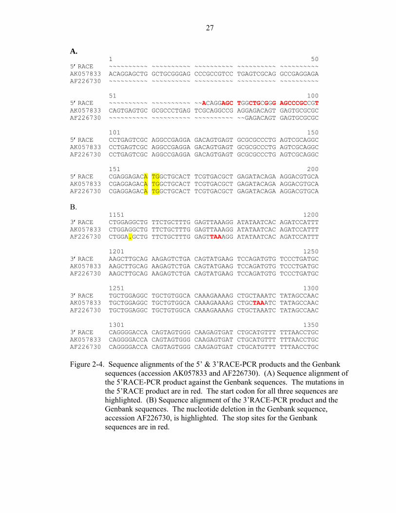
A. 1 50 5’RACE ~~~~~~~~~~ ~~~~~~~~~~ ~~~~~~~~~~ ~~~~~~~~~~ ~~~~~~~~~~ AK057833 ACAGGAGCTG GCTGCGGGAG CCCGCCGTCC TGAGTCGCAG GCCGAGGAGA AF226730 ~~~~~~~~~~ ~~~~~~~~~~ ~~~~~~~~~~ ~~~~~~~~~~ ~~~~~~~~~~ 51 100 5’RACE ~~~~~~~~~~ ~~~~~~~~~~ ~~ACAGGAGC TGGCTGCGGG AGCCCGCCGT AK057833 CAGTGAGTGC GCGCCCTGAG TCGCAGGCCG AGGAGACAGT GAGTGCGCGC AF226730 ~~~~~~~~~~ ~~~~~~~~~~ ~~~~~~~~~~ ~~GAGACAGT GAGTGCGCGC 101 150 5’RACE CCTGAGTCGC AGGCCGAGGA GACAGTGAGT GCGCGCCCTG AGTCGCAGGC AK057833 CCTGAGTCGC AGGCCGAGGA GACAGTGAGT GCGCGCCCTG AGTCGCAGGC AF226730 CCTGAGTCGC AGGCCGAGGA GACAGTGAGT GCGCGCCCTG AGTCGCAGGC 151 200 5’RACE CGAGGAGACA TGGCTGCACT TCGTGACGCT GAGATACAGA AGGACGTGCA AK057833 CGAGGAGACA TGGCTGCACT TCGTGACGCT GAGATACAGA AGGACGTGCA AF226730 CGAGGAGACA TGGCTGCACT TCGTGACGCT GAGATACAGA AGGACGTGCA B. 1151 1200 3’RACE CTGGAGGCTG TTCTGCTTTG GAGTTAAAGG ATATAATCAC AGATCCATTT AK057833 CTGGAGGCTG TTCTGCTTTG GAGTTAAAGG ATATAATCAC AGATCCATTT AF226730 CTGGA.GCTG TTCTGCTTTG GAGTTAAAGG ATATAATCAC AGATCCATTT 1201 1250 3’RACE AAGCTTGCAG AAGAGTCTGA CAGTATGAAG TCCAGATGTG TCCCTGATGC AK057833 AAGCTTGCAG AAGAGTCTGA CAGTATGAAG TCCAGATGTG TCCCTGATGC AF226730 AAGCTTGCAG AAGAGTCTGA CAGTATGAAG TCCAGATGTG TCCCTGATGC 1251 1300 3’RACE TGCTGGAGGC TGCTGTGGCA CAAAGAAAAG CTGCTAAATC TATAGCCAAC AK057833 TGCTGGAGGC TGCTGTGGCA CAAAGAAAAG CTGCTAAATC TATAGCCAAC AF226730 TGCTGGAGGC TGCTGTGGCA CAAAGAAAAG CTGCTAAATC TATAGCCAAC 1301 1350 3’RACE CAGGGGACCA CAGTAGTGGG CAAGAGTGAT CTGCATGTTT TTTAACCTGC AK057833 CAGGGGACCA CAGTAGTGGG CAAGAGTGAT CTGCATGTTT TTTAACCTGC AF226730 CAGGGGACCA CAGTAGTGGG CAAGAGTGAT CTGCATGTTT TTTAACCTGC Figure 2-4. Sequence alignments of the 5’ & 3’RACE-PCR products and the Genbank
sequences (accession AK057833 and AF226730). (A) Sequence alignment of the 5’RACE-PCR product against the Genbank sequences. The mutations in the 5’RACE product are in red. The start codon for all three sequences are highlighted. (B) Sequence alignment of the 3’RACE-PCR product and the Genbank sequences. The nucleotide deletion in the Genbank sequence, accession AF226730, is highlighted. The stop sites for the Genbank sequences are in red.
28
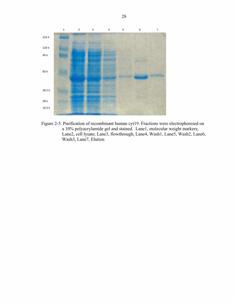
Figure 2-5. Purification of recombinant human cyt19. Fractions were electrophoresed on
a 10% polyacrylamide gel and stained. Lane1, molecular weight markers; Lane2, cell lysate; Lane3, flowthrough, Lane4, Wash1, Lane5, Wash2, Lane6, Wash3, Lane7, Elution
1 2 3 4 5 6 7
215 k 120 k 84 k
60 k
39.2 k 28 k 18.3 k
29
A.
0
200
400
600
800
1000
1200
1400
1600
1800
0 50 100 150 200 250
AsIII (uM)
Spec
ific
Act
ivity
(pm
ole/
mg/
min
)cyt19-WTcyt19R81S
B.
0
100200
300400
500
600700
800900
1000
0 200 400 600 800 1000 1200
MMAV (uM)
Spe
cific
Act
ivity
(pm
ole/
mg/
min
)
cyt19-WTcyt19R81S
Figure 2-6. The effects of AsIII & MMAV concentrations. All incubations were carried
out at 37°C for 30 min. in a final volume of 250 µl. A) Reaction mixtures contained 5 mM GSH, 1 mM DTT, 1 mM MgCl2, 13 pM [3H]SAM, 0.1 mM SAM, various [AsIII], and 5 µg of cyt19, in 100 mM Tris/100 mM Na phosphate, pH 7.4 B) Same as B but with various [MMAv].
30
0
100
200
300
400
500
600
700
5 6 7 8 9 10 11 12
pH
Spe
cific
Act
ivity
(fm
ole/
mg/
min
)
cyt19-WTcyt19R81S
Figure 2-7. The effect of pH on activity. All incubations were carried out at 37°C for 30
min. in a final volume of 250 µl. Reaction mixtures contained 5 mM GSH, 1 mM DTT, 1 mM MgCl2, 13 pM [3H]SAM, 50 µM AsIII, and 5 µg of cyt19, in 100 mM Tris/100 mM Na phosphate of the appropriate pH.
0100200300400500600700800900
1000
5mM GSH 5mMGSH+1mM
DTT
1mM TCEP 1mMTCEP+1mM
GSH
1mMTCEP+5mM
GSH
Reductants
Spec
ific
Act
ivity
(pm
ole/
mg/
min
)
cyt19-WTcyt19R81S
N.D.
Figure 2-8. The effects of reductants on methylation activity. All incubations were
carried out at 37°C for 30 min. in a final volume of 250 µl. Reaction mixtures contained 1 mM MgCl2, 13 pM [3H]SAM, 0.1 mM SAM, 50 µM AsIII, and 5 µg of cyt19-WT, in 100 mM Tris/100 mM Na phosphate, pH 7.4, with different reductants. The activity of cyt19R81S was not determined for 1 mM TCEP + 1 mM GSH.
31
0
50
100
150
200
250
300
350
400
0 200 400 600 800 1000 1200
SAM (uM)
Spe
cific
Act
ivity
(pm
ole/
mg/
min
)
Figure 2-9. The effect of SAM concentration on activity. All incubations were carried
out at 37°C for 30 min. in a final volume of 250 µl. Reaction mixtures contained 5 mM GSH, 1 mM DTT, 1 mM MgCl2, 13 pM [3H]SAM, 50 µM AsIII, and 5 µg of cyt19-WT, in 100 mM Tris/100 mM Na phosphate pH 7.4. with various [SAM].
32
A.
AsIII
0
500
1000
1500
2000
2500
0 5 10 15 20 25
Fraction Number
DPM
B.
MMAV
0
100
200
300
400
500
600
700
0 5 10 15 20 25
Fraction Number
DPM
Figure 2-10. Arsenical metabolites formed after incubation with [3H]SAM and cyt19 for
30 min at 37C. (A) Formation of MMA and DMA using AsIII as a substrate. (B) Formation of MMA and DMA using MMAV as the substrate.
33
CHAPTER 3 IDENTIFICATION OF A SPLICE VARIANT OF HUMAN CYT19 ARSENIC
METHYLTRANSFERASE
Introduction
Arsenic (As) is a naturally occurring element and ranks 20th in abundance in the
earth’s crust [85]. Arsenic is present in the environment in both organic and inorganic
forms and exists mainly in three valence states, -3, +3, and +5. Generally, inorganic
arsenic (iAs) is the more toxic form and people are exposed to iAs primarily through food
and potable water. In Taiwan, Mexico, western United States, western South America,
China, and Bangladesh, people are exposed to high levels of arsenic due to anthropogenic
and/or natural contamination of potable water [12, 62, 86]. In these areas, chronic
exposure to arsenic is associated with various tumors occurring in skin, liver, lung,
urinary bladder, and prostate [8, 87].
Once in the body, many mammals, including humans, methylate iAs to
monomethylarsonic acid (MMA) and dimethylarsinic acid (DMA) [49, 51, 54]. The
biotransformation of iAs alternates between the reduction of arsenate (iAsV) to arsenite
(iAsIII) followed by oxidative methylation (Figure 1) [88, 89]. Because pentavalent
methylated arsenicals are less toxic than inorganic arsenic, methylation has been
considered a detoxification mechanism. Recent studies indicate that trivalent methylated
arsenicals may be more acutely toxic and genotoxic than iAs suggesting that methylation
may actually be a bioactivation of iAs [20, 73, 74, 90]. For this reason the role of
methylation in acute and chronic arsenic toxicity remains unclear. There is significant
34
variation in human susceptibility to As induced toxicity, which may be related to
differences in arsenic biotransformation between individuals [82]. Epidemiological
studies have shown differences in the amount of MMA and DMA excreted in the urine of
exposed populations which may be associated with genetic polymorphisms [62]. Cyt19
has been identified as a human S-adenosyl-L-methionine:arsenic methyltransferase [52,
55] however, only a few coding region polymorphisms have been detected which may
alter the iAs methylation rate [65, 67]. In this study we identified an alternative splice
variant of human cyt19, which contains an upstream open reading frame (uORF)
followed by an internal start codon (AUG). This variant was expressed in 7 out of 7
human livers and represents another possible mechanism for regulating As methylation.
Materials and Methods
Molecular Cloning of cyt19 Splice Variants
Total RNA was isolated from HepG2 cells and human liver samples using Trizol
reagent according to the manufacturer’s instruction (Invitrogen, Carlsbad, USA). Total
RNA was treated with DNase I using the DNA-free™ kit from Ambion (Austin, TX) and
cDNA was made using the RETROscript™ Kit for RT-PCR and 2 µg of RNA as
template (Ambion). HepG2 cDNA was polymerase chain reaction (PCR)-amplified
using the following primers: forward primer (5’-
CACCATGGCTGCACTTCGTGACGCTGAGATACAG-3’) and the reverse primer (5’-
TTAACTCCAAAGCAGAACAGCTCCAGATGT-3’). The PCR reaction consisted of
2.5 U of Pfu DNA polymerase, 0.4 µM each primer, 5µl of the RT reaction, 0.2 mM
dNTP mix, 5µl of 10X PCR Buffer, and nuclease-free water to 50µl. The PCR
conditions were as follows: an initial denaturation at 94°C for 2min, followed by 35
cycles of denaturation at 94°C for 1min, annealing at 60°C for 1min, extension for 72°C
35
for 2min 30s and a final extension at 72°C for 7min. The PCR products, designated
cyt19 and cyt19∆E2, were ligated into the pET100/D-TOPO vector (Invitrogen) and
transformed into competent Escherichia coli (E. coli) One Shot TOP10 chemically
competent cells (Invitrogen). Ampicillin resistant colonies were analyzed by PCR and
visualized by agarose gel electrophoresis. Several clones containing inserts were
sequenced by the DNA Sequencing Core Laboratory at the University of Florida.
Human Liver Samples
Human liver samples were obtained from Vitron (Tucson, AZ). All the tissues
were from Caucasian males between the ages of 24 and 46. The tissues were preserved
in Viaspan after death. The tissue samples were stored at -80°C until use. All procedures
using human samples were approved by the Institutional Review Board at the University
of Florida and all identifying information has been removed.
qPCR of cyt19 Splice Variants
Total RNA was isolated and cDNA synthesized as described above. Real-time
quantitative PCR (qPCR) was carried out using a Bio-Rad iCycler with the following
primers: cyt19 forward primer: (5’-TTCGTGACGCTGAGATACAGAAG-3’); reverse
primer: (5’-TGGAGGTCTGCCGATCTCTT-3’); cyt19∆E2 forward primer: (5’-
GATACAGAAGGACGTGCAGATATTATG-3’); reverse primer: (5’-
CCAGATCCAAAATCCAGCAGTT-3’). Each PCR reaction consisted of 12.5 µl iTaq
SYBR Green Supermix with ROX (Bio-rad), 0.4 µM each primer, 5µl of the RT reaction,
and RNase/DNase-free water to 25 µl. The PCR cycling conditions included an initial
denaturation of 95°C for 3 min followed by cycling at 95°C for 15s, 60°C for 45s for 45
cycles. The constructs, pET100-cyt19 and pET100-cyt19∆E2 were used to generate
36
calibration curves for quantification of cyt19 and cyt19∆E2. A melting curve analysis
was performed after every run to determine product uniformity.
Results
When the full open reading frame of cyt19 was amplified using the primers
described in Molecular Cloning of cyt19 splice variants under Materials & Methods, two
products were generated, 1132 bp (cyt19) and 1005 bp (cyt19∆E2) products (Figure 3-2).
Sequencing of these two products revealed that the 1132 bp product is the reference cyt19
while the 1005 bp product is a splice variant (Figure 3-3). The reference cyt19 mRNA is
composed of 10 exons (Figure 3-4A) which encode a 375 amino acid protein with a
theoretical molecular weight of 41.747 kDa (Figure 3-5). The product, cyt19∆E2, is
missing 128 bp due to a deletion of exon 2 which could result in a protein that is about
102 amino acids shorter and the creation of a short 24 amino acid peptide as a product
from an upstream open reading frame (uORF) (Figure 3-3 & Figure 3-4B). This variant
may encode a 273 amino acid polypeptide chain that is identical to the reference cyt19
but lacks the first 102 amino acids present in the reference (Figure 3-5). Analysis of the
sequences surrounding the splice revealed that the splice occurs at conserved acceptor
and donor sites (Figure 3-4C). The individual information (Ri) technique and Shapiro’s
method were used to compare the splice-sites strength of exon 3 and exon 2 of cyt19
[91]. Exon3, a constitutive exon has a stronger splice site compared to exon2, the
alternative exon (Table 3-1).
The steady state mRNA levels of each transcript were determined in 7 human liver
samples as well as in HepG2 cells by qPCR (Table 3-2). Expression of cyt19 mRNA in
the human liver samples ranged from 41004.7 × to 61026.1 × copies per microgram of
37
RNA. The cyt19∆E2 splice variant was detected in all 7 human liver samples tested.
(Table 3-2). The amount of cyt19∆E2 mRNA was much lower than cyt19 mRNA
ranging from 31089.1 × through 31033.4 × copies per microgram of RNA. The HepG2
cells had an average of 61014.5 × copies/µg RNA and 41055.8 × copies/µg RNA of cyt19
and cyt19∆E2 mRNA, respectively.
Discussion
In this study, we identified an alternative splice variant of cyt19, which contains an
uORF. The variant mRNA contains a short ORF followed by an internal AUG codon
beginning 106bp downstream from the uORF (Figure 3-3). While this alternative variant
may encode a 273 amino acid protein it is unlikely that expression of the cyt19∆E2 splice
variant will result in production of an active protein. Studies have shown that SAM
dependent methyltransferases share 3 regions of sequence similarity (motif I, II, and III)
[92]. These motifs are found in the same order on the polypeptide chain and separated by
similar intervals [92]. It has been suggested that these conserved regions are important in
SAM binding [92]. Mutations of a conserved amino acid in rat guanidinoacetate
methyltransferase near motif I have resulted in an inactive enzyme. In addition,
mutations of motif II lead to reduced Kcat/Km values for substrates [93]. It is unlikely
that the protein translated from cyt19∆E2 would result in an active protein due to the
removal of motif I (Figure 3-5).
Whether the mRNA actually is translated into protein is not clear because the
internal AUG codon contains a relatively weak Kozak sequence suggesting that
translation may not reinitiate at the internal start codon. The sequence
(GCCA/GCCATGG) is a consensus Kozak sequence for the initiation of translation in
38
vertebrates [94, 95]. Deviation from the consensus sequence at position -3 and +4 would
be considered a weak initiator codon. The cyt19∆E2 transcript deviates from the
consensus sequence at position +4; the variant contains an A instead of a G (Figure 3-3).
It is also possible that cyt19∆E2 will not be translated but that this variant is a
substrate for the nonsense mediated decay (NMD) pathway due to the premature stop
codon. NMD is a pathway that recognizes and quickly degrades mRNAs containing
premature translation termination codons (PTC) in eukaryotes [96]. While cyt19∆E2
does contain a PTC, Zhang et al. identified a sequence motif which when present 3’ of a
nonsense codon promotes rapid decay of the mRNA transcript by the NMD pathway
[97]. This sequence motif (TGYYGATGYYYYY) is not found in the cyt19∆E2 mRNA
transcript and it remains unclear if this variant will undergo degradation by the NMD
pathway.
The cyt19∆E2 variant was present in all seven human liver samples tested,
suggesting that cyt19 mRNA exists both in the full length and alternatively spliced form
in most individuals. The cyt19∆E2 variant mRNA comprised 0.2 to 3.8% of the total
cyt19 transcript. The liver samples had lower copy numbers per microgram of RNA of
both reference and cyt19∆E2 variants compared to HepG2 cells. It is possible that some
degradation of cyt19 message occurred during collection and storage of the livers which
reduced apparent copy number.
Many mammalian species methylate arsenic through an enzymatic reaction that is
performed by cyt19. There are significant variations in the arsenic methylation capacity
between species and within species including humans [58, 59, 62, 81]. The reason for
this variation is unclear but has been attributed to cyt19 polymorphisms. However, only
39
a few polymorphisms have been found in the coding region of cyt19 to date [65, 67],
while the vast majority of mutations are found within the introns and the 5’ and 3’
untranslated region (UTR). Introns contain the acceptor site, branchpoint,
polypyrimidine tract, and the donor site, which are conserved sequences in which
splicesome assembly occurs [70]. While mutations within these sequence elements could
alter the constitutive splicing of a gene [98-100], there are differences in arsenic
methylation capacities among individuals, which are unlikely to be supported solely by
polymorphisms within the cyt19 coding region.
Alternative splicing is frequently used to regulate gene expression and to generate
tissue-specific mRNA and protein isoforms [68, 69]. Thirty-five to 60% of human genes
produce transcripts that are alternatively spliced, in addition 70-90% of these variants
alter the resulting protein products [101, 102]. Further studies should analyze the mRNA
expression levels of cyt19 splice variants in a larger number of fresh liver samples or
primary hepatocytes and correlate it to arsenic methylation activity. In addition, work to
determine if this transcript is a substrate for the NMD pathway or if a variant protein is
expressed will help clarify the role of cyt19∆E2 in human arsenic metabolism. Even
though the splice variant comprises a relatively small fraction of the total cyt19 transcript
in the livers tested it is possible that different population groups have varying amounts of
the cyt19 splice variant. It is also likely that the level of cyt19∆E2 in an individual will
change over time as alternative splice selection can be controlled by many variables
including developmental stage and xenobiotics [103, 104]. In conclusion, cyt19 appears
to be alternatively spliced in many individuals and may play a role in the observed
variation in arsenic methylation seen in individuals.
40
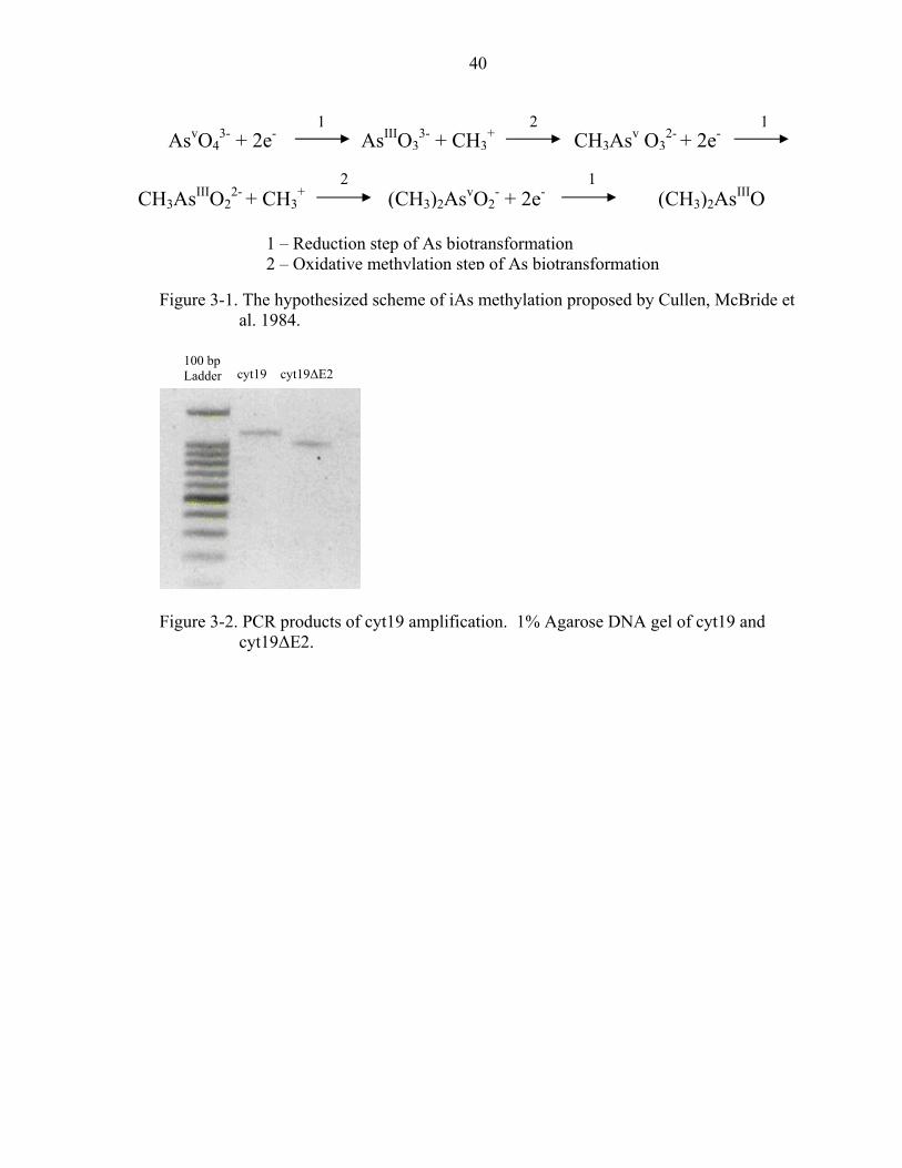
Figure 3-1. The hypothesized scheme of iAs methylation proposed by Cullen, McBride et
al. 1984.
Figure 3-2. PCR products of cyt19 amplification. 1% Agarose DNA gel of cyt19 and
cyt19∆E2.
1 – Reduction step of As biotransformation 2 – Oxidative methylation step of As biotransformation
CH3AsIIIO22- + CH3
+ (CH3)2AsvO2- + 2e- (CH3)2AsIIIO
2 1
AsvO43- + 2e- AsIIIO3
3- + CH3+ CH3Asv O3
2- + 2e- 1 2 1
cyt19 cyt19∆E2 100 bp Ladder
41
A.
$ cyt19∆E2 ATGGCTGCAC TTCGTGACGcyt19 ATGGCTGCAC TTCGTGACG cyt19∆E2 ~~~~~~~~~~ ~~~~~~~~~cyt19 CGGGCAGGTG CTGAAGAGA cyt19∆E2 ~~~~~~~~~~ ~~~~~~~~~cyt19 CCACAGCCAG GCCGGTCCC cyt19∆E2 ~~~~~~~~~~ ~~~~~~~~~cyt19 CACGAAGAAG TAGCCCTAA cyt19∆E2 GCATCTAGAA AACTGCTGGcyt19 GCATCTAGAA AACTGCTGG cyt19∆E2 ATTGCTATGT ACTTAGCCAcyt19 ATTGCTATGT ACTTAGCCA + cyt19∆E2 ATAGACATGA CCAAAGGCCcyt19 ATAGACATGA CCAAAGGCC Figure 3-3. Alignment of the reference
Represents the initial start cdue to the removal of exon The kozak sequence is undeposition +4 is highlighted in
1
ExonC TGAGATACAG AAGGACGTGC AG~~~C TGAGATACAG AAGGACGTGC AGACC
~ ~~~~~~~~~~ ~~~~~~~~~~ ~~~~~T CGGCAGACCT CCAGACCAAC GGCTG
~ ~~~~~~~~~~ ~~~~~~~~~~ ~~~~~C AAGCACATCC GGGAAGCCTT GCAAA
~ ATATTATGGC TGTGGTCTGG TGATCG ATATTATGGC TGTGGTCTGG TGATC
A TTTTGGATCT GGGTAGTGGA AGTGGA TTTTGGATCT GGGTAGTGGA CGTGG
G CTGGTTGGTG AAAAAGGACA CGTGAG CTGGTTGGTG AAAAAGGACA CGTGA
A GGTGGAAGTG GCTGAAAAGT ATCTTA GGTGGAAGTG GCTGAAAAGT ATCTT
cyt19 nucleotide sequence and cyt1odon. *Represents the putative PTC2. + Represents the putative downsrlined. The deviation from the koza grey.
3
4
Exon 2
~~~~~ TACTA 50
~~~~~ TGTCA 100
~~~~~ ATGTA 150
Exon
* CCTGA CCTGA 200
CAGAG CAGAG 250
CTGGA CTGGA 300
Exon
GACTA GACTA 3509∆E2. $ which results
tream start site. k sequence at
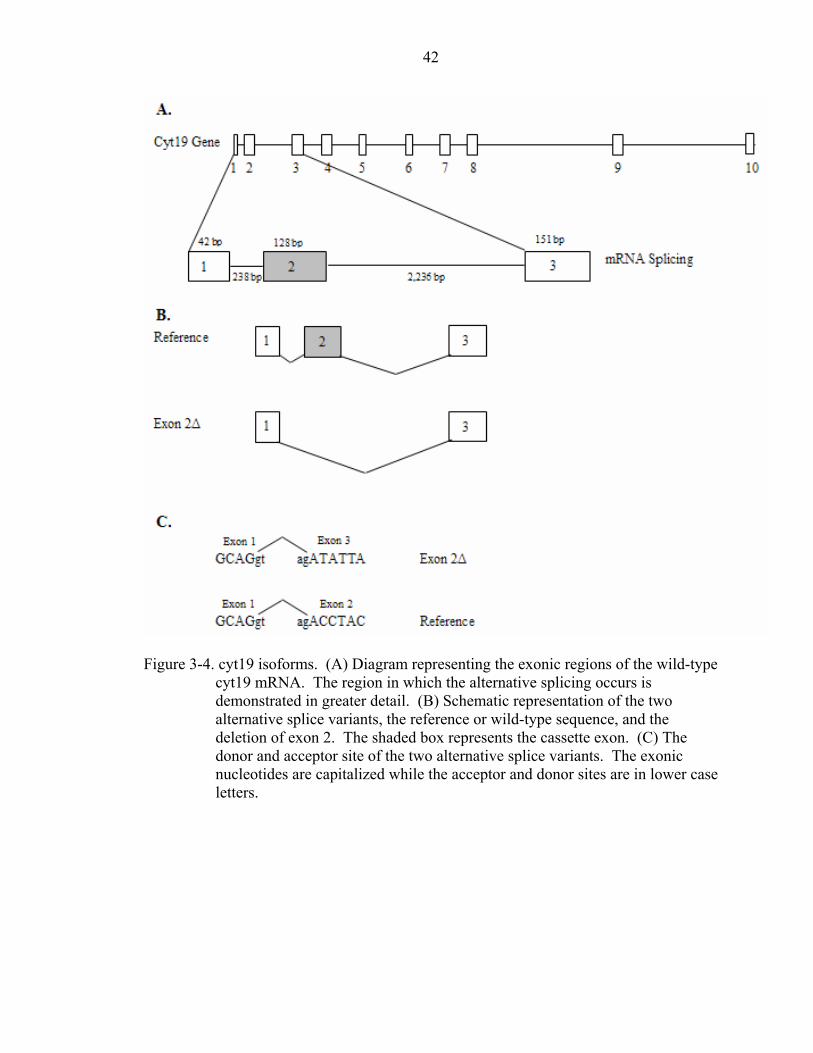
42
Figure 3-4. cyt19 isoforms. (A) Diagram representing the exonic regions of the wild-type
cyt19 mRNA. The region in which the alternative splicing occurs is demonstrated in greater detail. (B) Schematic representation of the two alternative splice variants, the reference or wild-type sequence, and the deletion of exon 2. The shaded box represents the cassette exon. (C) The donor and acceptor site of the two alternative splice variants. The exonic nucleotides are capitalized while the acceptor and donor sites are in lower case letters.
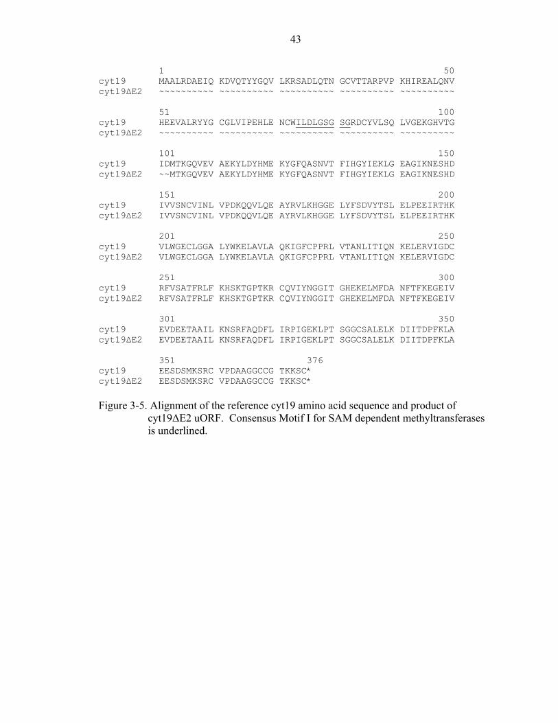
43
1 50 cyt19 MAALRDAEIQ KDVQTYYGQV LKRSADLQTN GCVTTARPVP KHIREALQNV cyt19∆E2 ~~~~~~~~~~ ~~~~~~~~~~ ~~~~~~~~~~ ~~~~~~~~~~ ~~~~~~~~~~ 51 100 cyt19 HEEVALRYYG CGLVIPEHLE NCWILDLGSG SGRDCYVLSQ LVGEKGHVTG cyt19∆E2 ~~~~~~~~~~ ~~~~~~~~~~ ~~~~~~~~~~ ~~~~~~~~~~ ~~~~~~~~~~ 101 150 cyt19 IDMTKGQVEV AEKYLDYHME KYGFQASNVT FIHGYIEKLG EAGIKNESHD cyt19∆E2 ~~MTKGQVEV AEKYLDYHME KYGFQASNVT FIHGYIEKLG EAGIKNESHD 151 200 cyt19 IVVSNCVINL VPDKQQVLQE AYRVLKHGGE LYFSDVYTSL ELPEEIRTHK cyt19∆E2 IVVSNCVINL VPDKQQVLQE AYRVLKHGGE LYFSDVYTSL ELPEEIRTHK 201 250 cyt19 VLWGECLGGA LYWKELAVLA QKIGFCPPRL VTANLITIQN KELERVIGDC cyt19∆E2 VLWGECLGGA LYWKELAVLA QKIGFCPPRL VTANLITIQN KELERVIGDC 251 300 cyt19 RFVSATFRLF KHSKTGPTKR CQVIYNGGIT GHEKELMFDA NFTFKEGEIV cyt19∆E2 RFVSATFRLF KHSKTGPTKR CQVIYNGGIT GHEKELMFDA NFTFKEGEIV 301 350 cyt19 EVDEETAAIL KNSRFAQDFL IRPIGEKLPT SGGCSALELK DIITDPFKLA cyt19∆E2 EVDEETAAIL KNSRFAQDFL IRPIGEKLPT SGGCSALELK DIITDPFKLA 351 376 cyt19 EESDSMKSRC VPDAAGGCCG TKKSC* cyt19∆E2 EESDSMKSRC VPDAAGGCCG TKKSC* Figure 3-5. Alignment of the reference cyt19 amino acid sequence and product of
cyt19∆E2 uORF. Consensus Motif I for SAM dependent methyltransferases is underlined.
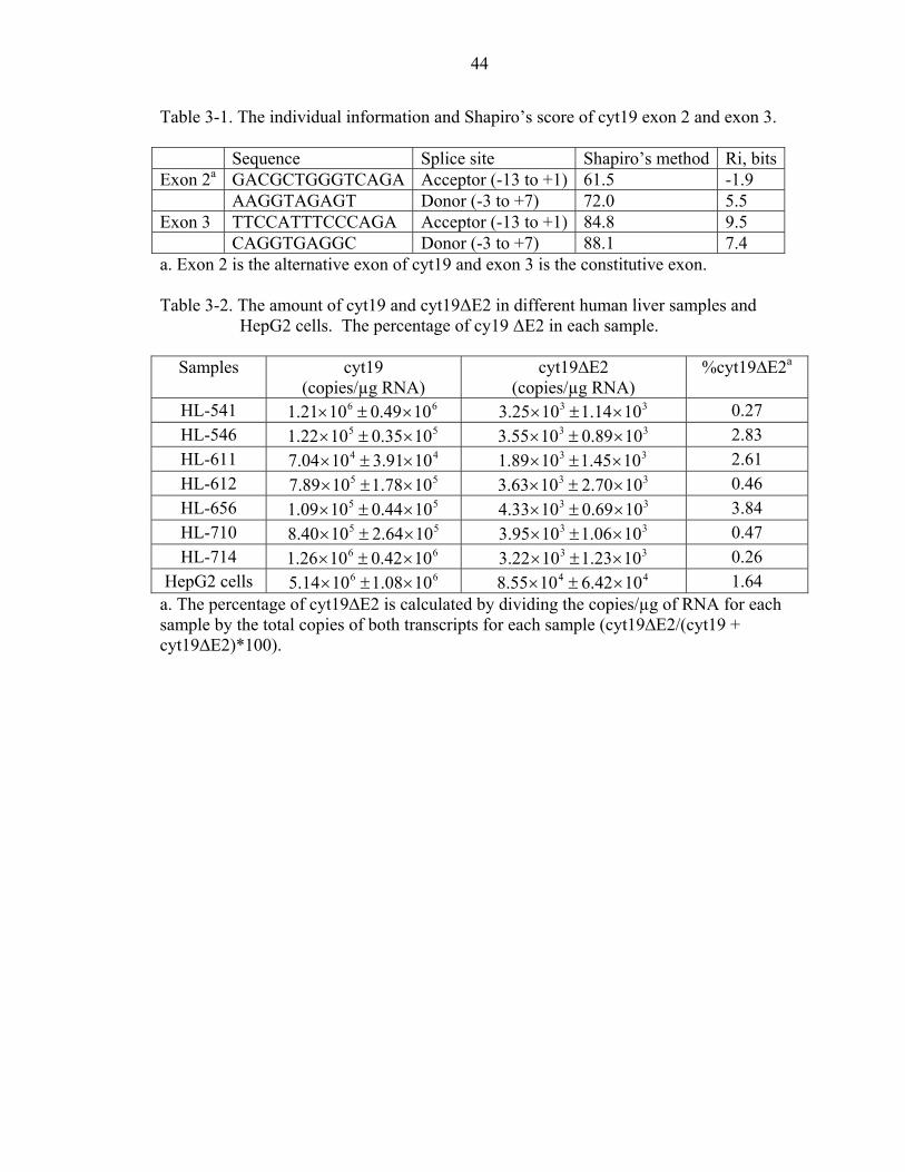
44
Table 3-1. The individual information and Shapiro’s score of cyt19 exon 2 and exon 3. Sequence Splice site Shapiro’s method Ri, bitsExon 2a GACGCTGGGTCAGA Acceptor (-13 to +1) 61.5 -1.9 AAGGTAGAGT Donor (-3 to +7) 72.0 5.5 Exon 3 TTCCATTTCCCAGA Acceptor (-13 to +1) 84.8 9.5 CAGGTGAGGC Donor (-3 to +7) 88.1 7.4 a. Exon 2 is the alternative exon of cyt19 and exon 3 is the constitutive exon. Table 3-2. The amount of cyt19 and cyt19∆E2 in different human liver samples and
HepG2 cells. The percentage of cy19 ∆E2 in each sample.
Samples
cyt19 (copies/µg RNA)
cyt19∆E2 (copies/µg RNA)
%cyt19∆E2a
HL-541 66 1049.01021.1 ×±× 33 1014.11025.3 ×±× 0.27 HL-546 55 1035.01022.1 ×±× 33 1089.01055.3 ×±× 2.83 HL-611 44 1091.31004.7 ×±× 33 1045.11089.1 ×±× 2.61 HL-612 55 1078.11089.7 ×±× 33 1070.21063.3 ×±× 0.46 HL-656 55 1044.01009.1 ×±× 33 1069.01033.4 ×±× 3.84 HL-710 55 1064.21040.8 ×±× 33 1006.11095.3 ×±× 0.47 HL-714 66 1042.01026.1 ×±× 33 1023.11022.3 ×±× 0.26
HepG2 cells 66 1008.11014.5 ×±× 44 1042.61055.8 ×±× 1.64 a. The percentage of cyt19∆E2 is calculated by dividing the copies/µg of RNA for each sample by the total copies of both transcripts for each sample (cyt19∆E2/(cyt19 + cyt19∆E2)*100).
45
CHAPTER 4 GENERAL CONCLUSIONS
The first study showed that cyt19 is an arsenic methyltransferase. In addition, we
have demonstrated that this enzyme does both methylation steps involved in arsenic
biotransformation seen in humans. We also showed that the C-terminus is critical in the
activity of the protein. In other SAM-dependent methyltransferases, the C-terminus is
important in substrate binding. This indicates that the cysteine rich C-terminus is
important for As binding and critical for activity. Others have shown that mutations can
change the methylation capacity of the protein. Here, we demonstrated that a single
mutation can drastically change the activity of the protein even though we believed that
the amino acid change would not have a significant effect on the activity of the
recombinant protein. This change occurs in motif I of the SAM-binding site which might
inhibit its ability to bind SAM therefore decreasing its activity. The mutation appears to
cause a change in the substrate affinity of the proteins as well as cause different
methylation profiles specifically for arsenite. However, the Vmax values of both cyt19-
WT and cyt19R81S are considerably higher than that seen among other mammals such as
the hamster, rabbit, and rhesus monkey. The kinetic analysis of these proteins may
explain the high levels of MMA excreted in human urine. Possibly, arsenite is converted
very quickly to MMA, allowing it to accumulate before the dimethylation step resulting
in the higher excretion of MMA seen in humans. The human arsenic methyltransferase
did have some similarities with the other mammalian arsenic methyltransferases. These
arsenic methyltransferases have been shown to increase in activity at basic pHs which
46
may be due to the deprotonation of cysteines at higher pHs, which increases the rate of
binding between arsenic and cysteines in the substrate binding domain. Our results
demonstrate that only a strong reductant is necessary for methylation of arsenic by cyt19,
however, the addition of GSH appears to increase the activity above the reductant alone.
The second study introduced another possible explanation for the variability in
arsenic methylation capacities among individuals, alternative splicing. Alternative
splicing is frequently used to regulate gene expression and to generate tissue-specific
mRNA and protein isoforms. Thirty-five to 60% of human genes produce transcripts that
are alternatively spliced, in addition 70-90% of these variants alter the resulting protein
products. In this study we identified an alternative splice variant of the human cyt19
(cyt19∆E2), in which exon 2 is removed creating a bicistronic transcript. The cyt19∆E2
variant was present in all seven human liver samples tested, suggesting that cyt19 mRNA
exists both in the full length and in alternatively spliced forms in most individuals. It is
unlikely that this variant would result in expression of an active protein. Studies have
shown that SAM dependent methyltransferases share 3 regions of sequence similarity
(motif I, II, and III). It has been suggested that these conserved regions are important in
SAM binding. Therefore, it is unlikely that the protein translated from cyt19∆E2 would
result in an active protein due to the removal of exon 2 which cotains motif I. Whether
the mRNA actually is translated into protein has not been determined. The majority of
mutations discovered within the cyt19 gene occur within the intron or untranslated
regions [66, 67]. In fact, only 4 mutations have been found within the coding region.
Introns contain sequence elements in which splicesome assembly occurs [66, 67].
Mutations within these elements could alter the constitutive splicing of a gene.
47
Future studies should focus on isolating the human AS3MT from human livers.
To this date, preparations from human livers, cytosolic or homogenates have shown no
activity in arsenic methylation. Reasons for this may be due to inhibitory factors present
in the liver preparation or the process of making the preparations may render the protein
inactive. Yet another explanation may be due to the possibility that cyt19 is an inducible
protein. Perhaps the average daily exposure does not cause high levels of expression of
the protein, making it difficult to purify from liver samples. It may be beneficial to
attempt protein purification from known higher than normal arsenic exposed populations.
Perhaps populations exposed to higher than normal levels have reached a threshold of
exposure resulting in higher expression levels of cyt19. Another possibility for
purification of cyt19 from normally exposed populations may be by immunoprecipitation
from human liver preparations using an antibody which is highly selective for cyt19.
Further studies should analyze the mRNA expression levels of cyt19 splice
variants in a larger number of fresh liver samples or primary hepatocytes and correlate it
to arsenic methylation activity. In addition, work to determine if this transcript is a
substrate for the NMD pathway or if a variant protein is expressed will help clarify the
role of cyt19∆E2 in human arsenic metabolism. In addition, more investigations should
look at mutations within the intron of cyt19 and determine if these alter splicing events.
Another important study would be to establish a method to test exposed populations at
both the mRNA and protein levels of cyt19. This study would help determine vulnerable
individuals in high arsenic exposed populations. Finally, the ultimate question which still
remains unanswered: is arsenic methylation a bioactivation or detoxification mechanism.

48
If time and funding were not a factor, I would enjoy working on solving these different
unanswered questions which may help identifying possible vulnerable populations.
49
APPENDIX ROLE OF CYT19 IN ACUTE ARSENIC TOXICITY. IS CYT19 THE ONLY HUMAN
ARSENIC METHYLTRANSFERASE?
While my work has clearly demonstrated that cyt19 is a human arsenic
methyltransferase, it is not clear that it is the only arsenic methyltransferase in humans.
In order to address this question, it is necessary to: 1) alter the expression of cyt19 and 2)
have the ability to measure cyt19 protein levels. The first challenge was addressed by the
use of small interfering RNA (siRNA) to reduce the expression of cyt19. The second
challenge was addressed by developing an antibody specific for human cyt19.
Small RNAs can theoretically be used to reduce the expression of any target gene.
There are two main categories of small RNA, microRNA (miRNA) and siRNA. These
small RNA have natural functions such as defense from viral and transposon invasion as
well as gene regulation. Scientists have used this new technology for several reasons
such as determination of gene function, validating drug targets, and treatment of diseases.
Small RNAs have two mechanisms by which protein translation is inhibited. If the small
RNA is 100% homologous to its mRNA target it results in degradation of the mRNA.
However, if the small RNA is not 100% identical to its mRNA target it results in
inhibition of translation without mRNA degradation [105].
Materials and Methods
Multiple siRNAs were designed and synthesized according to the Silencer™
siRNA Construction Kit (Ambion). Forty-five thousand HepG2 cells were plated on a 24
well plate in normal growth media overnight and transfected in duplicates with the
50
appropriate siRNA according to Silencer™ siRNA Trasnfection Kit. Total RNA was
isolated 2 days after transfection. The most efficient siRNA was assessed by measuring
the mRNA levels of cyt19 normalized to RNA polymerase II (RPII) by quantitative PCR
(qPCR). Once the most efficient siRNA is determined, further optimization steps can be
taken such as cell plating density, transfection agent, and siRNA amount. The siRNA
pool was further optimized in a 96-well plate using 16,000 cells per well and assayed.
The mRNA levels were measured by qPCR as above, and the viability was determined by
the XTT assay. GAPDH was used as negative control.
An antibody to cyt19 was developed in rabbits by sending purified recombinant
cyt19 to Cocalico. The antibody specificity was determined from western blots of
purified protein and HepG2 cell extracts. To increase the specificity of the antibody, it
was affinity purified using the AminoLink® Kit (Pierce) according the manufacturer’s
instructions. Briefly, the purified cyt19 protein was coupled to the gel followed by
affinity purification.
Results and Discussion
cyt19 mRNA Knockdown by siRNA
Three different siRNAs were designed (Table 1). The efficiency of all three
siRNAs as well as a pool of the three siRNAs was determined (Figure 1). The data
demonstrates that the knockdown was successful with the pool of siRNAs, however
further optimization is still required. It appears that the first three siRNAs had no effect
on the cyt19 mRNA levels. The pool of all three siRNAs appears to knockdown cyt19
mRNA levels by about 26%, relative to control. The second attempt at cyt19 mRNA
knockdown demonstrated that 16,000 cells per well in a 96-well plate resulted in a
greater knockdown of cyt19 mRNA of about 40% compared to 45,000 cells used in the
51
first attempt (Figure 2). It appears that cyt19 mRNA levels might have been slightly
reduced in the negative control. In addition, the siRNA transfection did not result in a
decrease of viability relative to control (Figure 3). The data demonstrates that the
optimal cell density and the transfection agent are important to optimize. Taken together
the results suggest that the concentration of the siRNA requires further optimization or
perhaps new siRNAs can be designed and tested.
Antibody Specificity and Purification
Crude antiserum was shown to be able to detect the antigen up to about 25 ng
(Figure 4). The antibody was not able to detect cyt19 in any of the human liver cytosol
preparations tested. In addition, the antibody appears not to be very specific (Figure 5).
Once purified, the antibody was characterized and found to be able to detect the antigen
up to about 40 ng (Figure 6). The specificity of the antibody was increased dramatically
and is able to detect cyt19 in all tested human liver samples and in HepG2 cytosol tested
(Figure 7). In order to determine if the antibody is in fact recognizing cyt19 in these
cytosol preparations, the protein must be immunoprecipitated and sequenced.
Future Experiments
Further studies are needed in order to better understand the role of arsenic
methylation in acute human arsenic exposures. However, two very important steps in
answering this question have been addressed, knockdown of cyt19 and the ability to
measure cyt19 protein levels through antibody specificity. We have to determine the
correlation between mRNA levels and protein levels. It may be possible to knockdown
the mRNA levels without affecting the protein levels if the protein has a long half-life.
The opposite may be true as well, we may not see much affect in the mRNA levels but
the siRNA may be able to suppress protein expression thereby reducing methylation.
52
Once we prove the protein levels are down, we can determine the effects of As exposure
as well as determine if cyt19 is the only arsenic methyltransferase in humans.
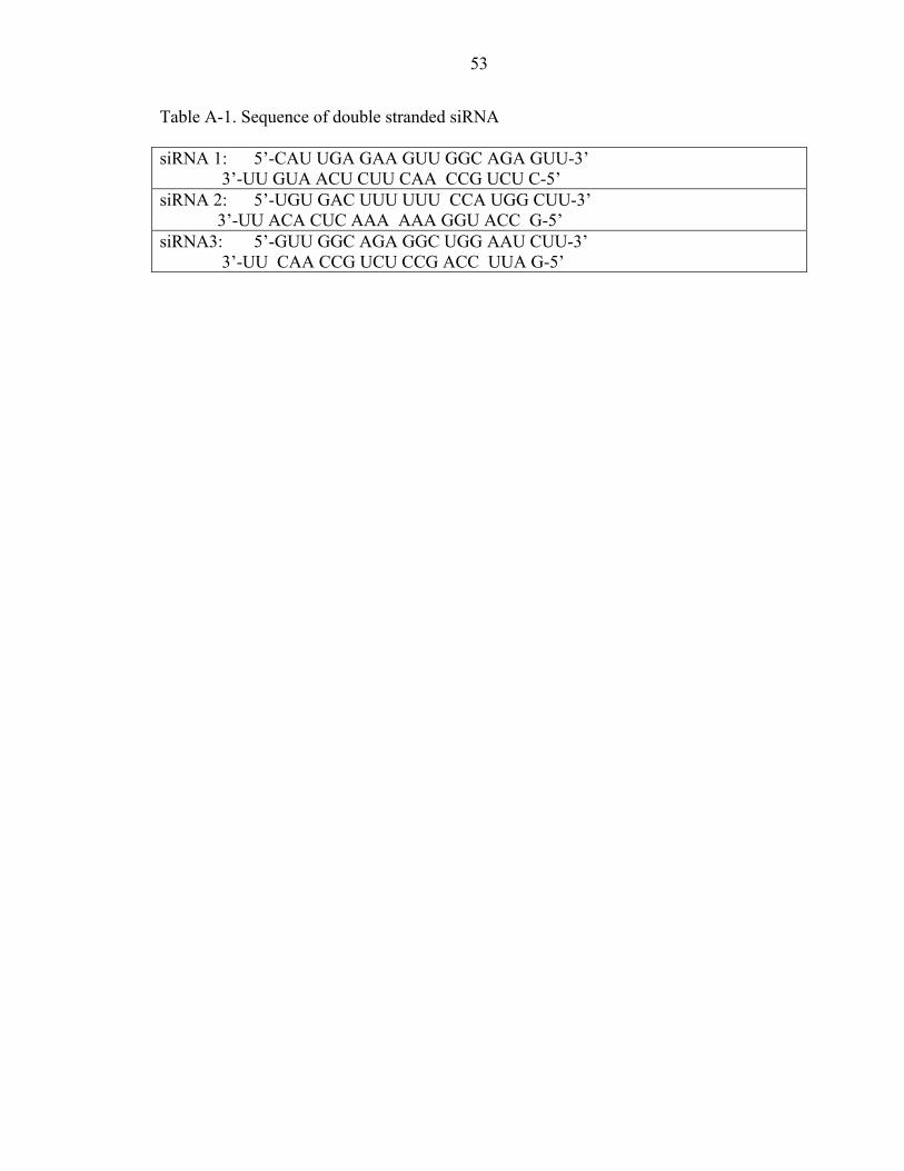
53
Table A-1. Sequence of double stranded siRNA siRNA 1: 5’-CAU UGA GAA GUU GGC AGA GUU-3’ 3’-UU GUA ACU CUU CAA CCG UCU C-5’ siRNA 2: 5’-UGU GAC UUU UUU CCA UGG CUU-3’ 3’-UU ACA CUC AAA AAA GGU ACC G-5’ siRNA3: 5’-GUU GGC AGA GGC UGG AAU CUU-3’ 3’-UU CAA CCG UCU CCG ACC UUA G-5’
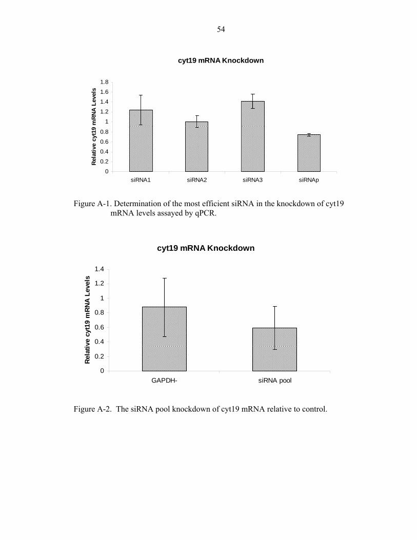
54
cyt19 mRNA Knockdown
0
0.2
0.4
0.6
0.8
1
1.2
1.4
1.6
1.8
siRNA1 siRNA2 siRNA3 siRNAp
Rel
ativ
e cy
t19
mR
NA
Lev
els
Figure A-1. Determination of the most efficient siRNA in the knockdown of cyt19
mRNA levels assayed by qPCR.
cyt19 mRNA Knockdown
0
0.2
0.4
0.6
0.8
1
1.2
1.4
GAPDH- siRNA pool
Rela
tive
cyt1
9 m
RNA
Leve
ls
Figure A-2. The siRNA pool knockdown of cyt19 mRNA relative to control.
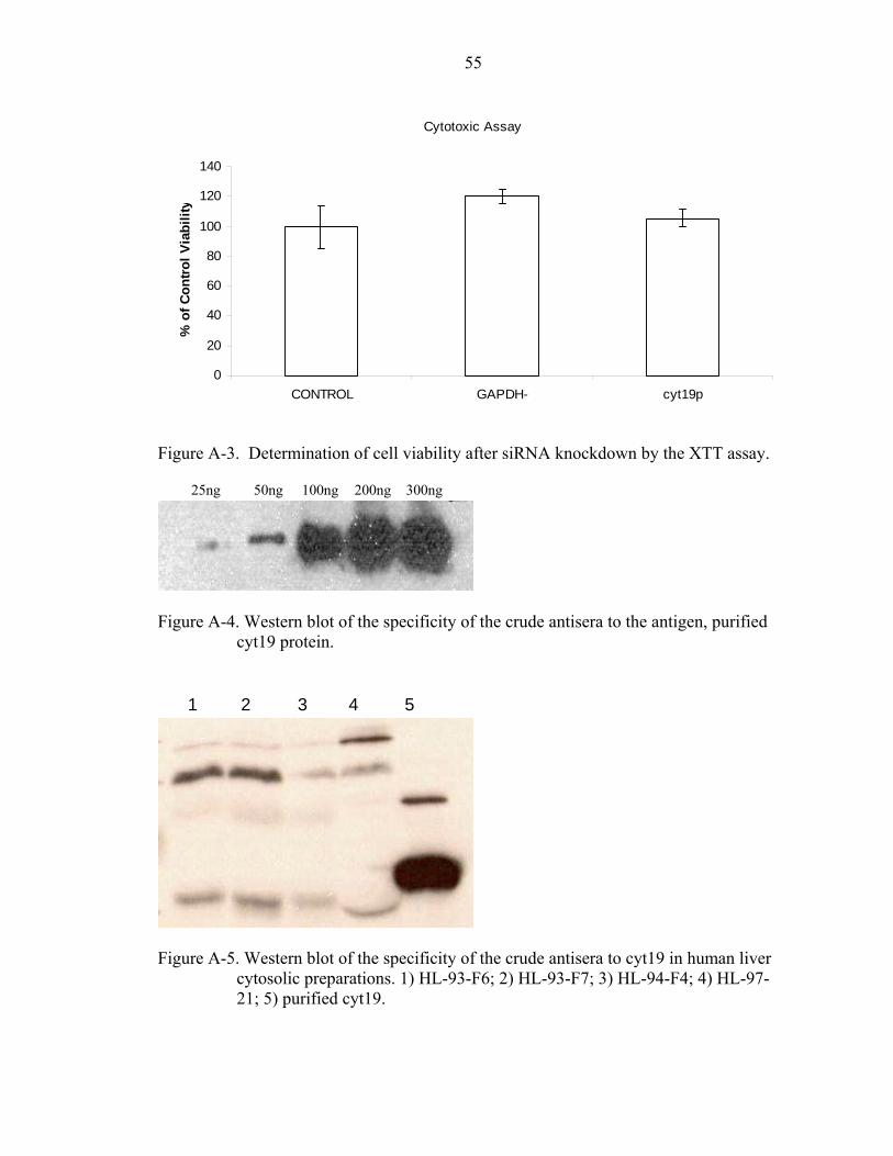
55
Cytotoxic Assay
0
20
40
60
80
100
120
140
CONTROL GAPDH- cyt19p
% o
f Con
trol V
iabi
lity
Figure A-3. Determination of cell viability after siRNA knockdown by the XTT assay.
Figure A-4. Western blot of the specificity of the crude antisera to the antigen, purified
cyt19 protein.
Figure A-5. Western blot of the specificity of the crude antisera to cyt19 in human liver
cytosolic preparations. 1) HL-93-F6; 2) HL-93-F7; 3) HL-94-F4; 4) HL-97-21; 5) purified cyt19.
1 4 53 2
25ng 50ng 100ng 200ng 300ng
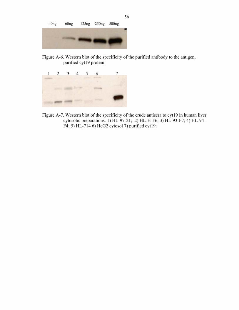
56
Figure A-6. Western blot of the specificity of the purified antibody to the antigen,
purified cyt19 protein.
Figure A-7. Western blot of the specificity of the crude antisera to cyt19 in human liver
cytosolic preparations. 1) HL-97-21; 2) HL-H-F6; 3) HL-93-F7; 4) HL-94-F4; 5) HL-714 6) HeG2 cytosol 7) purified cyt19.
40ng 125ng 60ng 250ng 500ng
1 3 2 4 5 76
57
LIST OF REFERENCES
1. NAS, Arsenic: Medical and Biologic Effects of Environmental Pollutants. 1977, Washington, D.C.
2. Maher, WA, The Presence of Arsenobetaine in Marine Animals. Comp Biochem Physiol C, 1985. 80(1): p. 199-201.
3. ATSDR, Toxicological Profile for Arsenic (Draft for Public Comment). 2005, Atlanta, GA: U.S. Department of Health and Human Services, Public Health Service.
4. Brooks, WE. Arsenic. U.S. Geological Survey, Mineral Commodity Studies. 2005 [accessed 2006 April]; Available from: http://minerals.usgs.gov/minerals/pubs/commodity/arsenic/arsenmcs05.pdf.
5. Jelinek, CF and Corneliussen, PE, Levels of Arsenic in the United States Food Supply. Environ Health Perspect, 1977. 19: p. 83-7.
6. Aronson, SM, Arsenic and Old Myths. R I Med, 1994. 77(7): p. 233-4.
7. Miller, WH, Jr., Schipper, HM, Lee, JS, Singer, J, and Waxman, S, Mechanisms of Action of Arsenic Trioxide. Cancer Res, 2002. 62(14): p. 3893-903.
8. Abernathy, CO, Thomas, DJ, and Calderon, RL, Health Effects and Risk Assessment of Arsenic. J Nutr, 2003. 133(5 Suppl 1): p. 1536S-8S.
9. EPA. Arsenic Treatment Technologies for Soil, Waste, and Water. 2002 [cited 2006 March 2].
10. NRC, Arsenic in Drinking Water. 2001, Washington, DC: National Academy Press.
11. Chowdhury, UK, Biswas, BK, Chowdhury, TR, Samanta, G, Mandal, BK, Basu, GC, Chanda, CR, Lodh, D, Saha, KC, Mukherjee, SK, Roy, S, Kabir, S, Quamruzzaman, Q, and Chakraborti, D, Groundwater Arsenic Contamination in Bangladesh and West Bengal, India. Environ Health Perspect, 2000. 108(5): p. 393-7.
58
12. Frost, FJ, Muller, T, Petersen, HV, Thomson, B, and Tollestrup, K, Identifying Us Populations for the Study of Health Effects Related to Drinking Water Arsenic. J Expo Anal Environ Epidemiol, 2003. 13(3): p. 231-9.
13. Yoshida, T, Yamauchi, H, and Fan Sun, G, Chronic Health Effects in People Exposed to Arsenic Via the Drinking Water: Dose-Response Relationships in Review. Toxicol Appl Pharmacol, 2004. 198(3): p. 243-52.
14. Tseng, WP, Chu, HM, How, SW, Fong, JM, Lin, CS, and Yeh, S, Prevalence of Skin Cancer in an Endemic Area of Chronic Arsenicism in Taiwan. J Natl Cancer Inst, 1968. 40(3): p. 453-63.
15. Chiou, HY, Hsueh, YM, Liaw, KF, Horng, SF, Chiang, MH, Pu, YS, Lin, JS, Huang, CH, and Chen, CJ, Incidence of Internal Cancers and Ingested Inorganic Arsenic: A Seven-Year Follow-up Study in Taiwan. Cancer Res, 1995. 55(6): p. 1296-300.
16. Tsuda, T, Babazono, A, Yamamoto, E, Kurumatani, N, Mino, Y, Ogawa, T, Kishi, Y, and Aoyama, H, Ingested Arsenic and Internal Cancer: A Historical Cohort Study Followed for 33 Years. Am J Epidemiol, 1995. 141(3): p. 198-209.
17. Smith, AH, Goycolea, M, Haque, R, and Biggs, ML, Marked Increase in Bladder and Lung Cancer Mortality in a Region of Northern Chile Due to Arsenic in Drinking Water. Am J Epidemiol, 1998. 147(7): p. 660-9.
18. Tseng, CH, Tai, TY, Chong, CK, Tseng, CP, Lai, MS, Lin, BJ, Chiou, HY, Hsueh, YM, Hsu, KH, and Chen, CJ, Long-Term Arsenic Exposure and Incidence of Non-Insulin-Dependent Diabetes Mellitus: A Cohort Study in Arseniasis-Hyperendemic Villages in Taiwan. Environ Health Perspect, 2000. 108(9): p. 847-51.
19. Li, JH and Rossman, TG, Inhibition of DNA Ligase Activity by Arsenite: A Possible Mechanism of Its Comutagenesis. Mol Toxicol, 1989. 2(1): p. 1-9.
20. Petrick, JS, Ayala-Fierro, F, Cullen, WR, Carter, DE, and Vasken Aposhian, H, Monomethylarsonous Acid (Mma(Iii)) Is More Toxic Than Arsenite in Chang Human Hepatocytes. Toxicol Appl Pharmacol, 2000. 163(2): p. 203-7.
21. Ding, W, Hudson, LG, and Liu, KJ, Inorganic Arsenic Compounds Cause Oxidative Damage to DNA and Protein by Inducing Ros and Rns Generation in Human Keratinocytes. Mol Cell Biochem, 2005. 279(1-2): p. 105-12.
22. Nesnow, S, Roop, BC, Lambert, G, Kadiiska, M, Mason, RP, Cullen, WR, and Mass, MJ, DNA Damage Induced by Methylated Trivalent Arsenicals Is Mediated by Reactive Oxygen Species. Chem Res Toxicol, 2002. 15(12): p. 1627-34.
59
23. Ray, BD, Moore, JM, and Rao, BD, 31p Nmr Studies of Enzyme-Bound Substrate Complexes of Yeast 3-Phosphoglycerate Kinase: Iii. Two Adp Binding Sites and Their Mg(Ii) Affinity; Effects of Vanadate and Arsenate on Enzymic Complexes with Adp and 3-P-Glycerate. J Inorg Biochem, 1990. 40(1): p. 47-57.
24. Wei, M, Wanibuchi, H, Morimura, K, Iwai, S, Yoshida, K, Endo, G, Nakae, D, and Fukushima, S, Carcinogenicity of Dimethylarsinic Acid in Male F344 Rats and Genetic Alterations in Induced Urinary Bladder Tumors. Carcinogenesis, 2002. 23(8): p. 1387-97.
25. Waalkes, MP, Ward, JM, Liu, J, and Diwan, BA, Transplacental Carcinogenicity of Inorganic Arsenic in the Drinking Water: Induction of Hepatic, Ovarian, Pulmonary, and Adrenal Tumors in Mice. Toxicol Appl Pharmacol, 2003. 186(1): p. 7-17.
26. Wanibuchi, H, Salim, EI, Kinoshita, A, Shen, J, Wei, M, Morimura, K, Yoshida, K, Kuroda, K, Endo, G, and Fukushima, S, Understanding Arsenic Carcinogenicity by the Use of Animal Models. Toxicol Appl Pharmacol, 2004. 198(3): p. 366-76.
27. Zhao, CQ, Young, MR, Diwan, BA, Coogan, TP, and Waalkes, MP, Association of Arsenic-Induced Malignant Transformation with DNA Hypomethylation and Aberrant Gene Expression. Proc Natl Acad Sci U S A, 1997. 94(20): p. 10907-12.
28. Chen, H, Li, S, Liu, J, Diwan, BA, Barrett, JC, and Waalkes, MP, Chronic Inorganic Arsenic Exposure Induces Hepatic Global and Individual Gene Hypomethylation: Implications for Arsenic Hepatocarcinogenesis. Carcinogenesis, 2004. 25(9): p. 1779-86.
29. Tully, DB, Collins, BJ, Overstreet, JD, Smith, CS, Dinse, GE, Mumtaz, MM, and Chapin, RE, Effects of Arsenic, Cadmium, Chromium, and Lead on Gene Expression Regulated by a Battery of 13 Different Promoters in Recombinant Hepg2 Cells. Toxicol Appl Pharmacol, 2000. 168(2): p. 79-90.
30. Binet, F, Cavalli, H, Moisan, E, and Girard, D, Arsenic Trioxide (at) Is a Novel Human Neutrophil Pro-Apoptotic Agent: Effects of Catalase on at-Induced Apoptosis, Degradation of Cytoskeletal Proteins and De Novo Protein Synthesis. Br J Haematol, 2006. 132(3): p. 349-58.
31. Yamanaka, K, Kato, K, Mizoi, M, An, Y, Takabayashi, F, Nakano, M, Hoshino, M, and Okada, S, The Role of Active Arsenic Species Produced by Metabolic Reduction of Dimethylarsinic Acid in Genotoxicity and Tumorigenesis. Toxicol Appl Pharmacol, 2004. 198(3): p. 385-93.
32. Cullen, WR, McBride, BC, and Reglinski, J, The Reduction of Trimethylarsine Oxide to Trimethylarsine by Thiols: A Mechanistic Model for the Biological Reduction of Arsenicals. Journal of Inorganic Biochemistry, 1984. 21(1): p. 45.
60
33. Gregus, Z and Nemeti, B, Purine Nucleoside Phosphorylase as a Cytosolic Arsenate Reductase. Toxicol Sci, 2002. 70(1): p. 13-9.
34. Radabaugh, TR, Sampayo-Reyes, A, Zakharyan, RA, and Aposhian, HV, Arsenate Reductase Ii. Purine Nucleoside Phosphorylase in the Presence of Dihydrolipoic Acid Is a Route for Reduction of Arsenate to Arsenite in Mammalian Systems. Chem Res Toxicol, 2002. 15(5): p. 692-8.
35. Nemeti, B, Csanaky, I, and Gregus, Z, Arsenate Reduction in Human Erythrocytes and Rats--Testing the Role of Purine Nucleoside Phosphorylase. Toxicol Sci, 2003. 74(1): p. 22-31.
36. Gregus, Z and Nemeti, B, The Glycolytic Enzyme Glyceraldehyde-3-Phosphate Dehydrogenase Works as an Arsenate Reductase in Human Red Blood Cells and Rat Liver Cytosol. Toxicol Sci, 2005. 85(2): p. 859-69.
37. Nemeti, B and Gregus, Z, Reduction of Arsenate to Arsenite by Human Erythrocyte Lysate and Rat Liver Cytosol - Characterization of a Glutathione- and Nad-Dependent Arsenate Reduction Linked to Glycolysis. Toxicol Sci, 2005. 85(2): p. 847-58.
38. Radabaugh, TR and Aposhian, HV, Enzymatic Reduction of Arsenic Compounds in Mammalian Systems: Reduction of Arsenate to Arsenite by Human Liver Arsenate Reductase. Chem Res Toxicol, 2000. 13(1): p. 26-30.
39. Zakharyan, RA and Aposhian, HV, Enzymatic Reduction of Arsenic Compounds in Mammalian Systems: The Rate-Limiting Enzyme of Rabbit Liver Arsenic Biotransformation Is Mma(V) Reductase. Chem Res Toxicol, 1999. 12(12): p. 1278-83.
40. Zakharyan, RA, Sampayo-Reyes, A, Healy, SM, Tsaprailis, G, Board, PG, Liebler, DC, and Aposhian, HV, Human Monomethylarsonic Acid (Mma(V)) Reductase Is a Member of the Glutathione-S-Transferase Superfamily. Chem Res Toxicol, 2001. 14(8): p. 1051-7.
41. Vasken Aposhian, H, Zakharyan, RA, Avram, MD, Sampayo-Reyes, A, and Wollenberg, ML, A Review of the Enzymology of Arsenic Metabolism and a New Potential Role of Hydrogen Peroxide in the Detoxication of the Trivalent Arsenic Species. Toxicol Appl Pharmacol, 2004. 198(3): p. 327-35.
42. Scott, N, Hatlelid, KM, MacKenzie, NE, and Carter, DE, Reactions of Arsenic(Iii) and Arsenic(V) Species with Glutathione. Chem Res Toxicol, 1993. 6(1): p. 102-6.
43. Delnomdedieu, M, Basti, MM, Otvos, JD, and Thomas, DJ, Reduction and Binding of Arsenate and Dimethylarsinate by Glutathione: A Magnetic Resonance Study. Chemico-Biological Interactions, 1994. 90(2): p. 139.
61
44. Mandal, BK, Ogra, Y, and Suzuki, KT, Identification of Dimethylarsinous and Monomethylarsonous Acids in Human Urine of the Arsenic-Affected Areas in West Bengal, India. Chem Res Toxicol, 2001. 14(4): p. 371-8.
45. Yamauchi, H and Yamamura, Y, Metabolism and Excretion of Orally Administrated Arsenic Trioxide in the Hamster. Toxicology, 1985. 34(2): p. 113-21.
46. Yoshida, K, Chen, H, Inoue, Y, Wanibuchi, H, Fukushima, S, Kuroda, K, and Endo, G, The Urinary Excretion of Arsenic Metabolites after a Single Oral Administration of Dimethylarsinic Acid to Rats. Archives of Environmental Contamination and Toxicology, 1997. 32(4): p. 416.
47. Marafante, E, Vahter, M, Norin, H, Envall, J, Sandstrom, M, Christakopoulos, A, and Ryhage, R, Biotransformation of Dimethylarsinic Acid in Mouse, Hamster and Man. J Appl Toxicol, 1987. 7(2): p. 111-7.
48. Francesconi, KA, Tanggaar, R, McKenzie, CJ, and Goessler, W, Arsenic Metabolites in Human Urine after Ingestion of an Arsenosugar. Clin Chem, 2002. 48(1): p. 92-101.
49. Zakharyan, R, Wu, Y, Bogdan, GM, and Aposhian, HV, Enzymatic Methylation of Arsenic Compounds: Assay, Partial Purification, and Properties of Arsenite Methyltransferase and Monomethylarsonic Acid Methyltransferase of Rabbit Liver. Chem Res Toxicol, 1995. 8(8): p. 1029-38.
50. Wildfang, E, Zakharyan, RA, and Aposhian, HV, Enzymatic Methylation of Arsenic Compounds. Vi. Characterization of Hamster Liver Arsenite and Methylarsonic Acid Methyltransferase Activities in Vitro. Toxicol Appl Pharmacol, 1998. 152(2): p. 366-75.
51. Lin, S, Shi, Q, Nix, FB, Styblo, M, Beck, MA, Herbin-Davis, KM, Hall, LL, Simeonsson, JB, and Thomas, DJ, A Novel S-Adenosyl-L-Methionine:Arsenic(Iii) Methyltransferase from Rat Liver Cytosol. J Biol Chem, 2002. 277(13): p. 10795-803.
52. Hayakawa, T, Kobayashi, Y, Cui, X, and Hirano, S, A New Metabolic Pathway of Arsenite: Arsenic-Glutathione Complexes Are Substrates for Human Arsenic Methyltransferase Cyt19. Arch Toxicol, 2004.
53. Healy, SM, Casarez, EA, Ayala-Fierro, F, and Aposhian, H, Enzymatic Methylation of Arsenic Compounds. V. Arsenite Methyltransferase Activity in Tissues of Mice. Toxicol Appl Pharmacol, 1998. 148(1): p. 65-70.
54. Wildfang, E, Radabaugh, TR, and Vasken Aposhian, H, Enzymatic Methylation of Arsenic Compounds. Ix. Liver Arsenite Methyltransferase and Arsenate Reductase Activities in Primates. Toxicology, 2001. 168(3): p. 213-21.
62
55. McNally, AJ and D.S., B, Molecular Cloning and Characterization of Human Cyt19, an S-Adenosyl-L-Methionine:As-Methyltransferase from Hepg2 Cells. Itinerary Planner. New Orleans, LA: Society of Toxicology, 2005: p. Abstract No. 131.
56. Vahter, M and Marafante, E, Intracellular Interaction and Metabolic Fate of Arsenite and Arsenate in Mice and Rabbits. Chem Biol Interact, 1983. 47(1): p. 29-44.
57. Vahter, M, Marafante, E, and Dencker, L, Tissue Distribution and Retention of 74as-Dimethylarsinic Acid in Mice and Rats. Arch Environ Contam Toxicol, 1984. 13(3): p. 259-64.
58. Healy, SM, Wildfang, E, Zakharyan, RA, and Aposhian, HV, Diversity of Inorganic Arsenite Biotransformation. Biol Trace Elem Res, 1999. 68(3): p. 249-66.
59. Vahter, M, Genetic Polymorphism in the Biotransformation of Inorganic Arsenic and Its Role in Toxicity. Toxicol Lett, 2000. 112-113: p. 209-17.
60. Concha, G, Vogler, G, Lezcano, D, Nermell, B, and Vahter, M, Exposure to Inorganic Arsenic Metabolites During Early Human Development. Toxicol Sci, 1998. 44(2): p. 185-90.
61. Vahter, M, Concha, G, Nermell, B, Nilsson, R, Dulout, F, and Natarajan, AT, A Unique Metabolism of Inorganic Arsenic in Native Andean Women. Eur J Pharmacol, 1995. 293(4): p. 455-62.
62. Concha, G, Nermell, B, and Vahter, MV, Metabolism of Inorganic Arsenic in Children with Chronic High Arsenic Exposure in Northern Argentina. Environ Health Perspect, 1998. 106(6): p. 355-9.
63. Concha, G, Vogler, G, Nermell, B, and Vahter, M, Low-Level Arsenic Excretion in Breast Milk of Native Andean Women Exposed to High Levels of Arsenic in the Drinking Water. Int Arch Occup Environ Health, 1998. 71(1): p. 42-6.
64. Chiou, HY, Hsueh, YM, Hsieh, LL, Hsu, LI, Hsu, YH, Hsieh, FI, Wei, ML, Chen, HC, Yang, HT, Leu, LC, Chu, TH, Chen-Wu, C, Yang, MH, and Chen, CJ, Arsenic Methylation Capacity, Body Retention, and Null Genotypes of Glutathione S-Transferase M1 and T1 among Current Arsenic-Exposed Residents in Taiwan. Mutat Res, 1997. 386(3): p. 197-207.
65. Drobna, Z, Waters, SB, Walton, FS, LeCluyse, EL, Thomas, DJ, and Styblo, M, Interindividual Variation in the Metabolism of Arsenic in Cultured Primary Human Hepatocytes. Toxicol Appl Pharmacol, 2004. 201(2): p. 166-77.
63
66. Meza, MM, Yu, L, Rodriguez, YY, Guild, M, Thompson, D, Gandolfi, AJ, and Klimecki, WT, Developmentally Restricted Genetic Determinants of Human Arsenic Metabolism: Association between Urinary Methylated Arsenic and Cyt19 Polymorphisms in Children. Environ Health Perspect, 2005. 113(6): p. 775-81.
67. Wood, TC, Salavaggione, OE, Mukherjee, B, Wang, L, Klumpp, AF, Thomae, BA, Eckloff, BW, Schaid, DJ, Wieben, ED, and Weinshilboum, RM, Human Arsenic Methyltransferase (As3mt) Pharmacogenetics: Gene Resequencing and Functional Genomics Studies. J Biol Chem, 2006.
68. Jeanteur, P, Regulation of Alternative Splicing. Progress in Molecular and Subcellular Biology, 31. 2003, Berlin; New York: Springer. xiii, 245.
69. Schoenberg, DR, Mrna Processing and Metabolism: Methods and Protocols. 2004, Totowa, NJ: Humana Press. xv, 270.
70. Black, DL, Mechanisms of Alternative Pre-Messenger Rna Splicing. Annu Rev Biochem, 2003. 72: p. 291-336.
71. Klaassen, CD, Casarett and Doull's Toxicology: The Basic Science of Poisons. 6th ed. 2001, New York: McGraw-Hill Medical Publishing Division. xix, 1236.
72. Mure, K, Uddin, AN, Lopez, LC, Styblo, M, and Rossman, TG, Arsenite Induces Delayed Mutagenesis and Transformation in Human Osteosarcoma Cells at Extremely Low Concentrations. Environ Mol Mutagen, 2003. 41(5): p. 322-31.
73. Petrick, JS, Jagadish, B, Mash, EA, and Aposhian, HV, Monomethylarsonous Acid (Mma(Iii)) and Arsenite: Ld(50) in Hamsters and in Vitro Inhibition of Pyruvate Dehydrogenase. Chem Res Toxicol, 2001. 14(6): p. 651-6.
74. Hirano, S, Kobayashi, Y, Cui, X, Kanno, S, Hayakawa, T, and Shraim, A, The Accumulation and Toxicity of Methylated Arsenicals in Endothelial Cells: Important Roles of Thiol Compounds. Toxicol Appl Pharmacol, 2004. 198(3): p. 458-67.
75. Healy, SM, Zakharyan, RA, and Aposhian, HV, Enzymatic Methylation of Arsenic Compounds: Iv. In Vitro and in Vivo Deficiency of the Methylation of Arsenite and Monomethylarsonic Acid in the Guinea Pig. Mutat Res, 1997. 386(3): p. 229-39.
76. Vahter, M, Couch, R, Nermell, B, and Nilsson, R, Lack of Methylation of Inorganic Arsenic in the Chimpanzee. Toxicol Appl Pharmacol, 1995. 133(2): p. 262-8.
77. Zakharyan, RA, Wildfang, E, and Aposhian, HV, Enzymatic Methylation of Arsenic Compounds. Iii. The Marmoset and Tamarin, but Not the Rhesus, Monkeys Are Deficient in Methyltransferases That Methylate Inorganic Arsenic. Toxicol Appl Pharmacol, 1996. 140(1): p. 77-84.
64
78. Aposhian, HV, Enzymatic Methylation of Arsenic Species and Other New Approaches to Arsenic Toxicity. Annu Rev Pharmacol Toxicol, 1997. 37: p. 397-419.
79. Peng, B, Sharma, R, Mass, MJ, and Kligerman, AD, Induction of Genotoxic Damage Is Not Correlated with the Ability to Methylate Arsenite in Vitro in the Leukocytes of Four Mammalian Species. Environ Mol Mutagen, 2002. 39(4): p. 323-32.
80. Cullen, WR, McBride, BC, Pickett, AW, and Reglinski, J, The Wood Preservative Chromated Copper Arsenate Is a Substrate for Trimethylarsine Biosynthesis. Appl Environ Microbiol, 1984. 47(2): p. 443-4.
81. Vahter, M, Methylation of Inorganic Arsenic in Different Mammalian Species and Population Groups. Sci Prog, 1999. 82 (Pt 1): p. 69-88.
82. Loffredo, CA, Aposhian, HV, Cebrian, ME, Yamauchi, H, and Silbergeld, EK, Variability in Human Metabolism of Arsenic. Environ Res, 2003. 92(2): p. 85-91.
83. Martin, JL and McMillan, FM, Sam (Dependent) I Am: The S-Adenosylmethionine-Dependent Methyltransferase Fold. Curr Opin Struct Biol, 2002. 12(6): p. 783-93.
84. Zakharyan, RA, Ayala-Fierro, F, Cullen, WR, Carter, DM, and Aposhian, HV, Enzymatic Methylation of Arsenic Compounds. Vii. Monomethylarsonous Acid (Mmaiii) Is the Substrate for Mma Methyltransferase of Rabbit Liver and Human Hepatocytes. Toxicol Appl Pharmacol, 1999. 158(1): p. 9-15.
85. NRC, Arsenic: Medical and Biologic Effects of Environmental Pollutants. 1977, Washington DC: National Academy of Sciences.
86. Brown, KG and Ross, GL, Arsenic, Drinking Water, and Health: A Position Paper of the American Council on Science and Health. Regul Toxicol Pharmacol, 2002. 36(2): p. 162-74.
87. ATSDR, Toxicological Profile for Arsenic (Update). August 1998.
88. Cullen, WR, K.J. Reimer, Arsenic Speciation in the Environment. Chemical Review, 1989. 89: p. 713-764.
89. Cullen, WR, McBride, BC, and Reglinski, J, The Reduction of Trimethlarsine Oxide to Trimethylarsine by Thiols: A Mechanistic Model for the Biological Reduction of Arsenicals. J. Inorg. Biochem., 1984. 21: p. 45-60.
90. Styblo, M, Del Razo, LM, Vega, L, Germolec, DR, LeCluyse, EL, Hamilton, GA, Reed, W, Wang, C, Cullen, WR, and Thomas, DJ, Comparative Toxicity of Trivalent and Pentavalent Inorganic and Methylated Arsenicals in Rat and Human Cells. Arch Toxicol, 2000. 74(6): p. 289-99.
65
91. Itoh, H, Washio, T, and Tomita, M, Computational Comparative Analyses of Alternative Splicing Regulation Using Full-Length Cdna of Various Eukaryotes. Rna, 2004. 10(7): p. 1005-18.
92. Kagan, RM and Clarke, S, Widespread Occurrence of Three Sequence Motifs in Diverse S-Adenosylmethionine-Dependent Methyltransferases Suggests a Common Structure for These Enzymes. Arch Biochem Biophys, 1994. 310(2): p. 417-27.
93. Hamahata, A, Takata, Y, Gomi, T, and Fujioka, M, Probing the S-Adenosylmethionine-Binding Site of Rat Guanidinoacetate Methyltransferase. Effect of Site-Directed Mutagenesis of Residues That Are Conserved across Mammalian Non-Nucleic Acid Methyltransferases. Biochem J, 1996. 317 (Pt 1): p. 141-5.
94. Kozak, M, Possible Role of Flanking Nucleotides in Recognition of the Aug Initiator Codon by Eukaryotic Ribosomes. Nucleic Acids Res, 1981. 9(20): p. 5233-52.
95. Kozak, M, Compilation and Analysis of Sequences Upstream from the Translational Start Site in Eukaryotic Mrnas. Nucleic Acids Res, 1984. 12(2): p. 857-72.
96. Baker, KE and Parker, R, Nonsense-Mediated Mrna Decay: Terminating Erroneous Gene Expression. Curr Opin Cell Biol, 2004. 16(3): p. 293-9.
97. Zhang, S, Ruiz-Echevarria, MJ, Quan, Y, and Peltz, SW, Identification and Characterization of a Sequence Motif Involved in Nonsense-Mediated Mrna Decay. Mol Cell Biol, 1995. 15(4): p. 2231-44.
98. Ketterling, RP, Drost, JB, Scaringe, WA, Liao, DZ, Liu, JZ, Kasper, CK, and Sommer, SS, Reported in Vivo Splice-Site Mutations in the Factor Ix Gene: Severity of Splicing Defects and a Hypothesis for Predicting Deleterious Splice Donor Mutations. Hum Mutat, 1999. 13(3): p. 221-31.
99. Moller, LB, Tumer, Z, Lund, C, Petersen, C, Cole, T, Hanusch, R, Seidel, J, Jensen, LR, and Horn, N, Similar Splice-Site Mutations of the Atp7a Gene Lead to Different Phenotypes: Classical Menkes Disease or Occipital Horn Syndrome. Am J Hum Genet, 2000. 66(4): p. 1211-20.
100. Faustino, NA and Cooper, TA, Pre-Mrna Splicing and Human Disease. Genes Dev, 2003. 17(4): p. 419-37.
101. Mironov, AA, Fickett, JW, and Gelfand, MS, Frequent Alternative Splicing of Human Genes. Genome Res, 1999. 9(12): p. 1288-93.
66
102. Kan, Z, Rouchka, EC, Gish, WR, and States, DJ, Gene Structure Prediction and Alternative Splicing Analysis Using Genomically Aligned Ests. Genome Res, 2001. 11(5): p. 889-900.
103. Hagiwara, M, Alternative Splicing: A New Drug Target of the Post-Genome Era. Biochim Biophys Acta, 2005. 1754(1-2): p. 324-31.
104. Hui, J and Bindereif, A, Alternative Pre-Mrna Splicing in the Human System: Unexpected Role of Repetitive Sequences as Regulatory Elements. Biol Chem, 2005. 386(12): p. 1265-71.
105. Zamore, PD and Haley, B, Ribo-Gnome: The Big World of Small Rnas. Science, 2005. 309(5740): p. 1519-24.
67
BIOGRAPHICAL SKETCH
Alex J. McNally is the youngest son of Eduaurdo and Ligia McNally, who
immigrated from Managua, Nicaragua. He was born on April 6, 1980, in Miami, FL. He
was raised in Hialeah, FL, where he attended North Miami Senior High and graduated
with honors on May 1998. He then attended the University of Florida where he dualed
major in animal science and microbiology and cell science. He further enhanced his
undergraduate experience working as a student lab assistant at the University of Florida
microbiology and cell science building. He worked with cloning arogenate
dehydrogenase from Arabidopsis thaliana under the guidance of Dr. Carol Bonner and
Dr. Nemat Keyhani. After earning his Bachelor of Science, on August 2003, Alex
continued his graduate studies at the University of Florida, pursuing a Master of Science
specializing in toxicology in May of 2004. He emphasized his graduate research on the
characterization of cyt19, an arsenic methyltransferase, under the guidance of Dr. David
S. Barber. He now plans to pursue a career in a research laboratory where he can
enhance his knowledge and practical experience.