Embed Size (px)
Citation preview
e at SciVerse ScienceDirect
Toxicon xxx (2013) 1–8
Contents lists availabl
Toxicon
journal homepage: www.elsevier .com/locate/ toxicon
Characterization of the enzymatic activity of Clostridiumperfringens TpeL
Serge Pauillac a, Jacques D’allayer b, Pascal Lenormand b,Jean Claude Rousselle b, Philippe Bouvet a, Michel R. Popoff a,*a Institut Pasteur, Unité des Bactéries anaérobies et Toxines, 25 rue du Dr Roux, 75724 Paris Cedex 15, Franceb Institut Pasteur, Plateforme Protéomique, 75724 Paris Cedex 15, France
a r t i c l e i n f o
Article history:Received 28 February 2013Received in revised form 25 June 2013Accepted 2 July 2013Available online xxxx
Keywords:Clostridium perfringensToxinTpeLRho-GTPasesRasRac
* Corresponding author. Tel.: þ33 1 45688307; faE-mail address: [email protected] (M.R. Popof
0041-0101/$ – see front matter � 2013 Elsevier Ltdhttp://dx.doi.org/10.1016/j.toxicon.2013.07.003
Please cite this article in press as: PauillacToxicon (2013), http://dx.doi.org/10.1016/j
a b s t r a c t
TpeL is a toxin produced by Clostridium perfringens which belongs to the large clostridialglucosylating toxin family. It was shown that TpeL modifies Ras using UDP-glucose or UDP-N-acetylglucosamine as cosubstrates (Guttenberg et al., 2012; Nagahama et al., 2011). Weconfirmed that TpeL preferentially glucosaminates the three isoforms of Ras (cH-Ras, N-Ras, and K-Ras) from UDP-N-acetylglucosamine and to a lower extent Rap1a and R-Ras3,and very weakly Rac1. In contrast to previous report, we observed that Ral was not asubstrate of TpeL. In addition, we confirmed by in vitro glucosylation and mass spec-trometry that TpeL modifies cH-Ras at Thr35.
� 2013 Elsevier Ltd. All rights reserved.
1. Introduction
Clostridium perfringens is an ubiquitous anaerobic bac-terium which is responsible for mild to severe diseases inhumans (gas gangrene, gastrointestinal diseases) and ani-mals (mainly gastrointestinal and enterotoxemic diseases).The pathogenicity of this microorganism is based on pro-tein toxins, which are secreted in the culture medium,gastrointestinal tract or tissues (Popoff and Bouvet, 2009).C. perfringens is among the bacteria which produce thegreatest number of toxins. According to the toxins, whichare synthesized, C. perfringens strains are divided intoseveral toxinotypes (Petit et al., 1999). Recently, a newtoxin, called TpeL (for toxin C. perfringens large cytotoxin),was identified in several strains of C. perfringens type B andC. TpeL was found to be lethal for mice and cytotoxic for
x: þ33 1 45613123.f).
. All rights reserved.
, S., et al., Characterizati.toxicon.2013.07.003
Vero cells and shows a significant homology of its N-ter-minal region with the catalytic domain of the large gluco-sylating clostridial toxins which encompass Clostridiumdifficile toxin A (TcdA) and toxin B (TcdB), Clostridium sor-dellii lethal toxin (TcsL) and hemorrhagic toxin (TcsH), andClostridium novyi alpha toxin (TcnA) (Amimoto et al., 2007).
Large glucosylating clostridial toxins (LGCTs) contain atleast three functional domains. The one third C-terminalpart exhibits multiple repeated sequences (31 short repeatsand 7 long repeats in TcdA), which are involved in therecognition of a cell surface receptor. The central part con-tains a hydrophobic segment and probably mediates thetranslocation of the toxin across the membrane. The enzy-matic site characterized by the DxD motif surrounded by ahydrophobic region, and the substrate recognition domainare localized within the 543 N-terminal residues corre-sponding to thenatural cleavage site inTcdB (Hofmannet al.,1998, 1997; Rupnik et al., 2005). The overall structure of theenzymatic domains of TcdB, TcsL and TcnA is conserved andconsists of a b-strain central core (about 235 amino acids)
on of the enzymatic activity of Clostridium perfringens TpeL,
S. Pauillac et al. / Toxicon xxx (2013) 1–82
forming an active center pocket surroundedbynumerous a-helices (Jank andAktories, 2008; Reinert et al., 2005; Ziegleret al., 2008). LGCTs catalyze the glucosylation of Rho- and/orRas-GTPases from UDP-glucose, except TcnA, which usesUDP-N-acetylglucosamine as cosubstrate. The glucosylatingactivity supports the main biological activities of thesetoxins. But, LGCTs differ in their substrates. Thereby, TcdAand TcdB glucosylate Rho, Rac and Cdc42 at Thr-37, whereasTcsL glucosylates Ras at Thr-35, Rap, Ral and Rac at Thr-37(Just and Gerhard, 2004; Popoff and Bouvet, 2009). Thestructural basis of the different recognition of substrates isnot well known. Differences in a-helices, insertions-deletions, probably account for the substrate specificity ofeach toxin (Reinert et al., 2005; Ziegleret al., 2008). Chimericmolecules betweenTcdB andTcsL havebeenused to identifythe sites of Rho-GTPase recognition. Amino acids 408 to 468of TcdB ensure the specificity for Rho, Rac and Cdc42,whereas in TcsL, the recognition of Rac and Cdc42 is medi-ated by residues 364 to 408, and that of Ras proteins byresidues 408 to 516 (Hofmann et al., 1998; Jank et al., 2007).Since TpeL seems to be a LCGT natural variant, the charac-terization of its enzymatic activity and substrate specificitywouldpermit to better understand the structure/function ofthis group of toxins.
In this study, we show that TpeL is a glucosylating toxinwhich targets Ras proteins and to a lower extent Rap and R-Ras3, and preferentially uses UDP-N-acetylglucosamine ascosubstrate. Similarly to C. sordellii TcsL (Popoff et al., 1996),TpeLmonoglucosylates Ras at Thr35. During the progress ofthis work, related results have been published (Guttenberget al., 2012).
2. Materials and methods
2.1. Reagents
Uridine-5-diphospho (UDP)-glucose, UDP-N-acetylglu-cosamine, UDP-galactose, UDP-glucuronic acid, UDP-galacturonic acid, UDP-mannose, ADP-glucose, ADP-ribose,CDP-glucose, GDP-glucose, TDP-glucose were from Sigma.PlasmidsencodingRho-,Rac-, Cdc42-GSTproteinswere fromA. Hall.
2.2. Production and purification of recombinant TpeL N-terminus (rN-TpeL)
The DNA encoding the N-terminal part of TpeL wasamplified from C. perfringens type B strain NCTC3181 by PCRwith primers adding a BamHI site at the 50 end and a SalI siteat the 30 end. The PCR productwas cloned into pCR2.1 vector(Invitrogen), and the digested insert with BamHI-SalI wassubcloned into pET28a (Novagen) at the correspondingsites. The recombinant protein was fused to an N-terminalextension containing a 6-histidine motif from pET28(MGSSHHHHHHSSGLVPRGSHMASMTGGQQMGRGS).
Escherichia coli BL21 CodonPlus (DE3)-RIL (Stratagene)transformed with recombinant plasmids respectively weregrown in LB medium containing kanamycine (50 mg/ml) at26 �C until a OD of 0.8. The expression was induced withIPTG (1 mM) and growth was continued overnight at 26 �C.The bacteria were harvested by centrifugation, suspended
Please cite this article in press as: Pauillac, S., et al., CharacterizatToxicon (2013), http://dx.doi.org/10.1016/j.toxicon.2013.07.003
in lysis buffer (50 mM Hepes, pH 7.5, 150 mM NaCl, andprotease inhibitors (cOmplete mini EDDTA-free, Roche)),and sonicated. The cell debris were separated from thesoluble fraction by centrifugation (18,000 g, 15 min). Thesoluble fraction was applied on a cobalt column (Talon,Qiagen). The column was washed with lysis buffer, andeluted with 2, 10 and 100 mM imidazole in the same buffer.The fractions containing highly purified recombinant pro-teins were pooled and dialyzed against 20 mM Hepes, pH7.5, 150 mM NaCl.
2.3. In vitro glucosylation
Glucosylation of small GTPases was achieved as fol-lowed according to Popoff et al. (1996). 2 ml of UDP-[14C]Glucose or UDP-[14C]-N-acetylglucosamine in ethanol(0.2 mCi, 300 mCi/mmol; DuPont NEN, les Ulis France) wasdried under vacuum; 1 mg of GST-GTPase protein in a finalvolume of 20 ml of glucosylation buffer (50 mM trietha-nolamine pH 7.5, 2 mMMgCl2, 0.3 mM GDP) were added tothe dried UDP-[14C]Glucose. One ml of rN-TpeL (2 mg/ml)was added to start the reaction, which was carried out for1 h at 37 �C. The reaction was stopped by adding 5 ml of 2�SDS sample buffer, boiled and electrophoresed on a 15%SDS-polyacrylamide gel. After staining with Coomassieblue, followed by distaining, the gel was dried andautoradiographed.
2.4. Peptide analysis by HPLC, sequencing and massspectrometry
cH-Ras (5 mg) was incubated without or with rN-TpeL(10�7 M) in glucosylation buffer containing UDP-N-acetyl-glucosamine (5 mM) for 3 h at 37 �C. Ras was subjected toSDS-(14%)PAGE and stained with Amido Black. Ras bandswere cut from the gel, washed with distilled water, dried,and resuspended in 150 ml of 50 mM Tris–HCl (pH 8.6)containing 0.001% Tween 20 and 0.2 mg of trypsin(Sequencing grade Trypsin, Promega). Following 18 h at30 �C, the preparations were centrifuged, the pellets werewashed with 100 ml of 50 mM Tris–HCl (pH 8.6) containing0.05% SDS and then with 100 ml 60% acetonitrile. The su-pernatants were recovered and concentrated to 100 ml bySpeed Vac. Samples were injected onto a DEAE-HPLC col-umn (InterchimHema Bio 1000 DEAE, 33�1mm) linked toa C18 reverse phase HPLC column (Interchim UptisphereODBD, 150 � 1 mm) eluted with 2–70% acetonitrilegradient in 0.1% trifluoroacetic acid and monitored at214 nm.
Separated peptides (1–2 ml on a ProteinChip gold array)were identified by their mass using SELDI-TOF-MS(PCS4000) from Bio-Rad (focus mass ¼ 4000 Da; laserintensity ¼ 1100 nl). The matrix was a-cyano-4-hydoxycinnamic acid saturated in 50% acetonitrile, 0.5%trifluoroacetic acid.
2.5. MS and MS/MS analysis
Purified HPLC fractions were diluted in 10 ml of CHCAmatrix (5 mg/ml in 70% ACN/30% H2O/0.1% TFA) and 0.5 mlof them were spotted on the MALDI plate.
ion of the enzymatic activity of Clostridium perfringens TpeL,
Fig. 1. Phylogenetic tree of the whole toxin sequence (A) and enzymaticdomain (543 N-terminal amino acids) (B) of TpeL (AB262081), TcdA(YP_001087137), TcdB (CAJ67492), TcdBF (CAC19891), TcsL6018 (X82638)and TcnA (CAA88565.1) according to the UP-GMAmethod. The percentage ofreplicate trees in which the associated taxa clustered together in the boot-strap test (100 replicates) are shown next to the branches. The tree is drawnto scale, with branch lengths in the same units as those of the evolutionarydistances used to infer the phylogenetic tree. The evolutionary distanceswere computed using the Poisson correction method and are in the units ofthe number of amino acid substitutions per site.
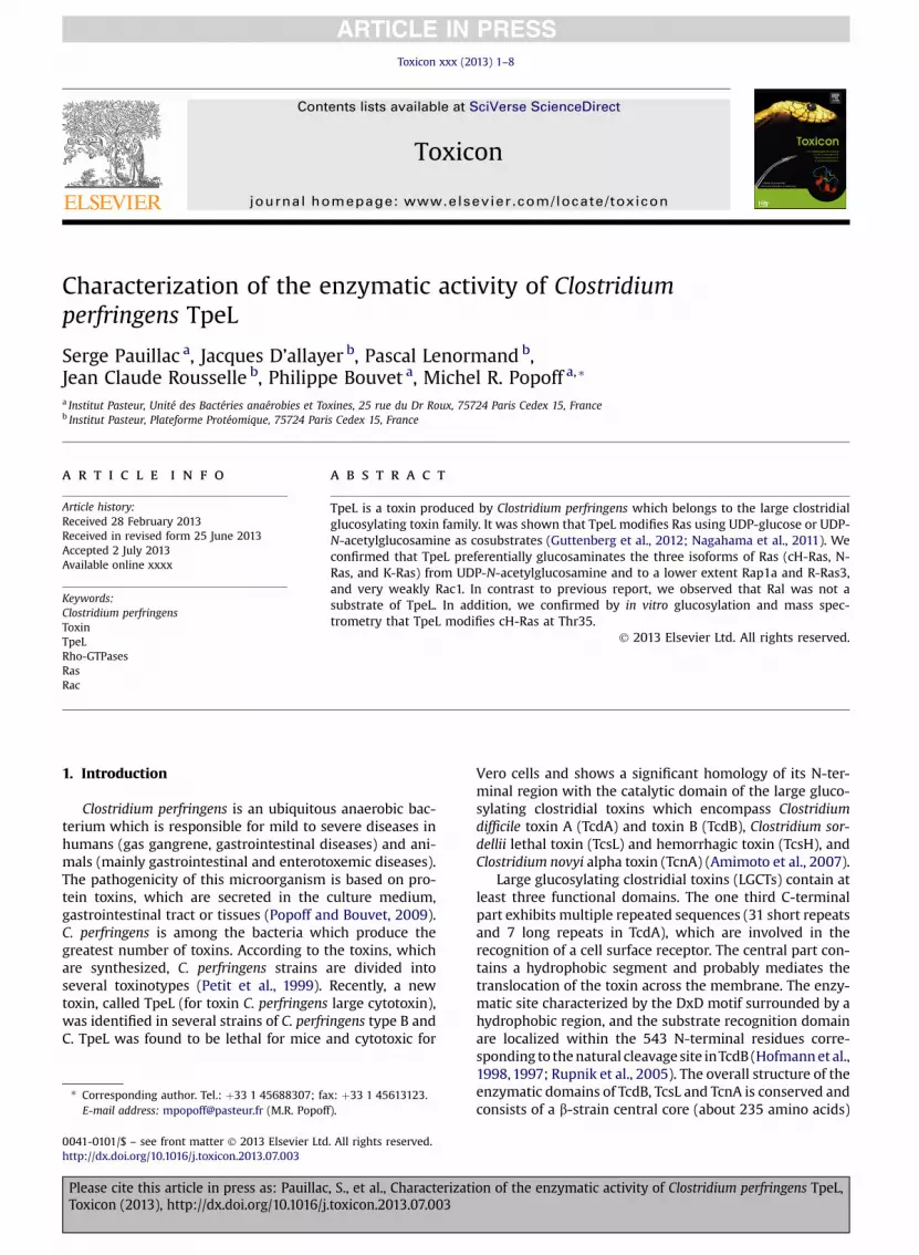
Fig. 2. TpeL N-terminal part glucosylates Ras but not Rho, Rac, and Cdc42.(A) SDS-PAGE of recombinant N-terminal part of TpeL (1 and 2) stained withBlue Coomassie R-250. (B) Glucosylation of cH-Ras, RhoA, Rac1, or Cdc42(1 mg recombinant protein) by TpeL N-terminal part (10�7 M) in the pres-ence of UDP-[14C]Glucose or UDP-[14C]-N-acetylglucosamine for 1 h at 37 �C.The proteins were run on a SDS-PAGE and autoradiographed.
S. Pauillac et al. / Toxicon xxx (2013) 1–8 3
MS spectra were acquired in reflector positive modeusing the 4800 MALDI TOF/TOF Analyzer (Applied Bio-systems, USA) within a mass range of 800–4000 m/z. MScalibration was first carried out to a final mass accuracy of1 ppm with a mixture of 5 standard peptides: des-Arg1Bradykinin [M þ H]þ ¼ 904.468; Angiotensin [M þH]þ ¼ 1296.685; Glu1-Fibrinopeptide [M þ H]þ ¼1570.677; ACTH 1–17 [M þ H]þ ¼ 2093.087; ACTH 18–39 [M þ H]þ ¼ 2465.199. MS spectra of the purified frac-tions were then acquired (3000 laser shots were averaged)and calibrated using the external default calibration(10 ppm mass accuracy).
MS/MS were performed in CID positive ion mode with a2 kV collision energy of and air as collision gas. MS/MScalibration was performed using 4 CID fragments of theGlu1-Fibrinopeptide ([M þ H]þ ¼ 175.119, 684.346,1056.475 and 1441.634) (mass accuracy of 10 ppm). 4000 to10,000 laser shots were averaged to acquired MS/MSspectra of the selected precursors. Each spectrumwas thencalibrated using the external default calibration (mass ac-curacy of 100 ppm).
MS/MS queries were performed using the MASCOTsearch engine 2.1 (Matrix Science Ltd., UK) and the Swis-sProt 57.4 database (470,369 sequences; 166,709,888 resi-dues) with the following parameters: 50 ppm masstolerance for the precursor and 0.3 Da for MS/MS frag-ments, trypsin cleavage. Hexose-N-acetylglucosamine(HexNAc) and oxidation of methionins were used as vari-able modifications. In these conditions, the minimalMASCOT score was 28 for a peptide confidence index �95%.
3. Results
3.1. TpeL is a glucosyltransferase which targets Ras proteins
As previously found (Amimoto et al., 2007), TpeL showsa significant overall sequence homology with the LCGTsand contains a conserved active enzymatic site (DxD) in theN-terminal part, except that the total protein length isshorter (190 kDa) than that of the glucosylating toxins fromC. difficile, C. sordellii or C. novyi (250–300 kDa) (Jank andAktories, 2008; Just and Gerhard, 2004; Popoff andBouvet, 2009; Rupnik and Just, 2006). The N-terminaldomain (amino acid 1–543) of LCGTs is the intracellularactive domain, which is released into the cytosol throughan autoproteolytic process during the toxin endocytosis(Jank and Aktories, 2008). Interestingly, the TpeL N-termi-nal domain shows a high level of sequence similarity withthat of the other LCGTs with an intermediate position be-tween the N-terminal domain of C. novyi alpha toxin andthose of the C. difficile and C. sordellii glucosylating toxins(Fig. 1). Therefore, we produced and purified the N-termi-nal domain of TpeL as a recombinant protein (Fig. 2A) tocheck its enzymatic activity. In a first series of experimentswe used RhoA, Rac1, Cdc42 and cH-Ras, which are therepresentative substrates of LGCTs, in a glucosylation assaywith TpeL. As shown in Fig. 2B, rN-TpeL glucosylated onlycH-Ras and no RhoA, Rac1 or Cdc42. In addition, the glu-cosylation of cH-Ras was more efficient by using UDP-N-acetylglucosamine than UDP-glucose as cosubstrate.
Please cite this article in press as: Pauillac, S., et al., Characterization of the enzymatic activity of Clostridium perfringens TpeL,Toxicon (2013), http://dx.doi.org/10.1016/j.toxicon.2013.07.003
S. Pauillac et al. / Toxicon xxx (2013) 1–84
Thereby, rN-TpeL contains a glucosyltransferase activityspecific of cH-Ras protein but not of Rho and Cdc42.
3.2. TpeL preferentially uses UDP-N-acetylglucosamine ascosubstrate
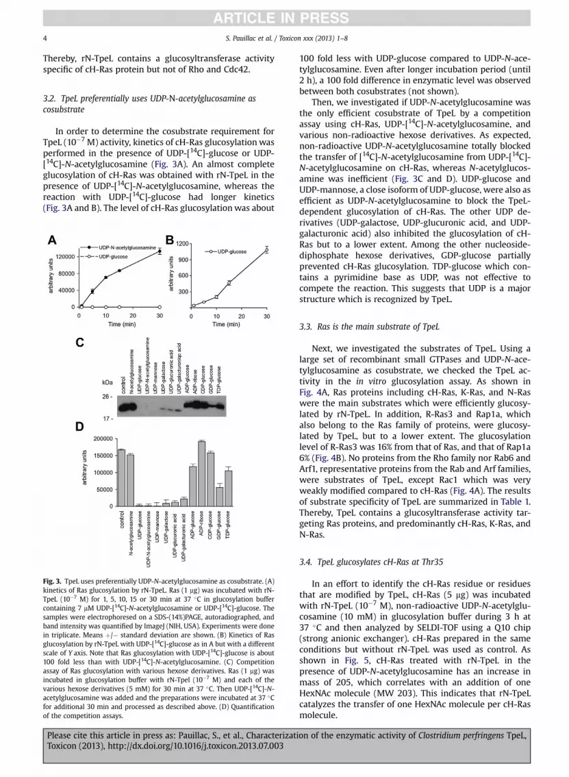
In order to determine the cosubstrate requirement forTpeL (10�7 M) activity, kinetics of cH-Ras glucosylationwasperformed in the presence of UDP-[14C]-glucose or UDP-[14C]-N-acetylglucosamine (Fig. 3A). An almost completeglucosylation of cH-Ras was obtained with rN-TpeL in thepresence of UDP-[14C]-N-acetylglucosamine, whereas thereaction with UDP-[14C]-glucose had longer kinetics(Fig. 3A and B). The level of cH-Ras glucosylationwas about
Fig. 3. TpeL uses preferentially UDP-N-acetylglucosamine as cosubstrate. (A)kinetics of Ras glucosylation by rN-TpeL. Ras (1 mg) was incubated with rN-TpeL (10�7 M) for 1, 5, 10, 15 or 30 min at 37 �C in glucosylation buffercontaining 7 mM UDP-[14C]-N-acetylglucosamine or UDP-[14C]-glucose. Thesamples were electrophoresed on a SDS-(14%)PAGE, autoradiographed, andband intensity was quantified by ImageJ (NIH, USA). Experiments were donein triplicate. Means þ/� standard deviation are shown. (B) Kinetics of Rasglucosylation by rN-TpeL with UDP-[14C]-glucose as in A but with a differentscale of Y axis. Note that Ras glucosylation with UDP-[14C]-glucose is about100 fold less than with UDP-[14C]-N-acetylglucosamine. (C) Competitionassay of Ras glucosylation with various hexose derivatives. Ras (1 mg) wasincubated in glucosylation buffer with rN-Tpel (10�7 M) and each of thevarious hexose derivatives (5 mM) for 30 min at 37 �C. Then UDP-[14C]-N-acetylglucosamine was added and the preparations were incubated at 37 �Cfor additional 30 min and processed as described above. (D) Quantificationof the competition assays.
Please cite this article in press as: Pauillac, S., et al., CharacterizatToxicon (2013), http://dx.doi.org/10.1016/j.toxicon.2013.07.003
100 fold less with UDP-glucose compared to UDP-N-ace-tylglucosamine. Even after longer incubation period (until2 h), a 100 fold difference in enzymatic level was observedbetween both cosubstrates (not shown).
Then, we investigated if UDP-N-acetylglucosamine wasthe only efficient cosubstrate of TpeL by a competitionassay using cH-Ras, UDP-[14C]-N-acetylglucosamine, andvarious non-radioactive hexose derivatives. As expected,non-radioactive UDP-N-acetylglucosamine totally blockedthe transfer of [14C]-N-acetylglucosamine from UDP-[14C]-N-acetylglucosamine on cH-Ras, whereas N-acetylglucos-amine was inefficient (Fig. 3C and D). UDP-glucose andUDP-mannose, a close isoform of UDP-glucose, were also asefficient as UDP-N-acetylglucosamine to block the TpeL-dependent glucosylation of cH-Ras. The other UDP de-rivatives (UDP-galactose, UDP-glucuronic acid, and UDP-galacturonic acid) also inhibited the glucosylation of cH-Ras but to a lower extent. Among the other nucleoside-diphosphate hexose derivatives, GDP-glucose partiallyprevented cH-Ras glucosylation. TDP-glucose which con-tains a pyrimidine base as UDP, was not effective tocompete the reaction. This suggests that UDP is a majorstructure which is recognized by TpeL.
3.3. Ras is the main substrate of TpeL
Next, we investigated the substrates of TpeL. Using alarge set of recombinant small GTPases and UDP-N-ace-tylglucosamine as cosubstrate, we checked the TpeL ac-tivity in the in vitro glucosylation assay. As shown inFig. 4A, Ras proteins including cH-Ras, K-Ras, and N-Raswere the main substrates which were efficiently glucosy-lated by rN-TpeL. In addition, R-Ras3 and Rap1a, whichalso belong to the Ras family of proteins, were glucosy-lated by TpeL, but to a lower extent. The glucosylationlevel of R-Ras3 was 16% from that of Ras, and that of Rap1a6% (Fig. 4B). No proteins from the Rho family nor Rab6 andArf1, representative proteins from the Rab and Arf families,were substrates of TpeL, except Rac1 which was veryweakly modified compared to cH-Ras (Fig. 4A). The resultsof substrate specificity of TpeL are summarized in Table 1.Thereby, TpeL contains a glucosyltransferase activity tar-geting Ras proteins, and predominantly cH-Ras, K-Ras, andN-Ras.
3.4. TpeL glucosylates cH-Ras at Thr35
In an effort to identify the cH-Ras residue or residuesthat are modified by TpeL, cH-Ras (5 mg) was incubatedwith rN-TpeL (10�7 M), non-radioactive UDP-N-acetylglu-cosamine (10 mM) in glucosylation buffer during 3 h at37 �C and then analyzed by SELDI-TOF using a Q10 chip(strong anionic exchanger). cH-Ras prepared in the sameconditions but without rN-TpeL was used as control. Asshown in Fig. 5, cH-Ras treated with rN-TpeL in thepresence of UDP-N-acetylglucosamine has an increase inmass of 205, which correlates with an addition of oneHexNAc molecule (MW 203). This indicates that rN-TpeLcatalyzes the transfer of one HexNAc molecule per cH-Rasmolecule.
ion of the enzymatic activity of Clostridium perfringens TpeL,
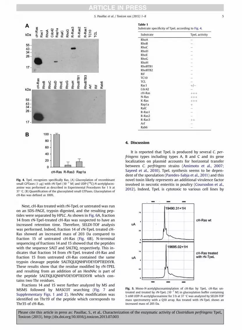
Fig. 4. TpeL recognizes specifically Ras. (A) Glucosylation of recombinantsmall GPTases (1 mg) with rN-Tpel (10�7 M) and UDP-[14C]-N-acetylglucos-amine was performed as described in Experimental Procedures for 1 h at37 �C. (B) Quantification of the glucosylated small GTPases. Glucosylation ofcH-Ras was defined as 100%.
Table 1Substrate specificity of TpeL according to Fig. 4.
Substrate TpeL activity
RhoA �RhoB �RhoC �RhoD �RhoE �RhoG �RhoH �RhoBTB1 �RhoBTB2 �Rif �TC10 �TCL �Rac1 þ/�Cdc42 �cH-Ras þþþN-Ras þþþK-Ras þþþRap1a þRalC �R-Ras1 �R-Ras2 �R-Ras3 þþArf �Rab6 �
Fig. 5. Mono-N-acetylglucosaminylation of cH-Ras by TpeL. cH-Ras un-treated and treated by rN-TpeL (10�7 M) in glucosylation buffer containing5 mM UDP-N-acetylglucosamine for 3 h at 37 �C was analyzed by SELDI-TOFmass spectrometry with a Q10 array. Ras treated with rN-TpeL shows anincreased mass of 205 Da.
S. Pauillac et al. / Toxicon xxx (2013) 1–8 5
Next, cH-Ras treated with rN-TpeL or untreated was runon an SDS-PAGE, trypsin digested, and the resulting pep-tides were separated by HPLC. As shown in Fig. 6A, fraction14 from rN-Tpel-treated cH-Ras was suspected to have anincreased retention time. Therefore, SELDI-TOF analysiswas performed. Indeed, fraction 14 of rN-TpeL treated cH-Ras showed an increased mass of 203 Da compared tofraction 15 of untreated cH-Ras (Fig. 6B). N-terminalsequencing of fractions 14 and 15 showed that the peptideswith the sequence SALT and SALTIQ, respectively. This in-dicates that fraction 14 from rN-TpeL treated cH-Ras andfraction 15 from untreated cH-Ras contained the sametrypsin cleavage peptide SALTIQLIQNHFVDEYDPTIEDSYR.These results show that the residue modified by rN-TPELand resulting from an addition of an HexNAc is part ofthe peptide SALTIQLIQNHFVDEYDPTIEDSYR which con-tains two Thr residues.
Fractions 14 and 15 were further analyzed by MS andMSMS followed by MASCOT searching (Fig. 7 andSupplementary Figs. 1 and 2). HexNAc modification wasidentified on Thr19 of the peptide which corresponds toThr35 of cH-Ras.
Please cite this article in press as: Pauillac, S., et al., CharacterizatiToxicon (2013), http://dx.doi.org/10.1016/j.toxicon.2013.07.003
4. Discussion
It is reported that TpeL is produced by several C. per-fringens types including types A, B and C and its genelocalization on plasmid accounts for horizontal transferbetween C. perfringens strains (Amimoto et al., 2007;Sayeed et al., 2010). TpeL synthesis seems to be depen-dent of the sporulation (Paredes-Sabja et al., 2011) and thisnovel toxin likely represents an additional virulence factorinvolved in necrotic enteritis in poultry (Coursodon et al.,2012). Indeed, TpeL is cytotoxic to various cell lines by
on of the enzymatic activity of Clostridium perfringens TpeL,
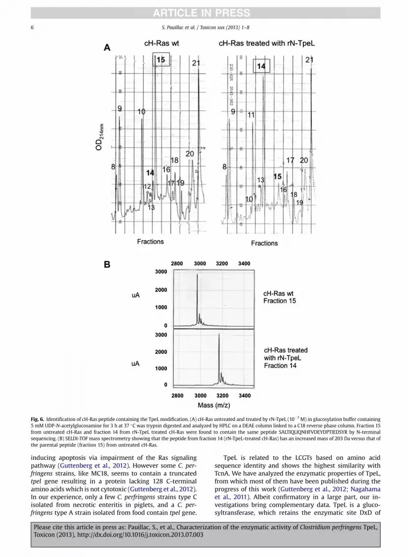
Fig. 6. Identification of cH-Ras peptide containing the TpeL modification. (A) cH-Ras untreated and treated by rN-TpeL (10�7 M) in glucosylation buffer containing5 mM UDP-N-acetylglucosamine for 3 h at 37 �C was trypsin digested and analyzed by HPLC on a DEAE column linked to a C18 reverse phase column. Fraction 15from untreated cH-Ras and fraction 14 from rN-TpeL treated cH-Ras were found to contain the same peptide SALTIQLIQNHFVDEYDPTIEDSYR by N-terminalsequencing. (B) SELDI-TOF mass spectrometry showing that the peptide from fraction 14 (rN-TpeL-treated cH-Ras) has an increased mass of 203 Da versus that ofthe parental peptide (fraction 15) from untreated cH-Ras.
S. Pauillac et al. / Toxicon xxx (2013) 1–86
inducing apoptosis via impairment of the Ras signalingpathway (Guttenberg et al., 2012). However some C. per-fringens strains, like MC18, seems to contain a truncatedtpel gene resulting in a protein lacking 128 C-terminalamino acids which is not cytotoxic (Guttenberg et al., 2012).In our experience, only a few C. perfringens strains type Cisolated from necrotic enteritis in piglets, and a C. per-fringens type A strain isolated from food contain tpel gene.
Please cite this article in press as: Pauillac, S., et al., CharacterizatToxicon (2013), http://dx.doi.org/10.1016/j.toxicon.2013.07.003
TpeL is related to the LCGTs based on amino acidsequence identity and shows the highest similarity withTcnA. We have analyzed the enzymatic properties of TpeL,from which most of them have been published during theprogress of this work (Guttenberg et al., 2012; Nagahamaet al., 2011). Albeit confirmatory in a large part, our in-vestigations bring complementary data. TpeL is a gluco-syltransferase, which retains the enzymatic site DxD of
ion of the enzymatic activity of Clostridium perfringens TpeL,
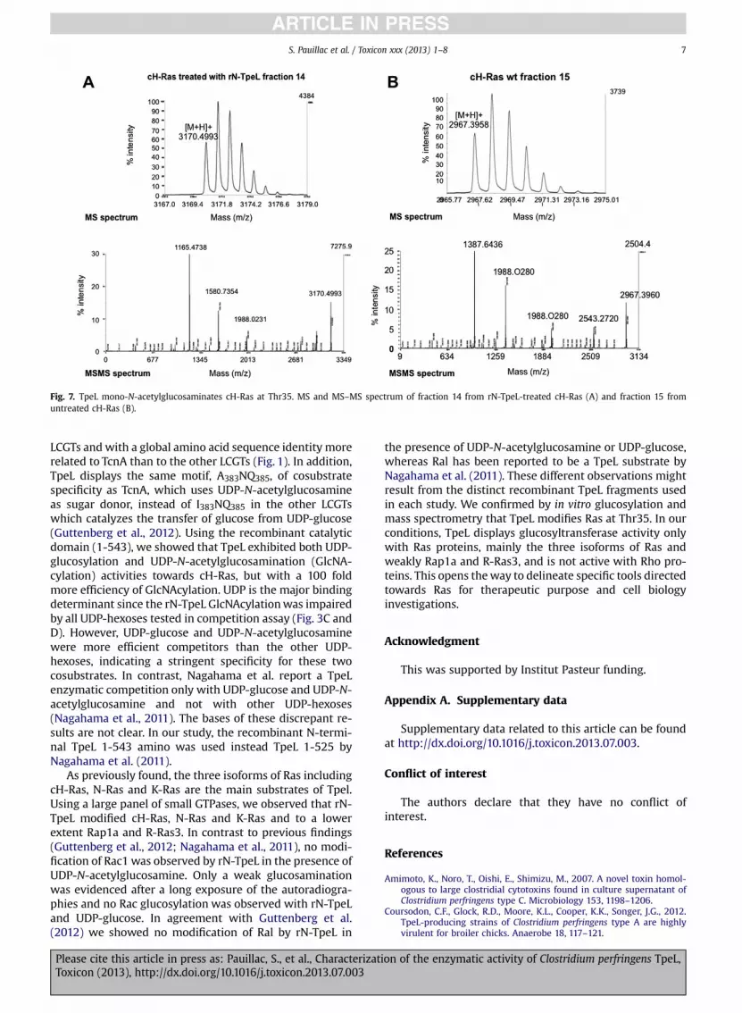
Fig. 7. TpeL mono-N-acetylglucosaminates cH-Ras at Thr35. MS and MS–MS spectrum of fraction 14 from rN-TpeL-treated cH-Ras (A) and fraction 15 fromuntreated cH-Ras (B).
S. Pauillac et al. / Toxicon xxx (2013) 1–8 7
LCGTs and with a global amino acid sequence identity morerelated to TcnA than to the other LCGTs (Fig. 1). In addition,TpeL displays the same motif, A383NQ385, of cosubstratespecificity as TcnA, which uses UDP-N-acetylglucosamineas sugar donor, instead of I383NQ385 in the other LCGTswhich catalyzes the transfer of glucose from UDP-glucose(Guttenberg et al., 2012). Using the recombinant catalyticdomain (1-543), we showed that TpeL exhibited both UDP-glucosylation and UDP-N-acetylglucosamination (GlcNA-cylation) activities towards cH-Ras, but with a 100 foldmore efficiency of GlcNAcylation. UDP is the major bindingdeterminant since the rN-TpeL GlcNAcylationwas impairedby all UDP-hexoses tested in competition assay (Fig. 3C andD). However, UDP-glucose and UDP-N-acetylglucosaminewere more efficient competitors than the other UDP-hexoses, indicating a stringent specificity for these twocosubstrates. In contrast, Nagahama et al. report a TpeLenzymatic competition only with UDP-glucose and UDP-N-acetylglucosamine and not with other UDP-hexoses(Nagahama et al., 2011). The bases of these discrepant re-sults are not clear. In our study, the recombinant N-termi-nal TpeL 1-543 amino was used instead TpeL 1-525 byNagahama et al. (2011).
As previously found, the three isoforms of Ras includingcH-Ras, N-Ras and K-Ras are the main substrates of Tpel.Using a large panel of small GTPases, we observed that rN-TpeL modified cH-Ras, N-Ras and K-Ras and to a lowerextent Rap1a and R-Ras3. In contrast to previous findings(Guttenberg et al., 2012; Nagahama et al., 2011), no modi-fication of Rac1 was observed by rN-TpeL in the presence ofUDP-N-acetylglucosamine. Only a weak glucosaminationwas evidenced after a long exposure of the autoradiogra-phies and no Rac glucosylation was observed with rN-TpeLand UDP-glucose. In agreement with Guttenberg et al.(2012) we showed no modification of Ral by rN-TpeL in
Please cite this article in press as: Pauillac, S., et al., CharacterizatiToxicon (2013), http://dx.doi.org/10.1016/j.toxicon.2013.07.003
the presence of UDP-N-acetylglucosamine or UDP-glucose,whereas Ral has been reported to be a TpeL substrate byNagahama et al. (2011). These different observations mightresult from the distinct recombinant TpeL fragments usedin each study. We confirmed by in vitro glucosylation andmass spectrometry that TpeL modifies Ras at Thr35. In ourconditions, TpeL displays glucosyltransferase activity onlywith Ras proteins, mainly the three isoforms of Ras andweakly Rap1a and R-Ras3, and is not active with Rho pro-teins. This opens theway to delineate specific tools directedtowards Ras for therapeutic purpose and cell biologyinvestigations.
Acknowledgment
This was supported by Institut Pasteur funding.
Appendix A. Supplementary data
Supplementary data related to this article can be foundat http://dx.doi.org/10.1016/j.toxicon.2013.07.003.
Conflict of interest
The authors declare that they have no conflict ofinterest.
References
Amimoto, K., Noro, T., Oishi, E., Shimizu, M., 2007. A novel toxin homol-ogous to large clostridial cytotoxins found in culture supernatant ofClostridium perfringens type C. Microbiology 153, 1198–1206.
Coursodon, C.F., Glock, R.D., Moore, K.L., Cooper, K.K., Songer, J.G., 2012.TpeL-producing strains of Clostridium perfringens type A are highlyvirulent for broiler chicks. Anaerobe 18, 117–121.
on of the enzymatic activity of Clostridium perfringens TpeL,
S. Pauillac et al. / Toxicon xxx (2013) 1–88
Guttenberg, G., Hornei, S., Jank, T., Schwan, C., Lu, W., Einsle, O.,Papatheodorou, P., Aktories, K., 2012. Molecular characteristics ofClostridium perfringens TpeL toxin and consequences of mono-O-GlcNAcylation of Ras in living cells. J. Biol. Chem. 287, 24929–24940.
Hofmann, F., Busch, C., Aktories, K., 1998. Chimeric clostridial cytotoxins:identification of the N-terminal region involved in protein substraterecognition. Infect. Immun. 66, 1076–1081.
Hofmann, F., Busch, C., Prepens, U., Just, I., Aktories, K., 1997. Localizationof the glucosyltransferase activity of Clostridium difficile toxin B to theN-terminal part of the holotoxin. J. Biol. Chem. 272, 11074–11078.
Jank, T., Aktories, K., 2008. Structure and mode of action of clos-tridial glucosylating toxins: the ABCD model. Trends Microbiol.16, 222–229.
Jank, T., Giesemann, T., Aktories, K., 2007. Clostridium difficile glucosyl-transferase toxin B-essential amino acids for substrate binding. J. Biol.Chem. 282, 35222–35231.
Just, I., Gerhard, R., 2004. Large clostridial cytotoxins. Rev. Physiol. Bio-chem. Pharmacol. 152, 23–47.
Nagahama, M., Ohkubo, A., Oda, M., Kobayashi, K., Amimoto, K.,Miyamoto, K., Sakurai, J., 2011. Clostridium perfringens TpeL glycosy-lates the Rac and Ras subfamily proteins. Infect. Immun. 79, 905–910.
Paredes-Sabja, D., Sarker, N., Sarker, M.R., 2011. Clostridium perfringenstpeL is expressed during sporulation. Microb. Pathog. 51, 384–388.
Please cite this article in press as: Pauillac, S., et al., CharacterizatToxicon (2013), http://dx.doi.org/10.1016/j.toxicon.2013.07.003
Petit, L., Gibert, M., Popoff, M.R., 1999. Clostridium perfringens: tox-inotype and genotype. Trends Microbiol. 7, 104–110.
Popoff, M.R., Bouvet, P., 2009. Clostridial toxins. Future Microbiol. 4, 1021–1064.
Popoff, M.R., Chaves-Olarte, E., Lemichez, E., Von Eichel-Streiber, C.,Thelestam, M., Chardin, P., Cussac, D., Antonny, B., Chavrier, P.,Flatau, G., Giry, M., de Gunzburg, J., Boquet, P., 1996. Ras, Rap, and racsmall GTP-binding proteins are targets for Clostridium sordellii lethaltoxin glucosylation. J. Biol. Chem. 271, 10217–10224.
Reinert, D.J., Jank, T., Aktories, K., Schulz, G.E., 2005. Structural basis forthe function of Clostridium difficile toxin B. J. Mol. Biol. 351, 973–981.
Rupnik, M., Just, I., 2006. Large clostridial cytotoxins modifying small GTPa-ses. In: Alouf, J.E., Popoff,M.R. (Eds.), The Sourcebook of Bacterial ProteinToxins, third ed. Elsevier, Academic Press, Amsterdam, pp. 409–429.
Rupnik, M., Pabst, S., Rupnik, M., von Eichel-Streiber, C., Urlaub, H.,Soling, H.D., 2005. Characterization of the cleavage site and functionof resulting cleavage fragments after limited proteolysis of Clostridiumdifficile toxin B (TcdB) by host cells. Microbiology 151, 199–208.
Sayeed, S., Li, J., McClane, B.A., 2010. Characterization of virulence plasmiddiversity among Clostridium perfringens type B isolates. Infect. Immun.78, 495–504.
Ziegler, M.O., Jank, T., Aktories, K., Schulz, G.E., 2008. Conformationalchanges and reaction of clostridial glycosylating toxins. J. Mol. Biol.377, 1346–1356.
ion of the enzymatic activity of Clostridium perfringens TpeL,